Article

Unraveling the Central Role of Sulfur-Oxidizing Acidiphilium multivorum LMS in Industrial Bioprocessing of Gold-Bearing Sulfide Concentrates

by
Anna Panyushkina
1,*,
Aleksandr Bulaev
1 and
Aleksandr V. Belyi
2
1
Winogradsky Institute of Microbiology, Research Centre of Biotechnology of the Russian Academy of Sciences, Leninsky Ave., 33, bld. 2, 119071 Moscow, Russia
2
JSC “Polyus Krasnoyarsk”, Research Center, Poligonnaya Str., 15, 660118 Krasnoyarsk, Russia
*
Author to whom correspondence should be addressed.
Microorganisms 2021, 9(5), 984; https://doi.org/10.3390/microorganisms9050984
Submission received: 15 March 2021 / Revised: 19 April 2021 / Accepted: 29 April 2021 / Published: 1 May 2021
(This article belongs to the Section Microbial Biotechnology)

Abstract

:
Acidiphilium multivorum LMS is an acidophile isolated from industrial bioreactors during the processing of the gold-bearing pyrite-arsenopyrite concentrate at 38–42 °C. Most strains of this species are obligate organoheterotrophs that do not use ferrous iron or reduced sulfur compounds as energy sources. However, the LMS strain was identified as one of the predominant sulfur oxidizers in acidophilic microbial consortia. In addition to efficient growth under strictly heterotrophic conditions, the LMS strain proved to be an active sulfur oxidizer both in the presence or absence of organic compounds. Interestingly, Ac. multivorum LMS was able to succeed more common sulfur oxidizers in microbial populations, which indicated a previously underestimated role of this bacterium in industrial bioleaching operations. In this study, the first draft genome of the sulfur-oxidizing Ac. multivorum was sequenced and annotated. Based on the functional genome characterization, sulfur metabolism pathways were reconstructed. The LMS strain possessed a complicated multi-enzyme system to oxidize elemental sulfur, thiosulfate, sulfide, and sulfite to sulfate as the final product. Altogether, the phenotypic description and genome analysis unraveled a crucial role of Ac. multivorum in some biomining processes and revealed unique strain-specific characteristics, including the ars genes conferring arsenic resistance, which are similar to those of phylogenetically distinct microorganisms.

1. Introduction

Gram-negative bacteria of the genus Acidiphilium are members of acidophilic microbial communities in ore deposits, commercial bioleaching operation sites, and acid mine drainage (AMD) [1,2,3,4]. The roles of phylogenetically close species Ac. cryptum and Ac. multivorum in acidophilic communities are considered similar to those of other acidophilic organoheterotrophs. The latter can utilize metabolites secreted by acidophilic auto-, hetero-, and mixotrophic microorganisms, as well as organic components of lysed cells, as carbon and energy sources [4]. One of the Acidiphilium species, Ac. acidophilum, has been shown to grow autotrophically with reduced inorganic sulfur compounds (RISCs) [5]. Acidiphilium cryptum strain DX1-1 isolated from AMD of the DeXing mine (Jiangxi Province, China) is able to grow with organic compounds and inorganic sulfur. However, when sulfur was the sole energy substrate, the growth and sulfur oxidation by the strain DX1-1 were negligible and improved when glucose was added to the culture medium [6]. Among strains assigned to different Acidiphilium species, the ferric iron reduction has been demonstrated in oxygen-containing and anoxic acidic environments [7]. Members of the genus Acidiphilium can also withstand high concentrations of toxic metals, such as copper, cadmium, nickel, and zinc [8]. These bacteria were recently characterized by a complex lifestyle granted by horizontal gene transfer. Photosynthetic and heavy metal resistance systems, as well as additional pathways for carbon metabolism, are associated with gene acquisitions that expand the genetic diversity of the genus [9]. Results obtained using genomic approaches have shed light on metabolic pathways and resistance determinants of Acidiphilium spp. strains. The latter included an environmental isolate Acidiphilium sp. PM from Rio Tinto [10,11], Acidiphilium sp. JA12-A1 from an iron-oxidizing mixed culture from a pilot plant for bioremediation of acid mine drainage [12], and an iron-reducing strain Acidiphilium sp. C61 isolated from iron-rich pelagic aggregates (“iron snow”) formed at the redoxcline of stratified iron-rich lignite mine Lake 77 (Lusatian mining area, east-central Germany) [13]. Complete sequences of the chromosome and eight plasmids of the strains Ac. cryptum JF-5 (BioProject PRJNA15753) and Ac. multivorum AIU301 (BioProjects PRJNA224116 and PRJDB503) have been deposited in the GenBank databases and compared [14]. Twelve more genomes of Acidiphilium spp. can be also found in the GenBank databases. Three Acidiphilium genomes (AccI, AccII, and ZJSH63) were recently sequenced, resulting in the complete nucleotide sequence of the strain AccI (a single chromosome and seven plasmids) and draft genomes of the strains AccII and ZJSH63, and deposited in the JGI IMG-ER database [9]. However, the genome of the only one reported sulfur-oxidizing strain DX1-1 assigned to the heterotrophic species Ac. cryptum has not been sequenced, although some of its features have been determined by the differential expression of several genes, and the upregulation of three genes involved in sulfur metabolism has been revealed [6].
The object of this research was an acidophilic bacterial strain isolated as a predominant sulfur oxidizer from a bioreactor during the processing of the gold-containing sulfidic concentrate at 38 °C [15]. Biotechnologies for tank biooxidation of sulfidic concentrates using sulfur- and iron-oxidizing acidophilic microorganisms were developed in the 1980s. Since then, biomining approaches have been successfully applied worldwide [16]. Gold-bearing sulfidic concentrates are classified as refractory concentrates, and direct gold recovery from them using cyanidation is insignificant. Tank biooxidation of gold-bearing arsenopyrite concentrates, using acidophilic microbial communities, is among the most efficient commercial biohydrometallurgical processes known to date [17]. Most arsenopyrite biooxidation processes operate at ~38–42 °C and are dominated by the communities of sulfur- and iron-oxidizing microorganisms, such as Acidithiobacillus (At.) spp., Leptospirillum (L.) spp., Sulfobacillus (Sb.) spp., Ferroplasma (F.) spp., and some other microorganisms [15,17,18,19,20,21,22]. The Olympiada biooxidation plant is located in the Krasnoyarsk Krai (Siberia, Russia) and has been operated by Polyus Krasnoyarsk JSC since 2001. It is one of the largest biooxidation factories in the world that processes from 8 to 9 million tons of gold-bearing ore annually using a high-performance BIONORD® technology developed under the conditions of Extreme North (Siberia, Russia) [15,20]. The average productivity of the Olimpiada plant is 1200–1300 tons of the flotation sulfidic concentrate per day, which allowed the recovery of 30 tons of gold in 2017 [20]. The main minerals composing sulfide concentrates are pyrite, arsenopyrite, pyrrhotite, and antimonite [15].
The goal of our research was to characterize the predominant sulfur-oxidizing bacterium that was isolated from the bioreactors of the Olimpiada plant and subsequently assigned to the heterotrophic species Ac. multivorum, as well as to determine its role in the microbial communities during the biooxidation of sulfidic concentrates. Within several years, this bacterium has become one of the predominant sulfur-oxidizing organisms, along with another sulfur-oxidizing species At. thiooxidans, which is commonly detected in bioreactors at the Olimpiada plant [21], or even succeeded the latter in some bioreactors. According to the previously obtained results of metabarcoding analysis (V3–V4 variable regions of the 16S rRNA gene), the proportion of the Acidiphilum bacteria in the microbial community reached 36% [15]. Therefore, we aimed to study the characteristic features of this acidophile, as well as to determine and analyze its genome sequence to provide new insights into its metabolism capacities, resistance mechanisms, and the role in the biooxidation of the refractory gold-bearing ores. The sulfur-oxidizing activity of this bacterium, together with the sulfur metabolism pathways predicted from the genome annotation, allowed us to conclude that the role of some heterotrophic acidophiles could be underestimated in acidophilic microbial consortia. The strain Ac. multivorum LMS proved to be an active sulfur oxidizer that possessed a complicated sulfur-oxidizing enzyme system, including the multi-enzyme Sox complex, to oxidize RISCs. Based on the genome analysis, a biochemical model for sulfur oxidation in Ac. multivorum LMS was proposed. A comparison to other Acidiphilium spp. genomes revealed additional strain-specific metal(loid) resistance determinants in the LMS strain. Probably, these features determined the crucial role of this microorganism in the industrial biooxidation of sulfidic raw materials.

2. Materials and Methods

2.1. Strain Isolation and Cultivation Conditions

The phylogenetically close sulfur-oxidizing Acidiphilium strains (including the LMS strain) were continuously isolated from the pulp samples collected from industrial bioreactors during the monitoring of the microbial population involved in the biooxidation of the gold-bearing pyrrhotite-containing pyrite-arsenopyrite concentrate at the Olimpiada biooxidation plant (38–42 °C; the pulp density of 15–20%; gold recovery of ~97% after biooxidation) in 2016 [15] and 2017–2019 [23]. The pure cultures of the strains designated LMS, PB-B3-4, PB-B1-3, and PB-B2-3 were isolated from the pulp samples collected from the bioreactors by serial tenfold dilutions in the selective liquid media (pH 2.5) containing elemental sulfur S0 (1%, w/v) and yeast extract (0.02%, w/v) or only S0 (autotrophic medium) at 40 °C. The amount of inoculum was 10% (v/v). The strains were cultured in 250 mL Erlenmeyer flasks (100 mL of the liquid medium) on Unimax-1010 rotor shakers (Heidolph Instruments, Schwabach, Germany; 180× rpm) in Inkubator-1000 thermostats (Heidolph Instruments, Schwabach, Germany). The salt base of the 9K medium was of the following composition (g/L): (NH4)2SO4, 3.0; KCl, 0.1; KH2PO4, 0.5; MgSO4 7H2O, 0.5; Ca(NO3)2 4H2O, 0.01 [24].
To determine the characteristics of the LMS strain growth and substrate oxidation, it was cultivated in the temperature range of 20–60 °C (pH 2.5) in the presence of S0 (1%, w/v) and yeast extract (0.02%, w/v). After determining the optimum temperature, the growth of the strain at different pH was tested. The initial ambient pH value of the medium was adjusted to 1.5–6.5 with 10 N H2SO4 depending on the experimental conditions, and the variants were cultivated at 38 °C. The 9K base salt media supplemented with Na2S2O3 (2%, w/v; pH 2.5), Na2S4O6 (2%, w/v; pH 2.5), or FeSO4·7H2O (20 mM Fe2+; pH 1.9) in the presence or absence of yeast extract (0.03%, w/v) were used to evaluate the ability of the strain to use RISCs and ferrous iron as energy sources. Yeast extract and/or glucose (0.01–0.5%) were used to test the growth of the LMS strain under heterotrophic conditions. Two series of experiments in each variant of the bacterial growth included three parallels (flasks) and three replicates for measured parameters. Statistical processing was performed using Microsoft Excel 2010. The standard deviation (SD) of the arithmetic mean was calculated, and the significance of the results was assessed using the Student’s t-test at the significance level p ≤ 0.05.
The biomass of the late-exponential cells grown under auto- and mixotrophic conditions was used to determine the phylogenetic position of the strain by subsequent sequencing of the 16S rRNA gene. For genome sequencing, the biomass of late-exponential cells was grown in the 9K medium containing both S0 and yeast extract in 2500 mL Erlenmeyer flasks (1500 mL of the inoculated medium).

2.2. Taxonomic Research and Phylogenetic Analysis

To carry out phylogenetic identification of the LMS strain pure culture, the microbial biomass was collected by centrifugation (8000× g, 20 min, 4 °C). The DNA extraction and purification, PCR amplification of the 16S rRNA gene fragment with standard primers for the bacterial 16S rRNA gene fragment, amplicon purification and subsequent sequencing, as well as phylogenetic identification of the pure microbial cultures, were carried out as previously described [21]. The 16S rRNA gene sequences of Acidiphilium sp. strains were deposited in the GenBank databases under accession numbers MW389315–MW389318. The phylogenetic tree was constructed in the MEGA X software package [25] using the sequences of the type strains and some other isolates identified as the closest Acidiphilium strains with the BlastN algorithm (https://blast.ncbi.nlm.nih.gov/Blast.cg; accessed on 8 September 2020). The percentage of replicate trees in which the associated taxa clustered together in the bootstrap test (1000 replicates) is shown next to the branches [26]. The evolutionary history was inferred using the Neighbor-Joining method [27]. The evolutionary distances were computed using the Maximum Composite Likelihood method [28] and were in the units of the number of base substitutions per site.

2.3. Genome Sequencing and Assembly

Late-exponential cells of the LMS strain were collected by centrifugation (8000× g, 20 min, 4 °C) and washed twice with acidified 9K medium without energy sources (pH 2.5) for the subsequent genome sequencing. Total DNA for genome sequencing was isolated using the FastDNA Spin kit (MP Biomedicals, Irvine, CA, USA) according to the manufacturer’s recommendations. DNA was fragmented using the NEBNext dsDNA Fragmentase (New England BioLabs, Inc., Ipswich, MA, USA). Subsequent steps of library preparation were carried out with the KAPA HyperPlus fragment library kit (Roche, Basel, Switzerland) according to the manufacturer’s protocol. The genome of the LMS strain was sequenced on the Illumina NextSeq platform (Illumina, Inc., San Diego, CA, USA) using the 2 × 100-cycles paired-end sequencing kit. Assembly of reads was performed with the SPAdes v. 3.13.0 (Genome Assembler; Center for Algorithmic Biotechnology: St. Petersburg, Russia, 2018) [29] and resulted in 139 contigs of the total length of 3,837,559 bp and an N50 value of 69,349 bp. The final assembly coverage was 500×.

2.4. Genome Annotation and Analysis

The draft genome of Ac. multivorum LMS was sequenced using the RAST (Rapid Annotation using Subsystem Technology) service v. 2.0 (RASTtk annotation scheme, https://rast.nmpdr.org/; accessed on 5 August 2020) for annotating microbial genomes [30] and the NCBI Prokaryotic Genome Annotation Pipeline (Bethesda, MD, USA, http://www.ncbi.nlm.nih.gov/genome/annotation_prok/; accessed on 8 September 2020). The genome sequence of Ac. multivorum LMS was deposited in the NCBI databases under the accession number JACVVW000000000 (BioProject PRJNA662208; BioSample SAMN16078398). Functional characterization of the genome was carried out using the KOALA (KEGG Orthology And Links Annotation) system [31]. Pathway mapping was carried out using tools of the KEGG software package. The annotation was manually improved by comparison with the type strain Ac. multivorum AIU301T (https://www.ncbi.nlm.nih.gov/genome/3581; accessed on 8 September 2020), Ac. cryptum JF5 (https://www.ncbi.nlm.nih.gov/genome/1383; accessed on 8 September 2020), and Acidiphilium spp. genomes (https://www.ncbi.nlm.nih.gov/genome/browse/#!/prokaryotes/13804/; accessed on 8 September 2020), available in the NCBI GenBank databases, as well as three recently published and characterized Acidiphilium genomes (strains AccI, AccII, and ZJSH63) that have been deposited in the JGI IMG-ER database (genome IDs 2824045439, 2824049744, and 2828882166, respectively) [9]. Genomes of Ac. multivorum LMS and other Acidiphilium strains were compared using the progressiveMauve program and the Mauve Contig Mover tool, which are part of the Mauve software package v. 2.4.0 for multiple genome alignments (The Darling lab at the University of Technology: Sydney, Australia, 2014) [32]. The average nucleotide identity (ANI) values between the genomes of Ac. multivorum LMS and other Acidiphilium strains were calculated with the ANI calculator tool (Kostas lab, Atlanta, GA, USA) using both best hits (one-way ANI) and reciprocal best hits (two-way ANI) between genomic datasets (http://enve-omics.ce.gatech.edu/ani/index; accessed on 16 April 2021). Circular visualization of the draft genome of the LMS strain and other Acidiphilium genomes used for comparisons was performed with the BRIG (BLAST (Basic Local Alignment Search Tool) Ring Image Generator v. 0.95) cross-platform application that can display circular comparisons between a large number of genomes, with a focus on handling genome assembly data (https://sourceforge.net/projects/brig/; accessed on 16 April 2021), and the NCBI BLAST+ database [33]. To identify the strain-specific genes and proteins (novel regions in the genome), we performed a comparison using the Panseq online tool (https://lfz.corefacility.ca/panseq; accessed on 4 December 2020) for analysis of core genome, pangenome, and accessory genomic regions [34]. The amino acid and nucleotide sequences of interest were compared using the NCBI BLAST (Bethesda, MD, USA, https://blast.ncbi.nlm.nih.gov/Blast.cgi; accessed on 16 April 2021).

2.5. Analytical Techniques

The pH value and concentrations of Fe3+ and Fe2+ were measured as previously described [18,21]. The quantitative assessment of the LMS strain was carried out by direct counts and by the method of serial terminal tenfold dilutions. Phase-contrast microscopy was performed with an Olympus CX41 microscope (Olympus Corporation, Tokyo, Japan) equipped with an Olympus C5060-ADU camera (Olympus Corporation, Tokyo, Japan); the ImageScope Lite software package (ImageScope Color Application, Moscow, Russia) was used. Microscopy was also used to determine morphological alterations within the cell population of the strain.

3. Results and Discussion

3.1. Isolation and Identification of the LMS Strain

Acidiphilium spp. strains were first isolated by us among the predominant sulfur oxidizers from a microbial community involved in the active process of biooxidation in different chains of bioreactors at the Olimpiada plant in 2016. The presence of this species in the bioleaching process has been also confirmed by metabarcoding analysis (V3–V4 variable regions of the 16S rRNA gene) of the microbial community structure during the monitoring of the microbial populations of commercial bioreactors in 2016–2019 [15,23]. In total, more than 200 samples have been collected from the bioreactors and analyzed by the 16S rRNA gene metabarcoding over this period, and the maximum proportion of Acidiphilium in the total microbial population reached 36–41% depending on the bioreactor [15,23].
Pure cultures of the predominant sulfur-oxidizing microorganisms were isolated under both autotrophic or mixotrophic conditions of cultivation from several bioreactors by a method of tenfold serial dilutions in selective liquid growth media supplemented with elemental sulfur or yeast extract and S0. The growth of the cultures was shown until the 10−7–10−9 dilution, which indicated that the strain abundance in the original pulp samples collected from the bioreactors reached 107–109 cells/mL. The similarity of the nucleotide sequences of the 16S rRNA gene fragments (1423–1456 bp) of the pure cultures isolated from the auto- and mixotrophic culture media containing S0 and inoculated with the pulp material from different reactors reached 99.93–100%. The isolates were designated Acidiphilium sp. strains PB-B3-4, PB-B1-3, PB-B2-3, and LMS. Their 16S rRNA gene sequences were deposited in the GenBank databases under accession numbers MW389315–MW389318. The closest phylogenetic relatives of these isolates belonged to the species Ac. multivorum. Thus, unexpectedly, Acidiphilium strains turned out to be sulfur oxidizers dominating the microbial communities during biooxidation of the pyrite-arsenopyrite concentrate. Although this species is frequently identified in the composition of various acidophilic chemolithotrophic microbial communities in AMD and at mining sites [3,4], it is considered to possess an organoheterotrophic type of metabolism [1,2]. One of the most abundant sulfur-oxidizing strains in the microbial community, the LMS strain (up to 109 cells/mL), was used for subsequent studies.
Based on the partial and complete 16S rRNA gene sequences of the pure culture of Acidiphilium sp. LMS, obtained by Sanger sequencing (GenBank accession number MW389318) and derived from the draft genome sequence of this isolate (GenBank accession number JACVVW000000000), respectively, and the 16S rRNA gene sequences of other Acidiphilium spp. available in the GenBank databases, a phylogenetic tree was constructed to show the position of the novel LMS strain among other Acidiphilium strains (Figure 1). All analyzed 16S rRNA gene sequences clustered into two distinct subgroups within the genus Acidiphilium. The novel strain Ac. multivorum LMS and the strains Ac. multivorum AIU301 and Ac. cryptum JF-5 belonged to the same subgroup (Figure 1). Li et al. have recently carried out a thorough phylogenetic analysis of the Acidiphilium spp. strains based on the draft or whole-genome sequences and the 16S rRNA gene sequences [9]. According to this classification, all strains have been grouped into four clades (I–IV) [9], and the LMS strain may be assigned to clade IV, which includes Ac. multivorum AIU301, Ac. cryptum JF-5, as well as Acidiphilium spp. strains AccI, AccII, and ZJSH63.

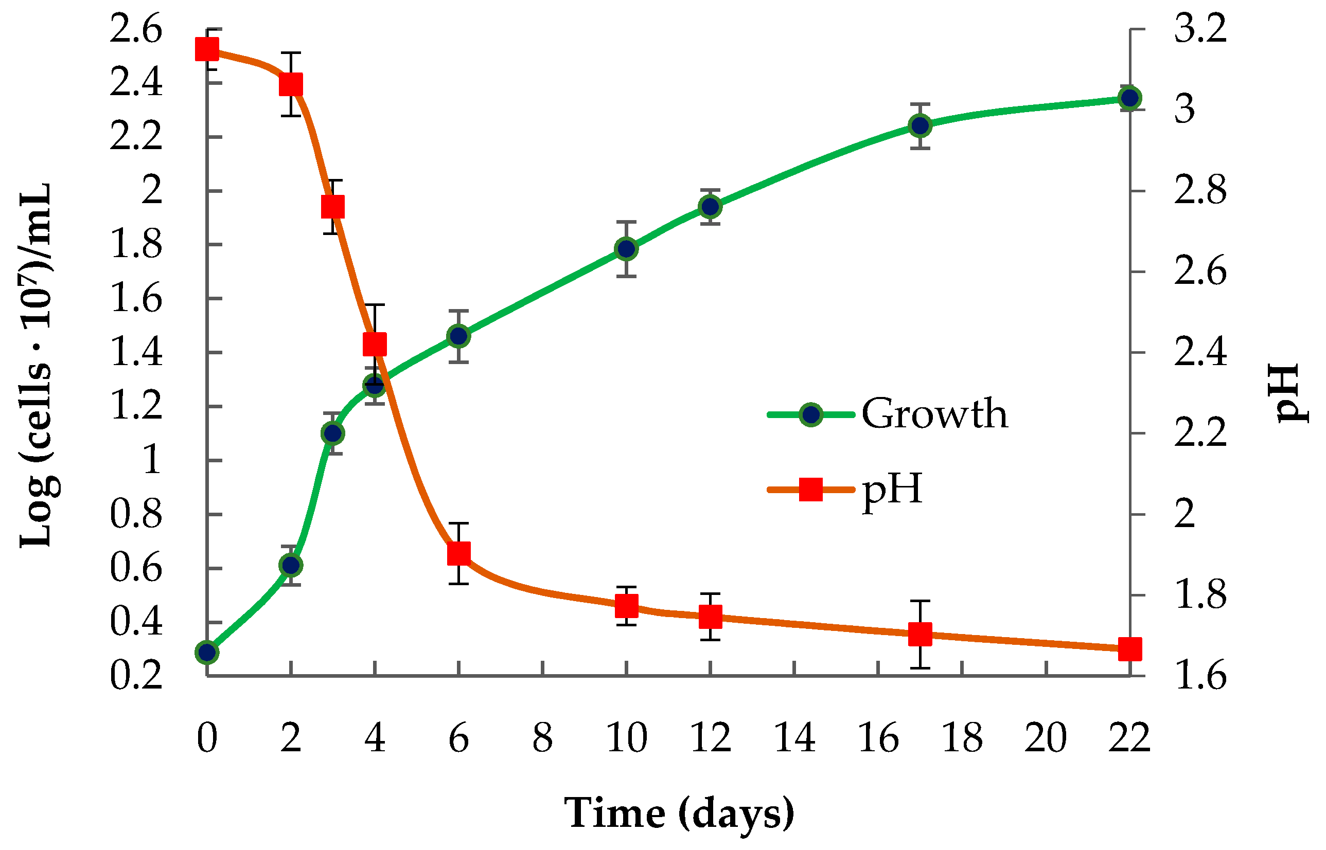
3.2. Phenotypic Features of the Strain Ac. multivorum LMS

Phenotypic characteristics of the novel LMS strain were studied and compared to those of the strains described in the scientific literature. However, the detailed phenotypic characterization is available only for its close phylogenetic relative: the type strain Ac. multivorum AIU301T [2]. The main phenotypic features of Ac. multivorum LMS are summarized and compared to characteristics of Ac. multivorum AIU301T in Table 1.
According to the results, the LMS strain had wider pH and temperature ranges for growth than the strain AIU301 (Table 1). A higher temperature optimum for growth of the LMS strain (30–40 °C) and a wider pH optimum (pH 2.5–4.0), as well as broader pH and temperature ranges (especially the upper-temperature limit of 47 °C and the lower pH limit of 1.6), may be associated with the competitive advantage of this strain under the conditions of the industrial processing of sulfidic concentrates (38–42 °C, pH 1.8–2.0). Moreover, the type of strain of the species Ac. multivorum was not shown to oxidize RISCs, while the novel LMS strain was able to grow in the media containing elemental sulfur or thiosulfate as sole energy sources. Curves for the growth and sulfur oxidation by the LMS strain under optimal mixotrophic conditions of cultivation are shown in Figure 2. Neither other Ac. multivorum strains nor Ac. cryptum isolates used S0 or other RISCs in their metabolism, except for the strain Ac. cryptum DX1-1, which showed 99.7% similarity to the LMS strain (according to the 16S rRNA gene sequence comparison). The strain DX1-1 can grow with either organic substances or inorganic sulfur energy substrates [6]. With sulfur as the sole energy source, the strain DX1-1 had a low growth rate and cell density, which, however, increased when a small amount of glucose was added to the medium [6].
Interestingly, the isolated LMS strain was represented by single or dividing cells (Figure 3a); however, after a series of culture transfers, cells tended to aggregate (Figure 3b–d). Under heterotrophic, autotrophic, and mixotrophic conditions, Ac. multivorum LMS formed aggregates that consisted of from three to several hundred cells. Such aggregation may be similar to that observed for Acidiphilium sp. C61, Ac. cryptum JF-5, and Acidiphilium SJH [13]. Extracellular polymeric substances (EPS) are considered to be among the major structural components of biofilm matrix and these aggregates, in particular. Genes encoding proteins involved in the synthesis and secretion of exopolysaccharides, such as glycosyltransferases, the polysaccharide biosynthesis/export protein (wza), and the capsular polysaccharide export protein (kps) were identified in Acidiphilium strains [13]. In the LMS strain, several glycosyltransferase genes were also found, and Wza homologs were identified: ICJ77_06775 (contig 9) and ICJ77_11025 (contig 20). The presence of different pathways involved in exopolysaccharide precursor production suggests that EPS biosynthesis plays a significant role in Acidiphilium sp. C61 aggregate formation [13]. In the case of Ac. multivorum LMS, the formation of aggregates may also be associated with sulfur oxidation. Cells can adhere to the sulfur particles forming aggregates on their surface (Figure 3b).

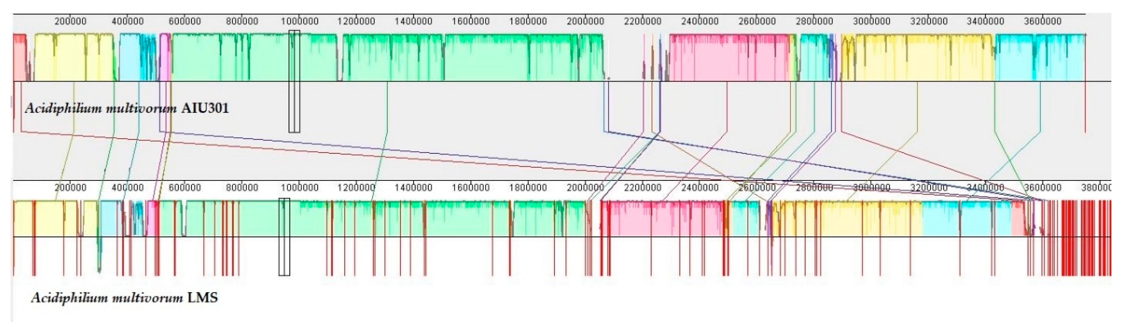
3.3. General Description and Specific Features of Ac. multivorum LMS Genome

The draft genome sequence of Ac. multivorum LMS consisted of 3,837,559 bp with an overall G + C content of 67.3% (Table 2). A total of 3676 genes were predicted. Among them, 3492 coding genes and 131 pseudogenes were identified. Genes with unclear functions were annotated as hypothetical proteins. Functional annotation of proteins encoded by the genes of the LMS draft genome sequence was carried out. According to the functional classification in clusters of orthologous groups (COGs), all identified proteins were grouped into 21 functional categories (Table 3). Figure 4 shows the alignment of the genomes of Ac. multivorum LMS and the type strain Ac. multivorum AIU301T, indicating the close relationship between them and the highly similar metabolic potential, as also previously reported for Acidiphilium strains [12].
The circular representation of the Ac. multivorum LMS draft genome and genomes of other Acidiphilium strains used for comparison is shown in Figure 5. Table 4 indicates the average nucleotide identity (ANI) between the genomes of Ac. multivorum LMS and other members of the genus Acidiphilium, showing the relatedness among Acidiphilium strains. Based on the results of this comparison (Figure 5 and Table 4), the genome of the LMS strain showed the highest homology to that of the strain Acidiphilium sp. AccII isolated from the acid mine drainage (AMD) water sample obtained in the mining area [9]. At the same time, the strain AccII contained more unique genes with adaptive functions than two other strains that were more similar to each other: AccI (isolated from the same sample as AccII) and strain ZJSH63 from an AMD water sample collected in the heap leaching area for copper ore [9].
Acidiphilium genomes that are currently available in databases have been recently compared by Li et al. to provide insights into the metabolism and evolution of the genus Acidiphilium [9]. Therefore, in our present study, we compared the genome of the LMS strain to other Acidiphilium spp. genomes by analysis of the pangenome, core genome, and accessory genomic regions to focus on the strain-specific features of Ac. multivorum LMS. As a result, a total of 32 unique genes and corresponding proteins were found in the genome of this strain (Table S1). The strain-specific genes mainly belonged to the mobile genetic elements (such as transposons, plasmids, and prophages) or encoded the proteins involved in transcription, replication, recombination, and repair (Table S1). A number of unique genes were associated with the metal(loid) resistance mechanisms. Among them, we identified the arsR, arsD, arsA, and arsB genes of the probable arsRDABC operon, which composed a 3785-bp contig 98 (Table S1, ICJ77_17765–ICJ77_17785).
In industrial operations, arsenic concentrations may reach >120 mM at pulp densities of 20 wt% [18]. In acidic conditions, arsenic is most commonly present as either arsenate (As5+/AsO43−) or arsenite (As3+/As(OH)3). Arsenate enters the cell via the phosphate uptake system and is toxic by acting as a phosphate analog, while the more toxic arsenite enters cells via aquaglyceroporins and some sugar transporters and is toxic by binding sulfhydryl groups [35]. Since arsenic concentrations reach high values during the processing of the gold-containing arsenopyrite concentrates and may be toxic to microorganisms involved in industrial operations, our research also focused on the arsenic resistance and unique arsenic resistance genes identified in the genome of the LMS strain.
The arsenic resistance (ars) operon in microorganisms encodes a transport system that extrudes arsenate, arsenite, and antimonite from the cells, lowering the intracellular concentration of toxic anions. The ars operons encoded by plasmids are known to confer plasmid-mediated resistance of Ac. multivorum AIU301, Escherichia (E.) coli, and Staphylococcus (St.) aureus to arsenic and antimony [36]. The arsenic resistance (ars) operon from plasmid pKW301 of Ac. multivorum AIU 301 conferred resistance of E. coli to arsenate and arsenite when overexpressed in E. coli cells [36]. Moreover, another arsenic resistance operon (arsRCB) and the gene encoding ArsH (arsenical resistance protein) are present in all clades of the genus Acidiphilium [9], including Ac. multivorum LMS (contig 3, ICJ77_02625, ICJ77_02630, and ICJ77_02635). The proteins encoded by the arsR, arsC, and arsB genes of the LMS strain showed the highest amino acid (a. a.) similarity to those of Ac. multivorum AIU301 and Ac. cryptum JF-5: 99.2, 100, and 99.8% (100% coverage), respectively. The percentage of identity (a. a.) to the corresponding proteins of other Acidiphilium spp. strains was 77.9–99.8% (95–100% coverage).
In general, Acidiphilium genomes were shown to harbor an abundant repertoire of horizontally transferred genes contributing to environmental adaption, which suggests that the genus Acidiphilium originated in mild conditions and adapted to extreme environments after the acquisition of diverse essential functions [9]. In contrast to the arsRCB operon, which is common for all studied Acidiphilium genomes, the arsR, arsD, arsA, and arsB genes of the probable arsRDABC operon of the LMS strain shared similarity with phylogenetically distinct microorganisms and were strain-specific within the genus Acidiphilium (Table S1). These genes were probably derived by the LMS strain via the horizontal gene transfer (HGT) from other metal-resistant microorganisms. The arsenical resistance operon repressor ArsR and the arsenical resistance operon trans-acting repressor ArsD showed the closest similarity to those of Pseudaminobacter (Ps.) arsenicus: 83.2 and 83.9% a. a. similarity, respectively. The arsenic-resistant strain Ps. arsenicus CB3T (pH range for growth 6.0–9.0) was isolated from arsenic-rich aquifers of the Jianghan Plain (Hubei, China), the groundwater of which is severely contaminated by arsenic [37]. The arsenical pump-driving ATPase ArsA involved in the removal of arsenate, antimonite, and arsenate from the cell showed the highest similarity to that of Acetobacteraceae bacterium SCN 69-10 (85.4% a. a. identity, 100% coverage). Other close homologs of the ArsA pump belonged to Ps. arsenicus, At. caldus, At. ferrivorans, At. ferrooxidans, and L. ferriphilum (72.0–85.4% a. a. similarity, 99% coverage). As mentioned above, Acidithiobacillus and Leptospirillum strains are common members of acidophilic communities in biomining operations, including the processing of gold-bearing pyrite-arsenopyrite concentrates [15,18,19,20,21]. The closest homolog of the arsenical efflux pump membrane protein ArsB was found to belong to the Gemmataceae bacterium (94% a. a. similarity, 100% coverage).
Research into the arsenic tolerance of biomining microorganisms has indicated that the arsenic efflux system is an important pathway of arsenite detoxification in L. ferriphilum and At. thiooxidans strains [38]. The overall response of Sb. thermotolerans cells to the toxic amounts of the gold-containing arsenopyrite concentrate was shown to involve mainly the mechanisms for resistance to arsenic [19]. The acquisition of additional arsenic resistance determinants by At. caldus and L. ferriphilum strains may be associated with their improved growth under arsenic stress [35]. In this study, Ac. multivorum LMS that was isolated from the pulp of the gold-containing sulfidic concentrate also carried additional arsenic resistance genes, which probably gave this strain an advantage over some other acidophiles in the microbial community and might explain its predominance in the oxidation processes of the pyrite-arsenopyrite concentrates.
Among other proteins specific for the LMS strain within the genus Acidiphilium, one protein related to heavy metal resistance was identified (contig 76, ICJ77_17235, Table S1). The closest homolog of this protein belonged to the copper and cadmium translocating P-type ATPase (ZntA) of Acidocella (Ad.) aminolytica (89.5% a. a. identity, 99% coverage). The genera Acidocella and Acidiphilium form two clusters within the alpha-I subclass of the Proteobacteria [39]. ZntA couples the hydrolysis of ATP with the export of zinc, cadmium, or lead; it can also bind nickel, copper, cobalt, and mercury [40].

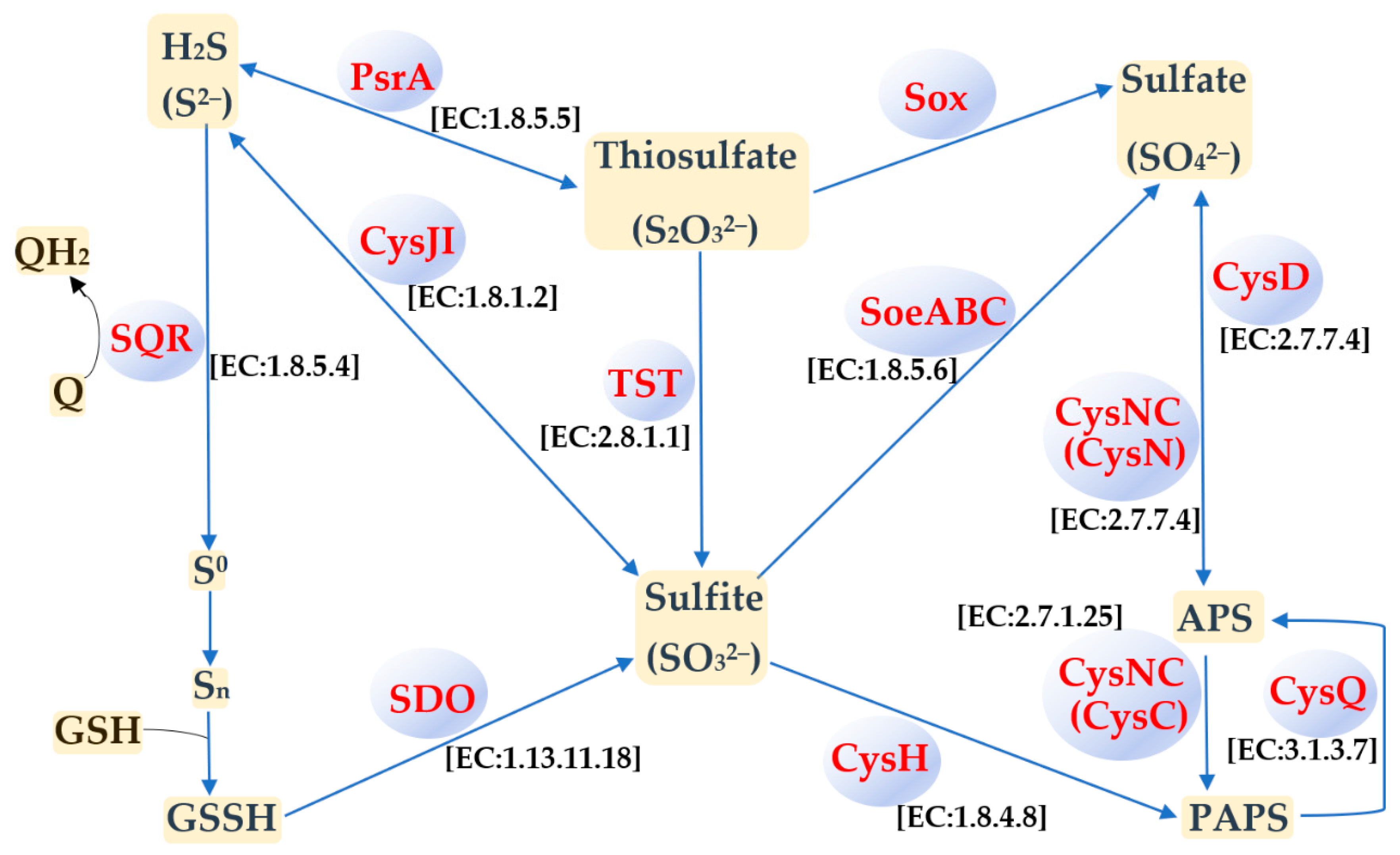
3.4. Sulfur Metabolism

Metabolic pathways of Acidiphilium strains assigned to different phylogenetic clades were compared in a recent study [9], and different components of the sulfur, nitrogen, and carbon metabolic pathways were shown to be acquired through HGT [9]. In the genome of Ac. multivorum LMS, we also determined the genes responsible for dissimilatory and assimilatory nitrate reduction, denitrification, and nitrification. KEGG pathways for carbon metabolism (including methane metabolism) in the LMS strain were also predicted. However, in this study, we focused on the metabolic ability of the LMS strain to oxidize RISCs, which is an unusual feature of the heterotrophic species Ac. multivorum. Analysis of the genome of Ac. multivorum LMS allowed the determination of the genes encoding different proteins involved in the oxidation of elemental sulfur, sulfite, thiosulfate, and sulfide (Table S2). Based on the NCBI and KEGG annotations, as well as the ability of the LMS strain to oxidize RISCs under mixotrophic and autotrophic conditions, a biochemical model for sulfur oxidation in Ac. multivorum LMS was proposed (Figure 6).
Previous analyses of the probable sulfur metabolic pathways predicted from the genome annotations have shown that certain components of sulfur metabolism are encoded by all Acidiphilium genomes [9]. In Ac. multivorum LMS, the Sox multi-enzyme complex encoded by the soxCDXYZAB gene cluster was found (Table S2). As in the case of other Acidiphilium strains assigned to clade IV [9], including the closest phylogenetic relative Acidiphilium sp. AccII, the Sox multi-enzyme complex of Ac. multivorum LMS was likely derived from the most recent common ancestor of clades III and IV. This conclusion was supported by the previous data [9], as well as the highest similarity of the Sox system components of the LMS strain (Table S2) to those of the members of the genus Acidiphilium (up to 98–100% a. a. identity, 100% coverage). In contrast to the Sox complexes of clades III and IV, those of clades I and II were probably acquired via independent HGT events [9]. The typical periplasmic multi-enzyme Sox system is involved in the oxidation of thiosulfate, sulfide, sulfite, and elemental sulfur to produce sulfate as the final product [41]. In Acidithiobacillus strains (the most well-studied sulfur- and iron-oxidizing acidophiles), the Sox-pathway-dependent sulfur oxidation is known to provide higher sulfur oxidation capacities (higher S0-oxidizing rate and biomass) compared to Sox-deficient strains [41].
Three genes coding for thiosulfate/3-mercaptopyruvate sulfurtransferase (TST) (EC:2.8.1.1 2.8.1.2) (ICJ77_05465 (contig 7), ICJ77_08765 (contig 13), and ICJ77_09760 (contig 16); Table S2) were also identified in the genome of the LMS strain. Rhodanase (TST) catalyzes the pathway of S2O32− utilization, producing sulfite [41] (Figure 6).
Among other enzymes involved in sulfur oxidation, sulfur dioxygenases (SDOs) were predicted from the annotated genome (Table S2; Figure 6). Possibly, SDOs play an essential role in sulfur oxidation by Ac. multivorum LMS. The oxidation of S0 to sulfite, catalyzed by SDO, was first reported in At. ferrooxidans [42]. Further studies showed that SDO used the sulfane sulfur atom of glutathione persulfide (GSSH) (and not S0) as a substrate to produce sulfite [43]. Although SDO activity was first reported in 1987 [42] and the actual substrate of SDO was determined in 2003 [43], the sdo genes in Acidithiobacillus spp. were identified only several years ago [44]. Moreover, a new subgroup of SDOs was recently identified in At. caldus. The previously identified SDO group [44] and the new one were designated Sdo2 and Sdo1, respectively [45].
As in the case of Acidiphilium spp. strains (clades I, III, and IV) [9], a homolog of the new Sdo1 associated with tetrathionate oxidation in At. caldus MTH-04 (Sdo1, A5904_0421) [45] was identified in the genome of Ac. multivorum LMS (Table S2). A comparison using the NCBI BLAST revealed a 49.4% a. a. identity (100% coverage) between Sdo1 of At. caldus MTH-04 and a metallo-beta-lactamase family protein of the LMS strain (ICJ77_14405, contig 38; Table S2). Moreover, we also identified a homolog of Sdo2 of At. caldus MTH-04 (A5904_0790, [45]) in the LMS strain (ICJ77_00340, contig 1; Table S2), with a 35% a. a. similarity (93% coverage) between them. Interestingly, a strong correlation between the sdo1 gene coding for Sdo1 and the tetrathionate intermediate pathway was previously revealed in At. caldus MTH-04 [45]. The deletion of sdo2 promoted bacterial growth on tetrathionate and thiosulfate, while the overexpression of sdo2 altered gene expression patterns of SQR and TST. Thus, sdo1 is essential for the survival of At. caldus when tetrathionate is used as the sole energy source, and sdo2 may also play a role in sulfur metabolism [45]. BLAST searching carried out in this study revealed homologs of Sdo2 of the LMS strain also in other Acidiphilium strains: Ac. cryptum JF-5, Acidiphilium sp. PM, and Acidiphilium sp. JA12-A1 (100% a. a. identity, 100% coverage); Ac. multivorum AIU301T (99.3% a. a. identity, 100% coverage); Acidiphilium spp. C61 and 37-67-22 (90.7–91.0% a. a. identity, 100% coverage); as well as Ac. rubrum (82% a. a. identity, 100% coverage) and Acidiphilium sp. 37-64-53 (81.7% a. a. identity, 100% coverage). According to the previously proposed phylogenetic classification by Li et al. [9], these strains belong to clades II–IV [9].
Thus, we propose an SDO-dependent and Sox-pathway-dependent sulfur oxidation model functioning in the LMS strain (Figure 6). According to this model, extracellular S0 is first activated by thiol-containing outer-membrane proteins to generate persulfide sulfane sulfur, and the latter is further oxidized by periplasmic SDO to produce sulfite as previously described [43]. Sulfite is oxidized in the APS (adenosine-5′-phosphosulfate)-mediated process, with the participation of Cys proteins, and by the heterotrimeric membrane-bound complex SoeABC (Figure 6). Genes encoding homologs of sulfite reductase subunit beta (CysI) and sulfate adenylyltransferase (CysD) of the LMS strain have been previously found to be upregulated in Acidiphilium cryptum DX1-1 that was grown under mixotrophic conditions in the culture medium containing glucose and sulfur [6].

4. Conclusions

In the present research, we isolated and characterized the LMS strain involved in the large-scale industrial processing of the gold-bearing pyrite-arsenopyrite ore concentrate at the Olimpiada biooxidation plant (Siberia, Russia). Surprisingly, the LMS strain that was assigned to the heterotrophic species Ac. multivorum proved to be among predominant sulfur oxidizers in the commercial acidophilic microbial communities in bioreactors. Summarizing data of several years of monitoring the industrial microbial populations, we concluded that Ac. multivorum was able to become a predominant sulfur oxidizer together with more common sulfur-oxidizing acidophiles, such as At. thiooxidans, or even succeed the latter in the commercial microbial communities. In this study, it was shown that:
  • Ac. multivorum LMS proved to be one of the predominant sulfur oxidizers in the microbial populations during industrial processing of the gold-bearing pyrite-arsenopyrite ore concentrates;
  • Phenotypic features of LMS strain included wide pH and temperature ranges for growth, as well as the ability to grow organotrophically, lithotrophically, or mixotrophically;
  • The first draft genome of the sulfur-oxidizing Ac. multivorum was sequenced, annotated, and compared to other Acidiphilium genomes available in the databases;
  • Based on the functional analysis of the genome and sulfur-oxidizing activity of the strain, a biochemical model for sulfur oxidation in Ac. multivorum LMS was proposed;
  • Strain-specific genes of Ac. multivorum LMS may confer additional resistance to arsenic and heavy metals, such as copper, cadmium, and zinc;
  • The commercial importance of Ac. multivorum LMS was proved by the efficient sulfur oxidation in the presence or absence of organic compounds, a complicated system of sulfur oxidation pathways, as well as additional metal resistance determinants specific for the LMS strain within the genus Acidiphilium.
Overall, the phenotypic features of Ac. multivorum LMS determined in this study, as well as functional analysis of its genome, made it possible to conclude that this microorganism plays a previously underestimated central role in acidophilic communities during biooxidation of gold-bearing sulfidic raw materials. Phenotypic characteristics, such as wide pH and temperature ranges for growth, the ability to oxidize RISCs, as well as versatile metabolic and resistance capacities, gave the LMS strain a competitive advantage in microbial communities of industrial stirred tank bioreactors and determined its predominance in commercial microbial populations.

Supplementary Materials

The following are available online at https://www.mdpi.com/article/10.3390/microorganisms9050984/s1, Table S1: Unique genes and corresponding proteins, identified in the genome of Ac. multivorum LMS. A comparison to other Acidiphilum genomes was carried out by analysis of core genome, pangenome, and accessory genomic regions, Table S2: Sulfur metabolism components predicted from the genome of Ac. multivorum LMS. Ortholog annotation was carried out using KOALA (KEGG Orthology And Links Annotation) system. Genome annotation was performed using the NCBI Prokaryotic Genome Annotation Pipeline and RAST (Rapid Annotation using Subsystem Technology service version 2.0 RASTtk annotation scheme).

Author Contributions

Conceptualization, A.P.; methodology, A.P. and A.B.; validation, A.P., A.B. and A.V.B.; investigation, A.P. and A.B.; formal analysis and resources, A.P., A.B. and A.V.B.; data curation, A.P., A.B. and A.V.B.; writing—original draft preparation, A.P.; writing—review and editing, A.P., A.B. and A.V.B.; visualization, A.P.; supervision and project administration, A.P.; funding acquisition, A.P. and A.B. All authors have read and agreed to the published version of the manuscript.

Funding

This research was funded by the Ministry of Science and Higher Education of the Russian Federation.

Institutional Review Board Statement

Not applicable.

Informed Consent Statement

Not applicable.

Data Availability Statement

The genome sequence of Ac. multivorum LMS was deposited in the NCBI databases under the accession number JACVVW000000000 (BioProject PRJNA662208; BioSample SAMN16078398). The 16S rRNA gene sequences of the Acidiphilium sp. strains PB-B3-4, PB-B1-3, PB-B2-3, and LMS were deposited in the GenBank databases under accession numbers MW389315–MW389318.

Conflicts of Interest

The authors declare no conflict of interest.

References

  1. Harrison, A.P., Jr. Acidiphilium cryptum gen. nov., sp. nov., heterotrophic bacterium from acidic mineral environments. Int. J. Syst. Evol. Microbiol. 1981, 31, 327–332. [Google Scholar] [CrossRef] [Green Version]
  2. Wakao, N.; Nagasawa, N.; Matsuura, T.; Matsukura, H.; Matsumoto, T.; Hiraishi, A.; Matsumoto, T.; Hiraishi, A.; Sakurai, Y.; Shiota, H. An acidophilic chemoorganotrophic bacterium from pyritic acid mine drainage. J. Gen. Appl. Microbiol. 1994, 40, 143–159. [Google Scholar] [CrossRef]
  3. Hallberg, K.B.; Johnson, D.B. Biodiversity of acidophilic prokaryotes. Adv. Appl. Microbiol. 2001, 49, 37–84. [Google Scholar] [CrossRef]
  4. Rawlings, D.E.; Johnson, D.B. The microbiology of biomining: Development and optimization of mineral-oxidizing microbial consortia. Microbiology 2007, 153, 315–324. [Google Scholar] [CrossRef] [Green Version]
  5. Hiraishi, A.; Nagashima, K.V.P.; Matsuura, K.; Shimada, K.; Takaichi, S.; Wakao, N.; Katayama, Y. Phylogeny and photosynthetic features of Thiobacillus acidophilus and related acidophilic bacteria: Its transfer to the genus Acidiphilium as Acidiphilium acidophilum comb. nov. Int. J. Syst. Bacteriol. 1998, 48, 1389–1398. [Google Scholar] [CrossRef]
  6. Xu, A.-I.; Xia, J.-I.; Song, Z.-W.; Jiang, P.; Xia, Y.; Wan, M.-X.; Zhang, R.-Y.; Yang, Y.; Liu, K.-K. The effect of energy substrates on PHB accumulation of Acidiphilium cryptum DX1-1. Curr. Microbiol. 2013, 67, 379–387. [Google Scholar] [CrossRef] [PubMed]
  7. Johnson, D.B.; Bridge, T.A.M. Reduction of ferric iron by acidophilic heterotrophic bacteria: Evidence for constitutive and inducible enzyme systems in Acidiphilium spp. J. Appl. Microbiol. 2002, 92, 315–321. [Google Scholar] [CrossRef]
  8. Mahapatra, N.R.; Benerjee, P.C. Extreme tolerance to cadmium and high resistance to copper, nickel and zinc in different Acidiphilium strains. Lett. Appl. Microbiol. 1996, 23, 393–397. [Google Scholar] [CrossRef]
  9. Li, L.; Liu, Z.; Zhang, M.; Meng, D.; Liu, X.; Wang, P.; Li, X.; Jiang, Z.; Zhong, S.; Jiang, C.; et al. Insights into the metabolism and evolution of the genus Acidiphilium, a typical acidophile in acid mine drainage. mSystems 2020, 5, e00867-20. [Google Scholar] [CrossRef]
  10. San Martin-Uriz, P.; Gomez, M.J.; Arcas, A.; Bargiela, R.; Amils, R. Draft genome sequence of the electricigen Acidiphilium sp. strain PM (DSM 24941). J. Bacteriol. 2011, 193, 5585–5586. [Google Scholar] [CrossRef] [PubMed] [Green Version]
  11. San Martin-Uriz, P.; Mirete, S.; Alcolea, P.J.; Gomez, M.J.; Amils, R.; Gonzalez-Pastor, J.E. Nickel-resistance determinants in Acidiphilium sp. PM identified by genome-wide functional screening. PLoS ONE 2014, 9, e95041. [Google Scholar] [CrossRef] [PubMed] [Green Version]
  12. Ullrich, S.R.; Poehlein, A.; Voget, S.; Hoppert, M.; Daniel, R.; Leimbach, A.; Tischler, J.S.; Schlömann, M.; Mühling, M. Permanent draft genome sequence of Acidiphilium sp. JA12-A1. Stand. Genom. Sci. 2015, 10, 56. [Google Scholar] [CrossRef]
  13. Li, Q.; Cooper, R.; Wegner, C.-E.; Küsel, K. Molecular mechanisms underpinning aggregation in Acidiphilium sp. C61 isolated from iron-rich pelagic aggregates. Microorganisms 2020, 8, 314. [Google Scholar] [CrossRef] [PubMed] [Green Version]
  14. Micciche, A.C.; Barabote, R.D.; Dittoe, D.K.; Ricke, S.C. In silico genome analysis of an acid mine drainage species, Acidiphilium multivorum, for potential commercial acetic acid production and biomining. J. Environ. Sci. Health B 2020, 55, 447–454. [Google Scholar] [CrossRef] [PubMed]
  15. Bulaev, A.; Belyi, A.; Panyushkina, A.; Solopova, N.; Pivovarova, T. Microbial population of industrial biooxidation reactors. Solid State Phenom. 2017, 262, 48–52. [Google Scholar] [CrossRef]
  16. Kaksonen, A.H.; Boxall, N.J.; Gumulya, Y.; Khaleque, H.N.; Morris, C.; Bohu, T.; Cheng, K.Y.; Usher, K.M.; Lakaniemi, A.-M. Recent progress in biohydrometallurgy and microbial characterisation. Hydrometallurgy 2018, 180, 7–25. [Google Scholar] [CrossRef]
  17. Tuffin, I.M.; Hector, S.B.; Deane, S.M.; Rawlings, D.E. Resistance determinants of a highly arsenic-resistant strain of Leptospirillum ferriphilum isolated from a commercial biooxidation tank. Appl. Environ. Microbiol. 2006, 72, 2247–2253. [Google Scholar] [CrossRef] [PubMed] [Green Version]
  18. Tsaplina, I.A.; Panyushkina, A.E.; Melamud, V.S.; Grigor’eva, N.V.; Kondrat’eva, T.F. Leaching of pyrite–arsenopyrite concentrate in bioreactors during continuous cultivation of a thermoacidophilic microbial community. Microbiology 2014, 83, 568–576. [Google Scholar] [CrossRef]
  19. Panyushkina, A.; Matyushkina, D.; Pobeguts, O. Understanding stress response to high-arsenic gold-bearing sulfide concentrate in extremely metal-resistant acidophile Sulfobacillus thermotolerans. Microorganisms 2020, 8, 1076. [Google Scholar] [CrossRef]
  20. Belyi, A.V.; Chernov, D.V.; Solopova, N.V. Development of BIONORD® technology on Olimpiada deposit refractory arsenic-gold ores treatment in conditions of Extreme North. Hydrometallurgy 2018, 179, 188–191. [Google Scholar] [CrossRef]
  21. Panyushkina, A.E.; Tsaplina, I.A.; Kondrat’eva, T.F.; Belyi, A.V.; Bulaev, A.G. Physiological and morphological characteristics of acidophilic bacteria Leptospirillum ferriphilum and Acidithiobacillus thiooxidans, members of a chemolithotrophic microbial consortium. Microbiology 2018, 87, 326–338. [Google Scholar] [CrossRef]
  22. Panyushkina, A.E.; Babenko, V.V.; Nikitina, A.S.; Selezneva, O.V.; Tsaplina, I.A.; Letarova, M.A.; Kostryukova, E.S.; Letarov, A.V. Sulfobacillus thermotolerans: New insights into resistance and metabolic capacities of acidophilic chemolithotrophs. Sci. Rep. 2019, 9, 15069. [Google Scholar] [CrossRef] [PubMed] [Green Version]
  23. Bulaev, A.; Belyi, A.; Panyushkina, A.; Solopova, N. Molecular–biological monitoring of microbial population of industrial bioleach bioreactors. In Proceedings of the 23rd International Biohydromatallurgy Symposium (IBS), Fukuoka, Japan, 20–23 October 2019; p. 13. [Google Scholar]
  24. Silverman, M.P.; Lundgren, D.G. Studies on the chemoautotrophic iron bacteria Ferrobacillus ferrooxidans. An improved medium and harvesting procedure for securing high yields. J. Bacteriol. 1959, 77, 642–647. [Google Scholar] [CrossRef] [Green Version]
  25. Kumar, S.; Stecher, G.; Li, M.; Knyaz, C.; Tamura, K. MEGA X: Molecular evolutionary genetics analysis across computing platforms. Mol. Biol. Evol. 2018, 35, 1547–1549. [Google Scholar] [CrossRef]
  26. Saitou, N.; Nei, M. The neighbor-joining method: A new method for reconstructing phylogenetic trees. Mol. Biol. Evol. 1987, 4, 406–425. [Google Scholar] [CrossRef] [PubMed]
  27. Felsenstein, J. Confidence limits on phylogenies: An approach using the bootstrap. Evolution 1985, 39, 783–791. [Google Scholar] [CrossRef]
  28. Tamura, K.; Nei, M.; Kumar, S. Prospects for inferring very large phylogenies by using the neighbor-joining method. Proc. Natl. Acad. Sci. USA 2004, 101, 11030–11035. [Google Scholar] [CrossRef] [PubMed] [Green Version]
  29. Bankevich, A.; Nurk, S.; Antipov, D.; Gurevich, A.A.; Dvorkin, M.; Kulikov, A.S.; Lesin, V.M.; Nikolenko, S.I.; Pham, S.; Prjibelski, A.D.; et al. SPAdes: A new genome assembly algorithm and its applications to single-cell sequencing. J. Comput. Biol. 2012, 19, 455–477. [Google Scholar] [CrossRef] [PubMed] [Green Version]
  30. Overbeek, R.; Olson, R.; Pusch, G.D.; Olsen, G.J.; Davis, J.J.; Disz, T.; Edwards, R.A.; Gerdes, S.; Parrello, B.; Shukla, M.; et al. The SEED and the Rapid Annotation of microbial genomes using Subsystems Technology (RAST). Nucleic Acids Res. 2014, 42, D206–D214. [Google Scholar] [CrossRef] [PubMed]
  31. Kanehisa, M.; Sato, Y.; Kawashima, M.; Furumichi, M.; Tanabe, M. KEGG as a reference resource for gene and protein annotation. Nucleic Acids Res. 2016, 44, D457–D462. [Google Scholar] [CrossRef] [PubMed] [Green Version]
  32. Darling, A.E.; Mau, B.; Perna, N.T. ProgressiveMauve: Multiple genome alignment with gene gain, loss and rearrangement. PLoS ONE 2010, 5, e11147. [Google Scholar] [CrossRef] [PubMed] [Green Version]
  33. Alikhan, N.-F.; Petty, N.K.; Ben Zakour, N.L.; Beatson, S.A. BLAST Ring Image Generator (BRIG): Simple prokaryote genome comparisons. BMC Genom. 2011, 12, 402. [Google Scholar] [CrossRef] [Green Version]
  34. Laing, C.; Buchanan, C.; Taboada, E.N.; Zhang, Y.; Kropinski, A.; Villegas, A.; Thomas, J.E.; Gannon, V.P.J. Pan-genome sequence analysis using Panseq: An online tool for the rapid analysis of core and accessory genomic regions. BMC Bioinform. 2010, 11, 461. [Google Scholar] [CrossRef] [PubMed] [Green Version]
  35. Dopson, M.; Holmes, D. Metal resistance in acidophilic microorganisms and its significance for biotechnologies. Appl. Microbiol. Biotechnol. 2014, 98, 8133–8144. [Google Scholar] [CrossRef]
  36. Suzuki, K.; Wakao, N.; Kimura, T.; Sakka, K.; Ohmiya, K. Expression and regulation of the arsenic resistance operon of Acidiphilium multivorum AIU 301 plasmid pKW301 in Escherichia coli. Appl. Environ. Microbiol. 1998, 64, 411–418. [Google Scholar] [CrossRef] [Green Version]
  37. Mu, Y.; Zhou, X.; Liu, L.; Zhou, X.-K.; Zeng, X.-C.; Li, W.-J. Pseudaminobacter arsenicus sp. nov., an arsenic-resistant bacterium isolated from arsenic-rich aquifers. Int. J. Syst. Evol. Microbiol. 2019, 69, 791–797. [Google Scholar] [CrossRef]
  38. Jiang, H.; Liang, Y.; Yin, H.; Xiao, Y.; Guo, X.; Xu, Y.; Hu, Q.; Liu, H.; Liu, X. Effects of arsenite resistance on the growth and functional gene expression of Leptospirillum ferriphilum and Acidithiobacillus thiooxidans in pure culture and coculture. BioMed Res. Int. 2015, 2015, 203197. [Google Scholar] [CrossRef] [Green Version]
  39. Kishimoto, N.; Kosako, Y.; Wakao, N.; Tano, T.; Hiraishi, A. Transfer of Acidiphilium facilis and Acidiphilium aminolytica to the genus Acidocella gen. nov, and emendation of the genus Acidiphilium. Syst. Appl. Microbiol. 1995, 18, 85–91. [Google Scholar] [CrossRef]
  40. Dutta, S.J.; Liu, J.; Stemmler, A.J.; Mitra, B. Conservative and nonconservative mutations of the transmembrane CPC motif in ZntA: Effect on metal selectivity and activity. Biochemistry 2007, 46, 3692–3703. [Google Scholar] [CrossRef]
  41. Wang, R.; Lin, J.-Q.; Liu, X.-M.; Pang, X.; Zhang, C.-J.; Yang, C.-L.; Gao, X.-Y.; Lin, C.-M.; Li, Y.-Q.; Li, Y.; et al. Sulfur oxidation in the acidophilic autotrophic Acidithiobacillus spp. Front. Microbiol. 2019, 9, 3290. [Google Scholar] [CrossRef] [PubMed]
  42. Sugio, T.; Mizunashi, W.; Inagaki, K.; Tano, T. Purification and some properties of sulfur: Ferric ion oxidoreductase from Thiobacillus ferrooxidans. J. Bacteriol. 1987, 169, 4916–4922. [Google Scholar] [CrossRef] [PubMed] [Green Version]
  43. Rohwerder, T.; Sand, W. The sulfane sulfur of persulfides is the actual substrate of the sulfur-oxidizing enzymes from Acidithiobacillus and Acidiphilium spp. Microbiology 2003, 149, 1699–1710. [Google Scholar] [CrossRef] [PubMed] [Green Version]
  44. Wang, H.; Liu, S.; Liu, X.; Li, X.; Wen, Q.; Lin, J. Identification and characterization of an ETHE1-like sulfur dioxygenase in extremely acidophilic Acidithiobacillus spp. Appl. Microbiol. Biotechnol. 2014, 98, 7511–7522. [Google Scholar] [CrossRef] [PubMed]
  45. Wu, W.; Pang, X.; Lin, J.; Liu, X.; Wang, R.; Lin, J.; Chen, L. Discovery of a new subgroup of sulfur dioxygenases and characterization of sulfur dioxygenases in the sulfur metabolic network of Acidithiobacillus caldus. PLoS ONE 2017, 12, e0183668. [Google Scholar] [CrossRef] [PubMed] [Green Version]
Figure 1. Dendrogram of Acidiphilium strains based on the 16S rRNA gene sequences, showing the phylogenetic position of Ac. multivorum LMS. The phylogenetic tree was constructed with MEGA X [25]. The evolutionary history was inferred using the Neighbor-Joining method [26]. The percentage of replicate trees in which the associated taxa clustered together in the bootstrap test (1000 replicates) is shown next to the branches [27]. The bootstrap values of ≥80 are shown. The tree is drawn to scale, with branch lengths in the same units as those of the evolutionary distances used to infer the phylogenetic tree. The evolutionary distances were computed using the Maximum Composite Likelihood method [28] and are in the units of the number of base substitutions per site. This analysis involved 11 nucleotide sequences. There were a total of 1511 positions in the final dataset. Kozakia baliensis DSM 14400T was used as an outgroup.
Microorganisms 09 00984 g001
Figure 2. Growth of Ac. multivorum LMS and pH decrease under mixotrophic conditions in the medium supplemented with 0.1% yeast extract and 1% S0.
Microorganisms 09 00984 g002
Figure 3. Micrographs of Ac. multivorum LMS cells grown under mixotrophic conditions in the medium containing 1% S0 and 0.1% yeast extract after isolation from the bioreactor (a) and after 5–7 culture transfers in the same medium (bd). Phase-contrast microscopy. Arrows indicate cell aggregates. S, sulfur. Scale bar, 10 µm.
Microorganisms 09 00984 g003
Figure 4. Alignment of reordered contigs of the Ac. multivorum LMS draft genome and the related reference genome of the type strain Ac. multivorum AIU301T, using the ProgressiveMauve software and Mauve Contig Mover tool [32]. The scale shows sequence coordinates. Colored blocks in the first genome are connected by lines to similarly colored blocks in the second genome. These lines indicate homologous regions in the genomes. Areas that are completely white and not aligned contain sequence elements specific to the genomes.
Microorganisms 09 00984 g004
Figure 5. Circular map of the aligned draft genome sequence of Ac. multivorum LMS and other Acidiphilium genomes, using the BLAST Ring Image Generator (BRIG) software program [33]. The panel on the right shows color codes for different Acidiphilium strains and the identity levels (%) with the Ac. multivorum LMS genome. ACC2, ACC1, and ZJSH63: Acidiphilium spp. AccII, AccI, and ZJSH63, respectively [9]; AIU301 and JF5: Ac. multivorum AIU301T and Ac. cryptum JF-5, respectively [14]; C61, Acidiphilium sp. C61 [13]; PM and JA12-A1: Acidiphilium spp. PM [10] and JA12-A1 [12], respectively.
Microorganisms 09 00984 g005
Figure 6. Sulfur metabolism pathways predicted from the genome of Ac. multivorum LMS annotated using the NCBI Prokaryotic Genome Annotation Pipeline and KOALA (KEGG Orthology And Links Annotation) tools [31]. SQR: Sulfide:quinone oxidoreductase (EC:1.8.5.4); SDO: Sdo and Sdo1 sulfur dioxygenases (EC:1.13.11.18); Cys JI: CysJ sulfite reductase (NADPH) flavoprotein alpha-component (EC:1.8.1.2) and CysI sulfite reductase (NADPH) hemoprotein beta-component (EC:1.8.1.2); TST: thiosulfate/3-mercaptopyruvate sulfurtransferase (EC:2.8.1.1 2.8.1.2); PsrA: Thiosulfate reductase/polysulfide reductase chain A (EC:1.8.5.5); SoeABC: Sulfite dehydrogenase (quinone) subunit SoeA (EC:1.8.5.6), sulfite dehydrogenase (quinone) subunit SoeB, and sulfite dehydrogenase (quinone) subunit SoeC; Sox: Sox multi-enzyme system that consists of sulfane dehydrogenase subunit SoxC, S-disulfanyl-l-cysteine oxidoreductase SoxD (EC:1.8.2.6), l-cysteine S-thiosulfotransferase SoxX (EC:2.8.5.2), sulfur-oxidizing protein SoxY, sulfur-oxidizing protein SoxZ, l-cysteine S-thiosulfotransferase SoxA (EC:2.8.5.2), and S-sulfosulfanyl-l-cysteine sulfohydrolase SoxB (EC:3.1.6.20); CysNC: Bifunctional enzyme CysN/CysC (EC:2.7.7.4 2.7.1.25); CysD: Sulfate adenylyltransferase subunit 2 (EC:2.7.7.4); CysH: Phosphoadenosine phosphosulfate reductase/phosphoadenylyl-sulfate reductase (EC:1.8.4.8 1.8.4.10); CysQ: 3′(2′),5′-Bisphosphate nucleotidase (EC:3.1.3.7); Q: Quinones; QH2: Quinol pool; GSH: Glutathione; GSSH: Glutathione persulfide; S0: Elemental sulfur; Sn: Polysulfide; APS: Adenosine-5′-phosphosulfate; PAPS: Phosphoadenosine phosphosulfate. Substrates, intermediates, and products are indicated in yellow, and proteins catalyzing the reactions are indicated in blue.
Microorganisms 09 00984 g006
Table 1. Main phenotypic features of Ac. multivorum LMS compared to characteristics of the type strain Ac. multivorum AIU301T.
ParameterLMS 1AIU301 2
Source of isolationIndustrial bioreactorAMD 3
MorphologyRods, single cells, and aggregatesRods, single cells, and chains
Cell size, µm0.3–0.5 × 0.8–1.00.5–1.2 × 0.9–3.8
Motility++
pH range1.6–5.51.9–5.6
pHopt 42.5–4.33.2–4.0
T5 range, °C<20–4717–42
Topt, °C30–4027–35
µmax6, h−10.02 (S0 + YE7)0.004–0.062 (organic substrates)
Growth with:
YE 7++
Glucose++
YE + S0+
S0+
YE + Na2S2O3+
Na2S2O3+
Fe2+
Reference(present study)[2]
1 LMS, Ac. multivorum LMS (present study); 2 AIU301, Ac. multivorum AIU301T; 3 AMD, acid mine drainage; 4 opt, optimum; 5 T, temperature; 6 µmax, maximum specific growth rate; 7 YE, yeast extract.
Table 2. Statistics of the genome of Ac. multivorum LMS.
AttributeValue
Genome size (bp)3,837,559
Number of contigs139
Number of contigs (≥1000 bp)135
Number of contigs (≥5000 bp)84
Number of contigs (≥10,000 bp)62
Number of contigs (≥25,000 bp)43
Number of contigs (≥50,000 bp)23
Largest contig308,818
DNA, G + C (%)67.3
Genes (total)3676
CDSs (total)3623
Genes (coding)3492
CDSs (with protein)3492
Genes (RNA)53
Complete rRNAs1, 1, 1 (5S, 16S, 23S)
tRNAs46
ncRNAs4
Pseudogenes (total)131
CRISPR arrays1
Table 3. Results of the ortholog annotation of the proteins encoded by the genome of Ac. multivorum LMS (56.1% annotated) using KOALA (KEGG Orthology And Links Annotation) tools [31].
Functional CategoryNumber of Proteins 1
Carbohydrate metabolism 242
Protein families: Genetic information processing235
Environmental information processing226
Protein families: Signaling and cellular processes213
Genetic information processing164
Amino acid metabolism157
Unclassified: Metabolism134
Energy metabolism117
Metabolism of cofactors and vitamins104
Unclassified103
Cellular processes100
Nucleotide metabolism63
Unclassified: Genetic information processing55
Protein families: Metabolism53
Lipid metabolism46
Glycan biosynthesis and metabolism39
Unclassified: Signaling and cellular processes39
Xenobiotics biodegradation and metabolism36
Metabolism of other amino acids28
Metabolism of terpenoids and polyketides14
1 Number of proteins encoded by Ac. multivorum LMS that are associated with the main functional categories.
Table 4. The average nucleotide identity (ANI) between the genomes of Ac. multivorum LMS and the most closely related Acidiphilium strains. The ANI values were calculated with the ANI calculator (Kostas lab, Atlanta, GA, USA) using reciprocal best hits (two-way ANI).
Acidiphilium StrainANI Value (SD 1), %
Acidiphilium sp. AccII99.29 (1.43)
Acidiphilium sp. JA12-A199.23 (0.83)
Ac. multivorum AIU301T99.12 (1.92)
Ac. cryptum JF-5 99.18 (1.02)
Acidiphilium sp. ZJSH63 99.04 (1.52)
Acidiphilium sp. AccI98.97 (1.84)
1 SD, standard deviation.
Publisher’s Note: MDPI stays neutral with regard to jurisdictional claims in published maps and institutional affiliations.

