Article

Characterisation of Phage Susceptibility Variation in Salmonellaenterica Serovar Typhimurium DT104 and DT104b

School of Life Sciences, College of Liberal Arts and Sciences, University of Westminster, 115 New Cavendish Street, Fitzrovia, London W1W 6XH, UK
*
Author to whom correspondence should be addressed.
Microorganisms 2021, 9(4), 865; https://doi.org/10.3390/microorganisms9040865
Submission received: 8 March 2021 / Revised: 6 April 2021 / Accepted: 13 April 2021 / Published: 17 April 2021
(This article belongs to the Special Issue Salmonella and Salmonellosis)

Abstract

:
The surge in mortality and morbidity rates caused by multidrug-resistant (MDR) bacteria prompted a renewal of interest in bacteriophages (phages) as clinical therapeutics and natural biocontrol agents. Nevertheless, bacteria and phages are continually under the pressure of the evolutionary phage–host arms race for survival, which is mediated by co-evolving resistance mechanisms. In Anderson phage typing scheme of Salmonella Typhimurium, the epidemiologically related definitive phage types, DT104 and DT104b, display significantly different phage susceptibility profiles. This study aimed to characterise phage resistance mechanisms and genomic differences that may be responsible for the divergent phage reaction patterns in S. Typhimurium DT104 and DT104b using whole genome sequencing (WGS). The analysis of intact prophages, restriction–modification systems (RMS), plasmids and clustered regularly interspaced short palindromic repeats (CRISPRs), as well as CRISPR-associated proteins, revealed no unique genetic determinants that might explain the variation in phage susceptibility among the two phage types. Moreover, analysis of genes coding for potential phage receptors revealed no differences among DT104 and DT104b strains. However, the findings propose the need for experimental assessment of phage-specific receptors on the bacterial cell surface and analysis of bacterial transcriptome using RNA sequencing which will explain the differences in bacterial susceptibility to phages. Using Anderson phage typing scheme of Salmonella Typhimurium for the study of bacteria-phage interaction will help improving our understanding of host–phage interactions which will ultimately lead to the development of phage-based technologies, enabling effective infection control.

1. Introduction

Non-typhoidal Salmonella (NTS) serovars predominantly cause a self-limiting diarrhoeal illness; however, they have recently observed to cause invasive extra-intestinal disease, including bacteraemia and focal systemic infections [1,2].
The global burden of disease caused by invasive NTS is significant and substantially exacerbated by the emergence of antibiotic resistant strains. The multidrug-resistant (MDR) Salmonella enterica serovar Typhimurium definitive phage type DT104 and the closely related DT104b are of considerable concern worldwide [3,4].
Interestingly, the emergence of MDR bacteria prompted a renewal of interest in bacteriophages (phages) as clinical therapeutics and natural biocontrol agents of foodborne pathogens, including Salmonella [5].
Although bacteria develop phage resistance mechanisms, phages continuously co-evolve to circumvent these anti-phage mechanisms [6,7,8]. Briefly, phage adsorption to a receptor (e.g., O-antigen lipopolysaccharide) on the cell surface is the initial step of the phage infection and host–phage interaction in S. Typhimurium [9]. Upon injecting its genetic material into the cytoplasm of the host cell, the phage follows either a lytic or lysogenic lifecycle. In the lytic cycle, the phage hijacks bacterial cell metabolic machinery to assemble virions and subsequently cause cell lysis. However, in the lysogenic cycle, the repressed phage genome integrates into the bacterial chromosome as a prophage, which provides the bacterium with a prophage-encoded phage resistance, superinfection exclusion (Sie), which prevents secondary infection with the same or closely related phage [8,10]. Nevertheless, the evolution of bacterial genomes allowed bacteria to acquire an array of mechanisms interfering with every step of phage infection. Blocking or modification of phage receptors, production of extracellular matrix, and production of competitive inhibitors enable bacteria to inhibit phage adsorption. However, when a phage succeeds in injecting its genome into a host cell, bacteria possess a variety of nucleic acid degrading systems that protect them from phage infection, such as restriction–modification systems (RMS) and clustered regularly interspaced short palindromic repeats (CRISPR), as well as CRISPR-associated (Cas) proteins [8,11,12,13].
Although MDR S. Typhimurium DT104 and DT104b are epidemiologically related, in the Anderson phage typing scheme [14], they display different phage susceptibility profiles, as shown in Table 1. We have previously shown that Anderson phage typing scheme of S. Typhimurium is a valuable model for study of phage host interaction [5] as whole genome sequencing (WGS) provided possible explanations for the difference in phage susceptibility among Salmonella Typhimurium DT8 and DT30. In this study, we performed comparative genome analysis on WGS data of representative strains of S. Typhimurium DT104 and DT104b to characterise phage resistance mechanisms and genomic differences that may impact the phenotypic lysis profiles. Understanding of host–phage interactions will ultimately lead to the development of phage-based technologies, enabling effective infection control.

2. Materials and Methods

2.1. Bacterial Strains

Sequences of nine representative S. Typhimurium DT104 (n = 5) and DT104b (n = 4) were selected for the study. In addition to a well-studied S. Typhimurium strain, LT2 (DT4) was included in the comparative analysis. The whole genome sequences (WGSs) of all phage types were obtained from EnteroBase (https://enterobase.warwick.ac.uk/). The EnteroBase Barcodes and accession numbers of all strains as well as the year and country of isolation are presented in Table 2. All DT104b strains were isolated in Ireland in 2006, from swine (n = 2), the environment (n = 1), and food sources (n = 1). The DT104 strains were collected in the United Kingdom (n = 1), Ireland (n = 1), Germany (n = 1), and France (n = 1) over the 1975–2004 period. The DT104 strains were of human (n = 3) and bovine (n = 1) origin. The resistance profiles of all strains are provided in Supplementary Table S1. ResFinder 4.1 [15] was used to identify acquired antimicrobial resistance genes (ARGs) and chromosomal mutations, rendering Salmonella isolates, genotypically, resistant to antibiotics.

2.2. Identification of SNPs and Phylogenomics

The CSI Phylogeny 1.4 tool at the Center for Genomic Epidemiology [16] was used to identify single nucleotide polymorphisms (SNPs) and infer a phylogeny. The input parameters were as follows: (a) minimum depth at SNP positions at 10X; (b) minimum relative depth at SNP position at 10%; (c) minimum distance between SNPs (prune) at 10 bp; (d) minimum SNP quality score at 30; (e) minimum read mapping quality of 25; and (f) minimum Z-score (standard score) at 1.96. A phylogenetic tree was constructed based on the identified SNPs using FastTree. The Newick tree data were visualised on the MEGA X software [17]. The Salmonella Typhimurium strain LT2 was used to outgroup the phylogenetic tree.

2.3. Identification of Prophages, Plasmids, and R-M and CRISPR/Cas Systems

Lysogenic prophages integrated into S. Typhimurium genomes were determined using the web-based tool Phage Search Tool Enhanced Release, PHASTER [18], applying default parameters.
We constructed maximum likelihood (ML) phylogenetic trees based on the SNPs of detected prophages in all strains of DT104 and DT104b.
The identification and classification of the R-M systems were performed using Restriction-ModificationFinder 1.1, REBASE [19,20]. A threshold of 95% was selected for minimum percent identity (%ID) between the sequence in the input genome and the restriction enzyme gene sequence in Restriction-ModificationFinder 1.1; the selected minimum length was set at 60%. The Basic Local Alignment Search Tool (BLAST) on the National Center for Biotechnology (NCBI) website [21] was used to confirm prophages and the R-M systems.
Detection of CRISPR arrays and subtyping of Cas systems were performed on CRISPRFinder [22] and CRISPRCasFinder [23] web servers using default settings. In addition, the plasmid database; PLSDB [24] was used to identify plasmids present within the S. Typhimurium genomes. The analysis was completed using Mash (search strategy: mash screen) with a maximal p-value of 0.1 and minimal identity of 0.99. To remove redundancy from the output data, the winner-takes-all strategy was applied.

3. Results

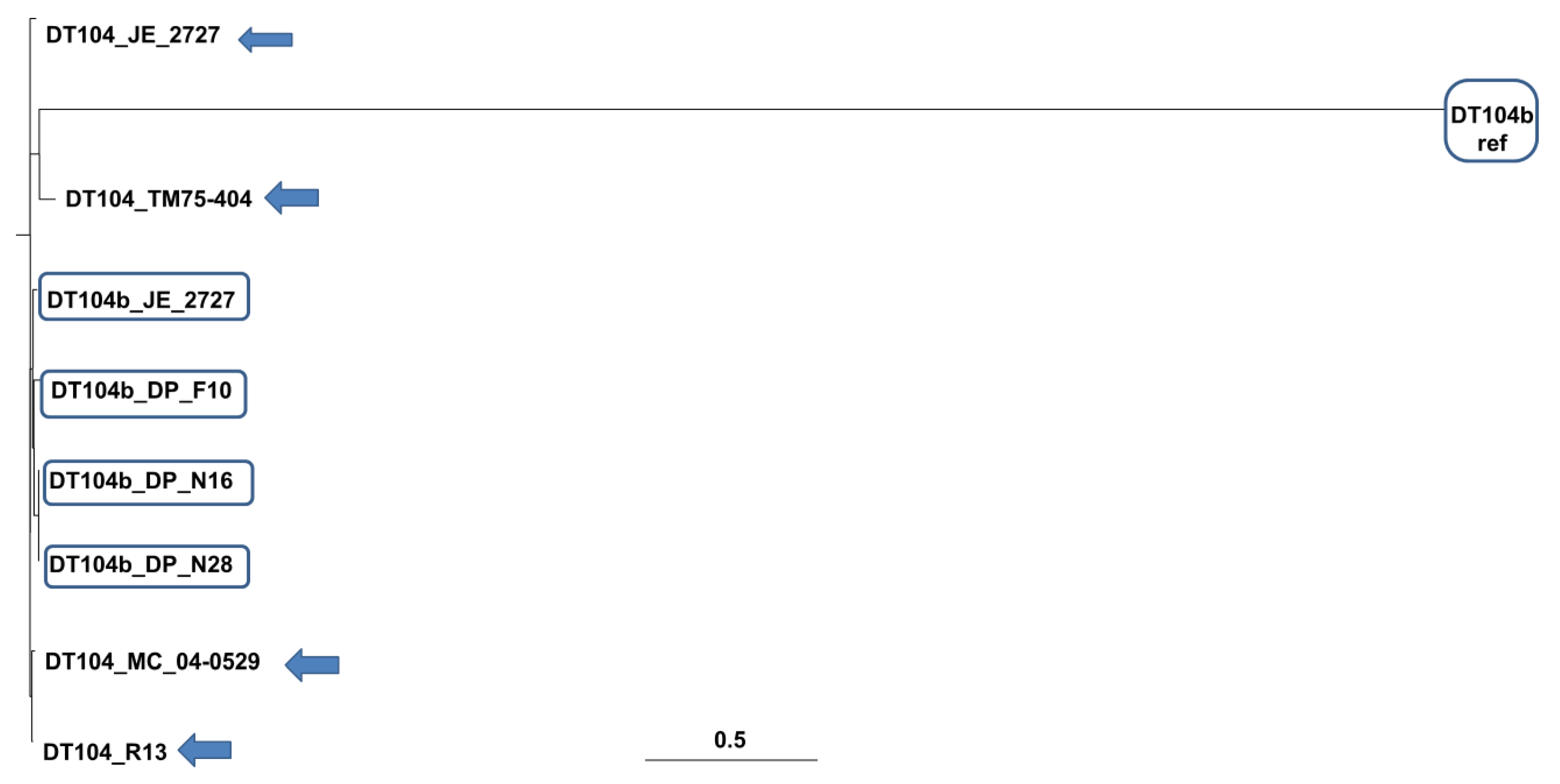
3.1. Phylogenomics of S. Typhimurium DT104 and DT104b

Phylogenetic analysis was performed to predict genetic relatedness between S. Typhimurium strains. Figure 1 illustrates the phylogenetic neighbourhood, and Table 3 contains the SNP distance matrix among the S. Typhimurium genomes. Strains DP_F10, DP_N16, DP_N28, JE_2727, JM_04.26, MC_04-0529, R13 and DT104 reference strain (DT104 ref.) were found to be closely related; notably, DP_N16 and DP_N28 showed a close relation with a bootstrap value of 100% (Figure 1). In contrast, the DT104b reference strain (DT104b ref.) as well as the S. Typhimurium strain LT2 (DT4) displayed significant divergence among the studied genomes and were located on the same clade. Moreover, the SNP distance matrix exhibited an SNP divergence between TM75-404 and other studied genome sequences (Table 3).

3.2. Prophages in S. Typhimurium DT104 and DT104b

The PHASTER prophage analysis web server identified intact (score > 90), questionable (score 70–90), and incomplete phages (score < 70) in S. Typhimurium genomes [18]. Intact and questionable phages were confirmed by the BLAST database [21]; the confirmed prophages are shown in Table 4.
Phages Salmon_118970_sal3 (NC_031940) and Salmon_ST64B (NC_004313) were detected in all DT104b and DT104 strains but were absent in S. Typhimurium str. LT2 (DT4) which, unlike other strains, harbours Fels-1 (NC_010391) and Fels-2 (NC_010463). Gifsy-2 (NC_010393) and Gifsy-1 (NC_010392) were commonly shared between the studied genomes, whereas phage Entero_ST104 (NC_005841) was absent in the DT104b reference strain (DT104b ref.) and LT2 (DT4). In contrast, phage Salmon_SP_004 (NC_021774) was present in only DT104b ref. and phage Entero_lato (NC_001422) in the TM75-404 strain.
Phylogenetic analyses based on the SNPs of prophages of all strains of DT104 and DT10b showed that strains of DT104 are intermixed with the strains of DT104b (Supplementary Figure S1).
Two different superinfection exclusion proteins (protein B and protein gp 17) were detected in all DT104 and DT104b strains, except one of the DT104b strains—DT104b ref (Supplementary Table S3).

3.3. R-M Systems S. Typhimurium DT104 and DT104b

As shown in Table 5, Restriction-ModificationFinder 1.1, REBASE [19,20] identified four R-M systems (Type I, Type II, Type III, and Type IV) in the studied genomes.
The Type I R-M system includes genes encoding restriction endonucleases, methyltransferases, and the specificity subunit (S.StyUK1II). The presence and location of the putative Type I restriction enzyme StyUK1IIP were confirmed by BLAST [21]. The StyUK1IIP gene resides in the close proximity to the cognate methyltransferases (M.SenTFII and M.Sen1899II); the percent identity was 99.97% (E value = 0.0) for the DT104b reference strain and 100% (E value = 0.0) for the remaining S. Typhimurium DT104b and DT104 strains. Putative Type I restriction enzyme SenLT2IIP was identified in the S. Typhimurium strain LT2 (DT4). The methyltransferase M.SenTFII was detected in all of the studied genomes except for the DT104b reference strain, which harbours M.Sen1899II. Nevertheless, both genes (M.SenTFII and M.Sen1899II) are closely related (99.94% percent identity, E value = 0.0) and code for the N6-adenine DNA methyltransferase subunit recognizing GAGNNNNNNRTAYG motifs.
The genes (M.SenAboDcm, M.Sen641III, M.StyUK1V) coding for methyltransferases, in the Type II R-M system, are common in all of the strains, except for LT2, which lacks methyltransferase M.StyUK1V. Moreover, Type IIG Sty13348III (restriction enzyme and methyltransferase) was identified in all of the genomes, excluding the LT2 and DT104b reference strains that instead harbour StyUK1IV Type IIG. The Type IIG Sty13348III and StyUK1IV genes are closely related (100% percent identity, E value = 0.0) and code for restriction endonuclease and methyltransferase recognising GATCAG motifs.
The genes of Type III and Type IV R-M systems were common for all studied S. Typhimurium genomes. The methyltransferase M.StyUK1I and restriction enzyme SenAZII were identified for Type III; for Type IV, only StyLT2Mrr was identified, which entails methyl-directed restriction.

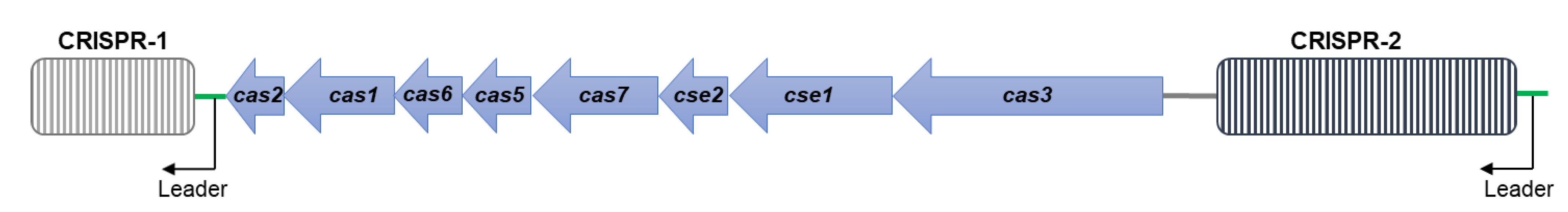
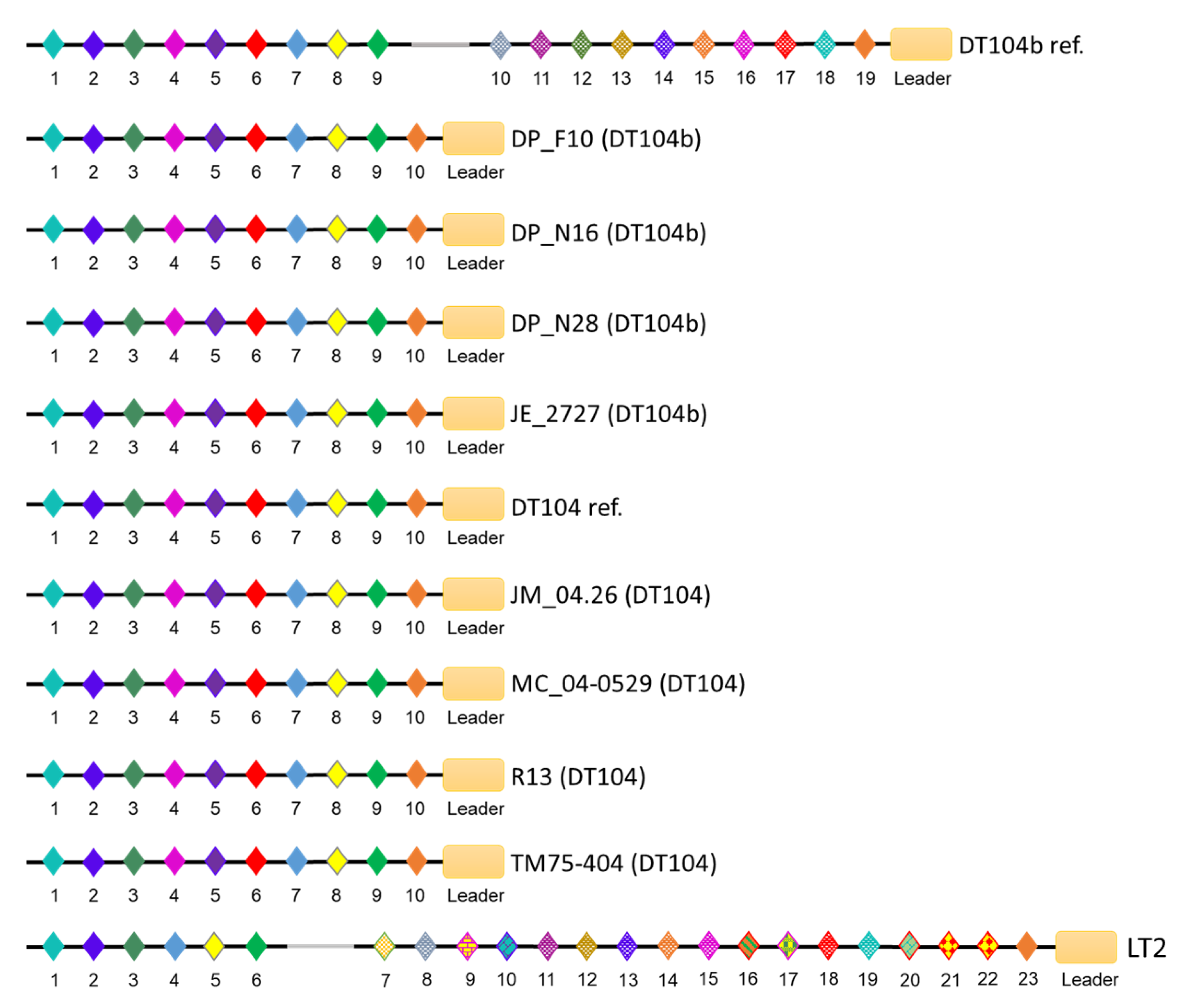
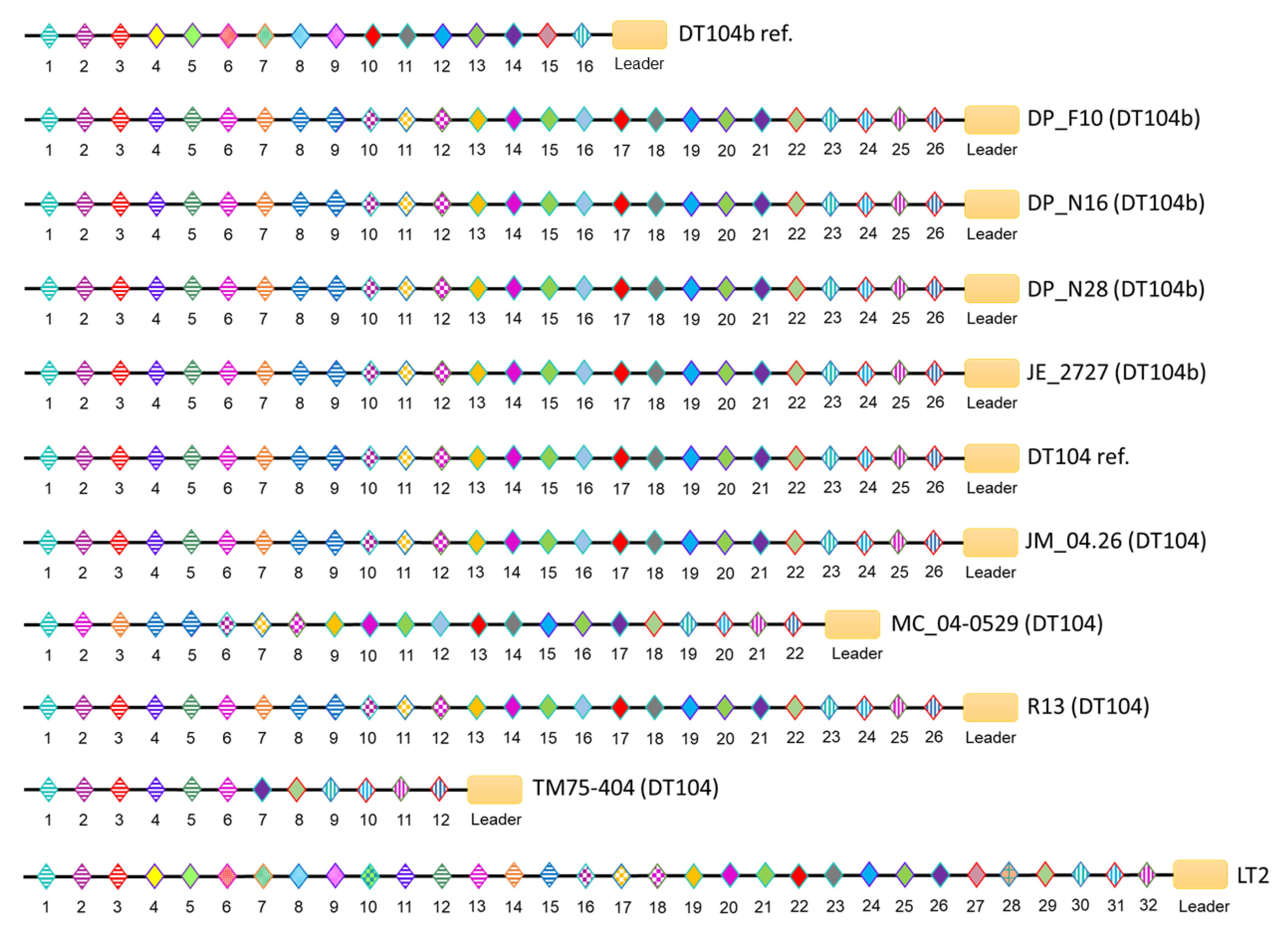
3.4. CRISPR/Cas Systems in S. Typhimurium DT104 and DT104b

Two CRISPR loci (CRISPR-1 and CRISPR-2) and a type I-E Cas cluster were detected in all studied S. Typhimurium strains (Table 6). The type I-E CRISPR/Cas encompasses eight cas genes: cas2, cas1, cas6, cas5, cas7, cse2, cse1 and cas3 (Figure 2).
The CRISPR arrays in both loci share the same direct repeat sequence (29 base pairs). The CRISPR-1 arrays were identical in all DT104 and DT104b genomes, only with spacer variation in the DT104b reference strain, which harbours a unique spacer. Additionally, the DT104b reference strain and LT2 (DT4) share eight unique spacers positioned at the leader proximal end of the array (Figure 3). Although spacers within the CRISPR-2 loci of S. Typhimurium DT104b and DT104 showed high similarities, the length of arrays differed among the genomes with n = 22 and n = 12 spacers in MC_04-0529 and TM75-404, respectively, as well as n = 26 in DP_F10, DP_N16, DP_N28, JE_2727, DT104 reference, JM_04.26, and R13. Moreover, the CRISPR-2 loci of the DT104b reference contain n = 16 spacers, of which n = 7 are unique to DT104b ref. and LT2. The unique spacers were positioned internally (n = 6) and at the leader proximal end of the array (n = 1), as shown in Figure 4. The arrays within CRISPR-1 and CRISPR-2 were commonly shared between DP_F10, DP_N16, DP_N28, JE_2727, DT104 reference, JM_04.26, and R13.

3.5. Plasmids in S. Typhimurium DT104 and DT104b

PLSDB [24] identified ten plasmids among S. Typhimurium genomes, of which nine were detected in S. Typhimurium DT104 and DT104b (Table 7). The 93,939 base pairs (bp) virulence pSLT (NC_003277.2) plasmid was only detected in the S. Typhimurium strain LT2 (DT4).
Similar to DT104 and DT104b strains, LT2 carries the 33,784 bp pSE81-1705-3 (NZ_CP018654.1) plasmid as well as a megaplasmid (147,787 bp) that was first identified in the Salmonella enterica subsp. Enterica serovar Senftenberg strain NCTC10384, plasmid 3 (NZ_LN868945.1). The 94,045 bp pSC-09-1 (NZ_CP028319.1) plasmid was conserved in all DT104 and DT104b strains, except for the DT104b reference that harbours a linear 93,862 bp plasmid (NZ_LT855377.1). Significantly, the 3319 bp p1PCN033 (NZ_CP006633.1), carried by all DT104b strains (excluding DT104 or DT4), was absent in the DT104b reference strain.

Potential Phage Receptors

Analysis of potential genes coding for phage receptors revealed no differences among DT104 and DT104b strains (Supplementary Table S2).

3.6. WGS-Based Identification of Antimicrobial Resistance Determinants

The chromosomally located AAC (6’)-Iaa gene that renders amikacin and tobramycin ineffective was identified in all of the studied genomes. Seven S. Typhimurium strains exhibit an ACSSuT (ampicillin, chloramphenicol, streptomycin, sulphonamides, and tetracycline) penta-resistance profile, and these are DP_F10, DP_N16, DP_N28, JE_2727, MC_04-0529, R13, and the DT104 reference strain (Supplementary Table S1).

4. Discussion

Worldwide, infections and outbreaks caused by non-typhoidal S. Typhimurium represent a significant public health concern and an economic burden [25]. Zoonotic S. Typhimurium DT104, as well as DT104b, has a broad-host-range, and it is primarily transmitted through food sources and the faecal–oral route. The infection ranges from self-limiting gastroenteritis to invasive disease that requires prompt antibiotic therapy [1,2].
Nonetheless, selective pressure led to the emergence and subsequent dissemination of MDR S. Typhimurium DT104 and DT104b strains that phenotypically display an ACSSuT resistance profile. Moreover, recently, it has been reported that Salmonella has become increasingly resistant to other antibiotics of clinical significance, such as ciprofloxacin [26]. Limited treatment options for invasive and extraintestinal salmonellosis can lead to treatment failure and a surge in mortality and morbidity. The epidemic incidence and the global dissemination of MDR Salmonella prompted a renewal of interest in phages as clinical therapeutics and natural biocontrol agents [7,27,28]. Nevertheless, bacteria and phages are continually under the pressure of the evolutionary phage–host arms race for survival, which is mediated by the evolving anti-phage mechanisms in bacteria and parallel co-evolution of phage genomes [7,8,27].
Until recently, surveillance and outbreak investigation of S. Typhimurium was widely performed by phenotypic phage typing that relies on phage–host interaction. Advances in molecular biology and the emergence of WGS significantly improved the surveillance and outbreak investigation as well as the ability to comprehensively characterise the causative pathogen in silico [29,30,31].
The focus of this study was to characterise phage resistance mechanisms and genomic differences that may be responsible for the divergent phage reaction patterns in S. Typhimurium DT104 and DT104b.
SNPs were used to infer phylogenetic relationships between S. Typhimurium genomes as well as to assess the genetic diversity of the DT104 and DT104b strains. The phylogenetic analysis could not unambiguously differentiate phage types. Notably, the DT104b reference strain (DT104b ref) displayed significant divergence among the DT104b studied genomes. This may have been caused by the accumulation of SNPs in prophage regions or the cas genes but also the spacer variation of the CRISPR arrays and acquisition or loss of prophages and plasmids that result in the diversification of closely related bacteria [32,33,34,35].
Repressed and integrated into the bacterial chromosome, temperate phage genomes (prophages) provide the lysogen with a superinfection exclusion immunity. Through mechanisms such as blocking of phage DNA entry into the lysogen’s cytoplasm or inhibition of phage lysozyme, the superinfection immunity resists secondary infection by the same or closely related phages, providing the lysogen with a survival advantage [36]. Additionally, many phages carry virulence and antibiotic resistance genes [37].
The genomes of the strains analysed in this study possess multiple intact prophages that may have been inherited vertically or acquired due to the exposure to a multitude of bacterial viruses. Phages Salmon_118970_sal3 (NC_031940), Salmon_ST64B (NC_004313), Gifsy-1 (NC_010392), and Gifsy-2 (NC_010393) were highly prevalent among the S. Typhimurium DT104b and DT104 genomes; however, none of the strains harboured Fels-1 (NC_010391) and Fels-2 (NC_010463), which were detected in the S. Typhimurium reference strain LT2 (DT4). Nonetheless, the lysogen analysis did not reveal a significant difference in prophage profiles of DT104 and DT104b.
Moreover, SNP analysis of the detected prophages showed that strains of DT104 are intermixed with the strains of DT104b, indicating that prophages cannot explain the difference in bacterial susceptibility to typing phages.
Analysis of the genes coding for potential phage receptors revealed no differences in phage binding sites among DT104 and DT104b strains.
R-M systems allow bacteria to resist phage infection by degrading their DNA if recognised as foreign. They consist of methyltransferase and a cognate restriction endonuclease. The methyltransferase catalyses methylation of DNA to protect the self-genome from degradation by the restriction enzyme, which recognises and cleaves unmethylated (foreign) DNA at specific recognition sites. Based on the subunit composition and protein complexes, the R-M systems are classified into four types (I–IV) [38,39]. The in silico analysis and comparison of S. Typhimurium DT104 and DT104b R-M systems did not uncover possible explanatory causes of different lysis profiles. Although there were some variations in enzymes of Type I and Type II R-M systems of the DT104b reference strain, the detected methyltransferases and endonucleases were homologous to those of other DT104 and DT104b strains as well as LT2.
The CRISPR/Cas systems provide bacteria with adaptive and sequence-specific immunity against phages and plasmids. A CRISPR locus comprises a CRISPR array flanked by a cas operon. The CRISPR array is made up of short palindromic repeats (identical in length and sequence) that are interspaced by segments of DNA (spacers) derived from previous exposures to phages [8,40,41]. CRISPR/Cas loci are categorised into two distinct classes, class 1 CRISPR/Cas and class 2 CRISPR/Cas, as well as 5 types and 16 subtypes. The classification is based on the signature genes encoding interference modules and distinctive architecture of cas loci. The interference modules of the class 1 CRISPR/Cas systems contain multi-subunit crRNA–effector complexes, whereas class 2 systems possess a single subunit crRNA–effector module with a signature cas9 gene [40]. S. Typhimurium DT104 and DT104b harbour two CRISPR loci: CRISPR-1 and CRISPR-2. Located upstream of CRISPR-1 loci, eight signature cas genes (cas2, cas1, cas6, cas5, cas7, cse2, cse1, and cas3) belong to the class 1 CRISPR/Cas systems, type I subtype E (I-E). The type I-E system utilises multiprotein effector crRNA complexes, known as Cascade (CRISPR-associated complex for antiviral defence), to mediate interference of incoming nucleic acids. The Cascade complex consists of Cse1, Cse2, Cas7, Cas5, and Cas6 proteins, as well as crRNA. In type I systems, Cas6 processes pre-crRNA, resulting in intermediate crRNAs, and Cas 3 induces cleavage of the target DNA. Cas 1 and Cas 2 proteins, which are prevalent amongst the majority of CRISPR/Cas types, form a protein complex responsible for the incorporation of protospacers into the CRISPR array [40,41].
A comparison of CRISPR arrays of S. Typhimurium DT104 to CRISPR arrays of the DT104 reference strain, as well as a comparison of DT104b to CRISPR arrays of the DT104b reference strain, revealed potential loss and gain of spacers within CRISPR loci of DT104 and DT104b. Although the CRISPR-1 arrays were identical in all DT104 and DT104b genomes, the comparison of CRISPR-1 arrays of DT104b to the DT104b reference implies that DT104b lost nine contiguous internal spacers. In contrast, the composition of CRISPR-2 arrays within DT104b genomes suggests greater exposure to multiple phages compared to the DT104b reference strain. Interestingly, spacer duplication in CRISPR-2 was observed in the majority of studied genomes. The analysis of CRISPRs did not provide a possible explanation for the differing phage susceptibility of DT104 and DT104b. However, the composition of CRISPR arrays partially reflects the phylogenetic distances between the S. Typhimurium genomes, where strains possessing identical CRISPR loci were closely related. Nonetheless, Shariat et al. (2015) proposed that Salmonella CRISPR/Cas systems ceased its immunogenic function [42].
Acquired through horizontal gene transfer (HGT), plasmids (the elements of an accessory genome) provide bacteria with adaptive traits that can advantage bacteria under certain circumstances and stressors, such as antibiotics. The literature suggests that besides carrying virulence and antibiotic resistance genes, plasmids may also encode active R-M systems against phages [5,43]. Alternatively, the possession of conjugative antibiotic resistance plasmids may render bacteria susceptible to phages [44]. In this study, the distribution of plasmids amongst the DT104 and DT104b strains was assessed to predict whether these plasmid profiles could have impacted phage susceptibility patterns. Notably, unlike LT2 (DT4), DT104, and the DT104b reference, the DT104b strains harbour a low-molecular-weight (3319 bp) p1PCN033 plasmid, which has been associated with virulence and resistance traits [45]. However, it is unknown whether the p1PCN033 plasmid has a role in bacterial phage susceptibility or resistance; further studies are required to determine genetic markers that may be responsible for host–phage interaction.

5. Conclusions

In silico analysis of WGS of well-documented S. Typhimurium phage types DT104 and DT104b revealed no unique genetic determinants that might explain the variation in phage susceptibility among the two different phage types. However, this pilot study corroborates the complex dynamics of bacteria–phage interaction that limits conventional phage therapy. It also implies the necessity for further research, such as a study of host receptors involved in recognition and adsorption of phages, as well as phage counterstrategies to circumvent bacterial anti-phage mechanisms. Also, analysis of bacterial transcriptome using RNA sequencing will explain the differences in bacterial susceptibility and resistance to phages. Using Anderson phage typing scheme of Salmonella Typhimurium for the study of bacteria-phage interaction will help improving improving our understanding of host–phage interactions which will ultimately lead to the development of phage-based technologies, enabling effective infection control.

Supplementary Materials

The following are available online at https://www.mdpi.com/article/10.3390/microorganisms9040865/s1, Figure S1: Phylogenetic analysis of prophages among DT104 and DT104b. Table S1: WGS-based identification of antimicrobial resistance determinants among MDR DT104 and DT104b, Table S2: Distribution of potential genes coding for phage receptors among all strains of DT104 and DT104b, Table S3: Distribution of prophages among DT104 and DT104b.

Author Contributions

Conceptualisation, M.M.; formal analysis, B.O. and M.M.; methodology, M.M.; writing—draft manuscript, B.O.; writing—reviewing and editing, M.M.; supervision, M.M. All authors have read and agreed to the published version of the manuscript.

Funding

Manal Mohammed is funded by a Quinton Hogg start-up award from the University of Westminster.

Institutional Review Board Statement

Not applicable.

Informed Consent Statement

Not applicable.

Data Availability Statement

The data that support the findings of this study are available from the corresponding author upon reasonable request.

Conflicts of Interest

The authors declare no conflict of interest.

References

  1. Mohammed, M.; Le Hello, S.; Leekitcharoenphon, P.; Hendriksen, R. The invasome of Salmonella Dublin as revealed by whole genome sequencing. BMC Infect. Dis. 2017, 17, 544. [Google Scholar] [CrossRef] [PubMed] [Green Version]
  2. Mohammed, M.; Thapa, S. Evaluation of WGS-subtyping methods for epidemiological surveillance of foodborne salmonellosis. One Health Outlook 2020, 2, 13. [Google Scholar] [CrossRef]
  3. Rajashekara, G.; Haverly, E.; Halvorson, D.A.; Ferris, K.E.; Lauer, D.C.; Nagaraja, K.V. Multidrug-Resistant Salmonella Typhimurium DT104 in Poultry. J. Food Prot. 2000, 63, 155–161. [Google Scholar] [CrossRef]
  4. Khan, A.; Nawaz, M.; Khan, S.; Cerniglia, C. Detection of multidrug-resistant Salmonella typhimurium DT104 by multiplex polymerase chain reaction. FEMS Microbiol. Lett. 2000, 182, 355–360. [Google Scholar] [CrossRef] [PubMed]
  5. Mohammed, M.; Cormican, M. Whole genome sequencing provides possible explanations for the difference in phage susceptibility among two Salmonella Typhimurium phage types (DT8 and DT30) associated with a single foodborne outbreak. BMC Res. Notes 2015, 8, 728. [Google Scholar] [CrossRef] [PubMed] [Green Version]
  6. Mohammed, M. Who Fights Whom? Understanding the Complex Dynamics of Bacteria Phage Interaction Using Anderson Phage Typing System. J. Infect. Dis. Ther. 2018, 6, 3. [Google Scholar] [CrossRef] [Green Version]
  7. Food Standards Agency 2016. Research Project: Systematic and Critical Review on the Potential Use of Bacteriophages on Foods. Food Standards Agency. Available online: https://www.food.gov.uk/research/research-projects/systematic-and-critical-review-on-the-potential-use-of-bacteriophages-on-foods (accessed on 6 May 2020).
  8. Orzechowska, B.; Mohammed, M. The War between Bacteria and Bacteriophages. In Growing and Handling of Bacterial Cultures; Mishra, M., Ed.; IntechOpen: London, UK, 2019. [Google Scholar]
  9. Andres, D.; Hanke, C.; Baxa, U.; Seul, A.; Barbirz, S.; Seckler, R. Tailspike interactions with lipopolysaccharide effect DNA ejection from phage P22 particles in vitro. J. Biol. Chem. 2010, 285, 36768–36775. [Google Scholar] [CrossRef] [Green Version]
  10. Bondy-Denomy, J.; Qian, J.; Westra, E.; Buckling, A.; Guttman, D.S.; Davidson, A.R.; Maxwell, K.L. Prophages mediate defense against phage infection through diverse mechanisms. ISME J. 2016, 10, 2854–2866. [Google Scholar] [CrossRef]
  11. Labrie, S.; Samson, J.; Moineau, S. Bacteriophage resistance mechanisms. Nat. Rev. Microbiol. 2010, 8, 317. [Google Scholar] [CrossRef]
  12. Wang, C.; Nie, T.; Lin, F.; Connerton, I.F.; Lu, Z.; Zhou, S.; Hang, H. Resistance mechanisms adopted by a Salmonella Typhimurium mutant against bacteriophage. Virus Res. 2019, 273, 197759. [Google Scholar] [CrossRef]
  13. Samson, J.; Magadán, A.; Sabri, M.; Moineau, S. Revenge of the phages: Defeating bacterial defences. Nat. Rev. Microbiol. 2013, 11, 675–687. [Google Scholar] [CrossRef]
  14. Anderson, E.S.; Ward, L.R.; Saxe, M.J.; de Sa, J.D. Bacteriophage-typing designations of Salmonella typhimurium. Epidemiol. Infect. 1977, 78, 297–300. [Google Scholar] [CrossRef] [PubMed] [Green Version]
  15. Bortolaia, V.; Kaas, R.S.; Ruppe, E.; Roberts, M.C.; Schwarz, S.; Cattoir, V.; Philippon, A.; Allesoe, R.L.; Rebelo, A.R.; Florensa, A.F.; et al. ResFinder 4.0 for predictions of phenotypes from genotypes. J. Antimicrob. Chemother. 2020, 75, 3491–3500. [Google Scholar] [CrossRef]
  16. Kaas, R.S.; Leekitcharoenphon, P.; Aarestrup, F.M.; Lund, O. Solving the Problem of Comparing Whole Bacterial Genomes across Different Sequencing Platforms. PLoS ONE 2014, 9, e104984. [Google Scholar] [CrossRef] [Green Version]
  17. Kumar, S.; Stecher, G.; Li, M.; Knyaz, C.; Tamura, K. MEGA X: Molecular Evolutionary Genetics Analysis across computing platforms. Mol. Biol. Evol. 2018, 35, 1547–1549. [Google Scholar] [CrossRef]
  18. Arndt, D.; Grant, J.R.; Marcu, A.; Sajed, T.; Pon, A.; Liang, Y.; Wishart, D.S. PHASTER: A better, faster version of the PHAST phage search tool. Nucleic Acids Res. 2016, 44, W16–W21. [Google Scholar] [CrossRef] [PubMed] [Green Version]
  19. CGE 2015. Restriction-ModificationFinder 1.1. Center for Genomic Epidemiology. Available online: https://cge.cbs.dtu.dk/services/Restriction-ModificationFinder/ (accessed on 20 February 2020).
  20. Roberts, R.J.; Vincze, T.; Posfai, J.; Macelis, D. REBASE-a database for DNA restriction and modification: Enzymes, genes and genomes. Nucleic Acids Res. 2015, 43, D298–D299. [Google Scholar] [CrossRef] [PubMed]
  21. Altschul, S.F.; Gish, W.; Miller, W.; Myers, E.W.; Lipman, D.J. Basic local alignment search tool. J. Mol. Biol. 1990, 215, 403–410. [Google Scholar] [CrossRef]
  22. Grissa, I.; Vergnaud, G.; Pourcel, C. CRISPRFinder: A web tool to identify clustered regularly interspaced short palindromic repeats. Nucleic Acids Res. 2007, 35, W52–W57. [Google Scholar] [CrossRef] [Green Version]
  23. Couvin, D.; Bernheim, A.; Toffano-Nioche, C.; Touchon, M.; Michalik, J.; Néron, B.; Rocha, E.P.C.; Vergnaud, G.; Gautheret, D.; Pourcel, C. CRISPRCasFinder, an update of CRISRFinder, includes a portable version, enhanced performance and integrates search for Cas proteins. Nucleic Acids Res. 2018, 46, W246–W251. [Google Scholar] [CrossRef] [Green Version]
  24. Galata, V.; Fehlmann, T.; Backes, C.; Keller, A. PLSDB: A resource of complete bacterial plasmids. Nucleic Acids Res. 2018, 47, D195–D202. [Google Scholar] [CrossRef] [PubMed]
  25. Mohammed, M.; Delappe, N.; O’Connor, J.; McKeown, P.; Garvey, P.; Cormican, M. Whole genome sequencing provides an unambiguous link between Salmonella Dublin outbreak strain and a historical isolate. Epidemiol. Infect. 2016, 144, 576–581. [Google Scholar] [CrossRef] [PubMed] [Green Version]
  26. Authority, E.F.S. The European Union Summary Report on Antimicrobial Resistance in zoonotic and indicator bacteria from humans, animals and food in 2017/2018. EFSA J. 2020, 18, e06007. [Google Scholar]
  27. Sillankorva, S.M.; Oliveira, H.; Azeredo, J. Bacteriophages and Their Role in Food Safety. Int. J. Microbiol. 2012, 2012, 863945. [Google Scholar] [CrossRef] [Green Version]
  28. Cooper, I.R. A review of current methods using bacteriophages in live animals, food and animal products intended for human consumption. J. Microbiol. Methods 2016, 130, 38–47. [Google Scholar] [CrossRef] [Green Version]
  29. Baggesen, D.L.; Sørensen, G.; Nielsen, E.M.; Wegener, H.C. Phage typing of Salmonella Typhimurium-is it still a useful tool for surveillance and outbreak investigation? Eurosurveillance 2010, 15, 12–14. [Google Scholar] [CrossRef]
  30. Ashton, P.M.; Peters, T.; Ameh, L.; McAleer, R.; Petrie, S.; Nair, S.; Muscat, I.; de Pinna, E.; Dallman, T. Whole Genome Sequencing for the Retrospective Investigation of an Outbreak of Salmonella Typhimurium DT 8. PLoS Curr. 2015, 10, 7. [Google Scholar] [CrossRef]
  31. Kwong, J.C.; Mccallum, N.; Sintchenko, V.; Howden, B.P. Whole genome sequencing in clinical and public health microbiology. Pathology 2015, 47, 199–210. [Google Scholar] [CrossRef] [Green Version]
  32. Rychlik, I.; Gregorova, D.; Hradecka, H. Distribution and function of plasmids in Salmonella enterica. Vet Microbiol. 2006, 112, 1–10. [Google Scholar] [CrossRef]
  33. Lang, A.S.; Zhaxybayeva, O.; Beatty, J.T. Gene transfer agents: Phage-like elements of genetic exchange. Nat. Rev. Microbiol. 2012, 10, 472. [Google Scholar] [CrossRef] [Green Version]
  34. Pang, S.; Octavia, S.; Feng, L.; Liu, B.; Reeves, P.R.; Lan, R.; Wang, L. Genomic diversity and adaptation of Salmonella enterica serovar Typhimurium from analysis of six genomes of different phage types. BMC Genom. 2013, 14, 718. [Google Scholar] [CrossRef] [Green Version]
  35. Louwen, R.; Staals, R.H.; Endtz, H.P.; van Baarlen, P.; van der Oost, J. The role of CRISPR-Cas systems in virulence of pathogenic bacteria. Microbiol. Mol. Biol. Rev. 2014, 78, 74–88. [Google Scholar] [CrossRef] [PubMed] [Green Version]
  36. Ramisetty, B.C.M.; Sudhakari, P.A. Bacterial ‘Grounded’ Prophages: Hotspots for Genetic Renovation and Innovation. Front. Genet. 2019, 10, 65. [Google Scholar] [CrossRef] [Green Version]
  37. Penadés, J.R.; Chen, J.; Quiles-Puchalt, N.; Carpena, N.; Novick, R.P. Bacteriophage-mediated spread of bacterial virulence genes. Curr. Opin. Microbiol. 2015, 23, 171–178. [Google Scholar] [CrossRef] [PubMed]
  38. Kim, J.S.; DeGiovanni, A.; Jancarik, J.; Adams, P.D.; Yokota, H.; Kim, R.; Kim, S.H. Crystal structure of DNA sequence specificity subunit of a type I restriction-modification enzyme and its functional implications. Proc. Natl. Acad. Sci. USA 2005, 102, 3248–3253. [Google Scholar] [CrossRef] [PubMed] [Green Version]
  39. Vasu, K.; Nagaraja, V. Diverse Functions of Restriction-Modification Systems in Addition to Cellular Defense. Microbiol. Mol. Biol. Rev. 2013, 77, 53. [Google Scholar] [CrossRef] [PubMed] [Green Version]
  40. Makarova, K.S.; Wolf, Y.I.; Alkhnbashi, O.S.; Costa, F.; Shah, S.A.; Saunders, S.J.; Barrangou, R.; Brouns, S.J.; Charpentier, E.; Haft, D.H.; et al. An updated evolutionary classification of CRISPR–Cas systems. Nat. Rev. Microbiol. 2015, 13, 722–736. [Google Scholar] [CrossRef] [PubMed] [Green Version]
  41. Hille, F.; Charpentier, E. CRISPR-Cas: Biology, mechanisms and relevance. Philos Trans. R. Soc. Lond. B Biol. Sci. 2016, 371, 20150496. [Google Scholar] [CrossRef] [PubMed]
  42. Shariat, N.; Timme, R.E.; Pettengill, J.B.; Barrangou, R.; Dudley, E.G. Characterization and evolution of Salmonella CRISPR-Cas systems. Microbiology 2015, 161, 374–386. [Google Scholar] [CrossRef]
  43. Gregorová, D.; Pravcova, M.; Karpíšková, R.; Rychlik, I. Plasmid pC present in Salmonella enterica serovar Enteritidis PT14b strains encodes a restriction modification system. FEMS Microbiol. Lett. 2002, 214, 195–198. [Google Scholar] [CrossRef] [Green Version]
  44. Jalasvuori, M.; Friman, V.P.; Nieminen, A.; Bamford, J.K.; Buckling, A. Bacteriophage selection against a plasmid-encoded sex apparatus leads to the loss of antibiotic-resistance plasmids. Biol. Lett. 2011, 7, 902–905. [Google Scholar] [CrossRef] [PubMed] [Green Version]
  45. Liu, C.; Zheng, H.; Yang, M.; Xu, Z.; Wang, X.; Wei, L.; Tang, B.; Liu, F.; Zhang, Y.; Ding, Y.; et al. Genome analysis and in vivo virulence of porcine extraintestinal pathogenic Escherichia coli strain PCN033. BMC Genom. 2015, 16, 717. [Google Scholar] [CrossRef] [PubMed] [Green Version]
Figure 1. Maximum likelihood (ML) phylogenetic tree based on single nucleotide polymorphisms (SNPs) determined from the whole genome scheme 2. DT4 was used as a reference strain to construct the tree. The tree shows a close relation among DT104 strains (with arrows) and DT104b strains (boxed) with no significant divergence among them.
Microorganisms 09 00865 g001
Figure 2. Schematic representation of the clustered regularly interspaced short palindromic repeats (CRISPRs) in S. Typhimurium DT4 (LT2), DT104, and DT104b. The eleven studied S. Typhimurium strains harbour two CRISPR loci, CRISPR-1 and CRISPR-2. The type I-E CRISPR/Cas system is encoded by an operon harbouring eight cas genes (blue boxed arrows) that are located upstream of CRISPR-1. Each CRISPR locus also contains a leader region (green horizontal lines). The black arrows show transcriptional orientation.
Microorganisms 09 00865 g002
Figure 3. Schematic representation of CRISPR-1 locus in S. Typhimurium. The direction of spacers and repeats is shown, 5’ → 3’, with respect to the leader region (orange rectangle). The palindromic repeats (CGGTTTATCCCCGCTGGCGCGGGGAACAC) are shown as black horizontal lines. A distinctly coloured diamond represents each spacer. Spacer sequences and length were commonly shared between the DT104b and DT104 S. Typhimurium genomes, only with spacer variation in the DT104b reference (DT104b ref.) strain that possesses nine unique (in respect to all other strains) spacers represented as numbers 10–18. Additionally, n = 15 spacers in the DT104b reference genome and n = 7 spacers in the genomes of DP_F10, DP_N16, DP_N28, JE_2727, JM_04.26, MC_04-0529, R13, TM75-404, and DT104 ref. are identical to the spacers within the LT2 CRISPR-1 array.
Microorganisms 09 00865 g003
Figure 4. Schematic representation of CRISPR-2 locus in S. Typhimurium. The direction of spacers and repeats is shown, 5’ → 3’, with respect to the leader region (orange rectangle). The palindromic repeats (CGGTTTATCCCCGCTGGCGCGGGGAACAC) are shown as black horizontal lines. A distinctly coloured diamond represents each spacer. Strains DP_F10, DP_N16, DP_N28, JE_2727, DT104 reference (DT104 ref.), JM_04.26, and R13 harbour the same spacers (n = 26), of which n = 24 (spacers 1–8, 10–25; spacer 9 is a duplicate of spacer 8) are commonly shared with LT2 (DT4). The DT104 strains MC_04-0529 and TM75-404 possess shorter CRISPR-2 arrays, n = 22 and n = 12 spacers, respectively; n = 20 spacers (1–11, 21; spacer 5 is a duplicate of spacer 4) of MC_04-0529 and n = 11 spacers (1–11) of TM75-404 are identical to spacers within the LT2 CRISPR-2 locus. The DT104b reference (DT104b ref.) strain possesses n = 16 spacers, of which n = 7 (spacers 4–9, 15) were unique to DT104b ref. and LT2.
Microorganisms 09 00865 g004
Table 1. Phage reaction pattern of the panel of 30 typing phages in Salmonella Typhimurium DT104 and DT104b. Variable degrees of reaction: −, no reaction; ±, weak reaction (1–20 plaques); +, 21–40 plaques; +++, 81–100 plaques; CL, confluent clear lysis; ±/−, variable reaction.
123456789101112131415161718192021222324252627282930
DT104----------CLCL----CL-+------±/−−/±-−/±+/+++
DT104b----------------CL-±------±±--+
Table 2. List of S. Typhimurium strains used in the study.
Strain IDPhage TypeEnteroBase BarcodeAccession
Number
Collection YearCountry of DetectionSource
DP_F10DT104bSAL_KA4333AAERS36556512006IrelandSwine
DP_N16DT104bSAL_KA4331AAERS36556492006IrelandEnvironment
DP_N28DT104bSAL_KA4341AA-2006IrelandSwine
JE_2727DT104bSAL_KA4322AA-2006IrelandFood
DT104b ref.DT104b-----
JM_04.26DT104SAL_KA4067AA-2004UKHuman
MC_04-0529DT104SAL_KA3878AA-2004IrelandHuman
R13DT104SAL_KA3845AA-1999GermanyBovine
TM75-404DT104SAL_EA5197AA-1975FranceHuman
DT104 ref.DT104SAL_EA9332AAHF937208.1---
LT2DT4-AE006468.2---
Table 3. SNP distance matrix among the S. Typhimurium genomes. The SNP differences between each pair of S. Typhimurium were determined by the CSI Phylogeny 1.4 tool [16].
DP_F10DP_N16DP_N28JE_2727DT104b ref.JM_04.26MC_04-0529R13TM75-404DT104 ref.LT2
DP_F100596053109572765913988655
DP_N16590550108869655213477652
DP_N28605049108770665313378651
JE_27275350490108257614412271644
DT104b ref.1095108810871082010751065106410721059862
JM_04.267269705710750583911766639
MC_04-05297665666110655802712362627
R135952534410643927010451626
TM75-40413913413312210721171231040105636
DT104 ref.8877787110596662511050617
LT26556526516448626396276266366170
Table 4. List of confirmed prophages in S. Typhimurium strains. +, present; − absent.
Strain IDPhage TypeProphages, Accession Number
Salmon_118970_sal3
NC_031940
Salmon_ST64B
NC_004313
Gifsy−2
NC_010393
Gifsy−1
NC_010392
Entero_ST104
NC_005841
Salmon_SP_004
NC_021774
Entero_lato
NC_001422
Fels−1
NC_010391
Fels−2
NC_010463
DP_F10DT104b+++++
DP_N16DT104b+++++
DP_N28DT104b+++++
JE_2727DT104b+++++
DT104b ref.DT104b+++++
JM_04.26DT104+++++
MC_04−0529DT104+++++
R13DT104+++++
TM75−404DT104++++++
DT104 ref.DT104+++++
LT2DT4++++
Table 5. Four distinct types of restriction–modification systems in the studied S. Typhimurium genomes. +, identified; − unidentified; *, undetermined.
Type I Restriction Modification System
GeneFunctionRecognition Sequence DP_F10
(DT104b)
DP_N16
(DT104b)
DP_N28
(DT104b)
JE_2727
(DT104b)
DT104b ref.
(DT104b)
JM_04.26
(DT104)
MC_04-0529
(DT104)
R13
(DT104)
TM75-404
(DT104)
DT104 ref.
(DT104)
LT2
(DT4)
StyUK1IIPRestriction enzymeGAGNNNNNNRTAYG++++++++++
SenLT2IIPRestriction enzymeGAGNNNNNNRTAYG+
M.SenTFIIMethyltransferaseGAGNNNNNNRTAYG++++++++++
M.Sen1899IIMethyltransferaseGAGNNNNNNRTAYG+
S.StyUK1IISpecificity subunitGAGNNNNNNRTAYG+++++++++++
Type II Restriction Modification System
Sty13348III Type IIGRestriction enzyme/MethyltransferaseGATCAG+++++++++
M.SenAboDcmMethyltransferaseCCWGG+++++++++++
M.Sen641IIIMethyltransferaseATGCAT+++++++++++
M.StyUK1VMethyltransferase*++++++++++
StyUK1IV Type IIGRestriction enzyme/MethyltransferaseGATCAG++
Type III Restriction Modification System
M.StyUK1IMethyltransferaseCAGAG+++++++++++
SenAZIIRestriction enzymeCAGAG+++++++++++
Type IV Restriction Modification System
StyLT2MrrMethyl-directed restriction enzyme*+++++++++++
Table 6. CRISPR/Cas systems detected in the studied S. Typhimurium genomes.
Strain IDPhage TypeSpacer NumberCas Cluster Subtype
CRISPR-1CRISPR-2Total
DP_F10 (DT104b)DT104b102636I-E
DP_N16 (DT104b)DT104b102636I-E
DP_N28 (DT104b)DT104b102636I-E
JE_2727 (DT104b)DT104b102636I-E
DT104b ref. (DT104b)DT104b191635I-E
JM_04.26 (DT104)DT104102636I-E
MC_04-0529 (DT104)DT104102232I-E
R13 (DT104)DT104102636I-E
TM75-404 (DT104)DT104101222I-E
DT104 ref. (DT104)DT104102636I-E
LT2 (DT4)DT4233255I-E
Table 7. Distribution of plasmids identified among the studied S. Typhimurium genomes using the PLSDB webserver [24].
PlasmidAccession
Number
Length (bp)DP_F10
(DT104b)
DP_N16
(DT104b)
DP_N28
(DT104b)
JE_2727
(DT104b)
DT104b ref.
(DT104b)
JM_04.26
(DT104)
MC_04-0529
(DT104)
R13
(DT104)
TM75-404
(DT104)
DT104 ref.
(DT104)
LT2
(DT4)
pSC-09-1NZ_CP028319.194,045+++++++++
p1PCN033NZ_CP006633.13319++++
unnamed 2 *NZ_CP043666.14593+++
pSE81-1705-3NZ_CP018654.133,784+++++++++++
plasmid 3 *NZ_LN868945.1147,787+++++++++++
pCERC1NC_019070.16790++
Punnamed 4 *NZ_CP036207.14149+
plasmid: 2 **NZ_LT855377.193,862+
pAUSMDU00010534_03NZ_CP045935.157,073+
pSLTNC_003277.2 93,939+ ***
+, identified; − unidentified; unnamed 2 *, Salmonella enterica subsp. enterica serovar Kentucky strain 161,365 plasmid unnamed 2; plasmid 3 *, Salmonella enterica subsp. enterica serovar Senftenberg strain NCTC10384 plasmid 3; punnamed 4 *, Escherichia coli strain L725 plasmid unnamed 4; plasmid: 2 **, linear topology, Salmonella enterica subsp. enterica serovar Typhimurium isolate STMU2UK genome assembly, plasmid: 2 *** confirmed by PlasmidFinder-2.0 Server (https://cge.cbs.dtu.dk/services/PlasmidFinder/, accessed on 1 March 2021).
Publisher’s Note: MDPI stays neutral with regard to jurisdictional claims in published maps and institutional affiliations.

