Article

Xerotolerance: A New Property in Exiguobacterium Genus

1
Microbial and Plant Biotechnology Department, Centro de Investigaciones Biológicas Margarita Salas-CSIC, Ramiro de Maeztu 9, 28040 Madrid, Spain
2
Department of Biochemistry and Molecular Biology, Facultad de Ciencias Biológicas, Universidad Complutense de Madrid, Av. Complutense s/n, 28040 Madrid, Spain
3
Program for Applied Systems Biology and Synthetic Biology, Instituto de Biología Integrativa de Sistemas (I2SYSBIO) (UV-CSIC), Carrer del Catedràtic Agustín Escardino Benlloch s/n, 46980 Paterna, Spain
4
Department of Biochemistry and Molecular Biology, University of Valencia, 46100 Burjassot, Spain
5
Department of Molecular Biology, Facultades de Veterinaria y Biología, Universidad de León, 24007 León, Spain
6
Department of Applied Mathematics, Materials Science and Engineering and Electronic Technology, School of Experimental Sciences and Technology, Rey Juan Carlos University, 28933 Móstoles, Spain
*
Author to whom correspondence should be addressed.
Microorganisms 2021, 9(12), 2455; https://doi.org/10.3390/microorganisms9122455
Submission received: 27 September 2021 / Revised: 24 November 2021 / Accepted: 25 November 2021 / Published: 28 November 2021
(This article belongs to the Section Microbial Biotechnology)

Abstract

:
The highly xerotolerant bacterium classified as Exiguobacterium sp. Helios isolated from a solar panel in Spain showed a close relationship to Exiguobacterium sibiricum 255-15 isolated from Siberian permafrost. Xerotolerance has not been previously described as a characteristic of the extremely diverse Exiguobacterium genus, but both strains Helios and 255-15 showed higher xerotolerance than that described in the reference xerotolerant model strain Deinococcus radiodurans. Significant changes observed in the cell morphology after their desiccation suggests that the structure of cellular surface plays an important role in xerotolerance. Apart from its remarkable resistance to desiccation, Exiguobacterium sp. Helios strain shows several polyextremophilic characteristics that make it a promising chassis for biotechnological applications. Exiguobacterium sp. Helios cells produce nanoparticles of selenium in the presence of selenite linked to its resistance mechanism. Using the Lactobacillus plasmid pRCR12 that harbors a cherry marker, we have developed a transformation protocol for Exiguobacterium sp. Helios strain, being the first time that a bacterium of Exiguobacterium genus has been genetically modified. The comparison of Exiguobacterium sp. Helios and E. sibiricum 255-15 genomes revealed several interesting similarities and differences. Both strains contain a complete set of competence-related DNA transformation genes, suggesting that they might have natural competence, and an incomplete set of genes involved in sporulation; moreover, these strains not produce spores, suggesting that these genes might be involved in xerotolerance.

1. Introduction

The genus Exiguobacterium belongs to the order Bacillales of the low G + C phylum Firmicutes, including a diverse group of pigmented Gram-positive facultative anaerobic bacteria with variable morphologies, from small rods to cocci. Exiguobacterium was proposed as a new genus almost 40 years ago [1], including 17 validly recognized species at the moment of writing this article [2,3]. Strains belonging to this genus have been isolated from a globally diverse array of environmental samples such as soil, sediments, seawater, permafrost, plant rhizosphere, glaciers, industrial effluents, and hydrothermal vents [4,5,6,7,8,9,10,11,12]. This genus is currently divided into two major groups based on taxonomic and phylogenetic analysis using 16S rRNA gene sequences: Group I comprises strains isolated from cold environments, whereas Group II includes strains from alkaline marine environments and hot springs [2].
A distinctive feature of the isolates belonging to this genus is their ability to grow under extreme environmental conditions, such as at temperatures ranging from −12 °C to 55 °C (and under nutrient-limiting situations). Some Exiguobacterium strains are also tolerant to other abiotic stresses, such as UV radiation, H2O2, high salt concentrations, osmotic stress, and heavy metals [13,14,15,16]. Due to these interesting properties, various isolates have been explored for biotechnological and industrial purposes, including enzyme production, agriculture applications, or bioremediation [3,17,18,19,20,21,22,23,24,25,26]. In this last sense, the genus has acquired some relevance since some strains are able to degrade 4-chloroindole and polystyrene [27,28,29].
However, none of the Exiguobacterium strains isolated to date has been described as xerotolerant, i.e., tolerant to desiccation. Anhydrobiosis is a phenomenon related to the partial or total desiccation of some living organisms that are capable of recovering their vital functions after rehydration. The desiccated state in prokaryotes has been widely studied, mainly due to the broad spectrum of anhydrobiosis applications [30]. In this sense, Deinococcus radiodurans has been considered a reference strain for xerotolerance, and it has been proposed as a biotechnological chassis and a model system to study the metabolic factors that support polyextremophilic and, particularly, irradiation and desiccation tolerance [31]. The remarkable capacity of D. radiodurans to survive high doses of ionizing radiation has been proposed to be an adaptation to periodic desiccation because both stress factors lead to the formation of DNA double-strand breaks [32]. Nevertheless, there are many other microorganisms that are able to colonize a wide range of extreme natural and artificial environments that have been less studied, and they might also be promising sources of bacterial chassis useful as cell factories for new biotechnological applications. In this regard, there is hardly any data on the microbial ecology of one the most currently widespread manufactured extreme structures, such as solar panels [33]. We have recently shown that solar panels in a Mediterranean city (Valencia, Spain) harbor a highly diverse microbial community with more than 500 different species per panel, most of which belong to drought-, heat-, and radiation-adapted bacterial genera [33,34,35].
In this work, we show that, using the bacterial communities of a solar panel, it is possible to isolate a large number of polyextremophilic bacteria, in particular, new xerotolerant strains. One of these new isolated bacteria was Exiguobacterium sp. Helios, which showed several extremophile properties, but especially high xerotolerance. Its genome sequence and several physiological characteristics are described. These findings, together with the description of the first transformation protocol for this genus, lead us to consider this new strain as a potential cell factory for biotechnological applications under conditions of extreme dryness.

2. Materials and Methods

2.1. Sample Collection

Sampling of solar panels was performed as described [33]. Briefly, harvesting of microbiota was carried out by pouring sterile phosphate-buffered saline (PBS) (pH 7.4) on the solar panel and scraping the surface with a window cleaner attached to an autoclaved silicone tube (5 mm in diameter). The resulting liquid suspension was collected by using a pipette and transferred to Falcon tubes, placed on ice, and immediately transported to the lab, where it was filtered by a hydrophilic nylon membrane (Millipore, Burlington, MA, USA) (20 µm pore size) to discard particles, most of the fungi, and inorganic debris.

2.2. Strains and Culture Conditions

The bacterial strains used in this work were Escherichia coli DH10B, E. coli W, E. sibiricum 255-15 (DSM 17290T), and D. radiodurans (DSM 20539T/46620T). Exiguobacterium sp. Helios and Exiguobacterium sp. HE 26.4 were isolated in this work in different screening of solar panels (see below). For routine cultures, Exiguobacterium and E. coli strains were grown aerobically in LB medium [36] at 30 °C or 37 °C, respectively, and D. radiodurans was grown in TGY medium (Tryptone (1%), glucose (0.1%), yeast extract (0.5%), pH 7.2) at 30 °C, with orbital shaking at 200 rpm. For the salinity resistance test, LB medium was supplemented with increasing concentrations of NaCl (10–150 g/L). Resistance test to polyethylene glycol (PEG) was performed in LB medium supplemented with 0.5–35% of Mw 8000 PEG. Resistance to antibiotics was tested with kanamycin (1–50 µg/mL), gentamycin (1–20 µg/mL), ampicillin (12.5–100 µg/mL), chloramphenicol (4–34 µg/mL), or spectinomycin (25–100 µg/mL), which were purchased from Sigma-Aldrich (St. Louis, MO, USA).
Minimal media M63 [37] was used for growing the cells in single carbon sources, supplemented when necessary with trace elements (nitrilotriacetic acid (1.5 mg/L); MgSO4·7H2O (3.0 mg/L); MnSO4·2H2O (0.5 mg/L); NaCl (1.0 mg/L); FeSO4·7H2O (0.1 mg/L); CoSO4·7H2O (0.18 mg/L); CaCl2·2H2O (0.1 mg/L); ZnSO4·7H2O (0.18 mg/L); CuSO4·7H2O (0.01 mg/L); KAl(SO4)2 12H2O (0.02 mg/L); H3BO3 (0.01 mg/L); Na2MoO4·2H2O (0.01 mg/L); NiCl2 (0.025 mg/L); Na2SeO4 (0.3 mg/L)), vitamins (cobalamin (0.05 mg/L); pantothenic acid (0.05 mg/L); riboflavin (0.05 mg/L); pyridoxamine HCl (0.01 mg/L); biotin (0.02 mg/L); folic acid (0.02 mg/L); nicotinic acid (0.025 mg/L); p-aminobenzoic acid (0.05 mg/L); thiamine HCl (0.05 mg/L)), goodies (FeSO4 7H2O (2.78 mg/L); MnCl2 4H2O (1.98 mg/L); CoSO4 7H2O (2.81 mg/L); CaCl2 2H2O (1.47 mg/L); CuCl2 2H2O (0.17 mg/L); ZnSO4 7H2O (0.29 mg/L)), and casamino acids (0.02%). The carbons tested were arabinose (10 mM), fructose (3 mM), galactose (10 mM), glucose (0.2%), lactose (10 mM), maltose (10 mM), ribose (3 mM), sucrose (10 mM), xylose (10 mM), 3-hydroxybenzoic acid (3 mM), 3,4-dihydroxybenzoic acid (3 mM), 4-hydroxybenzoic acid (3 mM), gentisic acid (3 mM) benzoic acid (3 mM), catechol (3 mM), citric acid (0.2%), pyruvic acid (3 mM), phenylacetic acid (3 mM), and succinic acid (0.2%). All products were purchased from Merck. Growth was monitored measuring the optical density of the culture at 600 nm (OD600).

2.3. Xerotolerance Test for the Isolation of Xerotolerant Bacterial Strains

The isolation of xerotolerant strains was performed as follows: A number of 100 µL aliquots of microbiota samples isolated from solar panels were spread into Millipore™ membrane filters (0.45 µm pore size, 47 mm diameter, mixed cellulose esters, hydrophilic) and incubated in a stove at 37 °C with 10–15% humidity for 10 days. Filters were hydrated with 1 mL of PBS and the bacterial suspension was plated on LB agar and incubated overnight at 37 °C. The isolated colonies were cultured individually in LB and subjected to successive desiccation tests cycles in order to obtain the most xerotolerant strains. The final isolated colonies were selected and conserved at −80 °C in 20% glycerol for future uses. Validation of the xerotolerance test was performed using E. coli W, as a reference for low xerotolerance; and D. radiodurans, for high xerotolerance. To carry out the comparative tests, 100 µL of the strain cultures in rich media with an OD600 of 0.05 were deposited on the filters and incubated in a stove at 37 °C with 10–15% humidity for several days. The filters were hydrated with 1 mL of PBS after 3, 7, and 15 days. To quantify the survival ratio, viability cell count was performed on LB agar plates.

2.4. Identification of Xerotolerant Isolated Strains

The identification of the xerotolerant isolated strains was made by 16S rRNA sequencing. A 500 bp conserved fragment of 16S rRNA gene was amplified by PCR from genomic DNA, using universal primers 28F (5′-GAGTTTGATCNTGGCTCAG-3′) and 519R (5′-GTNTTACNGCGGCKGCTG-3′). PCR products were checked in 1.5% agarose gel and purified with QIAquick PCR Purification Kit. Sequencing was carried out by Secugen S.L. (Madrid, Spain). The resulting sequences were compared to the nucleotide collection at NCBI using the BLAST tool (http://blast.ncbi.nlm.nih.gov/Blast.cgi) (accessed on 23 November 2021) optimized for highly similar sequences (megablast).

2.5. Sequencing, Assembly, and Bioinformatic Analyses of Genome

Total DNA extraction of Exiguobacterium sp. Helios was performed as described [38]. The genome was sequenced by Illumina sequencer and was assembled de novo by Microbes NG (http://www.microbesng.uk) (accessed on 23 November 2021) using its standard pipeline. Briefly, the closest available reference genome was identified by using Kraken (jhu.edu), and the reads were mapped to the reference genome using BWA mem (Burrows–Wheeler Aligner, sourceforge.net) to assess the quality of the data. De novo assembly of the reads was performed using SPAdes, and the reads were mapped back to the resultant contigs, using BWA mem to obtain more quality metrics. The number of reads was 1,472,423 with a median insert size of 526 bp and mean coverage of 198.5. The number of contigs delivered was 46 with an N50 of 64,238, and the largest contig size obtained was 1,262,877 bp. A total of 1,466,367 reads were mapped back to the final assembly, being 99.58% of the total number of reads. To achieve higher genome assembly, the resulting contigs were aligned with the genome of the E. sibiricum 255-15 using the default settings from Mauve with Geneious v 2020.0 software (http://www.geneious.com) (accessed on 23 November 2021). To enhance genome quality, we further sequenced the genome by Nanopore. A genomic library was created with the 1D Native Barcoding genomic DNA Barcode kit and run through the flow cell FLO-MIN-106D v R9 in a MinION equipment. The number of reads obtained was 105,656 with a median insert size of 5,722 and average quality of 11.47. The assembly was performed using the Galaxy Community Hub (https://galaxyproject.org/) (accessed on 23 November 2021), first selecting the reads longer than 1 kb and with a quality bigger than 10 using Filtlong software (v 0.2.0) (https://github.com/rrwick/Filtlong) (accessed on 16 November 2021) and comparing them with the Illumina reads using Unicycler (v 0.4.8) (https://github.com/rrwick/Unicycler/releases/tag/v0.4.8) (accessed on 16 November 2021) with the standard parameters. This resulted in two contigs of 3,042,803 and 110,506 bp. The genome was structurally annotated using the RAST Server [39], and automated genome annotation system, functions, names, and general properties of gene products were predicted using this method. Average nucleotide identity (ANI) was calculated using reciprocal best hits (two-way ANI) between two genomic datasets in an online tool developed at Kostas lab [40]. Phylogenetic analyses were performed using Geneious v 2020.0 software (http://www.geneious.com) (accessed on 23 November 2021). The circular map of plasmid pMCEX was created using SnapGene software (GSL Biotech LLC, San Diego, CA, USA). Comparative analyses were carried out using BLAST software at NCBI (https://blast.ncbi.nlm.nih.gov/Blast.cgi) (accessed on 23 November 2021). The genome project has been deposited at GenBank under the accession numbers CP053557 (Exiguobacterium sp. Helios chromosome) and CP053558.1 (Exiguobacterium sp. Helios plasmid pMCEX).

2.6. Genetic Manipulations
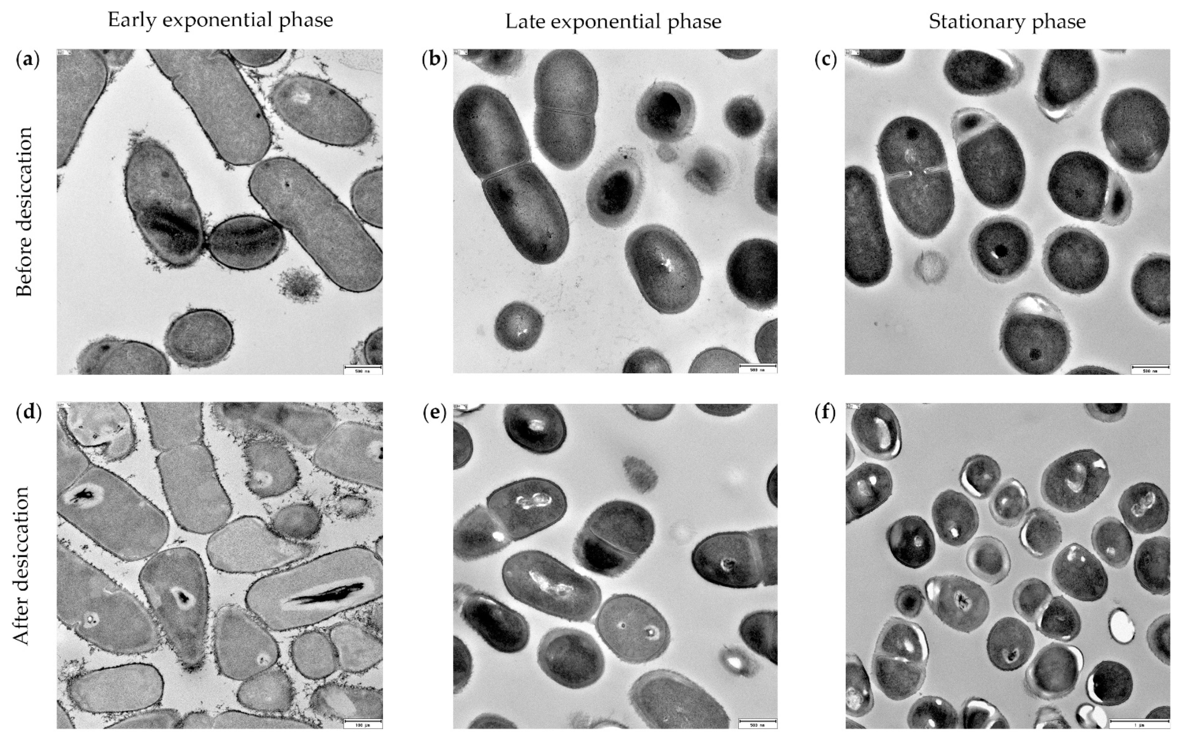
The shuttle vector pRCR12 (Solmeglas S. L., Madrid, Spain) was transformed successfully into competent cells of Exiguobacterium sp. Helios. pRCR12 contains the replicon of plasmid pSH71 from Lactococcus lactis and carries the mrfp gene encoding the mCherry protein under the control of the Px promoter of Streptococcus pneumoniae and a cat gene conferring chloramphenicol resistance [41]. The transformation was carried out as follows: Stationary-phase cultures were collected and washed with sterile cold water three times to prepare the electrocompetent cells. Electroporation was carried out in a Bio-Rad Gene Pulser/Pulse Controller (200 Ω, 25 μF and 1.5 kV/cm) using 1 μg of plasmid pRCR12, extracted from E. coli DH5α. Cells were recovered after 2 h in LB at 30 °C with agitation at 200 rpm and selected on LB plates supplemented with chloramphenicol at 20 µg/mL for Exiguobacterium sp. Helios strain and at 5 µg/mL for E. coli DH10B.

2.7. Microscopy

Cultures of Exiguobacterium sp. Helios were harvested at early exponential phase (OD600 = 1.5) and stationary phase (OD600 = 6.0); preparations of these cultures were photographed with a Leica DFC345 FX optical microscopy and processed by the LAS V4.2 software. We used ImageJ software [42] to measure the cell size.
To prepare the cells for transmission electron microscopy (TEM), the cultures of Exiguobacterium sp. Helios were harvested, washed twice in PBS, and fixed in 3% (w/v) glutaraldehyde in PBS for 1 h. Afterwards, cells were washed again several times in PBS and then suspended in 1% (w/v) OsO4 and 0.8% (w/v) C6N6FeK3 for 1 h at 4 °C. After that, cells were washed in PBS until the supernatant was clear and then gradually dehydrated in ethanol (30%, 50%, 70%, 90%, and 100% (v/v); 30 min each) and finally embedded in Spurr ERL-4221 resin, first in a 1:1 ethanol: resin suspension for 30 min and then twice in resin alone, 30 min each time. Finally, the cells embedded in the resin were transferred to clean and dry beads and polymerized at 60 °C overnight. Ultrathin sections (thickness 70 nm) were cut with a microtome using a Diatome diamond knife. The sections were picked up with 400 mesh cupper grids coated with a layer of carbon and subsequently observed in a JEOL JEM-1230 electron microscope (Jeol Ltd., Akishima, Japan).

2.8. Metal Resistance and Formation of SeNPs

To study the metal and metalloid resistance of strains, we used Tris-Minimal Medium (6.06 g/L Tris-HCl; 4.68 g/L NaCl; 1.49 g/L KCl; 1.07 g/L NH4Cl; 0.43 g/L Na2SO4; 0.2 g/L MgCl2 6H2O; 0.03 g/L Ca2Cl 2H2O; 0.23 g/L Na2HPO4 12H2O; 0.005 g/L Fe(III)NH4 citrate; 1 µL/L 25% HCl; 70 µg/mL ZnCl2; 100 µg/mL MnCl2 4H2O; 60 µg/mL H3BO3; 200 µg/mL CoCl2 6H2O; 20 µg/mL CuCl2 2H2O; 20 µg/mL NiCl2 6H2O; 40 µg/mL Na2MoO4 2H2O) [43] supplemented with 1 g/L of yeast extract and the appropriate metal (Sigma-Aldrich). The concentrations of metals used were 0.62–5.0 mM NiCl2, 0.62–5.0 mM ZnCl2, 0.62–5.0 mM K2TeO3, 0.62–5.0 mM NaAsO2, 5–50 mM Na3AsO4, 0.1–1.2 mM CuSO4, 0.62–5.0 mM CdCl2, 0.62–5.0 mM AgNO3, 0.62–10.0 mM Pb(NO3)2, or 0.05–50.0 mM K2SeO3.
To establish the selenite tolerance of Exiguobacterium sp. Helios strain, cells were grown in 10 mL of LB in 50 mL Falcon at 30 °C with orbital shaking at 200 rpm using 0–50 mM Na2SeO3. After 48 h of incubation, selenite resistance was monitored and cell growth was determined. In addition, we also tested the ability of Exiguobacterium sp. Helios to reduce selenite to elemental selenium Se (0) monitored by red color formation. Exiguobacterium sp. Helios was grown in LB medium supplemented with 1 mM selenite for 24 h at 30 °C with orbital shaking at 200 rpm, with the appropriate controls.

2.9. Characterization of Selenium Nanoparticles

For transmission electron microscopy (TEM) observation, the samples were prepared by placing drops of the Exiguobacterium sp. Helios cell cultures onto carbon-coated copper grids and allowing the solvent to evaporate. TEM observations were performed on a JEOL model JEM-2100 instrument operated at an accelerating voltage of 200 kV. The chemical composition of the SeNPs observed was determined by energy-dispersive X-ray spectroscopy (EDX) as previously described [44]. The size of the SeNPs was determined by using the ImageJ software [42].

3. Results

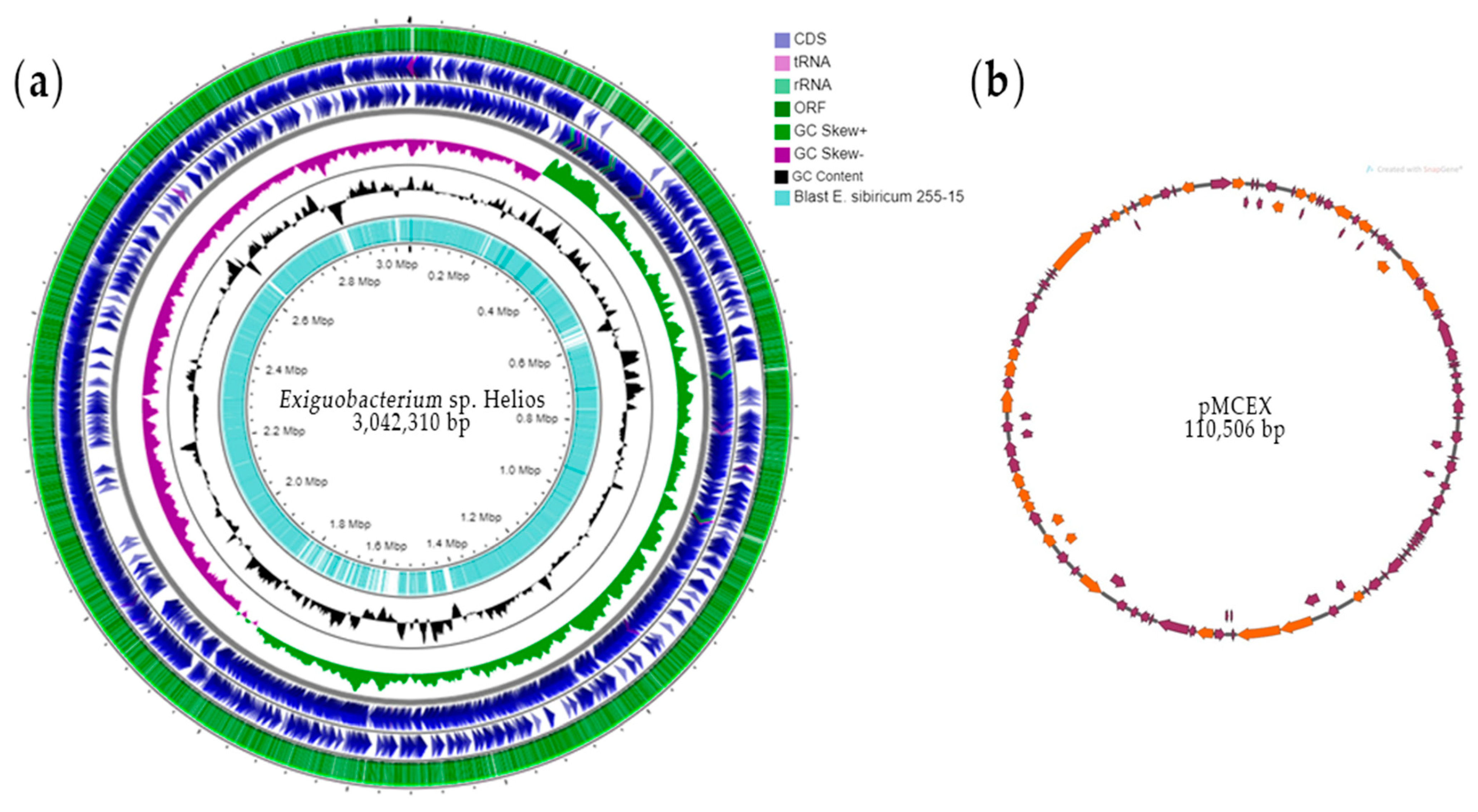
3.1. Analysis of Exiguobacterium sp. Helios Genome

Exiguobacterium sp. Helios strain genome was sequenced by Illumina and assembled in a two-scaffolds chromosome of 3,042,310 bp with a G + C content of 47.1% and one plasmid named pMCEX of 110,560 bp with a G + C content of 41.6% (Figure 1).
The whole Exiguobacterium sp. Helios genome contains 3241 open reading frames (ORFs), of which 2144 (66%) can be assigned to putative functions, 69 tRNAs, and 26 rRNA operons.
Plasmid pMCEX contains 95 ORFs, but remarkably, only 37 ORFs display homology at the NCBI protein database (Table S1). Among them, HNY42_RS1605 contains an LD-carboxypeptidase domain encoding a putative muramoyl tetrapeptide carboxypeptidase involved in recycling of peptidoglycan (PG). Gene HNY42_RS16150 codes a putative ParE toxin of type II toxin-antitoxin system RelE/ParE consisting of a stable toxin and a small, labile antitoxin. Under unfavorable conditions, the antitoxin is degraded, leading to activation of the toxin and resulting in growth arrest, possibly also in bacterial programmed cell death. Gene HNY42_RS15810 codes a ComEC-like protein that could be involved in late natural competence.
Exiguobacterium sp. Helios genome has two large regions involved in the synthesis of flagella (Table S2) that are organized in two clusters: HNY42_RS11055-HNY42_RS11200 and HNY42_RS13935-HNY42_RS14085 covering a 23.9 kb and 29.4 kb region, respectively. The presence of these genes suggests that this strain is motile as E. sibiricum.
Using as reference the genes of Bacillus subtilis involved in natural competence, we detected the existence of a complete DNA-uptake machinery in Exiguobacterium sp. Helios, suggesting that this strain has the ability to acquire exogenous DNA from the medium. These predicted competence genes are not clustered together and are scattered throughout the chromosome (Table 1). The HNY42_RS05970-HNY42_RS05985 region encodes homologous proteins to ComGC known to form pseudopilins responsible for binding the exogenous dsDNA and some additional proteins required for DNA internalization in Firmicutes, such as ComGA and ComGB. HNY42_RS05435 and HNY42_RS05440 encode ComEA and ComEC homologous proteins. In Gram-positive microorganisms, ComEA delivers the dsDNA to a protein that generates ssDNA, presumably an AddAB, before its internalization through a transmembrane pore formed by ComEC. HNY42_RS05235 and HNY42_RS05240 genes encode the putative AddAB homologous proteins. Internalization of ssDNA is presumably driven by the ATP-dependent translocase ComFA (coded by HNY42_RS14095). The RecA homologous protein that polymerizes on ssDNA and promotes a homology search along the chromosome is encoded by HNY42_RS06690. Moreover, there are two most probably paralogous genes, HNY42_RS05215 and HNY42_RS13920, encoding the ComK regulator responsible of activating the expression of genes involved in natural competence in B. subtilis.
On the other hand, it has been described that a conserved genomic signature of about 50 genes coding the minimal machinery for endosporulation enables to distinguish endospore-forming organisms [45]. Exiguobacterium sp. Helios strain contains only 17 genes of this genomic signature, i.e., this strain only has 34% of the genes that are currently required to sporulate, suggesting that it is not an endospore-forming bacterium, but this observation raises a question about the possible association of these genes to xerotolerance (Table 2).
The large number of genes related to carbohydrate metabolism annotated in the chromosome of Exiguobacterium sp. Helios suggest a remarkable ability to use several sugars and polymeric carbohydrates as carbon sources. For instance, among them, HNY42_RS05125 and HNY42_RS00620 genes encode a putative sucrose-6-phosphate hydrolase and a putative fructokinase, respectively, most probably involved in sucrose catabolism. Table 1 shows the sugars that can be used as carbon and energy sources by this strain.
The genome analysis predicted that Exiguobacterium sp. Helios contains an 11,679 kb cluster (HNY42_RS03440-HNY42_RS03510) encoding the complete degradation pathway of phenylacetic acid. HNY42_00705, HNY42_07190, HNY42_08240, HNY42_08800, and HNY42_14205 genes annotated as VOC family proteins and/or class I extradiol dioxygenases could be involved in the degradation of other aromatic compounds, such as 3,4-hydroxybenzoate, 4-hydroxybenzoate and benzoate (Table 3).
Regarding stress-resistance genes, Exiguobacterium sp. Helios genome contains HNY42_RS02725, coding a putative transport system of glycine betaine (N,N,N-trimethylglycine) which is a very efficient osmolyte that accumulates at high cytoplasmic concentrations in response to osmotic stress [46]. HNY42_RS02725 encodes a homologous protein to OpuD from B. subtilis (50% identity in amino acid sequence) that is a BCCT type carrier that uptakes glycine betaine from the environment. Locus HNY42_RS05230 encodes a putative bacterial globine. Globin-coupled sensors (GCSs) were first described as regulators of the aerotactic responses in B. subtilis and Halobacterium salinarum [47]. Some of the main roles assigned to bacterial globins are in oxygen and nitric oxide (NO) metabolism similar to the protection from the stresses caused by exposure to nitric oxide [48].
The sigma B factor coded by the HNY42_RS00075 gene and its accessory regulatory proteins anti-sigma factors RsbW and RsbV, coded by the HNY42_RS00080 and HNY42_RS00085 genes, respectively, are present in Exiguobacterium sp. Helios genome. The sigma B factor is known to control general stress regulons in different bacteria that are activated by the exposure to either physical or nutritional stress.
Exiguobacterium sp. Helios genome encodes four specific DinB homologous (HNY42_RS08860, HNY42_RS08570, HNY42_RS01380, and HNY42_RS13495 genes). The DNA damage-inducible (din) protein family is overrepresented in D. radiodurans, with 13 homologous genes, whose expression is highly induced in response to gamma radiation (γ-radiation) and mitomycin C (MMC) exposure [49].
The HNY42_RS04520 gene encodes a PspA/IM30 family protein whose members appear to have membrane protective activity by membrane binding to preserve membrane integrity [50].
There are five genes encoding small (66 amino acids) putative cold shock proteins (Csp) (HNY42_RS10205-HNY42_RS10210-HNY42_RS10215; HNY42_RS13925 and HNY42_00710 genes). Many bacteria produce these small proteins as a response to rapid temperature downshift (cold shock). However, some Csps are non-cold-inducible and they are reported to be involved in cellular processes to promote normal growth and stress adaptation responses. Csps have been shown to contribute to osmotic, oxidative, starvation, pH, and ethanol stress tolerance, as well as to host cell invasion [51].
Concerning the genes that confer xerotolerance in other organisms, we have not found in Exiguobacterium sp. Helios similar genes to those described in the five pathways described for the synthesis of trehalose [52], nor those described for the synthesis of betaine [53] and sucrose [54]. Therefore, we assume that this strain does not produce trehalose, betaine, or sucrose to become xerotolerant. However, Exiguobacterium sp. Helios has the genes responsible for the synthesis of proline, and thus, we cannot discard the production of proline as a xerotolerance mechanism. On the other hand, Exiguobacterium sp. Helios does not have genes encoding proteins similar to the late embryogenesis abundant (LEA) proteins that protect some Azotobacter vinelandii [55] or D. radiodurans [56] against drought and other stresses. Nevertheless, Helios contains a gene HNY42_14175 that encodes a hypothetical protein containing a small LEA_2 region.

3.2. Genome Comparison of Exiguobacterium sp. Helios and E. sibiricum 255-15

As mentioned above, the closest genome sequence of Exiguobacterium sp. Helios strain is that from E. sibiricum 255-15, which was isolated from the Siberian permafrost [4]. E. sibiricum 255-15 genome consists of 3,040,786 bp (accession number CPO01022) and two plasmids: pEXIG01 of 4.8 kb (accession number CPO01023) and pEXIG02 of 1.7 kb (accession number CPO01024) encoding five and two unknown proteins, respectively. The annotation in RAST server reveals 3102 coding regions, 904 of which are hypothetical proteins. The typical Mauve alignment (Figure S1) revealed that Exiguobacterium sp. Helios shares similar genomic arrangements with E. sibiricum but has some significant differences that are shown in Tables S3 and S4. Several regions within the Exiguobacterium sp. Helios strain genome are not found in E. sibiricum, according to Mauve alignment and the basic local alignment search tool (Blast) at NCBI. One example is the existence of the biosynthetic cluster HNY42_RS07465-HNY42_RS07515 that contains the genes responsible for the synthesis of the iron siderophore petrobactin. The two genes forming a putative operon, HNY42_RS07660-HNY42_RS07700, encode spore coat proteins that could be involved in the desiccation tolerance of the strain. HNY42_RS08215 gene encodes a cupin, also called seed storage protein, that usually serves as biological reserve of amino acids and metals. The conserved domain, comprising a six-stranded beta barrel structure, is typically found in the phyla Firmicutes. The cupin superfamily is a group of functionally diverse proteins that are found in all three domains of life: Archaea, Eubacteria, and Eukaryota. Regarding carbohydrate catabolism, Exiguobacterium sp. Helios contains 38 glycoside hydrolases, 22 glycosyl transferases, 1 polysaccharide lyase, 3 carbohydrate esterase, and 12 carbohydrate-binding module family proteins, according to CAZy Database. The number of enzymes that degrade, modify, or create glycosidic bonds found in E. sibiricum is slightly lower, being 26 glycoside hydrolases, 20 glycosyl transferases, 1 polysaccharide lyase, 3 carbohydrate esterase, and 11 carbohydrate-binding module family protein.
Curiously, another interesting genomic difference is that Exiguobacterium sp. Helios strain does not have a CRISPR-CAS system, as is E. sibiricum coded by the EXIG_RS00985-EXIG_RS1015 genes (Table S4). The CRISPR-CAS system is also present in other related strains as E. antarticum.
Finally, E. sibiricum genome contains 27 putative transposases that are not present in Exiguobacterium sp. Helios strain. A lower number of transposases is beneficial in order to use it as a biotechnological robust chassis, since transposases pose a threat to the organisms as the sequence specificity of transposase insertions is usually low, and transpositions may disrupt vital genes.

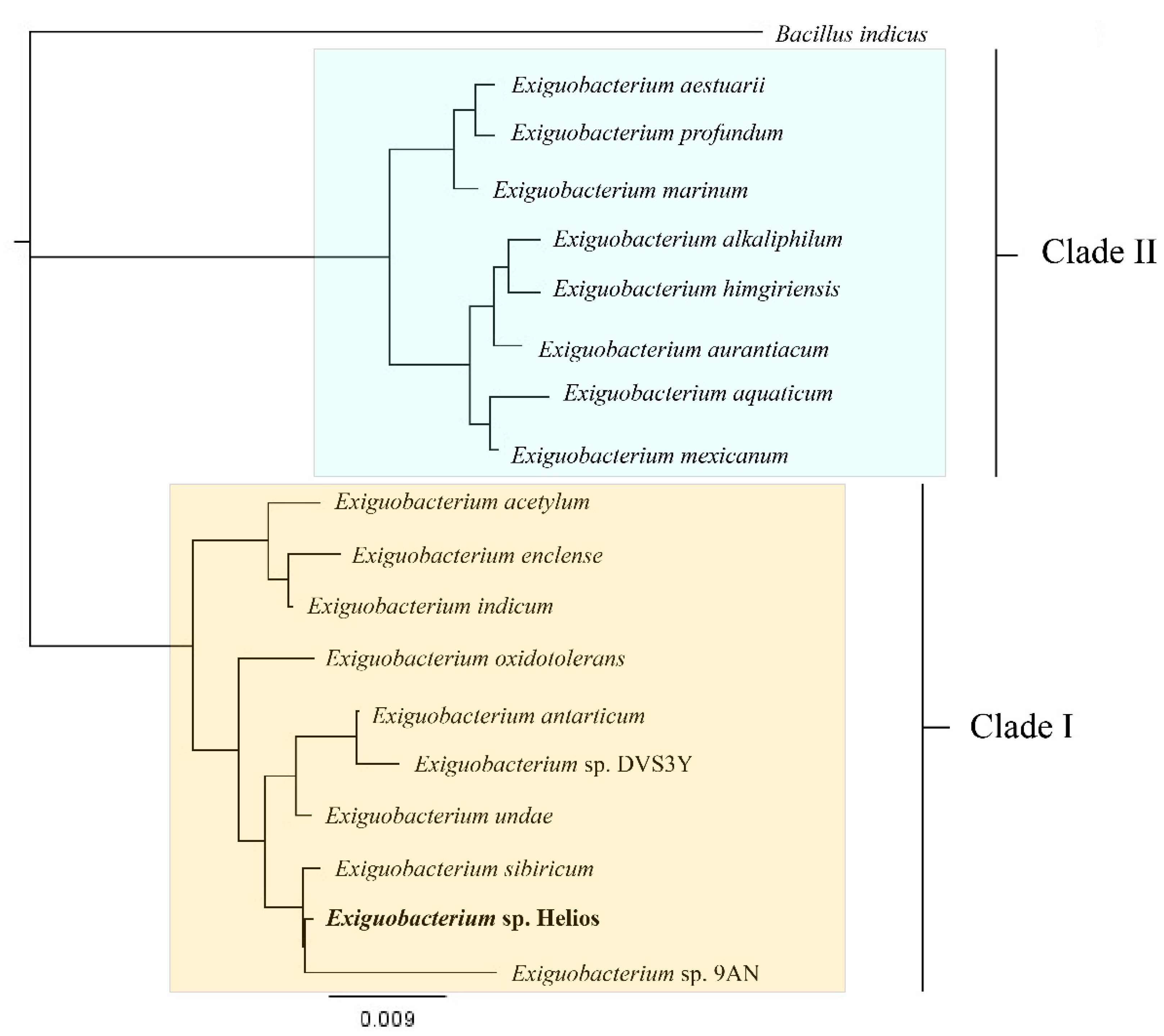
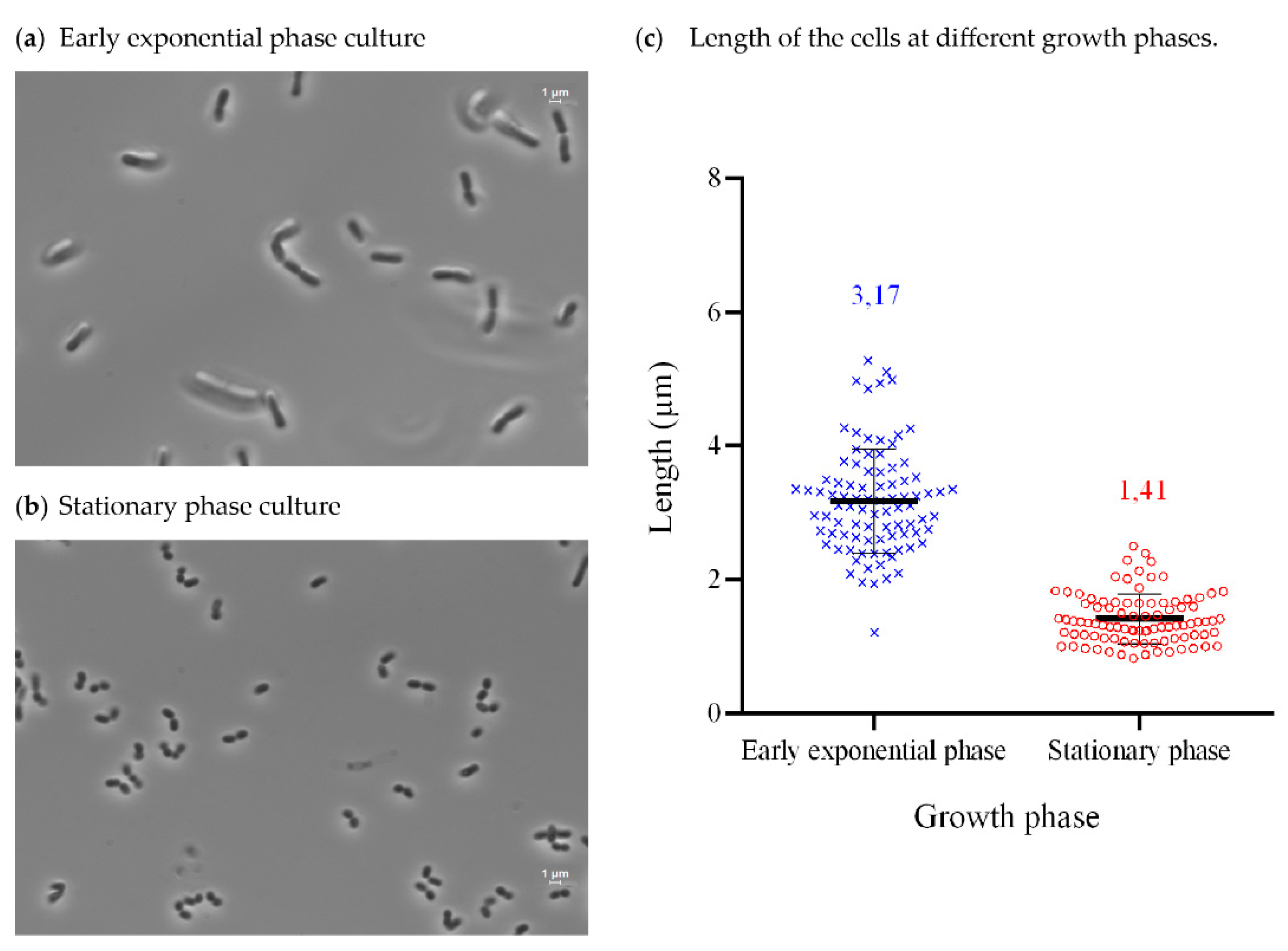
3.3. Isolation, Identification, Growth Conditions, and Phenotypical Characterization of Exiguobacterium sp. Helios

One of the xerotolerant strains isolated from the solar panels was identified as Exiguobacterium sp. according to its 16S rRNA. This strain named Helios was selected for this study since no representative of this genus has been described as xerotolerant bacterium to date. Present work was performed with the strain Exiguobacterium sp. Helios due to its higher resistance to desiccation.
We generated a maximum-likelihood phylogeny of 16S rRNA gene sequences from eighteen representative strains of Exiguobacterium spp., for which complete genomes are available at the NCBI, and one typed strain of Bacillus indicus as outgroup. Exiguobacterium sp. Helios strain was positioned in Clade I, along with Exiguobacterium sp. 9AN and E. sibiricum 255-15 (Figure 2).
E. sibiricum 255-15 and Exiguobacterium sp. Helios strain were analyzed for average nucleotide identity (ANI) and average amino acid identity (AAI) to provide interspecies relationship. The E. sibiricum 255-15 strain was the closest strain which had a complete genome uploaded to the NCBI database. The comparison renders 89.75% average nucleotide identity (ANI) and 94.75% average amino acid identity (AAI). Based on these data, these strains do not seem to belong to the same species [40].
Exiguobacterium sp. Helios is a Gram-positive bacillary rod-shaped and nonsporulating facultative anaerobe. Its optimal growth temperature is 30 °C, although it can grow efficiently between 4 °C and 42 °C. When observed by optic microscopy, Exiguobacterium sp. Helios strain showed different phenotypes depending on the growth phase. Thus, cells at exponential phase have a bacillary/rod shape (Figure 3a), while they become coccoid at stationary phase (Figure 3b). Moreover, a difference in the length of the cells can be observed at exponential phase (average of 3.17 μm) and at stationary phase (average of 1.41 μm) (Figure 3c).
This strain is able to grow in rich and minimal media using different substrates as sole carbon and energy sources (Table 3). Exiguobacterium sp. Helios cells cannot grow at concentrations of NaCl higher than 60 g/L, such as other Exiguobacterium strains from Clade I that are only moderately resistant to salt stress. However, it can grow up to an OD600 of 2.0 on LB medium containing 35% PEG 8000, i.e., under a low water activity of 0.982 [57].
In order to investigate the susceptibility to antibiotics to carry out genetic modifications of Exiguobacterium sp. Helios, we determined the minimum inhibitory concentration (MIC) for some commonly used antibiotics (Table 4).
Taking into account the antibiotic resistances of Exiguobacterium sp. Helios, we selected the plasmid pRCR12 carrying a cat gene conferring chloramphenicol resistance to develop a method to transform this strain. This expression vector contains the replicon of plasmid pSH71 from L. lactis and carries the mrfp gene encoding the mCherry protein under the control of the Px promoter of S. pneumoniae that allows a rapid detection of the plasmid. Using this plasmid, we obtained recombinant Exiguobacterium sp. Helios (pRCR12) colonies showing a red phenotype, indicating that the mrfp gene was expressed under the Px promoter in this bacterium (Figure S2).

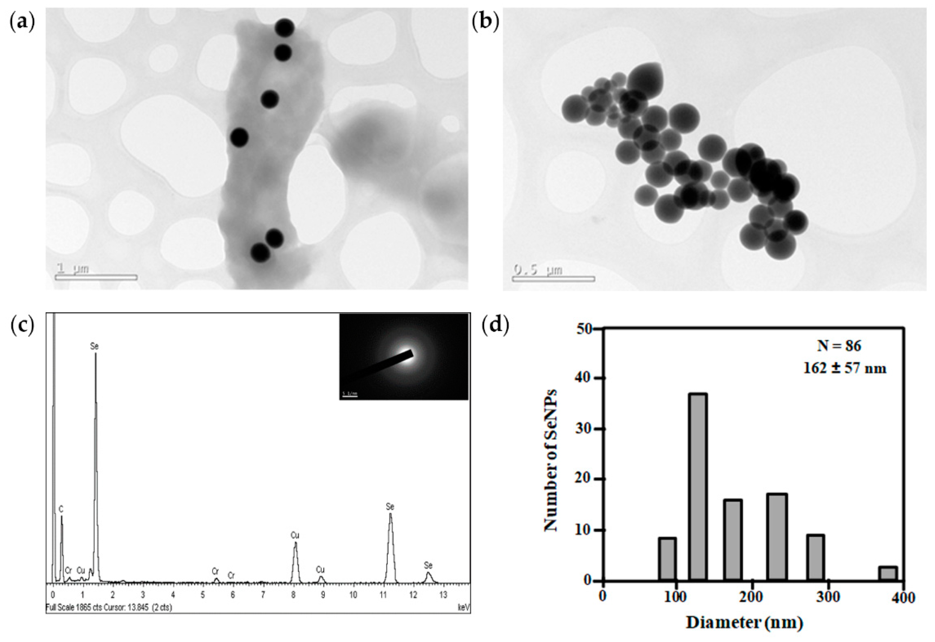
3.4. Metal Resistance of Exiguobacterium sp. Helios

Bacteria that resist extreme conditions often present other capacities, for instance, the ability to resist high concentrations of some toxic metals and metalloids. In this sense, we tested the capacity of Exiguobacterium sp. Helios strain to grow in the presence of these compounds. Table 5 shows that the strain has moderate resistance to some metals and metalloids, especially selenite.
The resistance of Exiguobacterium sp. Helios to selenite was analyzed in detail. When Exiguobacterium sp. Helios was grown in LB containing 1 mM selenite at 37 °C, the culture acquired a red color, suggesting the reduction of selenite to elemental selenium (Figure S3). No color was observed in the absence of bacterial cells, suggesting a role of this strain in selenite reduction. In fact, Exiguobacterium sp. Helios was able to grow in the presence of selenite up to 20 mM at 37 °C, indicating that the resistance is close to that reported for highly tolerant selenite strains such as Comamonas testosteroni S44 [58], Pseudomonas moraviensis [59], or Vibrio natriegens [60].
In addition, we also tested the ability of this strain to reduce selenite to elemental Se (0) as a proof of concept for developing a useful biotechnological process producing Se nanoparticles (SeNPs). We observed electron-dense nanospheres in cells of Exiguobacterium sp. Helios after 24 h of growth at 30 °C in LB containing 1 mM selenite (Figure 4a,b). The EDX analysis showed that the nanoparticles presented the specific Se peak (Figure 4c). The selected area electron diffraction (SAED) pattern of the nanoparticles showed a diffuse halo, indicating that selenium is present in its amorphous form (Figure 4c, inset). SeNPs appeared as spherical nanoparticles with an average size of 162 ± 57 nm (Figure 4d).

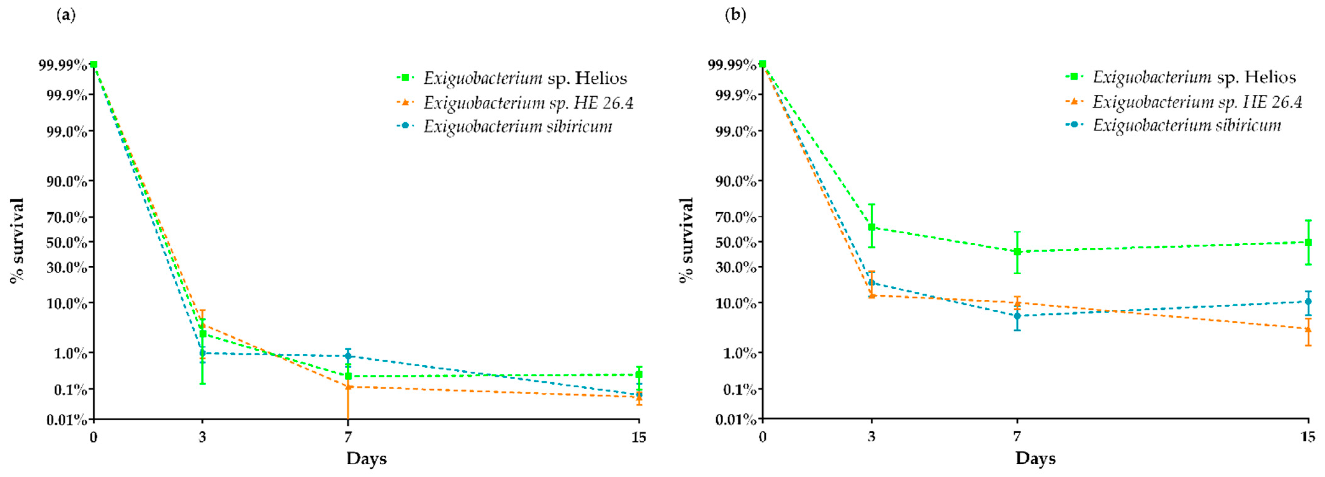
3.5. Xerotolerance of Exiguobacterium sp. Helios

A desiccation assay was performed with Exiguobacterium sp. Helios, E. sibiricum, E. coli, and D. radiodurans. In addition, we tested the performance of another Exiguobacterium sp. HE 26.4 strain isolated from the same solar panel, but that was classified within Clade II. In these assays, we used the cells obtained from cultures at early exponential and stationary phases to see the influence of the growth phase (Figure 5a,b). The results revealed that the xerotolerance of Exiguobacterium sp. Helios strain was very high, and similar to that exhibited by the model xerotolerant strain D. radiodurans, which, in our conditions, showed a survival score of around 80% after 15 days (data not shown). Interestingly, although E. sibiricum and Exiguobcaterium sp. HE 26.4 showed a high xerotolerance, it was lower than that of Exiguobacterium sp. Helios strain. As expected for the non-xerotolerant control, after 3 days, no survival of E. coli cells was detected (data not shown). Figure 5b shows that xerotolerance is higher at stationary phase on the Exiguobacterium strains.
Samples of Exiguobacterium sp. Helios obtained from different growth phases and before or after desiccation were observed by TEM (Figure 6). The images showed a large number of coccoid cells at the stationary phase, and remarkably, the cell envelope of these coccoid cells was thicker than that of bacillary-shaped cells. Taking into account that we have observed a higher xerotolerance when cells enter the stationary phase, the acquisition of a coccoid thickened-cell envelope phenotype might be required to improve their xerotolerance.

4. Discussion

The genus Exiguobacterium was first described by Collins et al. [1], who characterized the species E. aurantiacum. Since then, many other species have been described, but a fundamental issue that contributes to the interest for this genus was the discovery of E. sibiricum from a 43.6 m deep geological layer in the permafrost core of Kolyma Lowland (northeast of Siberia) that is dated 2–3 million years old, i.e., deposited from late Pliocene and Pleistocene periods [2,4]. Earth’s permafrost is characterized by low carbon availability, low water availability, and exposure to gamma radiation produced by soil minerals. Therefore, only special microorganisms are able to survive in these conditions [61]. Whereas extremophiles are usually defined by one extreme, many natural environments, for instance permafrost, have several extreme conditions that act as selection pressures, and thus, an increasing number of species and strains isolated from these extreme environments have been found to tolerate multiple extremes [62]. Such organisms are known as polyextremophiles, and one of the best-known examples is D. radiodurans, recognized for its ability to withstand a multitude of extreme environments including severe cold, dehydration, vacuum, acid, and intense radiation [62]. The removal of water through heat and air-drying damages membranes, proteins, and nucleic acids and should be lethal to the majority of organisms. Nevertheless, in addition to the resistance to desiccation exhibited by spores and akinetes developed by some bacteria and cyanobacteria, respectively, it is interesting to know that also some vegetative cells can survive extreme desiccation without forming these structures [63].
Although many natural complex extreme environments can be found on the Earth, we have tried to investigate if the microorganisms can be adapted to manufactured structures supporting extreme environmental conditions, such as solar panels [33]. Surprisingly, solar panels harbor a highly diverse microbial community, which can provide potential polyextremophiles [34,35]. For many hours a day, solar panels are subjected to absolute dryness and irradiation and, therefore, one might expect that highly desiccation-tolerant bacteria can be found there.
A simple screening procedure allowed us to isolate several bacteria from solar panels that were able to survive after several cycles of extreme desiccation. The procedure was validated using D. radiodurans as a positive control and E. coli as a negative one. Although the global microbiome analysis of the solar panels did not reveal the presence of a significant number of bacteria of Exiguobacterium genus [33], one of the most xerotolerant isolated bacteria from the solar panels was classified by 16S rRNA analysis as Exiguobacterium sp. Helios, phylogenetically very close to E. sibiricum. It is worth mentioning that additional screening tests of solar panels have allowed us to isolate another Exiguobacterium strain named HE 26.4 as well as many other xerotolerant strains that still remain to be fully characterized (Table S5).
Considering that none of the Exiguobacterium species have been described as xerotolerant so far, we investigated whether this property was specific to Exiguobacterium strains isolated from solar panels or if it could be shared by Exiguobacterium sp. Helios closest relative, i.e., E. sibiricum. Figure 4 shows that, while the xerotolerance of Exiguobacterium sp. Helios is higher than that of E. sibiricum, this emblematic strain can be also considered as xerotolerant, adding a new extremophile property to E. sibiricum. Although it has been shown that E. sibiricum can survive under low water activity conditions, this skill does not necessarily confer it the possibility to survive extreme desiccation conditions. In fact, Exiguobacterium sp. Helios can grow on a medium containing 35% PEG 8000 (i.e., under matric water stress), but can only grow very poorly under salt concentrations higher than 60 g/L (i.e., under osmotic water stress). This means than the adaptation to low water activity conditions depends on the type of osmotic perturbations (i.e., electrolyte or nonelectrolyte osmotic stress (matrix stress) caused to the cells), since the adaptation mechanisms are different. On the other hand, ice formation increases solute concentration by decreasing the amount of free water available, resulting in an environment with low water activity, similar to desiccated and salt-stressed environments. The low water activity of 0.90 encountered in permafrost corresponds to an increase in solute concentration to 2.79 M NaCl (5 osm) [64]. However, in these conditions neither Exiguobacterium sp. Helios strain nor E. sibiricum [4] can grow.
Low water activity requires an increased amount of turgor pressure to maintain cellular respiration and provide the required energy for cellular processes to take place. In this sense, it has been observed that E. sibiricum has a thicker cell envelope, allowing withstanding high turgor pressures [65]. This appears to be also the case for Exiguobacterium sp. Helios, since the cells surviving desiccation have a coccoid morphology with a thick cellular envelope. We hypothesize that Exiguobacterium sp. Helios is able to generate this morphology during its growth, particularly during the stationary phase, and only the cells having a thicker cellular envelope will stand the desiccation conditions. This hypothesis agrees with the observation that Exiguobacterium sp. Helios cells recovered by rehydration show a coccoid phenotype that soon changes to a bacillary phenotype when the cells start growing again. The development of thick, multilayered envelopes rich in polysaccharides, lipids, and proteins was described in the field- and laboratory-desiccated cells of the cyanobacteria resistant to desiccation, Chroococcidiopsis spp. [66]. The genes involved in this phenotype remain unknown, but it was interesting to notice that Exiguobacterium sp. Helios and E. sibiricum strains contain a number of genes related to sporulation, in spite of the fact that Exiguobacterium bacteria do not produce spores. In addition, the analysis of the Exiguobacterium sp. Helios genome allowed the identification of several genes that could be involved in stress resistance. Whether all these genes are responsible for the formation of the thick envelope phenotype and the xerotolerance is a matter of further research to identify the genetic basis of anhydrobiotic adaptation.

Supplementary Materials

The following are available online at https://www.mdpi.com/article/10.3390/microorganisms9122455/s1, Figure S1: Scheme of the alignment of Exiguobacterium sp. Helios vs. E. sibiricum 255-15 genomes by Mauve Alignment with Geneious 2020.0.; Figure S2: Transformation of Exiguobacterium sp. Helios.; Figure S3: Selenite reduction by Exiguobacterium cells.; Table S1: Plasmid pMCEX annotation at NCBI.; Table S2: Genes from Exiguobacterium sp. Helios putatively involved in flagellum synthesis and functionality.; Table S3: Exiguobacterium sp. Helios genes that are not present in E. sibiricum 255-15.; Table S4: E. sibiricum 255-15 genes that are not present in Exiguobacterium sp. Helios.; Table S5: Other strains isolated by the desiccation assay.

Author Contributions

J.L.G., B.G. and J.M.N.L. designed the study. J.L.G. and B.G. wrote the manuscript. M.C.L. performed the experiments. B.G. performed the data analysis. M.C. and L.C. designed and performed the selenium nanoparticles experiments. L.G., E.R.O. and J.M.L. contributed to the phylogenetic studies. M.C.L., B.G., J.L.G., J.M.N.L., M.C., E.R.O., J.M.L., J.P. and M.P. contributed to revise the manuscript. All authors have read and agreed to the published version of the manuscript.

Funding

This research was supported by grants BIO2015-66960-C3-1-R, BIO2016-79736-R and PID2019-110612RB-I00 from the Ministry of Economy and Competitiveness of Spain, RTI2018-095584-B-C41-42-43-44 from the Ministry of Science and Innovation of Spain, and by Grants CSIC 2017 2 0I 015. María Castillo has a fellowship from the State Program for the Promotion of Talent and its Employability in R&D&I supported by grant PRE2019-087933.

Institutional Review Board Statement

Not applicable.

Informed Consent Statement

Not applicable.

Data Availability Statement

The datasets generated in this work can be found in the NCBI under the BioProject PRJNA632476.

Acknowledgments

We thank A. Valencia for technical assistance and Secugen S.L. for DNA sequencing. The bioinformatics support of the Bioinformatics and Biostatistics Service at CIB-MS, particularly Guillermo Padilla Alonso, is greatly appreciated.

Conflicts of Interest

The authors declare that the research was conducted in the absence of any commercial or financial relationships that could be construed as a potential conflict of interest.

References

  1. Collins, M.D.; Lund, B.M.; Farrow, J.A.E.; Schleifer, K.H. Chemotaxonomic Study of an Alkalophilic Bacterium, Exiguobacterium aurantiacum gen. nov., sp. nov. J. Gen. Microbiol. 1983, 129, 2037–2042. [Google Scholar] [CrossRef] [Green Version]
  2. Vishnivetskaya, T.A.; Kathariou, S.; Tiedje, J.M. The Exiguobacterium Genus: Biodiversity and Biogeography. Extremophiles 2009, 13, 541–555. [Google Scholar] [CrossRef] [PubMed]
  3. Kasana, R.C.; Pandey, C.B. Exiguobacterium: An Overview of a Versatile Genus with Potential in Industry and Agriculture. Crit. Rev. Biotechnol. 2018, 38, 141–156. [Google Scholar] [CrossRef] [PubMed]
  4. Rodrigues, D.F.; Goris, J.; Vishnivetskaya, T.; Gilichinsky, D.; Thomashow, M.F.; Tiedje, J.M. Characterization of Exiguobacterium Isolates from the Siberian Permafrost. Description of Exiguobacterium sibiricum sp. nov. Extremophiles 2006, 10, 285–294. [Google Scholar] [CrossRef]
  5. Chaturvedi, P.; Shivaji, S. Exiguobacterium Indicum sp. nov., a Psychrophilic Bacterium from the Hamta Glacier of the Himalayan Mountain Ranges of India. Int. J. Syst. Evol. Microbiol. 2006, 56, 2765–2770. [Google Scholar] [CrossRef] [Green Version]
  6. Crapart, S.; Fardeau, M.L.; Cayol, J.L.; Thomas, P.; Sery, C.; Ollivier, B.; Combet-Blanc, Y. Exiguobacterium profundum sp. nov., a Moderately Thermophilic, Lactic Acid-Producing Bacterium Isolated from a Deep-Sea Hydrothermal Vent. Int. J. Syst. Evol. Microbiol. 2007, 57, 287–292. [Google Scholar] [CrossRef]
  7. Kasana, R.C.; Yadav, S.K. Isolation of a Psychrotrophic Exiguobacterium sp. SKPB5 (MTCC 7803) and Characterization of Its Alkaline Protease. Curr. Microbiol. 2007, 54, 224–229. [Google Scholar] [CrossRef] [PubMed]
  8. Alam, M.Z.; Malik, A. Chromate Resistance, Transport and Bioreduction by Exiguobacterium sp. ZM-2 Isolated from Agricultural Soil Irrigated with Tannery Effluent. J. Basic Microbiol. 2008, 48, 416–420. [Google Scholar] [CrossRef]
  9. Chaturvedi, P.; Prabahar, V.; Manorama, R.; Pindi, P.K.; Bhadra, B.; Begum, Z.; Shivaji, S. Exiguobacterium soli sp. nov., a Psychrophilic Bacterium from the McMurdo Dry Valleys, Antarctica. Int. J. Syst. Evol. Microbiol. 2008, 58, 2447–2453. [Google Scholar] [CrossRef] [Green Version]
  10. Carneiro, A.R.; Ramos, R.T.J.; Dall’Agnol, H.; Pinto, A.C.; Soares, S.d.C.; Santos, A.R.; Guimarães, L.C.; Almeida, S.S.; Baraúna, R.A.; das Graças, D.A.; et al. Genome Sequence of Exiguobacterium antarcticum B7, Isolated from a Biofilm in Ginger Lake, King George Island, Antarctica. J. Bacteriol. 2012, 194, 6689–6690. [Google Scholar] [CrossRef] [Green Version]
  11. Singh, N.K.; Raichand, R.; Kaur, I.; Kaur, C.; Pareek, S.; Mayilraj, S. Exiguobacterium himgiriensis sp. nov. A Novel Member of the Genus Exiguobacterium, Isolated from the Indian Himalayas. Antonie Van Leeuwenhoek 2013, 103, 789–796. [Google Scholar] [CrossRef]
  12. Dastager, S.G.; Mawlankar, R.; Sonalkar, V.V.; Thorat, M.N.; Mual, P.; Verma, A.; Krishnamurthi, S.; Tang, S.K.; Li, W.J. Exiguobacterium enclense sp. nov., Isolated from Sediment. Int. J. Syst. Evol. Microbiol. 2015, 65, 1611–1616. [Google Scholar] [CrossRef] [Green Version]
  13. Etemadifar, Z.; Gholami, M.; Derikvand, P. UV-Resistant Bacteria with Multiple-Stress Tolerance Isolated from Desert Areas in Iran. Geomicrobiol. J. 2016, 33, 1–7. [Google Scholar] [CrossRef]
  14. Hanaoka, Y.; Takebe, F.; Nodasaka, Y.; Hara, I.; Matsuyama, H.; Yumoto, I. Growth-Dependent Catalase Localization in Exiguobacterium oxidotolerans T-2-2T Reflected by Catalase Activity of Cells. PLoS ONE 2013, 8, e76862. [Google Scholar] [CrossRef] [Green Version]
  15. Remonsellez, F.; Castro-Severyn, J.; Pardo-Esté, C.; Aguilar, P.; Fortt, J.; Salinas, C.; Barahona, S.; León, J.; Fuentes, B.; Areche, C.; et al. Characterization and Salt Response in Recurrent Halotolerant Exiguobacterium sp. SH31 Isolated from Sediments of Salar de Huasco, Chilean Altiplano. Front. Microbiol. 2018, 9, 2228. [Google Scholar] [CrossRef] [PubMed]
  16. Castro-Severyn, J.; Pardo-Esté, C.; Mendez, K.N.; Morales, N.; Marquez, S.L.; Molina, F.; Remonsellez, F.; Castro-Nallar, E.; Saavedra, C.P. Genomic Variation and Arsenic Tolerance Emerged as Niche Specific Adaptations by Different Exiguobacterium Strains Isolated From the Extreme Salar de Huasco Environment in Chilean—Altiplano. Front. Microbiol. 2020, 11, 1632. [Google Scholar] [CrossRef] [PubMed]
  17. Selvakumar, G.; Joshi, P.; Nazim, S.; Mishra, P.K.; Kundu, S.; Gupta, H.S. Exiguobacterium acetylicum Strain 1P (MTCC 8707) a Novel Bacterial Antagonist from the North Western Indian Himalayas. World J. Microbiol. Biotechnol. 2009, 25, 131–137. [Google Scholar] [CrossRef]
  18. Selvakumar, G.; Kundu, S.; Joshi, P.; Nazim, S.; Gupta, A.D.; Gupta, H.S. Growth Promotion of Wheat Seedlings by Exiguobacterium acetylicum 1P (MTCC 8707) a Cold Tolerant Bacterial Strain from the Uttarakhand Himalayas. Indian J. Microbiol. 2010, 50, 50–56. [Google Scholar] [CrossRef] [PubMed] [Green Version]
  19. Gangoiti, J.; Pijning, T.; Dijkhuizen, L. The Exiguobacterium sibiricum 255-15 GtfC Enzyme Represents a Novel Glycoside Hydrolase 70 Subfamily of 4,6-α-Glucanotransferase Enzymes. Appl. Environ. Microbiol. 2016, 82, 756–766. [Google Scholar] [CrossRef] [PubMed] [Green Version]
  20. Qiao, Y.; Peng, Q.; Yan, J.; Wang, H.; Ding, H.; Shi, B. Gene Cloning and Enzymatic Characterization of Alkali-Tolerant Type I Pullulanase from Exiguobacterium acetylicum. Lett. Appl. Microbiol. 2015, 60, 52–59. [Google Scholar] [CrossRef]
  21. Crespim, E.; Zanphorlin, L.M.; de Souza, F.H.M.; Diogo, J.A.; Gazolla, A.C.; Machado, C.B.; Figueiredo, F.; Sousa, A.S.; Nóbrega, F.; Pellizari, V.H.; et al. A Novel Cold-Adapted and Glucose-Tolerant GH1 β-Glucosidase from Exiguobacterium antarcticum B7. Int. J. Biol. Macromol. 2016, 82, 375–380. [Google Scholar] [CrossRef] [PubMed]
  22. Lee, C.W.; Kwon, S.; Park, S.H.; Kim, B.Y.; Yoo, W.; Ryu, B.H.; Kim, H.W.; Shin, S.C.; Kim, S.; Park, H.; et al. Crystal Structure and Functional Characterization of an Esterase (EaEST) from Exiguobacterium antarcticum. PLoS ONE 2017, 12, e0169540. [Google Scholar] [CrossRef]
  23. Castro-Severyn, J.; Remonsellez, F.; Valenzuela, S.L.; Salinas, C.; Fortt, J.; Aguilar, P.; Pardo-Esté, C.; Dorador, C.; Quatrini, R.; Molina, F.; et al. Comparative Genomics Analysis of a New Exiguobacterium Strain from Salar de Huasco Reveals a Repertoire of Stress-Related Genes and Arsenic Resistance. Front. Microbiol. 2017, 8, 456. [Google Scholar] [CrossRef] [PubMed] [Green Version]
  24. Aburto, C.; Castillo, C.; Cornejo, F.; Arenas-Salinas, M.; Vásquez, C.; Guerrero, C.; Arenas, F.; Illanes, A.; Vera, C. Β-Galactosidase from Exiguobacterium acetylicum: Cloning, Expression, Purification and Characterization. Bioresour. Technol. 2019, 277, 211–215. [Google Scholar] [CrossRef]
  25. Yin, B.; Gu, H.; Mo, X.; Xu, Y.; Yan, B.; Li, Q.; Ou, Q.; Wu, B.; Guo, C.; Jiang, C. Identification and Molecular Characterization of a Psychrophilic GH1 β-Glucosidase from the Subtropical Soil Microorganism Exiguobacterium sp. GXG2. AMB Expr. 2019, 9, 159. [Google Scholar] [CrossRef] [Green Version]
  26. Wang, Y.; Le, L.T.H.L.; Yoo, W.; Lee, C.W.; Kim, K.K.; Lee, J.H.; Kim, T.D. Characterization, Immobilization, and Mutagenesis of a Novel Cold-Active Acetylesterase (EaAcE) from Exiguobacterium antarcticum B7. Int. J. Biol. Macromol. 2019, 136, 1042–1051. [Google Scholar] [CrossRef]
  27. Arora, P.K.; Bae, H. Biodegradation of 4-Chloroindole by Exiguobacterium sp. PMA. J. Hazard. Mater. 2015, 284, 261–268. [Google Scholar] [CrossRef] [PubMed]
  28. Yang, Y.; Yang, J.; Wu, W.M.; Zhao, J.; Song, Y.; Gao, L.; Yang, R.; Jiang, L. Biodegradation and Mineralization of Polystyrene by Plastic-Eating Mealworms: Part 1. Chemical and Physical Characterization and Isotopic Tests. Environ. Sci. Technol. 2015, 49, 12080–12086. [Google Scholar] [CrossRef]
  29. Chauhan, D.; Agrawal, G.; Deshmukh, S.; Roy, S.S.; Priyadarshini, R. Biofilm Formation by Exiguobacterium sp. DR11 and DR14 Alter Polystyrene Surface Properties and Initiate Biodegradation. RSC Adv. 2018, 8, 37590–37599. [Google Scholar] [CrossRef] [Green Version]
  30. García, A.H. Anhydrobiosis in Bacteria: From Physiology to Applications. J. Biosci. 2011, 36, 939–950. [Google Scholar] [CrossRef]
  31. Gerber, E.; Bernard, R.; Castang, S.; Chabot, N.; Coze, F.; Dreux-Zigha, A.; Hauser, E.; Hivin, P.; Joseph, P.; Lazarelli, C.; et al. Deinococcus as New Chassis for Industrial Biotechnology: Biology, Physiology and Tools. J. Appl. Microbiol. 2015, 119, 1–10. [Google Scholar] [CrossRef] [PubMed] [Green Version]
  32. Mattimore, V.; Battista, J.R. Radioresistance of Deinococcus Radiodurans: Functions Necessary to Survive Ionizing Radiation Are Also Necessary to Survive Prolonged Desiccation. J. Bacteriol. 1996, 178, 633–637. [Google Scholar] [CrossRef] [Green Version]
  33. Dorado-Morales, P.; Vilanova, C.; Peretó, J.; Codoñer, F.M.; Ramón, D.; Porcar, M. A Highly Diverse, Desert-like Microbial Biocenosis on Solar Panels in a Mediterranean City. Sci. Rep. 2016, 6, 29235. [Google Scholar] [CrossRef] [PubMed] [Green Version]
  34. Porcar, M.; Louie, K.B.; Kosina, S.M.; van Goethem, M.W.; Bowen, B.P.; Tanner, K.; Northen, T.R. Microbial Ecology on Solar Panels in Berkeley, CA, United States. Front. Microbiol. 2018, 9, 3043. [Google Scholar] [CrossRef] [PubMed] [Green Version]
  35. Tanner, K.; Martí, J.M.; Belliure, J.; Fernández-Méndez, M.; Molina-Menor, E.; Peretó, J.; Porcar, M. Polar Solar Panels: Arctic and Antarctic Microbiomes Display Similar Taxonomic Profiles. Environ. Microbiol. Rep. 2018, 10, 75–79. [Google Scholar] [CrossRef]
  36. Sambrook, J.; Russell, D.W. Molecular Cloning: A Laboratory Manual, 3rd ed.; Cold Spring Harbor Laboratory Press: Cold Spring Harbor, NY, USA, 2001; Volume 1, ISBN 978-087969577-4. [Google Scholar]
  37. Cohen, G.N.; Rickenberg, R.H. Reversible Specific Concentration of Amino Acids in Escherichia coli. Ann. Inst. Pasteur Paris. 1956, 91, 693–720. [Google Scholar]
  38. Uhía, I.; Galán, B.; Morales, V.; García, J.L. Initial Step in the Catabolism of Cholesterol by Mycobacterium smegmatis MC2155. Environ. Microbiol. 2011, 13, 943–959. [Google Scholar] [CrossRef] [PubMed]
  39. Aziz, R.K.; Bartels, D.; Best, A.; DeJongh, M.; Disz, T.; Edwards, R.A.; Formsma, K.; Gerdes, S.; Glass, E.M.; Kubal, M.; et al. The RAST Server: Rapid Annotations Using Subsystems Technology. BMC Genom. 2008, 9, 75. [Google Scholar] [CrossRef] [PubMed] [Green Version]
  40. Rodriguez-R, L.M.; Konstantinidis, K.T. The Enveomics Collection: A Toolbox for Specialized Analyses of Microbial Genomes and Metagenomes. PeerJ. Prepr. 2016, 4, e1900v1. [Google Scholar] [CrossRef]
  41. Russo, P.; Iturria, I.; Mohedano, M.L.; Caggianiello, G.; Rainieri, S.; Fiocco, D.; Angel Pardo, M.; López, P.; Spano, G. Zebrafish Gut Colonization by MCherry-Labelled Lactic Acid Bacteria. Appl. Microbiol. Biotechnol. 2015, 99, 3479–3490. [Google Scholar] [CrossRef]
  42. Collins, T.J. ImageJ for Microscopy. Biotechniques 2007, 43, 25–30. [Google Scholar] [CrossRef] [PubMed]
  43. Mergeay, M.; Nies, D.; Schlegel, H.G.; Gerits, J.; Charles, P.; van Gijsegem, A.F. Alcaligenes eutrophus CH34 Is a Facultative Chemolithotroph with Plasmid-Bound Resistance to Heavy Metals. J. Bacteriol. 1985, 162, 328–334. [Google Scholar] [CrossRef] [PubMed] [Green Version]
  44. Li, D.B.; Cheng, Y.Y.; Wu, C.; Li, W.W.; Li, N.; Yang, Z.C.; Tong, Z.H.; Yu, H.Q. Selenite Reduction by Shewanella oneidensis MR-1 Is Mediated by Fumarate Reductase in Periplasm. Sci. Rep. 2014, 4, 3735. [Google Scholar] [CrossRef] [PubMed]
  45. Abecasis, A.B.; Serrano, M.; Alves, R.; Quintais, L.; Pereira-Leal, J.B.; Henriques, A.O. A Genomic Signature and the Identification of New Sporulation Genes. J. Bacteriol. 2013, 195, 2101–2115. [Google Scholar] [CrossRef] [Green Version]
  46. Kappes, R.M.; Kempf, B.; Bremer, E. Three Transport Systems for the Osmoprotectant Glycine Betaine Operate in Bacillus subtilis: Characterization of OpuD. J. Bacteriol. 1996, 178, 5071–5079. [Google Scholar] [CrossRef] [Green Version]
  47. Hou, S.; Larsen, R.W.; Boudko, D.; Riley, C.W.; Karatan, E.; Zimmer, M.; Ordal George, W.; Alam, M. Myoglobin-like Aerotaxis Transducers in Archaea and Bacteria. Nature 2000, 403, 540–544. [Google Scholar] [CrossRef]
  48. Poole, R.K.; Hughes, M.N. MicroReview New Functions for the Ancient Globin Family: Bacterial Responses to Nitric Oxide and Nitrosative Stress. Mol. Microbiol. 2000, 36, 775–783. [Google Scholar] [CrossRef]
  49. Appukuttan, D.; Seo, H.S.; Jeong, S.; Im, S.; Joe, M.; Song, D.; Choi, J.; Lim, S. Expression and Mutational Analysis of DinB-like Protein DR0053 in Deinococcus radiodurans. PLoS ONE 2015, 10, e0118275. [Google Scholar] [CrossRef] [Green Version]
  50. Thurotte, A.; Brüser, T.; Mascher, T.; Schneider, D. Membrane Chaperoning by Members of the PspA/IM30 Protein Family. Commun. Integr. Biol. 2017, 10, e1264546. [Google Scholar] [CrossRef]
  51. Keto-Timonen, R.; Hietala, N.; Palonen, E.; Hakakorpi, A.; Lindström, M.; Korkeala, H. Cold Shock Proteins: A Minireview with Special Emphasis on Csp-Family of Enteropathogenic yersinia. Front. Microbiol. 2016, 7, 1151. [Google Scholar] [CrossRef]
  52. Avonce, N.; Mendoza-Vargas, A.; Morett, E.; Iturriag, G. Insights on the Evolution of Trehalose Biosynthesis. BMC Evol. Biol. 2006, 6, 109. [Google Scholar] [CrossRef] [PubMed] [Green Version]
  53. Zou, H.; Chen, N.; Shi, M.; Xian, M.; Song, Y.; Liu, J. The Metabolism and Biotechnological Application of Betaine in Microorganism. Appl. Microbiol. Biotechnol. 2016, 100, 3865–3876. [Google Scholar] [CrossRef]
  54. Billi, D.; Wright, D.J.; Helm, R.F.; Prickett, T.; Potts, M.; Crowe, J.H. Engineering Desiccation Tolerance in Escherichia coli. Appl. Environ. Microbiol. 2000, 66, 1680–1684. [Google Scholar] [CrossRef] [PubMed] [Green Version]
  55. Rodriguez-Salazar, J.; Moreno, S.; Espín, G. LEA Proteins Are Involved in Cyst Desiccation Resistance and Other Abiotic Stresses in Azotobacter vinelandii. Cell Stress Chaperones 2017, 22, 397–408. [Google Scholar] [CrossRef] [PubMed] [Green Version]
  56. Liu, Y.; Zhang, C.; Wang, Z.; Lin, M.; Wang, J.; Wu, M. Pleiotropic Roles of Late Embryogenesis Abundant Proteins of Deinococcus radiodurans against Oxidation and Desiccation. Comput. Struct. Biotechnol. J. 2021, 19, 3407–3415. [Google Scholar] [CrossRef] [PubMed]
  57. Èliassi, A.; Modarress, H.; Ali Mansoori, G. Measurement of Activity of Water in Aqueous Poly(Ethylene Glycol) Solutions (Effect of Excess Volume on the Flory-Huggins χ-Parameter). J. Chem. Eng. Data 1999, 44, 52–55. [Google Scholar] [CrossRef]
  58. Zheng, S.; Su, J.; Wang, L.; Yao, R.; Wang, D.; Deng, Y.; Wang, R.; Wang, G.; Rensing, C. Selenite Reduction by the Obligate Aerobic Bacterium Comamonas testosteroni S44 Isolated from a Metal-Contaminated Soil. BMC Microbiol. 2014, 14, 204. [Google Scholar] [CrossRef]
  59. Staicu, L.C.; Ackerson, C.J.; Cornelis, P.; Ye, L.; Berendsen, R.L.; Hunter, W.J.; Noblitt, S.D.; Henry, C.S.; Cappa, J.J.; Montenieri, R.L.; et al. Pseudomonas moraviensis subsp. stanleyae, a Bacterial Endophyte of Hyperaccumulator Stanleya pinnata, Is Capable of Efficient Selenite Reduction to Elemental Selenium under Aerobic Conditions. J. Appl. Microbiol. 2015, 119, 400–410. [Google Scholar] [CrossRef] [PubMed]
  60. Fernández-Llamosas, H.; Castro, L.; Blázquez, M.L.; Díaz, E.; Carmona, M. Speeding up Bioproduction of Selenium Nanoparticles by Using Vibrio natriegens as Microbial Factory. Sci. Rep. 2017, 7, 16046. [Google Scholar] [CrossRef] [Green Version]
  61. Lebre, P.H.; de Maayer, P.; Cowan, D.A. Xerotolerant Bacteria: Surviving through a Dry Spell. Nat. Rev. Microbiol. 2017, 15, 285–296. [Google Scholar] [CrossRef] [Green Version]
  62. Seckbach, J.; Rampelotto, P.H. Polyextremophiles. In Microbial Evolution under Extreme Conditions; Bakermans, C., Ed.; De Gruyter Editorial: Berlin, Germany, 2015; pp. 153–170. ISBN 978-3-11-033506-4. [Google Scholar]
  63. Billi, D.; Potts, M. Life and Death of Dried Prokaryotes. Res. Microbiol. 2002, 153, 7–12. [Google Scholar] [CrossRef]
  64. Ponder, M.A.; Thomashow, M.F.; Tiedje, J.M. Metabolic Activity of Siberian Permafrost Isolates, Psychrobacter arcticus and Exiguobacterium sibiricum, at Low Water Activities. Extremophiles 2008, 12, 481–490. [Google Scholar] [CrossRef] [PubMed]
  65. Rodrigues, D.F.; Ivanova, N.; He, Z.; Huebner, M.; Zhou, J.; Tiedje, J.M. Architecture of Thermal Adaptation in an Exiguobacterium sibiricum Strain Isolated from 3 Million Year Old Permafrost: A Genome and Transcriptome Approach. BMC Genom. 2008, 9, 547. [Google Scholar] [CrossRef] [PubMed] [Green Version]
  66. Caiola, M.G.; Billi, D.; Friedmann, E.I. Effect of Desiccation on Envelopes of the Cyanobacterium Chroococcidiopsis sp. (Chroococcales). Eur. J. Phycol. 1996, 31, 97–105. [Google Scholar] [CrossRef] [Green Version]
Figure 1. Circular map of the genome of Exiguobacterium sp. Helios and its plasmid pMCEX. (a) From outer to inner circles: The first four circles show the coding sequence (CDS), transfer ribonucleic acid (tRNA), ribosomal ribonucleic acid (rRNA), and open reading frames (ORF). The fifth circle demonstrates the GC skew curve (positive GC skew, green; negative GC skew, violet). The sixth circle represents the GC content (black). The seventh circle represents a Blast against the E. sibiricum 255-15 genome. The genome position scaled in 500 kb from base 1 is shown on the inner circle. (b) The circular map of plasmid pMCEX (SnapGene, GSL Biotech LLC, Chicago, IL, USA). Orange arrows represent genes coding for proteins with homology in other species from NCBI database, and purple represents hypothetical proteins.
Microorganisms 09 02455 g001
Figure 2. Phylogenetic tree of 16S rRNA genes created by using Geneious v2020.0. Accession numbers are as follows: Exiguobacterium acetylicum (X70313), Exiguobacterium aestuarii (AY594264), Exiguobacterium alkaliphilum (EU379016), Exiguobacterium antarticum (DQ019164), Exiguobacterium aquaticum (JF775503), Exiguobacterium aurantiacum (DQ019166), Exiguobacterium enclense (JF893462), Exiguobacterium himgiriensis (JX999056), Exiguobacterium indicum (AJ846291), Exiguobacterium marinum (AY594266), Exiguobacterium mexicanum (AM072764), Exiguobacterium oxidotolerans (AB105164), Exiguobacterium profundum (AY818050), Exiguobacterium sibiricum (CP001022), Exiguobacterium sp. 9AN (AM072763), Exiguobacterium sp. HELIOS (CP053557), Exiguobacterium sp. DVS3Y (AY864633), and Exiguobacterium undae (DQ019165). Clade I is indicated in pale orange, whereas Clade II is indicated in pale blue.
Microorganisms 09 02455 g002
Figure 3. Exiguobacterium sp. Helios cells grown in LB at early exponential phase (a) and stationary phase (b) observed by phase contrast microscopy, 100×. Length of the cells measured with ImageJ in each growth phase (c).
Microorganisms 09 02455 g003
Figure 4. Analysis of the SeNPs production by Exiguobacterium sp. Helios cells. (a,b) TEM analysis showing nanoparticles produced by the bacterial cells. (c) EDX analysis of one SeNP from panel a. In the inset are shown the diffuse rings in the SAED (selected area electron diffraction) pattern of one SeNP. White line on the inset represents 5 1/nm. (d) Size distribution of SeNPs produced by Exiguobacterium sp. Helios obtained using ImageJ (N = number of nanoparticles analyzed).
Microorganisms 09 02455 g004
Figure 5. Desiccation test’s survival for Exiguobacterium sp. Helios, E. sibiricum 255-15, and Exiguobacterium sp. HE 26.4 at (a) early exponential phase and (b) stationary phase.
Microorganisms 09 02455 g005
Figure 6. Cells of Exiguobacterium sp. Helios observed by TEM. Cells from a culture at early exponential phase before (a) and after (d) desiccation. Cells from a culture at late exponential phase before (b) and after (e) desiccation. Cells from a culture at stationary phase before (c) and after (f) desiccation.
Microorganisms 09 02455 g006
Table 1. Genes from Exiguobacterium sp. Helios putatively involved in competence. The percentage of amino acids identity (%ID aa) is shown.
GeneLocus Function Defined in B. subtilis subsp. subtilis Str. 168%ID aaAccession #
clcC/mecBHNY42_RS01815Negative regulator of genetic competence clcC/mecB 80.62%P37571.1
comECHNY42_RS05440DNA internalization-related competence protein ComEC/Rec228.05%P39695.2
comKHNY42_RS05215
HNY42_RS13920
Competence transcription factor 24.84%
27.70%
P40396.1
addABbHNY42_RS05235ATP-dependent helicase/nuclease AddAB, subunit B36.23%P23477.2
addABaHNY42_RS05240ATP-dependent helicase/nuclease AddAB, subunit A40.87%P23478.2
comEAHNY42_RS05435Late competence protein ComEA, DNA receptor 37.44%P39694.1
comGAHNY42_RS05970Late competence protein ComGA, access of DNA to ComEA 30.68%P25953.2
comGBHNY42_RS05975Late competence protein ComGB, access of DNA to ComEAND
comGCHNY42_RS05980Late competence protein ComGC, access of DNA to ComEAND
mecA1HNY42_RS12035Adapter protein MecA 142.67%P37958.1
mecA2HNY42_RS10860Adapter protein MecA 229.29%P50734.1
coiAHNY42_RS12020Competence protein CoiAND
yhgHHNY42_RS14090Competence protein F homolog. Protein YhgH required for utilization of DNA as sole source of carbon and energy 32.61%P39147.1
comFHNY42_RS14095ComF operon protein A, DNA transporter ATPase.41.42%P39145.1
cinAHNY42_RS06685competence/damage-inducible protein A46.68%P46323.3
Table 2. Similar sporulation genes found in the genome of Exiguobacterium sp. Helios. The percentage of amino acids identity (%ID aa) is shown.
GeneLocusAccession #Function Defined in B. subtilis subsp. subtilis Str. 168%ID aaAccession #
sigBHNY42_RS00075QNR19439.1RNA polymerase σB factor. General stress protein63.18%P06574.3
spoIIAAHNY42_RS00085QNR19441.1Regulation of the sigma factor (σF) activity.48.62%P17903.1
ykvlHNY42_RS00585QNR19540.17-carboxy-7-deazaguanine (CDG) synthase. tRNA modification.49.79%O31677.1
yyaCHNY42_RS01240QNR22337.1Spore specific protease.48.90%P37521.1
spoVT/abrBHNY42_RS01560QNR19702.1Transition state regulatory protein AbrB 81.11%P08874.1
spoVGHNY42_RS01605QNR19711.1 Regulator required for spore cortex synthesis.65.59%P28015.1
mcsAHNY42_RS01805QNR19736.1Activator of protein kinase McsB.26.70%P37569.1
mcsBHNY42_RS01810QNR19737.1Protein arginine kinase.49.13%P37570.1
sigHHNY42_RS01870QNR19748.1RNA polymerase σH. Transcription of early stationary phase genes (sporulation, competence).71.50%P17869.1
dapGHNY42_RS03930QNR20129.1Aspartokinase 40.30%P08495.2
yqhQHNY42_RS06055QNR20509.1Survival to stress conditions. σB and σF regulons.55.16%P54515.2
spo0AHNY42_RS06130QNR20524.1Coordinates DNA replication and initiation of sporulation by binding to sites close to the oriC.47.47%P06534.1
spoVSHNY42_RS06705QNR20638.1Regulator required for dehydration of the spore core and assembly of the coat.87.21%P45693.1
spoIIIAAHNY42_RS12180QNR21664.1Uncharacterized AAA domain-containing protein YrvN48.12%O34528.1
ytvIHNY42_RS13260QNR21869.1Unknown38.58%O34991.1
cwlDHNY42_RS14150QNR22035.1N-acetylmuramoyl-L-alanine amidase, spore cortex peptidoglycan synthesis. 24.19%O32041.1
Table 3. Growth of Exiguobacterium sp. Helios and E. sibiricum 255-15 in different carbon sources. (+) Cells reach an OD600 > 0.4. (+/−) Cells reach an OD600 > 0.2 and <0.4. (−) Cells reach an OD600 < 0.2.
SubstratesExiguobacterium sp. HeliosE. sibiricum 255-15
Arabinose+
Fructose++
Galactose++
Glucose++
Lactose++/−
Maltose++
Ribose++
Sucrose++
Xylose+
3,4-Dihydroxybenzoic acid++
3-Hydroxybenzoic acid
4-Hydroxybenzoic acid++
Benzoic acid++
Gentisic acid
Phenylacetic acid++
Catechol
Citric acid++
Pyruvic acid++
Succinic acid++
Table 4. Antibiotic resistance. MIC concentrations for Exiguobacterium sp. Helios.
AntibioticsMIC (µg/mL)
Kanamycin6
Gentamycin10
Ampicillin50
Chloramphenicol8
Spectinomycin75
Table 5. Metal resistance. MIC concentrations for Exiguobacterium sp. Helios.
Metal and MetalloidMIC (mM)
NiCl21.25
ZnCl22.50
K2TeO30.62
K2SeO320.00
NaAsO21.25
Na3AsO425.00
CdCl20.62
AgNO30.62
Pb(NO3)25.00
Publisher’s Note: MDPI stays neutral with regard to jurisdictional claims in published maps and institutional affiliations.

