Article

Heterogeneity of Molecular Characteristics among Staphylococcus argenteus Clinical Isolates (ST2250, ST2793, ST1223, and ST2198) in Northern Taiwan

1
Department of Clinical Laboratory Sciences and Medical Biotechnology, National Taiwan University College of Medicine, Taipei 100, Taiwan
2
Department of Laboratory Medicine, National Taiwan University Hospital, National Taiwan University College of Medicine, Taipei 100, Taiwan
3
Department of Medical Laboratory Science and Biotechnology, China Medical University, Taichung 404, Taiwan
4
Department of Microbiology and Immunology, School of Medicine, College of Medicine, Taipei Medical University, Taipei 110, Taiwan
5
Department of Internal Medicine National Taiwan University Hospital, National Taiwan University College of Medicine, Taipei 100, Taiwan
*
Author to whom correspondence should be addressed.
These authors contributed equally to this work.
Microorganisms 2020, 8(8), 1157; https://doi.org/10.3390/microorganisms8081157
Submission received: 30 June 2020 / Revised: 24 July 2020 / Accepted: 28 July 2020 / Published: 30 July 2020
(This article belongs to the Section Medical Microbiology)

Abstract

:
Staphylococcus argenteus is an emerging pathogen that is recognized as non-pigmented Staphylococcus aureus. However, the molecular characteristics of S. argenteus and its virulence factors have not been well studied. The present study analyzed 96 isolates of S. argenteus recovered from blood. Identification of S. argenteus was based on results of MALDI-TOF MS and lacking crtM gene. All 96 isolates were methicillin-susceptible. Multilocus sequence typing (MLST) revealed four sequence types: ST2250 (n = 72), ST2793 (n = 12), ST1223 (n = 10), and ST2198 (n = 2). All 72 ST2250 isolates harbored CRISPR loci with polymorphism of direct repeats and spacers, but no other STs carried CRISPR loci. To date, ST2793 isolates have rarely been reported in other countries. Collagen-binding adhesin gene (cna) and staphylococcal enterotoxin type C (sec) were detected in 12 (100%) and 8 (67%) ST2793 isolates, respectively. ST1223 has been reported as food poisoning pathogens, and enterotoxin gene clusters (egc) were detected in all 10 isolates, while seb gene was detected in three isolates. Two ST2198 isolates carried bone sialoprotein-binding protein gene (bbp), belonging to agr type IV. Our focus on the heterogeneity of molecular characterization in four ST types of S. argenteus revealed that S. argenteus had been isolated as early as 2000. Each ST type of S. argenteus harbors particular genetic markers that may contribute to their virulence.

1. Introduction

Staphylococcus argenteus is an emerging pathogen that has been recognized as non-pigmented Staphylococcus aureus displaying white colonies on chocolate agar plates owing to lack of the crtOPQMN gene operon required for staphyloxanthin pigment production. [1,2]. To date, S. argenteus clinical isolates have been reported in many countries, including Belgium, France, Thailand, Japan, China, and Taiwan [3,4,5,6,7,8]. However, the molecular characterization and the virulence factors of S. argenteus have not been well studied.
S. argenteus was proposed in 2015, and it is a divergent lineage branching from S. aureus, with approximately 10% nucleotides divergence or 87% average nucleotides identity (ANI) [2]. S. argenteus is a Gram-positive, catalase-positive, and coagulase positive cocci and demonstrates β-hemolysis on blood agar [2]. The type strain of S. argenteus MSHR1132T (= DSM 28299T) was isolated from the blood culture of an indigenous patient in 2006 in Darwin, Northern Territory, Australia, and it belongs to ST1850, methicillin-resistant and initially recognized as clonal complex 75 (CC75) [2]. Phenotypic identification of S. argenteus is difficult since most biochemical phenotypes of S. argenteus are similar to those of S. aureus, including coagulase activity.
To date, characteristics such as the lack of pigment have been applied to identify S. argenteus along with multilocus sequence typing (MLST), rpoB, nonribosomal peptide synthetase (NRPS) gene molecular typing, and/or MALDI-TOF MS [5,7,9]. Recently, molecular characterization of whole genome sequencing or genotyping of S. argenteus with more than ten clinical isolates of S. argenteus have been reported in Denmark, Thailand, China, Sweden, and Japan [10,11,12,13,14]. Previous studies report the prevalence rate of S. argenteus as ranging from <1% in European countries and Japan to 19% of community onset Staphylococcus sepsis in Thailand [10,14,15]. ST2250 lineage is the predominant clone and is found worldwide [10,11,12,13,14]. The second most prevalent is ST1223, and its prevalence rate is apparently lower than ST2250 [14]. However, ST2793 or ST2198 are found only sporadically in Europe and Asia [10,14]. In addition, S. argenteus infections in humans may be linked mainly to community-onset with high mortality [8,15].
Previous studies have indicated that S. argenteus is generally more susceptible to antibiotics compared to S. aureus. [15] Some S. argenteus isolates may harbor virulence factors such as Panton-Valentine leucocidin (PVL) or enterotoxin (-like) genes [4,10,14,15,16,17]. S. argenteus has been isolated in Taiwan as reported by Chen et al. [8,9] and Chu et al. [18]. The most prevalent lineage is that of ST2250 [9], which is similar to that shown in other counties. However, it is unclear whether each ST of S. argenteus may display different characterization and may delineate its clinical significance and pathogenicity.
The purpose of this study was to examine molecular characteristics of 96 S. argenteus comprising four STs isolated from northern Taiwan. The results indicated that each ST type of S. argenteus harbors particular genetic markers that may contribute to their virulence.

2. Materials and Methods

2.1. Bacterial Isolates and Identification of S. argenteus by Detection of MALDI-TOF MS, MLST Typing, and Detection of CrtM

Bacterial isolates were collected from the National Taiwan University Hospital (NTUH) in 2000, 2005, and 2010–2012 in prior studies [8,9]. In brief, NTUH is a 2500-bed teaching hospital providing both primary and tertiary care in northern Taiwan. For all the preserved S. argenteus (previously misidentified as S. aureus) isolates used in this study, species identification was performed by colony morphology, Gram staining results, a positive slide or tube coagulase test, and using the Vitek 2 identification system (bioMerieux, Marcy l’Etoile, France). Antimicrobial susceptibility was determined using the standard disk diffusion method and Vitek 2 identification system. In total, 96 methicillin-susceptible S. argenteus (MSSAg) bloodstream isolates were further confirmed by MALDI-TOF MS, multilocus sequence typing (MLST), and detection of the crtM gene. MLST was conducted as previously described [19], and STs were assigned using the S. aureus MLST database (http://saureus.mlst.net). The previously described amplification PCR primers were crtMupF (5′-TTAGGAAGTGCATATACTTCAC-3′) and crtMdownR (5′-GGCACCGTTATACGATCATCGT-3′), and conditions were established to amplify a partial crtM gene involved in staphyloxanthin production [20]. The 1660-bp amplicon was generated in isolates containing crtM. The existence of the crt operon was detected by PCR with primers crtOp-F (5′-CCATGAAAAGCACCATATTT-3′) and crtOp-R (5′-GTTAACAGCAACGGTTCTGT-3′), whose targets on the crt operon are upstream and downstream, respectively. Amplification was performed using the following conditions: 7 min at 94 °C, followed by 30 cycles at 94 °C for 30 s, 45 °C for 30 s, and 68 °C for 6 min, and ending with a final extension time of 10 min at 68 °C. A 6.4 kb amplicon was obtained in pigmented isolates, while a 977 bp amplicon was generated from non-pigmented isolates. For matrix-assisted laser desorption/ionization time-of-flight mass spectrometer (MALDI-TOF MS, Bruker Daltonik GmbH, Bremen, Germany) identification, all S. argenteus isolates were prepared and analyzed as previously described [9].

2.2. Pulsed-Field Gel Electrophoresis and Spa Typing

The genetic associations of 96 S. argenteus isolates were determined by pulsed-field gel electrophoresis (PFGE). The DNA in gel plugs was digested with SmaI (New England BioLabs, Ipswich, MA, USA) and then separated in a CHEF-DR III apparatus (Bio-Rad Laboratories, Inc., Hercules, CA, USA). The plugs were applied to wells of 0.8% (w/v) agarose gels (Bio-Rad Laboratories, Inc., Hercules, CA, USA). PFGE was carried out at 200 V and 12 °C for 20 h, with a pulse angle of 120° and pulse times ranging from 5 to 60 s. The pulsotypes were analyzed using BioNumerics software version 4.0. (Applied Maths, Sint-MartensLatem, Belgium) In addition, the spa typing was determined by PCR and sequencing, as previously described [21]. The X-region of the staphylococcal protein A gene (spa gene) was amplified with primer pair spa-1095F and spa-1517R [21]. The resulting spa types were assigned using the Ridom Spaserver website (http://www.spaserver.ridom.de) [22].

2.3. Coa, DnaJ, GroEL Gene and Spacer Sequencing, and AgrD Type

The previously described universal amplification PCR primers SA-(F) (5′-GCCAAAAGAGACTATTATGA-3′), SA-(R) (5′-ATTGYTTACCYGTTTGTGTACC-3′), Gor600F (5′-GGNGAYGGNACNACNACNGCNACNGT-3′), Gor600R (5′-TCNCCRAANCCNGGYGCNTTNACNGC-3′), groESL-F (CACCACGTAACATWGMTTGWC), groESL-R (TCGTSTTCCAACAATWYGCWGG) and conditions were established to amplify the partial dnaJ (hsp40), groEL (hsp60), and spacer gene [23,24]. In addition, the amplified product was sequenced. For the selected isolates, the accessory gene regulator (agr) group was determined by the PCR with specific primers and sequenced for agrB region [25]. Staphylocoagulase genotype (coa-type) was determined by multiplex PCR assay as previously described and sequenced for D1 region [26].

2.4. Detection of Virulence Factors

For all S. argenteus isolates, the presence of 12 staphylococcal enterotoxin (SE) genes (sea-see, seg-sej, and sem-seo), the TSST-1 gene (tst-1), exfoliative toxin genes (eta and etb), leukocidins (lukDE and lukM), PVL genes, hemolysins (hla, hlb, hld and hlg), adhesin genes (cna, and bbp), and modulators of host defense (sak, chp and scn) were analyzed by multiplex or uniplex PCRs [27,28,29]. Isolates carrying SE genes were further checked to identify the SEs produced using the commercially available Enterotox-F reversed passive latex agglutination (RPLA) kit (Denka Seiken, Tokyo, Japan). This kit incorporates a monovalent antiserum for detecting SEA, SEB, SEC, SED, and SEE. Each culture supernatant was assayed according to the protocol and agglutination was checked after incubation at 25 °C for 18–24 h.

2.5. Sequencing of CRISPR/cas Loci of S. argenteus ST2250

Specific primers (Table S4) based on S. argenteus type strain MSHR1132 were designed to amplify the CRISPR/cas Loci (clustered regularly interspaced short palindromic repeats (CRISPR)-CRISPR associated proteins (Cas)). CRISPR/cas Loci of selected S. argenteus ST2250 isolates were determined by PCR and direct sequencing. Direct repeats (DRs, leader end and inner repeat), degenerated DRs (trailer end repeat), and spacers of the CRISPR region of all 72 S. argenteus ST2250 isolates were sequenced to see the polymorphism.

2.6. Phylogenetic Analysis

DNA sequences were aligned using the GeneWorks software (IntelliGenetics, Mountain View, CA, USA). The phylogenetic relationships between the species were analyzed using the neighbor-joining method of phylogenetic tree construction, as shown in the MEGA (molecular evolutionary genetic analysis) analytical package [30]. For neighbor-joining analysis, distances between the sequences were calculated using Kimura’s two-parameter model. Levels of similarity were determined between the species. Bootstrap values were obtained for 500 randomly generated trees.

2.7. Nucleotide Sequence Accession Numbers

Nucleotide sequences for dnaJ, groEL, groESL spacer, agrD, coa, and CRIPSR have been deposited in GenBank under accession numbers KY995170 to KY995177, and MT542641 to MT542686 (Table S5).

3. Results

3.1. Identification of S. argenteus by MALDI-TOF MS, MLST Typing and Lack of CrtM

All 96 isolates were recovered from blood cultures and first identified as S. aureus by Vitek2 but displayed white colonies (non-pigmented) and lacked the crtM gene, pigment production-associated genes, and whole crt operon (crtOPQMN). All 96 isolates were identified as S. argenteus by MALDI-TOF MS using a previously established database [9]. MLST typing identified four sequence types with ST2250 (n = 72), ST2793 (n = 12), ST1223 (n = 10), and ST2198 (n = 2) (Table 1). S. argenteus ST2250 lineage accounted for 75% (72/96) of isolates. The second most frequent ST was ST2793 (12.5%), and the third one was ST1223.

3.2. Antimicrobial Susceptibility and Biochemical Characteristics

All 96 S. argenteus isolates were susceptible to methicillin, oxacillin, clindamycin, trimethoprim/sulfamethoxazole, and teicoplanin. Resistance rates for erythromycin (2 isolates), fusidic acid (1 isolate), and gentamicin (2 isolates) were low. Comparison of biochemical activities of the 96 S. argenteus isolates tested by VITEK 2 indicated similar results to those of S. aureus and a previous study [2], except for urease, N-acetyl-d-glucosamine, and d-ribose (Table S1). Of the 96 S. argenteus isolates, 62 (65%) were positive for urease, while the overall positivity rate of clinical S. aureus (which may include S. argenteus) was approximately 2% in the VITEK 2 database and 8.4% in the National Taiwan University Hospital (NTUH) database. For N-acetyl-d-glucosamine, 94 (97.5%) isolates were negative, but only 59.8% of S. aureus (including S. argenteus) were negative based on the results of VITEK 2 in the NTUH. ST1223, which displayed a more than 20% difference from the average positive rate in six biochemical test items, may have exhibited its distinct biochemical profiles more than ST2250 and ST2793 isolates.

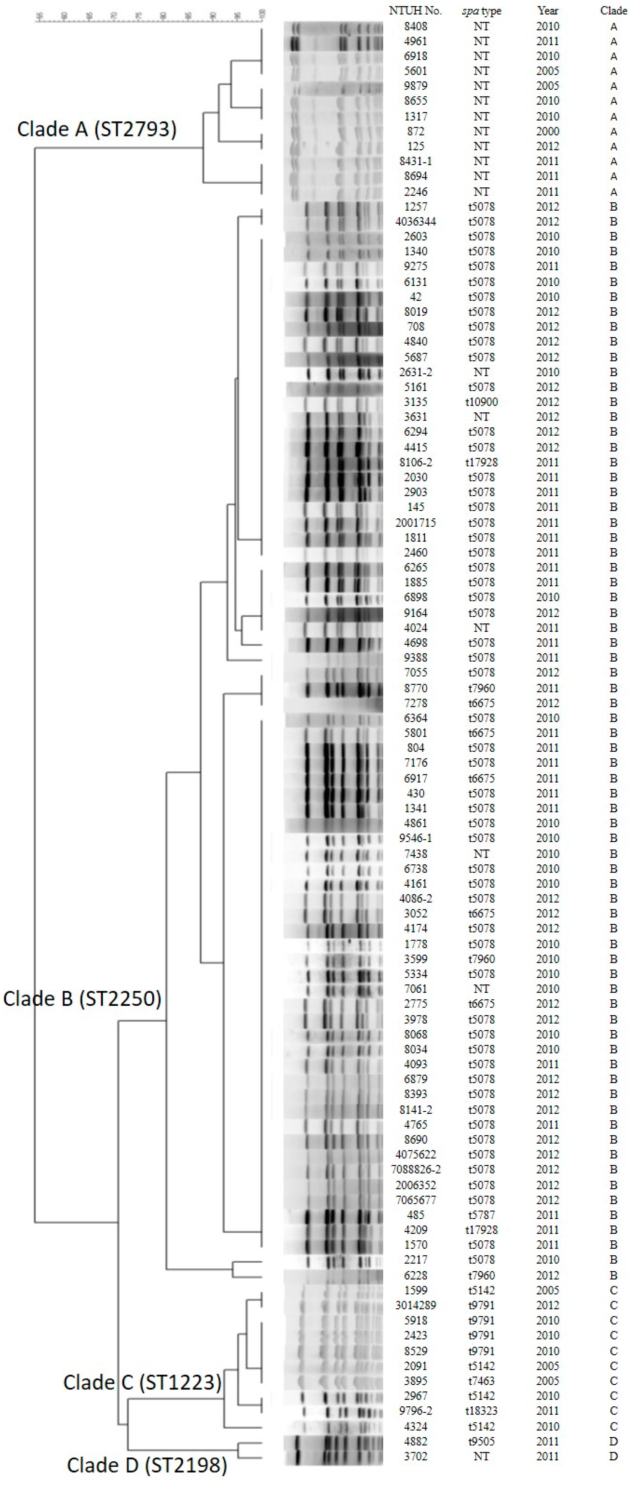
3.3. Genotyping by Pulsed-Field Gel Electrophoresis (PFGE) and Spa Types

PFGE analysis revealed four major pulsotypes (Figure 1). Isolates of the same ST always clustered together. The spa types are listed in Table 1 and Table 2. Most spa types are t5078 and t6675 among ST2250 and t5142 among ST1223. The other spa types have not been reported, and non-typeable (N.T.) (new spa types) in S. argenteus. t5078 was the most frequent spa type (55 isolates and 57%), followed by t6675 (5 isolates and 5%). However, spa was not typeable in 18 isolates, including in all of 12 ST2793 isolates that contain eight isolates with one identical spa repeat (t19483).

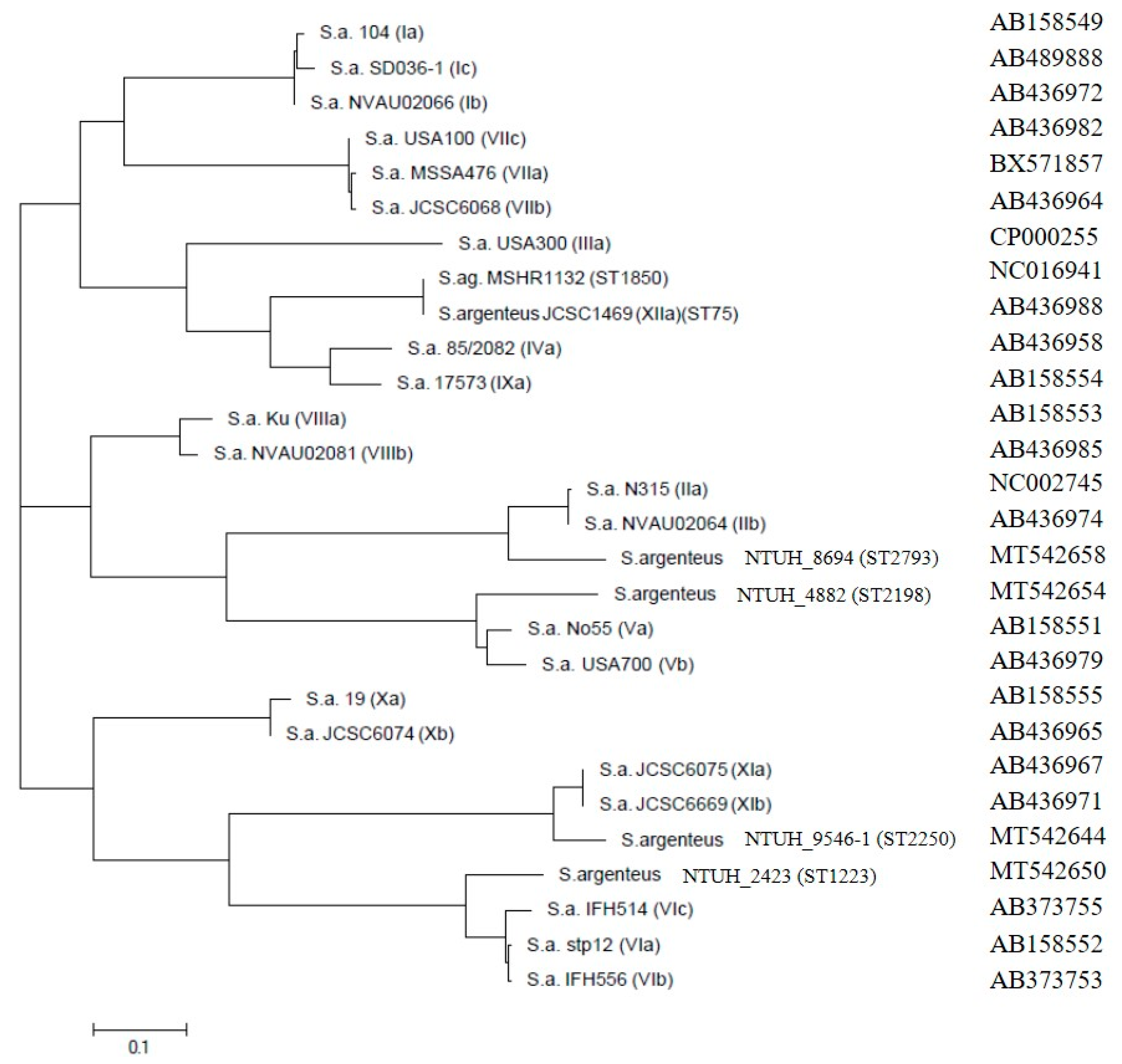
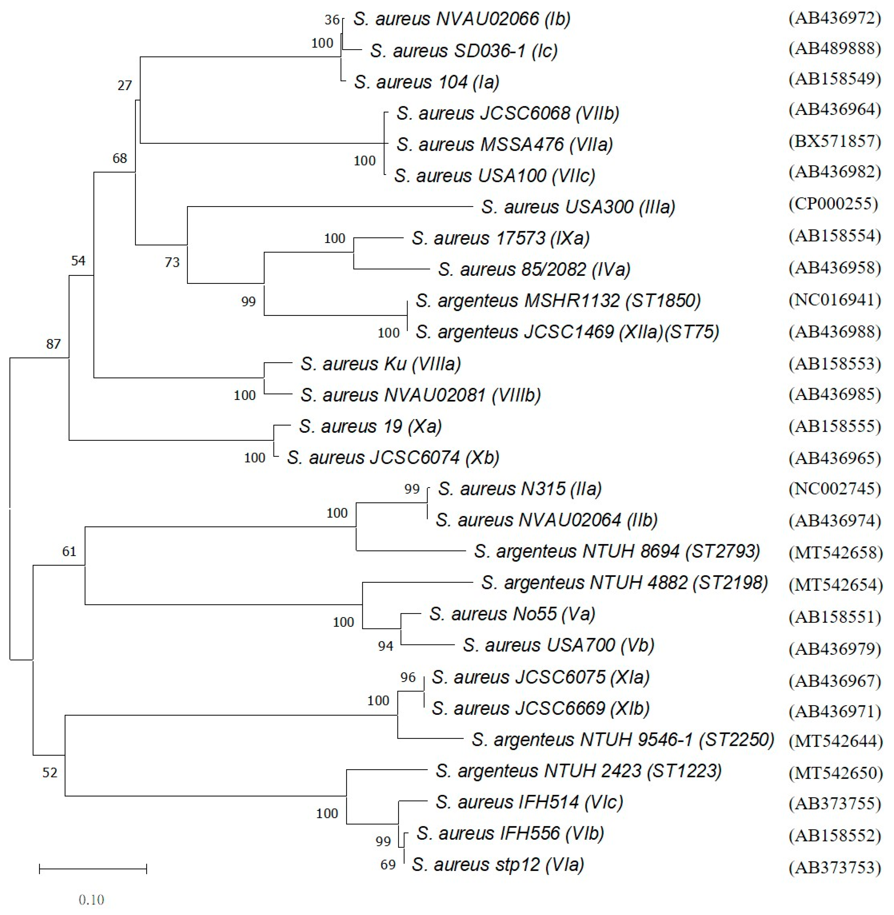
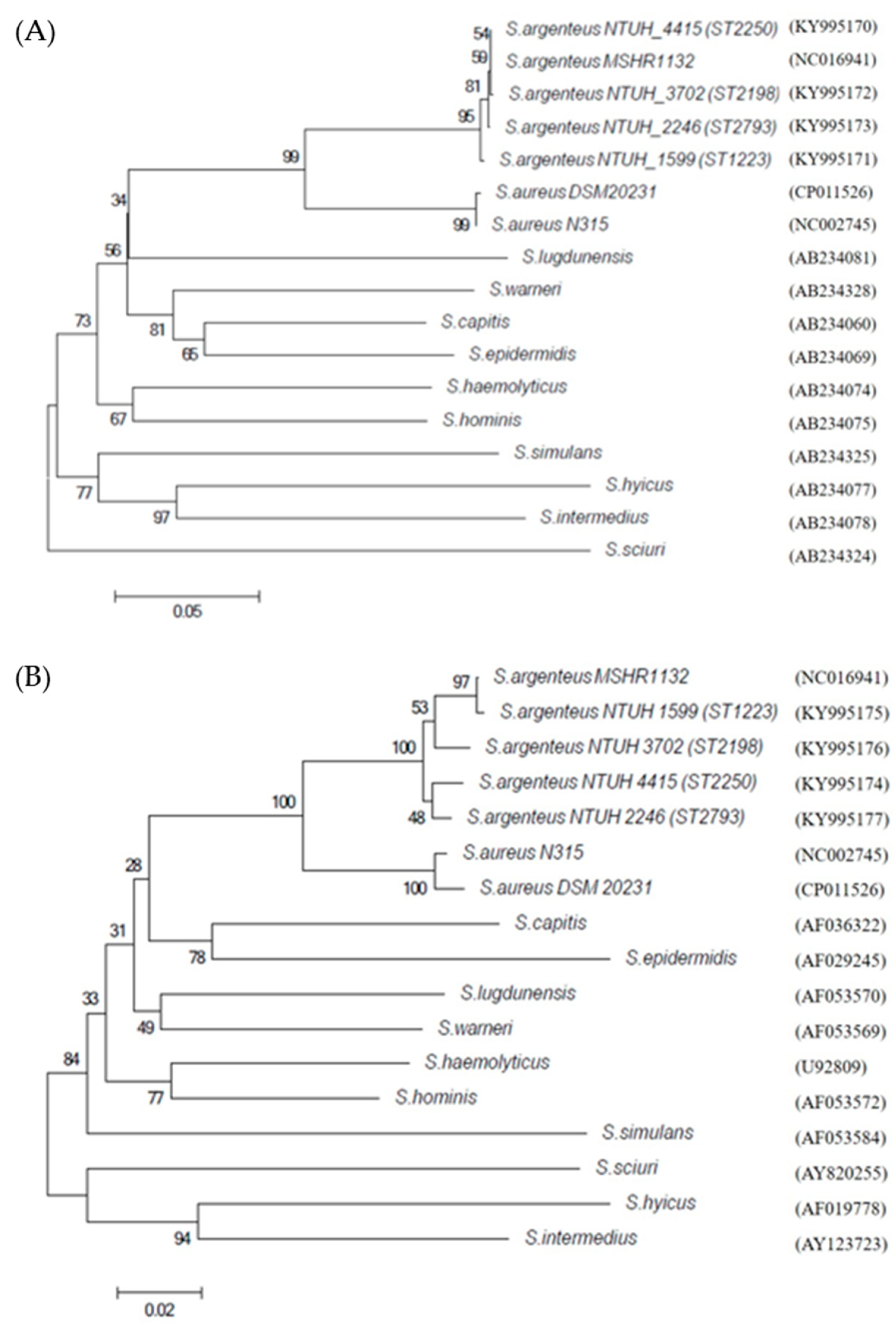
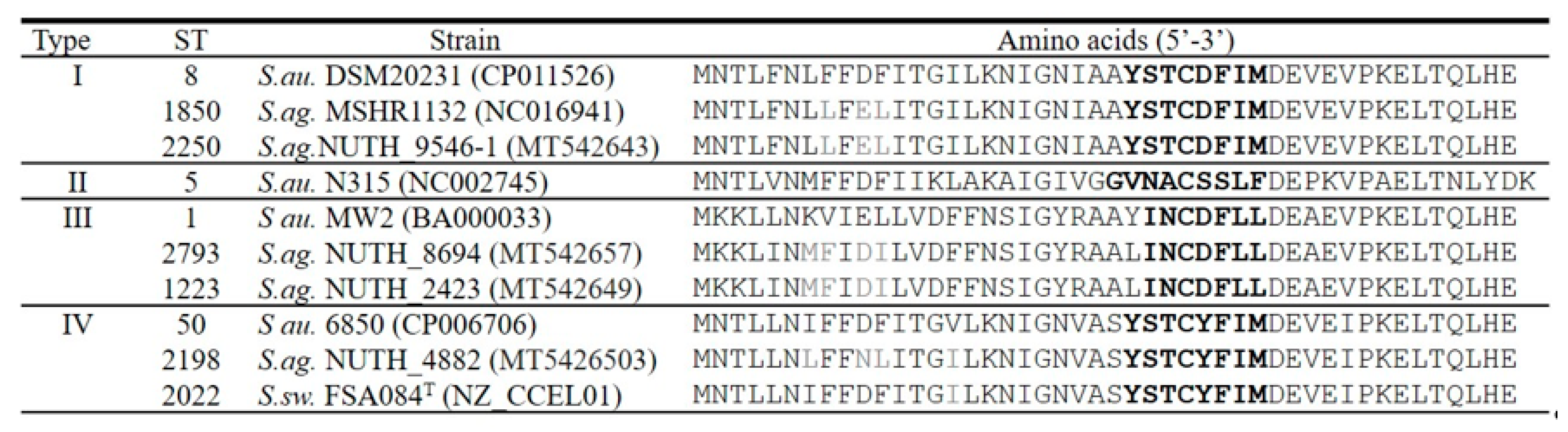
3.4. Phylogenetic Trees of Coa, DnaJ and GroESL and Agr Type

Selected isolates of each ST of S. argenteus were examined for the D1 region sequence of the staphylocoagulase (SC) gene and the (coa) gene. Phylogenetic trees showed that each ST type displayed a distinct type (Figure 2). SC genes of S. argenteus ST2250, ST2793, ST1223, and ST2198 were close to genotype type XI, type II, type VI, and type V but were assigned to type XId, type XVI, type XV, and type XIV according to the criteria, respectively [31]. In particular, ST1223 has been reported as serotype VI, and the phylogenetic tree of the coa gene showed that ST1223 is also close to genotype VI [32].
To further characterize S. argenteus, analyses of partial dnaJ and groESL sequences were performed in selected isolates for each ST. Based on the dnaJ and the groEL sequences, in this study, the S. argenteus clinical isolates and the S. argenteus MSHR1132 were clustered together and separated from S. aureus, forming a stable clade with bootstrap values of 95% and 100% in the phylogenetic trees, respectively (Figure 3). The BLAST results indicated that dnaJ and groEL showed the highest similarity (>98%) to that of non-pigmented S. argenteus MSHR1132 and approximately 90% similarity to S. aureus N315 (Tables S2 and S3). In addition, the spacers between groES and groEL were analyzed. S. argenteus displayed 71 nucleotides of spacer length, while S. aureus and S. argenteus ST1223 had 75 nucleotides of spacer length (Table 3).
Gene agrD produces a ribosomal propeptide of which the middle section encodes the seven to nine residue autoinducing peptide (AIP) used as a quorum sensing (QS) signal molecule. The agrD alignment is shown in Figure 4. S. argenteus harbors S. aureus arg types I, III, and IV. The most dominant agr are type I (ST1850 and ST2250) and type III (ST1223 and ST2793). S. argenteus ST2198 and S. schweitzeri ST2022 are known to be of animal origin, and these two isolates belong to type IV. Although S. argenteus harbors identical AIP to that of S. aureus, S. argenteus displays its unique agrD amino acid sequences, which are different from those of S. aureus or S. schweitzeri.

3.5. Examination of Virulence Factors

The results of virulence factors are shown in Table 4. Each ST type displayed distinct characteristics. The hlb, hlg, lukPVL, eta, etb, edin, sea, sed, see, seh, and tst genes were not detected in all S. argenteus isolates, but the hemolysin genes (hla and hld) were distributed universally. CRISPR/cas, collagen-binding adhesin gene (cna), enterotoxin gene cluster (egc), or sialoprotein-binding protein gene (bbp) were found exclusively in S. argenteus ST2250, ST2793, ST1223, or ST2198 lineage, respectively. Immune evasion cluster (IEC) genes were detected in ~90% isolates, and most of them harbored staphylococcal complement inhibitor gene (scn) and staphylokinase (sak). In addition, the gene encoding staphylococcal enterotoxin type C (sec) was found in 8 out of 12 (67%) ST2793 isolates, and that of staphylococcal enterotoxin type B (seb) was found in three S1223 isolates (3/10, 30%) and one ST2198 (1/2, 50%). The expression of seb or sec in these isolates was further tested by the rapid latex agglutination test. All nine isolates carrying sec were exclusively reactive with enterotoxin type C-specific antibodies, but no positive reactions were seen in the isolates carrying seb.

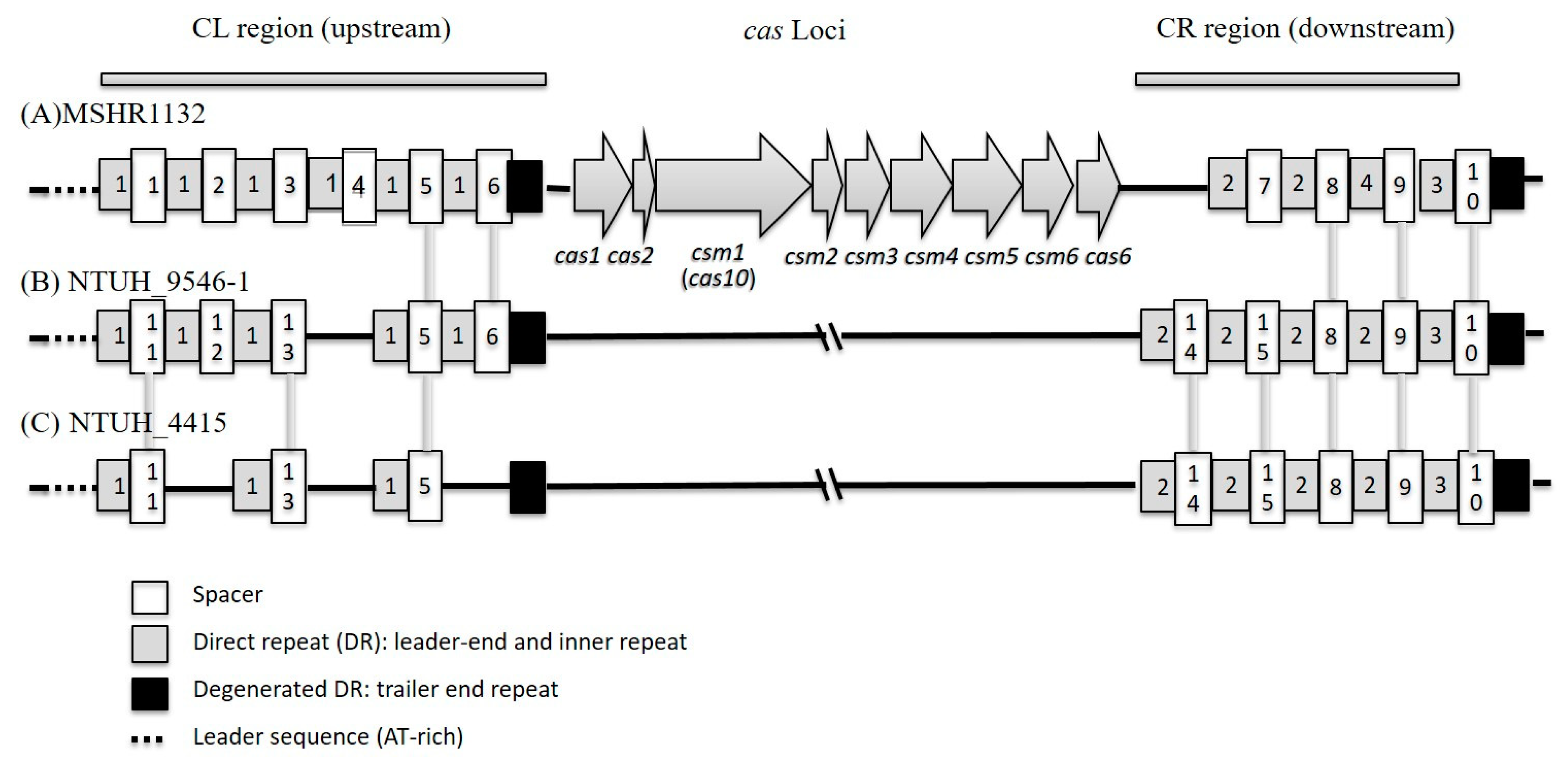
3.6. Polymorphism of CRISPR/Cas System in ST2250 Isolates

Since S. argenteus MSHR1132 harbored the CRISPR/cas system, we tried to determine whether S. argenteus isolates have the same one. The whole CRISPR/cas system of selected S. argenteus ST2250 isolates was determined by PCR and direct sequencing. S. argenteus NTUH_9546-1 and NTUH_4415 (ST2250) and MSHR1132 (ST1850) shared the same type III CRISPR/cas system, and the cas loci was cas1, cas2, csm1(cas10), csm2, csm3, csm4, csm5, csm6, and cas6 in order. (Figure 5) These isolates did not possess the SCCmec found in MSHR1132 and displayed different direct repeats (DRs, leader end and inner repeat) and spacers. Upstream (CL region) and downstream (CR region) regions of cas loci were sequenced for all 72 ST2250 isolates. A total of four DRs, two degenerated DRs (DDR, trailer end repeat), and 16 different spacers were found in these isolates (Table 5). Half isolates (36 isolates, 50%) displayed the same spacers of CL and CR regions as NTUH_9546-1 isolates (Table 6).

4. Discussion

Recent studies have displayed the increasing emergence of S. argenteus from many countries, which has been isolated from both humans and animals [33,34,35]. Isolation of S. argenteus emphasizes its clinical significance and the importance of correct identification in clinical laboratories [8,15]. MALDI-TOF MS is a simple and accurate method for phenotypic screening and identification of S. argenteus [8]. Furthermore, in the present study, the biochemical results of VITEK 2 for the 96 S. argenteus isolates showed a higher percentage of urease and were almost negative for N-acetyl-d-glucosamine. The results suggest that these two tests also could be screening phenotypes for S. argenteus. In the present study, all 96 S. argenteus isolates with bacteremia were methicillin-susceptible and comparable to recent reports from Thailand, Japan, and Myanma, while isolates from Europe and Australia were more methicillin-resistant [5,10,14,15,16].
Notably, the earliest and the only S. argenteus isolate in the year 2000 was ST2793 with novel spa type (t19483). ST2793 lineage is rarely reported in other countries [10,11,12,13,14,36]. To date, ST2793 has been found in Europe and USA but not in Asian counties other than Taiwan [10,13,36]. In addition, a total of 11 new spa types across three ST types were first found in the present study. Some known spa types were identical to those in Japan and Myanmar isolates [14,16] (Table 2). The t5078 clone occupied more than half of S. argenteus ST2250 isolates in Asian countries.
Besides MLST typing and whole genome sequencing, several genetic classification methods have been proposed to distinguish S. argenteus from S. aureus. PCR-based method targeting the NRPS gene, sequencing of the rpoB or the nuc gene, and lack of the crtM gene have been used previously to determine S. argenteus [3,7]. The dnaJ and the groEL genes have been used previously to identify Staphylococcus species [23,37]. Phylogenetic analysis based on the dnaJ and the groEL sequences indicates that these genes may be used as candidate genes for species identification.
Detection of SE-like toxin genes revealed that nine of 12 ST2793 S. argenteus isolates carry sec. A report from Denmark using whole genome sequencing in four ST2793 and 21 non-ST2793 revealed that none of the isolates carried sec [10]. Moreover, the sec partial sequence (245 bp) from ST2793 is identical to that of S. aureus in the Genbank database and almost identical to sec2 or sec3 genes of S. argenteus ST2250 from Japan [14]. ST2250 was described in Thailand as carrying an enterotoxin gene cluster composed of seC-bov (enterotoxin C bovine) and entQ (staphylococcal enterotoxin Q), and these two genes should be the two novel superantigens sel27 and sel26 [14,38]. In addition, some of the ST1223 and the ST2198 isolates in the present study carried enterotoxin type b gene (seb) and others did not, which shows the diversity in Taiwan isolates. However, no isolates carrying seb were found in Hokkaido, Japan, or Myanmar [14,16]. Interestingly, all ST1223 isolates harbored egc clusters as in other studies, but only a small portion carried seb gene, which may cause Staphylococcal food poisoning [32,39].
IEC (immune evasion cluster) was carried by bacteriophage and into hemolysin b gene (hlb) of Staphylococci [29]. Additionally, 60 isolates of ST2250 carried both sak and scn (IEC type E), and only one ST2198 isolate harbored sak, scn, and chp (IEC type B), which is comparable to previous studies [11,40]. Regarding other virulence factors analyzed in this study, collagen binding gene (cna) and spa (protein A) belong to microbial surface components recognizing the adhesive matrix molecules (MSCRAMM) family, and these are associated with bacterial adhesion and pathogenesis [41]. We found that all ST2793 isolates, but not other S. argenteus lineages, harbored the cna gene and carried new and different spa sequences (Table 2). In addition, the ST2793 isolates are methicillin-susceptible, while isolates from Europe carrying SCCmec are methicillin-resistant [10]. This may indicate that the ST2793 isolates in Taiwan reveal genetically diverse clones, which is definitely worth further study.
In the present study, all 72 ST2250 isolates harbored the CRISPR/cas system. Previous studies have mentioned the polymorphism of CRISPR/cas system in S. aureus and S. argenteus [42,43]. However, the present study is the first to study the CRISPR/cas system solely for S. argenteus ST2250 lineage, including four direct repeats with 37 bp (DRs, one upstream and three downstream) and 16 different spacers with 33–39 bp (nine upstream and seven downstream) (Table 5). S. argenteus ST2250 showed greater diversity of CRISPR/cas than that of most other S. aureus ST types and may account for the prevalence and the success of S. argenteus ST2250 clones.
In summary, this study focused on the molecular characterization of four ST types of S. argenteus isolates in northern Taiwan, finding that S. argenteus was isolated as early as the year 2000. Each ST type of S. argenteus harbors particular genetic markers that may contribute to their virulence

Supplementary Materials

Supplementary materials can be found at https://www.mdpi.com/2076-2607/8/8/1157/s1. Table S1: Key biochemical tests used for identification of S. argenteus (N = 96), Table S2: Partial dnaJ (889bp) nucleotide and amino acid sequence similarities, Table S3: Partial groEL (780bp) nucleotide and amino acid sequence similarities, Table S4: Primers list for CRIPSR/cas, Table S5: GenBank accession numbers in the present study.

Author Contributions

Conceptualization: L.-J.T., Methodology: J.-C.H., T.-W.W., H.L., X.-M.W., Y.-T.L., C.-J.J., L.-J.T., Validation: J.-C.H., T.-W.W., Formal Analysis: J.-C.H., T.-W.W., Investigation: J.-C.H., T.-W.W., H.L., X.-M.W., C.-J.J., Resources: J.-C.H., T.-W.W., H.L., X.-M.W., Y.-T.L., T.-F.L., P.-R.H., Data Curation: J.-C.H., T.-W.W., Writing—Original Draft Preparation: J.-C.H., T.-W.W., Writing—Review & Editing: Y.-T.L., L.-J.T., Visualization: J.-C.H., T.-W.W., Supervision: L.-J.T., Project Administration: L.-J.T., Funding Acquisition: L.-J.T., All authors have read and agreed to the published version of the manuscript.

Funding

This work was supported by the grant MOST 106-2320-B-002-039-MY3 from the Ministry of Science and Technology of Taiwan.

Conflicts of Interest

The authors declare that they have no competing interests.

References

  1. Holt, D.C.; Holden, M.T.; Tong, S.Y.; Castillo-Ramirez, S.; Clarke, L.; Quail, M.A.; Currie, B.J.; Parkhill, J.; Bentley, S.D.; Feil, E.J.; et al. A very early-branching Staphylococcus aureus lineage lacking the carotenoid pigment staphyloxanthin. Genome Biol. Evol. 2011, 3, 881–895. [Google Scholar] [CrossRef] [PubMed] [Green Version]
  2. Tong, S.Y.; Schaumburg, F.; Ellington, M.J.; Corander, J.; Pichon, B.; Leendertz, F.; Bentley, S.D.; Parkhill, J.; Holt, D.C.; Peters, G.; et al. Novel staphylococcal species that form part of a Staphylococcus aureus-related complex: The non-pigmented Staphylococcus argenteus sp. nov. and the non-human primate-associated Staphylococcus schweitzeri sp. nov. Int. J. Syst. Evol. Microbiol. 2015, 65, 15–22. [Google Scholar] [CrossRef] [PubMed]
  3. Argudin, M.A.; Dodemont, M.; Vandendriessche, S.; Rottiers, S.; Tribes, C.; Roisin, S.; de Mendonca, R.; Nonhoff, C.; Deplano, A.; Denis, O. Low occurrence of the new species Staphylococcus argenteus in a Staphylococcus aureus collection of human isolates from Belgium. Eur. J. Clin. Microbiol. Infect. Dis. 2016, 35, 1017–1022. [Google Scholar] [CrossRef]
  4. Dupieux, C.; Blonde, R.; Bouchiat, C.; Meugnier, H.; Bes, M.; Laurent, S.; Vandenesch, F.; Laurent, F.; Tristan, A. Community-acquired infections due to Staphylococcus argenteus lineage isolates harbouring the Panton-Valentine leucocidin, France, 2014. Eurosurveillance 2015, 20, 21154. [Google Scholar] [CrossRef] [PubMed]
  5. Thaipadungpanit, J.; Amornchai, P.; Nickerson, E.K.; Wongsuvan, G.; Wuthiekanun, V.; Limmathurotsakul, D.; Peacock, S.J. Clinical and molecular epidemiology of Staphylococcus argenteus infections in Thailand. J. Clin. Microbiol. 2015, 53, 1005–1008. [Google Scholar] [CrossRef] [PubMed] [Green Version]
  6. Ohnishi, T.; Shinjoh, M.; Ohara, H.; Kawai, T.; Kamimaki, I.; Mizushima, R.; Kamada, K.; Itakura, Y.; Iguchi, S.; Uzawa, Y.; et al. Purulent lymphadenitis caused by Staphylococcus argenteus, representing the first Japanese case of Staphylococcus argenteus (multilocus sequence type 2250) infection in a 12-year-old boy. J. Infect. Chemother. 2018, 24, 925–927. [Google Scholar] [CrossRef]
  7. Zhang, D.-F.; Xu, X.; Song, Q.; Bai, Y.; Zhang, Y.; Song, M.; Shi, C.; Shi, X. Identification of Staphylococcus argenteus in Eastern China based on a nonribosomal peptide synthetase (NRPS) gene. Future Microbiol. 2016, 11, 1113–1121. [Google Scholar] [CrossRef]
  8. Chen, S.Y.; Lee, H.; Wang, X.M.; Lee, T.F.; Liao, C.H.; Teng, L.J.; Hsueh, P.R. High mortality impact of Staphylococcus argenteus on patients with community-onset staphylococcal bacteraemia. Int. J. Antimicrob Agents 2018, 52, 747–753. [Google Scholar] [CrossRef]
  9. Chen, S.-Y.; Lee, H.; Teng, S.-H.; Wang, X.-M.; Lee, T.-F.; Huang, Y.-C.; Liao, C.-H.; Teng, L.-J.; Hsueh, P.-R. Accurate differentiation of novel Staphylococcus argenteus from Staphylococcus aureus using MALDI-TOF MS. Future Microbiol. 2018, 13, 997–1006. [Google Scholar] [CrossRef]
  10. Hansen, T.A.; Bartels, M.D.; Hogh, S.V.; Dons, L.E.; Pedersen, M.; Jensen, T.G.; Kemp, M.; Skov, M.N.; Gumpert, H.; Worning, P.; et al. Whole Genome Sequencing of Danish Staphylococcus argenteus Reveals a Genetically Diverse Collection with Clear Separation from Staphylococcus aureus. Front. Microbiol. 2017, 8, 1512. [Google Scholar] [CrossRef]
  11. Moradigaravand, D.; Jamrozy, D.; Mostowy, R.; Anderson, A.; Nickerson, E.K.; Thaipadungpanit, J.; Wuthiekanun, V.; Limmathurotsakul, D.; Tandhavanant, S.; Wikraiphat, C.; et al. Evolution of the Staphylococcus argenteus ST2250 Clone in Northeastern Thailand Is Linked with the Acquisition of Livestock-Associated Staphylococcal Genes. MBio 2017, 8. [Google Scholar] [CrossRef] [PubMed] [Green Version]
  12. Zhang, D.F.; Zhi, X.Y.; Zhang, J.; Paoli, G.C.; Cui, Y.; Shi, C.; Shi, X. Preliminary comparative genomics revealed pathogenic potential and international spread of Staphylococcus argenteus. BMC Genom. 2017, 18, 808. [Google Scholar] [CrossRef] [PubMed] [Green Version]
  13. Giske, C.G.; Dyrkell, F.; Arnellos, D.; Vestberg, N.; Fang, H. Transmission events and antimicrobial susceptibilities of methicillin-resistant Staphylococcus argenteus in Stockholm. Clin. Microbiol. Infect. 2019, 25, 1289.e5–1289.e8. [Google Scholar] [CrossRef] [PubMed]
  14. Aung, M.S.; Urushibara, N.; Kawaguchiya, M.; Sumi, A.; Takahashi, S.; Ike, M.; Ito, M.; Habadera, S.; Kobayashi, N. Molecular Epidemiological Characterization of Staphylococcus argenteus Clinical Isolates in Japan: Identification of Three Clones (ST1223, ST2198, and ST2550) and a Novel Staphylocoagulase Genotype XV. Microorganisms 2019, 7, 389. [Google Scholar] [CrossRef] [Green Version]
  15. Chantratita, N.; Wikraiphat, C.; Tandhavanant, S.; Wongsuvan, G.; Ariyaprasert, P.; Suntornsut, P.; Thaipadungpanit, J.; Teerawattanasook, N.; Jutrakul, Y.; Srisurat, N.; et al. Comparison of community-onset Staphylococcus argenteus and Staphylococcus aureus sepsis in Thailand: A prospective multicentre observational study. Clin. Microbiol. Infect. 2016, 22, 458.e11–458.e19. [Google Scholar] [CrossRef] [PubMed] [Green Version]
  16. Aung, M.S.; San, T.; San, N.; Oo, W.M.; Ko, P.M.; Thet, K.T.; Urushibara, N.; Kawaguchiya, M.; Sumi, A.; Kobayashi, N. Molecular characterization of Staphylococcus argenteus in Myanmar: Identification of novel genotypes/clusters in staphylocoagulase, protein A, alpha-haemolysin and other virulence factors. J. Med. Microbiol. 2019, 68, 95–104. [Google Scholar] [CrossRef] [PubMed]
  17. Aung, M.S.; San, T.; Aye, M.M.; Mya, S.; Maw, W.W.; Zan, K.N.; Htut, W.H.W.; Kawaguchiya, M.; Urushibara, N.; Kobayashi, N. Prevalence and Genetic Characteristics of Staphylococcus aureus and Staphylococcus argenteus Isolates Harboring Panton-Valentine Leukocidin, Enterotoxins, and TSST-1 Genes from Food Handlers in Myanmar. Toxins 2017, 9, 241. [Google Scholar] [CrossRef] [Green Version]
  18. Chu, C.; Wong, M.Y.; Tseng, Y.H.; Lin, C.L.; Tung, C.W.; Kao, C.C.; Huang, Y.K. Vascular access infection by Staphylococcus aureus from removed dialysis accesses. Microbiologyopen 2019, 8, e800. [Google Scholar] [CrossRef] [Green Version]
  19. Ng, J.W.; Holt, D.C.; Lilliebridge, R.A.; Stephens, A.J.; Huygens, F.; Tong, S.Y.; Currie, B.J.; Giffard, P.M. Phylogenetically distinct Staphylococcus aureus lineage prevalent among indigenous communities in northern Australia. J. Clin. Microbiol. 2009, 47, 2295–2300. [Google Scholar] [CrossRef] [Green Version]
  20. Liu, G.Y.; Essex, A.; Buchanan, J.T.; Datta, V.; Hoffman, H.M.; Bastian, J.F.; Fierer, J.; Nizet, V. Staphylococcus aureus golden pigment impairs neutrophil killing and promotes virulence through its antioxidant activity. J. Exp. Med. 2005, 202, 209–215. [Google Scholar] [CrossRef]
  21. Koreen, L.; Ramaswamy, S.V.; Graviss, E.A.; Naidich, S.; Musser, J.M.; Kreiswirth, B.N. spa typing method for discriminating among Staphylococcus aureus isolates: Implications for use of a single marker to detect genetic micro-and macrovariation. J. Clin. Microbiol. 2004, 42, 792–799. [Google Scholar] [CrossRef] [PubMed] [Green Version]
  22. Harmsen, D.; Claus, H.; Witte, W.; Rothgänger, J.; Claus, H.; Turnwald, D.; Vogel, U. Typing of methicillin-resistant Staphylococcus aureus in a university hospital setting by using novel software for spa repeat determination and database management. J. Clin. Microbiol. 2003, 41, 5442–5448. [Google Scholar] [CrossRef] [PubMed] [Green Version]
  23. Hauschild, T.; Stepanovic, S. Identification of Staphylococcus spp. by PCR-restriction fragment length polymorphism analysis of dnaJ gene. J. Clin. Microbiol. 2008, 46, 3875–3879. [Google Scholar] [CrossRef] [PubMed] [Green Version]
  24. Hung, W.-C.; Tseng, S.-P.; Chen, H.-J.; Tsai, J.-C.; Chang, C.-H.; Lee, T.-F.; Hsueh, P.-R.; Teng, L.-J. Use of groESL as a target for identification of Abiotrophia, Granulicatella, and Gemella species. J. Clin. Microbiol. 2010, 48, 3532–3538. [Google Scholar] [CrossRef] [Green Version]
  25. Strommenger, B.; Cuny, C.; Werner, G.; Witte, W. Obvious lack of association between dynamics of epidemic methicillin-resistant Staphylococcus aureus in central Europe and agr specificity groups. Eur. J. Clin. Microbiol. Infect. Dis. 2004, 23, 15–19. [Google Scholar] [CrossRef] [PubMed]
  26. Kinoshita, M.; Kobayashi, N.; Nagashima, S.; Ishino, M.; Otokozawa, S.; Mise, K.; Sumi, A.; Tsutsumi, H.; Uehara, N.; Watanabe, N.; et al. Diversity of staphylocoagulase and identification of novel variants of staphylocoagulase gene in Staphylococcus aureus. Microbiol. Immunol. 2008, 52, 334–348. [Google Scholar] [CrossRef]
  27. Jarraud, S. Relationships between Staphylococcus aureus Genetic Background, Virulence Factors, agr Groups (Alleles), and Human Disease. Infect. Immun. 2002, 70, 631–641. [Google Scholar] [CrossRef] [Green Version]
  28. Peacock, S.J.; Moore, C.E.; Justice, A.; Kantzanou, M.; Story, L.; Mackie, K.; O’Neill, G.; Day, N.P. Virulent combinations of adhesin and toxin genes in natural populations of Staphylococcus aureus. Infect. Immun. 2002, 70, 4987–4996. [Google Scholar] [CrossRef] [Green Version]
  29. van Wamel, W.J.; Rooijakkers, S.H.; Ruyken, M.; van Kessel, K.P.; van Strijp, J.A. The innate immune modulators staphylococcal complement inhibitor and chemotaxis inhibitory protein of Staphylococcus aureus are located on beta-hemolysin-converting bacteriophages. J. Bacteriol. 2006, 188, 1310–1315. [Google Scholar] [CrossRef] [Green Version]
  30. Tamura, K.; Dudley, J.; Nei, M.; Kumar, S. MEGA4: Molecular evolutionary genetics analysis (MEGA) software version 4.0. Mol. Biol. Evol. 2007, 24, 1596–1599. [Google Scholar] [CrossRef]
  31. Thoendel, M.; Kavanaugh, J.S.; Flack, C.E.; Horswill, A.R. Peptide signaling in the staphylococci. Chem. Rev. 2011, 111, 117–151. [Google Scholar] [CrossRef] [PubMed] [Green Version]
  32. Suzuki, Y.; Kubota, H.; Ono, H.K.; Kobayashi, M.; Murauchi, K.; Kato, R.; Hirai, A.; Sadamasu, K. Food poisoning outbreak in Tokyo, Japan caused by Staphylococcus argenteus. Int. J. Food Microbiol. 2017, 262, 31–37. [Google Scholar] [CrossRef] [PubMed]
  33. Neyaz, L.; Karki, A.B.; Fakhr, M.K. The Whole-Genome Sequence of Plasmid-Bearing Staphylococcus argenteus Strain B3-25B from Retail Beef Liver Encodes the Type VII Secretion System and Several Virulence Factors. Microbiol. Resour. Announc. 2019, 8. [Google Scholar] [CrossRef] [Green Version]
  34. Lia, Q.; Lia, Y.; Tang, Y.; Meng, C.; Ingmer, H.; Jiao, X. Prevalence and characterization of Staphylococcus aureus and Staphylococcus argenteus in chicken from retail markets in China. Food Control. 2019, 96, 158–164. [Google Scholar] [CrossRef]
  35. Pumipuntu, N.; Tunyong, W.; Chantratita, N.; Diraphat, P.; Pumirat, P.; Sookrung, N.; Chaicumpa, W.; Indrawattana, N. Staphylococcus spp. associated with subclinical bovine mastitis in central and northeast provinces of Thailand. PeerJ 2019, 7, e6587. [Google Scholar] [CrossRef] [PubMed] [Green Version]
  36. Long, S.W.; Beres, S.B.; Olsen, R.J.; Musser, J.M. Absence of patient-to-patient intrahospital transmission of Staphylococcus aureus as determined by whole-genome sequencing. mBio 2014, 5, e01692-14. [Google Scholar] [CrossRef] [Green Version]
  37. Kwok, A.Y.; Chow, A.W. Phylogenetic study of Staphylococcus and Macrococcus species based on partial hsp60 gene sequences. Int. J. Syst. Evol. Microbiol. 2003, 53, 87–92. [Google Scholar] [CrossRef] [Green Version]
  38. Zhang, D.F.; Yang, X.Y.; Zhang, J.; Qin, X.; Huang, X.; Cui, Y.; Zhou, M.; Shi, C.; French, N.P.; Shi, X. Identification and characterization of two novel superantigens among Staphylococcus aureus complex. Int. J. Med. Microbiol. 2018, 308, 438–446. [Google Scholar] [CrossRef]
  39. Wakabayashi, Y.; Umeda, K.; Yonogi, S.; Nakamura, H.; Yamamoto, K.; Kumeda, Y.; Kawatsu, K. Staphylococcal food poisoning caused by Staphylococcus argenteus harboring staphylococcal enterotoxin genes. Int. J. Food Microbiol. 2018, 265, 23–29. [Google Scholar] [CrossRef]
  40. Schuster, D.; Rickmeyer, J.; Gajdiss, M.; Thye, T.; Lorenzen, S.; Reif, M.; Josten, M.; Szekat, C.; Melo, L.D.; Schmithausen, R.M.; et al. Differentiation of Staphylococcus argenteus (formerly: Staphylococcus aureus clonal complex 75) by mass spectrometry from S. aureus using the first strain isolated from a wild African great ape. Int. J. Med. Microbiol. 2017, 307, 57–63. [Google Scholar] [CrossRef] [Green Version]
  41. Cao, L.; Gao, C.H.; Zhu, J.; Zhao, L.; Wu, Q.; Li, M.; Sun, B. Identification and functional study of type III-A CRISPR-Cas systems in clinical isolates of Staphylococcus aureus. Int. J. Med. Microbiol. 2016, 306, 686–696. [Google Scholar] [CrossRef] [PubMed]
  42. Mao, T.; Long, J.; Duan, G.; Yang, H. Commentary: Study the Features of 57 Confirmed CRISPR Loci in 38 Strains of Staphylococcus aureus. Front. Microbiol. 2019, 10, 59. [Google Scholar] [CrossRef] [PubMed]
  43. Zhao, X.; Yu, Z.; Xu, Z. Study the Features of 57 Confirmed CRISPR Loci in 38 Strains of Staphylococcus aureus. Front. Microbiol. 2018, 9. [Google Scholar] [CrossRef] [PubMed] [Green Version]
Figure 1. Pulsed-field gel electrophoresis (PFGE) dendrogram of 96 S. argenteus clinical isolates. PFGE cluster was assigned to isolates having 80% or greater similarity from the dendrogram. For unknown spa type (NT): see Table 2.
Microorganisms 08 01157 g001
Figure 2. Phylogenetic tree based on D1 region of coa gene. The phylogenetic tree was generated using the unrooted neighbor-joining method in the MEGA6 package. The numbers at the nodes are confidence levels, expressed as percentages of occurrence in 1000 bootstrapped resamplings. The scale bar indicates the evolutionary distance between sequences, as determined by measuring the lengths of the horizontal lines connecting two organisms. Each sequence type (ST) type of S. argenteus belonged to different clades. The results showed identical partial coa sequences at least two isolates for each ST. GenBank accession numbers are given in parentheses with one isolate for each ST in this study.
Microorganisms 08 01157 g002aMicroorganisms 08 01157 g002b
Figure 3. Phylogenetic tree based on partial dnaJ (A) and groEL (B). The phylogenetic tree was generated using the unrooted neighbor-joining method in the MEGA6 package. The numbers at the nodes are confidence levels, expressed as percentages of occurrence in 1000 bootstrapped resamplings. The scale bar indicates the evolutionary distance between sequences, as determined by measuring the lengths of the horizontal lines connecting two organisms. The results showed identical partial dnaJ or groEL sequences at least two isolates for each ST. GenBank accession numbers are given in parentheses with one isolate for each ST in this study.
Microorganisms 08 01157 g003
Figure 4. Comparisons of the predicted AgrD amino acid sequences and agr types. S. au.: S. aureus. S.ag.: S. argenteus. S.sw.: S. schweitzeri. AIP (autoinducing peptide) sequences are bolded. The sequences different from S. au are grey bottom. The results showed identical partial AgrD sequences at least two isolates for each ST.
Microorganisms 08 01157 g004
Figure 5. The schematic diagrams of S. argenteus CRISPR/cas systems. Sequences of numbers in squares, see Table 4.
Microorganisms 08 01157 g005
Table 1. Staphylococcus argenteus clinical isolates by lineage (N = 96).
STNo. of Isolates Each Year
Spa Type20002005201020112012Total (%)
ST22500019252872 (75.0)
t5078 a0015182255 (57.3)
t5787000101 (1.0)
t6675000235 (5.2)
t7960001113 (3.1)
t10900000011 (1.0)
t17928000202 (2.1)
Unknown b003115 (5.3)
ST27931244112 (12.5)
Unknown c1244112 (12.5)
ST12230351110 (10.4)
t5142022004 (4.2)
t7463010001 (1.0)
t9791003014 (4.2)
t18323000101 (1.0)
ST2198001102 (2.1)
t9505000101 (1.0)
Unknown d001001 (1.0)
Total (%)1 (1.0)5 (5.2)29 (30.2)31 (32.3)30 (31.3)96 (100)
a One isolate (NTUH_2217) corresponded to a new single locus variant (pta locus 180A > G) of ST2250. b,c,d See Table 2 for new spa type.
Table 2. New spa types for Staphylococcus argenteus clinical isolates.
ST TypeSpa Repeat ProfileNo. of Isolates
ST2250299-31-25-17-16-16-16-161
ST2250299-31-25-16-16-16-16-161
ST2250299-31-25-17-119-16-16-16-16 1
ST2250299-31-17-17-16-16-16-16-16-16-161
ST2250299-31-31-31-25-17-17-16-16-16-161
ST2793259-31-16-16-16-23-17-360-360-25 (t19483)8
ST2793259-31-16-16-23-307-360-360-251
ST2793259-31-307-16-23-17-360-360-251
ST2793259-25-251
ST2793259-251
ST2198259-23-23-17-17-17-23-23-23-17-17-161
Table 3. Nucleotide sequences and length of spacers between groES and groEL among species. “-” indicated the same nucleotide with MSHR1132 and “.” Indicated blank. The results showed identical spacer sequences at least two isolates for each ST. GenBank accession numbers are given in parentheses with one isolate for each ST in this study.
StrainSpacer Length (bp)Spacer Sequence (5′-3′)
S.ag. MSHR1132
(ST1850) (NC016941)
71TACAGAACTTAATTCATAAATAAATTATTAAGAACAATAATCAAACA....TTAAAAAATGGAGGTTTATTATAT
S.ag. NTUH_9546-1
(ST2250) (MT542642)
71-----------------------------------------------....------------------------
S.ag. NTUH_8694
(ST2793) (MT542656)
71--g---g----------------------------------------....------------------------
S.ag. NTUH_2423
(ST1223) (MT542648)
75---------------------------aa------g-a---g-----caat-a--c-------------------
S.ag. NTUH_4882
(ST2198) (MT542652)
71-----------------------------------------------....------------------------
S.a. N315
(ST5) (NC002745)
75--t-a--t-a--------g-------g-a------g-a---g---t-tgac-a--c--------------c--t-
S.a. DSM20231
(ST8) (CP011526)
75---------------------------aat-----g-a---g-----caac-a--c--------------c--t-
Table 4. Virulence genes of Staphylococcus argenteus clinical isolates by lineage a (N = 96).
ST Type
(No. of Isolates)
ST2250 (72)ST2793 (12)ST1223 (10)ST2198 (2)
agr type bIIIIIIIIV
coa genotype bXIdXVIXV
(serotype VI)
XIV
CRISPR+ (72)NTNTNT
IEC type Csak+, scn+ (60)
(Type E)
sak+ (1)
scn+ (1)
NT (10)
scn+ (11)
NT(1)
scn+ (9)
NT(1)
sak+, scn+, chp+ (1)
scn+ (1)
hla & hld+ (72)+ (12)+ (10)+ (2)
cnaNT+ (12, type III)NTNT
bbpNTNTNT+ (2)
sebNTNT+ (3)+ (1)
secNT+ (8)NTNT
egc clusterNTNT+ (10)NT
a NT, Non-typeable or not detected; The following genes were not detected in any strain: crtM, hlb, hlg, lukPVL, eta, etb, edin, sea, sed, see, seh,and tst. b selected strains (NTUH-9546-1, 8694, 2423 and 3702) for agr and coa typing, coa genotype was based on D1 region and previous described (17) and serotype was according to previous described (27). C IEC (immune evasion cluster) type B contains sak+, scn+, and chp (60 isolates) and type E contains sak+ and scn+ (one isolate).
Table 5. Direct repeat (DR) and spacer of CRISPR region. DR was classified by core region (underlined, 36 bp).
TypeSequenceSize(bp)Note
DR1GATCGATAACTACCCCGAAGAATAGGGGACGAGAACX37upstream
DR2XATTCGATAACTACCCCGAAGAAGAGGGGACGAGAAC37downstream
DR3TATTCGATAACTACCCCGAAGAAAAGGGGACGAGAAC37downstream
DR4TATTCGATAACTACCCCGAAGAAGAGGAGACGAGAAC37downstream
DDRTGATCGATAACTACCCCGAAGAATAGGGGACAGAGTG37Degenerated DR
DDRTATTCGATAAATACCCCGGAGAACAGGGGGCGAAAAC37Degenerated DR
S1CTACTAAAAAGTTATATGTTTCAACAATTTCGTCA35upstream
S2GGTTTAAGTTTGTCATTATAATCAATCCTTTTTCTT36upstream
S3GATTAAAACGGTTTGCTTTATTTGCATTTAAAATAG36upstream
S4TTTTTCATAGTTAATCAATCCCTTTTCTTTTTT33upstream
S5TAAATCTTTGATTGCTCTTAGCTCTAGTTATGTAT35upstream
S6ACGCTGTAGTGAAGTATAGAAACGGCATGAGTACAAT37upstream
S7TTTTACTGTGTTTTTCATAATTAATCAATCCTTT34downstream
S8TGCCCACTTAATTAATTCATCTAGTCTCATTTCTT35downstream
S9CATCAACTGACTTTTTAACTGTTTTAGTGAATTCGTC37downstream
S10TTAAAGATCTCAACAATAGCGTCCCATATTTTCTG35downstream
S11TAATTGCATTATCAAATGTATATGCTGGATTCCAT35upstream
S12GTACTTAAGATTTCATCAACTTTCTTTTGTACT33upstream
S13CGAATTTTGATTCTTTGTTTGTAAATAATGCTCT34upstream
S14CTATAATAGTTACTGCTTTTGTAACCGTCCATAT34downstream
S15AAATGCTTATCCATTCTAATCATATTTTCAATTTGTTTA39downstream
S16TGCCCACTTAATTAATTCATCTAGTCTTATTTCTT35downstream
similar to S8
Table 6. Staphylococcus argenteus Type III-A CRISPR/cas systems.
Isolate ID a (Accession No.)Number of SpacersDirect Repeat
UpstreamDownstream
MSHR 1132 (ST1850) (NC016941)6 (S1~6)4 (S7~10)DR1, 2, 3, 4
TD13 (ST2854) (MH513583)5 (S11~13,5~6)-DR1
TD162 (ST2250) (MF167423)5 (S11~13,5~6)- aDR1
SH3 (ST2250) (PRJEB8900)5 (S11~13,5~6)5 (S14~15,8~10)DR1, 2, 3
36 isolates (NTUH_9546-1) (MT542645)5 (S11~13,5~6)5 (S14~15,8~10)DR1, 2, 3
9 isolates (MT542659~60)4 (S11~13,5)5 (S14~15,8~10)DR1, 2, 3
6 isolates (MT542661~62)4 (S11,13,5~6)5 (S14~15,8~10)DR1, 2, 3
5 isolates (MT542663~64)3 (S13,5~6)5 (S14~15,8~10)DR1, 2, 3
3 isolates (MT542665~66)5 (S11~13,5~6)4 (S14,8~10)DR1, 2, 3
2 isolates (MT542667~68) 2 (S11,6)5 (S14~15,8~10)DR1, 2, 3
2 isolates (MT542669~70)4 (S11~12,5~6)4 (S15,8~10)DR1, 2, 3
1 isolate (MT542671~72)5 (S11~13,5~6)7 (S14~15, 8~9, 8~10)DR1, 2, 3
1 isolate (MT542673~74)4 (S11~13,5)5 (S14~16, 9~10)DR1, 2, 3
1 isolate (MT542675~76)4 (S11~13,6)5 (S14~15,8~10)DR1, 2, 3
1 isolate (MT542677~78)3 (S12~13,5)5 (S14~15,8~10)DR1, 2, 3
1 isolate (NTUH_4415) (MT542646)3 (S11,13,5)5 (S14~15,8~10)DR1, 2, 3
1 isolate (MT542679~80)3 (S11,5~6)5 (S14~15,8~10)DR1, 2, 3
1 isolate (MT542681~82)2 (S11,5)5 (S14~15,8~10)DR1, 2, 3
1 isolate (MT542683~84)2 (S5,6)5 (S14~15,8~10)DR1, 2, 3
1 isolate (MT542685~86)4 (S11~13,6)3 (S8~10)DR1, 2, 3
a No downstream sequence data of TD13 and TD162.

