Article

Investigation into In Vitro and In Vivo Caenorhabditis elegans Models to Select Cheese Yeasts as Probiotic Candidates for their Preventive Effects against Salmonella Typhimurium

1
Université Clermont Auvergne, INRAE, VetAgro Sup, F-15000 Aurillac, France
2
Université Clermont Auvergne, Laboratoire Météorologie Physique, CNRS, F-15000 Aurillac, France
*
Author to whom correspondence should be addressed.
Microorganisms 2020, 8(6), 922; https://doi.org/10.3390/microorganisms8060922
Submission received: 27 May 2020 / Revised: 12 June 2020 / Accepted: 17 June 2020 / Published: 18 June 2020

Abstract

:
The design of multiscale strategies integrating in vitro and in vivo models is necessary for the selection of new probiotics. In this regard, we developed a screening assay based on the investigation of the potential of yeasts from cheese as probiotics against the pathogen Salmonella Typhimurium UPsm1 (ST). Two yeasts isolated from raw-milk cheese (Saccharomyces cerevisiae 16, Sc16; Debaryomyces hansenii 25, Dh25), as well as S. cerevisiae subspecies boulardii (CNCM I-1079, Sb1079), were tested against ST by applying in vitro and in vivo tests. Adherence measurements to Caco-2 and HT29-MTX intestinal cells indicated that the two tested cheese yeasts presented a better adhesion than the probiotic Sb1079 as the control strain. Further, the Dh25 was the cheese yeast most likely to survive in the gastrointestinal tract. What is more, the modulation of the TransEpithelial Electrical Resistance (TEER) of differentiated Caco-2 cell monolayers showed the ability of Dh25 to delay the deleterious effects of ST. The influence of microorganisms on the in vivo model Caenorhabditis elegans was evaluated by measuring the longevity of the worm. This in vivo approach revealed that this yeast increased the worm’s lifespan and protected it against ST infection, confirming that this in vivo model can be useful for screening probiotic cheese yeasts.

1. Introduction

The gut microbiota is dominated by bacteria, but eukaryotic microorganisms also contribute to the human microbiome [1,2]. This complex community interacts with the host, greatly impacts human health and physiology, and is essential for gastrointestinal homeostasis [3]. Perturbations in the gut microbiota composition may represent an important risk factor for disease [4]. Food, and particularly cheese, obtained by the activity of bacteria and yeasts, should be a source of eukaryotic microorganisms, some of which may have a probiotic potential. The Food and Agriculture Organization of the United Nations (FAO) and the World Health Organization (WHO) [5] defined probiotics as live microorganisms that, when administered in adequate amounts, confer a health benefit on the host. The majority of the probiotic strains that have been studied are lactic bacteria. However, more recently, research into yeasts with potentially beneficial influences on human health has mainly revolved around Saccharomyces cerevisiae subspecies boulardii CNCM I-1079. This is one of the most well-known probiotic yeasts for human use and available on the market [6] and has been included in numerous controlled trials. Yeasts are not affected by antibacterial treatments, and this is one of the reasons why S. boulardii is prescribed for the prevention of antibiotic-associated diarrhea [7]. This strain has shown a beneficial effect on inflammatory bowel diseases such as Crohn’s disease [8] by impacting the inflammatory cytokine production of the intestinal epithelial cells [9]. Little is known about the interaction between non-Saccharomyces yeasts and the human microbiota, but other yeast species have been reported to have potential probiotic properties, including Debaryomyces hansenii, Kluyveromyces lactis, Yarrrowia lipolytica, and Issatchenkia occidentalis [10,11,12]. These species are frequently associated with dairy products such as raw-milk cheeses [13,14]. Furthermore, these species have the Qualified Presumption of Safety (QPS) status in the European Union and are commonly found and used in food fermentation [15]. These facts could support and help the applications for human use.
Different approaches for new probiotic screening can highlight different physiological characteristics and mechanisms of action. Indeed, potentially probiotic microorganisms generally need to be able to survive in the gastrointestinal tract in order to function in the gut environment. They must withstand acid gastric juice, bile salts, low pH, and internal human body temperature. Moreover, it is preferable that they adhere to the epithelial cells in order to better exert their effect against intestinal pathogens. They can function as a microbial barrier by preventing pathogen binding [16] and modulation of the immune system [17,18], as well as by the production of inhibitory compounds, such as organic acid, oxygen catabolites, and proteinaceous compounds [19]. Adhesion ability is strain-dependent, and an evaluation of this characteristic is recommended for the selection of a probiotic [19,20]. Recent research has shown the need to combine in vitro and in vivo approaches with animal models as a preface to human studies in order to decipher probiotic functions and mechanisms of action [21]. However, in vivo assays are expensive and time-consuming as they require a large number of animals with the ensuing ethical problems. This would explain why, recently, there has been a noticeable increase in the use of the soil nematode Caenorhabditis elegans in microbiology studies. Another advantage of this model is that its intestinal cells are structurally similar to those in humans [22], and its body is transparent [23]. This worm has already been used successfully as a model organism for the simple, rapid and reliable screening of potential probiotic bacteria [23] and, more especially, to evaluate the protective effect of Lactobacillus spp. against S. Typhimurium [24]. C. elegans has also emerged as a valuable model host in which to study the pathogenicity of yeasts [25,26,27,28] but has rarely been used to evaluate the possible probiotic activities of yeasts [29]. Recently, Kunyeit et al. [12] chose this model to highlight the protective effect of yeasts against pathogenic non-albicans Candida species. C. elegans has never been before considered for the evaluation of the preventive effects of yeasts against pathogenic bacteria.
Salmonella enterica serovar Typhimurium is a major cause of infectious disease in animals (i.e., poultry, pig, human). Human contamination can occur via goat milk and cheese consumption [30], as a substantial percentage of animals carry this microorganism in their rumen and feces [31]. The pathogenesis of Salmonella requires, in particular, the adhesion to and the invasion of the intestinal epithelial cells [32]. This bacterium then induces the disruption of the host cell tight junctions, leading to a decrease in the TransEpithelial Electrical Resistance (TEER) [33]. Indeed, TEER is a widely accepted quantitative technique to measure the integrity of tight junctions. TEER values are strong indicators of the integrity of cellular barriers. They can be performed in real time without cell damage and generally are based on measuring ohmic resistance. To investigate Salmonella adhesion and invasion, Gagnon et al. [34] used human colonic Caco-2 and mucus-secreting HT29-MTX cell lines. The results showed the influence of Salmonella serovar on cell adhesion as well as the positive effects of mucus on the infection. These in vitro cell models have also been widely selected for in vitro attachment and mechanistic studies [35]. This pathogen can survive for a long time in stored cheese, and one approach for inhibiting it is the use of lactic acid bacteria. Yeasts can also be considered for this purpose.
In this study, two yeasts isolated from raw-milk cheese (Saccharomyces cerevisiae 16, Sc16; Debaryomyces hansenii 25, Dh25), as well as Saccharomyces cerevisiae subspecies boulardii (CNCM I-1079, Sb1079) were experimented against Salmonella Typhimurium (UPsm1 strain, ST) by applying in vitro recognized tests (human cells) and an innovative C. elegans in vivo test. Tolerance to low pH and bile was evaluated, and the ability of the yeasts to adhere to human epithelial intestinal cells was observed using the Caco-2 and HT29-MTX cell lines. We also assessed the impact of the yeasts on the TEER of the differentiated monolayers of the Caco-2 cells, and the ability of Dh25 to delay the deleterious effects of ST on the integrity of the monolayers cells. An in vivo approach using the nematode C. elegans was performed by testing its ability to survive and to extend its lifespan in the presence of the yeasts and/or the pathogen and, thus, to consider its possible use for the screening of potential probiotic yeasts.

2. Materials and Methods

2.1. Yeasts, Bacteria Strains, and Growth Conditions

All of the strains—Salmonella enterica serovar Typhimurium UPsm1 (ST), Saccharomyces cerevisiae (Sc16 and Sc52 strains), Debaryomyces hansenii (Dh14 and Dh25 strains), Yarrowia lypolitica (3CP5 and 4PO1 strains) and Kluyveromyces lactis (Kl1 and Kl6 strains), isolated from cheese—came from the UMRF (Unité Mixte de Recherche sur le Fromage) collection. Saccharomyces cerevisiae subspecies boulardii CNCM I-1079 (S. boulardii, Sb1079) was obtained from Ultralevure (Lallemand, Toulouse, France). Escherichia coli OP50 (standard food for nematode) was obtained from the Caenorhabditis Genetics Center (CGC, Minneapolis, USA). All strains were stored at −80 °C in their culture media with 20% glycerol (Biosolve, Dieuze, France). For all experiments, S. Typhimurium was aerobically grown at 37 °C for 24 h in 50 mL Brain–Heart Infusion (BHI) broth (Biokar Diagnostics, Pantin, France), and yeasts were aerobically grown at 25 °C for 48 h, in 50 mL Yeast Extract–Peptone–Glucose (YPG) broth, at pH = 5.2 (40 g/L glucose (Sigma-Aldrich, St-Louis, USA), 5 g/L yeast extract (Biokar Diagnostics) and 5 g/L peptone (CONDA pronadisa, Madrid, Spain)). E. coli OP50 was aerobically grown at 37 °C for 24 h, in 50 mL Lysogeny Broth (LB) (Biokar Diagnostics).

2.2. Human Cell Cultures

The human colon carcinoma cell line, Caco-2 [36] and the mucus-secreting HT29-MTX cell line, provided by Dr. Thécla Lesuffleur (INSERM UMR S 938 Paris, France), were grown in Dulbecco’s Modified Eagle’s Minimal essential medium (DMEM) (Life Technologies, Villebon sur Yvette, France) supplemented with 20% or 10%, respectively, of heat-inactivated fetal calf serum (Life Technologies). The cells were maintained at 37 °C with 5% CO2 in air atmosphere.

2.3. Determination of Acid pH and Bile Tolerance of Yeasts

As described previously [37], acid tolerance at pH 2.5 was determined in gastric juice (Bacto Peptone 30 g/L (FISHER Bioblock Scientific, Illkirch, France), sodium chloride 99.5% 15 g/L (ACROS ORGANICS, Geel, Belgium), Pepsin A 3.2 g/L (SIGMA, Saint Quentin-Fallavier, France)) adjusted with HCl 0.1 M (DUTSCHER, Brumath, France). A suspension of the yeast strains from a 20 h culture, corresponding to approximately 107 colony-forming units (CFU)/mL, was added to 10 mL of the gastric juice solution. Viable cells were measured after 0, 45, and 90 min by plating serial dilutions of aliquots onto Oxytetracycline Glucose Agar (OGA) base (Biokar, France), supplemented with 0.1% (w/v) oxytetracycline. The Petri dishes were incubated for 3 days at 25 °C.
Bile tolerance was determined in 20 mL of YPG broth with or without 3% of bile salts (SIGMA). The number of viable cells was determined after 0, 60, 120, 180, and 240 min of incubation by plating the serial dilutions of aliquots onto OGA Petri dishes incubated for 3 days at 25 °C. All assays were performed in triplicate.

2.4. In Vitro Yeasts Adhesion Assay

The adhesion capacities of the yeast strains were examined using the Caco-2 and the HT29-MTX cell lines. Human cells were seeded at a concentration of 3.5 × 10⁵ cells per well in 24-well plates (Dutscher, Brumath, France) and placed in growth conditions overnight and for 15 days.
Overnight cultures of yeast strains were harvested by centrifugation (1500g for 15 min), washed once with phosphate-buffered saline (PBS), and suspended in 9 mL of DMEM supplemented with 20% (Caco-2) or 10% (HT29-MTX) fetal calf serum. The final multiplicities of infection (MOIs, ratios between yeasts and human cells) tested were 0.1, 1, 10, and 100. The plates were incubated for 3 h at 37 °C under 5% CO2. After incubation, the wells were washed three times with 1 mL of PBS, and the cell monolayer with adhering yeasts was detached by the addition of 1 mL of trypsin solution (Life Technologies) and resuspended in sterile water. The number of viable yeasts was determined by plating serial dilutions of the suspensions onto YPG agar plates after 48 h at 25 °C. Each adhesion assay was performed in triplicate.

2.5. Growth Inhibition of Salmonella Typhimurium by Yeasts

For the ST and yeast growth monitoring in pure culture and co-cultures, cells were harvested at 1500g for 15 min. Microbial cell pellets were resuspended in 2 mL of their respective medium culture, and cell concentrations were then determined using a Petroff–Hausser Counting Chamber. Mixtures of 5 mL of BHI and 5 mL of YPG were then co-inoculated with ST or yeasts, respectively, to finally obtain 106 cells per mL of each microorganism in 50 mL conical bottom tubes. Pure culture controls were made in the same conditions. They were then aerobically incubated (250 rpm) for 0 h, 24 h, and 48 h at 37 °C. After these incubations, serial dilutions were performed and 100 µL were plated onto BHI and YPG agar plates for ST and yeast numerations, respectively. The number of viable pathogens and yeasts was determined by counting the colony-forming units (CFU) after an overnight incubation at 37 °C for the ST and a 48 h incubation at 25 °C for the yeasts. Each assay was performed in triplicate with at least three independent cultures.

2.6. Caco-2 TransEpithelial Electrical Resistance (TEER) Assay

Caco-2 cells (400 µL at a density of 3.5 × 10⁵ cells/mL) were seeded on Merck-Millipore® Transwell® polycarbonate cell culture inserts with a 0.4 µm filter membrane and a 0.6 cm2 surface area. They were placed in Cellstar® 24-well plates. The values of TEER were determined by measuring the potential difference between the two sides of the cell monolayer using a Millicell® ERS-2 m (Millipore®, Billerica, MA, USA) connected to a pair of chopstick electrodes. TEER measurements and medium changes were carried out every two days to maintain optimal culture conditions and to make sure of the complete maturity of the cell monolayer i.e., 6 days of culture in our experimental conditions.
To evaluate the influence of yeasts and ST on the epithelial cell barrier, mature cell cultures were washed with PBS prior to the addition of 400 µL of each microorganism, which was resuspended in DMEM at a final concentration of 106 CFU/mL. TEER measurements were made every hour for 8 h and then once again at 24 h.
In order to evaluate the influence of Dh25 on the TEER modulation by ST, co-cultures of 24 h, 48 h, 72 h, and 96 h were made and resuspended in DMEM. A volume of 400 µL of these four co-cultures was inoculated into the monolayer cells. TEER measurements were made every hour for 16 h, and once at 24 h, in comparison with 400 µL of ST alone, at a concentration of 6.3 log CFU/mL.
The resistance value of each individual Transwell was calculated by subtracting the resistance of a coated Transwell without cells from the total resistance (cell epithelium with or without microorganism plus filter insert). Values at 0 h were normalized to 100%. Data were presented as the average of three independent experiments.

2.7. C. elegans Maintenance and Synchronization

C. elegans Bristol strain N2 (wild-type) and the mutant AU37 (glp-4(bn2) I; sek-1(km4) X) with enhanced sensitivity to pathogens were obtained from the Caenorhabditis Genetics Center (CGC). The worms were maintained at 20 °C and 15 °C, respectively, on Nematode Growth Medium agar containing 4 g/L Yeast extract (NGMY), seeded at least 24 h at 37 °C with E. coli OP50 [38]. For synchronization, eggs and gravid worms were washed with M9 buffer and centrifuged at 190g for 2 min. The pellet was resuspended in 5 mL of Worm Bleach (2.5 mL, 1.5 pure sodium hypochlorite and 1 mL sodium hydroxide 5M), mixed vigorously for 3 min until adult worm body disruption. Sodium hypochlorite action was then immediately stopped by using 10 mL of M9 buffer and afterward centrifuged at 190 g for 2 min. The pellet containing the eggs was washed twice with 10 mL of M9 buffer and then, finally, resuspended in 20 mL of M9 buffer. The eggs were incubated on a shaker (150 rpm) at 25 °C for 24 h to obtain L1 larvae. These larvae were settled by centrifugation at 190 g for 2 min and were finally resuspended in 1 mL of the remaining supernatant. One hundred microliters were then transferred onto NGMY plates, seeded with E. coli OP50, and incubated until the worms reached the L4/young adult stage.

2.8. C. elegans Longevity Assay

In a solid medium (Petri dishes of 5 cm diameter), to evaluate the effect of yeasts and S. Typhimurium on the host (C. elegans N2), the lifespans of the nematode were measured according to the method of Lee et al. [24] and Poupet et al. [39]. At the L4/young adult stage, the worms were divided into several groups. The control group was fed ad libitum on E. coli OP50, and the experimental groups on one microbial strain. The medium used was NGMY supplemented with 0.12 mM 5-fluorodeoxyuridine (FUdR, Sigma, Saint-Louis, USA) to avoid egg hatching. Worms were kept at 20 °C. The number of living nematodes was scored every day until the death of the last worm. A worm was considered dead when it failed to respond to gentle touch. This assay was performed in three independent experiments with three Petri dishes per condition.

2.9. C. elegans Survival Assay

For the liquid medium condition, nematodes (L4/young adult stage N2 strain) were washed from E. coli OP50-seeded plates with M9 buffer and placed on NGMY + 0.12 mM FUdR plates containing Dh25, Sc16 or Sb1079 for 24 h. The medium used for S. Typhimurium was BHI. The worms were then washed twice with M9 medium and transferred into the wells of a 6-well microtiter plate (approximately 50 worms per well). Each well contained 2 mL of liquid assay medium (20% BHI and 80% M9) + 0.12 mM FUdR. The plates were incubated at 20 °C. The nematodes were observed daily until the death of the last worm. They were considered dead when they did not move after mechanical stimulation on the dish. This assay was performed in three independent experiments with n = 3 wells per group.

2.10. Effects of D. hansenii on S. Typhimurium Infection in the C. elegans Survival Assay

To evaluate the preventive effects of D. hansenii against S. Typhimurium, AU37 nematode survival was measured in a liquid medium according to the work of de Barros et al. [40] and Poupet et al. [39] with some modifications. A synchronous population of AU37 worms (stage L1) was cultured on NGMY plates, with a 24 h culture of E. coli OP50, at 25 °C for 48 h. For the preventive tests, the nematodes were washed with M9 buffer and placed on NGMY plates containing Dh25 or E. coli OP50 for 24 h. They were then washed twice with M9 buffer and transferred onto BHI agar containing ST for 4 h. Infected worms were washed twice with M9 medium and transferred into the wells of a Corning® 6-well microtiter plate (50 worms per well). Each well contained 2 mL of liquid assay medium (20% BHI and 80% M9). For the non-preventive treatment groups, the worms were placed on Dh25 and E. coli OP50 for 24 h or for 4 h with ST. They were then washed and transferred onto microtiter plates and incubated at 25 °C. The nematodes were observed until the death of the last worm. They were considered dead when they did not move after mechanical stimulation on the dish. This assay was performed in three independent experiments with n = 3 wells per group.

2.11. Statistical Analysis

The C. elegans survival assay was examined by using the Kaplan–Meier method, and differences were determined by using the log-rank test with R software version 3.5.0 [41] survival [42] and survminer [43] R packages. Each experiment was performed in three independent experiments with n = 3 wells per group. The null hypothesis H0 is that of the equality of the survival functions between two groups. A p-value ≤ 0.05 was considered as significant. Other statistical analyses were performed using R software version 3.5.0 [40] with a single-factor analysis of variance (ANOVA) followed by Fisher’s Least Significant Difference (LSD) test.

3. Results and discussion

3.1. Acidic pH and Bile Tolerance

The survival of probiotics during gastric transit is important for the colonization of the gastrointestinal tract. The ability to survive at pH 2.5 for 45 and 90 min, or a bile concentration of 3%, was the first step to determine the probiotic potential of the selected isolates of the following species: D. hansenii, S. cerevisiae, Y. lipolytica, and K. lactis from cheese. All the yeast strains tested were able to grow under conditions simulating the passage through the gastrointestinal tract (Table S1). Based on their capabilities to resist both acid and bile, Dh25 and Sc16 were chosen as probiotic candidates to be tested on our in vivo model. The well-known probiotic Sb1079 was included as the control strain [44,45,46,47]. Usually, the acid tolerance test is performed at pH 3, but a pH value of 2.5 is more selective [48]. At this pH, the highest percentage of survival was observed (Table 1a) for Sb1079 (91.1 ± 9.0% after 90 min). Dh25 also presented a high resistance with 85.5 ± 3.0% survival. In the same conditions, Sc16 presented a 50% decrease in viability between 45 and 90 min, to finally reach 54.2 ± 2.7%.
Bile is an amphiphilic substance secreted into the small intestine. It can damage the membranes of microorganisms [49]. The percentage of survival of all the strains tested (Table 1b) decreased in relation to the length of contact time with the 3% bile condition (from 1 to 4 h). Sb1079 and Dh25 showed high resistance to bile, with a 73.5 ± 4.9% and 68.9 ± 16.1% survival rate, respectively, at 4 h. In comparison, Sc16 was the most sensitive strain, with 39.2 ± 6.7% survival.
In general, yeasts associated with food show a high survival rate in gastrointestinal tract conditions [50,51]. In our study, Dh25 was the most suitable yeast strain for gut colonization. However, we can hypothesize that a sufficient number of viable cells of the other two species could also reach the small intestine and, once there, exhibit their functional activity.

3.2. In Vitro Adhesion Assay

The adhesion of probiotics to the epithelial intestinal cells is also an important prerequisite for mucosal colonization. It is an effective way to exclude pathogens. The adhesive capacity of the selected yeasts was examined in vitro with the Caco-2 and HT29-MTX cell lines. Four multiplicities of infection (MOIs) were tested (100, 10, 1, and 0.1). The more the MOI decreased, the more the adhesion capacities increased (Table 2). Indeed, the best adhesion percentage was obtained at an MOI of 0.1 for both of the cell types. In addition, when the concentration of yeasts was high (high MOIs), the lower adhesion may be due to yeast auto-aggregation reducing the amount of available space on the cell it has adhered to [52]. The lowest adhesion capacity at the MOI of 0.1 was observed with Sb1079 (3.60 ± 0.028% with Caco-2 cell line and 1.15 ± 0.270% with HT29-MTX). With Dh25, the adhesion to the Caco-2 and to the HT29-MTX cells reached 4.12 ± 1.701% and 1.56 ± 0.010%, respectively. The adhesion ability of Sc16 was the highest in the Caco-2 (7.26 ± 0.088%) compared with the HT29-MTX (2.61 ± 0.24%) cells. Although the three tested yeasts tended to follow the same trend in both of the intestinal cell lines, they exhibited a lower percentage of adhesion to the mucus-producing cell line HT29-MTX. These results are in agreement with Zivkovic et al. [10], who mentioned that, generally, yeasts isolated from traditional cheeses manufactured in Serbia and Croatia show better adhesion to the Caco-2 cell line than to the HT29-MTX. They suggested that the presence of mucin hinders the availability of the cells as receptors for yeasts. Ochangco et al. [11] also showed that Dh25 adhered better to Caco-2 cells, which was explained by the fact that these cells had a smooth cell surface, creating easier access for the yeasts [53]. The two tested cheese yeasts presented a better adhesion capacity than the control yeast, Sb1079. These results show that it is of interest to screen these microorganism strains for their probiotic properties, especially Dh25, which was found to survive better under anaerobic conditions than under aerobic conditions, as does S. boulardii [11]. Indeed, the conditions become increasingly anaerobic during the passage through the gastrointestinal tract [54].

3.3. Growth Inhibition of Salmonella Typhimurium by Yeasts

Whatever the yeast tested, the number of ST was always lower in a pure culture than in a co-culture, whether 24 h or 48 h (Table 3). In a pure culture, with an initial cell concentration of ST of 6.47 log CFU/mL, the bacterial population increased and reached 8.88 log CFU/mL at 24 h and 8.71 log CFU/mL at 48 h. When ST was co-cultured with Sb1079, no significant difference was observed at 24 h. As for the other two cheese yeasts, the ST population was significantly lower than in a pure culture. At 48 h, the three yeasts clearly decreased the ST population by over 2 log CFU/mL. These results showed the inhibitory activity of the yeasts against the ST and were in accordance with Lima et al. [55], who demonstrated that S. cerevisiae strains from Brazilian kefir-fermented milk presented an antagonistic activity against Gram-negative bacteria. Similar results were obtained by Roussel et al. [56] with a S. cerevisiae strain on an enterotoxigenic E. coli. On the contrary, Binetti et al. [57] showed that yeasts from autochthonal cheese starters did not inhibit the growth of Salmonella Enteritidis, E. coli, or Staphylococcus aureus. Perricone et al. [58] also demonstrated that yeasts isolated from Altamura sourdough did not have antagonistic activity against Listeria monocytogenes, Yersinia enterocolitica, E. coli O157:H7, or S. aureus. All these results show that, as in the case of bacteria, the antipathogenic properties are species- and strain-dependent in yeasts and that the method used is likely to influence these results.

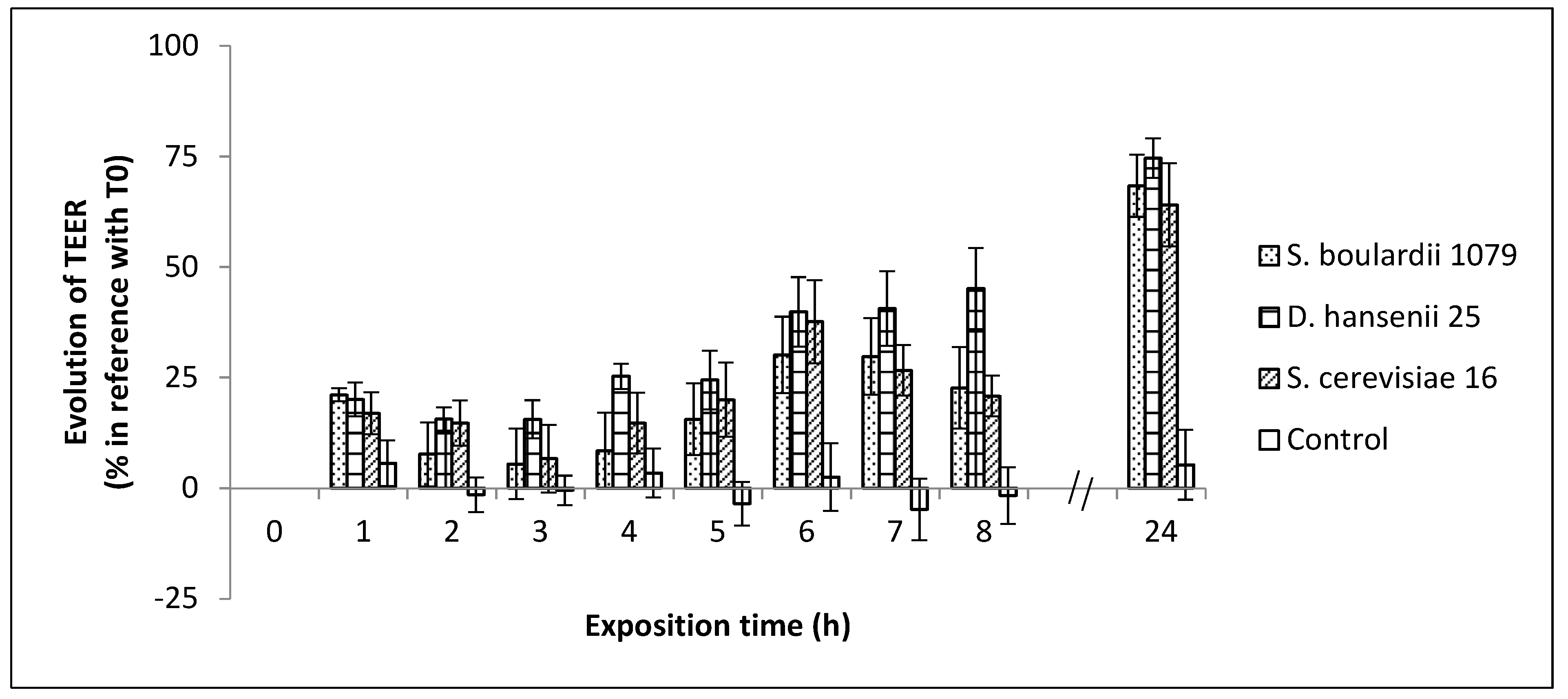
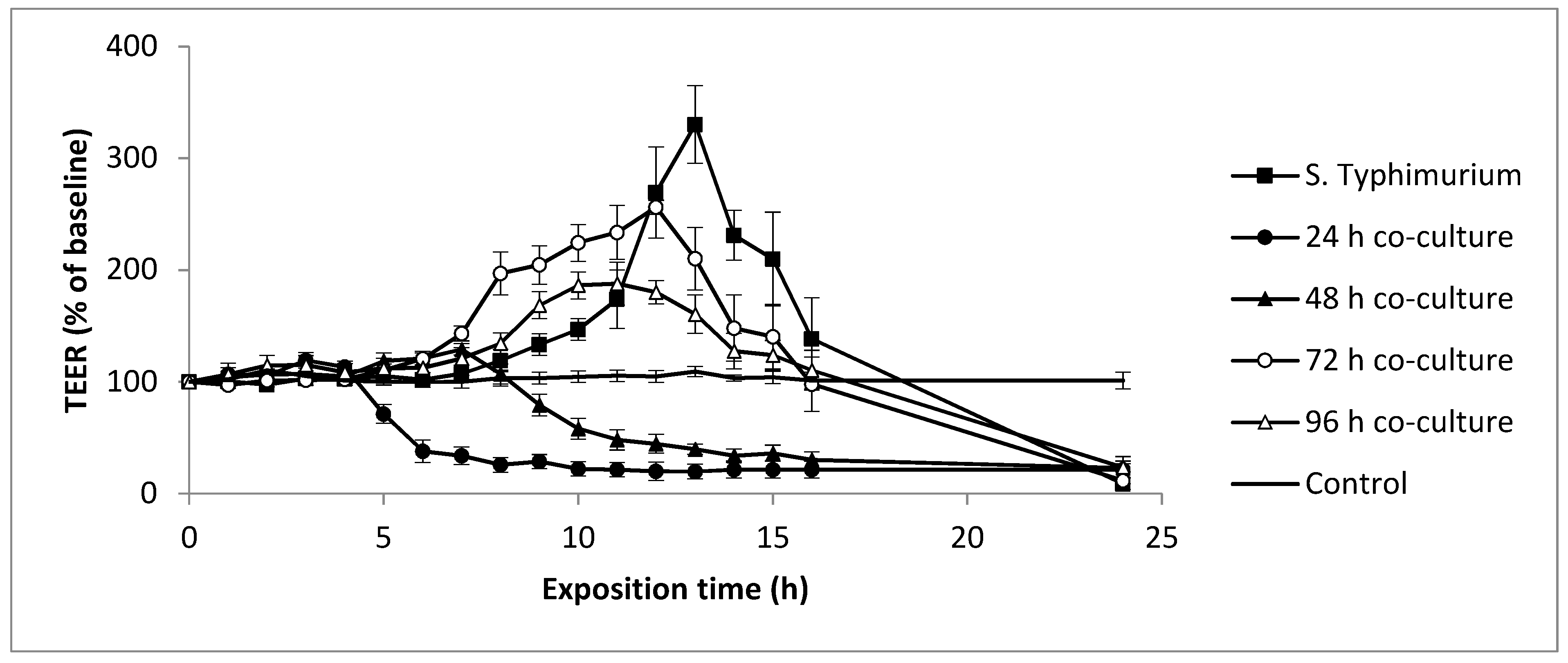
3.4. Evolution of Yeasts and Salmonella Typhimurium-Induced Epithelial Caco-2 Cell Barrier Function

As a measure of epithelial cell barrier function in vitro, we assessed the TEER across the differentiated cell monolayers of the human colonic cell line Caco-2; an established approach often applied for the evaluation of potential probiotic properties of microorganisms [59]. It is now well established that a multiplicity of factors can affect TEER measurements [60,61]. Indeed, in our experimental conditions, monitoring of intestinal epithelium resistance showed that the TEER values stabilized after 6 days (Figure S1) when the formation of the tight junctions in the Caco-2 cells was optimal. The differentiated monolayers of Caco-2 cells were then exposed to the three strains of yeasts (106 CFU/mL) in order to evaluate the influence of these microorganisms on the integrity of the epithelial cell barrier. Caco-2 cells were used instead of HT29-MTX because yeasts have greater adhesion properties on the first cell line, which could promote the anti-Salmonella action. Using S. boulardii as a control, we wanted to determine if Dh25 and Sc16 were good candidates to promote cell barrier integrity. Figure 1 shows that after only one hour of cell exposition, the transepithelial electrical resistance increased significantly, whatever the tested yeast, when compared to the control cells. The TEER values continued to increase progressively until the end of the experiment (24 h). It clearly appears that the three tested yeasts significantly enhanced transepithelial resistance. These results are in accordance with similar studies using non-Saccharomyces yeasts [50,62].
Taking into account the above findings, the Dh25 was considered as the best candidate among the tested cheese yeasts and was thus selected to study its antimicrobial properties. To determine if Dh25 had an influence on cell barrier function and/or on the virulence of ST, we exposed the Caco-2 cell monolayers to ST on its own, and to 24, 48, 72, and 96 h co-cultures (Table 4). After 24 h of co-culture, ST and Dh25 concentrations increased from 6.40 ± 0.10 and 5.93 ± 0.21 log CFU/mL to 8.08 ± 0.58 and 6.60 ± 0.35 log CFU/mL, respectively. From 48 h to 96 h of co-culture, the population of viable ST and Dh25 decreased progressively to finally reach 6.79 ± 0.09 and 4.15 ± 0.85 log CFU/mL, respectively.
When 6.3 log CFU/mL of ST alone were applied to the Caco-2 cell barrier (Figure 2), no change in TEER values was observed during the first six hours of the exposition. Values then began to progressively increase up to 330% of the initial value after 13 h of contact. After this period, TEER values rapidly decreased, reaching 138% at 16 h, and finally 8% after 24 h of exposition. These results are in accordance with those of Smith et al. [62]. S. Typhimurium is an intracellular bacterial pathogen that resides and proliferates within a membrane-bound vacuole in the epithelial cells of the gut, or freely within the cell cytosol, depending on the host cells [63]. Cellular invasion and intracellular survival are critical virulence stages for this pathogen. It is possible that the increase in the volume of the Caco-2 cells, induced by the proliferation of the ST, enhanced the TEER values until 13 h. It is then essential for the ST to exit the infected cell for its dissemination and transmission to other hosts. These cells are extruded from the monolayer and break, explaining the strong decrease in TEER observed from 13 h of contact with the pathogen to the end of the experiment. It would be interesting to use scanning electron microscopy during TEER experiments to verify this hypothesis.
When Caco-2 cells were exposed to 24 h of a Dh25/ST co-culture, right after the 4 h point of exposition, there was a drastic decrease in the TEER values over the next 4 h, finally reaching a constant value, approximately 20% of the initial value, until the end of the experiment. This can be explained by the enhanced virulence of ST, which is correlated with its multiplication, as mentioned above (from 6.40 ± 0.10 log CFU/mL to 8.08 ± 0.58 log CFU/mL, Table 4). When a 48 h co-culture was applied onto cell layers, the TEER values also decreased, albeit progressively, but not until after 8 h. Indeed, the viable population of ST decreased from 8.08 ± 0.58 to 6.90 ± 0.13 log CFU/mL from 24 h to 48 h of co-culture (Table 4), suggesting that, in this condition, Dh25 exerted a negative effect on the viability of ST. The co-cultures of 72 h and 96 h globally showed a similar pattern. The TEER values increased until 12 h and 11 h, respectively, and subsequently decreased to 20% at the 24 h point (Figure 2). In co-cultures of 48 h, 72 h, and 96 h, the ST viable population was constant (Table 4). The delays in the decrease of the TEER values could be explained by the diminution of virulence at 72 h and 96 h, either due to the presence of the Dh25 or to starvation.

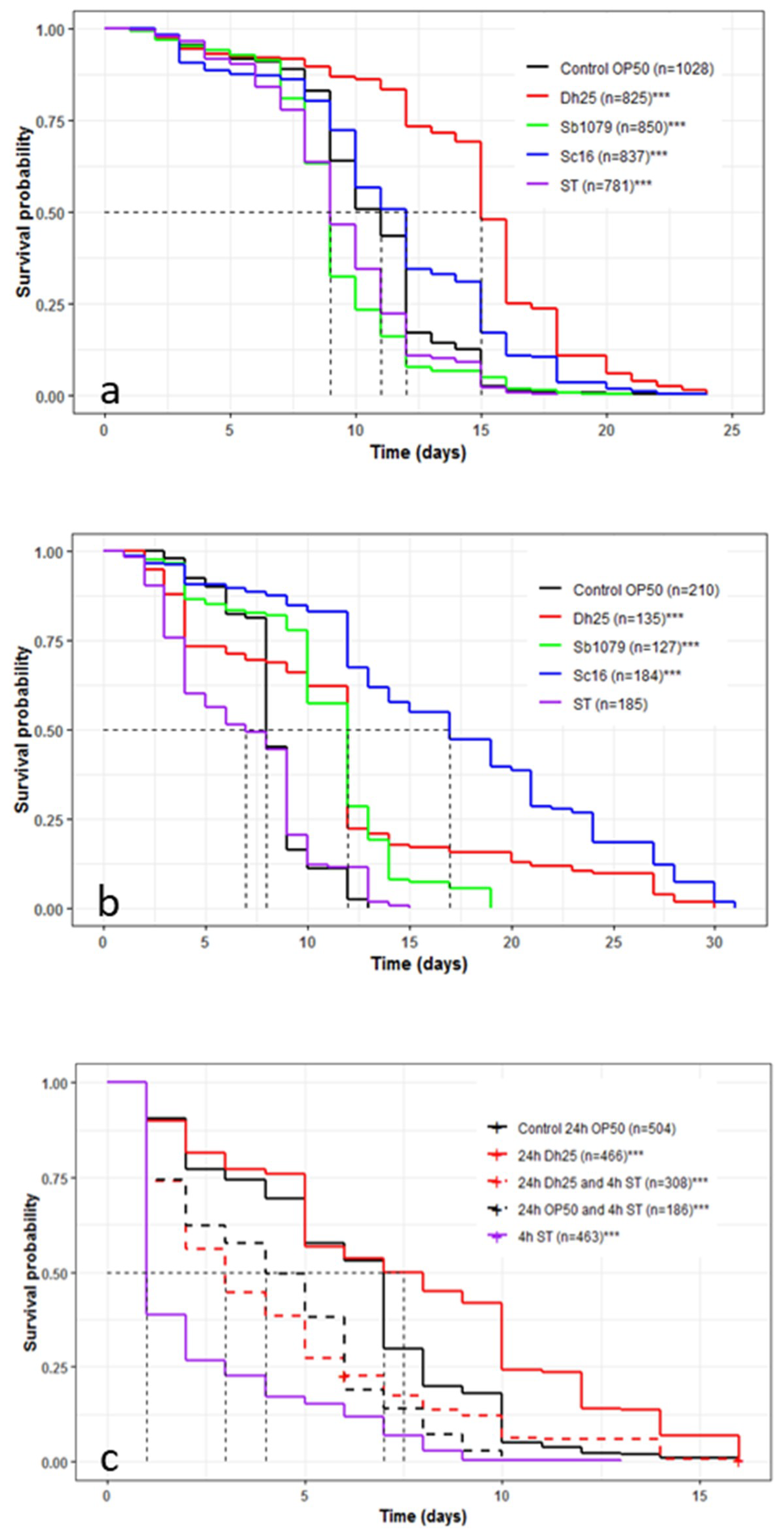
3.5. Influence of Yeasts and Salmonella Typhimurium on the Longevity and Survival of Caenorhabditis elegans and the Preventive Effects of Debaryomyces hansenii 25

We observed whether feeding the C. elegans N2 strain with Sb1079, Sc16, Dh25, and ST, instead of E. coli OP50 (normally used as food to maintain the nematode), affected the longevity of the C. elegans in a solid medium (Figure 3a). Sb1079 and ST reduced the mean lifespan by 2 days compared to the worms fed on E. coli OP50 (9 days versus 11 days), whereas Sc16 and Dh25 increased it by 1 day and 4 days, respectively. On the other hand, the three tested yeasts increased longevity from 4 to 7 days, in comparison with E. coli OP50. The two cheese yeasts had a beneficial effect on C. elegans wild-type (p < 0.001), whereas ST presented a low pathogenicity (p < 0.001). Recently, Kunyeit et al. [12] showed that Saccharomyces cerevisiae (strain KTP) and Issatchenkia occidentalis (strain ApC), had a negative effect on the mean lifespan. These results are not in accordance with our data, suggesting a yeast-strain-dependent effect on C. elegans.
In a liquid medium (Figure 3b), the mean lifespans of the worm strain N2 fed on E. coli OP50 and ST were equal to 7 and 8 days, respectively. The mean lifespan was equal to 12 days with Sb1079 and Dh25, while increasing to 17 days for Sc16. The three yeasts increased survival (19, 30, and 31 days for Sb1079, Dh25, and Sc16, respectively) in comparison with OP50 and ST (13 and 15 days, respectively). The variation in the lifespan of C. elegans, between cheese yeasts and E. coli OP50, might be explained by the different sources of nutritional components. Thus, it could be interesting to measure the body sizes, the lipid accumulation, and the fertility of C. elegans in line with the different nutrition sources. In our conditions, the three tested yeasts showed a significant pro-survival effect on C. elegans (p < 0.001). The C. elegans N2 lifespan was not significantly (p = 0.313) different between the ST and E. coli OP50 conditions. The serovar Typhimurium could be virulent in C. elegans [64,65], but in our in vivo experimental conditions, the Salmonella cheese strain may be considered avirulent.
We then used the worm strain AU37 for its high sensitivity to pathogens. When this C. elegans was fed on ST, the mean lifespan decreased to one day, compared to 7 days with E. coli OP50 (Figure 3c). This result showed the high virulence of ST on this mutant worm strain. Dh25 increased (p < 0.001) mean lifespan and longevity significantly, confirming the beneficial effects of this cheese yeast. When E. coli OP50 and Dh25 were tested as a preventive treatment against ST, the lifespan increased from 1 day with ST alone, to 3 and 4 days with Dh25 and E. coli OP50, respectively. It could be interesting to further investigate whether the protection procured by E. coli OP50 and Dh25 arose from the inhibition of the intestinal colonization of S. Typhimurium. Zhou et al. [66] showed that the action of Lactobacillus against Enterotoxigenic Escherichia coli in C. elegans was not due to colonization inhibition but to the suppression of pathogen enterotoxin gene expression. To further characterize the molecular mechanism involved in the observed increase in lifespan, we could follow the nuclear translocation of the DAF-16 transcription factor using the transgenic TJ-356 worm strain. Indeed, Tullet et al. [67] reported the importance of DAF-16 in the extension of lifespan through the insulin/IGF-1 metabolic signaling pathway. In addition, the DAF-16 implication has been demonstrated by Poupet et al. [39] in the preventive effects of a probiotic bacterium against a pathogenic yeast, Candida albicans.

4. Conclusions

In the present study, we investigated the relevance of in vitro and in vivo methods in the screening of cheese yeasts as probiotic candidates in addition to the well-known S. cerevisiae and S. boulardii. Our data showed that the yeast Debaryomyces hansenii 25, isolated from cheese, exhibits interesting probiotic traits. This strain shows a clear resistance to low pH (pH 2.5) and to bile contact. It also expresses a capacity to adhere to Caco-2 and HT29-MTX intestinal epithelial cells and leads to induced TEER enhancement similar to the probiotic Saccharomyces cerevisiae subspecies boulardii. Furthermore, the lengthening of the co-culture duration between D. hansenii 25 and S. Typhimurium UPsm1 delays Salmonella-induced TEER disruption and could protect human epithelial cells from pathogen invasion. This result is further confirmed by the protective effects of the yeast against S. Typhimurium UPsm1 in C. elegans. We also highlight the importance of the choice of the nematode strain according to the virulence of the pathogen tested. Thus, the combination of in vitro and in vivo methods, before human clinical trials, like the measurement of epithelial cell barrier function and C. elegans lifespan assay, can improve efficiency when selecting probiotic yeast candidates. The nematode can be used as a preclinical model to study the preventive effect of probiotic yeasts against pathogens. Additional investigations to decipher the mechanisms involved in the resistance provided by D. hansenii 25 against S. Typhimurium UPsm1 in C. elegans are however needed. Transcriptomic approaches and/or the use of loss-of-function mutant worms targeting signal transduction pathways implicated in immunity could be of great interest.
The fact that the strain D. hansenii 25 has probiotic characteristics and that the other cheese yeasts tested resist the conditions simulating passage through the gastrointestinal tract suggests that the consumption of cheeses could have a favorable impact on human health via the activity of yeasts.

Supplementary Materials

The following are available online at https://www.mdpi.com/2076-2607/8/6/922/s1. Table S1: Survival rates of yeasts strains in the presence of acid pH (2.5). a, acid pH tolerance (%) = number of CFUs in broth with pH = 2.5/number of CFUs in broth with pH = 7) X 100. b, the survival rate of S. cerevisiae subspecies boulardii CNCM I/079, D. hansenii 25 and S. cerevisiae 16 in the presence of bile salts (3%). Bile tolerance (%) = number of CFUs in broth with bile/number of CFUs in broth without bile) X 100. The values are mean ± SD (error bars) of three independent experiments. Results show a high resistance to acidic pH and bile salts suggesting the strains will be able to resist to gastric environment. Figure S1: Evolution of Caco-2 cells TEER during maturation of the cell layer.

Author Contributions

S.B., M.B., P.V., C.P., and T.S. designed the protocols and scientific approaches. P.V., M.B., T.S., C.P., O.C., M.G., and C.C. (Cécile Callon) carried out the experiments. G.F. programmed the software. P.V. and M.B. wrote the manuscript. C.P., C.C. (Cécile Callon), S.B., and C.C. (Christophe Chassard) provided critical feedback. S.B. supervised the project. All authors have read and agreed to the published version of the manuscript.

Funding

The authors acknowledge the European funds (FEDER), the University of Clermont Auvergne and the Technological University Institute for their financial support.

Acknowledgments

Some strains (C. elegans and E. coli OP50) were provided by the CGC, which is funded by the NIH Office of Research Infrastructure Programs (P40 OD010440). The authors thank Patricia Mabrut and Kate James for English expert proofreading.

Conflicts of Interest

The authors declare no conflict of interest. The funders had no role in the design of the study; in the collection, analyses, or interpretation of data; in the writing of the manuscript; or in the decision to publish the results.

Abbreviation

TEERTransEpithelial Electrical Resistance

References

  1. Huffnagle, G.B.; Noverr, M.C. The emerging world of the fungal microbiome. Trends Microbiol. 2013, 21, 334–341. [Google Scholar] [CrossRef] [PubMed] [Green Version]
  2. Lukeš, J.; Stensvold, C.R.; Jirků-Pomajbíková, K.; Wegener Parfrey, L. Are human intestinal eukaryotes beneficial or commensals? PLoS Pathog. 2015, 11, e1005039. [Google Scholar]
  3. Peterson, L.W.; Artis, D. Intestinal epithelial cells: Regulators of barrier function and immune homeostasis. Nat. Rev. Immunol. 2014, 14, 141–153. [Google Scholar] [CrossRef] [PubMed]
  4. Wang, B.; Yao, M.; Lv, L.; Ling, Z.; Li, L. The human microbiota in health and disease. Engineering 2017, 3, 71–82. [Google Scholar] [CrossRef]
  5. FAO; WHO. Health and Nutritional Properties of Probiotics in Food including Powder Milk with Live Lactic Acid Bacteria. Food Nutr. Pap. 2001, 85, 71. [Google Scholar]
  6. Didari, T.; Solki, S.; Mozaffari, S.; Nikfar, S.; Abdollahi, M. A systematic review of the safety of probiotics. Expert Opin. Drug Saf. 2014, 13, 227–239. [Google Scholar] [CrossRef]
  7. Dinleyici, E.C.; Eren, M.; Ozen, M.; Yargic, Z.A.; Vandenplas, Y. Effectiveness and safety of Saccharomyces boulardii for acute infectious diarrhea. Expert Opin. Biol. Ther. 2012, 12, 395–410. [Google Scholar] [CrossRef]
  8. McFarland, L.V. Systematic review and meta-analysis of Saccharomyces boulardii in adult patients. World J. Gastroenterol. 2010, 16, 2202–2222. [Google Scholar] [CrossRef]
  9. Zanello, G.; Meurens, F.; Berri, M.; Chevaleyre, C.; Melo, S.; Auclair, E.; Salmon, H. Saccharomyces cerevisiae decreases inflammatory responses induced by F4+ enterotoxigenic Escherichia coli in porcine intestinal epithelial cells. Vet. Immunol. Immunopathol. 2011, 141, 133–138. [Google Scholar] [CrossRef]
  10. Živković, M.; Čadež, N.; Uroić, K.; Miljković, M.; Tolinački, M.; Doušova, P.; Kos, B.; Šušković, J.; Raspor, P.; Topisirović, L.; et al. Evaluation of probiotic potential of yeasts isolated from traditional cheeses manufactured in Serbia and Croatia. J. Intercult. Ethnopharmacol. 2015, 4, 12–18. [Google Scholar] [CrossRef]
  11. Ochangco, H.S.; Gamero, A.; Smith, I.M.; Christensen, J.E.; Jespersen, L.; Arneborg, N. In vitro investigation of Debaryomyces hansenii strains for potential probiotic properties. World J. Microbiol. Biotechnol. 2016, 32, 141. [Google Scholar] [CrossRef]
  12. Kunyeit, L.; Kurrey, N.K.; Anu-Appaiah, K.A.; Rao, R.P. Probiotic yeasts inhibit virulence of non-albicans Candida Species. mBIO 2019, 10, 1–10. [Google Scholar] [CrossRef] [Green Version]
  13. Retureau, E.; Callon, C.; Didienne, R.; Montel, M.-C. Is microbial diversity an asset for inhibiting Listeria monocytogenes in raw milk cheeses? Dairy Sci. Technol. 2010, 90, 375–398. [Google Scholar] [CrossRef]
  14. Callon, C.; Retureau, E.; Didienne, R.; Montel, M.-C. Microbial biodiversity in cheese consortia and comparative Listeria growth on surfaces of uncooked pressed cheeses. Int. J. Food Microbiol. 2014, 174, 98–109. [Google Scholar] [CrossRef] [PubMed]
  15. Andreoletti, O.; Budka, H.; Buncic, S.; Colin, P.; Collins, J.D.; De Koeijer. The maintenance of the list of QPS microorganisms intentionally added to food or feed - Scientific Opinion of the Panel on Biological Hazards. EFSA J. 2008, 923, 1–48. [Google Scholar]
  16. Martins, F.S.; Vieira, A.T.; Elian, S.D.A.; Arantes, R.M.E.; Tiago, F.C.P.; Sousa, L.P.; Araújo, H.R.; Pimenta, P.F.; Bonjardim, C.A.; Nicoli, J.R.; et al. Inhibition of tissue inflammation and bacterial translocation as one of the protective mechanisms of Saccharomyces boulardii against Salmonella infection in mice. Microbes Infect. 2013, 15, 270–279. [Google Scholar] [CrossRef] [PubMed]
  17. Matsubara, V.H.; Ishikawa, K.H.; Ando-Suguimoto, E.S.; Bueno-Silva, B.; Nakamae, A.E.M.; Mayer, M.P.A. Probiotic bacteria alter pattern-recognition receptor expression and cytokine profile in a human macrophage model challenged with Candida albicans and lipopolysaccharide. Front. Microbiol. 2017, 8, 2280. [Google Scholar] [CrossRef] [Green Version]
  18. Leão, M.V.P.; Tavares, T.A.A.; Gonçalves, E.; Silva, C.R.; Dos Santos, S.S.F.; Junqueira, J.C.; de Oliveira, L.D.; Jorge, A.O.C. Lactobacillus rhamnosus intake can prevent the development of candidiasis. Clin. Oral Investig. 2018, 22, 2511–2518. [Google Scholar] [CrossRef] [Green Version]
  19. Tallon, R.; Arias, S.; Bressollier, P.; Urdaci, M.C. Strain- and matrix-dependent adhesion of Lactobacillus plantarum is mediated by proteinaceous bacterial compounds. J. Appl. Microbiol. 2007, 102, 442–451. [Google Scholar] [CrossRef]
  20. Zielińska, D.; Długosz, E.; Zawistowska-Deniziak, A. Functional properties of food origin Lactobacillus in the gastrointestinal ecosystem—In vitro study. Probiotics Antimicrob. Proteins 2019, 11, 820–829. [Google Scholar] [CrossRef]
  21. Lacroix, C.; de Wouters, T.; Chassard, C. Integrated multi-scale strategies to investigate nutritional compounds and their effect on the gut microbiota. Curr. Opin. Biotechnol. 2015, 32, 149–155. [Google Scholar] [CrossRef] [PubMed]
  22. McGhee, J. The C. elegans Intestine; WormBook, Ed.; The C. elegans Research Community, WormBook, 2007; Available online: http://www.wormbook.org/chapters/www_intestine/intestine.html (accessed on 19 May 2020).
  23. Park, M.R.; Yun, H.S.; Son, S.J.; Oh, S.; Kim, Y. Short communication: Development of a direct in vivo screening model to identify potential probiotic bacteria using Caenorhabditis elegans. J. Dairy Sci. 2014, 97, 6828–6834. [Google Scholar] [CrossRef] [Green Version]
  24. Lee, J.; Choe, J.; Kim, J.; Oh, S.; Park, S.; Kim, S.; Kim, Y. Heat-killed Lactobacillus spp. cells enhance survivals of Caenorhabditis elegans against Salmonella and Yersinia infections. Lett. Appl. Microbiol. 2015, 61, 523–530. [Google Scholar] [CrossRef]
  25. Mylonakis, E.; Ausubel, F.M.; Perfect, J.R.; Heitman, J.; Calderwood, S.B. Killing of Caenorhabditis elegans by Cryptococcus neoformans as a model of yeast pathogenesis. Proc. Natl. Acad. Sci. USA 2002, 99, 15675–15680. [Google Scholar] [CrossRef] [PubMed] [Green Version]
  26. Jain, C.; Yun, M.; Politz, S.M.; Rao, R.P. A Pathogenesis Assay Using Saccharomyces cerevisiae and Caenorhabditis elegans reveals novel roles for yeast AP-1, Yap1, and host dual oxidase BLI-3 in fungal pathogenesis. Eukaryot. Cell 2009, 8, 1218–1227. [Google Scholar] [CrossRef] [PubMed] [Green Version]
  27. Pukkila-Worley, R.; Ausubel, F.M.; Mylonakis, E. Candida albicans infection of Caenorhabditis elegans induces antifungal immune defenses. PLoS Pathog. 2011, 7, e1002074. [Google Scholar] [CrossRef] [PubMed] [Green Version]
  28. Madende, M.; Albertyn, J.; Sebolai, O.; Pohl, C.H. Caenorhabditis elegans as a model animal for investigating fungal pathogenesis. Med. Microbiol. Immunol. 2020, 209, 1–13. [Google Scholar] [CrossRef]
  29. Romanin, D.E.; Llopis, S.; Genovés, S.; Martorell, P.; Ramón, V.d.; Garrote, G.l.; Rumbo, M. Probiotic yeast Kluyveromyces marxianus CIDCA 8154 shows anti-inflammatory and anti-oxidative stress properties in in vivo models. Benef. Microbes 2015, 7, 83–93. [Google Scholar] [CrossRef]
  30. Oliveira, C.J.B.; Hisrich, E.R.; Moura, J.F.P.; Givisiez, P.E.N.; Costa, R.G.; Gebreyes, W.A. On farm risk factors associated with goat milk quality in Northeast Brazil. Small Ruminant Res. 2011, 98, 64–69. [Google Scholar] [CrossRef]
  31. Pointon, A.; Kiermeier, A.; Fegan, N. Review of the impact of pre-slaughter feed curfews of cattle, sheep and goats on food safety and carcase hygiene in Australia. Food Control. 2012, 26, 313–321. [Google Scholar] [CrossRef]
  32. Haraga, A.; Ohlson, M.B.; Miller, S.I. Salmonellae interplay with host cells. Nat. Rev. Microbiol. 2008, 6, 53–66. [Google Scholar] [CrossRef]
  33. Guttman, J.A.; Finlay, B.B. Tight junctions as targets of infectious agents. Biochim. Biophys. Acta – Biomembr. 2009, 1788, 832–841. [Google Scholar] [CrossRef] [Green Version]
  34. Gagnon, M.; Berner, A.Z.; Chervet, N.; Chassard, C.; Lacroix, C. Comparison of the Caco-2, HT-29 and the mucus-secreting HT29-MTX intestinal cell models to investigate Salmonella adhesion and invasion. J. Microbiol. Meth. 2013, 94, 274–279. [Google Scholar] [CrossRef] [PubMed]
  35. Zweibaum, A.; Laburthe, M.; Grasset, E.; Louvard, D. Use of cultured cell lines in studies of intestinal cell differentiation and function. Compr. Physiol. 2011, 223–255. [Google Scholar]
  36. Pinto, M.; Robine-Leon, S.; Appay, M.D.; Kedinger, M.; Triadou, N.; Dussaulx, E.; Lacroix, B.; Simon-Assmann, P.; Haffen, K.; Fogh, J.; et al. Enterocyte-like differentiation and polarization of the human colon carcinoma cell line Caco-2 in culture. Biol. Cell. 1983, 47, 323–330. [Google Scholar]
  37. Nivoliez, A.; Camares, O.; Paquet-Gachinat, M.; Bornes, S.; Forestier, C.; Veisseire, P. Influence of manufacturing processes on in vitro properties of the probiotic strain Lactobacillus rhamnosus Lcr35®. J. Biotechnol. 2012, 160, 236–241. [Google Scholar] [CrossRef] [PubMed]
  38. Brenner, S. The genetics of Caenorhabditis elegans. Genetics 1974, 77, 71–94. [Google Scholar] [PubMed]
  39. Poupet, C.; Saraoui, T.; Veisseire, P.; Bonnet, M.; Dausset, C.; Gachinat, M.; Camarès, O.; Chassard, C.; Nivoliez, A.; Bornes, S. Lactobacillus rhamnosus Lcr35 as an effective treatment for preventing Candida albicans infection in the invertebrate model Caenorhabditis elegans: First mechanistic insights. PLoS ONE 2019, 14, e0216184. [Google Scholar]
  40. de Barros, P.; Scorzoni, L.; Ribeiro, F.; Fugisaki, L.; Fuchs, B.; Mylonakis, E.; Jorge, A.; Junqueira, J.; Rossoni, R. Lactobacillus paracasei 28.4 reduces in vitro hyphae formation of Candida albicans and prevents the filamentation in an experimental model of Caenorhabditis elegans. Microb. Pathog. 2018, 117, 80–87. [Google Scholar] [CrossRef] [Green Version]
  41. R Core Team. R: A Language and Environnement for Statistical Computing; R Foundation for Statistical Computing: Vienna, Austria, 2018; Available online: https://www.r-project.org/ (accessed on 19 May 2020).
  42. Therneau, T.M. A package for survival analysis in R. 2015. Available online: https://github.com/therneau/survival (accessed on 19 May 2020).
  43. Kassambara, A.; Kosinski, M. Survminer: Drawing Survival Curves Using “ggplot2”. 2017. Available online: http://www.sthda.com/english/rpkgs/survminer/ (accessed on 19 May 2020).
  44. Edwards-Ingram, L.; Gitsham, P.; Burton, N.; Warhurst, G.; Clarke, I.; Hoyle, D.; Oliver, S.G.; Stateva, L. Genotypic and physiological characterization of Saccharomyces boulardii, the probiotic strain of Saccharomyces cerevisiae. Appl. Environ. Microbiol. 2007, 73, 2458–2467. [Google Scholar] [CrossRef] [Green Version]
  45. Villar-Garcia, J.; Güerri-Fernandez, R.; Moya, A.; Gonzalez, A.; Hernandez, J.J.; Lerma, E.; Guelar, A.; Sorli, L.; Horcajada, J.P.; Artacho, A.; et al. Impact of probiotic Saccharomyces boulardii on the gut microbiome composition in HIV-treated patients: A double-blind, randomised, placebo-controlled trial. PLoS ONE 2017, 12, e0173802. [Google Scholar] [CrossRef]
  46. Arévalo-Villena, M.; Fernandez-Pacheco, P.; Castillo, N.; Bevilacqua, A.; Briones Pérez, A. Probiotic capability in yeasts: Set-up of a screening method. LWT 2018, 89, 657–665. [Google Scholar] [CrossRef]
  47. Asmat, S.; Shaukat, F.; Asmat, R.; Faiq Siddique Gul Bakhat, H.; Asmat, T.M. Clinical efficacy comparison of Saccharomyces boulardii and lactic acid as probiotics in acute pediatric diarrhea. J. Coll. Physicians Surg. Pak. 2018, 28, 214–217. [Google Scholar] [CrossRef] [PubMed]
  48. Argyri, A.A.; Zoumpopoulou, G.; Karatzas, K.-A.G.; Tsakalidou, E.; Nychas, G.-J.E.; Panagou, E.Z.; Tassou, C.C. Selection of potential probiotic lactic acid bacteria from fermented olives by in vitro tests. Food Microbiol. 2013, 33, 282–291. [Google Scholar] [CrossRef] [PubMed]
  49. Hyronimus, B.; Le Marrec, C.; Sassi, A.H.; Deschamps, A. Acid and bile tolerance of spore-forming lactic acid bacteria. Int. J. Food Microbiol. 2000, 61, 193–197. [Google Scholar] [CrossRef]
  50. Pedersen, L.L.; Owusu-Kwarteng, J.; Thorsen, L.; Jespersen, L. Biodiversity and probiotic potential of yeasts isolated from Fura, a West African spontaneously fermented cereal. Int. J. Food Microbiol. 2012, 159, 144–151. [Google Scholar] [CrossRef]
  51. Trotta, F.; Caldini, G.; Dominici, L.; Federici, E.; Tofalo, R.; Schirone, M.; Corsetti, A.; Suzzi, G.; Cenci, G. Food borne yeasts as DNA-bioprotective agents against model genotoxins. Int. J. Food Microbiol. 2012, 153, 275–280. [Google Scholar] [CrossRef] [PubMed]
  52. Chen, L.-S.; Ma, Y.; Maubois, J.-L.; He, S.-H.; Chen, L.-J.; Li, H.-M. Screening for the potential probiotic yeast strains from raw milk to assimilate cholesterol. Dairy Sci. Technol. 2010, 90, 537–548. [Google Scholar] [CrossRef] [Green Version]
  53. Lange, K. Fundamental role of microvilli in the main functions of differentiated cells: Outline of a universal regulating and signaling system at the cell periphery. J. Cell Physiol. 2011, 226, 896–927. [Google Scholar] [CrossRef]
  54. Espey, M.G. Role of oxygen gradients in shaping redox relationships between the human intestine and its microbiota. Free Radic. Biol. Med. 2013, 55, 130–140. [Google Scholar] [CrossRef]
  55. Lima, M.D.S.F.; de Souza, K.M.S.; de Albuquerque, W.W.C.; Teixeira, J.A.C.; Cavalcanti, M.T.H.; Porto, A.L.F. Saccharomyces cerevisiae from Brazilian kefir-fermented milk: An in-vitro evaluation of probiotic properties. Microb. Pathog. 2017, 110, 670–677. [Google Scholar] [CrossRef] [Green Version]
  56. Roussel, C.; Sivignon, A.; de Vallée, A.; Garrait, G.; Denis, S.; Tsilia, V.; Ballet, N.; Vandekerckove, P.; Van de Wiele, T.; Barnich, N.; et al. Anti-infectious properties of the probiotic Saccharomyces cerevisiae CNCM I-3856 on enterotoxigenic E. coli (ETEC) strain H10407. Appl. Microbiol. Biotechnol. 2018, 102, 6175–6189. [Google Scholar] [CrossRef]
  57. Binetti, A.; Carrasco, M.; Reinheimer, J.; Suárez, V. Yeasts from autochthonal cheese starters: Technological and functional properties. J. Appl. Microbiol. 2013, 115, 434–444. [Google Scholar] [CrossRef] [PubMed]
  58. Perricone, M.; Bevilacqua, A.; Corbo, M.R.; Sinigaglia, M. Technological characterization and probiotic traits of yeasts isolated from Altamura sourdough to select promising microorganisms as functional starter cultures for cereal-based products. Food Microbiol. 2014, 38, 26–35. [Google Scholar] [CrossRef] [PubMed]
  59. Klingberg, T.D.; Pedersen, M.H.; Cencic, A.; Budde, B.B. Application of Measurements of Transepithelial Electrical Resistance of Intestinal Epithelial Cell Monolayers to Evaluate Probiotic Activity. Appl. Environ. Microbiol. 2005, 71, 7528–7530. [Google Scholar] [CrossRef] [PubMed] [Green Version]
  60. Sambuy, Y.; Angelis, I.D.; Ranaldi, G.; Scarino, M.L.; Stammati, A.; Zucco, F. The Caco-2 cell line as a model of the intestinal barrier: Influence of cell and culture-related factors on Caco-2 cell functional characteristics. Cell. Biol. Toxicol. 2005, 21, 1–26. [Google Scholar] [CrossRef]
  61. Srinivasan, B.; Kolli, A.R.; Esch, M.B.; Abaci, H.E.; Shuler, M.L.; Hickman, J.J. TEER measurement techniques for in vitro barrier model systems. J. Lab. Autom. 2015, 20, 107–126. [Google Scholar] [CrossRef] [Green Version]
  62. Smith, I.M.; Baker, A.; Arneborg, N.; Jespersen, L. Non-Saccharomyces yeasts protect against epithelial cell barrier disruption induced by Salmonella enterica subsp. enterica serovar Typhimurium. Lett. Appl. Microbiol. 2015, 61, 491–497. [Google Scholar] [CrossRef]
  63. Knodler, L.A.; Vallance, B.A.; Celli, J.; Winfree, S.; Hansen, B.; Montero, M.; Steele-Mortimer, O. Dissemination of invasive Salmonella via bacterial-induced extrusion of mucosal epithelia. Proc. Natl. Acad. Sci. USA 2010, 107, 17733–17738. [Google Scholar] [CrossRef] [PubMed] [Green Version]
  64. Oza, J.P.; Yeh, J.B.; Reich, N.O. DNA methylation modulates Salmonella enterica serovar Typhimurium virulence in Caenorhabditis elegans. FEMS Microbiol. Lett. 2005, 245, 53–59. [Google Scholar] [CrossRef] [Green Version]
  65. Sahu, S.N.; Anriany, Y.; Grim, C.J.; Kim, S.; Chang, Z.; Joseph, S.W.; Cinar, H.N. (2013) Identification of virulence properties in Salmonella Typhimurium DT104 using Caenorhabditis elegans. PLoS ONE 2005, 8, e76673. [Google Scholar]
  66. Zhou, M.; Yu, H.; Yin, X.; Sabour, P.M.; Chen, W.; Gong, J. Lactobacillus zeae protects Caenorhabditis elegans from Enterotoxigenic Escherichia coli-caused death by inhibiting enterotoxin gene expression of the pathogen. PLoS ONE 2014, 9, e89004. [Google Scholar] [CrossRef]
  67. Tullet, J.M.A. DAF-16 target identification in C. elegans: Past, present and future. Biogerontology 2015, 16, 221–234. [Google Scholar] [CrossRef] [PubMed] [Green Version]
Figure 1. Change in TransEpithelial Electrical Resistance (TEER) level of yeast-exposed Caco-2 cells during apical incubation with S. boulardii 1079, D. hansenii 25, S. cerevisiae 16, and Dulbecco’s Modified Eagle’s Minimal essential medium (DMEM) control medium throughout the 24 h incubation period, expressed by reference to resistance measured at time 0. The values are mean ± SD (error bars) of three independent experiments. A high enhancement of the TEER is observed in the presence of the yeasts.
Microorganisms 08 00922 g001
Figure 2. S. Typhimurium culture and D. hansenii 25/S. Typhimurium co-culture (24, 48, 72, and 96 h) modulations of human epithelial cell barrier function. Control with DMEM medium only. The values are mean ± SD (error bars) of three independent experiments. An increased co-culture duration delayed the deleterious effect of Salmonella on the TEER.
Microorganisms 08 00922 g002
Figure 3. (a) Influence of D. hansenii 25 (Dh25), E. coli OP50, S. boulardii 1079 (Sb1079), S. cerevisiae 16 (Sc16), and S. Typhimurium (ST) on the longevity of the C. elegans wild-type N2 strain in a solid medium. (b) Influence of Dh25, E. coli OP50, Sb1079, Sc16, and ST on survival of the C. elegans wild-type N2 strain in a liquid medium. (c) Preventive effects of Dh25 on ST survival of the C. elegans mutant AU37 strain in a liquid medium. Mean survival, with half of the population dead, is represented on the abscissa. The asterisks indicate the p-values (log-rank test) against E. coli OP50 (***, p < 0.001). C. elegans AU37 mutant is more sensible to the pathogenic bacterium Salmonella Typhimurium and is protected by the yeasts Debaryomyces hansenii 25.
Microorganisms 08 00922 g003
Table 1. D. hansenii 25 and S. cerevisiae 16 in the presence of acid pH (2.5). (a) Acid pH tolerance (%) = (number of colony-forming units (CFU) in broth with pH = 2.5/number of CFUs in broth with pH = 7) X 100. (b) The survival rate of S. cerevisiae subspecies boulardii CNCM I/079, D. hansenii 25 and S. cerevisiae 16 in the presence of bile salts (3%). Bile tolerance (%) = (number of CFUs in broth with bile/number of CFUs in broth without bile) X 100. The values are mean ± SD (error bars) of three independent experiments. Results show a high resistance to acidic pH and bile salts, suggesting the strains will be able to resist to gastric environment.
a. Survival rates in the presence of acidic pH (%)
Strains45 min90 min
S. boulardii 107993.6 ± 8.391.1 ± 9.0
D. hansenii 2596.6 ± 3.485.5 ± 3.0
S. cerevisiae 16101.3 ± 3.454.2 ± 2.7
b. Survival rates in the presence of bile salts (%)
Strains1 h2 h3 h4 h
S. boulardii 107985.3 ± 4.570.3 ± 4.463.1 ± 2.773.5 ± 4.9
D. hansenii 2594.1 ± 7.491.3 ± 7.772.8 ± 13.568.9 ± 16.1
S. cerevisiae 1689.2 ± 2.977.5 ± 15.156.0 ± 9.439.2 ± 6.7
Table 2. Determination of the number of viable adhering S. boulardii 1079, D. hansenii 25, and S. cerevisiae 16 strains after 1 h of contact with (a) HT29-MTX and with (b) Caco-2, cell lines at multiplicities of infection (MOI) of 0.1, 1, 10, and 100. Results are expressed as percentages of adhering yeasts compared to the initial inoculum (mean ± SD). a, b superscripts represent groups determined by Least Significant Difference (LSD) test, the same letter indicating values not significantly different (p < 0.05 by ANOVA1) across a column. Results indicate yeast adhesion is higher on Caco-2 cells than on HT29-MTX cells.
a. Multiplicity of infection (MOI)
HT29-MTXMOI 100MOI 10MOI 1MOI 0.1
S. boulardii 10790.51 ± 0.046% a,b0.39 ± 0.123% a,b0.46 ± 0.189% a,b1.15 ± 0.270% a,b
D. hansenii 250.18 ± 0.005% b0.29 ± 0.005% b0.44 ± 0.007% b1.56 ± 0.010% b
S. cerevisiae 160.91 ± 0.015% a0.90 ± 0.013% a1.24 ± 0.020% a2.61 ± 0.024% a
b. Multiplicity of infection (MOI)
Caco-2MOI 100MOI 10MOI 1MOI 0.1
S. boulardii 10790.59 ± 0.006% a1.87 ± 0.011% a1.77 ± 0.013% a,b3.60 ± 0.028% a
D. hansenii 252.27 ± 0.011% b2.50 ± 0.060% a4.10 ± 0.325% b4.12 ± 1.701% a
S. cerevisiae 160.81 ± 0.009% a0.86 ± 0.007% b2.56 ± 0.032% a7.26 ± 0.088% a
Table 3. Effect of the control yeast strain, S. cerevisiae subspecies boulardii CNCM I-1079 (Sb1079), D. hansenii 25 (Dh25) and S. cerevisiae 16 (Sc16), isolated from cheese, on S. Typhimurium (ST) growth. a, b superscripts represent groups determined by LSD test, the same letter indicating values not significantly different (p <0.05 by ANOVA1) across a column. The yeasts tested have an inhibition activity against Salmonella growth.
Time (h)02448
ST6.47 ± 0.48 a8.88 ± 0.30 a8.71 ± 0.20 a
ST + Sb10796.63 ± 0.38 a8.39 ± 0.50 a,b6.46 ± 0.92 b
ST + Dh256.58 ± 0.46 a8.34 ± 0.62 b6.90 ± 0.97 b
ST + Sc166.60 ± 0.43 a8.21 ± 0.43 b6.91 ± 0.97 b
Table 4. Effect of D. hansenii 25 (Dh25), isolated from cheese, on S. Typhimurium (ST) growth after four co-culture durations (24 h, 48 h, 72 h, and 96 h). a, b, and c superscripts represent groups determined by LSD test, a same letter indicating values not significantly different (p < 0.05 by ANOVA1) with t = 0 h as the control.
Co-Culture DurationST (log CFU/mL)Dh25 (log CFU/mL)
0 h6.40 ± 0.10 a5.93 ± 0.21 a
24 h8.08 ± 0.58 b6.60 ± 0.35 a
48 h6.90 ± 0.13 a5.90 ± 0.56 a
72 h6.85 ± 0.30 a4.87 ± 0.81 b
96 h6.79 ± 0.09 a4.15 ± 0.85 c

