Article

New Antimicrobial Phenyl Alkenoic Acids Isolated from an Oil Palm Rhizosphere-Associated Actinomycete, Streptomyces palmae CMU-AB204T

1
Research Center of Microbial Diversity and Sustainable Utilization, Faculty of Science, Chiang Mai University, Chiang Mai 50200, Thailand
2
Kitasato Institute for Life Sciences, Kitasato University, 5-9-1 Shirokane, Minato-ku, Tokyo 108-8641, Japan
3
Graduate School of Infection Control Sciences, Kitasato University, 5-9-1 Shirokane, Minato-ku, Tokyo 108-8641, Japan
4
Department of Biology, Faculty of Science, Chiang Mai University, Chiang Mai 50200, Thailand
5
Academy of Science, The Royal Society of Thailand, Bangkok 10300, Thailand
*
Authors to whom correspondence should be addressed.
Microorganisms 2020, 8(3), 350; https://doi.org/10.3390/microorganisms8030350
Submission received: 15 January 2020 / Revised: 24 February 2020 / Accepted: 28 February 2020 / Published: 1 March 2020

Abstract

:
Basal stem rot (BSR), or Ganoderma rot disease, is the most serious disease associated with the oil palm plant of Southeast Asian countries. A basidiomycetous fungus, Ganoderma boninense, is the causative microbe of this disease. To control BSR in oil palm plantations, biological control agents are gaining attention as a major alternative to chemical fungicides. In the course of searching for effective actinomycetes as potential biological control agents for BSR, Streptomyces palmae CMU-AB204T was isolated from oil palm rhizosphere soil collected on the campus of Chiang Mai University. The culture broth of this strain showed significant antimicrobial activities against several bacteria and phytopathogenic fungi including G. boninense. Antifungal and antibacterial compounds were isolated by antimicrobial activity-guided purification using chromatographic methods. Their structures were elucidated by spectroscopic techniques, including Nuclear Magnetic Resonance (NMR), Mass Spectrometry (MS), Ultraviolet (UV), and Infrared (IR) analyses. The current study isolated new phenyl alkenoic acids 16 and three known compounds, anguinomycin A (7), leptomycin A (8), and actinopyrone A (9) as antimicrobial agents. Compounds 1 and 2 displayed broad antifungal activity, though they did not show antibacterial activity. Compounds 3 and 4 revealed a strong antibacterial activity against both Gram-positive and Gram-negative bacteria including the phytopathogenic strain Xanthomonas campestris pv. oryzae. Compounds 79 displayed antifungal activity against Ganoderma. Thus, the antifungal compounds obtained in this study may play a role in protecting oil palm plants from Ganoderma infection with the strain S. palmae CMU-AB204T.

1. Introduction

Oil palm (Elaeis guineensis Jacq.) is an important economic crop in many tropical areas. In particular, Indonesia, Malaysia, and Thailand are the leading palm oil producing countries of this region. The oil palm plant typically has a productive life of 20 or more years, and oil can be harvested several times each year. Consequently, it holds an advantage over all other oil-producing crops [1]. However, the plant is often damaged by fungal infections, and these can cause a decrease of crop yields and result in the death of oil palm trees.
Fungal pathogens mainly infect the stems and leaves of oil palm trees during all stages of growth, from seedlings to the mature stage, and consequently can affect both the quality and quantity of palm oil. Basal stem rot (BSR), or Ganoderma rot disease, is the most severe disease of oil palm trees in Southeast Asian countries, especially Malaysia and Indonesia [2]. In addition to these countries, BSR has also destroyed oil palm plantations in Africa, Colombia [3], Papua New Guinea [4], and Thailand [5]. The causative fungus Ganoderma boninense is a basidiomycetous fungus and belongs to the order Polyporales and the family of Ganodermataceae. Fruiting bodies of Ganoderma typically form on the exterior of the oil palm trunk and then release and spread the spores to the soil. The usual method of controlling BSR in oil palm plantations is the use of chemical fungicides. Many fungicides, such as azoxystrobin, benomyl, carbendazim, carboxin, cycloheximide, cyproconazole, drazoxolone, hexaconazole, methfuroxam, nystatin, penconazole, thiram, triadimefon, triadimenol, tridemorph, and quintozene, could inhibit the growth of Ganoderma [6,7,8,9,10]. However, the fungicides cannot actually cure infected palm trees; they can only delay the spreading of the disease [9]. Furthermore, the applications of these chemical treatments have some worrying effects on human health and ecosystems. Examples of this would be toxicity to organisms and the suppression of beneficial microbes [9,11,12,13]. Nowadays, raising concerns about the high cost of chemicals, and the environmental problems they are associated with, have encouraged researchers to seek alternative strategies for BSR suppression.
The use of biological control agents represents a major alternative approach in the management of oil palm diseases. Fungal species, such as Trichoderma harzianum, Trichoderma viride, and Gliocladium viride, have been studied for their anti-Ganoderma activity, and their effectiveness against Ganoderma in a glasshouse and in a field trial [2,14,15,16]. Certain Trichoderma species are known as mycoparasites and have been utilized to control fungal pathogens. One of the biocontrol mechanisms of Trichoderma spp. is the release of glucanase and chitinase enzymes that are involved in the cell-wall degradation of G. boninense, and these can be elicitors in inducing a plant defense response [17,18]. Several strains of bacteria, especially Pseudomonas aeruginosa, Pseudomonas syringae, and Burkholderia cepacia, have also been studied for their potential to be applied as biological control agents [19,20,21,22]. Their potential abilities to inhibit the spread of G. boninense and to reduce the incidence of the disease have been documented [19,20,21,22]. Although the control mechanisms of these bacteria have not yet been clarified, they may control Ganoderma by producing antifungal secondary metabolites. In addition, several actinomycetes were screened for their antagonistic activity against G. boninense. Actinomycetes, especially the genus Streptomyces, are well known for their ability to produce a wide variety of bioactive metabolites [23,24,25,26]. Many Streptomyces species, such as Streptomyces hygroscopicus, Streptomyces ahygroscopicus, Streptomyces abikoensis, and Streptomyces angustmyceticus, were found to be promising biocontrol agents for BSR disease [27,28]. Streptomyces violaceorubidus released not only secondary metabolites towards G. boninense but also released cell-wall degrading enzymes involved in the control of this pathogen [29,30].
Actinomycetes associated with the oil palm rhizosphere may have an important role in protecting plants from Ganoderma infection by releasing antibiotics and enzymes. Thus, we isolated actinomycetes from the rhizosphere of healthy oil palm plants and screened the antifungal activities of their culture broth against G. boninense. One actinomycete strain, CMU-AB204T, showed significant antimicrobial activities against, not only G. boninense but also phytopathogenic fungi and several bacteria. We had previously identified this strain and proposed that it could serve as a novel species, namely Streptomyces palmae CMU-AB204T [31]. This actinomycete was selected to investigate antimicrobial secondary metabolites. This report describes the results of the isolation, structural elucidation, and antimicrobial activities of six new compounds, AB204-A–F (16), and three known compounds, anguinomycin A (7), leptomycin A (8), and actinopyrone A (9), that were produced by S. palmae CMU-AB204T.

2. Materials and Methods

2.1. Microbial Material

Streptomyces palmae CMU-AB204T was previously isolated from the rhizosphere of an oil palm tree collected from the oil palm plantation at Chiang Mai University, Chiang Mai Province, Thailand, in October 2012. This strain has been characterized using a polyphasic approach and was previously proposed as S. palmae (type strain CMU-AB204T = JCM 31289T = TBRC 1999T) [31].

2.2. Culture Conditions

S. palmae CMU-AB204T was grown in the International Streptomyces Project medium 2 (ISP2) agar [32] at 28 °C. For seed culture, 100 mL of ISP2 medium, consisting of 0.4% yeast extract (Becton, Dickinson and Company, Sparks, MD, USA), 1.0% malt extract (Becton, Dickinson and Company, Sparks, MD, USA), and 0.4% glucose, was prepared in an Erlenmeyer flask and the pH was adjusted to 7.0 before sterilization. The slant culture of S. palmae was scraped by an inoculating loop and inoculated into ISP2 medium. The inoculated flask was incubated at 30 °C for three days on a rotary shaker at 150 rpm. Two mL portions of this seed culture were transferred into 500 mL Erlenmeyer flasks containing 150 mL of ISP2 medium, which was followed by fermentation using a rotary shaker at 150 rpm, 30 °C for seven days.

2.3. Compound Extraction and Isolation Procedure

The mycelia were separated from fermentation broth (40.0 L) by filtration. The culture filtrate and mycelium were separately extracted twice with an equal volume of EtOAc. The organic layer was evaporated using a rotary evaporator. Extracts from culture filtrate and mycelium were combined and concentrated to dryness in vacuo to obtain a crude extract as a brown oil. The active secondary metabolites were isolated by biological activity-guided purification. The crude extract (4.9 g) was separated using an open column with silica gel (silica gel 60, 0.063–0.200 mm, Merck, Darmstadt, Germany, 150 g of silica gel, Ø40 mm × 240 mm) and eluted with a stepwise gradient of CHCl3/MeOH: 100:0, 99:1, 98:2, 95:5, 90:10, 80:20, 50:50 and 0:100 (v/v), with 1.0 L each. Each eluent was collected in two 500 mL Erlenmeyer flasks (S1–S16) and concentrated in vacuo. The components of each fraction were analyzed using thin-layer chromatography (TLC, silica gel F254, Merck, Darmstadt, Germany) plates with a thickness of 0.25 mm, developed with the CHCl3/MeOH solvent system. Compounds were detected by UV light and phosphomolybdic acid reagent and followed by heating. The active fractions S3 (580.4 mg) and S4 (608.8 mg) eluted with 99:1 (v/v) of CHCl3/MeOH were dissolved in a small amount of MeOH and then separately subjected to Sephadex LH-20 column chromatography (GE Healthcare Bio-Sciences, USA, Ø20 mm × 650 mm) with MeOH as the eluent. The eluate was automatically fractionated into 100 fractions (L1–L100) by a fraction collector (CHF100AA, Advantec, Tokyo, Japan). The active materials were detected from fractions L52–L64. From fractions S3 and S4, 59.6 mg of yellow semi-solid substance was obtained as an active material. Analytical and preparative HPLC of these fractions were carried out on a JASCO HPLC system (JASCO, Tokyo, Japan); pump, PU-2080 Plus; solvent mixer, LG-2808-04; UV detector, MD-1510. The HPLC columns included an analytical column (Pegasil ODS SP100, Ø4.6 mm × 250 mm; Senshu Scientific, Tokyo, Japan) and a preparative column (Pegasil ODS SP100, Ø20 mm × 250 mm; Senshu Scientific). This dried material (59.6 mg) was subjected to preparative HPLC developed with a gradient system of CH3CN aqueous solution containing 0.1% trifluoroacetic acid (60–90% CH3CN for 20 min, 90% CH3CN for 20 min) at flow rate of 7.0 mL/min, and detection was achieved at 254 nm. The eluates at retention times of 16, 21, 32, 33, and 34 min were collected and concentrated in vacuo to dryness in order to afford AB204-A (1), AB204-B (2), AB204-E (5), AB204-F (6), and a mixture of AB204-C (3) and D (4), respectively. Compound 9 was obtained from side fractions (L36–L49) of LH-20 column chromatography of S3. The combined fractions (L36–L49 of S3) were purified by preparative TLC (silica gel, Merck, Darmstadt, Germany) with a developing solvent of CHCl3/MeOH (20:1) to obtain 9. Compounds 7 and 8 were isolated from the active fraction that was eluted with 98:2 (v/v) of CHCl3/MeOH. The fraction was subjected to silica gel column chromatography with the CHCl3/MeOH solvent system, and active compounds were obtained from the 95:5 (v/v) fraction. This fraction was purified by preparative HPLC with a linear gradient system of 60–90% CH3CN–H2O containing 0.1% trifluoroacetic acid for 30 min at a flow rate of 7 mL/min and at room temperature. Detection was achieved at 254 nm. Compounds 7 and 8 were eluted at 24 min and 27 min, respectively.

2.4. Analyses of the Chemical Structure and Physicochemical Properties

The purified compounds were prepared at a concentration of 1 mg/mL in MeOH for the measurement of optical rotation, UV spectra, and IR spectra. An optical rotation [α]D of the compound suspension was measured using a P-2200 polarimeter (JASCO, Tokyo, Japan). UV spectra of each compound were recorded with a U-2810 spectrophotometer (Hitachi High-Tech Science Co., Tokyo, Japan), and IR spectra (ATR) were measured using a FT–IR 4600 (JASCO, Tokyo, Japan). The isolated compounds were dissolved in chloroform-d (CDCl3) or methanol-d4 (CD3OD) for NMR analyses. NMR spectra of each compound were obtained on a JNM ECP500 NMR spectrometer (JEOL, Tokyo, Japan) with 500 MHz for 1H NMR and 125 MHz for 13C NMR. Chemical shifts (ppm) of CDCl3 (δH 7.26, δC 77.0) and CD3OD (δH 3.30, δC 49.0) were used as references. The accurate mass and molecular formulas of the isolated compounds were established by liquid chromatography–mass spectrometry (LC–MS) analyses. Spectra of electron ionization mass spectrometry (EI–MS) were analyzed using a JMS-AX505 HA spectrometer (JEOL, Tokyo, Japan), while the spectra of electrospray ionization mass spectrometry (ESI–MS) were obtained by a JMS-T100LP spectrometer (JEOL, Tokyo, Japan) equipped with an Agilent1100 HPLC system (Agilent, CA, USA).

2.5. Measurement of Antimicrobial Activity

In the purification process of antimicrobial compounds, every fraction obtained from each fractionation step was tested with representative microbes, Xanthomonas campestris pv. oryzae KB88, Kocuria rhizophila ATCC 9341, Mucor racemosus IFO 4581, and G. boninense BCC 21330, by paper disk diffusion assay. The antimicrobial activity of the purified compounds was analyzed using the paper disk diffusion method (Ø8 mm disk, Advantec, Co., Ltd., Tokyo, Japan) against fourteen microorganisms. Cell suspensions of Bacillus subtilis ATCC 6633 (5 × 10⁵ cfu/mL), Escherichia coli NIHJ (5 × 10⁵ cfu/mL), K. rhizophila ATCC 9341 (2 × 10⁵ cfu/mL), Mycobacterium smegmatis ATCC 607 (5 × 10⁵ cfu/mL), Staphylococcus aureus ATCC 6538p (5 × 10⁵ cfu/mL), Klebsiella pneumonia ATCC 10031 (5 × 10⁵ cfu/mL), Proteus vulgaris NBRC 3167 (5 × 10⁵ cfu/mL), Pseudomonas aeruginosa IFO 3080 (1 × 10⁶ cfu/mL), and X. campestris pv. oryzae KB88 (1 × 10⁶ cfu/mL) were individually mixed into the medium containing 0.5% peptone, 0.5% meat extract, and 0.8% agar, while Aspergillus niger ATCC 6275 (1 × 10⁶ spores/mL), Candida albicans ATCC 64548 (2 × 10⁵ cfu/mL), G. boninense BCC 21330 (2 × 10⁵ cfu/mL), Mu. racemosus IFO 4581 (2 × 10⁵ spores/mL), and Saccharomyces cerevisiae ATCC 9763 (1 × 10⁶ cfu/mL) were individually mixed into the medium containing 1.0% glucose, 0.5% yeast extract, and 0.8% agar, and poured into Petri dishes. After that, paper disks containing the purified compounds at 50 µg/disk were put onto agar plates of each microorganism with three replicates. All bacterial plates, except M. smegmatis ATCC 607 and X. campestris pv. oryzae KB 88, were incubated at 37 °C for 24 h. M. smegmatis ATCC 607 was incubated at the same temperature for 48–72 h., whereas X. campestris, yeasts, and fungi were incubated at 27 °C for 24–48 h. The diameter of the inhibition zone was measured in mm units.

3. Results

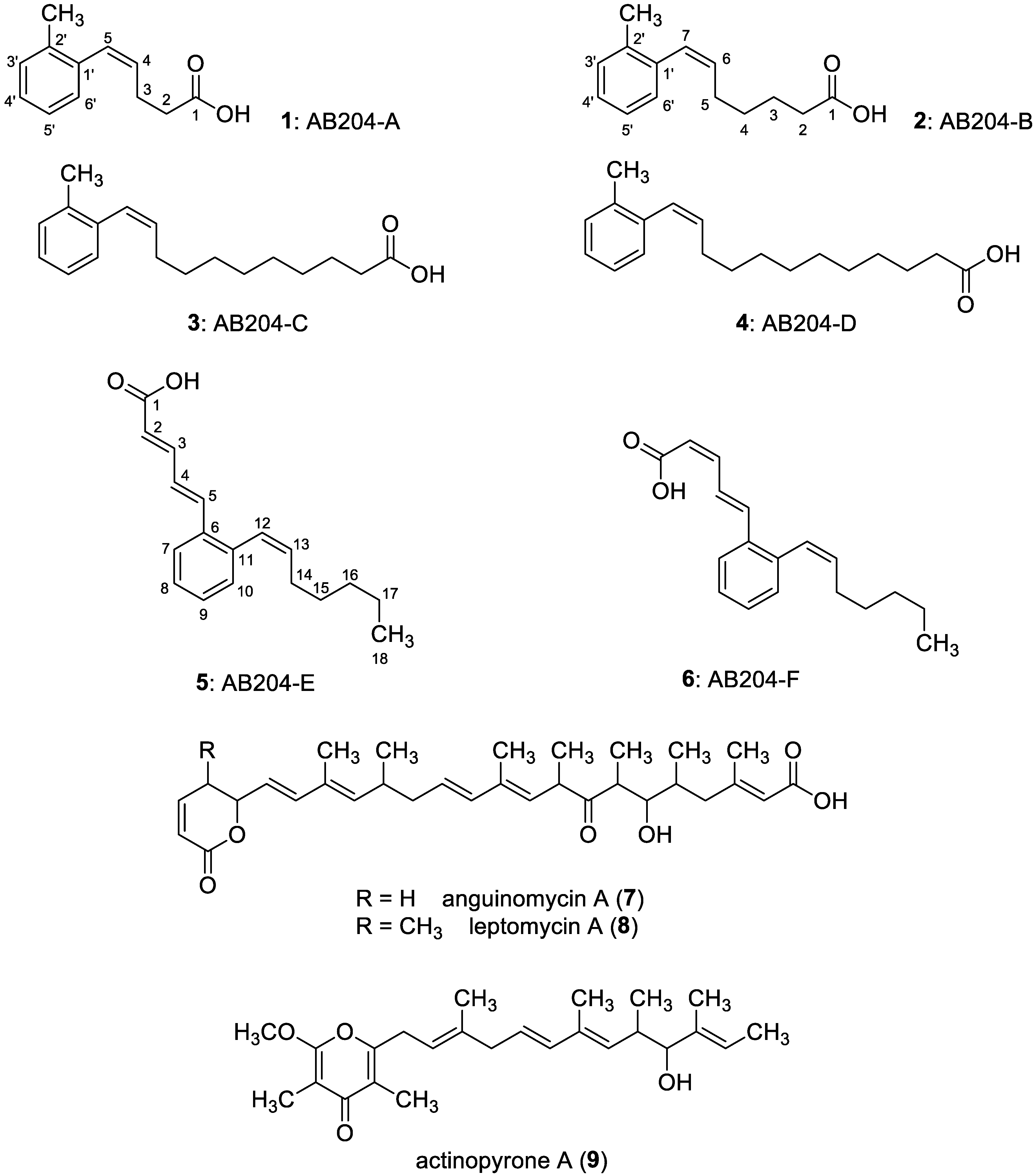
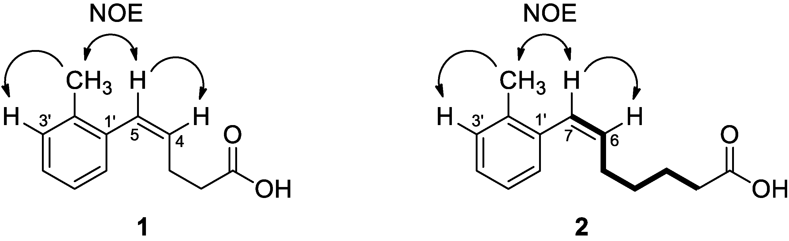
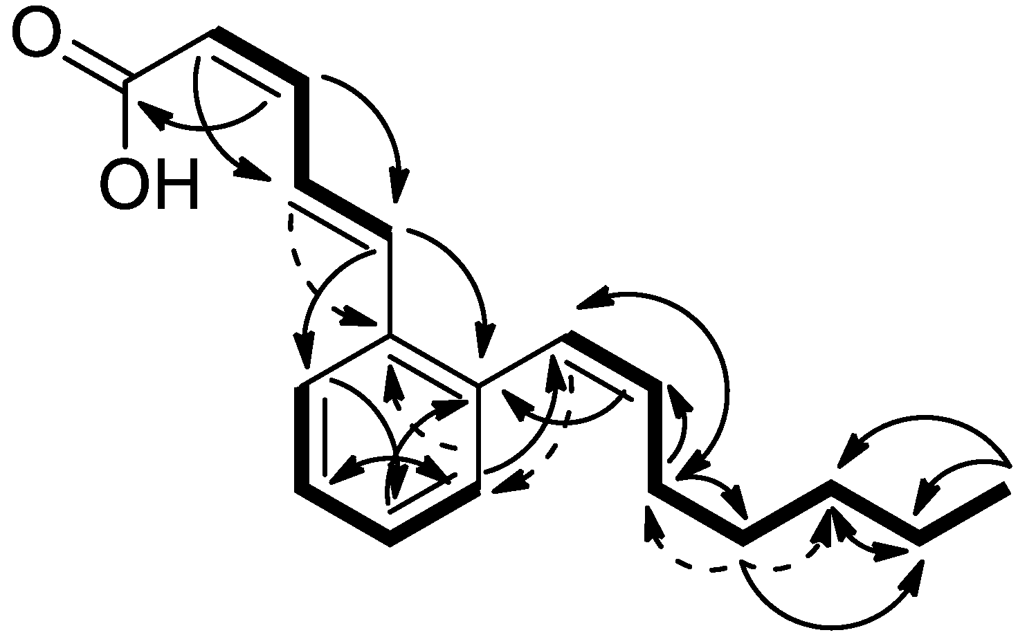
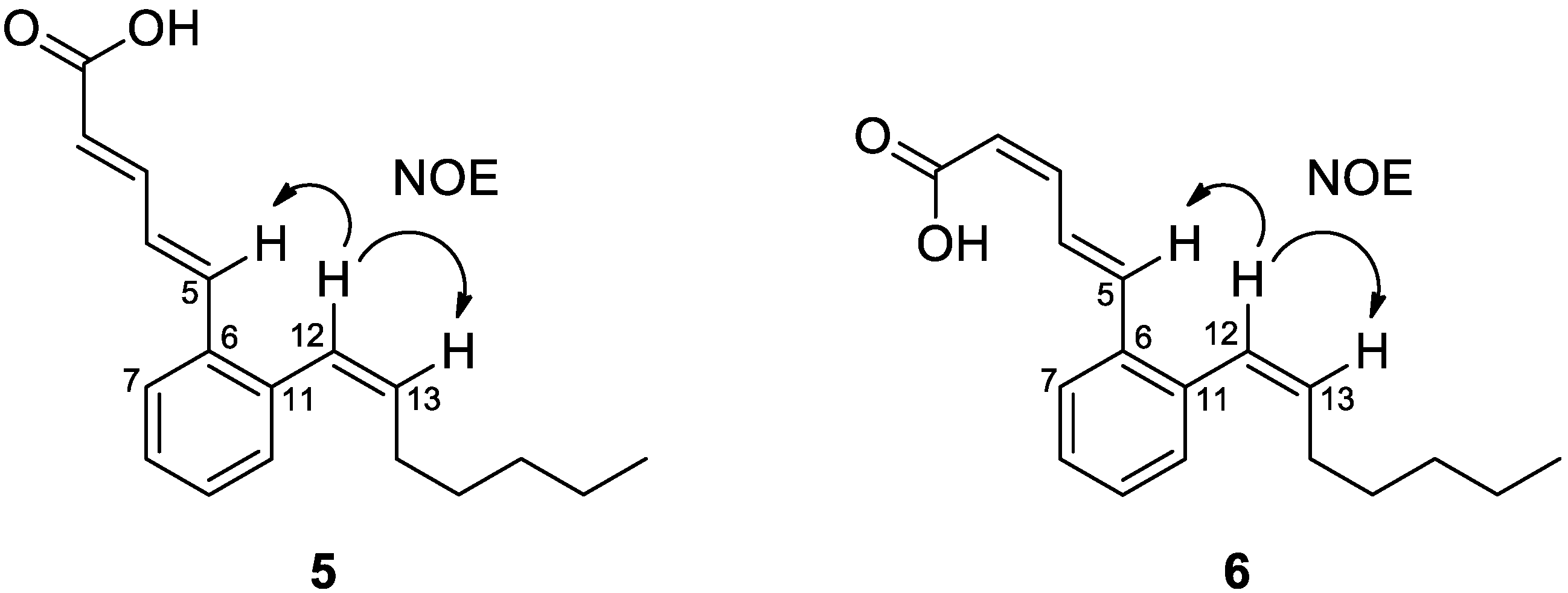
3.1. Biological Activity-Guided Purification of Active Components from Culture Broth of S. palmae CMU-AB204T and Structure Determination of Active Components

S. palmae CMU-AB204T was cultured in 40 L of ISP2 medium at 28 °C for seven days, and the broth and mycelia were extracted with EtOAc. The active components in culture broth extract of strain CMU-AB204T were isolated by biological activities-guided purification using paper disk assay. The extract was purified by silica gel column chromatography, Sephadex LH-20 column chromatography, preparative TLC, and preparative HPLC. The eluates were concentrated in vacuo to yield nine compounds, AB204-A (1, 10.2 mg), AB204-B (2, 8.2 mg), AB204-E (5, 14.2 mg), AB204-F (6, 9.9 mg), a mixture (6.5 mg) of AB204-C (3) and AB204-D (4), 7 (7.0 mg), 8 (1.7 mg), and 9 (17.0 mg) as depicted in Scheme 1.
AB204-A (1) was obtained as a pale yellow amorphous solid. It was found to be readily soluble in acetonitrile, MeOH, CHCl3, and was observed to be less soluble in water. As the HREIMS analysis showed m/z 190.1000 [M]+, the molecular formula of 1 was elucidated as C12H14O2 (calculated value of 190.0994, Figure S1). The intense band at 1706 cm−1 of the IR spectrum in MeOH solution was assigned as C=O stretching frequency of dimeric carboxylic acid moiety (Figure S2). Based on 1H NMR analysis, 1 revealed four aromatic protons stacked at 7.14–7.18 ppm and a pair of olefinic protons stacked at 5.70 ppm and 6.51 ppm (Figure S3). Coupling constants (J = 11.5 Hz) of the two olefinic protons showed Z-configuration of the olefin moiety. Compound 1 had four additional methylene protons at 2.41–2.51 ppm and one methyl singlet signal at 2.24 ppm (Table 1). The 13C NMR spectrum showed 12 carbon signals: one carbonyl carbon at 177.2 ppm that indicated a carboxylic acid, eight aromatic or olefinic carbons, two methylene carbons at 23.5 and 33.7 ppm, and one methyl carbon at 19.8 ppm (Table 1, Figure S4).
MS, HMBC, and HMQC analyses suggested 1 contained one disubstituted aromatic ring, one methyl, and pentenoic acid moieties (Figures S5 and S6). HMBC correlations were observed from two methylene protons (2.41–2.44 ppm and 2.46–2.51 ppm) to a carboxylic carbon at 177.2 ppm, and two olefinic carbons of C-4 and C-5 (129.7 and 129.8 ppm), as are given in Table 1. A correlation between the Z-olefinic proton at 6.51 ppm (H-5) and one methylene carbon (C-3) at 23.5 ppm was also observed; thus 1 was believed to possess 4,5-Z-pentenoic acid moiety in the structure. An HMBC correlation between one methyl proton at 2.24 ppm and three aromatic carbons of C-1’, C-2’, and C-3’ (136.2, 136.2, and 129.8 ppm, respectively), and between Z-olefinic protons and aromatic carbons, H-4 (5.70 ppm) and C-1’ (136.2 ppm), and H-5 (6.51 ppm) and C-6’ (128.8 ppm), indicated 1 was an ortho-methyl phenyl alkenoic acid compound, (Z)-5-(2-methylphenyl)-4-pentenoic acid (Figure 1). Differential NOE of 1 was observed between a methyl proton and both an aromatic 3’-proton and an olefinic proton of H-5 as well as between the two olefinic protons (Figure S7). The geometry of two substitutes of the aromatic ring was confirmed by NOE correlations, as is shown in Figure 2. From some Streptomyces strains, E-isomer of 1, (E)-5-(2-methylphenyl)-4-pentenoic acid was identified [33,34,35]; however, there was no report on the Z-isomer (1) obtained from natural sources. Therefore, it was concluded that 1 was a novel natural product.
AB204-B (2) was isolated as a pale yellow amorphous solid. It was readily soluble in the same solvent as 1 and less soluble in water. The molecular formula of 2 was established as C14H18O2 (calculated value of 218.1329) based on NMR data and the HREIMS ion peak of m/z 218.1301 [M] + (Figure S8), indicating compound 2 had one more C2H4 unit when compared to the molecular formula of 1. The IR spectrum of 2 revealed the presence of C=O stretching frequency of dimeric carboxylic acid moiety at 1706 cm−1, which was similar to the spectrum of 1 (Figure S9). The 1H NMR spectrum of 2 revealed four aromatic protons at 7.14–7.16 ppm, a pair of olefinic protons at 5.69 and 6.45 ppm, eight methylene protons at 1.45, 1.63, 2.17, and 2.30 ppm and one methyl singlet signal at 2.25 ppm (Table 1, Figure S10). The coupling constant of two olefinic protons (11.5 Hz) indicated a Z-configuration. The 13C NMR spectrum of 2 showed 14 carbon signals: one carbonyl carbon at 177.6 ppm, eight aromatic or olefinic carbons, four methylene carbons at 24.2, 27.8, 29.1, and 33.4 ppm, and one methyl carbon at 19.9 ppm (Table 1, Figure S11). These data support the conclusion that the compound had a closely related structure to 1. Each methylene signal was assigned by COSY, as is shown in Figure 2. Eight methylene protons constructed a C4 alkyl chain, and COSY correlation confirmed the connection between this C4 alkyl chain and Z-olefin (Figure S12). This connection was supported by HMBC and HMQC spectra (Figures S13 and S14). HMBC correlations were observed from one olefinic proton H-6 (5.69 ppm) to an aromatic carbon at C-1’ (136.7 ppm) and from singlet methyl proton at 2.25 ppm to aromatic carbons at C-1’, C-2’, and C-3’. Therefore, one methyl moiety and one alkene chain were substituted for an aromatic ring in the ortho position. HMBC correlation from two methylene protons of the alkene chain at 1.63 and 2.30 ppm to the carbonyl carbon at 177.6 ppm, and a molecular formula of 2, suggested that this compound had a carboxylic acid at the end of the alkene chain. The same NOE correlation was observed in 1 and 2 (Figure 2 and Figure S15). Thus, the structure of 2 was assigned as (Z)-7-(2-methylphenyl)-6-heptenoic acid, as is shown in Figure 1.
The structures of AB204-C (3) and AB204-D (4) were elucidated as a mixture of both compounds because of the difficulty associated with further purification. MS spectra showed the molecular formulas of 3 and 4 were C18H26O2 and C19H28O2, respectively (Figure S16). 1H NMR data suggested compounds 3 and 4 were analogs of 1 and 2, thus 3 and 4 might be (Z)-11-(2-methylphenyl)-10-undecenoic acid and (Z)-12-(2-methylphenyl)-11-dodecenoic acid, respectively (Figure 1 and Figure S17).
AB204-E (5) and AB204-F (6) were obtained as a pale yellow amorphous solid. The accurate mass and molecular formula of compounds 5 and 6 were analyzed by both HRESIMS and HREIMS. Molecular ion peaks were exhibited at m/z 271.1705 [M + H]+ and 270.1616 [M]+ for 5, and m/z 271.1689 [M + H]+ and 270.1633 [M]+ for 6 (Figures S18–S21). These data suggest that both compounds had the same molecular formula as C18H22O2 (calcd. 270.1620 for C18H22O2 and calcd. 271.1698 for C18H23O2). The C=O stretching frequency band at 1685 cm−1 in the IR spectrum of 5 and 1684 cm−1 in the spectrum of 6 was assigned as a carboxylic acid moiety (Figures S22 and S23). NMR spectra of both compounds demonstrated a structural similarity. The assignment of 1H and 13C NMR spectra of 5 and 6 are given in Table 2.
1H NMR spectrum of 5 showed ten aromatic/olefinic protons, eight methylene protons, and one methyl triplet proton at 0.83 ppm (Figure S24). In 13C NMR spectrum of 5, one calboxylic carbon at 170.5 ppm, twelve olefinic/aromatic carbons, four methylene carbons, and one methyl carbon were measured (Figure S25). 1H NMR spectrum of 5 suggested the existence of two pairs of E-olefin assigned by large coupling constants (15.5 Hz for each) and one pair of Z-olefin whose coupling constant was 11.5 Hz. Three partial structures were assigned by COSY correlation; one 1,2-substituted aromatic ring, one 1,2-Z-heptene group, and one diene group (Figure S26). HMBC correlations between diene protons of 5.99 and 7.41 ppm and a carbonyl carbon at 170.5 ppm, and between diene protons of 6.95 and 7.13 ppm and three aromatic carbons at positions C-6, C-7, and C-11 (135.6, 126.7, and 138.7 ppm, respectively) indicated that one end of the diene was connected to a carboxylic carbon and the other end of the diene was attached to an aromatic ring at position 6 (Figures S27 and S28). HMBC correlation between Z-olefinic protons (5.83 and 6.54 ppm) of 1,2-Z-heptene and aromatic ring carbons at C-10 and C-11 (131.0 and 138.7 ppm, respectively) suggested 1,2-Z-heptene moiety was connected to the aromatic ring at C-11. These data support the structure of 5 as (2E,4E)-5-(2-(1Z)-heptenylphenyl)-2,4-pentadienoic acid (Figure 1).
1H NMR and 13C NMR spectra of 6 suggested that the structure was almost the same as 5 (Figures S29 and S30). However, the 1H NMR spectrum clarified that only one pair of E-olefin existed, while the other two pairs of olefin were identified as Z-configuration by analysis of coupling constants. These data indicated that 6 was a stereoisomer of 5. COSY correlations revealed that two partial structures of 6, one 1,2-substituted aromatic ring and one 1,2-Z-heptene moiety, were identical to those of 5; however, a diene structure was constituted of both E and Z-olefins (Figure S31). The connection of 1,2-Z-heptene moiety to the aromatic ring at C-11 was confirmed by the HMBC spectrum (Figure 3, Figures S32 and S33). HMBC correlations between Z-olefinic protons of the diene moiety and a carboxylic carbon at 169.5 ppm and between one E-olefinic proton (7.08 ppm) of the diene moiety and aromatic ring carbons at C-7 and C-11 (126.2 and 137.5 ppm, respectively) established the structure of 6 as (2Z,4E)-5-(2-(1Z)-heptenylphenyl)-2,4-pentadienoic acid, as is depicted in Figure 1.
To clarify the geometry of two substituted chains of 5 and 6, the differential NOE experiment was conducted. NOE correlations between H-5 and H-12 were observed in both compounds (Figures S34 and S35), which supported the geometry of two side chains in 5 and 6, as is shown in Figure 4. This is the first report of 5 and 6 obtained from natural sources. Qureshi et al. [36] found structurally related compounds, MF-EA-705a and b, along with actinopyrone A from a broth extract of Streptomyces MF-EA-705. The most related structures were found as cinnamoyl moieties of rare peptide compounds, pepticinnamin E, WS9326A, and RP-1776 (skyllamycin A) [37,38,39].
Compounds 7 and 8 were pale-yellow amorphous solids that were determined by HRESI-MS analyses to have molecular formulas of C31H44O6 and C32H46O6, respectively (Figures S36 and S37). The planar structures of 7 and 8 (Figure 1) were confirmed by NMR spectra (Figures S38 and S39) as known compounds, anguinomycin A (7) [40] and leptomycin A (8) [41], which were known to be relative structures. Compound 9 was a colorless oil, and the molecular formula of 9 was determined to be C25H36O4 on the basis of HRESI-MS data and the signals of 1H NMR spectrum (Figures S40 and S41). These data supported the structure of 9 to be actinopyrone A (9) as is shown in Figure 1 [42].
Physicochemical Properties of 1, 2, 5, and 6
Compound 1 (AB204-A): pale yellow amorphous solid; [α]D –3.4 (c 0.1, MeOH); UV (MeOH) λmax (log ε) 204.5 (4.09), 235.5 (3.63) nm; IR (ATR) νmax 3014, 2923, 1706, 1654, 1409 cm−1; 1H and 13C NMR (chloroform-d) see Table 1; HREIMS m/z 190.1000 [M]+ (calcd for C12H14O2, 190.0994).
Compound 2 (AB204-B): pale yellow amorphous solid; [α]D –0.3 (c 0.1, MeOH); UV (MeOH) λmax (log ε) 204.5 (3.98), 235.5 (3.53) nm; IR (ATR) νmax 3011, 2925, 1706, 1653, 1457 cm−1; 1H and 13C NMR (chloroform-d) see Table 1; HREIMS m/z 218.1301 [M]+ (calcd for C14H18O2, 218.1329).
Compound 5 (AB204-E): pale yellow amorphous solid; [α]D –13.4 (c 0.1, MeOH); UV (MeOH) λmax (log ε) 204 (4.04), 251 (3.90), 309.5 (4.26) nm; IR (ATR) νmax 2956, 2924, 2857, 1684, 1617, 1277, 1000 cm−1; 1H and 13C NMR (methanol-d4) see Table 2; HREIMS m/z 270.1616 [M]+ (calcd for C18H22O2, 270.1620).
Compound 6 (AB204-F): pale yellow amorphous solid; [α]D –14.1 (c 0.1, MeOH); UV (MeOH) λmax (log ε) 204 (3.76), 251 (3.51), 309.5 (3.86) nm; IR (ATR) νmax 2955, 2924, 2855, 1684, 1615, 1244, 958 cm−1; 1H and 13C NMR (chloroform-d) see Table 2; HREIMS m/z 270.1633 [M]+ (calcd for C18H22O2, 270.1620).

3.2. Antimicrobial Activities of Isolated Compounds

Antimicrobial activities of the purified compounds, except for the mixture of AB204-C (3) and AB204-D (4), were tested against four Gram-positive bacteria, five Gram-negative bacteria, two yeasts, and three fungi using paper disk diffusion assay with an equal amount of each compound at 50 µg/disk. The results are shown in Table 3. All compounds did not show activity against E. coli NIHJ, K. pneumonia ATCC 10031, P. vulgaris NBRC 3167, Ps. aeruginosa IFO 3080, and Sa. cerevisiae ATCC 9763. AB204-A (1) and B (2) displayed a weak activity towards C. albicans ATCC 64548, A. niger ATCC 6275, and G. boninense BCC 21330, with a clear zone range from 10.4 to 13.2 mm. However, they did not affect the Gram-positive and Gram-negative bacteria. AB204-E (5) and AB204-F (6) displayed no antifungal and antiyeast activities but showed good antibacterial activity against Gram-positive bacteria and weak activity against the Gram-negative bacterium X. campestris pv. oryzae KB88, which is a phytopathogenic strain. AB204-E (5) strongly inhibited K. rhizophila ATCC 9341, B. subtilis ATCC 6633, M. smegmatis ATCC 607, and S. aureus ATCC 6538p with inhibition zones of 41.3, 35.3, 32.7, and 26.0 mm, respectively, while AB204-F (6) showed a slightly lower activity against the same pathogens as is presented in Table 3. Anguinomycin A (7) revealed potent inhibitory activity against the Gram-positive bacterium K. rhizophila ATCC 9341 (19.1 mm), and two fungi, Mu. racemosus IFO 4581 (16.9 mm), and G. boninense BCC 21330 (19.6 mm), while leptomycin A (8) showed stronger activity against these pathogens at 30.6, 49.0, and 21.2 mm, respectively. Actinopyrone A (9) exhibited potent antifungal activity against C. albicans ATCC 64548 and three fungal strains with the inhibition zone in a range of 11.9 to 23.9 mm (Table 3). These results suggest that the antibacterial and antifungal activities of the S. palmae CMU-AB204T may have been displayed as a consequence of the contribution of all these antimicrobial secondary metabolites.

4. Discussion

Several mechanisms have been proposed to control G. boninense causing BSR disease in oil palm trees. However, none of them have successfully been treated or been shown to suppress the disease [9]. The search for antifungal alternatives is representative of a potential solution that has drawn significant interest. In this study, new compounds were identified during the isolation of anti-Ganoderma substances from S. palmae CMU-AB204T. The assessment of antimicrobial activity of four new phenyl alkenoic acids showed that AB204-A (1) and B (2) mildly inhibited the growth of fungi, G. boninense BCC 21330, Mu. racemosus IFO 4581, and A. niger ATCC 6275, while AB204-E (5) and F (6) displayed a positive degree of activity against Gram-positive bacteria, B. subtilis ATCC 6633, K. rhizophila ATCC 9341, M. smegmatis ATCC 607, and S. aureus ATCC 6538p. New antifungal compounds, AB204-A (1) and B (2), possessed similar structures to phenylethyl alcohol (PEA), an antifungal aromatic compound that was obtained from Trichoderma virens 7b, which had significant potential as a biological control agent for BSR [43]. These compounds may exhibit a mechanism in inhibiting fungi similar to PEA which inhibits protein, DNA, RNA, and aminoacyl tRNA syntheses of fungi [44,45]. However, a mode of action of novel compounds in controlling fungi should be confirmed in the future.
AB204-B (2) contained more C2H4 units than AB204-A (1), but it displayed antimicrobial activity against the same pathogenic strains with similar inhibition zone sizes. This result indicated that the presence of a longer chain of the carboxylic group in 2 had not been involved in the antimicrobial activity. Biological activities of the E-isomer of AB204-A (1), (E)-5-(2-methylphenyl)-4-pentenoic acid was previously reported to be an inactive compound against bacteria and fungi but the tested concentration and the strain of tested microorganisms have not been indicated [33]. However, the difference of an antimicrobial activity between (E)-5-(2-methylphenyl)-4-pentenoic acid and new compounds, AB204-A (1) and AB204-B (2), revealed that the existence of Z-olefin in 1 and 2 had been involved in their antifungal activity. The structures of a mixture of AB204-C (3) and AB204-D (4) were predicted based on HREI-MS and 1H NMR spectra. In the future, the mixture should be reseparated using other techniques, and additional data is needed to confirm their structures and antimicrobial activities. AB204-E (5) and F (6) have an 1,2-Z-heptene moiety connected to the aromatic ring. These metabolites have not shown antifungal activity but exhibited strong antibacterial activity when associated with this moiety. Moreover, the existence of one pair of Z-olefin in the chain of the carboxylic group of AB204-E (5), instead of the E and Z-olefins of AB204-F (6), increased the antibacterial activity of this compound.
Previously, Thong et al. [35] found two closely related compounds of 14, and they were isolated from a Streptomyces that had been spontaneously acquired rifampicin resistance. These compounds contained E-olefins and have both a methylbenzene unit and a 2-amino-3-hydroxycyclopent-2-enone (C5N) moiety. The phenyl alkenoic acid-associated metabolites discovered by Thong et al. did not display antibacterial activity against E. coli, M. luteus, S. aureus, and B. subtilis in testing with a microplate assay at 100 μM or approximately 28.5 and 33.5 mg/mL, thus revealing similar results to AB204-A (1) and B (2). Notably, the presence of a carboxylic acid moiety in the novel compounds, and a C5N moiety in the known compounds, may not be involved in the antimicrobial activity. Based on draft genome sequences of the rifampicin-resistant mutant (TW-R50–13), the methylbenzene moiety may be biosynthesized by the expression of polyketide synthase (PKS) genes that are located at a different locus from the biosynthetic genes for the C5N moiety [35]. The genes encoding for PKS have been disclosed to complex biosynthetic mechanisms, which were involved in the production of many metabolites in microorganisms [46]. Genome sequences would provide the data of potential gene clusters to understand the metabolic pathways of S. palmae CMU-AB204T. Thus, the genome sequences of this strain should be further studied to determine the presence of both silent and cryptic secondary metabolite biosynthetic gene clusters that are able to synthesize the corresponding novel natural products.
In addition to 1 and 2, other antifungal compounds, anguinomycin A (7), leptomycin A (8), and actinopyrone A (9) obtained from the same broth of S. palmae CMU-AB204T, also displayed anti-Ganoderma activity. The ability of S. palmae to produce a variety of antifungal compounds was proven. This strain might produce each antifungal secondary metabolite depending on the prevailing environmental conditions, such as nutritional source, incubation period, pH value, and temperature [47,48]. Hence, the optimization of culture conditions should be studied in order to obtain high yields of the antifungal metabolites. The protecting effect of S. palmae CMU-AB204T against BSR has also been confirmed in a glasshouse experiment [49]. The results obtained from this study strongly suggest that the antimicrobial secondary metabolites were involved in the mechanism exhibiting anti-BSR effects by this Streptomyces strain.
Although the new compounds obtained in this study showed moderate activity towards G. boninense, they inhibited clinical bacterial pathogens and other phytopathogenic fungi, suggesting a possible utility of the four new antimicrobial substances in both agricultural and medical treatments. However, cytotoxicity to mammalian cell of both new compounds and three known compounds should be tested before being applied to these compounds. The recovery of novel actinomycetes species, especially the genus Streptomyces, has the potential to be a rich source of both new and known natural products [50,51]. Notably, S. palmae CMU-AB204T was found to produce various bioactive metabolites.

Supplementary Materials

The following are available online at https://www.mdpi.com/2076-2607/8/3/350/s1.

Author Contributions

The project approach was designed by K.S., W.P.-a., S.L.; Streptomyces strain was selected by K.S.; activity-guided purification was conducted by K.S., M.M., K.D.; structural determination was conducted by K.S., M.M., K.D.; antimicrobial activity was measured by K.S.; the research was supervised by K.S., W.P.-a., S.L.; funding was acquired by M.M. and S.L. All authors have read and agreed to the published version of the manuscript.

Funding

This research work was partially supported by Chiang Mai University, the Thailand Research Fund (TRF) within the program entitled Research and Researchers for Industries (RRi) PHD56I0048 with the cooperation of Trang Palm Oil Co., Ltd., Thailand, and the Institute for Fermentation, Osaka (IFO), Japan.

Acknowledgments

We thank Kenichiro Nagai, School of Pharmacy, Kitasato University, for help in obtaining mass data, and Toshiyuki Tokiwa, Kitasato Institute for Life Sciences, Kitasato University, for providing the microbes used to measure antimicrobial activity.

Conflicts of Interest

The authors declare no conflicts of interest.

References

  1. Rees, R.W.; Flood, J.; Hasan, Y.; Cooper, R.M. Effects of inoculum potential, shading and soil temperature on root infection of oil palm seedlings by the basal stem rot pathogen Ganoderma boninense. Plant. Pathol. 2007, 56, 862–870. [Google Scholar] [CrossRef]
  2. Susanto, A.; Sudharto, P.S.; Purba, R.Y. Enhancing biological control of basal stem rot disease (Ganoderma boninense) in oil palm plantations. Mycopathologia 2005, 159, 153–157. [Google Scholar] [CrossRef] [PubMed]
  3. Nieto, L. Incidence of oil palm stem rots in Colombia. Palmas 1995, 16, 227–232. [Google Scholar]
  4. Turner, P.D. Oil Palm Diseases and Disorders; Oxford University Press: Oxford, UK, 1981; pp. 88–110. [Google Scholar]
  5. Likhitekaraj, S.; Tummakate, A. Basal stem rot of oil palm in Thailand caused by Ganoderma. In Ganoderma Diseases of Perennial Crops; Flood, J., Bridge, P.D., Holderness, M., Eds.; CABI Publishing: Wallingford, UK, 2000; pp. 66–70. [Google Scholar]
  6. Jollands, P. Laboratory investigations on fungicides and biological agents to control three diseases of rubber and oil palm and their potential applications. Trop. Pest. Manag. 1983, 29, 33–38. [Google Scholar] [CrossRef]
  7. Idris, A.S.; Ismail, S.; Ariffin, D.; Ahmed, D. Control of Ganoderma infected palm-development of pressure injection and field application. MPOB Info. Ser. 2002, 148, 2. [Google Scholar]
  8. Idris, A.S.; Arifurrahman, R. Determination of 50% effective concentration (EC50) of fungicides against pathogenic Ganoderma. MPOB Info. Ser. 2008, 449, 2. [Google Scholar]
  9. Naher, L.; Siddiquee, S.; Yusuf, U.K.; Mondal, M.M.A. Issues of Ganoderma spp. and basal stem rot disease management in oil palm. Am. J. Agri. Sci. 2015, 2, 103–107. [Google Scholar]
  10. Shariffah-Muzaimah, S.; Idris, A.; Madihah, A.; Dzolkhifli, O.; Kamaruzzaman, S.; Cheong, P. Isolation of actinomycetes from rhizosphere of oil palm (Elaeis guineensis Jacq.) for antagonism against Ganoderma boninense. J. Oil Palm Res. 2015, 27, 19–29. [Google Scholar]
  11. Wightwick, A.; Walters, R.; Allinson, G.; Reichman, S.; Menzies, N. Environmental risks of fungicides used in horticultural production systems. In Fungicides; Carisse, O., Ed.; IntechOpen: London, UK, 2010; pp. 273–304. [Google Scholar]
  12. Ji, X.Y.; Zhong, Z.J.; Xue, S.T.; Meng, S.; He, W.Y.; Gao, R.M.; Li, Y.H.; Li, Z.R. Synthesis and antiviral activities of synthetic Glutarimide derivatives. Chem. Pharm. Bull. 2010, 58, 1436–1441. [Google Scholar] [CrossRef] [Green Version]
  13. Gupta, P.K. Toxicity of fungicides. In Veterinary Toxicology, Basic and Clinical Principles, 3rd ed.; Gupta, R.C., Ed.; Academic Press: London, UK, 2018; pp. 569–580. [Google Scholar]
  14. Nur Ain Izzati, M.Z.; Abdullah, F. Disease suppression in Ganoderma-infected oil palm seedlings treated with Trichoderma harzianum. Plant. Protect. Sci. 2008, 44, 101–107. [Google Scholar] [CrossRef] [Green Version]
  15. Sundram, S.; Abdullah, F.; Ahmad, Z.A.M.; Yusuf, U.K. Efficacy of single and mixed treatments of Trichoderma harzianum as biocontrol agents of Ganoderma basal stem rot in oil palm. J. Oil Palm Res. 2008, 20, 470–483. [Google Scholar]
  16. Naher, L.; Tan, S.G.; Yusuf, U.K.; Ho, C.-L.; Abdullah, F. Biocontrol agent Trichoderma harzianum strain FA 1132 as an enhancer of oil palm growth. Pertanika J. Trop. Agric. Sci. 2012, 35, 173–182. [Google Scholar]
  17. Harman, G.E.; Howell, C.R.; Viterbo, A.; Chet, I.; Lorito, M. Trichoderma species-opportunistic, avirulent plant symbionts. Nat. Rev. Microbiol. 2004, 2, 43–56. [Google Scholar] [CrossRef]
  18. Naher, L.; Ho, C.-L.; Tan, S.G.; Yusuf, U.K.; Abdullah, F. Cloning of transcripts encoding chitinases from Elaeis guineensis Jacq. and their expression profiles in response to fungal infections. Physiol. Mol. Plant. Pathol. 2011, 76, 96–103. [Google Scholar] [CrossRef]
  19. Sapak, Z.; Meon, S.; Ahmad, Z.A.M. Effect of endophytic bacteria on growth and suppression of Ganoderma infection in oil palm. Int. J. Agric. Biol. 2008, 10, 127–132. [Google Scholar]
  20. Bivi, M.R.; Farhana, M.; Khairulmazmi, A.; Idris, A. Control of Ganoderma boninense: A causal agent of basal stem rot disease in oil palm with endophyte bacteria in vitro. Int. J. Agric. Biol. 2010, 12, 833–839. [Google Scholar]
  21. Sundram, S.; Meon, S.; Seman, I.A.; Othman, R. Symbiotic interaction of endophytic bacteria with arbuscular mycorrhizal fungi and its antagonistic effect on Ganoderma boninense. J. Microbiol. 2011, 49, 551–557. [Google Scholar] [CrossRef]
  22. Nurrashyeda, R.; Maizatul, S.; Idris, A.; Madihah, A.; Nasyaruddin, M. The potential of endophytic bacteria as a biological control agent for Ganoderma disease in oil palm. Sains Malaysiana 2016, 45, 401–409. [Google Scholar]
  23. Katz, L.; Baltz, R.H. Natural product discovery: past, present, and future. J. Ind. Microbiol. Biotechnol. 2016, 43, 155–176. [Google Scholar] [CrossRef]
  24. Procópio, R.E.; Silva, I.R.; Martins, M.K.; Azevedo, J.L.; Araújo, J.M. Antibiotics produced by Streptomyces. Braz. J. Infect. Dis. 2012, 16, 466–471. [Google Scholar] [CrossRef] [Green Version]
  25. Ōmura, S.; Ikeda, H.; Ishikawa, J.; Hanamoto, A.; Takahashi, C.; Shinose, M.; Takahashi, Y.; Horikawa, H.; Nakazawa, H.; Osonoe, T.; et al. Genome sequence of an industrial microorganism Streptomyces avermitilis: Deducing the ability of producing secondary metabolites. PNAS 2001, 98, 12215–12220. [Google Scholar]
  26. Watve, M.G.; Tickoo, R.; Jog, M.M.; Bhole, B.D. How many antibiotics are produced by the genus Streptomyces? Arch. Microbiol. 2001, 176, 386–390. [Google Scholar] [CrossRef]
  27. Pithakkit, S.; Petcharat, V.; Chuenchit, S.; Pornsuriya, C.; Sunpapao, A. Isolation of antagonistic actinomycetes species from rhizosphere as effective biocontrol against oil palm fungal diseases. Walailak J. Sci. Tech. 2014, 12, 481–490. [Google Scholar]
  28. Shariffah-Muzaimah, S.; Idris, A.; Madihah, A.; Dzolkhifli, O.; Kamaruzzaman, S.; Maizatul-Suriza, M. Characterization of Streptomyces spp. isolated from the rhizosphere of oil palm and evaluation of their ability to suppress basal stem rot disease in oil palm seedlings when applied as powder formulations in a glasshouse trial. World J. Microbiol. Biotech. 2018, 34, 15. [Google Scholar] [CrossRef]
  29. Ting, A.S.Y.; Hermanto, A.; Peh, K.L. Indigenous actinomycetes from empty fruit bunch compost of oil palm: evaluation on enzymatic and antagonistic properties. Biocat. Agric. Biotech. 2014, 3, 310–315. [Google Scholar] [CrossRef]
  30. Pal, K.K.; Gardener, B.M. Biological control of plant pathogens. Plant. Health Instructor 2006, 2, 1117–1142. [Google Scholar] [CrossRef] [Green Version]
  31. Sujarit, K.; Kudo, T.; Ohkuma, M.; Pathom-Aree, W.; Lumyong, S. Streptomyces palmae sp. nov., isolated from oil palm (Elaeis guineensis) rhizosphere soil. Int. J. Syst. Evol. Microbiol. 2016, 66, 3983–3988. [Google Scholar] [CrossRef]
  32. Shirling, E.B.; Gottlieb, D. Methods for characterization of Streptomyces species. Int. J. Syst. Bacteriol. 1966, 16, 313–340. [Google Scholar] [CrossRef] [Green Version]
  33. Mukku, V.J.R.V.; Maskey, R.P.; Monecke, P.; Grün-Wollny, I.; Laatsch, H. 5-(2-Methylphenyl)-4-pentenoic acid from a terrestrial Streptomycete. Z. Naturforsch. 2002, 57b, 335–337. [Google Scholar] [CrossRef]
  34. Shaaban, K.A.; Helmke, E.; Kelter, G.; Fiebig, H.H.; Laatsch, H. Glucopiericidin C: A cytotoxic piericidin glucoside antibiotics produced by a marine Streptomyces isolate. J. Antibiot. 2011, 64, 205–209. [Google Scholar] [CrossRef] [Green Version]
  35. Thong, W.L.; Shin-ya, K.; Nishiyama, M.; Kuzuyama, T. Methylbenzene-containing polyketides from a Streptomyces that spontaneously acquired rifampicin resistance: Structural elucidation and biosynthesis. J. Nat. Prod. 2016, 79, 857–864. [Google Scholar] [CrossRef] [PubMed]
  36. Qureshi, A.; Mauger, J.B.; Cano, R.J.; Galazzo, J.L.; Lee, M.D. MF-EA-705a & MF-EA-705b, new metabolites from microbial fermentation of a Streptomyces sp. J. Antibiot. 2001, 54, 1100–1103. [Google Scholar] [PubMed] [Green Version]
  37. Shiomi, K.; Yang, H.; Inokoshi, J.; Van der Pyl, D.; Nakagawa, A.; Takeshima, H.; Ōmura, S. Pepticinnamins, new farnesyl-protein transferase inhibitors produced by an actinomycete. II. Structural elucidation of pepticinnamin E. J. Antibiot. 1993, 46, 229–234. [Google Scholar] [CrossRef] [PubMed] [Green Version]
  38. Shigematsu, N.; Hayashi, K.; Kayakiri, N.; Takase, S.; Hashimoto, M.; Tanaka, H. Structure of WS9326A, a novel tachykinin antagonist from a Streptomyces. J. Org. Chem. 1993, 58, 170–175. [Google Scholar] [CrossRef]
  39. Toki, S.; Agatsuma, T.; Ochiai, K.; Saitoh, Y.; Ando, K.; Nakanishi, S.; Lokker, N.; Giese, N.A.; Matsuda, Y. RP-1776, a novel cyclic peptide produced by Streptomyces sp., inhibits the binding of PDGF to the extracellular domain of its receptor. J. Antibiot. 2001, 54, 405–414. [Google Scholar] [CrossRef] [Green Version]
  40. Hayakawa, Y.; Adachi, K.; Komeshima, N. New antitumor antibiotics, anguinomycins A and B. J. Antibiot. 1987, 40, 1349–1352. [Google Scholar] [CrossRef] [Green Version]
  41. Hamamoto, T.; Seto, H.; Beppu, T. Leptomycins A and B, new antifungal antibiotics. II. Structure elucidation. J. Antibiot. 1983, 36, 646–650. [Google Scholar] [CrossRef]
  42. Yano, K.; Yokoi, K.; Sato, J.; Oono, J.; Kouda, T.; Ogawa, Y.; Nakashima, T. Actinopyrones A, B, and C, new physiologically active substances. II. Physicochemical properties and chemical structures. J. Antibiot. 1986, 39, 38–43. [Google Scholar] [CrossRef]
  43. Angel, L.P.L.; Yusof, M.T.; Ismail, I.S.; Ping, B.T.Y.; Mohamed Azni, I.N.A.; Kamarudin, N.H.; Sundram, S. An in vitro study of the antifungal activity of Trichoderma virens 7b and a profile of its non-polar antifungal components related against Ganoderma boninense. J. Microbiol. 2016, 54, 732–744. [Google Scholar] [CrossRef]
  44. Lester, G. Inhibition of growth, synthesis, and permeability in Neurospora crassa by phenethyl alcohol. J. Bacteriol. 1965, 90, 29–37. [Google Scholar] [CrossRef] [Green Version]
  45. Liu, P.; Cheng, Y.; Yang, M.; Liu, Y.; Chen, K.; Long, C.A.; Deng, X. Mechanisms of action for 2-phenylethanol isolated from Kloeckera apiculata in control of Penicillium molds of citrus fruits. BMC Microbiol. 2014, 14, 242. [Google Scholar] [CrossRef] [Green Version]
  46. Staunton, J.; Weissman, K.J. Polyketide biosynthesis: a millennium review. Nat. Prod. Rep. 2001, 18, 380–416. [Google Scholar] [CrossRef]
  47. Banga, J.; Praveen, V.; Singh, V.; Tripathi, C.K.M.; Bihari, V. Studies on medium optimization for the production of antifungal and antibacterial antibiotics from a bioactive soil actinomycete. Med. Chem. Res. 2008, 17, 425–436. [Google Scholar] [CrossRef]
  48. Ruiz, B.; Chávez, A.; Forero, A.; García-Huante, Y.; Romero, A.; Sánchez, M.; Rocha, D.; Sánchez, B.; Rodríguez-Sanoja, R.; Sánchez, S.; et al. Production of microbial secondary metabolites: Regulation by the carbon source. Crit. Rev. Microbiol. 2010, 36, 146–167. [Google Scholar] [CrossRef]
  49. Sujarit, K. Selection and Characterization of Actinomycetes for Growth Promotion of Oil Palm and Biological Control of Oil Palm Diseases. Ph.D. Thesis, Chiang Mai University, Chiang Mai, Thailand, 2018. [Google Scholar]
  50. Bérdy, J. Thoughts and facts about antibiotics: where we are now and where we are heading. J. Antibiot. 2012, 65, 385–395. [Google Scholar] [CrossRef] [Green Version]
  51. Lucas, X.; Senger, C.; Erxleben, A.; Grüning, B.A.; Döring, K.; Mosch, J.; Flemming, S.; Günther, S. StreptomeDB: A resource for natural compounds isolated from Streptomyces species. Nucleic Acids Res. 2013, 41, D1130–D1136. [Google Scholar] [CrossRef]
Scheme 1. Purification procedures for compounds 19.
Microorganisms 08 00350 sch001
Figure 1. Antimicrobial compounds isolated from the broth extract of Streptomyces palmae CMU-AB204T.
Microorganisms 08 00350 g001
Figure 2. Observed COSY correlation (bold lines, in 2) and NOE correlation in AB204-A (1) and B (2).
Microorganisms 08 00350 g002
Figure 3. Observed COSY (bold lines) and HMBC correlations (arrows) in AB204-F (6). The same correlations were observed in AB204-E (5).
Microorganisms 08 00350 g003
Figure 4. NOE correlations in AB204-E (5) and AB204-F (6).
Microorganisms 08 00350 g004
Table 1. NMR spectroscopic data of AB204-A (1) and B (2) (δH, 500 MHz; δC, 125 MHz).
Carbon No.1 (in CDCl3)2 (in CDCl3)
δC, TypeδH, Mult (J in Hz) HMBCδC, typeδH, Mult (J in Hz)HMBC
1177.2, C 177.6, C
233.7, CH22.41–2.44, mC1, C3, C433.4, CH22.30, t (7.5)C1, C3, C4
323.5, CH22.46–2.51, mC1, C2, C524.2, CH21.63, tt (7.5, 7.5)C1, C2, C5
4129.7 or 129.8, CH5.70, dt (11.5, 7.0)C1′29.1, CH21.45, tt (7.5, 7.5)C2, C5, C6
5129.7 or 129.8, CH6.51, d (11.5)C3, C2′, C6′27.8, CH22.17, dtd (7.5, 7.5, 1.5)C3, C4, C6, C7
6 132.0, CH5.69, dt (11.5, 7.5)C1′
7 128.5, CH6.45, br.d (11.5)C5, C6′
1′136.2, C 136.7, C
2′136.2, C 136.2, C
3′129.8, CH7.14–7.18 *, m 129.8, CH7.13–7.18 *, m-
4′125.4 or 127.1, CH7.14–7.18 *, m 125.3 or 126.8, CH7.13–7.18 *, mC2′
5′125.4 or 127.1, CH7.14–7.18 *, mC3′125.3 or 126.8, CH7.13–7.18 *, mC3′
6′128.8, CH7.14–7.18 *, m 128.9, CH7.13–7.18 *, mC2′
2′-Me19.8, CH32.24, sC1′, C2′, C3′19.9, CH32.25, sC1′, C2′, C3′
* overlapped.
Table 2. NMR spectroscopic data of AB204-E (5) and F (6) (δH, 500 MHz; δC, 125 MHz).
Carbon No.5 (in CD3OD)6 (in CDCl3)
δC, TypeδH, Mult (J in Hz)HMBCδC, TypeδH, Mult (J in Hz)HMBC
1170.5, C 169.5, C
2122.3, CH5.99, d (15.5)C1, C4115.8, CH5.74, d (11.5)C1, C4
3146.8, CH7.41, dd (15.5, 11.0)C1, C5147.3, CH6.85, t (11.5)C1, C5
4128.2, CH6.95, dd (15.5, 11.0)C2, C5, C6125.4, CH8.04, dd (15.5, 11.5)C6
5140.0, CH7.13, d (15.5)C3, C7, C11140.6, CH7.08, d (15.5)C3, C7, C11
6135.6, C 134.3, C
7126.7, CH7.67, mC9, C11126.2, CH7.72, mC9, C11
8128.4, CH7.26 *, m 127.1, CH7.26 *, mC10
9129.5, CH7.26 *, m 128.6, CH7.26 *, mC11
10131.0, CH7.15, mC8, C12129.9, CH7.17, mC6, C8, C12
11138.7, C 137.5, C
12128.3, CH6.54, br.d (11.5)C6, C10, C14127.0, CH6.51, br.d (11.5)C10, C14
13135.7, CH5.83, dt (11.5, 7.5)C11134.9, CH5.81, dt (11.5, 7.5)C11
1429.4, CH22.02, dtd (7.5, 7.5, 1.5)C12, C13, C15, C1628.4, CH22.04, dtd (7.5, 7.5, 1.0)C12, C13, C15, C16
1530.2, CH21.38, mC13, C14, C16, C1729.2, CH21.37, tt (7.5, 7.5)C14, C16, C17
1632.6, CH21.22, mC1731.4, CH21.23 *, mC17
1723.5, CH21.21, mC1622.5, CH21.23 *, mC16
1813.3, CH30.83, t (7.0)C16, C1714.0, CH30.84, t (7.0)C16, C17
* overlapped.
Table 3. Antimicrobial activities of the pure compounds against fourteen microorganisms using an equal amount of each compound at 50 µg/disk. Inhibition zone (mm) (Mean ± SD; n = 3) including the diameter of the paper disk (8 mm) was measured after 24 and 48 h of incubation. 1, AB204-A; 2, AB204-B; 5, AB204-E; 6, AB204-F; 7, anguinomycin A; 8, leptomycin A; 9, actinopyrone A.
MicroorganismInhibition Zone (mm) of Seven Pure Compounds
1256789
Gram-positive bacteria
Bacillus subtilis ATCC 6633 --35.3 ± 1.412.3 ± 1.8---
Kocuria rhizophila ATCC 9341--41.3 ± 2.017.5 ± 1.519.1 ± 1.930.6 ± 2.3-
Mycobacterium smegmatis ATCC 607--32.7 ± 1.214.0 ± 2.2---
Staphylococcus aureus ATCC 6538p--26.0 ± 1.613.2 ± 1.9---
Gram-negative bacteria
Escherichia coli NIHJ-------
Klebsiella pneumonia ATCC 10031-------
Proteus vulgaris NBRC 3167-------
Pseudomonas aeruginosa IFO 3080-------
Xanthomonas campestris pv. oryzae KB 88--10.6 ± 1.311.0 ± 2.2 --
Yeasts
Candida albicans ATCC 6454813.1 ± 1.610.4 ± 0.9----20.8 ± 1.5
Saccharomyces cerevisiae ATCC 9763-------
Fungi
Mucor racemosus IFO 4581----16.9 ± 1.749.0 ± 2.323.1 ± 1.6
Aspergillus niger ATCC 627511.5 ± 1.111.1 ± 1.2----23.9 ± 1.8
Ganoderma boninense BCC 2133011.0 ± 1.413.2 ± 2.0--19.6 ± 1.622.1 ± 1.711.9 ± 1.0

