Article

Differential Distribution of the wlaN and cgtB Genes, Associated with Guillain-Barré Syndrome, in Campylobacter jejuni Isolates from Humans, Broiler Chickens, and Wild Birds

by
Pedro Guirado
1,
Sonia Paytubi
1,†,
Elisenda Miró
2,
Yaidelis Iglesias-Torrens
2,3,
Ferran Navarro
2,3,*,
Marta Cerdà-Cuéllar
4,
Camille Stephan-Otto Attolini
5,
Carlos Balsalobre
1 and
Cristina Madrid
1,*
1
Departament de Genètica, Microbiologia i Estadística, Facultat de Biología, Universitat de Barcelona. Avda. Diagonal 643, 08028 Barcelona, Spain
2
Hospital de la Santa Creu i Sant Pau and Institut d’Investigació Biomèdica Sant Pau (IIB Sant Pau), Sant Quintí 89, 08041 Barcelona, Spain
3
Departament de Genètica i Microbiologia. Universitat Autònoma de Barcelona, 08193 Cerdanyola del Vallès (Barcelona), Spain
4
IRTA, Centre de Recerca en Sanitat Animal (CReSA-IRTA-UAB). Campus de la Universitat Autònoma de Barcelona, Bellaterra, 08193 Barcelona, Spain
5
Institute for Research in Biomedicine (IRB Barcelona), The Barcelona Institute of Science and Technology, Baldiri Reixac 10, 08028 Barcelona, Spain
*
Authors to whom correspondence should be addressed.
Current address: Infections and Cancer Laboratory. Cancer Epidemiology Research Program (CERP). Institut Català d’Oncologia (ICO)–IDIBELL. Avda. de la Gran Via 199, 08908 L’Hospitalet de Llobregat (Barcelona), Spain.
Microorganisms 2020, 8(3), 325; https://doi.org/10.3390/microorganisms8030325
Submission received: 23 January 2020 / Revised: 21 February 2020 / Accepted: 24 February 2020 / Published: 26 February 2020
(This article belongs to the Special Issue Foodborne Pathogen Campylobacter)

Abstract

:
Campylobacter jejuni causes campylobacteriosis, a bacterial gastroenteritis with high incidence worldwide. Moreover, C. jejuni infection can trigger the polyneuropathic disorder denominated Guillain-Barré syndrome (GBS). The C. jejuni strains that can elicit GBS carry either wlaN or cgtB, coding both genes for a β-1,3-galactosyltransferase enzyme that is required for the production of sialylated lipooligosaccharide (LOSSIAL). We described a differential prevalence of the genes wlaN and cgtB in C. jejuni isolates from three different ecological niches: humans, broiler chickens, and wild birds. The distribution of both genes, which is similar between broiler chicken and human isolates and distinct when compared to the wild bird isolates, suggests a host-dependent distribution. Moreover, the prevalence of the wlaN and cgtB genes seems to be restricted to some clonal complexes. Gene sequencing identified the presence of new variants of the G- homopolymeric tract within the wlaN gene. Furthermore, we detected two variants of a G rich region within the cgtB gene, suggesting that, similarly to wlaN, the G-tract in the cgtB gene mediates the phase variation control of cgtB expression. Caco-2 cell invasion assays indicate that there is no evident correlation between the production of LOSSIAL and the ability to invade eukaryotic cells.

1. Introduction

Campylobacter jejuni causes the most common bacterial food-borne disease in Europe, a gastroenteritis named campylobacteriosis, which can range from mild, watery to bloody diarrhea [1,2]. Campylobacteriosis is mostly transmitted to humans by the consumption and improper handling of poultry meat [3]. Wild birds play a relevant role in the C. jejuni transmission route by being carriers that can infect both poultry and humans [4,5]. Moreover, campylobacteriosis is the most frequent infection preceding Guillain-Barré syndrome (GBS), an acute polyneuropathic disorder [6]. The strains of C. jejuni that can potentially elicit GBS are those that produce a sialylated LOS (LOSSIAL), due to the well-documented molecular mimicry between the LOSSIAL and the saccharide component of the human GM1 ganglioside which is present in peripheral nerves [7]. Antibodies generated against Campylobacter LOSSIAL structures might cross-react with GM1 found in nerve tissue [1]. Consistently, during the acute-phase of C. jejuni-associated GBS infection, the sera contain high titers of antibodies against LOSSIAL that cross-react with GM1 gangliosides [8].
The operon responsible for the biosynthesis of LOS is highly diverse among C. jejuni isolates, with up to 19 different locus classes, named from A to S [9,10]. Only classes A, B, and C carry genes coding for a β-1,3-galactosyltransferase catalyzing the addition of a galactose molecule, which is required for the production of a GM1-like LOSSIAL structure [11]. Two different genes have been identified to encode for this enzymatic activity: cgtB (in class A and B) and wlaN (in class C) [10,12]. The amino acid identity between CgtB and WlaN is about 58% [12].
Genomic analyses revealed the presence of hypervariable sequences in C. jejuni genes coding for proteins involved in the biosynthesis of surface structures [13]. Most of these hypervariable sequences consist of short homopolymeric nucleotide tracts. The wlaN gene contains a G-tract located within the coding sequence, rendering the expression of the wlaN gene under phase variation control, presumably by a slipped-strand mechanism [12]. Three different G-tract variants have been described, carrying either six, eight, or nine residues [12,14]. The presence of an 8G-tract results in a full-length product (ON-phase), whereas either a 6G- or a 9G-tract results in truncated proteins (OFF-phase).
In this work, we describe the prevalence of the genes responsible for LOSSIAL production, cgtB and wlaN, in C. jejuni isolates from different ecological origin: humans, broiler chickens, and wild birds. The presence and diversity of homopolymeric G-tract regions within both LOSSIAL-related genes are studied. The correlation between the production of LOSSIAL and the ability to invade eukaryotic cells is evaluated.

2. Materials and Methods

2.1. Bacterial Strains and Culture Conditions

The C. jejuni strain collection, previously described [15], is composed of 150 isolates obtained from faeces of three different sources: human patients suffering from symptomatic gastroenteritis (50 isolates), broiler chickens (50 isolates), and wild birds (50 isolates). Human isolates were obtained from the Santa Creu i Sant Pau Hospital (Barcelona) strain collection. Broiler chicken isolates belong to a Campylobacter strain collection at IRTA-CReSA, collected at different slaughterhouses located in Barcelona, Lleida, and Tarragona (Catalonia). The wild bird samples, collected in Catalonia and Alboran Island, were obtained from six different species (Spatula clypeata, Ciconia ciconia, Corvus corax, Columba livia, Larus michahellis, and Larus audouinii). The strain 81-176 (ATCC BAA-2151) was used as the reference strain [13]. Complete genomes of Campylobacter jejuni strains were recovered from an NCBI database.
Isolates were cultured onto Columbia blood agar (CBA) plates (Scharlau) and incubated at 42 °C for 48 h in microaerophilic conditions (CampyGen, Oxoid), unless otherwise stated.

2.2. PCR Amplification

Genomic DNA was extracted from cultures grown onto CBA plates using the InstaGene Matrix Kit (Bio-Rad Laboratoires). PCR reactions (PCR Master Mix x2, Thermo Scientific) were performed using 35 ng of DNA as a template and the presence of wlaN and cgtB genes were tested using the specific primers indicated in Table 1. Primers used to amplify the housekeeping gene gltA were included in the PCR mixtures as an internal control of the PCR reaction. Amplified fragments of wlaN and cgtB were purified and sequenced with the same primers that were used in the PCR reaction.

2.3. Analysis of LOS

Cell biomass from one CBA plate was resuspended in 1 ml of PBS and the OD600 of the cell suspension determined. After centrifugation, the cells were resuspended in Laemmli buffer, adjusting the final volume accordingly to the OD600 of the cell suspension (OD600 × 0.5) (whole cell extracts). LOS was isolated as described previously [18]. Briefly, samples were incubated at 100 °C for 10 min and centrifuged at 10,000× g for 10 min. A 50 µl aliquot of the supernatant was treated with proteinase K (20 mg/ml; 2 h at 65 °C). LOS samples were fractionated by 15% SDS-PAGE and visualized by a carbohydrate specific silver staining method [16]. LOSSIAL was detected by Western blot using a peroxidase-conjugated labelled cholera toxin B (HRP-CT), as previously described [12]. Coomasie staining of whole cell extracts was used as the loading control.

2.4. Invasion Assay

Adherence and invasion assays were performed using human colonic carcinoma (Caco-2) cells, as previously described [19]. Caco-2 cells were seeded in 24-well plates at 2 × 10⁴ cells per well and incubated for 8 days at 37 °C. Bacteria, grown on CBA plates under microaerophilic conditions for 24 h at 37 °C, were resuspended in PBS plus 1% inactivated-FBS (PBS-F) and the bacterial concentration was adjusted at approximately 2 × 10⁸ cfu/ml (OD600 of 0.04). Confluent monolayers of Caco-2 cells were washed once with PBS and infected with 0.5 ml of the bacterial suspension. To allow bacterial adherence and internalization, monolayers and bacteria were incubated for 3 h at 37 °C and 5% CO2 in a humified atmosphere. For total cell-associated bacteria (intracellular and adhered) quantification, the unbound bacteria were removed from cell monolayers by washing with PBS, the cells were lysed with 0.5 ml Triton X-100 (1%) for 10 min, and the total cell-associated bacteria was determined by serial dilutions on CBA plates. For intracellular bacteria quantification, infected monolayers were washed with PBS and incubated in 0.5 ml of fresh PBS-F with 150 ml/ml gentamycin (Sigma) to kill extracellular bacteria. After 2 h at 37 °C, cells were washed and lysed following the same procedure as for the total cell-associated bacteria. The amount of both intracellular and total cell-associated bacteria (intracellular and adhered) was determined in triplicate assays. The invasion index was calculated as the percentage of intracellular bacteria relative to the total cell-associated bacteria.

3. Results

3.1. Differential Prevalence of wlaN and cgtB Genes in C. jejuni Isolates from Human Patients, Broiler Chickens, and Wild Birds

The presence of the wlaN and cgtB genes in a collection of 150 C. jejuni strains from human patients (50), broiler chickens (50), and wild birds (50) was determined by PCR. A similar percentage of strains were positive for LOSSIAL related genes (wlaN+ and cgtB+) among human (28%) and broiler chicken (22%) strains. The percentage increased to 40% among wild bird strains. Interestingly, more striking differences exist in the prevalence of each specific gene depending on the origin of the strains (Figure 1). The wlaN gene was more frequently detected among human and broiler chicken strains (20% and 16%, respectively) than the cgtB gene (8% and 6%, respectively). In contrast, an inverse distribution was found among wild bird strains, with 34% of the strains cgtB+ and only 6% wlaN+. Therefore, among the LOSSIAL proficient strains, the wlaN gene was responsible for the LOS modification in 72% of the human and broiler chicken strains, whereas the cgtB gene was responsible in 85% of the wild bird strains.

3.2. The Presence of the cgtB and wlaN Genes is Associated with Certain MLST Clonal Complexes

The phylogenetic population structure of the C. jejuni collection used in this work has previously been reported [15]. The most frequent clonal complexes (CC) detected were ST-21, ST-1275, ST-45, and ST-257, each CC with 24, 16, 13, and 12 strains, respectively.
The LOSSIAL producing strains grouped within certain CC (Table 2). The most predominant clonal complex, the ST-21 CC, which was only found among human and broiler strains, showed the highest occurrence (78%) of LOSSIAL related genes, with 14 wlaN+ and 3 cgtB+ isolates. Remarkably, the three cgtB+ strains belong to the ST-883. Within the ST-1275 CC, which was only found in wild bird strains, 44% were LOSSIAL (3 wlaN+ and 4 cgtB+). It should be highlighted that the only three wlaN+ strains identified among wild bird isolates belong to this CC.
The ST-45 CC is a multihost complex found among the three populations. However, all LOSSIAL strains from this clonal complex (54%) were isolated only from wild birds and they carry the cgtB gene. The strains belonging to the ST-257 CC, isolated from human patients and broiler chickens, were negative for the presence of LOSSIAL-related genes. Among the non-predominant CC (containing 3 to 10 strains per CC), the ST-179 showed the highest percentage of LOSSIAL strains (57.5%). This CC was only found among wild bird strains and accordingly, all LOSSIAL strains were cgtB+. The 37.5% of the ST-607 CC strains, found in humans and broiler chickens, carry LOSSIAL-related genes (1 wlaN+ and 2 cgtB+). None of the strains belonging to the ST-48, 61, 354, 464, and 952 CC were positive for LOSSIAL-related genes.
The fact that within the same sequence type we found strains carrying different LOS determinants is consistent with previous studies, suggesting that the LOS locus is one of the hypervariable regions within the C. jejuni genome [20].

3.3. Homopolymeric G-tract variants in wlaN and cgtB genes

As described earlier, the wlaN gene carries an intragenic homopolymeric G-tract [12]. It is assumed that the number of G-residues can vary after DNA replication by a slipped strand mechanism. It has been identified that wlaN alleles carry homolopymeric G-tract with different numbers of G residues. So far, G-tracts with 6, 8, and 9G-residues have been described [12,14]. From those, the 8G-tract variant is the only one rendering a full-length product (ON). The homopolymeric G-tract was characterized for all wlaN+ strains of our collection (Figure 2A,B). Variants carrying the previously identified 8 and 9G-tracts were found in both broiler chicken and human isolates. Additionally, new G-tract variants were identified during our study. In the broiler chicken isolate B50, the wlaN sequence reveals a mixed population, with 10G- (OFF) and 11G-tracts (ON) (Figure 2B). Strikingly, the three unique wlaN+ isolates among the wild bird strains (W09, W20, and W25), belonging to the ST-1275 CC, carry a 5G-tract variant, which was not previously described (Table 2). Detection of LOSSIAL in extracts of the W20 strain corroborate that the newly described wlaN 5G-tract variant renders expression of a functional β-1,3-galactosyltransferase (Figure 2D). It is worth mentioning that the nucleotide sequence surrounding the G-tract has very high identity among the sequenced wlaN variants (Figure S1).
It has not yet been reported whether cgtB expression is under phase variation control. Interestingly, the cgtB gene carries a G-tract which is located in a different relative position within the coding sequence as compared to wlaN. Within the wlaN coding sequence, the G-tract is located at position 331 from 912 nt; whereas, within the cgtB coding sequence, the newly described G-tract is located at position 476 from 906 nt (Figure S2). In most cgtB+ C. jejuni sequenced strains, the cgtB gene carries a 5G-tract, which renders a full length protein (Table 2, [21]). The length of the homopolymeric G-tract in the cgtB+ isolates was determined. Although most cgtB+ strains carry a 5G-tract (ON), the human isolate H58 carries a 6G-tract, which will generate a truncated protein (Figure 2C). These results suggest that the homopolymeric G-tract which is present within the cgtB gene may also be mediating phase variation control. As for wlaN, the sequences surrounding the G tract of the cgtB variants show very high identity (Figure S3).
The phase variation control involves phenotypic diversity (LOS and LOSSIAL) within a clonal population. The presence of LOSSIAL on C. jejuni surface can be detected by its ability to bind cholera toxin subunit B (CT) [12]. Phenotypic characterization of the LOSSIAL production in wlaN+ and cgtB+ strains was performed. Representative results are shown in Figure 2D and Figure 3. LOSSIAL was detected in all extracts from either wlaN+ or cgtB+ strains carrying “ON” G-tracts and in most of the genotypically “OFF” characterized strains, as in the case of H63 strain (Figure 3). Exceptionally, in extracts from H11 and H58 strains, carrying “OFF” G-tracts were negative for LOSSIAL detection (Figure 2D). LOS silver staining indicates that both strains indeed produce LOS structures, although it does not carry the modification recognized by the cholera toxin (Figure 2E).

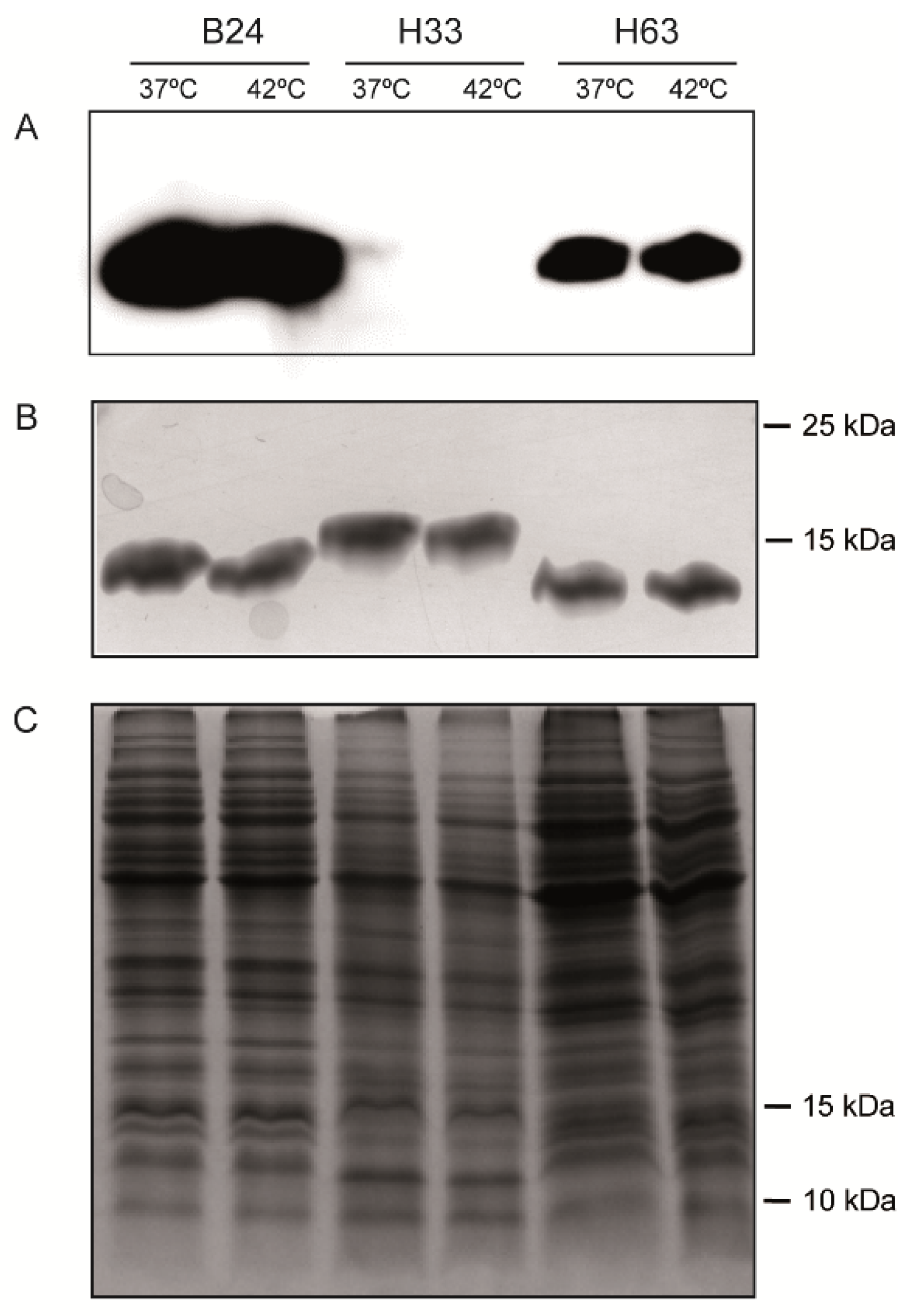
3.4. The production of LOSSIAL is Not Affected by Temperature

C. jejuni colonizes different hosts, including birds as broilers and mammals as humans. The LOSSIAL production at 42 °C and 37 °C, resembling the gastrointestinal tract temperature in broiler chickens and humans, respectively, was monitored. No difference was detected in the amount of LOS in extracts from the LOSSIAL (strains B24 and H63) or non-sialylated LOS (H33) from cultures grown at 42 °C and 37 °C (Figure 3).

3.5. Sialylation is Not Needed for Invasiveness

The role of LOSSIAL structures in C. jejuni pathogenicity is not fully understood. It was proposed that the LOSSIAL presence on the surface of C. jejuni might promote invasion of eukaryotic cells [14,22,23]. However, controversial data is reported by different authors [24,25].
The ability to invade Caco-2 cells by 38 C. jejuni strains (37 from our collection and the pathogenic 81-176 strain) was tested. Half of the strains were LOSSIAL proficient (wlaN+ or cgtB+). The invasion index was calculated and the results indicate that no correlation exists between the presence of LOSSIAL structures and invasiveness (Figure 4).

4. Discussion

The presence of LOSSIAL-related genes—wlaN and cgtB—coding for β-1,3-glycosyltransferases is correlated with the ability to trigger GBS in patients suffering from campylobacteriosis. In our study, the prevalence of these genes was determined among C. jejuni isolates from human patients and broiler chickens since the consumption of undercooked chicken meat is the most common transmission route of C. jejuni to humans. C. jejuni isolates from wild birds were also analyzed since circulation of C. jejuni strains among poultry and wild birds has been reported [26]. Furthermore, wild birds are potential sources of human campylobacteriosis [4,27]. Previous studies focused mostly on comparing human and broiler chicken isolates and no correlation between the origin of the strains and the presence of wlaN and cgtB genes was found. It has been described that 40% to 60% of human isolates and 28% to 90% of broiler chicken isolates carry LOSSIAL related genes [21,28,29,30,31,32]. Similarly, in our collection, no significant differences were found in the prevalence of LOSSIAL-related genes among human and broiler chicken isolates (28% and 22%, respectively). Remarkably, our data indicate that the prevalence of LOSSIAL-related genes is higher among the wild bird isolates (40%).
Among the LOSSIAL proficient strains, a differential distribution of the cgtB and wlaN genes between the human/broiler chicken isolates and those from wild birds was found. The wlaN gene is detected in 72% of the LOSSIAL strains from humans and broiler chickens, whereas the most prevalent LOSSIAL-related gene in wild bird isolates is cgtB (85%). The information available on the prevalence of LOSSIAL-related genes among wild bird isolates is limited. No previous data exist on the prevalence of the cgtB gene, and the presence of the wlaN gene has been estimated between 11% and 17% of wild bird isolates [33,34]. Although several reports indicated that cgtB and wlaN may coexist [14,31], our data suggest the contrary, since none of the isolates carry both genes.
A search for the presence of LOSSIAL-related genes among 136 C. jejuni genome sequences available in the NCBI database was performed (Table S1). In agreement with our data, the presence of LOSSIAL-related genes was confirmed in 40% of the strains, with wlaN and cgtB present in 22% and 18%, respectively. It should be noted that 87% of LOSSIAL positive strains were isolated from human or broiler chicken hosts and the remaining 13% from other domestic animals. Moreover, none of the genome sequences were found to simultaneously carry wlaN and cgtB.
The wlaN gene is under phase variation control [12], meaning that within a clonal population, cells can express sialylated and unsialylated LOS. This phenomenon is achieved since the number of G residues in a G-tract within the wlaN coding sequence can be randomly altered during replication, rendering a truncated or a fully functional protein. Three variants were previously described (6G, 8G, and 9G) [12,13]. Here, we revealed three new variants: a 5G-tract found in three wild bird isolates and 10- and 11G-tracts, which were both found in a broiler chicken isolate (Figure 2B and Figure S1). Remarkably, the cgtB gene carries a G-tract within the coding sequence, but a potential phase variation regulation was not reported. Here, two cgtB variants were found (Figure 2C, Figure S3): a 5G-tract, detected in most cgtB+ strains, rendering a full protein and a 6G-tract, detected only in one isolate, rendering a truncated peptide. Interestingly, the genomic sequence of the well-characterized strain 81–176 indicates the presence of a truncated cgtB gene with a 6G-tract. Overall, our data suggest that, similar to the wlaN gene, cgtB is under phase variation regulation by altering the number of residues in the G-tract. Further studies will be required to fully characterize the mechanism behind the described phenomenon.
Despite the well-established role of the cgtB and wlaN genes in triggering GBS [6], the relevance of these genes in the pathogenesis of C. jejuni during gastrointestinal infection remains unclear. The lack of thermoregulation of LOS and/or LOSSIAL suggests that its production is neither promoted nor repressed in a specific host temperature. Some authors reported a link between LOS sialylation and the ability to invade eukaryotic cells [14,22,23]. In agreement with other reports [24,25], our data suggest that no correlation exists between these two processes since no differences in the invasion index were detected between LOSSIAL and non-sialylated strains.
Overall, our data reveal a closer relationship between human and broiler chicken isolates as compared to wild bird isolates, which is in agreement with the previous characterization of the strain collection in terms of population structure, drug resistance, and virulence factor profiling [15]. The differential distribution of wlaN and cgtB genes may also indicate a host-dependent distribution of the LOSSIAL-related genes, with wlaN positively selected among broiler chickens and consequently, also among human isolates, and cgtB positively selected among wild birds.

Supplementary Materials

The following are available online at https://www.mdpi.com/2076-2607/8/3/325/s1, Figure S1: wlaN nucleotide sequence surrounding the position of G-tract in the wlaN positive strains, Figure S2: Alignment of wlaN and cgtB nucleotide sequences, Figure S3: cgtB nucleotide sequence surrounding the position of the G-tract in the cgtB positive strains, Table S1: Presence of the wlaN and cgtB genes in complete genome sequences of C. jejuni from the NCBI database.

Author Contributions

P.G.: Investigation, Formal analysis; S.P.: Investigation, Writing—original draft; Y.I.-T.: Investigation, E.M.: Resources, Writing manuscript—original draft, Writing manuscript—review and editing; F.N.: Resources, Writing manuscript—original draft, Writing manuscript—review and editing; M.C.-C.: Resources, Writing manuscript—original draft, Writing manuscript—review and editing; C.S.-O.A.: Investigation; C.B.: Conceptualization, Resources, Writing manuscript—original draft, Writing manuscript—review and editing, Funding acquisition; C.M.: Conceptualization, Investigation, Resources, Writing manuscript—original draft, Writing manuscript—review and editing, Funding acquisition. All authors have read and agreed to the published version of the manuscript.

Funding

This study was supported by the Spanish Ministry of Economy and Competitiveness (grant AGL2013-45339R), Spanish Ministry of Science, Innovation and Universities (MCIU), State Bureau of Investigation (AIE), and European Regional Development Fund (FEDER) (grant PGC2018-096958-B-I00), and the Catalan Government (grant 2017SGR499). During this work, PG was supported by a trainee teaching and research staff grant (Universitat de Barcelona). The CERCA Program from the Generalitat de Catalunya is also acknowledged.

Conflicts of Interest

The authors declare no conflict of interest.

References

  1. Blaser, M.J.; Engberg, J. Clinical and Epidemiological Aspects of Campylobacter Infections. In Campylobacter, 3rd ed.; Nachamkin, I., Szymanski, C., Eds.; ASM Press: Washington, DC, USA, 2008; pp. 99–121. [Google Scholar]
  2. EFSA. The European Union summary report on trends and sources of zoonoses, zoonotic agents and food-borne outbreaks in 2016. EFSA J. 2017, 15, e05077. [Google Scholar]
  3. Pires, S.M.; Vigre, H.; Makela, P.; Hald, T. Using outbreak data for source attribution of human salmonellosis and sampylobacteriosis in Europe. Foodborne Pathog. Dis. 2010, 7, 1351–1361. [Google Scholar] [CrossRef]
  4. French, N.P.; Midwinter, A.; Holland, B.; Collins-Emerson, J.; Pattison, R.; Colles, F.; Carter, P. Molecular epidemiology of Campylobacter jejuni isolates from wild-bird fecal material in children’s playgrounds. Appl. Environ. Microbiol. 2009, 75, 779–783. [Google Scholar] [CrossRef] [PubMed] [Green Version]
  5. Moore, J.E.; Corcoran, D.; Dooley, J.S.G.; Fanning, S.; Lucey, B.; Matsuda, M.; Mcdowell, D.A.; Mégraud, F.; Millar, B.C.; O’mahony, R.; et al. Campylobacter. Vet. Res. 2005, 36, 351–382. [Google Scholar] [CrossRef] [PubMed] [Green Version]
  6. Koga, M.; Gilbert, M.; Takahashi, M.; Li, J.; Koike, S.; Hirata, K.; Yuki, N. Comprehensive analysis of bacterial risk factors for the development of Guillain-Barré syndrome after Campylobacter jejuni enteritis. J. Infect. Dis. 2006, 193, 547–555. [Google Scholar] [CrossRef] [Green Version]
  7. Ang, C.W.; De Klerk, M.A.; Endtz, H.P.; Jacobs, B.C.; Laman, J.D.; van der Meche, F.G.A.; van Doorn, P.A. Guillain-Barre syndrome- and Miller Fisher syndrome-associated Campylobacter jejuni lipopolysaccharides induce anti-GM1 and anti-GQ1b antibodies in rabbits. Infect. Immun. 2001, 69, 2462–2469. [Google Scholar] [CrossRef] [PubMed] [Green Version]
  8. Yuki, N. Infectious origins of, and molecular mimicry in, Guillain-Barré and Fisher syndromes. Lancet Infect. Dis. 2001, 1, 29–37. [Google Scholar] [CrossRef]
  9. Parker, C.T.; Gilbert, M.; Yuki, N.; Endtz, H.P.; Mandrell, R.E. Characterization of lipooligosaccharide-biosynthetic loci of Campylobacter jejuni reveals new lipooligosaccharide classes: Evidence of mosaic organizations. J. Bacteriol. 2008, 190, 5681–5689. [Google Scholar] [CrossRef] [PubMed] [Green Version]
  10. Parker, C.T.; Horn, S.T.; Gilbert, M.; Miller, W.G.; Woodward, D.L.; Mandrell, R.E. Comparison of Campylobacter jejuni lipooligosaccharide biosynthesis loci from a variety of sources. J. Clin. Microbiol. 2005, 43, 2771–2781. [Google Scholar] [CrossRef] [Green Version]
  11. Gilbert, M.; Karwaski, M.-F.; Bernatchez, S.; Young, N.M.; Taboada, E.; Michniewicz, J.; Cunningham, A.-M.; Wakarchuk, W.W. The genetic bases for the variation in the lipo-oligosaccharide of the mucosal pathogen, Campylobacter jejuni biosynthesis of sialylated ganglioside mimics in the core oligosaccharide. J. Biol. Chem. 2002, 277, 327–337. [Google Scholar] [CrossRef] [Green Version]
  12. Linton, D.; Gilbert, M.; Hitchen, P.G.; Dell, A.; Morris, H.R.; Wakarchuk, W.W.; Gregson, N.A.; Wren, B.W. Phase variation of a beta-1,3 galactosyltransferase involved in generation of the ganglioside GM1-like lipo-oligosaccharide of Campylobacter jejuni. Mol. Microbiol. 2000, 37, 501–514. [Google Scholar] [CrossRef] [PubMed]
  13. Parkhill, J.; Wren, B.W.; Mungall, K.; Ketley, J.M.; Churcher, C.; Basham, D.; Chillingworth, T.; Davies, R.M.; Feltwell, T.; Holroyd, S.; et al. The genome sequence of the food-borne pathogen Campylobacter jejuni reveals hypervariable sequences. Nature 2000, 403, 665–668. [Google Scholar] [CrossRef] [Green Version]
  14. Müller, J.; Meyer, B.; Hänel, I.; Hotzel, H. Comparison of lipooligosaccharide biosynthesis genes of Campylobacter jejuni strains with varying abilities to colonize the chicken gut and to invade Caco-2 cells. J. Med. Microbiol. 2007, 56, 1589–1594. [Google Scholar] [CrossRef] [Green Version]
  15. Iglesias-Torrens, Y.; Miró, E.; Guirado, P.; Llovet, T.; Muñoz, C.; Cerdà-Cuéllar, M.; Madrid, C.; Balsalobre, C.; Navarro, F. Population structure, antimicrobial resistance, and virulence-associated genes in Campylobacter jejuni isolated from three ecological niches: Gastroenteritis patients, broilers, and wild birds. Front. Microbiol. 2018, 9, 1676. [Google Scholar] [CrossRef] [PubMed] [Green Version]
  16. Koolman, L.; Whyte, P.; Burgess, C.; Bolton, D. Distribution of virulence-associated genes in a selection of Campylobacter isolates. Foodborne Pathog. Dis. 2015, 12, 424–432. [Google Scholar] [CrossRef] [PubMed]
  17. Harrison, J.W.; Dung, T.T.N.; Siddiqui, F.; Korbrisate, S.; Bukhari, H.; Tra, M.P.V.; Hoang, N.V.M.; Carrique-Mas, J.; Bryant, J.; Campbell, J.I.; et al. Identification of possible virulence marker from Campylobacter jejuni isolates. Emerg. Infect. Dis. 2014, 20, 1026–1029. [Google Scholar] [CrossRef] [Green Version]
  18. Tsai, C.M.; Frasch, C.E. A sensitive silver stain for detecting lipopolysaccharides in polyacrylamide gels. Anal. Biochem. 1982, 119, 115–119. [Google Scholar] [CrossRef]
  19. Hänel, I.; Müller, J.; Müller, W.; Schulze, F. Correlation between invasion of Caco-2 eukaryotic cells and colonization ability in the chick gut in Campylobacter jejuni. Vet. Microbiol. 2004, 101, 75–82. [Google Scholar] [CrossRef]
  20. Taboada, E.N.; Acedillo, R.R.; Carrillo, C.D.; Findlay, W.A.; Medeiros, D.T.; Mykytczuk, O.L.; Roberts, M.J.; Valencia, C.A.; Farber, J.M.; Nash, J.H.E. Large-scale comparative genomics meta-analysis of Campylobacter jejuni isolates reveals low level of genome plasticity. J. Clin. Microbiol. 2004, 42, 4566–4576. [Google Scholar] [CrossRef] [Green Version]
  21. Gilbert, M.; Brisson, J.R.; Karwaski, M.F.; Michniewicz, J.; Cunningham, A.M.; Wu, Y.; Young, N.M.; Wakarchuk, W.W. Biosynthesis of ganglioside mimics in Campylobacter jejuni OH4384. Identification of the glycosyltransferase genes, enzymatic synthesis of model compounds, and characterization of nanomole amounts by 600-mhz (1)h and (13)c NMR analysis. J. Biol. Chem. 2000, 275, 3896–3906. [Google Scholar] [CrossRef] [Green Version]
  22. Naito, M.; Frirdich, E.; Fields, J.A.; Pryjma, M.; Li, J.; Cameron, A.; Gilbert, M.; Thompson, S.A.; Gaynor, E.C. Effects of sequential 81-176 lipooligosaccharide core truncations on biofilm formation, stress survival, and pathogenesis. J. Bacteriol. 2010, 192, 2182–2192. [Google Scholar] [CrossRef] [PubMed] [Green Version]
  23. Habib, I.; Louwen, R.; Uyttendaele, M.; Houf, K.; Vandenberg, O.; Nieuwenhuis, E.E.; Miller, W.G.; van Belkum, A.; De Zutter, L. Correlation between genotypic diversity, lipooligosaccharide gene locus class variation, and Caco-2 cell invasion potential of Campylobacter jejuni isolates from chicken meat and humans: Contribution to virulotyping. Appl. Environ. Microbiol. 2009, 75, 4277–4288. [Google Scholar] [CrossRef] [PubMed] [Green Version]
  24. Ellström, P.; Feodoroff, B.; Hänninen, M.-L.; Rautelin, H. Characterization of clinical Campylobacter jejuni isolates with special emphasis on lipooligosaccharide locus class, putative virulence factors and host response. Int. J. Med. Microbiol. 2013, 303, 134–139. [Google Scholar] [CrossRef] [PubMed]
  25. Asakura, H.; Brü Ggemann, H.; Sheppard, S.K.; Ekawa, T.; Meyer, T.F.; Yamamoto, S.; Igimi, S. Molecular Evidence for the thriving of Campylobacter jejuni ST-4526 in Japan. PLoS ONE 2012, 7, e48394. [Google Scholar] [CrossRef]
  26. Hald, B.; Skov, M.N.; Nielsen, E.M.; Rahbek, C.; Madsen, J.J.; Wainø, M.; Chriél, M.; Nordentoft, S.; Baggesen, D.L.; Madsen, M. Campylobacter jejuni and Campylobacter coli in wild birds on Danish livestock farms. Acta Vet. Scand. 2015, 58, 11. [Google Scholar] [CrossRef] [Green Version]
  27. Kwan, P.S.L.; Xavier, C.; Santovenia, M.; Pruckler, J.; Stroika, S.; Joyce, K.; Gardner, T.; Fields, P.I.; McLaughlin, J.; Tauxe, R.V.; et al. Multilocus sequence typing confirms wild birds as the source of a Campylobacter outbreak associated with the consumption of raw peas. Appl. Environ. Microbiol. 2014, 80, 4540–4546. [Google Scholar] [CrossRef] [Green Version]
  28. Datta, S.; Niwa, H.; Itoh, K. Prevalence of 11 pathogenic genes of Campylobacter jejuni by PCR in strains isolated from humans, poultry meat and broiler and bovine faeces. J. Med. Microbiol. 2003, 52, 345–348. [Google Scholar] [CrossRef]
  29. Ellström, P.; Hansson, I.; Nilsson, A.; Rautelin, H.; Olsson Engvall, E. Lipooligosaccharide locus classes and putative virulence genes among chicken and human Campylobacter jejuni isolates. BMC Microbiol. 2016, 16, 1–6. [Google Scholar] [CrossRef]
  30. Khoshbakht, R.; Tabatabaei, M.; Hosseinzadeh, S.; Shekarforoush, S.S.; Aski, H.S. Distribution of nine virulence-associated genes in Campylobacter jejuni and C. coli isolated from broiler feces in Shiraz, Southern Iran. Foodborne Pathog. Dis. 2013, 10, 764–770. [Google Scholar] [CrossRef]
  31. Kordinas, V.; Nicolaou, C.; Ioannidis, A.; Papavasileiou, E.; John Legakis, N.; Chatzipanagiotou, S. Prevalence of four virulence genes in Campylobacter jejuni determined by PCR and sequence analysis. Mol. Diagn. 2005, 9, 211–215. [Google Scholar] [CrossRef]
  32. Mortensen, N.P.; Kuijf, M.L.; Ang, C.W.; Schiellerup, P.; Krogfelt, K.A.; Jacobs, B.C.; van Belkum, A.; Endtz, H.P.; Bergman, M.P. Sialylation of Campylobacter jejuni lipo-oligosaccharides is associated with severe gastro-enteritis and reactive arthritis. Microbes Infect. 2009, 11, 988–994. [Google Scholar] [CrossRef] [PubMed]
  33. Wei, B.; Kang, M.; Jang, H.-K. Genetic characterization and epidemiological implications of Campylobacter isolates from wild birds in South Korea. Transbound. Emerg. Dis. 2018, 66, 56–65. [Google Scholar] [CrossRef] [PubMed] [Green Version]
  34. Gargiulo, A.; Sensale, M.; Marzocco, L.; Fioretti, A.; Menna, L.F.; Dipineto, L. Campylobacter jejuni, Campylobacter coli, and cytolethal distending toxin (CDT) genes in common teals (Anas crecca). Vet. Microbiol. 2011, 150, 401–404. [Google Scholar] [CrossRef] [PubMed] [Green Version]
Figure 1. Prevalence of wlaN and cgtB genes in C. jejuni strains from different origins. H, human patients; B, broiler chickens; W, wild birds. Statistical analyses were performed using Pearson’s chi-squared test (R Studio software). p < 0.005 was considered statistically significant (indicated by an asterisk).
Microorganisms 08 00325 g001
Figure 2. G-tract characterization within the wlaN and cgtB genes. (A) Homopolymeric G-tracts identified in wlaN and cgtB genes from human (H), broiler chicken (B), and wild bird strains (W). The different homopolymeric variants identified are indicated and the number of strains is shown between parentheses. (B) Sequence of the intragenic homopolymeric G-tracts (indicated by red lines) of the wlaN gene of the strains H11, H52, W20, and B50. The number of G residues within the G-tract are indicated between parentheses. (C) Sequence of the intragenic homopolymeric G-tracts (indicated by red lines) of the cgtB gene of the strains H58 and W27. The number of G residues within the G-tracts are indicated between parentheses. (D) Detection of LOSSIAL in purified LOS samples from cultures of the indicated strains grown onto CBA plates for 48 h at 42 °C. LOSSIAL was detected by Western blot using HRP-CT. Below, the presence for each strain of either the wlaN or cgtB gene is indicated and the G-tract variant detected. (E) Silver-stained 15% SDS-PAGE of the same samples of purified LOS as in D. (F) Coomassie-stained 15% SDS-PAGE of whole cell extracts from the cultures used to obtain the purified LOS samples analyzed in D and E. In E and F, the migration of the 10 and 15 kDa proteins from the molecular mass marker are indicated.
Microorganisms 08 00325 g002
Figure 3. Analysis of LOS from C. jejuni strains grown at either 37 °C or 42 °C. (A). Detection of LOSSIAL in purified LOS samples from cultures of the indicated strains grown onto CBA plates for 48 h at either 37 °C or 42 °C. LOSSIAL was detected by Western blot using HRP-CT. (B). Silver-stained 15% SDS-PAGE of the same samples of purified LOS as in (A). (C). Coomassie-stained 15% SDS-PAGE of whole cell extracts from the cultures used to obtain the purified LOS samples analyzed in (A,B). In (B,C), the migration of the 10 and 15 kDa proteins from the molecular mass marker is indicated.
Microorganisms 08 00325 g003
Figure 4. Relation between invasion index and LOSSIAL production in C. jejuni strains. The invasion index was calculated as the percentage of intracellular cells from the total cell-associated bacteria after 3 h infection of Caco-2 cells with the indicated strains. The average and standard deviation of three independent experiments are shown. Grey and black bars indicate strains that express or do not express LOSSIAL, respectively.
Microorganisms 08 00325 g004
Table 1. Primers used in this work.
PrimerSequence 5’–3’PCR product (bp)Reference
cgtB-FTTAAGAGCAAGATATGAAGGT740[12]
cgtB-RGCACATAGAGAACGCTACAA
wlaN-FTGCTGGGTATACAAAGGTTGTG561[16]
wlaN-RAGGTCCATTACCGCATACCA
gltA-FGCCCAAAGCCCATCAAGCGGA141[17]
gltA-RGCGCTTTGGGGTCATGCACA
Table 2. Collection of C. jejuni isolated from human patients (H), broiler chikens (B), and wild birds (W). Strains are organized attending to their ST Clonal Complexes (ST CC) and sequence types (ST). S means a singlenton ST. cgtB and wlaN positive strains are indicated (black squares) and the number of G residues found in the G-tract is indicated.
StraincgtBwlaNST-CC (ST)StraincgtBwlaNST-CC (ST)StraincgtBwlaNST-CC (ST)
H01 S (441)H62 ST-206 (572)B45 ST-354 (354)
H72 S (441)H68 ST-21 (19)W28 ST-354 (354)
B44 S (441)H06 9ST-21 (19)W54 ST-354 (354)
H53 S (531)H11 9ST-21 (21)W55 ST-354 (354)
B16 S (531)H32 9ST-21 (21)W56 ST-354 (354)
W025 S (996)H52 8ST-21 (21)B37 ST-354 (8498)
W11 S (1261)H59 8ST-21 (21)H615 ST-42 (459)
W13 S (1343)B20 9ST-21 (21)H586 ST-42 (4016)
W23 S (1343)B36 8ST-21 (21)H46 ST-443 (51)
H07 S (1710)B46 8ST-21 (21)H19 ST-443 (5799)
B10 S (1710)H12 ST-21 (50)W12 ST-446 (3552)
B26 S (1710)H34 9ST-21 (50)H48 ST-45 (45)
B35 S (1710)H35 ST-21 (50)B27 ST-45 (45)
B02 S (2331)H66 8ST-21 (50)B31 ST-45 (45)
W175 S (2351)H73 9ST-21 (50)W30 ST-45 (45)
W06 S (4355)B07 8ST-21 (50)W325 ST-45 (45)
W07 S (4355)B14 8ST-21 (50)W365 ST-45 (45)
W08 S (4355)B50 10/11ST-21 (50)W375 ST-45 (45)
B39 S (7114)H375 ST-21 (883)W405 ST-45 (45)
H33 S (8479)H605 ST-21 (883)W435 ST-45 (45)
W53 S (8514)B065 ST-21 (883)W445 ST-45 (45)
W05 ST-1034 (4001)B17 ST-21 (883)B42 ST-45 (137)
W24 ST-1275 (637)H54 ST-21 (1214)B22 ST-45 (652)
W275 ST-1275 (637)H08 ST-21 (3769)W475 ST-45 (8512)
W03 ST-1275 (1223)H02 ST-21 (4664)H18 ST-464 (464)
W105 ST-1275 (1223)H03 ST-257 (257)B08 ST-464 (464)
W155 ST-1275 (1223)H50 ST-257 (257)B15 ST-464 (464)
W185 ST-1275 (1223)H56 ST-257 (257)H38 ST-48 (48)
W21 ST-1275 (1223)B52 ST-257 (367)B09 ST-48 (48)
W22 ST-1275 (1268)B53 ST-257 (367)W29 ST-48 (48)
W04 ST-1275 (1275)B54 ST-257 (367)H71 ST-49 (49)
W09 5ST-1275 (1275)B55 ST-257 (367)H51 ST-52 (52)
W20 5ST-1275 (1275)B56 ST-257 (367)B21 ST-574 (305)
W14 ST-1275 (1292)B57 ST-257 (367)B25 ST-574 (305)
W16 ST-1275 (1292)H67 ST-257 (2254)B045 ST-607 (607)
W26 ST-1275 (3049)H69 ST-257 (2254)B415 ST-607 (607)
W19 ST-1275 (3629)B13 ST-257 (2254)H04 ST-607 (904)
W25 5ST-1275 (8511)B33 ST-283 (267)B38 ST-607 (904)
W33 ST-179 (179)B28 ST-353 (5)B47 ST-607 (904)
W41 ST-179 (220)B29 ST-353 (5)B24 8ST-607 (1707)
W345 ST-179 (2209)H63 9ST-353 (353)B19 ST-607 (7110)
W395 ST-179 (2209)B48 ST-353 (356)B51 ST-607 (7110)
W465 ST-179 (2209)H36 ST-353 (400)H05 ST-61 (61)
W48 ST-179 (2209)B05 ST-353 (400)H40 ST-61 (61)
W495 ST-179 (2209)B18 ST-353 (400)H57 ST-61 (61)
B49 9ST-206 (46)B30 ST-353 (400)H65 ST-61 (61)
H64 8ST-206 (227)B40 ST-353 (400)H70 ST-61 (61)
H09 ST-206 (572)H49 ST-354 (354)W50 ST-952 (8513)
H13 ST-206 (572)H74 ST-354 (354)W51 ST-952 (8513)
H14 ST-206 (572)B23 ST-354 (354)W52 ST-952 (8513)

