Article

Temporal Resistome and Microbial Community Dynamics in an Intensive Aquaculture Facility with Prophylactic Antimicrobial Treatment

1
Institute of Soil, Water and Environmental Sciences, Volcani Center, Agricultural Research Organization, P.O. Box 15159, Rishon LeZion 7528809, Israel
2
Department of Plant Pathology and Microbiology, The Robert H. Smith Faculty of Agriculture, Food and Environment, The Hebrew University of Jerusalem, Rehovot 91905, Israel
3
Dor Aquaculture Research Station, Fisheries Department, Israel Ministry of Agriculture and Rural Development, Dor 3082000, Israel
4
The Dead Sea and Arava Science Center, Masada 86900, Israel
5
Department of Marine Biology, Leon H. Charney School of Marine Sciences, University of Haifa, Haifa 3498838, Israel
6
CosmosID Inc., Rockville, MD 20742, USA
7
Center for Bioinformatics and Computational Biology, University of Maryland, College Park, MD 20742, USA
8
Maryland Pathogen Research Institute, University of Maryland, College Park, MD 20742, USA
*
Author to whom correspondence should be addressed.
Microorganisms 2020, 8(12), 1984; https://doi.org/10.3390/microorganisms8121984
Submission received: 5 November 2020 / Revised: 9 December 2020 / Accepted: 10 December 2020 / Published: 13 December 2020

Abstract

:
Excessive use of antimicrobials in aquaculture is concerning, given possible environmental ramifications and the potential contribution to the spread of antimicrobial resistance (AR). In this study, we explored seasonal abundance of antimicrobial resistance genes and bacterial community composition in the water column of an intensive aquaculture pond stocked with Silver Carp (Hypophthalmichthys molitrix) prophylactically treated with sulfamethoprim (25% sulfadiazine; 5% trimethoprim), relative to an adjacent unstocked reservoir. Bacterial community composition was monitored using high-throughput sequencing of 16S rRNA gene amplicons in eight sampling profiles to determine seasonal dynamics, representing principal stages in the fish fattening cycle. In tandem, qPCR was applied to assess relative abundance of selected antimicrobial resistance genes (sul1, sul2, dfrA1, tetA and blaTEM) and class-1 integrons (int1). Concomitantly, resistomes were extrapolated from shotgun metagenomes in representative profiles. Analyses revealed increased relative abundance of sulfonamide and tetracycline resistance genes in fishpond-03, relative to pre-stocking and reservoir levels, whereas no significant differences were observed for genes encoding resistance to antimicrobials that were not used in the fishpond-03. Seasons strongly dictated bacterial community composition, with high abundance of cyanobacteria in summer and increased relative abundance of Flavobacterium in the winter. Our results indicate that prophylactic use of sulfonamides in intensive aquaculture ponds facilitates resistance suggesting that prophylactic use of these antimicrobials in aquaculture should be restricted.

1. Introduction

The global spread of antimicrobial resistance (AMR) has severe implications for human health [1]. For example, approximately 70% of the agents causing hospital acquired infections in the United States are resistant to at least one antimicrobial family [2], demonstrating the severe public health risk associated with AMR [3]. The dynamics of environmental AMR are currently not well understood, but there is evidence that anthropogenic activities such as animal husbandry, aquafarming and wastewater treatment may contribute to this phenomenon [4,5,6]. AMR can propagate across microbial communities via horizontal gene transfer (HGT) through mobile genetic elements such as plasmids, transposons and integrons [7,8]. As a result, anthropogenic- and environmentally-derived bacteria can exchange antimicrobial resistance genes (ARGs), and these HGT events can be maintained under selective pressure.
The use of antimicrobial compounds in aquaculture has become a common practice throughout the world [9,10,11,12,13]. Although clinical and prophylactic application of antimicrobials have proven valuable in preventing and treating fish disease, excessive application of antimicrobials in this rapidly growing industry may facilitate increased abundance of antimicrobial resistant bacteria (ARB) and antimicrobial resistance genes (ARGs) [11]. ARB and ARGs from aquaculture can disseminate to food webs and enter the water cycle, and alter microbial community structure. Furthermore, ARGs harboring mobile genetic elements can mobilize to clinically relevant bacteria resulting into global propagation of AMR (FAO/OIE/WHO, 2006) [14]. Nakayama et al. [13] reported the ubiquitous presence of sulfonamide resistance genes coupled with remarkably low bacterial diversity at interfaces of wastewater streams from backyard-based aquaculture units in Vietnam, associated with high load of sulfonamide antimicrobials used in those systems. Tang et al. [15] suggested temperature as a key driver of the bacterial community structure in freshwater aquaculture systems. Moreover, other studies have provided valuable information about AMR in relation to microbial community composition and temporal changes. Despite several past studies, comprehensive assessment of AMR dynamics in conventional intensive aquaculture that apply antimicrobials has not been investigated.
Recently, shotgun metagenomics has contributed significantly to microbial ecology by describing structure (who is there?) and function (what are they doing?) of complex environmental microbial communities [16]. In contrast to conventional 16S rRNA gene-amplicon sequencing, shotgun genomics adds the capacity to classify and quantify specific microbial genes in real time as a function of environmental parameters, thereby elucidating associations among specific microbial communities and the host genotype/phenotypes comprising the community [17,18,19]. For example, metagenomics was applied to detect co-occurrence of specific ARGs on both viromes and bacterial genomes in an experimental aquaculture facility, suggesting that transduction of ARGs takes place in these systems [20].
In a previous culture-based study targeting Aeromonas in the water columns of two commercial-scale aquaculture facilities, we demonstrated that sulfonamide and tetracycline gene abundance were positively correlated to sulfadiazine/trimethoprim and oxytetracycline that were applied prophylactically and clinically, respectively. In contrast, these isolates displayed lower resistance to antimicrobials (chloramphenicol, ceftriaxone and gentamicin) that were not used [12]. The aim of this study was to understand the structural and functional dynamics of the resistome and microbiome in the water column of an intensive aquaculture system employing antimicrobials prophylactically. Standard water monitoring methods were integrated with advanced culture-independent molecular and metagenomic platforms.

2. Materials and Methods

2.1. Sampling and DNA Extraction

Water samples for 16SrRNA gene amplicon and qPCR analysis were collected from fishpond-03 (FP-03) and reservoir (Res) of the Dor aquaculture research station, Israel, details of which have previously been described [12] and sample wise analysis details were tabulated below (Table S1). Altogether, eight profiles were sampled for the duration of a Silver Carp (Hypophthalmichthys molitrix) fattening cycle: 6 July 2015; 4 August 2015; 18 August 2015; 31 August 2015; 14 October 2015; 22 December 2015, 10 February and 9 March 2016. All samples were collected in triplicate (except 6 July, 4 August, and 18 August, when for logistical reasons samples were collected in duplicate and 10 February where only a single replicate was sampled). Samples were immediately filtered (25–30 mL depending on turbidity) onto glass microfiber filters (Whatman GF/F; 0.7µ; dia 25 mm) for nutrient analyses, and subsequently onto 0.22µ polycarbonate filters for DNA extraction and subsequent molecular analyses. Filters were preserved in conservation buffer (50 mM Tris, pH = 8.3, 0.75 M Sucrose, 40 mM EDTA), transferred on ice to the laboratory within 2 h and stored at −80 °C until DNA extraction. Subsequently, frozen filters with conservation buffer added to the tubes were transferred on ice to the Biomedical Core Facility of the Technion for robotic DNA extraction. The first step of extraction included chemical and mechanical extraction as follows: filters were thawed, centrifuged for 10 min at 15,000× g and the conservation buffer removed. Next, Blood and Tissue Kit lysis buffer (Qiagen) was added, and the samples were mechanically extracted using two 3 mm stainless steel beads and a speed of 30/s for 1.5 min on the TissueLyser LT (Qiagen). An amount of 30 µL of lysozyme was added and the tubes incubated at 37 °C for 30 m, after which 25 µL and 200 µL proteinase K and buffer AL (Qiagen) were added, respectively, and the tubes incubated at 56 °C for 1 h on a shaker. Finally, the tubes were centrifuged for 10 min at 5000× g and the upper liquid was transferred to a new 2 mL Eppendorf tube for further extraction using the Qiacube robot (Qiagen) and the DNeasy Blood and Tissue Kit. Extracted DNA was maintained at −20 °C serving as working stock, and at −80 °C as preserved stock for 16S rRNA gene amplicon sequencing and shotgun metagenomic analyses described below.

2.2. Physiochemical Analyses

Analysis of ammonia, nitrite and nitrate was performed using an autoanalyzer continuous flow system (Lachat QuikChem® 8500 Series 2 Flow Injection Analyzer, Milwaukee, WI, USA). The pH was measured with a pH meter (EUTECH Instruments pH 700), while average temperature and precipitation at the target site during the study period was obtained from the Israel Meterological Service database (https://ims.data.gov.il/).

2.3. Sequencing of 16S rRNA Gene Amplicons

All 38 (19 FP-03 and19 Res) environmental DNA samples were amplified separately targeting the V3-V4 16S rRNA gene region using primer set CS1_341F and CS2_816R [21] with a two-stage “targeted amplicon sequencing (TAS)” protocol [22,23]. The primers contained 5′ common sequence tags (known as common sequence 1 and 2 or CS1 and CS2) as described previously by Moonsamy et al. [24]. The PCR reaction was conducted in a final volume of 25 µL and PCR conditions were as follows: 1 cycle of 95 °C for 5 min, followed by 28 cycles of 95 °C for 30 s, 50 °C for 30 s and 72 °C for 60 s with final elongation of 72 °C for 5 min. Subsequently, sond PCR amplification was performed in 10µl reactions in 96-well plates. A mastermix for the entire plate was prepared using MyTaq HS 2X mastermix. To each well, a separate primer pair with a unique 10-base barcode was added, obtained from the Access Array Barcode Library for Illumina (Fluidigm, South San Francisco, CA; Item# 100-4876). These Access Array primers contained CS1 and CS2 linkers at the 3′ ends of the oligonucleotides. Cycling conditions were as follows: 95 °C for 5 min, followed by 8 cycles of 95 °C for 30 s, 60 °C for 30 s and 72 °C for 30 s. A final, 7-min elongation step was performed at 72 °C. PCR products were purified using SequalPrep plates (Life Technologies) according to manufacturer’s instructions. Subsequently, the PCR products were quantified using a Quant-iT PicoGreen dsDNA Assay Kit (Thermo Fisher) implemented on a Genios Pro Fluorescence microplate reader (Tecan). PCR products were pooled using PicoGreen quantification on an epMotion5075 liquid handling workstation (Eppendorf).
The pooled libraries, along with a 15% phiX spike-in, were loaded to a MiSeq v3 flow cell, and sequenced using an Illumina MiSeq sequencer. Fluidigm sequencing primers, targeting CS1 and CS2 linker regions, were used to initiate sequencing. De-multiplexing of reads was performed on the instrument. Library preparation and pooling was performed at the DNA Services (DNAS) facility, Research Resources Center (RRC), University of Illinois at Chicago (UIC). Sequencing was performed at the W.M. Keck Center for Comparative and Functional Genomics at the University of Illinois at Urbana-Champaign (UIUC). Raw sequences were processed through downstream analysis, using the below mentioned bioinformatic tools.

2.4. Metagenome Analysis Employing Shotgun Sequencing

Extracted DNA was prepared and processed for sequencing using Nextera XT kit (Illumina, San Diego, CA, USA) according to the manufacturer’s instructions, and sequenced at the Genome Research Division Sequencing Core, University of Illinois, Chicago, USA. After processing, libraries were assessed for size using an Agilent TapeStation 2000 automated electrophoresis device (Agilent Technologies, Santa Clara, CA, USA) and for concentration by a Qubit flurometer (Thermo Fisher Scientific Inc., Waltham, MA, USA). Libraries were pooled in equimolar ratio and sequenced on a mid-output kit using an Illumina NextSeq500 sequencer, with paired-end 2 × 150 base reads.

2.5. Bioinformatic Analysis

2.5.1. 16S rRNA Gene Amplicon Analysis

Amplicon sequences were subjected to an initial quality control step followed by bioinformatic analysis using a pipeline that integrated tools from QIIME v.1.91 [25] and MOTHUR v.1.33.3 [26]. Briefly, sequences containing more than one ambiguous base, those having a homopolymer length longer than 8bp, and sequences with an average quality score below 25 were removed from analysis. We included fragments ranging from 440 to 475 bp after adjacent PCR primer prioritizing quality of the sequences. Sequences were aligned using the SILVA reference database [27] and potential chimeric sequences were detected and removed using the chimera.uchime of MOTHUR. Sequences were assigned to operational taxonomic units (OTUs), based on ≥97% sequence similarity cutoff. Alpha diversity of the samples was calculated using the Simpson diversity index, while the Bray–Curtis dissimilarity matrix, which incorporates both membership and abundance, was used to interpret beta diversity among the samples and was calculated using the QIIME and MOTHUR combined pipeline.

2.5.2. Shotgun Metagenome Analysis

Four selectedFP-03 (July 2015; August 2015; December 2015, and March 2016) and four previously sampled reservoir/Res (July 2013; August 2013; January 2014, and March 2014) profiles were selected for shotgun metagenome analysis. Two annotation pipelines were used to interpret the shotgun metagenomics data. The COSMOSID pipeline (app.cosmosid.com; [28,29,30]) was used to elucidate microbial (bacteria, virus, fungus and protist) community composition of the FP-03 water column samples, identify specific potential pathogens and pinpoint dominant ARGs in the water column samples. This pipeline facilitates rapid identification and quantifies microbial species, even from unassembled short NGS reads. A non-automated custom pipeline was also employed in the analysis for characterizing ARGs in the targeted samples. For the latter pipeline, raw reads were quality checked using FastQC, followed by trimmomatic [31]. BLAST analysis was performed using a downloaded version (1.1.3) of the Comprehensive Antibiotic Resistance Database (CARD). All reads were annotated and identified as ARG, according to its best BLAST hit [32], with minor modifications as follows: BLASTx with E-value cut-off set at ≤10−6, considering ≥80% similarity and ≥30 bit score to establish the ARG profile as well as abundance. The metagenomic data were deposited in the NCBI Sequence Read Archive under accession number SRP101485.

2.6. Quantification of ARGs Using qPCR

Amongst extracted DNA, eight samples (one each from all sampling profile as detailed in Table S1) of FP-03 were prepared as described above, and subjected to quantitative PCR amplification. The analysis was targeting five ARGs: sul1, sul2, dfrA1, tetA and blaTEM; the class-1 integron integrase gene intI1; and the 16S rRNA gene (which provides a rough estimate of total bacterial abundance in a given sample). Fast Real Time PCR was employed (Applied Biosystems). PCR was conducted using a final volume of 20 μL with master mix reagents as follows. Amplification programs were: 1 cycle of 95 °C for 10 min, 40 cycles of 95 °C for 15 s and 60 °C for 60 s with POWER and SELECT master mix reagents (Thermo Scientific), following 1 cycle of 95 °C for 5 min, 40 cycles of 95 °C for 5 s and 60 °C for 30 s with FAST SYBR green reaction master mix reagents (Thermo Scientific). Standards for respective genes were in a tenfold dilution series (gene copy numbers of 1 × 101, 1 × 10², 1 × 10³, 1 × 10⁴, 1 × 10⁵ and 1 × 10⁶/reaction) run with prepared Dor FP-03DNA samples. Each sample was analyzed using three technical replicates for each run. Primer details are provided in Table 1. Quantitative analysis of unknown samples was performed using standard curves generated from the amplification plot of known concentrations of the respective standard [33]. Efficiencies of all qPCR reactions ranged from 95% to 105%, except for sul1. Limit of detection for all of the screened ARGs was 10 copies.

2.7. Statistical Analyses

Statistical analyses were performed to determine correlations between abiotic factors, such as temperature, pH and average precipitation, with relative abundance of ARGs estimates made by qPCR and the bacterial communities delineated as OTUs at phylum as well as genus level. Pearson Correlation Coefficient was calculated to reflect variance among the datasets with respect to significance. Similarly, Canonical Correspondence Analysis (CCA) was performed to determine relative impact among the above-mentioned factors on each other. The relative abundance of ARGs was transformed to log (base 10) for ease of analysis. All statistical analyses were performed by PAleontologicalSTatistics software (PAST, version 3.08). Heatmaps were generated using the Python 3.1 Seaborn data visualization package. PERMANOVA test was conducted by “vegan” package in “R” [39].
The 16S rRNA gene amplicon sequencing data from nineteen FP-03 and Res samples and the shotgun metagenomic data from the eight FP-03 and Res samples were collectively deposited to the NCBI Bioproject repository as project # PRJNA375891entitled “Temporal analysis of intensive aquaculture water column”.

3. Results

3.1. Nutrient Analysis of Water Samples

Variable levels of ammonia (0.05–0.96 mg/L), nitrite (0.01–0.19 mg/L) and nitrate (0.04–5.73 mg/L) were observed in both FP-03 and Res water columns (Figure S1, Table S2). Despite significantly higher fish densities, ammonia and nitrate levels measured in FP-03 were similar or only slightly higher than those measured in the Res.
Precipitation, temperature and pH profiles for samples collected in this study are shown in Table S2. Rainfall was highest in February 2016 (63.7 mm), and there was no rainfall in July and August, 2015. Maximum and minimum temperatures were recorded in August 2015 (27.8 °C) and December 2015 (14.7 °C), respectively. The pH of the water samples ranged from 7.7 to 8.2.

3.2. Microbial Community Analysis

Evaluation of bacterial community structure in the FP-03 water column at eight selected time points during the fish fattening cycle was achieved by targeting 16S rRNA gene amplicon sequences. A total of 2,429,487 Illumina MiSeq-generated reads were obtained for 19 FP-03 and 19 Res samples in eight temporal profiles. Each sample contained between 40,000 and 160,000 reads with an average of 51,600 good quality reads per sample. Alpha diversity of the bacterial communities in the FP-03 and Res water columns at various sampling times was estimated using the Simpson diversity index and species richness (Figure 1). In general, we did not observe statistically significant differences in diversity for water column samples. However, the diversity of FP-03 was generally lower in the summer and relatively higher in the fall and winter. In the reservoir, lower diversity and species richness was observed in the summer and winter, respectively, relative to the same seasonal profiles in FP-03.
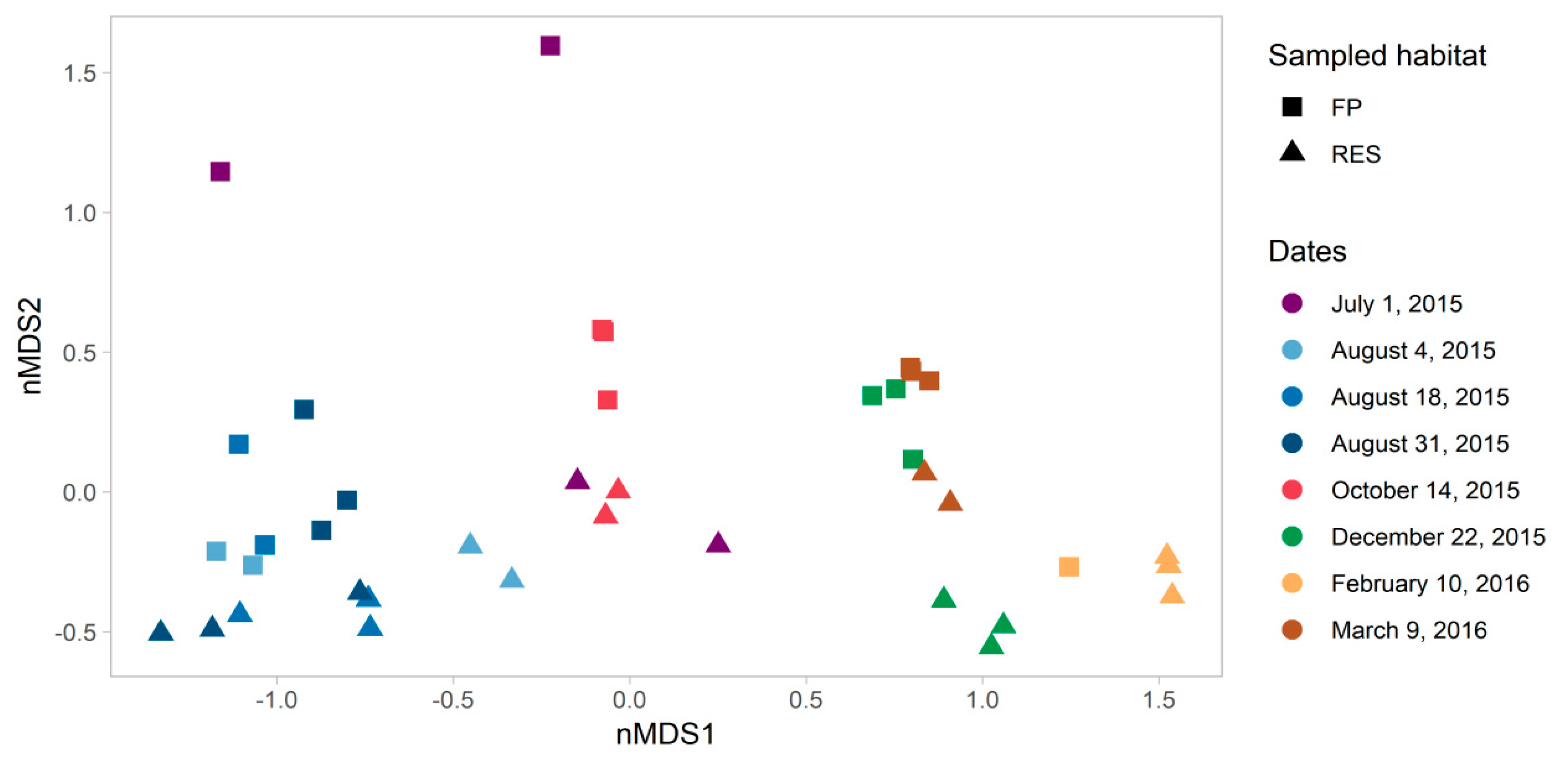
We applied non-metric multidimensional scaling (NMDS) to observe dissimilarities in microbial community composition among all the sampling profiles (Figure 2). Samples were grouped based on date of sampling (PERMANOVA of Bray–Curtis dissimilarity matrix, F = 8.59, p value = 0.001) and specifically segregated based on seasonality (summer and autumn—from July to October, versus winter and spring—December to March), at the first NMDS axis. This seasonal trend is supported by shifts in specific bacterial taxa described in the paragraph below. Although seasonality was the stronger driver, we also observed a statistically significant difference between the FP-03 vs. Res samples (FP vs. RES, F = 2.866, p value = 0.017).
At the phylum level, Proteobacteria, Actinobacteria, Bacteriodetes [40] and Verrucomicrobia were abundant in the water columns of both the FP-03 and Res, and Plantomycetes was also present at lower levels (Figure S2). Cyanobacteria were abundant in the summer profiles, but almost completely absent in the winter. Genus level taxonomical analysis revealed that these were primarily Microcystis, although Synechococcus were present in several Res profiles, inversely correlated to the relative abundance of Microcystis (Figure 3). The facultative phototrophic purple sulfur bacterium Rhodobacter was abundant in both water column samples throughout the year, as was the metabolically versatile genus Sphingomonas. Pseudomonas spp. was ubiquitous in both water column systems, but apparently inversely correlated with Microcystis blooms. Novosphingobium, Bradyrhizobium and Fluviicola spp. were present in all samples, but at lower relative abundances. Flavobacterium spp. were abundant in winter in both water column microbiomes, but almost undetectable in summer.
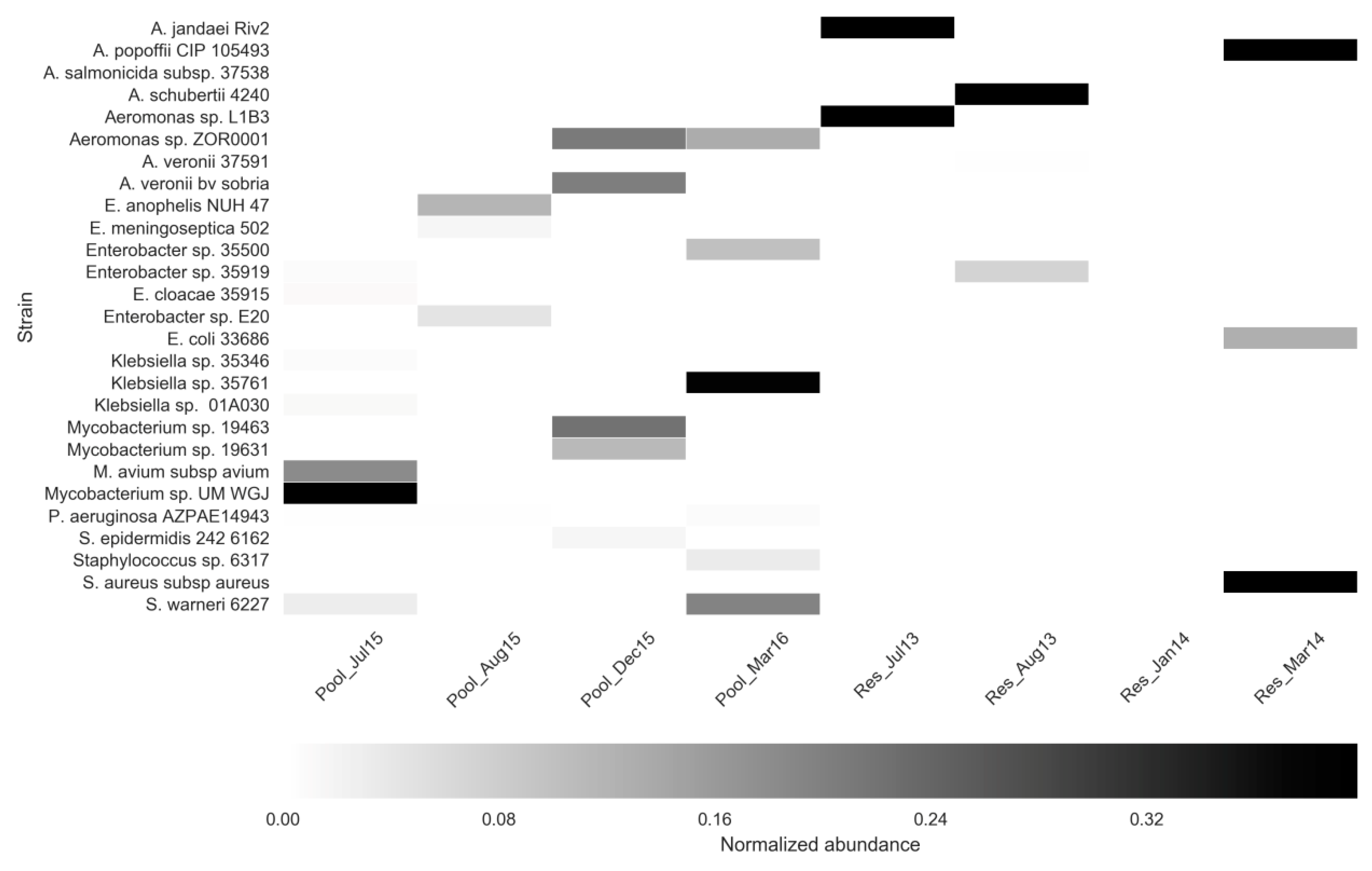
To identify specific clinically-associated bacteria, eukaryotes and viruses in the FP-03 and Res water columns, as mentioned above in the materials and methods section, four FP-03 (July 2015; August 2015; December 2015, and March 2016) and four previously sampled Res (July 2013; August 2013; January 2014, and March 2014) samples were analyzed by shotgun metagenome analysis using the COSMOSID pipeline. Abundance of human/animal-associated bacterial pathogens and commensals (Figure 4) included Mycobacterium, Aeromonas, Elizabethkingia and Enterobacter spp., as well as Escherichia coli and Staphylococcus spp. With the exception of Mycobacterium UM WGJ (isolated from a suspected tuberculosis patient in Malaysia) of these potential pathogens comprised less than 1% of the total bacterial community. Most of these were unique to a specific profile, suggesting these bacteria represent transient components of the water column microbiome.
COSMOSID bioinformatics were further applied to assess composition of viruses (Figure S3), protists (Figure S4) and virulence genes in the eight targeted FP-03 and Res water column samples. Almost all of the viruses that were detected were bacteriophages and the vast majority were Microcystis phages, corresponding to the Microcystis blooms observed in the summer in both the FP-03 and Res. Bacteriophages associated with Pelagibacter and Enterobacteriacae also were ubiquitous in most of the samples. Protist diversity was significantly higher in FP-03 water. While Thalassiosira, Pseudoperonospora cubensis and Paramecium biaurelia were ubiquitous in water at both sites, Acanthamoeba palestinensis, Hammondia hammondi, Reticulomyxa filosa and Acanthamoeba mauritaniensis were highly abundant and unique in the FP-03. A myriad of virulence genes associated with the Klebsiella pneumonia, Salmonella typhimurium, Pseudomonas aeruginosa, Escherichia coli and Proteus mirabilis were detected in all of the samples, but were for the most part substantially more abundant in the FP-03 than in the Res samples.

3.3. Assessment of ARGs Using Shotgun Metagenomics and qPCR

Relative abundance of ARGs in FP-03 and Res water was assessed using shotgun metagenomics and analyzed with both the COSMOSID and the CARD bioinformatic platforms, respectively. Subsequently, quantitative estimates of selected clinically relevant ARGs in the FP-03 water samples were conducted using qPCR.
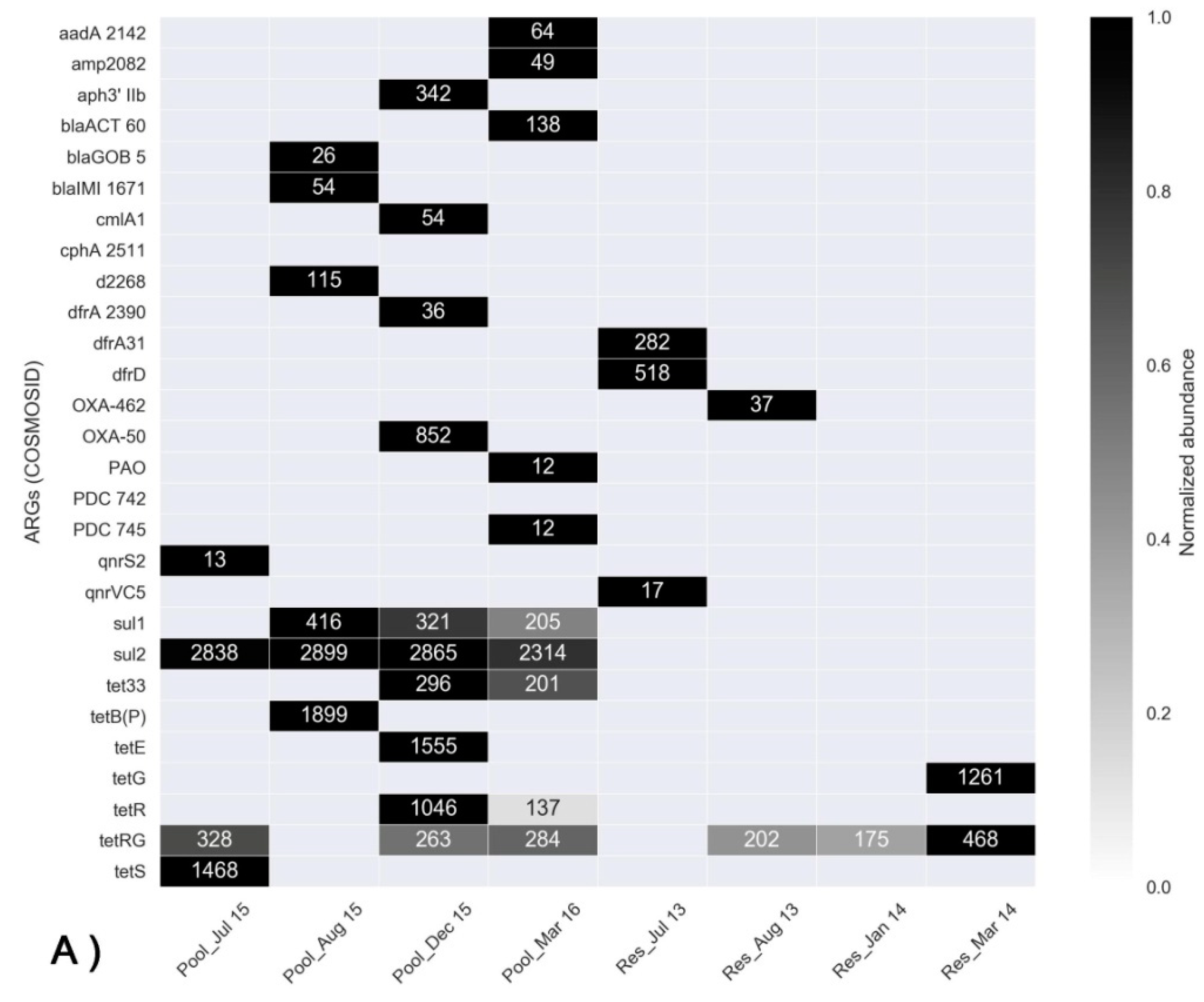
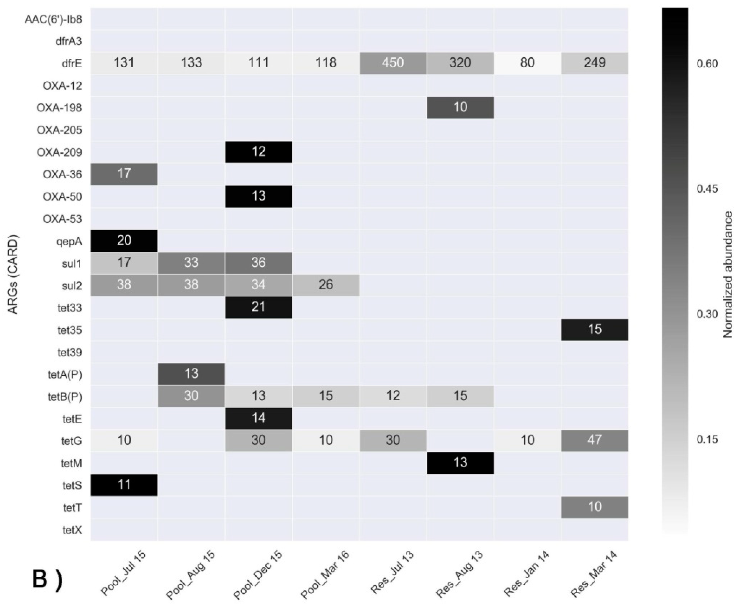
A large fraction of the identified ARGs (Table S3) encode for efflux pumps, which are often intrinsic and generally not associated with mobile genetic elements. We, therefore, focused on genes that can be potentially mobile. The most abundant of these ARGs identified using the COSMOSID and CARD bioinformatic pipelines are shown in the heatmaps in Figure 5A,B, respectively. Collectively, more ARGs were detected in the FP-03 compared to the Res water column samples. Sulfonamide resistance genes (sul1 and sul2) were ubiquitous in the FP-03 samples, but were almost completely absent in the Res samples. While sul2 was present in all of the profiles including July 2015, prior to antimicrobial administration, sul1 was most abundant subsequent to the prophylactic treatment of fish with trimethoprim-sulfamethoxazole. In contrast, trimethoprim (administered prophylactically, along with sulfamethoxazole) resistance genes were not correlated with antimicrobial use and a substantial level of discrepancy in the detection of trimethoprim resistance genes occurred between the applied bioinformatic pipelines. COSMOSID analysis identified trimethoprim resistance genes dfrA31 and dfrD, but only in the July 2013 Res samples, whereas CARD analysis showed high relative abundance of dfrE in all of the FP-03 and Res water samples. Based on COSMOSID analyses, tetracycline resistance genes were substantially more abundant in the FP-03 (with the exception of tetG in the March 2014 Res profile). This was, however, not the case in the CARD analysis, where a substantially higher number of tetracycline resistance genes was identified. The β-lactamase gene blaOXA was detected in all samples by CARD analyses, but only in a few samples by COSMOSID analysis and no substantial differences were observed between the FP-03 and Res samples. Collectively, these analyses demonstrate that different bioinformatic platforms can produce different results on the same data. However, both platforms clearly indicated profuse and ubiquitous presence of sulphonamide resistance genes (sul1 and sul2) in the FP-03 and almost complete absence of these genes in the Res. This suggests a correlation between these genes and the prophylactic antimicrobial use in the FP-03.
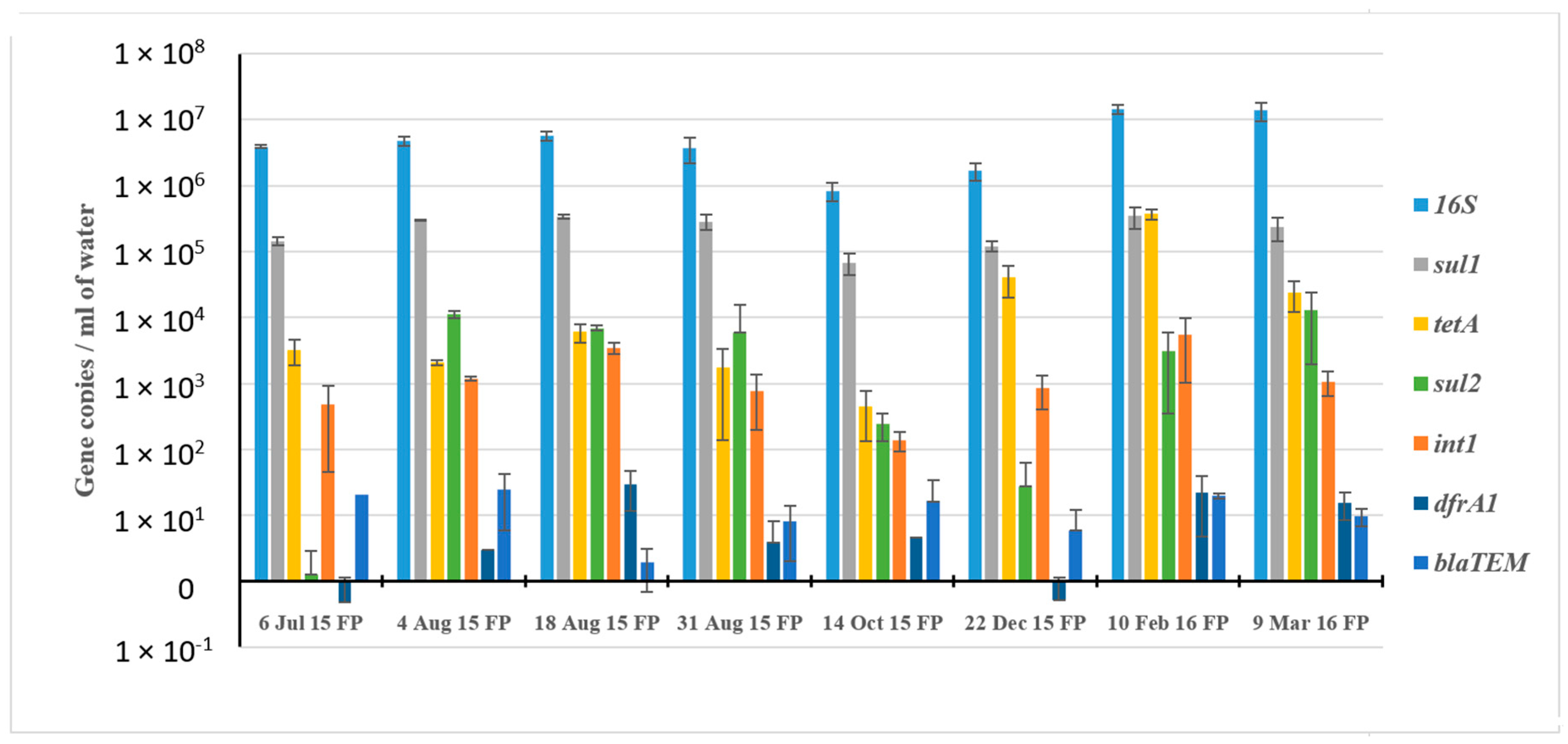
We subsequently applied quantitative PCR (qPCR) to assess the relative abundance of selected ARGs in water samples from FP-03 in eight profiles spanning the duration of the fish fattening cycle from July 2015 to March 2016 (Figure 6). Efficiencies of all of the qPCR reactions ranged from 95% to 105%, except for sul1. Limit of detection for all ARGs was 10 copies. The abundance of sul1 (ranging from 6.8 × 10⁴ to 3.5 × 10⁵ copies mL−1) substantially fluctuated during the fish fattening cycle, whereas sul2 abundance was extremely low in the pre-stocked June profile, but was markedly increased in the post-stocking water column samples. The abundance of tetA and int1 (ranging from 1.4 × 10² to 3.7 × 10⁵ gene copy numbers) varied in the different sampling profiles, but the abundance of tetA was increased substantially in the December and February profiles. Levels of dfrA1 and blaTEM were much lower than the other ARGs (4.9 × 10⁻¹ to 3 × 101), and no clear temporal trend in the abundance of these genes was observed.

3.4. Correlation Analysis

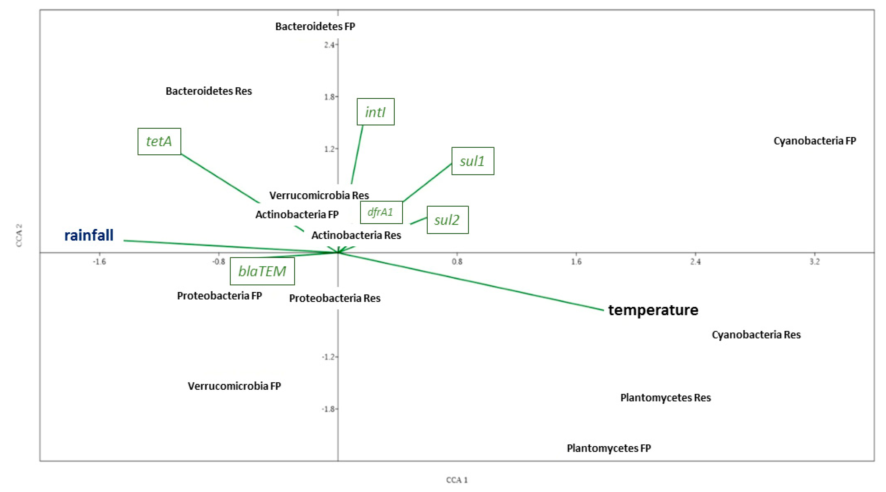
Canonical Correspondence Analysis showed complex relationships between and among specific environmental parameters (temperature, pH, average precipitation), ARG abundance based on qPCR and bacterial community composition from amplicon sequencing analyses (Figure 7, Table S4), of both FP-03 and Res water samples. A positive correlation was observed between targeted ARGs encoding resistance to antimicrobials used in FP-03 (sul1, tetA and dfrA1) and the class 1 integron integrase gene (int1; p < 0.01; R = 0.7282/sul1, 0.6322/tetA, 0.7945/dfrA1; n = 8 for each gene). Furthermore, positive correlation was observed between members of Bacteroidetes spp. in FP-03 water and ARGs sul1, tetA and int1 (p < 0.05; R = 0.7172/sul1, 0.869/tetA, 0.9279/int1; n = 8). Presence of Flavobacterium spp. was strongly correlated with tetA abundance in both water columns (p < 0.01; R = 0.9618/FP-3, 0.8819/Res; n = 8), and was positively correlated with int1 in FP-03 water (p < 0.05; R = 0.755; n = 8). Similarly, Fluviicola spp. was positively correlated with tetA (p < 0.01; R = 0.8485; n = 8) and int1 in FP-03 (p < 0.05; R = 0.8209; n = 8), whereas in Res water Pseudomonas spp were positively correlated with tetA (p < 0.01; R = 0.9171; n = 8). Cyanobacteria were positively correlated with temperature (R = 0.8011/FP-3, 0.8961/Res; n = 8), while relative abundance of Proteobacteria (p < 0.01; R = 0.836; n = 8) in FP-03 water was positively correlated with precipitation. At the genus level, Microcystis was positively correlated with temperature at both sites (p < 0.05; R = 0.8166/FP-3, 0.7163/Res; n = 8), while the opposite held for Flavobacterium (p < 0.05; R = -0.7584/FP-3, -0.8618/Res; n = 8).

4. Discussion

The objective of this study was to evaluate ARG and microbial community dynamics in a freshwater aquaculture pond for the duration of a full annual fish fattening cycle, following prophylactic administration of antimicrobials. Similar to previous studies that assessed ARG abundance in aquaculture systems [41,42], shotgun metagenomic analyses indicated that sulfonamide resistance genes were strongly correlated to antimicrobial use, as both sul1 and sul2 were relatively copious in FP-03 water columns, but almost completely absent in the non-stocked Res water columns. While sul2 was abundant in all of the FP-03 profiles, the relative abundance sul1 substantially increased subsequent to prophylactic application of trimethoprim/sulfomethoxazole at the beginning of the fish fattening cycle, suggesting that the relative abundance of bacteria harboring this gene increases upon exposure to sub-therapeutic sulphonamide concentrations. Correlations between prophylactic antimicrobial use in aquaculture and enhanced abundance of ARGs has been previously reported by Gao et al. [43] and Xiong et al. [1]. This phenomenon was supported by a previous study we conducted that linked prophylactic trimethoprim/sulfomethoxazole use in aquaculture ponds to sulphonamide resistance in Aeromonas spp. [12].
The ubiquitous presence of sul2 in all of the FP-03 profiles (including the pre-stocked Jul15 profile not exposed to antimicrobials) and its complete absence in the adjacent non-stocked Res, suggests that the FP-03 sediment may be a reservoir for sulphonamide-resistant bacteria. Previous studies have reported significant accumulation of ARGs in sediments impacted by aquaculture, which were subsequently characterized as potential ARG repositories [1]. Kobayashi et al. [44] and Suzuki et al. [45] reported similar augmented resistomes in Mekong river sediments impacted by aquaculture.
Contrary to sulphonamide resistance genes, we did not observe a clear correlation between prophylactic trimethoprim/sulfomethoxazole use and the propagation of trimethoprim resistance genes. Overall, there were significant discrepancies in the distribution and relative abundance of trimethoprim resistance genes in the water column samples depending on the type of analysis conducted. COSMOSID detected high abundances of dfrD in the July 13 Res water profile (but not elsewhere), whereas qPCR and CARD analyses reported dfrA and dfrE, respectively, in all profiles with no clear trend in relative abundance. These discrepancies can stem from a wide range of factors discussed below.
Class 1 integrons play an important role in carriage and dissemination of ARGs [46], and sul1 is often ubiquitous in these elements. The strong positive correlation between sul1 and dfrA1 and class-1 integron integrase genes (int1) observed in the qPCR analysis, suggests that these two ARGs are harbored on class-1 integrons. Nonetheless, more robust analyses specifically targeting integron gene cassettes [47] need to be conducted in order to confirm this assumption.
Results of both the qPCR and the COSMOSID analysis of metagenomic data showed increased abundance of tetracycline resistance genes in Dec15 and Feb16 FP-03 water samples (not reflected in the CARD metagenomics analysis). While oxytetracycline was applied to quarantined fish in some of the fishponds linked to FP-03, and has been used in quarantined fish in FP-03 in previous seasons, to the best of our knowledge, unlike trimethoprim and sulfomethoxazole, it was not used directly during this study. Therefore, it is probable that oxytetracycline concentrations in the water column are extremely low and most likely do not select for tetracycline resistance.
While the study provides strong evidence linking prophylactic use of trimethoprim/sulfomethoxazole to increased abundance of sulfonamide resistance genes, discrepancies between the relative abundances of sul1 and sul2, the various trimethoprim resistance genes and other ARGs between qPCR and the shotgun metagenomic analysis highlights the complexity of elucidating resistome dynamics in complex environments. Inconsistencies in relative abundance of ARGs detected using shotgun metagenomics rather than PCR have been reported previously [48]. Andersen et al. [49] suggested that they may stem from different detection sensitivity and potential inhibition associated with either of the methods, but concluded that shotgun metagenomics was more accurate in detecting spiked DNA within complex microbial communities. Collectively, we believe that the shotgun metagenomic analyses of the ARGs is more accurate than the qPCR, considering the potential biases associated with PCR amplification and the fact that primers can potentially miss a large fraction of the targeted ARGs.
Interestingly, we also found substantial inconsistencies between the CARD and COSMOSID outputs from the same metagenomic data. This is clearly associated with differences in both the ARG databases used for comparison and the stringencies of the algorithms used in the two annotation pipelines. The COSMOSID pipeline applies a highly curated database and positive hits are based on multiple targets, and therefore, it can be assumed that the results obtained using the COSMOSID pipeline are more reliable for identifying clinically relevant ARGs. Conversely, the CARD database is more exhaustive and includes a broader range of ARGs from environmental sources, and therefore, it may provide a more holistic overview of environmental resistomes.
When assessing ARG distribution and abundance in the analysed water columns, it is vital to consider the microbial community composition in addition to selective pressure. Certain phyla harbor specific ARGs and, therefore, fluctuations in ARG abundance can be dictated by shifts in bacterial community composition regardless of selective pressure. It is highly probable that the cyanobacterial blooms observed in the summer profiles influence microbial community composition and henceforth the scope and abundance of detected ARGs. Guo et al. [50] suggested that cyanobacterial blooms have an inhibitory effect on ARG abundance in freshwater microbiomes, but considering the above, it is not clear if this is a cause or effect.
Seasonal factors (i.e., temperature and radiation) were the primary drivers of the microbial communities in the FP-03 and Res water columns. However, significant differences in the microbiomes of the two water columns suggests other “fish-associated” factors such as oxygen saturation, organic load and residual concentrations of antimicrobials also play a role in defining the microbial community composition. Nakayama et al. [13] observed that the water column microbiota of different aquaculture systems are strongly dictated by aquaculture management practices. In one report, Fouladkhah et al. [51] stated that the rise in environmental temperatures facilitates an increase in infectious disease agents. The multifactorial complexity of aquaculture ponds makes it difficult to determine precisely which specific factors affect microbial community dynamics in the water column. Nonetheless, we were able to identify trends and compile hypotheses, based on associated metadata. For example, at the genus level, algal blooms in FP-03 predominantly comprised Microcystis, whereas Res water also contained high levels of Synechococcus. This finding is supported by previous reports indicating that Microcystis thrives in ecosystems rich in nutrients (such as intensive aquaculture ponds), whereas Synechococcus proliferates in more oligotrophic ecosystems [52,53].
Sphingomonas was positively correlated with Microcystis blooms, based on its higher relative abundance in August profiles from FP-03 as compared to the Res water column. This may be associated with the capacity of this genus to detoxify microcystins [54]. A previously study that investigated temperature effects on microbiomes of German lakes found that Sphingomonadales were an integral element of Microcystis sp. blooms, corroborating results from this study [55].
Specific screening of the shotgun metagenomes using the curated COSMOSID platform identified substantially higher loads of potential human and animal pathogens in the FP-03 water column relative to that of the Res. These included species belonging to Mycobacterium, Aeromonas, Elizabethkingia, Klebsiella, Staphylococcus and Enterobacter. Nonetheless, none of these strains were identified in more than one profile, and generally, they were detected at very low concentrations, suggesting that they may not have significant epidemiological relevance. This was true for Aeromonas and salmonicida (detected in the July profile), the causative agent of skin lesions in carp and koi [56], and a major pathogen in fishponds in the sampled region.
To the best of our knowledge, this is the first study to assess comprehensively the temporal microbiome and resistome dynamics of intensive aquaculture ponds that use antimicrobials. Although data are quite complex, we provide strong evidence that prophylactic use of trimethoprim/sulfomethoxazole in aquaculture systems facilitates increased relative abundance of sulfonamide resistance genes. Furthermore, we provide evidence of a highly dynamic microbial community in aquaculture ponds that are strongly influenced by both photosynthetic and heterotrophic bacterial communities in the water column. To fully understand the clinical risks associated with intensive aquaculture systems using antimicrobials, future studies should assess presence of other clinically-relevant ARGs in class 1 integrons and their association with mobile genetic elements.

Supplementary Materials

The following are available online at https://www.mdpi.com/2076-2607/8/12/1984/s1, Figure S1: Temporal analysis of ammonia, nitrite and nitrate in the fishpond-3 (FP) and the reservoir (Res) water columns at the Dor aquaculture research station. Figure S2: Relative abundance of dominant bacterial phyla in the Dor fishpond-3 (FP) and reservoir (Res) water columns. Figure S3: Relative abundance of predominant viruses in fishpond-3 (Pool) and the reservoir, based on shotgun metagenomic data analysed using the highly curated COSMOSID pipeline. Figure S4: Relative abundance of predominant protists in fishpond-3 (P) and the reservoir (R), based on shotgun metagenomic data analysed using the highly curated COSMOSID pipeline. Table S1: Samples description. Table S2: Physicochemical parameters at the study site during sampling. Table S3: Antibiotic resistance gene hits extrapolated from fishpond-3 and reservoir shotgun metagenomes using COSMOSID and CARD bioinformatic pipelines. Genes with less than ten hits are not shown. Table S4: Pearson’s correlation coefficients followed by Canonical Correspondence Analysis (CCA) between phyla (with relative abundance >5.0% at any of the sampling profile), ARGs, integrase genes and environmental parameters (temperature and precipitation).

Author Contributions

H.J.P. participated in experimental design, performed most of the experiments and actively participated in data analysis and in manuscript preparation; J.G., A.Z. and B.D. assisted with data analysis, A.B.-P. and A.N. managed aquaculture system and assisted with experimental design; A.A.A. and S.M. assisted with experimental design and data management; N.A.H. and R.R.C. assisted with data analysis and edited manuscript; D.S. and D.M. managed project and reviewed manuscript; E.C. managed project, participated in experimental design and wrote and reviewed manuscript. All authors have read and agreed to the published version of the manuscript.

Funding

This research received no external funding.

Acknowledgments

This study was supported by Israeli Ministry of Science and Technology (MOST) Infrastructure grant 3-10342. HP was funded by the Planning and Budgeting Committee (PBC) postdoctoral fellowship fund.

Conflicts of Interest

The authors declare no conflict of interest

References

  1. Xiong, W.; Sun, Y.; Zhang, T.; Ding, X.; Zhenling, Z.; Wang, M.; Zeng, Z.-L. Antibiotics, Antibiotic Resistance Genes, and Bacterial Community Composition in Fresh Water Aquaculture Environment in China. Microb. Ecol. 2015, 70, 425–432. [Google Scholar] [CrossRef]
  2. Leeb, M. A shot in the arm. Nature 2004, 431, 892–893. [Google Scholar] [CrossRef]
  3. Cabello, F.C. Heavy use of prophylactic antibiotics in aquaculture: A growing problem for human and animal health and for the environment. Environ. Microbiol. 2006, 8, 1137–1144. [Google Scholar] [CrossRef] [PubMed]
  4. Zhu, Y.-G.; Johnson, T.A.; Su, J.-Q.; Qiao, M.; Guo, G.-X.; Stedtfeld, R.D.; Hashsham, S.A.; Tiedje, J.M. Diverse and abundant antibiotic resistance genes in Chinese swine farms. Proc. Natl. Acad. Sci. USA 2013, 110, 3435–3440. [Google Scholar] [CrossRef] [PubMed] [Green Version]
  5. Yang, Y.; Li, B.; Zou, S.; Fang, H.H.; Zhang, T. Fate of antibiotic resistance genes in sewage treatment plant revealed by metagenomic approach. Water Res. 2014, 62, 97–106. [Google Scholar] [CrossRef] [PubMed]
  6. Shah, S.Q.A.; Cabello, F.C.; L’Abée-Lund, T.M.; Tomova, A.; Godfrey, H.P.; Buschmann, A.H.; Sørum, H. Antimicrobial resistance and antimicrobial resistance genes in marine bacteria from salmon aquaculture and non-aquaculture sites. Environ. Microbiol. 2014, 16, 1310–1320. [Google Scholar] [CrossRef] [PubMed]
  7. Allen, H.K.; Donato, J.; Wang, H.H.; Cloud-Hansen, K.A.; Davies, J.; Handelsman, J. Call of the wild: Antibiotic resistance genes in natural environments. Nat. Rev. Genet. 2010, 8, 251–259. [Google Scholar] [CrossRef]
  8. Da Silva, G.J.; Domingues, S. Insights on the Horizontal Gene Transfer of Carbapenemase Determinants in the Opportunistic Pathogen Acinetobacter baumannii. Microorganisms 2016, 4, 29. [Google Scholar] [CrossRef] [Green Version]
  9. Alderman, D.J.; Hastings, T.S. Antibiotic use in aquaculture: Development of antibiotic resistance—Potential for consumer health risks. Int. J. Food Sci. Technol. 1998, 33, 139–155. [Google Scholar] [CrossRef]
  10. Kemper, N. Veterinary antibiotics in the aquatic and terrestrial environment. Ecol. Indic. 2008, 8, 1–13. [Google Scholar] [CrossRef]
  11. Cabello, F.C.; Godfrey, H.P.; Tomova, A.; Ivanova, L.; Dölz, H.; Millanao, A.; Buschmann, A.H. Antimicrobial use in aquaculture re-examined: Its relevance to antimicrobial resistance and to animal and human health. Environ. Microbiol. 2013, 15, 1917–1942. [Google Scholar] [CrossRef] [PubMed]
  12. Patil, H.J.; Benet-Perelberg, A.; Naor, A.; Smirnov, M.; Ofek, T.; Nasser, A.; Minz, D.; Cytryn, E. Evidence of Increased Antibiotic Resistance in Phylogenetically-Diverse Aeromonas Isolates from Semi-Intensive Fish Ponds Treated with Antibiotics. Front. Microbiol. 2016, 7, 1875. [Google Scholar] [CrossRef] [PubMed] [Green Version]
  13. Nakayama, T.; Hoa, T.T.T.; Harada, K.; Warisaya, M.; Asayama, M.; Hinenoya, A.; Lee, J.W.; Phu, T.M.; Ueda, S.; Sumimura, Y.; et al. Water metagenomic analysis reveals low bacterial diversity and the presence of antimicrobial residues and resistance genes in a river containing wastewater from backyard aquacultures in the Mekong Delta, Vietnam. Environ. Pollut. 2017, 222, 294–306. [Google Scholar] [CrossRef] [PubMed]
  14. World Health Organization: Report of a Joint FAO/OIE/WHO Expert Consultation on Antimicrobial Use in Aquaculture and Antimicrobial Resistance. Seoul, Republic of Korea. 2006. Available online: http://apps.who.int/iris/bitstream/10665/133869/1/9241595124_eng.pdf (accessed on 27 December 2016).
  15. Tang, Y.; Tao, P.; Tan, J.; Mu, H.; Peng, L.; Yang, D.; Tong, S.; Chen, L. Identification of bacterial community composition in freshwater aquaculture system farming of Litopenaeusvannamei reveals distinct temperature-driven patterns. Int. J. Mol. Sci. 2014, 15, 13663–13680. [Google Scholar] [CrossRef] [PubMed]
  16. Martínez-Porchas, M.; Vargas-Albores, F. Microbial metagenomics in aquaculture: A potential tool for a deeper insight into the activity. Rev. Aquac. 2017, 9, 42–56. [Google Scholar] [CrossRef]
  17. Monchy, S.; Sanciu, G.; Jobard, M.; Rasconi, S.; Gerphagnon, M.; Chabé, M.; Cian, A.; Meloni, D.; Niquil, N.; Christaki, U.; et al. Exploring and quantifying fungal diversity in freshwater lake ecosystems using rDNA cloning/sequencing and SSU tag pyrosequencing. Environ. Microbiol. 2011, 13, 1433–1453. [Google Scholar] [CrossRef]
  18. Gilbert, J.A.; Steele, J.A.; Caporaso, J.G.; Steinbrück, L.; Reeder, J.; Temperton, B.; Huse, S.M.; McHardy, A.C.; Knight, R.T.; Joint, I.; et al. Defining seasonal marine microbial community dynamics. ISME J. 2012, 6, 298–308. [Google Scholar] [CrossRef] [Green Version]
  19. Quince, C.; Lundin, E.E.; Andreasson, A.N.; Greco, D.; Rafter, J.; Talley, N.J.; Agreus, L.; Andersson, A.F.; Engstrand, L.; D’Amato, M. The impact of Crohn’s disease genes on healthy human gut microbiota: A pilot study. Gut 2013, 62, 952–954. [Google Scholar] [CrossRef]
  20. Colombo, S.; Arioli, S.; Guglielmetti, S.; Lunelli, F.; Mora, D. Virome-associated antibiotic-resistance genes in an experimental aquaculture facility. FEMS Microbiol. Ecol. 2016, 92, fiw003. [Google Scholar] [CrossRef] [Green Version]
  21. Kennedy, K.; Hall, M.W.; Lynch, M.D.J.; Moreno-Hagelsieb, G.; Neufeld, J.D. Evaluating Bias of Illumina-Based Bacterial 16S rRNA Gene Profiles. Appl. Environ. Microbiol. 2014, 80, 5717–5722. [Google Scholar] [CrossRef] [Green Version]
  22. Green, S.; Venkatramanan, R.; Naqib, A. Deconstructing the Polymerase Chain Reaction: Understanding and Correcting Bias Associated with Primer Degeneracies and Primer-Template Mismatches. PLoS ONE 2015, 10, e0128122. [Google Scholar] [CrossRef] [PubMed]
  23. Bybee, S.M.; Bracken-Grissom, H.; Haynes, B.D.; Hermansen, R.A.; Byers, R.J.; Clement, M.J.; Udall, J.A.; Wilcox, E.R.; Crandall, K.A. Targeted Amplicon Sequencing (TAS): A Scalable Next-Gen Approach to Multilocus, Multitaxa Phylogenetics. Genome Biol. Evol. 2011, 3, 1312–1323. [Google Scholar] [CrossRef] [PubMed] [Green Version]
  24. Moonsamy, P.; Williams, T.; Bonella, P.; Holcomb, C.L.; Höglund, B.N.; Hillman, G.; Goodridge, D.; Turenchalk, G.S.; Blake, L.A.; Daigle, D.A.; et al. High throughput HLA genotyping using 454 sequencing and the Fluidigm Access Array™ system for simplified amplicon library preparation. Tissue Antigens 2013, 81, 141–149. [Google Scholar] [CrossRef] [PubMed]
  25. Caporaso, J.G.; Kuczynski, J.; Stombaugh, J.; Bittinger, K.; Bushman, F.D.; Costello, E.K.; Fierer, N.; Peña, A.G.; Goodrich, J.K.; Gordon, J.I.; et al. QIIME allows analysis of high-throughput community sequencing data. Nat. Methods 2010, 7, 335–336. [Google Scholar] [CrossRef] [PubMed] [Green Version]
  26. Schloss, P.D.; Westcott, S.L.; Ryabin, T.; Hall, J.R.; Hartmann, M.; Hollister, E.B.; Lesniewski, R.A.; Oakley, B.B.; Parks, D.H.; Robinson, C.J.; et al. Introducing mothur: Open-Source, Platform-Independent, Community-Supported Software for Describing and Comparing Microbial Communities. Appl. Environ. Microbiol. 2009, 75, 7537–7541. [Google Scholar] [CrossRef] [Green Version]
  27. Pruesse, E.; Quast, C.; Knittel, K.; Fuchs, B.M.; Ludwig, W.; Peplies, J.; Glöckner, F.O. SILVA: A comprehensive online resource for quality checked and aligned ribosomal RNA sequence data compatible with ARB. Nucleic Acids Res. 2007, 35, 7188–7196. [Google Scholar] [CrossRef] [Green Version]
  28. Hasan, N.A.; Young, B.A.; Minard-Smith, A.T.; Saeed, K.; Li, H.; Heizer, E.M.; McMillan, N.J.; Isom, R.; Abdullah, A.S.; Bornman, D.M.; et al. Microbial Community Profiling of Human Saliva Using Shotgun Metagenomic Sequencing. PLoS ONE 2014, 9, e97699. [Google Scholar] [CrossRef]
  29. Leddy, M.B.; Hasan, N.A.; Subramanian, P.; Heberling, C.; Cotruvo, J.; Colwell, R.R. Characterization of Microbial Signatures From Advanced Treated Wastewater Biofilms. J. Am. Water Work. Assoc. 2017, 109, E503–E512. [Google Scholar] [CrossRef]
  30. Connelly, S.; Subramanian, P.; Hasan, N.A.; Colwell, R.R.; Kaleko, M. Distinct consequences of amoxicillin and ertapenem exposure in the porcine gut microbiome. Anaerobe 2018, 53, 82–93. [Google Scholar] [CrossRef]
  31. Bolger, A.M.; Lohse, M.; Usadel, B. Trimmomatic: A flexible trimmer for Illumina sequence data. Bioinformatics 2014, 30, 2114–2120. [Google Scholar] [CrossRef] [Green Version]
  32. Kristiansson, E.; Fick, J.; Janzon, A.; Grabic, R.; Rutgersson, C.; Weijdegård, B.; Söderström, H.; Larsson, D.G.J. Pyrosequencing of Antibiotic-Contaminated River Sediments Reveals High Levels of Resistance and Gene Transfer Elements. PLoS ONE 2011, 6, e17038. [Google Scholar] [CrossRef] [PubMed]
  33. Bockelmann, U.; Dorries, H.H.; Ayuso-Gabella, M.N.; de Marcay, M.S.; Tandoi, V.; Levantesi, C.; Masciopinto, C.; Van Houtte, E.; Szewzyk, U.; Wintgens, T.; et al. Quantitative PCR Monitoring of Antibiotic Resistance Genes and Bacterial Pathogens in Three European Artificial Groundwater Recharge Systems. Appl. Environ. Microbiol. 2009, 75, 154–163. [Google Scholar] [CrossRef] [PubMed] [Green Version]
  34. Pei, R.; Kim, S.-C.; Carlson, K.H.; Pruden, A. Effect of River Landscape on the sediment concentrations of antibiotics and corresponding antibiotic resistance genes (ARG). Water Res. 2006, 40, 2427–2435. [Google Scholar] [CrossRef]
  35. Muziasari, W.I.; Managaki, S.; Pärnänen, K.; Karkman, A.; Lyra, C.; Tamminen, M.; Suzuki, S.; Virta, M. Sulfonamide and trimethoprim resistance genes persist in sediments at Baltic Sea aquaculture farms but are not detected in the surrounding environment. PLoS ONE 2014, 9, e92702. [Google Scholar] [CrossRef] [PubMed]
  36. Wu, N.; Qiao, M.; Zhang, B.; Cheng, W.-D.; Zhu, Y.-G. Abundance and Diversity of Tetracycline Resistance Genes in Soils Adjacent to Representative Swine Feedlots in China. Environ. Sci. Technol. 2010, 44, 6933–6939. [Google Scholar] [CrossRef] [PubMed]
  37. Bibbal, D.; Dupouy, V.; Ferré, J.-P.; Toutain, P.-L.; Fayet, O.; Prère, M.-F.; Bousquet-Mélou, A. Impact of Three Ampicillin Dosage Regimens on Selection of Ampicillin Resistance in Enterobacteriaceae and Excretion of blaTEM Genes in Swine Feces. Appl. Environ. Microbiol. 2007, 73, 4785–4790. [Google Scholar] [CrossRef] [Green Version]
  38. Levesque, C.; Roy, P.H. PCR Analysis of Integrons. In Diagnostic Molecular Microbiology: Principles and Applications; Persing, D.H., Smith, T.F., Tenover, F.C., White, T.J., Eds.; American Society for Microbiology: Washington, DC, USA, 1993; pp. 590–594. [Google Scholar]
  39. Dixon, P. VEGAN, a package of R functions for community ecology. J. Veg. Sci. 2003, 14, 927–930. [Google Scholar] [CrossRef]
  40. Klase, G.; Lee, S.; Liang, S.; Kim, J.; Zo, Y.-G.; Lee, J. The microbiome and antibiotic resistance in integrated fishfarm water: Implications of environmental public health. Sci. Total. Environ. 2019, 649, 1491–1501. [Google Scholar] [CrossRef]
  41. Zhang, X.-X.; Zhang, T.; Fang, H.H.P. Antibiotic resistance genes in water environment. Appl. Microbiol. Biotechnol. 2009, 82, 397–414. [Google Scholar] [CrossRef]
  42. Ng, C.; Le, T.-H.; Goh, S.G.; Liang, L.; Kim, Y.; Rose, J.B.; Yew-Hoong, K.G. A Comparison of Microbial Water Quality and Diversity for Ballast and Tropical Harbor Waters. PLoS ONE 2015, 10, e0143123. [Google Scholar] [CrossRef]
  43. Gao, P.; Mao, D.; Luo, Y.; Wang, L.; Xu, B.; Xu, L. Occurrence of sulfonamide and tetracycline-resistant bacteria and resistance genes in aquaculture environment. Water Res. 2012, 46, 2355–2364. [Google Scholar] [CrossRef] [PubMed]
  44. Kobayashi, T.; Suehiro, F.; Tuyen, B.C.; Suzuki, S. Distribution and diversity of tetracycline resistance genes encoding ribosomal protection proteins in Mekong river sediments in Vietnam. FEMS Microbiol. Ecol. 2007, 59, 729–737. [Google Scholar] [CrossRef] [PubMed] [Green Version]
  45. Suzuki, S.; Kobayashi, T.; Suehiro, F.; Tuyen, B.C.; Tana, T.S. High Occurrence Rate of Tetracycline (TC)-Resistant Bacteria and TC Resistance Genes Relates to Microbial Diversity in Sediment of Mekong River Main Waterway. Microbes Environ. 2008, 23, 149–152. [Google Scholar] [CrossRef] [PubMed] [Green Version]
  46. Yu, H.S.; Lee, J.C.; Kang, H.Y.; Jeong, Y.S.; Lee, E.Y.; Choi, C.H.; Tae, S.H.; Lee, Y.C.; Seol, S.Y.; Cho, D.T. Prevalence of dfr genes associated with integrons and dissemination of dfrA17 among urinary isolates of Escherichia coli in Korea. J. Antimicrob. Chemother. 2004, 53, 445–450. [Google Scholar] [CrossRef] [PubMed] [Green Version]
  47. Gatica, J.; Tripathi, V.; Green, S.; Manaia, C.M.; Berendonk, T.; Cacace, D.; Merlin, C.; Kreuzinger, N.; Schwartz, T.; Fatta-Kassinos, D.; et al. High Throughput Analysis of Integron Gene Cassettes in Wastewater Environments. Environ. Sci. Technol. 2016, 50, 11825–11836. [Google Scholar] [CrossRef] [PubMed]
  48. Munk, P.; Andersen, V.D.; De Knegt, L.; Jensen, M.S.; Knudsen, B.E.; Lukjancenko, O.; Mordhorst, H.; Clasen, J.; Agersø, Y.; Folkesson, A.; et al. A sampling and metagenomic sequencing-based methodology for monitoring antimicrobial resistance in swine herds. J. Antimicrob. Chemother. 2016, 72, 385–392. [Google Scholar] [CrossRef] [PubMed]
  49. Andersen, S.C.; Fachmann, M.S.R.; Kiil, K.; Nielsen, E.M.; Hoorfar, J. Gene-Based Pathogen Detection: Can We Use qPCR to Predict the Outcome of Diagnostic Metagenomics? Genes 2017, 8, 332. [Google Scholar] [CrossRef] [Green Version]
  50. Guo, Y.; Liu, M.; Liu, L.; Liu, X.; Chen, H.; Yang, J. The antibiotic resistome of free-living and particle-attached bacteria under a reservoir cyanobacterial bloom. Environ. Int. 2018, 117, 107–115. [Google Scholar] [CrossRef]
  51. Fouladkhah, A.; Thompson, B.; Camp, J.S. The Threat of Antibiotic Resistance in Changing Climate. Microorganisms 2020, 8, 748. [Google Scholar] [CrossRef]
  52. Fukushima, M.; Tomioka, N.; Jutagate, T.; Hiroki, M.; Murata, T.; Preecha, C.; Avakul, P.; Phomikong, P.; Imai, A. The dynamics of pico-sized and bloom-forming cyanobacteria in large water bodies in the Mekong River Basin. PLoS ONE 2017, 12, e0189609. [Google Scholar] [CrossRef] [Green Version]
  53. Marmen, S.; Blank, L.; Al-Ashhab, A.; Malik, A.; Ganzert, L.; Lalzar, M.; Grossart, H.-P.; Sher, D. The Role of Land Use Types and Water Chemical Properties in Structuring the Microbiomes of a Connected Lake System. bioRxiv 2018. [Google Scholar] [CrossRef] [PubMed] [Green Version]
  54. Okano, K.; Shimizu, K.; Kawauchi, Y.; Maseda, H.; Utsumi, M.; Zhang, Z.; Neilan, B.A.; Sugiura, N. Characteristics of a Microcystin-Degrading Bacterium under Alkaline Environmental Conditions. J. Toxicol. 2009, 2009, 1–8. [Google Scholar] [CrossRef] [PubMed]
  55. Dziallas, C.; Grossart, H.-P. Temperature and biotic factors influence bacterial communities associated with the cyanobacterium Microcystis sp. Environ. Microbiol. 2011, 13, 1632–1641. [Google Scholar] [CrossRef] [PubMed]
  56. Kodama, H.; Nakagawa, T. Protection against atypical Aeromonas salmonicida infection in carp (Cyprinuscarpio L.) by oral administration of humus extract. J. Vet. Med Sci. 2007, 69, 405–408. [Google Scholar] [CrossRef] [PubMed] [Green Version]
Figure 1. Temporal (alpha) diversity in the FP-03 (FP) and Res (Res) water columns at the Dor aquaculture research station based on analyses of 16S rRNA gene amplicon sequencing reads. (A) Simpson index and (B) Species Richness.
Microorganisms 08 01984 g001
Figure 2. Non-metric multidimensional scaling (NMDS) biplot of a Bray–Curtis dissimilarity matrix of 16S rRNA gene amplicon sequencing data, showing the microbial community structure of 8 sampling points from FP-03 (FP- squares) and the Res (Res- triangles) at the Dor aquaculture research station. Stress = 0.06.
Microorganisms 08 01984 g002
Figure 3. Relative abundance of predominant bacterial genera in FP-03 (FP) and the Res (Res) based on 16S rRNA gene amplicon sequencing analyses.
Microorganisms 08 01984 g003
Figure 4. Heat map showing the relative abundance of predominant bacterial pathogens in FP-03 (Pool) and the Res (Res), based on shotgun metagenomic data analyzed using the highly curated COSMOSID pipeline.
Microorganisms 08 01984 g004
Figure 5. Heat maps showing the relative abundance of predominant ARGs in FP-03 (Pool) and the Res (Res) based on shotgun metagenomic data analyzed with the highly curated COSMOSID (A) and CARD (B) bioinformatic pipelines, respectively. Numbers indicate the absolute abundance of the targeted genes in the individual samples. Only ARGs potentially associated with mobile genetic elements were included.
Microorganisms 08 01984 g005aMicroorganisms 08 01984 g005b
Figure 6. Abundance of selected qPCR-targeted ARGs in the FP-03 (FP) water column.
Microorganisms 08 01984 g006
Figure 7. Canonical Correspondence Analysis (CCA) depicting the correlation between environmental factors (bold letters), ARGs (green text boxes) and prominent bacterial phyla.
Microorganisms 08 01984 g007
Table 1. qPCR primers and conditions used in this study.
Gene TargetPrimer SequenceAmplicon (bp)SYBR Green Master Mix Kit (Applied Biosystems)Reference
16S rRNA (CS1_341F and CS2_806R)5′-ACACTGACGACATGGTTCT
ACANNNNCCTACGGGAGGCAGCAG-3′
195FAST[21]
5′-TACGGTAGCAGAGACTTG
GTCTGGACTACHVGGGTWTCTAAT-3′
sul15′-CGCACCGGAAACATCGCT
GCAC-3′
163FAST[34]
5′-TGAAGTTCCGCCGCAAGG
CTCG-3′
sul25′-TCCGATGGAGGCCGGTATC
TGG-3′
191POWER[34]
5′-CGGGAATGCCATCTGCCTT
GAG-3′
dfrA15′-TTCAGGTGGTGGGGAGAT
ATAC-3′
150POWER[35]
5′-TTAGAGGCGAAGTCTTGG
GTAA-3′
tetA5′-GCTACATCCTGCTTGCCT
TC-3′
210SELECT[36]
5′-CATAGATCGCCGTGAAGA
GG-3′
blaTEM5′-TTCCTGTTTTTGCTCACCC
AG-3′
113SELECT[37]
5′-CTCAAGGATCTTACCGCTG
TTG-3′
intI15′-CCTCCCGCACGATGATC-3′293POWER[38]
5′-TCCACGCATCGTCAGGC-3′
Publisher’s Note: MDPI stays neutral with regard to jurisdictional claims in published maps and institutional affiliations.