Article

Deletion of the Aspergillus niger Pro-Protein Processing Protease Gene kexB Results in a pH-Dependent Morphological Transition during Submerged Cultivations and Increases Cell Wall Chitin Content

1
Institute of Biology Leiden, Microbial Sciences, Leiden University, Sylviusweg 72, 2333 BE Leiden, The Netherlands
2
Institute of Biology Leiden, Animal Sciences, Leiden University, Einsteinweg 55, 2333 CC Leiden, The Netherlands
3
Centre for Structural and Functional Genomics, Concordia University, Montreal, QC H4B1R6, Canada
4
TERRA Teaching and Research Centre, Gembloux Agro-Bio Tech, University of Liège, Avenue de la Faculté, 2B, 5030 Gembloux, Belgium
5
Dutch DNA Biotech, Hugo R Kruytgebouw 4-Noord, Padualaan 8, 3584 CH Utrecht, The Netherlands
*
Author to whom correspondence should be addressed.
Microorganisms 2020, 8(12), 1918; https://doi.org/10.3390/microorganisms8121918
Submission received: 9 November 2020 / Revised: 30 November 2020 / Accepted: 30 November 2020 / Published: 2 December 2020
(This article belongs to the Special Issue Microbes for Production of Biofuels and Bio-Products)

Abstract

:
There is a growing interest in the use of post-fermentation mycelial waste to obtain cell wall chitin as an added-value product. In the pursuit to identify suitable production strains that can be used for post-fermentation cell wall harvesting, we turned to an Aspergillus niger strain in which the kexB gene was deleted. Previous work has shown that the deletion of kexB causes hyper-branching and thicker cell walls, traits that may be beneficial for the reduction in fermentation viscosity and lysis. Hyper-branching of ∆kexB was previously found to be pH-dependent on solid medium at pH 6.0, but was absent at pH 5.0. This phenotype was reported to be less pronounced during submerged growth. Here, we show a series of controlled batch cultivations at a pH range of 5, 5.5, and 6 to examine the pellet phenotype of ΔkexB in liquid medium. Morphological analysis showed that ΔkexB formed wild type-like pellets at pH 5.0, whereas the hyper-branching ΔkexB phenotype was found at pH 6.0. The transition of phenotypic plasticity was found in cultivations at pH 5.5, seen as an intermediate phenotype. Analyzing the cell walls of ΔkexB from these controlled pH-conditions showed an increase in chitin content compared to the wild type across all three pH values. Surprisingly, the increase in chitin content was found to be irrespective of the hyper-branching morphology. Evidence for alterations in cell wall make-up are corroborated by transcriptional analysis that showed a significant cell wall stress response in addition to the upregulation of genes encoding other unrelated cell wall biosynthetic genes.

1. Introduction

Filamentous fungi are industrially used to produce a range of products, from organic acids, antibiotics and other metabolites to enzymes and (heterologous) proteins. However, industrial-scale fermentation using filamentous fungi is typically limited by limited oxygen supply due to the high viscosity of the fermentation broth at high mycelial growth densities. These conditions impair homogeneous mixing, are very energy-demanding, and cause stress to the fungus due to the high amounts of hyphal shearing. Fluctuations in nutrient and oxygen levels caused by sub-optimal mixing within the large-scale fermentation vessels can subsequently lead to additional metabolic stress and shifts, diminishing batch-to-batch consistency and product quality [1,2].
In addition to high-viscosity problems, intensive use of filamentous fungi as cell factories at an industrial scale produces large amounts of spent mycelium left over as a by-product. Spent mycelium has been suggested to be a source of chitin and chitosan products that can be extracted from the fungal cell wall to be used for many applications in medicine and agriculture [3,4,5]. To both outcompete the current supply of chitin from crustacean shell waste and to make chitin yields a profitable option, the optimization of extraction and high chitin levels is required [3,6]. Efforts have been made to do so by genetic modification of the chitin biosynthetic pathway or through alterations in fermentation conditions [7,8,9]. Additionally, we recently reported on the identification of two Aspergillus niger UV-mutants that showed increased cell wall chitin [10,11]. These efforts contribute to exploring the use of spent mycelium as an added-value product rather than waste output.
In the endeavor to address both the issue of fermentation (mycelial) viscosity and increase chitin production, we turned to an A. niger kexB deletion strain that is already known for impaired pro-protein processing. The deletion of kexB has already been shown to be beneficial for the secretion of fusion proteins consisting of a well secreted carrier and proteolytically sensitive proteins [12]. In A. niger, kexB (also named pclA in the literature) was shown to be implicated in the processing of dibasic cleavage sites of secretory proteins [12,13]. The A. niger KexB protein is the homologue of Saccharomyces cerevisiae Kex2p, a Ca2+-dependent serine protease that is responsible for processing dibasic Lys-Arg or Arg-Arg cleavage sites for the maturation of secreted proteins [14]. Designated proteins that pass through the secretory pathway are processed in the Golgi apparatus where kexin proteins reside due to their Golgi-retention signal [15,16]. These Kex enzymes are important in ascomycete alpha-pheromone processing [17,18], first discovered in yeast, where two to four copies are the alpha-pheromone, processed into peptides by kex2 [19,20,21]. Additionally, Kex2 was shown to be important in subsequent steps of mating during cell fusion [22]. Furthermore, in filamentous fungi, KexB is implicated in the processing of cyclic and modified peptides [23]. Previous reports have also shown that the A. niger ∆kexB strain displayed shorter, visibly thicker hyphae and a hyper-branching morphology [12,13,24]. Shorter hyphae and smaller pellets are ideal traits to reduce stirring viscosity in fermenter conditions, but the actual performance of this strain under fermentation conditions has not been fully explored. Cell wall compositional assessments have never been performed in any kexB mutant strain in A. niger. Besides the above-mentioned processing targets of yeast and fungal kexins, many more putatively kexin-processed proteins can be found in fungal proteomes and inferred from the corresponding genomes.
A knockout of KEX2 in Candida albicans resulted in reduced virulence with 147 predicted proteins that were identified as potential Kex2p targets that relate to cell wall construction and modification, including hydrolases, adhesins, cell wall components, and outer membrane proteins [25]. Previous work in A. oryzae has also shown that the deletion of kexB affects cell wall synthesis and activation of the CWI pathway by MAPK phosphorylation assays [24,26,27]. Using prediction algorithms such as v.1.0b ProPeptide Cleavage Site Prediction (ProP), a plethora of putative KexB targets can be identified, which are only indicative for potential targets and lack biological interpretation of any relation to the observed pleiotropic effects, such as shorter hyphae and a hyperbranching phenotype. Interestingly, this growth phenotype has clearly been shown on plates at pH 6.0, but not at pH 5.0, suggesting a pH-dependent phenotype [28], but a detailed study how the ΔkexB strain behaves during submerged growth is lacking. Here, we investigated the role of KexB on hyphal morphology in pH-controlled batch cultivations. In doing so, we aimed to investigate the impact of deleting kexB in A. niger on the cell wall composition with respect to its shorter and thicker hyphae.

2. Materials and Methods

2.1. Strains, Media, Growth Conditions

The strains used in this study can be found in Table 1. All media were prepared as described [29]. In all cases, minimal medium (MM) contained 1% (w/v) glucose, 1.5% agar (Scharlau, Barcelona, Spain) and was not supplemented unless otherwise specified. Complete medium (CM) contained 0.1% (w/v) casamino acids and 0.5% (w/v) yeast extract in addition to MM. Strains were inoculated from −80 °C glycerol stocks onto fresh CM plates and were allowed to grow and sporulate for 5–7 days at 30 °C, prior to spore harvesting. Spores were harvested by the addition of 15 mL of 0.9% (w/v) NaCl to CM spore plates and were carefully scraped from the surface with a cotton swab. In case of harvesting spore plates for bioreactor cultivations, 0.05% Tween-80 was added to a 0.9% (w/v) NaCl solution to prevent spore clumping. Spore solutions were poured over sterile cotton filters (Amplitude™ Ecocloth™ Wipes, Contec Inc., Spartanburg, SC, USA) to remove large mycelial debris. Spore solutions were counted using Bio-Rad TC20™ Automated Cell Counter (Bio-Rad Laboratories, Inc. Hercules, CA, USA) using Counting Slides, Dual Chamber for Cell Counter (Cat#145-0011, Bio-Rad Laboratories, Inc. Hercules, CA, USA).

2.2. Bioreactor Cultivation

Controlled batch cultivations for A. niger strains N402 and the ΔkexB strain were performed in 6.6 L BioFlo bioreactors (New Brunswick Scientific, Edison, NJ, USA), as previously described [32]. A batch of 21 L MM containing 0.75% D-glucose was made by adding 1 L filter-sterilized (0.2 µm pore) glucose (15.75% w/v) solution to a freshly autoclaved volume of 20 L MM (no carbon source) as described above. Allowing 1 day of dissolving and a check for contamination, 5 L of MM 0.75% glucose was added to each bioreactor directly after autoclaving. Temperature, acidity and stir speed were set to and kept at 30 °C, pH 3 and 250 rpm, respectively. The pH was controlled by addition of titrants (2 M NaOH and 1 M HCl). Sparger aeration of 1 L/min was left on to allow oxygen saturation of the medium prior to inoculation. Next, aeration was set to headspace only and 1.5 mL 10% w/v Yeast Extract was added to the medium to promote homogeneous germination for the to-be-added spores. Subsequently, a total of 5 × 10⁹ (106 sp/mL) spores were added to the medium using a concentrated spore solution. A germination time of approximately 4–5 h was maintained, preceding the addition of polypropylene glycol P2000 anti-foam agent, increasing agitation to 750 rpm and changing aeration from headspace to sparger only (1 L/min). Oxygen, base and acid consumption were monitored, and samples were taken at regular intervals to obtain biomass, culture filtrate and microscopy samples. Biomass was harvested by applying a vacuum over Whatman™ Glass Microfiber Filter (GF/C™) (diameter 47 mm, CAT No.1822-047, Buckinghamshire, UK). Samples were all quickly frozen in liquid nitrogen prior to storage at −80 °C. Biomass accumulation through time was gravimetrically determined by lyophilizing designated samples from the corresponding broth culture mass.

2.3. Biofilm Cultivations

Biofilm cultivations were performed in a 4 stirred-tank mini-bioreactors platform (DASGIP DASbox Reactor SR0250ODLS, Eppendorf AG, Hamburg, Germany). For promoting biofilm formation, the stirring device was completely removed in order to leave space for two sheets of stainless-steel 316 L wire gauze. These metal sheets were used as a support promoting biofilm growth. Each bioreactor was filled with 200 mL of MM and operated at 30 °C. The pH level was maintained at 5 or 6 depending on the experiment (controlled by the addition of NH4OH or H3PO4) and the air flow rate was adjusted to 200 mL/min. Dissolved oxygen was measured using PSt1 optical sensors linked to an OXY-4 oxygen meter (Presens Precision Sensing, Regensburg, Germany). Each bioreactor was initially inoculated with spores in order to reach 106 sp/mL. At the end of the cultivation, metal sheets were removed from the bioreactor for estimating the biofilm wet (in this case, the sheets were left for 30 min in a beaker for removing excess of liquid before mass measurement) and dry (estimated after keeping the sheets at 105 °C for 24 h) weight.

2.4. Microscopy

Pellet morphology samples were taken at 100% biomass and visualized in a Zeiss Observer confocal laser-scanning microscope (Zeiss, Jena, Germany). Images were processed and analyzed using FIJI (ImageJ) software [33].

2.5. Cell Wall Isolation and Chitin Analysis

Cell wall samples were isolated as previously described [11]. In short, dried mycelium was frozen in liquid N2 and were ground to break open the cells. Samples were washed to remove intracellular debris and proteins, three times with 1 M NaCl and three times with MilliQ ultrapure water (MQ). Supernatant was carefully discarded prior to the next washing step. Cell wall samples were lyophilized after washing steps for 48 h. Cell wall isolation, hydrolysis and chitin content analysis, measured as total glucosamine, were performed as described previously [11]. Cell wall glucosamine measurements from independent replicate experiments are expressed as means ± SE.

2.6. RNA Isolation and RNA-Sequencing

RNA was isolated from mycelial biomass samples obtained from batch-cultivated A. niger strains N402 and ∆pclA (∆kexB), using TRIzol (Invitrogen). RNA was purified afterwards with NucleoSpin RNA Clean-up kit (Macherey-Nagel) with DNase treatment. Concentration and quality of the RNA was determined using a NanoDrop 2000 spectrophotometer (Thermo Scientific, Breda, The Netherlands) and by gel-electrophoresis, respectively. RNA sample were sent to Centre d’expertise et de services Genome Québec for sequencing using the HiSeq4000 technology. Sequencing data are available under GEO accession number GSE151618.

2.7. Transcriptomic Analysis

Raw RNA-seq read sets were retrieved from the Centre d’expertise et de services Génome Québec’s Nanuq portal, and pre-processed with BBDuk from the BBTools package (https://sourceforge.net/projects/bbmap) to trim sequencing adapters and remove reads derived from PhiX and ribosomal RNA. The transcriptome of A. niger NRRL3 (v. 20140311) was retrieved from the jgi Genome portal [34], and the raw reads were mapped to the transcriptome using Salmon v0.14.1 [35]. The libraries were imported in RStudio 1.2.5001 (RStudio: Integrated Development for R. RStudio, Inc., Boston) running R 3.6.1 (R Development Core Team 3.6.1) using txtimport v.1.12.3 [36]. Differential gene expression was assessed via pairwise comparisons using DESeq2 v1.24.0 [37], using the design~mutation (Padj ≤ 0.05). Updated gene length data for the NRRL3 genome were retrieved from the JGI Genome portal. The full code is available at https://github.com/gabrifc/rnaseq_analysis_kexB.

2.8. Gene Knockout of kexB in TLF39

A deletion of kexB was introduced in the seven-fold crh knockout strain (TLF39) using a split marker approach [29]. TLF39 (Table 1) was transformed after protoplastation as described previously [29]. Using the split marker approach for single-gene knockout, the entire kexB ORFs was deleted by replacement with the hygromycin B selection marker [38]. Primer sequences are available upon request. Approximately 2 µg of DNA per flank was added to protoplasts for transformation. Transformation plates were incubated on MMS for 6 days at 30 °C. Transformed colonies were single-streaked on MM twice for purification and were genotyped using diagnostic PCR (data not shown).

2.9. Cell Wall Sensitivity Assays

Cell-wall-disturbing compounds Calcofluor White (CFW), Congo Red (CR), Caspofungin (CA), sodium dodecyl sulfate (SDS, 0.004% and 0.005%), were added to MM plates. Spores were harvested as described above, counted, and serially diluted into 2000, 200, 20 and 2 spores/µL, and 5 µL of respective dilutions was spotted on MM plates containing cell-wall-disturbing compounds. Plates were incubated for 3–5 days at 30 °C.

3. Results

3.1. Disruption of kexB Shows a pH-Dependent Phenotype between pH 5.0 and pH 6.0 in Fermenters

The deletion of kexB in A. niger is known to result in a pH-dependent morphological phenotype on solid medium [13]. On pH 6.0 buffered agar plates, the ΔkexB strain displays a hyper-branching and compact phenotype, whereas at pH 5.0 growth is like wild type [28]. To analyze the effect of the pH on the morphology during stirred submerged growth, we cultivated both the parental strain (N402) and ΔkexB at pH 5.0, 5.5 and 6.0. Batch cultivations were performed at pH-controlled conditions using 0.75% glucose as carbon source. Dry weights were used to determine both the maximal specific growth rate (µmax) and maximum biomass, which are listed in Table 2. At pH 5.0, both the growth rate (0.200 · h−1) and maximum biomass (4.15 gDW · kg−1) of the ΔkexB strain were found to be similar to the wild type (0.187 · h−1, 3.66 gDW · kg−1). The hyphal morphology of the ΔkexB strain shows a wild type-like phenotype at pH 5.0, although the ΔkexB strain showed a slightly more open pellet morphology (Figure 1). Submerged growth at pH 5.5 showed slightly shorter hyphae and increased branching in the case of the ΔkexB strain compared to wild type, as can be seen in Figure 1. The growth rate and maximum acquired biomass at pH 5.5 for the ΔkexB strain (0.194·h−1, 4.19 gDW·kg−1) was similar to wild type (0.175·h−1, 3.68 gDW·kg−1) and also similar to the conditions at pH 5.0. When grown at pH 6.0, the ΔkexB strain again showed a similar growth rate (0.211·h−1) and maximum biomass accumulation (4.31 gDW·kg−1) as at other pH conditions, but importantly, showed a very clear compact pellet phenotype (Figure 1). Cultivation of N402 at pH 6.0 resulted in severe biofilm formation on the walls of the fermenters. As a result, submerged biomass samples represented an incorrect measurement of both the submerged growth rate (0.063·h−1) and amount of in-broth maximum biomass (0.64 gDW·kg−1). Microscopic analysis of the N402 pellets at pH 6.0 showed a similar hyphal morphology compared to pH 5.0 and pH 5.5. In general, base consumption to maintain the desired culture pH in all cultures was largely similar, as was published earlier for N402 [39].

3.2. Disruption of kexB Displays Reduced Biofilm Formation and More Compact Biofilm Structure

As observed in the previous section, classical filamentous growth (i.e., without hyper-filamentation) displayed by wild type (N402) strain leads to severe biofilm proliferation on bioreactor walls. It thus seems that the hyper-filamentous phenotype exhibited by ∆kexB strain did not promote biofilm formation. In order to challenge this hypothesis, both strains were cultivated in a biofilm reactor at two different pH values (i.e., 5 and 6). This cultivation mode involves a standard lab-scale bioreactor where the mechanical stirring device is removed and replaced by two sheets made of stainless-steel wire gauze (Figure 2A). This device has been evaluated for the cultivation of Aspergillus oryzae in a previous study and observations have pointed out that fungal growth occurs exclusively on the metal sheets in this cultivation device [40], making the growth process fully dependent on the adhesion capacity of the strains. Qualitatively, wild type strain (N402) at both pH levels and ∆kexB strain at pH 5 displayed similar biofilm morphologies, i.e., mycelium with a relatively low ramification frequency. On the other hand, at pH 6, a totally different biofilm morphology was displayed, with this one being composed of several hyper-ramified mycelial clumps instead of a continuous layer of mycelium (see microscopy images at Figure 2A).
The two strains also exhibited differences at the quantitative level (Figure 2B), with biofilm dry weights for wild type strain (N402) cultivated at pH 5 and 6 of 2.95 and 2.39, respectively (mean values), whereas the mean dry weights of 1.74 and 1.31, respectively, were observed for the ∆kexB strain. The pH-dependent hyper-filamentation phenotype of the ∆kexB strain is thus also observed in the biofilm mode of cultivation, leading to reduced attachment to the metal supports and a much thicker biofilm structure with reduced water sorption capacity (Figure 2C).

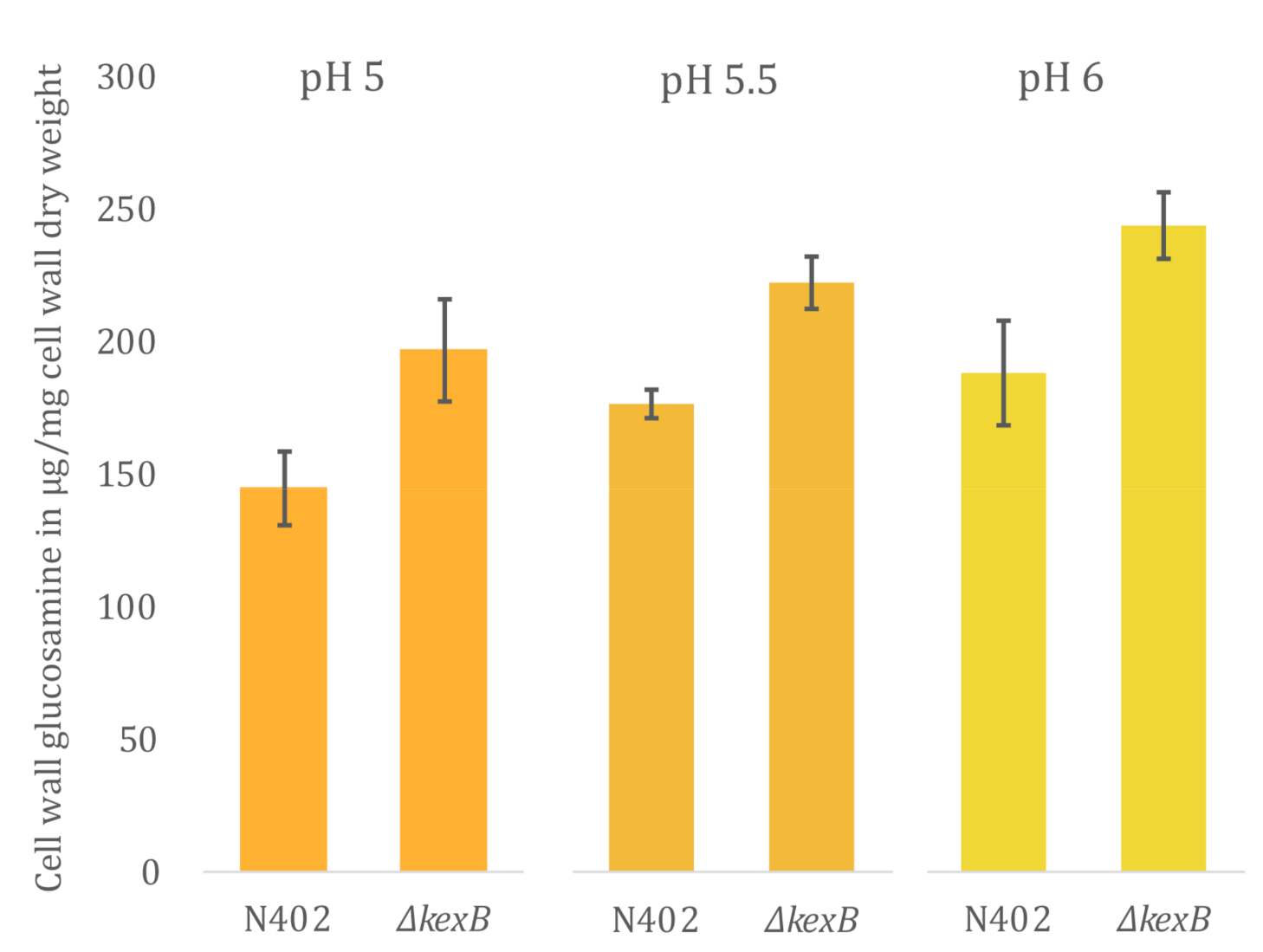
3.3. Cell Wall Chitin Content Is Increased in ΔkexB Irrespective of Morphology

To assess the relationship of different hyphal branching morphologies at pH 5.0, 5.5 and 6.0, and cell wall chitin content, we isolated cell walls from maximum biomass samples that were obtained from the bioreactor cultivations described above. Cell wall samples were hydrolyzed in triplicate and total glucosamine content was assessed using a colorimetric assay (Section 2.5). The results of glucosamine measurements are shown in Figure 3. During all cultivations, the ΔkexB strain displayed an increase in chitin content compared to the wild type. The increase in percentage of cell wall glucosamine for the ΔkexB strain compared to the wild type were found to be 35.0 ± 13.3%, 27.3 ± 5.51% and 29.6 ± 6.62% at pH 5.0, 5.5 and 6.0, respectively. Looking at absolute numbers of cell wall glucosamine per cell wall dry weight, we find that total glucosamine levels increase proportionally with the culture medium pH for both strains. Hence, cell wall glucosamine increases with increasing pH with about 20% between pH 5.0 and 6.0, and at all pH values the chitin content of the ΔkexB strain is about 30% higher compared the wild type.

3.4. Genome-Wide Expression Profiling Reveals Changes in Expression of Cell Wall Biosynthetic Genes

To gain better insight to transcriptomic responses in relation to cell wall biosynthetic changes that occur as a result of lacking KexB, we decided to look into the transcriptome by performing RNA-sequencing on the RNA extracted from wild type and the ΔkexB mutant grown at pH 5.5. A pH of 5.5 resulted in the most comparable growth conditions for both the wild type and the ∆kexB strain. To obtain biological duplicates, additional bioreactor cultivations at pH 5.5 were performed and resulted in similar growth rates and maximum biomass accumulation, as shown before for both wild type (0.167·h−1, 3.54 gDW·kg−1) and the ∆kexB strain (0.180·h−1, 4.30 gDW·kg−1) (Table 2). RNA was isolated from culture samples in the exponential growth phase at 90% of the maximum attained biomass. Duplicate batch-culture cultivations for both wild type and mutant were used to obtain a total of four RNA-sequencing samples. Following DeSeq2 analysis (Section 2.7), we found 2461 transcripts (1163 up, 1298 down)—approximately 21% of the 11846 total transcripts—to be differentially expressed between the mutant strain and the wild type (adjusted p-value ≤ 0.05) (expression data in Table S1).
For this study, we focused our interest on investigating to what extent cell wall biosynthesis was affected by the deletion of kexB. To assess this, we checked for differential expression of all genes involved in 82 cell wall biosynthesis in A. niger, as reported by Pel et al., 2007 [41]. Table 3 summarizes all cell-wall-related genes that were differentially expressed in the ΔkexB strain compared to N402. Differential expression was found for 29 out of 82 cell-wall-related genes involved in synthesis of all major components of the cell well, including α-glucan, β-glucan and chitin, and the modification thereof (Table 3, Table S1). The results for increased cell wall chitin (Figure 3) were corroborated by transcriptional upregulation of gene encoding the rate-limiting step in chitin precursor synthesis (UPD-N-acetylglucosamine), gfaA (FC 1.19) and gfaB (FC 6.22). Other genes involved in the synthesis of UPD-N-acetylglucosamine (gnaA, pcmA and ugnA) were not differentially expressed. Additionally, five chitin synthases were found to be upregulated in the ∆kexB strain (chsB, chsC, chsD, chsE and chsG), whereas four out of seven putative chitin-to-glucan crosslinking enzymes were found to be differentially expressed (crhA, and crhD upregulated, and crhB and crhF downregulated) along with four differentially expressed chitinases (cfcD and NRRL3_09653 upregulated, and cfcG and NRRL3_04221 downregulated).
Next to the upregulation of chitin-related transcripts, we observed the upregulation of agsA (FC 5.52), a gene known to be induced upon activation of the cell wall integrity (CWI) pathway in an RlmA-dependent manner [42]. We checked for upregulation of additional genes involved in the CWI response, as previously published [43,44]. Many of the reported genes were found to be represented in the differential dataset including bxgA, gelA, chsE, crhD, chsB, dfgC and wscB (highlighted by asterisks in Table 3). However, some cell-wall stress-related genes, such as chitin synthesis genes gfaA and gfaB, are not listed in this table. Taken together, the transcription data corroborate the observed increase in cell wall chitin and indicated that most of the CWI pathway is induced in a ΔkexB strain.

3.5. Cell Wall Integrity Is Affected by Disruption of kexB

To test whether the deletion of kexB affects the integrity of the cell wall, we exposed both the parental strain (N402) and ΔkexB to a series of cell-wall-disturbing compounds around pH 6.0 on plates—where the growth effect of ΔkexB is most prominent—that respond to changes in chitin content: Sodium dodecyl Sulfate (SDS), Caspofungin (CA), CalcoFluor White (CFW) and Congo Red (CR). As can be seen from Figure 4, the ΔkexB strain is more sensitive to SDS, CFW and CR than the wild type, while slight resistance towards CA was observed for the ΔkexB strain in this assay.
In addition to an increase in chitin content for the ΔkexB strain, the transcriptional response showed differential expression for the majority of the chitin-to-glucan crosslinking crh family (Table 3). To investigate the importance of these enzymes in the construction and modification of existing chitin in the cell walls of the ΔkexB strain, we opted for knockout kexB in an existing seven-fold crh deletion [31]. We found that deleting kexB in the seven-fold crh deletion strain resulted sporulation deficiency. Despite the impact on sporulation, we neither observed a vegetative growth deficiency nor did we find a significant exacerbation of sensitivity towards any of the tested compounds in the ΔcrhA-G + ΔkexB double deletion mutant (Figure 4).

4. Discussion

In this study, we invoked the impact of deleting kexB in A. niger on hyphal morphology and cell wall composition by performing phenotypic, cell wall and transcriptomic analysis. We show that the characteristic hyper-branching pH-dependent phenotype of the ΔkexB strain, as reported on solid agar plates, is also prevalent in submerged solid support growth and submerged pH-controlled batch cultivations at pH 6.0. The phenotypic plasticity of the ΔkexB strain transitions between pH 5.0 and pH 6.0; pH-controlled conditions revealed that the ΔkexB strain morphology resembles the wild type at pH 5.0, shows slightly shorter hyphae and smaller pellets at pH 5.5, and shows severe hyper-branching and tiny pellets at pH 6.0 (Figure 1). Despite the differences in morphology, both the growth rate and maximum biomass of the ΔkexB strain were not impaired and were even slightly higher than the wild type across all pH conditions. It is interesting to note, however, that wild type growth at pH 6.0 resulted in severe biofilm formation at the fermenter walls, whereas the ΔkexB strain did not.
To extend the observation that kexB may be involved in surface attachment (i.e., biofilm formation), we grew both wild type and ΔkexB in specific biofilm reactors. Under these very specific cultivation conditions, it was shown before that fungal growth occurs only on the metal sheets and not (or in very limited amount) in the liquid phase [40,45]. On these metal sheets, the ΔkexB strain showed reduced biofilm formation compared to the wild type at both pH 5.0 and pH 6.0. These observations showed that the deletion of kexB limits the proliferation of the fungal biomass on the metal support. Additionally, we found that the hyper-branching phenotype of the ΔkexB strain was only observed at pH 6.0 and is very similar to the batch cultivation conditions at pH 6.0. Additionally, the biofilm of the ΔkexB strain produced at pH 6.0 is of a totally different structure to the one observed for the wild type, as well as for the ΔkexB strain at pH 5.0. This mycelial layer of hyper-branching phenotype growing on the metal support is more compact and displayed reduced water sorption capacity. It is important to note that it remains unknown to what extent the morphology and biofilm composition will affect secretion and total production parameters, and these may be topics of future research. Despite these different mycelial properties of the hyper-branching morphology, a deletion of kexB reduced the ability to form biofilms at both pH conditions compared to the wild type. Furthermore, the increase in pH from 5.0 and 6.0 appeared to affect both wild type and ΔkexB, as a similar percentile drop in biofilm dry weight was observed between pH 5.0 and 6.0 (Figure 2B). Taken together, these data show that the kexB deletion shows a pH-dependent morphology at pH 6.0 without affecting growth rate or maximum biomass formation under submerged batch cultivation conditions. Additionally, the hyper-branching morphology showed less mycelial clumping and will reduce the stirring viscosity due to tinier pellets. As such, the deletion of kexB may provide a valuable industrial candidate strain for fermentation in near-neutral pH conditions. Additionally, we observed that the kexB deletion resulted in visibly thicker cell walls that could be used as an added-value post-fermentation product.
Next, we investigated the cell wall composition of the ∆kexB strain and found an increase in cell wall chitin compared to the wild type; a trait that is often coupled with activation of the cell wall integrity to increase the strength of the cell wall [10,11,46,47,48,49,50], and increased susceptibility towards CFW and resistance towards CA [47,48,51]. The observed increase in chitin content from bioreactor cultivations was corroborated by the transcriptional upregulation of gfaA, and its paralogue gfaB, that are required for chitin precursor (UDP-N-acetylglucosamine) synthesis, known as the rate-limiting step for chitin synthesis [46,52]. In addition, upregulation of multiple chitin synthases and downregulation of chitinases cfcD, cfcG and the N402 lineage-specific chitinase NRRL3_04221 were found. This transcriptional response is similar to that of the UDP-galactopyranose mutase A (ugmA) cell wall mutant, lacking cell wall galactofuranose, that also increased cell wall chitin and chitin modifications by upregulation of gfaA and gfaB, chsB and chsE and severe downregulation of chitinase ctcA [44,53].
Aside from the increased chitin synthesis in the ΔkexB strain, chitin-modifying enzymes also appear to play a role in the response to KexB-deficiency, as can be inferred from the upregulation of crhA and crhD, and downregulation of crhB and crhF (Table 3). However, the combined deletion of all crh genes and kexB did not result in increased susceptibility to cell-wall-disturbing compounds. We did observe reduced sporulation, an effect that was also observed when the deletions of the seven-fold crh family and ugmA were combined [31]. In case of the ugmA strain, it has clearly been described that the CWI pathway is activated. Here, the ΔkexB strain also showed a large number of differentially expressed cell wall biosynthetic genes that are known to be upregulated during the CWI response. Moreover, the kexB deletion has been reported to induce the CWI response pathway by means of MAPK phosphorylation in A. oryzae and A. fumigatus [24,27,54]. In addition, the A. oryze kexB deletion has been reported to cause hyper-branching similar to A. niger, and increased amylase and protease production [24]. In A. fumigatus, a kexB deletion strain showed increased sensitivity towards CFW and CR, similar to our observations for the ΔkexB strain here (Figure 3). The sensitivity towards these compounds combined with the genome-wide expression profile confirm activation of the CWI pathway in ΔkexB strains.
The observed changes in cell wall chitin content in the A. niger ΔkexB strain are in congruence with A. oryzae, where the deletion of kexB was shown to increase cell wall chitin along with a reduction in α-glucan. This reduction in cell wall α-glucan was suggested to be caused by a lack of KexB-processed α-glucan synthases [26]. In this study, we did not analyze cell wall content for levels of α-glucan; however, we did observe a very strong positive transcriptional response for both agsA (5.52 FC) and agsC (26.48 FC) in the ΔkexB strain. Similar to upregulation of chitin-modifying enzymes, we found increased transcriptional expression of agnD, an alpha-1,3-glucanase [43] and agtB, an α-glucan glycanosyl transferase [55] in conjunction with the two α-glucan synthases. Upregulation of α-glucan metabolism may either indicate an attempted compensatory response to a lack of correctly processed α-glucan synthases [56], or be a sign of general cell wall stress resulting from other improperly processed (cell wall) enzymes. Previously, we showed the relevance of proper α-glucan deposition in the cell wall for integrity; when α-glucan synthases agsA and agsE are disrupted in A. niger, we showed that crh-facilitated chitin-β-glucan crosslinking genes become important for maintenance of cell wall integrity, while these genes are redundant when α-glucan synthesis is undisturbed [31]. Interestingly, α-glucan has also been described in relation to pathogenesis and hyphal aggregation for filamentous fungi [56], by showing conidial and hyphal pellet clumping in α-glucan synthase knockouts of A. fumigatus, A. oryzae and A. nidulans [57,58,59]. Hyphal aggregation of the A. niger ΔkexB strain appeared to be reduced across all three pH conditions (Figure 1) which may be related to cell wall α-glucan levels. Further analysis of α-glucan-dependent hyphal aggregation and biofilm formation may be a topic of future research.
Next to upregulation of α-glucan and chitin metabolism, fksA, the sole β-1,3-glucan synthase in A. niger was also found to be upregulated. Along with fksA, differential expressions of glucanases and glucanosyltransferases (Table 2) were observed, that are predicted to cause rearrangements of β-1,3-glucan polymer length and branching. This transcriptional response in A. niger is very similar to that observed in the kexB deletion in A. fumigatus, that also resulted in upregulation of gel glucanosyltransferases [54], and the results described on the β-1,3-glucan composition in A. oryzae, where the AoΔkexB strain showed a higher degree of β-glucan polymerization combined with slightly less branching [26]. Together with the overall cell wall biosynthetic gene response, a deletion of kexB appears to bring about major cell wall content and compositional changes for all major constituents of the cell wall.
This study shows that the cell wall integrity of the ΔkexB strain is clearly affected, however, it is affected without impairing the growth rate or the maximally attained biomass during submerged batch cultivations. Contrarily, we did observe that kexB is important in the formation of biofilms, as this mode of cultivation in biofilm reactors led to a reduction in the ability to colonize the metal support. For now, understanding the characteristic phenotype of the ΔkexB strain during growth at pH 5.5 and 6 seems a consequence of pleiotropic effects of several hitherto unrelated responses. The morphology at pH 5.5/6.0 is possibly the result of loss of polarity due to the inability to synthesize or modify the cell wall correctly. Many possible cell wall synthesis or cell-wall-modifying enzymes could be targets of KexB processing. Explanations for the pH-dependent effect have been postulated and they suggest that at a lower pH, other proteases can perform KexB-like dibasic processing [28]. In yeast, yapsins, aspartic proteolytic enzymes involved in processing cell wall proteins including Scw4p, Utr2p, Pir4p and Gas1p [60,61,62], show a pH-dependent dependency on Kex2. For Scw4p, it was shown that only at a neutral pH (7.0), but not at an acidic pH (4.0), Kex2p is required for correct processing of the cell wall protein prior to yapsin processing, in order to activate the protein [62]. It is likely that the pH-dependent morphology of the ∆kexB mutant in A. niger is caused by improper processing of such enzymes/proteins. Here, we showed that—despite the clear morphological changes in liquid between pH 5.0 and 6.0—the respective increase in cell wall chitin compared to wild type was observed across all pH conditions. From this, we infer that the increase in chitin content is unlikely to dictate cell shape, however, we cannot rule out chitin-modifying enzymes could still play a role in the distinct phenotype. Taken together, the transcriptional data suggest activation of the CWI pathway that results in cell wall compositional rearrangements. Despite these findings, it remains to be elucidated how and if the KexB-dependent cell wall modifications are related to the pH-dependent growth phenotype.

Supplementary Materials

The following are available online at https://www.mdpi.com/2076-2607/8/12/1918/s1, Table S1: Expression data of differentially expressed genes ∆kexB vs. wild type.

Author Contributions

T.M.v.L.: Conceptualization, Investigation, Validation, Formal Analysis, Methodology, Writing—Original draft preparation, Visualization. M.A.: Investigation. G.F.-C.: Software, Formal Analysis. N.G.: Software, Resources A.T.: Resources and editing. F.D.: Investigation, Formal analysis, Writing—Original draft preparation. A.H.M.: Resources and editing. A.F.J.R.: Conceptualization, Writing—Reviewing and Editing, Funding Acquisition, Data curation, Supervision. P.J.P.: Conceptualization, Writing—Reviewing and Editing, Funding Acquisition. All authors have read and agreed to the published version of the manuscript.

Funding

This work is part of the “FunChi” ERA-IB project with project number ERA-IB-15-080, which is (partly) financed by the Dutch Research Council (NWO). G.F.-C. was funded by the European Marie Skłodowska-Curie program (H2020-COFUND-2015-FP-707404).

Acknowledgments

We would like to thank Bruno M. Moerschbacher for the coordination of the FunChi project.

Conflicts of Interest

The authors declare no conflict of interest.

Availability of Data and Materials

The DNA reads described in this study are deposited in the GEO accession database under accession number GSE151618. All other data are available on request by contacting the corresponding author.

References

  1. Orzali, L.; Corsi, B.; Forni, C.; Riccioni, L. Chitosan in Agriculture: A New Challenge for Managing Plant Disease. In Biological Activities and Application of Marine Polysaccharides; InTech: London, UK, 2017. [Google Scholar]
  2. Naveed, M.; Phil, L.; Sohail, M.; Hasnat, M.; Baig, M.M.F.A.; Ihsan, A.U.; Shumzaid, M.; Kakar, M.U.; Khan, T.M.; Akabar, M.D.; et al. Chitosan oligosaccharide (COS): An overview. Int. J. Biol. Macromol. 2019, 129, 827–843. [Google Scholar] [CrossRef] [PubMed]
  3. Cai, J.; Yang, J.; Du, Y.; Fan, L.; Qiu, Y.; Li, J.; Kennedy, J.F. Enzymatic preparation of chitosan from the waste Aspergillus niger mycelium of citric acid production plant. Carbohydr. Polym. 2006, 64, 151–157. [Google Scholar] [CrossRef]
  4. Nwe, N.; Stevens, W.F. Effect of urea on fungal chitosan production in solid substrate fermentation. Process. Biochem. 2004, 39, 1639–1642. [Google Scholar] [CrossRef]
  5. Deng, M.-D.; McMullin, T.W.; Grund, A.D. Metabolic engineering for enhanced production of chitin and chitosan in microorganisms. U.S. Patent 2005/0042735A1, 24 February 2005. [Google Scholar]
  6. Ja’Afaru, M.I. Screening of Fungi Isolated from environmental samples for Xylanase and Cellulase production. ISRN Microbiol. 2013, 283423, 1–7. [Google Scholar] [CrossRef] [Green Version]
  7. Van Leeuwe, T.M.; Arentshorst, M.; Punt, P.J.; Ram, A.F.J. Interrogation of the cell wall integrity pathway in Aspergillus niger identifies a putative negative regulator of transcription involved in chitin deposition. Gene X 2020, 5, 100028. [Google Scholar] [CrossRef]
  8. Van Leeuwe, T.M.; Gerritsen, A.; Arentshorst, M.; Punt, P.J.; Ram, A.F.J. Rab GDP-dissociation inhibitor gdiA is an essential gene required for cell wall chitin deposition in Aspergillus niger. Fungal Genet. Biol. 2020, 136, 103319. [Google Scholar] [CrossRef]
  9. Punt, P.J.; Drint-Kuijvenhoven, A.; Lokman, B.C.; Spencer, J.A.; Jeenes, D.; Archer, D.A.; Van Den Hondel, C.A.M.J.J. The role of the Aspergillus niger furin-type protease gene in processing of fungal proproteins and fusion proteins: Evidence for alternative processing of recombinant (fusion-) proteins. J. Biotechnol. 2003, 106, 23–32. [Google Scholar] [CrossRef]
  10. Jalving, R.; Van De Vondervoort, P.J.I.; Visser, J.; Schaap, P.J. Characterization of the Kexin-Like Maturase of Aspergillus niger. Appl. Environ. Microbiol. 2000, 66, 363–368. [Google Scholar] [CrossRef] [Green Version]
  11. Fuller, R.S.; Brake, A.; Thorner, J. Yeast prohormone processing enzyme (KEX2 gene product) is a Ca2+-dependent serine protease. Proc. Natl. Acad. Sci. USA 1989, 86, 1434–1438. [Google Scholar] [CrossRef] [Green Version]
  12. Wilcox, C.A.; Fuller, R.S. Posttranslational processing of the prohormone-cleaving Kex2 protease in the Saccharomyces cerevisiae secretory pathway. J. Cell Biol. 1991, 115, 297–307. [Google Scholar] [CrossRef]
  13. Bryant, N.J.; Stevens, T.H. Two Separate Signals Act Independently to Localize a Yeast Late Golgi Membrane Protein through a Combination of Retrieval and Retention. J. Cell Biol. 1997, 136, 287–297. [Google Scholar] [CrossRef] [PubMed]
  14. Martin, S.H.; Wingfield, B.D.; Wingfield, M.; Steenkamp, E. Causes and Consequences of Variability in Peptide Mating Pheromones of Ascomycete Fungi. Mol. Biol. Evol. 2011, 28, 1987–2003. [Google Scholar] [CrossRef] [PubMed] [Green Version]
  15. Le Marquer, M.; San Clemente, H.; Roux, C.; Savelli, B.; Frei Dit Frey, N. Identification of new signalling peptides through a genome-wide survey of 250 fungal secretomes. BMC Genom. 2019, 20, 64. [Google Scholar]
  16. Leibowitz, M.J.; Wickner, R.B. A chromosomal gene required for killer plasmid expression, mating, and spore maturation in Saccharomyces cerevisiae. Proc. Natl. Acad. Sci. USA 1976, 73, 2061–2065. [Google Scholar] [CrossRef] [Green Version]
  17. Wagner, J.C.; Wolf, D.H. Hormone (pheromone) processing enzymes in yeast The carboxy-terminal processing enzyme of the mating pheromone α-factor, carboxypeptidase yscα, is absent in α-factor maturation-defective kex1 mutant cells. FEBS Lett. 1987, 221, 423–426. [Google Scholar] [CrossRef] [Green Version]
  18. Achstetter, T. Regulation of alpha-factor production in Saccharomyces cerevisiae: A-factor pheromone-induced expression of the MF alpha 1 and STE13 genes. Mol. Cell. Biol. 1989, 9, 4507–4514. [Google Scholar] [CrossRef] [Green Version]
  19. Heiman, M.G.; Engel, A.; Walter, P. The Golgi-resident protease Kex2 acts in conjunction with Prm1 to facilitate cell fusion during yeast mating. J. Cell Biol. 2007, 176, 209–222. [Google Scholar] [CrossRef] [Green Version]
  20. Yoshimi, A.; Umemura, M.; Nagano, N.; Koike, H.; Machida, M.; Abe, K. Expression of ustR and the Golgi protease KexB are required for ustiloxin B biosynthesis in Aspergillus oryzae. AMB Express. 2016, 9. [Google Scholar] [CrossRef] [Green Version]
  21. Te Biesebeke, R.T.; Record, E.; Van Biezen, N.; Heerikhuisen, M.; Franken, A.; Punt, P.J.; van den Hondel, C.A.M.J.J. Branching mutants of Aspergillus oryzae with improved amylase and protease production on solid substrates. Appl. Microbiol. Biotechnol. 2005, 69, 44–50. [Google Scholar] [CrossRef]
  22. Newport, G.; Kuo, A.; Flattery, A.; Gill, C.; Blake, J.J.; Kurtz, M.B.; Abruzzo, G.K.; Agabian, N. Inactivation of Kex2p Diminishes the Virulence of Candida albicans. J. Biol. Chem. 2002, 278, 1713–1720. [Google Scholar] [CrossRef] [Green Version]
  23. Mizutani, O.; Shiina, M.; Yoshimi, A.; Sano, M.; Watanabe, T.; Yamagata, Y.; Nakajima, T.; Gomi, K.; Abe, K. Substantial decrease in cell wall α-1,3-glucan caused by disruption of the kexB gene encoding a subtilisin-like processing protease in Aspergillus oryzae. Biosci. Biotechnol. Biochem. 2016, 80, 1781–1791. [Google Scholar] [CrossRef] [PubMed] [Green Version]
  24. Mizutani, O.; Nojima, A.; Yamamoto, M.; Furukawa, K.; Fujioka, T.; Yamagata, Y.; Abe, K.; Nakajima, T. Disordered cell integrity signaling caused by disruption of the kexB gene in Aspergillus oryzae. Eukaryot. Cell 2004, 3, 1036–1048. [Google Scholar] [CrossRef] [PubMed] [Green Version]
  25. Jalving, R. Proteolytic Processing in the Secretory Pathway of Aspergillus Niger; Wageningen Universiteit: Wageningen, The Netherlands, 2005. [Google Scholar]
  26. Arentshorst, M.; Niu, J.; Ram, A.F.J. Efficient generation of Aspergillus niger knock out strains by combining NHEJ mutants and a split marker approach. In Genetic Transformation Systems in Fungi; van den Berg, M.A., Maruthachalam, K., Eds.; Springer International Publishing: Cham, Switzerland, 2015; Volume 1, pp. 263–272. [Google Scholar]
  27. Bos, C.J.; Debets, A.J.M.; Swart, K.; Huybers, A.; Kobus, G.; Slakhorst, S.M. Genetic analysis and the construction of master strains for assignment of genes to six linkage groups in Aspergillus niger. Curr. Genet. 1988, 14, 437–443. [Google Scholar] [CrossRef] [PubMed]
  28. Van Leeuwe, T.M.; Wattjes, J.; Niehues, A.; Forn-Cuní, G.; Geoffrion, N.; Mélida, H.; Arentshorst, M.; Molina, A.; Tsang, A.; Meijer, A.H.; et al. A seven-membered cell wall related transglycosylase gene family in Aspergillus niger is relevant for cell wall integrity in cell wall mutants with reduced α-glucan or galactomannan. Cell Surf. 2020, 6, 100039. [Google Scholar] [CrossRef]
  29. Jørgensen, T.R.; Nitsche, B.M.; Lamers, G.E.; Arentshorst, M.; Hondel, C.A.V.D.; Ram, A.F. Transcriptomic insights into the physiology of Aspergillus niger approaching a specific growth rate of zero. Appl. Environ. Microbiol. 2010, 76, 5344–5355. [Google Scholar] [CrossRef] [Green Version]
  30. Schindelin, J.; Arganda-Carreras, I.; Frise, E.; Kaynig, V.; Longair, M.; Pietzsch, T.; Preibisch, S.; Rueden, C.; Saalfeld, S.; Schmid, B.; et al. Fiji: An open-source platform for biological-image analysis. Nat. Methods 2012, 9, 676–682. [Google Scholar] [CrossRef] [Green Version]
  31. Aguilar-Pontes, M.; Brandl, J.; McDonnell, E.; Strasser, K.; Nguyen, T.; Riley, R.; Mondo, S.; Salamov, A.; Nybo, J.; Vesth, T.; et al. The gold-standard genome of Aspergillus niger NRRL 3 enables a detailed view of the diversity of sugar catabolism in fungi. Stud. Mycol. 2018, 91, 61–78. [Google Scholar] [CrossRef]
  32. Patro, R.; Duggal, G.; Love, M.I.; Irizarry, M.I.L.R.A.; Kingsford, C. Salmon provides fast and bias-aware quantification of transcript expression. Nat. Methods 2017, 14, 417–419. [Google Scholar] [CrossRef] [Green Version]
  33. Soneson, C.; Love, M.I.; Robinson, M.D. Differential analyses for RNA-seq: Transcript-level estimates improve gene-level inferences [version 2; referees: 2 approved]. F1000Research 2016, 4, 1521. [Google Scholar] [CrossRef]
  34. Love, M.I.; Huber, W.; Anders, S. Moderated estimation of fold change and dispersion for RNA-seq data with DESeq2. Genome Biol. 2014, 15, 550. [Google Scholar] [CrossRef] [Green Version]
  35. Punt, P.J.; Oliver, R.P.; Dingemanse, M.A.; Pouwels, P.H.; Hondel, C.A.V.D. Transformation of Aspergillus based on the hygromycin B resistance marker from Escherichia coli. Gene 1987, 56, 117–124. [Google Scholar] [CrossRef]
  36. Niu, J.; Arentshorst, M.; Nair, P.D.S.; Dai, Z.; Baker, S.E.; Frisvad, J.C.; Nielsen, K.F.; Punt, P.J.; Ram, A.F.J. Identification of a classical mutant in the industrial host Aspergillus niger by systems genetics: LaeA is required for citric acid production and regulates the formation of some secondary metabolites. G3 Genes Genomes Genet. 2016, 6, 193–204. [Google Scholar]
  37. Zune, Q.; Delepierre, A.; Gofflot, S.; Bauwens, J.; Twizere, J.-C.; Punt, P.J.; Francis, F.; Toye, D.; Boukraa, S.; Delvigne, F. A fungal biofilm reactor based on metal structured packing improves the quality of a Gla:GFP fusion protein produced by Aspergillus oryzae. Appl. Microbiol. Biotechnol. 2015, 99, 6241–6254. [Google Scholar] [CrossRef] [PubMed] [Green Version]
  38. Pel, H.J.; De Winde, J.H.; Archer, D.B.; Dyer, P.S.; Hofmann, G.; Schaap, P.J.; Turner, G.; De Vries, R.P.; Albang, R.; Albermann, K.; et al. Genome sequencing and analysis of the versatile cell factory Aspergillus niger CBS 513.88. Nat. Biotechnol. 2007, 25, 221–231. [Google Scholar] [CrossRef] [Green Version]
  39. Damveld, R.A.; Arentshorst, M.; Franken, A.; vanKuyk, P.A.; Klis, F.M.; van den Hondel, C.A.; Ram, A.F. The Aspergillus niger MADS-box transcription factor RlmA is required for cell wall reinforcement in response to cell wall stress. Mol. Microbiol. 2005, 58, 305–319. [Google Scholar] [CrossRef]
  40. Meyer, V.; Damveld, R.A.; Arentshorst, M.; Stahl, U.; Van Den Hondel, C.A.M.J.J.; Ram, A.F.J. Survival in the presence of antifungals: Genome-wide expression profiling of Aspergillus niger in response to sublethal concentrations of caspofungin and fenpropimorph. J. Biol. Chem. 2007, 282, 32935–32948. [Google Scholar] [CrossRef] [Green Version]
  41. Park, J.; Hulsman, M.; Arentshorst, M.; Breeman, M.; Alazi, E.; Lagendijk, E.L.; Rocha, M.C.; Malavazi, I.; Nitsche, B.M.; Hondel, C.A.V.D.; et al. Transcriptomic and molecular genetic analysis of the cell wall salvage response of Aspergillus niger to the absence of galactofuranose synthesis. Cell. Microbiol. 2016, 18, 1268–1284. [Google Scholar] [CrossRef]
  42. Khalesi, M.; Zune, Q.; Telek, S.; Riveros-Galan, D.; Verachtert, H.; Toye, D.; Gebruers, K.; Derdelinckx, G.; Delvigne, F. Fungal biofilm reactor improves the productivity of hydrophobin HFBII. Biochem. Eng. J. 2014, 88, 171–178. [Google Scholar] [CrossRef]
  43. Ram, A.F.J.; Arentshorst, M.; Damveld, R.A.; Vankuyk, P.A.; Klis, F.M.; van den Hondel, C.A.M.J.J. The cell wall stress response in Aspergillus niger involves increased expression of the glutamine: Fructose-6-phosphate amidotransferase-encoding gene (gfaA) and increased deposition of chitin in the cell wall. Microbiology 2004, 150, 3315–3326. [Google Scholar] [CrossRef] [Green Version]
  44. Walker, L.A.; Munro, C.A.; De Bruijn, I.; Lenardon, M.D.; McKinnon, A.D.; Gow, N.A.R. Stimulation of chitin synthesis rescues Candida albicans from Echinocandins. PLoS Pathog. 2008, 4, e1000040. [Google Scholar] [CrossRef] [Green Version]
  45. Walker, L.A.; Lee, K.K.; Munro, C.A.; Gow, N.A.R.R. Caspofungin treatment of Aspergillus fumigatus results in ChsG-dependent upregulation of chitin synthesis and the formation of chitin-rich microcolonies. Antimicrob. Agents Chemother. 2015, 59, 5932–5941. [Google Scholar] [CrossRef] [Green Version]
  46. Fortwendel, J.R.; Juvvadi, P.R.; Perfect, B.Z.; Rogg, L.E.; Perfect, J.R.; Steinbach, W.J. Transcriptional regulation of chitin synthases by calcineurin controls paradoxical growth of Aspergillus fumigatus in response to Caspofungin. Antimicrob. Agents Chemother. 2010, 54, 1555–1563. [Google Scholar] [CrossRef] [Green Version]
  47. Heilmann, C.J.; Sorgo, A.G.; Mohammadi, S.; Sosinska, G.J.; De Koster, C.G.; Brul, S.; De Koning, L.J.; Klis, F.M. Surface stress induces a conserved cell wall stress response in the pathogenic fungus Candida albicans. Eukaryot. Cell 2012, 12, 254–264. [Google Scholar] [CrossRef] [Green Version]
  48. Ram, A.F.J.; Klis, F.M. Identification of fungal cell wall mutants using susceptibility assays based on Calcofluor white and Congo red. Nat. Protoc. 2006, 1, 2253–2256. [Google Scholar] [CrossRef]
  49. Lagorce, A.; Le Berre-Anton, V.; Aguilar-Uscanga, B.; Martin-Yken, H.; Dagkessamanskaia, A.; François, J. Involvement ofGFA1, which encodes glutamine-fructose-6-phosphate amidotransferase, in the activation of the chitin synthesis pathway in response to cell-wall defects in Saccharomyces cerevisiae. JBIC J. Biol. Inorg. Chem. 2002, 269, 1697–1707. [Google Scholar] [CrossRef]
  50. Damveld, R.A.; Franken, A.; Arentshorst, M.; Punt, P.J.; Klis, F.M.; van den Hondel, C.A.M.J.J.; Ram, A.F.J. A novel screening method for cell wall mutants in Aspergillus niger identifies UDP-Galactopyranose mutase as an important protein in fungal cell wall biosynthesis. Genetics 2008, 178, 873–881. [Google Scholar] [CrossRef] [Green Version]
  51. Wang, J.; Zhou, H.; Lu, H.; Du, T.; Luo, Y.; Wilson, I.B.H.; Jingyang, W. Kexin-like endoprotease KexB is required for N-glycan processing, morphogenesis and virulence in Aspergillus fumigatus. Fungal Genet. Biol. 2015, 76, 57–69. [Google Scholar] [CrossRef] [Green Version]
  52. van der Kaaij, R.M.; Yuan, X.L.; Franken, A.; Ram, A.F.; Punt, P.J.; van der Maarel, M.J.; Dijkhuizen, L. Two novel, putatively cell wall-associated and glycosylphosphatidylinositol-anchored alpha-glucanotransferase enzymes of Aspergillus niger. Eukaryot. Cell 2007, 6, 1178–1188. [Google Scholar] [CrossRef] [Green Version]
  53. Yoshimi, A.; Miyazawa, K.; Abe, K. Function and biosynthesis of cell wall α-1,3-glucan in fungi. J. Fungi 2017, 3, 63. [Google Scholar] [CrossRef]
  54. Henry, C.; Latgé, J.-P.; Beauvais, A. α1,3 Glucans Are Dispensable in Aspergillus fumigatus. Eukaryot. Cell 2012, 11, 26–29. [Google Scholar] [CrossRef] [Green Version]
  55. Miyazawa, K.; Yoshimi, A.; Kasahara, S.; Sugahara, A.; Koizumi, A.; Yano, S.; Kimura, S.; Iwata, T.; Sano, M.; Abe, K. Molecular Mass and Localization of α-1,3-Glucan in Cell Wall Control the Degree of Hyphal Aggregation in Liquid Culture of Aspergillus nidulans. Front. Microbiol. 2018, 9, 2623. [Google Scholar] [CrossRef] [PubMed]
  56. Yoshimi, A.; Sano, M.; Inaba, A.; Kokubun, Y.; Fujioka, T.; Mizutani, O.; Hagiwara, D.; Fujikawa, T.; Nishimura, M.; Yano, S.; et al. Functional analysis of the α-1,3-Glucan synthase genes agsA and agsB in Aspergillus nidulans: AgsB Is the Major α-1,3-Glucan synthase in this fungus. PLoS ONE 2013, 8, e54893. [Google Scholar] [CrossRef] [Green Version]
  57. Gagnon-Arsenault, I.; Parisé, L.; Tremblay, J.; Bourbonnais, Y. Activation mechanism, functional role and shedding of glycosylphosphatidylinositol-anchored Yps1p at the Saccharomyces cerevisiae cell surface. Mol. Microbiol. 2008, 69, 982–993. [Google Scholar] [CrossRef]
  58. Miller, K.A.; DiDone, L.; Krysan, D.J. Extracellular secretion of overexpressed glycosylphosphatidylinositol-Linked Cell Wall Protein Utr2/Crh2p as a novel protein quality control mechanism in Saccharomyces cerevisiae. Eukaryot. Cell 2010, 9, 1669–1679. [Google Scholar] [CrossRef] [Green Version]
  59. Grbavac, A.; Čanak, I.; Stuparević, I.; Teparić, R.; Mrša, V. Proteolytic processing of the Saccharomyces cerevisiae cell wall protein Scw4 regulates its activity and influences its covalent binding to glucan. Biochim. Biophys. Acta (BBA) Bioenerg. 2017, 1864, 507–515. [Google Scholar] [CrossRef]
  60. Cai, M.; Zhang, Y.; Hu, W.; Shen, W.; Yu, Z.; Zhou, W.; Jiang, T.; Zhou, X.; Zhang, Y. Genetically shaping morphology of the filamentous fungus Aspergillus glaucus for production of antitumor polyketide aspergiolide A. Microb. Cell Factories 2014, 13, 73. [Google Scholar] [CrossRef] [Green Version]
  61. Papagianni, M. Fungal morphology and metabolite production in submerged mycelial processes. Biotechnol. Adv. 2004, 22, 189–259. [Google Scholar] [CrossRef]
  62. Dhillon, G.S.; Kaur, S.; Brar, S.K.; Verma, M. Green synthesis approach: Extraction of chitosan from fungus mycelia. Crit. Rev. Biotechnol. 2012, 33, 379–403. [Google Scholar] [CrossRef]
Figure 1. Pellet morphology of pH-controlled batch cultivations wild type (N402) and ∆kexB strain microscopy samples taken at maximum biomass under different pH conditions: pH 5.0, pH 5.5 and pH 6.0.
Microorganisms 08 01918 g001
Figure 2. Submerged biofilm reactor cultivations of wild type (N402) and ∆kexB. (A) Scheme of the biofilm reactor set-up used in this work and pictures displaying biofilm layer for each strain at two pH levels. Representative microscopy pictures (brightfield, 20×) are displayed for each type of biofilm structure. (B) Dry fungal biomass weight developed on the stainless-steel sheets in biofilm reactor and (C) biofilm water sorption capacity (n = 2). Biofilm (water) sorption capacity is the difference between biofilm’s wet and dry weight.
Microorganisms 08 01918 g002
Figure 3. Cell wall glucosamine content of wild type (N402) and ∆kexB from pH-controlled batch cultivations. Cell wall glucosamine content maximum biomass samples that originate from single bioreactor cultivations, run at either pH 5.0, pH 5.5 (1) or pH 6.0. Measurements were performed in technical triplicates (n = 3); error bars are the standard error (SE) and represent the variation of technical replicates.
Microorganisms 08 01918 g003
Figure 4. Susceptibility against cell wall disturbing compounds of wild type, ΔkexB, ΔcrhA-G and ΔcrhA-G + ΔkexB. Spore dilution assay on a range of concentrations of SDS, Caspofungin (CA), CalcoFluor White (CFW) and Congo Red (CR). The number of spores plated, from left to right, are 104, 103, 102 and 101 spores, respectively. All growth was performed in minimal medium (MM), pH 6.0. Strains were allowed to grow at 30 °C for 96 h prior to recordings, with the exception of the control (65 h).
Microorganisms 08 01918 g004
Table 1. All strains used in this study.
NameGenotypeReference
N402cspA1[30]B
∆kexBcspA1, pyrG-, ΔkexB::AOpyrG (AB4.1∆pclA)[12]
TLF39cspA1, ΔkusA::DR-amdS-DR, ΔcrhA-G[31]
TLF69cspA1, ΔkusA::DR-amdS-DR, ΔcrhA-G, ΔkexB::hygBThis study
Table 2. Growth parameters of wild type and ΔkexB. Growth is shown as maximum growth rate per hour (µmax) and maximum biomass (gDW·kg−1) at different pH-controlled batch-cultivations.
Wild TypeΔkexB
pH Conditionµmax (h−1)Max Biomass
(gDW·kg−1)
µmax (h−1)Max Biomass
(gDW·kg−1)
pH 5.00.1873.660.2004.15
pH 5.5 (1)0.1753.680.1944.19
pH 5.5 (2)0.1673.540.1804.30
pH 6.00.063 *0.64 *0.2114.31
* Cultivation of N402 at pH 6.0 resulted in severe biofilm formation on the walls of the fermenters resulting in underestimation of both the submerged growth rate and amount of in-broth maximum biomass.
Table 3. Differentially expressed cell wall biosynthesis genes in A. niger. 29 genes were differentially expressed (Padj ≤ 0.05) based on all 82 cell wall biosynthesis genes described by Pel et al., 2007 [41].
An IDNRRL3 IDGene DescriptionGeneWild Type (Normalized Read Counts)ΔkexB (Normalized Read Counts)FCUp- or Down-RegulatedPadj
A-glucan biosynthesis and modification
An12g02460NRRL3_09001Putative GPI-anchored amylase-like protein (GH13-family) with possible function in alpha1,3-1,4-glucan processing agtB30111438.06up1.43 × 10−69
An12g02450NRRL3_09002Putative catalytic subunit alpha1,3-glucan synthase complex; SpAgs1-like agsC64168826.48up5.91 × 10−67
An04g09890NRRL3_07454Putative catalytic subunit alpha1,3-glucan synthase complex; SpAgs1-like agsA *1045745.52up8.90 × 10−25
An08g09610NRRL3_11494Putative alpha-1,3-glucanase GH71; member of the SpAgn1-family agnD149665444.37up1.55 × 10−71
Β-glucan biosynthesis and modification
An09g00670NRRL3_00054Predicted GPI-anchored protein. Putative 1,3-β-glucanosyltransferase GH72; member of the Gel-family gelD4092022.95up3.83 × 10−73
An03g05290NRRL3_08399Predicted GPI-anchored protein. Putative beta-1,3-glucanosyltransferase GH17; member of the AfBgt1-family bgtB20,95629,2351.40up7.26 × 10−4
An01g12450NRRL3_02657Putative exo-beta-1,3-glucanase (GH55-family); related to Coniothyrium minitans exo-1,3-glucanase (Cmg1) bxgA *474364071.35up8.95 × 10−3
An06g01550NRRL3_11624Putative catalytic subunit beta1,3-glucan synthase complex; ScFks1-like fksA18,18923,4021.29up6.29 × 10−4
An07g04650NRRL3_04586Putative beta-1,3-glucanosyltransferase GH17; member of the AfBgt1-family bgtC1161847−1.37down2.46 × 10−3
An10g00400NRRL3_06317Predicted GPI-anchored protein. Putative 1,3-β-glucanosyltransferase GH72; A. fumigatus Gel1-like gelA *14,0859570−1.47down4.10 × 10−8
An03g06220NRRL3_08332Predicted GPI-anchored protein. Putative 1,3-β-glucanosyltransferase GH72; member of the Gel-family gelE2008192−10.50down6.04 × 10−120
An19g00090NRRL3_01223Putative exo-beta-1,3-glucanase (GH55-family); related to Coniothyrium minitans exo-1,3-glucanase (Cmg1) bgxC1101102−10.83down3.28 × 10−50
Chitin biosynthesis and modification
N/ANRRL3_09653Putative chitinase (N402 specific)-1733619.52up2.11 × 10−17
An12g10380NRRL3_02932Putative chitin synthase ClassIII; EnChsB-like chsE *376465171.73up1.35 × 10−15
An08g05290NRRL3_11152Putative chitin synthase ClassVI; chsG1472521.71up4.56 × 10−3
An14g00650
An14g00660
NRRL3_00641Putative chitin synthase ClassI; EnChsC-like chsC239838141.59up2.44 × 10−7
An11g01540NRRL3_10021Putative transglycosidase of GH16-family involved in cell wall biosynthesis; ScCrh1-like crhA73710971.49up8.45 × 10−4
An01g11010NRRL3_02532Predicted GPI-anchored protein. Putative transglycosidase of GH16-family involved in cell wall biosynthesis; member of the ScCrh1-family crhD *363752341.44up7.65 × 10−4
An09g02290NRRL3_00179Putative chitin synthase ClassIV; EnChsD-like chsD227831041.36up1.99 × 10−3
An09g04010NRRL3_00331Putative chitin synthase ClassIII; EnChsB-like chsB *677485531.26up5.98 × 10−3
An01g05360NRRL3_02063Putative ClassV Chitinase (GH18); ScCts2-like cfcD1384818−1.69down3.96 × 10−5
An16g02850NRRL3_07085Putative transglycosidase of GH16-family involved in cell wall biosynthesis; member of the ScCrh1-family crhF1468812−1.81down4.92 × 10−8
An07g07530NRRL3_04809Predicted GPI-anchored protein. Putative transglycosidase of GH16-family involved in cell wall biosynthesis; ScCrh2-like crhB2205988−2.23down1.53 × 10−9
N/ANRRL3_04221Putative chitinase (N402 specific)-17458−2.99 3.09 × 10⁻³
An19g00100NRRL3_01224Putative ClassV Chitinase (GH18); ScCts2-like cfcG4152−197.88 8.05 × 10⁻²⁰
GH76 family proteins
An11g01240NRRL3_10041Putative endo-mannanase (GH76-family) with a possible role in GPI-CWP incorporation; ScDfg5-like dfgH28911033.82up2.58 × 10−35
An14g03520NRRL3_00897Predicted GPI-anchored protein. Putative endo-mannanase (GH76-family) with a possible role in GPI-CWP incorporation; ScDfg5-like dfgC *113319961.76up6.95 × 10−10
An16g08090NRRL3_06700Predicted GPI-anchored protein. Putative endo-mannanase (GH76-family) with a possible role in GPI-CWP incorporation; ScDfg5-like dfgE88611691.32up2.14 × 10−2
An02g02660NRRL3_06048Putative endo-mannanase (GH76-family) with a possible role in GPI-CWP incorporation; ScDfg5-like dfgG25871693−1.53down3.75 × 10−5
Rho-GAPs
An18g06730NRRL3_10703Putative Cdc42-GTPase Activating protein (GAP) with similarity to ScBem3p capB137917831.29up5.54 × 10−3
An13g00850NRRL3_01500Putative Rho1-GTPase Activating protein (GAP) with strong similarity to ScRgd2 rapE225627551.22up1.91 × 10−2
CWI signaling
An04g10140NRRL3_07436Putative plasma membrane sensor required for cell wall integrity signaling; ScMtl1like mtlB935756.16up6.51 × 10−22
An07g04070NRRL3_04545Putative plasma membrane sensor-transducer of the stress-activated PKC1-MPK1 kinase pathway involved in maintenance of cell wall integrity; ScWsc1-like wscB *207329631.43up4.60 × 10−5
An18g02400NRRL3_10351Protein kinase C with putative function in CWI signaling pkcA360245181.25up5.43 × 10−3
An08g10670NRRL3_11584MAPK with putative function in Pheromone response/pseudohyphal growth pathway; ScFus3-like fusC226928081.24up1.39 × 10−2
* Identified as induced upon cell wall stress [42,43].
Publisher’s Note: MDPI stays neutral with regard to jurisdictional claims in published maps and institutional affiliations.

