Review

Cryptosporidium and Colon Cancer: Cause or Consequence?

1
Université de Lille, CNRS, INSERM, CHU Lille, Institut Pasteur de Lille, U1019-UMR 9017-CIIL-Centre d’Infection et d’Immunité de Lille, F-59000 Lille, France
2
INSERM, CHU-Lille, U1189-ONCO-THAI-Assisted Laser Therapy and Immunotherapy for Oncology, Université de Lille, F-59000 Lille, France
3
Service d’Anatomie et de Cytologie Pathologiques, Groupement des Hôpitaux de l’Institut Catholique de Lille, F-59000 Lille, France
4
Faculté de Pharmacie, Université de Lille, F-59000 Lille, France
5
Délégation à la Recherche Clinique et à l’Innovation, Groupement des Hôpitaux de l’Institut Catholique de Lille, F-59462 Lomme, France
6
Equipe Ecologie et Biodiversité, Unité de Recherche Smart and Sustainable Cities, Faculté de Gestion, Economie et Sciences, Institut Catholique de Lille, F-59000 Lille, France
*
Author to whom correspondence should be addressed.
Microorganisms 2020, 8(11), 1665; https://doi.org/10.3390/microorganisms8111665
Submission received: 28 September 2020 / Revised: 16 October 2020 / Accepted: 23 October 2020 / Published: 27 October 2020

Abstract

:
The number of cancers attributable to infectious agents represents over 20% of the global cancer burden. The apicomplexan intracellular parasite Cryptosporidium is currently considered one of the major causes of mild and severe diarrhea worldwide. However, less attention has been paid to its tumorigenic potential despite the high exposure of humans and animals to this ubiquitous parasite. Herein, we discuss the potential causal link between Cryptosporidium infection and digestive cancer, with particular emphasis on colon cancer, based on increasing clinical, epidemiological and experimental pieces of evidence supporting this association. In addition, we highlight the current knowledge about the potential mechanisms by which this parasite may contribute to cell transformation and parasite-induced cancer.

1. Introduction

One of the biggest obstacles to increase life expectancy in the 21st century is cancer since this disease causes about 13% of human mortality [1]. Behind the strategy of treatment and early detection, it is important to work on cancer prevention, considering that between 40% and 45% of cancers are associated with preventable risk factors, including tobacco smoke, lack of physical activity, obesity, dietary factors, exposition to solar ultraviolet (UV) radiation or infectious agents [2,3]. Subsequently, reducing the burden of cancer is possible if these risk factors could be identified and if population exposure to them could be avoided or at least reduced [3]. In particular, the role of some infectious agents as carcinogens has been already recognized by the International Agency for Research on Cancer (IARC). However, additional pathogens are probably involved in specific human cancers. This review will thus be focused on the causal link between infection and cancer, including an update on the association between the infection by the protozoan Cryptosporidium and digestive cancer. Besides the several experimental and epidemiological studies that have revealed this link, mechanistic studies have shown that this parasite is able to hijack the host-cell machinery, potentially leading to a transformation of the host cell.

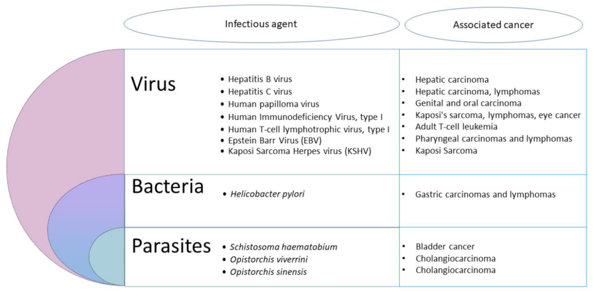
2. Infection, an Important Cause of Cancer

Causal associations between infectious agents and the development of human cancers have already been highlighted [4]. Overall, the total number of cancer attributable to infectious agents in 2002 was estimated at 1.9 million cases, standing for 17.8% of the global cancer burden. Moreover, it has been hypothesized that, by 2050, the majority of human cancers could be due to infections [5,6]. However, proving that infectious agents are causative factors of human cancer remains difficult for many reasons, such as:
  • The periods between primary infection and malignant transformation are frequently very long [7].
  • Even if the majority of the infectious agents associated to human cancers are ubiquitous and common in the human population, only a small proportion of infected individuals develops cancer.
  • Some infections are linked to cancer development as associated risk factors [8].
  • Infectious agents act mainly as indirect oncogenes, without persistence of their genes within the respective cancer host cells. The most common indirect infectious carcinogens are agents causing immunosuppression, such as Human Immunodeficiency Virus (HIV) leading to Kaposi’s sarcoma, or inflammation caused by the bacteria Helicobater pylori, the trematode Schistosoma hematobium and the Hepatitis C and B viruses [7].
  • The main mechanisms by which infectious agents promote cancer are not necessarily involving direct mutagenesis, but instead are due to the complex interactions between hosts and pathogens [8].
  • An infectious agent may trigger the initial events of oncogenesis while being absent in the final tumor [7].
  • Pathogens associated with cancer are directing pathogen-driven processes leading to cell transformation. However, many non-oncogenic pathogens can also regulate these processes, indicating that other factors must be involved [8].
  • In the cases of viruses, oncogenesis can occur through the persistence of the viral genome in a latent form in an infected host cell, either without replication or through integration of the viral genome into a host-cell chromosome [8].
  • Koch’s postulates for proving a causal connection between a particular infectious agent and a disease cannot be applied to many human diseases as it would be unethical to experimentally infect humans with a potentially lethal infectious agent [8].
  • Existing diagnostic tools may not be sensitive enough to link infectious agents with cancer development or testing may occur too long after the exposure [9].
Nevertheless, at least 11 biological agents have presently been recognized by the IARC as major contributors to the global number of cancers in humans (Figure 1).
These agents include viruses, bacteria and helminths. The most important infectious agents worldwide are H. pylori (5.5% of all cancer), the human papilloma viruses (HPVs) (5.2%), the hepatitis B and C viruses (4.9%), the Epstein–Barr virus (EBV) (1%), the HIV together with the human herpes virus 8 (0.9%) and the human T-cell leukemia/lymphoma virus type 1 (HTLV-I) (0.03%) [10]. Other pathogens, including parasites, are also considered carcinogenic agents in human beings. Among helminths, the widespread digenetic trematode S. haematobium has been associated to urinary bladder cancer, and the flukes Opisthorchis viverrini and Clonorchis sinensis are causally linked with cholangiocarcinoma [10]. So, the idea of parasites as a cause of cancer in vertebrates is slowly developing [11]. However, the contribution of intracellular eukaryotic parasites to cancer development has been largely neglected until now [2]. Yet, based on clinical and epidemiological pieces of evidence, many reports underlined a potential association between parasitic protozoan infections and cancer. Hence, the flagellate Trichomonas vaginalis was suspected to be associated with prostate [12] and cervical cancers [13], while the apicomplexan Toxoplasma gondii was suggested to be linked with ocular tumor, meningioma, leukemia and lymphomas [14]. It was also suggested that Plasmodium falciparum could play a co-factor role in the development of Burkitt lymphoma [14]. Nevertheless, only the apicomplexan genera Cryptosporidium and Theileria have been shown to induce cell transformation experimentally [2].
Pathogens use several strategies to target cellular processes during their parasitic interactions with the host cell. The identification of microbial proteins manipulating host functions to promote infection, proliferation and escape defenses has led to great progress in understanding the host’s cellular processes. Continued persistent infection by a pathogen requires host-cell survival, host-cell proliferation, and evasion of the immune system by the pathogen. These pathogen-driven processes are achieved through various mechanisms that interfere with normal cell physiology. Alterations in these normally highly regulated pathways can lead to transforming events that have been described as the ‘hallmarks of cancer’ [5]. Carcinogenic pathogens are also able to target epigenetic mechanisms to divert the host cellular machinery [2]. Interestingly, recent mechanistic studies suggest that apicomplexan eukaryotic intracellular parasites are indeed capable of reproducing some mechanistic aspects of tumorigenesis leading to cancers, either by their infection alone, or by the combination of the parasitic infection with environmental factors [15,16]. There is a growing number of pharmacological studies analyzing the effect of anti-cancer molecules on parasitic diseases and vice versa [17]. Overall, infection seems to play a crucial role in the etiology of cancer. Actually, it was estimated that there would be 26.3% fewer cancers in developing countries and 7.7% in developed countries if cancers associated with infectious diseases were prevented [18].

3. The Special Case of Cryptosporidium: A Public Health Issue

Cryptosporidium is the agent of cryptosporidiosis, an infection resulting from the ingestion of parasite oocysts mainly through the consumption of fecally contaminated food or water, or through direct contact with the infected host [19,20]. Cryptosporidium parvum and C. hominis are the two species responsible for the majority of human cases of cryptosporidiosis. This parasite is considered a major cause of diarrhea worldwide. It causes self-limited watery diarrhea in immunocompetent individuals, but has devastating effects in those who are immunocompromised. In young children, malnutrition, growth and cognitive deficits were reported as sequels of cryptosporidiosis [21,22]. Most strikingly, a cohort study (GEMS) involving 22,500 children in Africa and Asia revealed that Cryptosporidium is one of the four main pathogens responsible for severe diarrhea and mortality in infants and toddlers [23]. This parasite was then considered the second leading cause of death in children due to diarrhea [24]. More recently, the Global Burden of Diseases, Injuries, and Risk Factors Study (GBD) analyzed in 2016 the burden of diarrhea in 195 countries and reported that Cryptosporidium is the fourth leading cause of diarrhea mortality among children under 5 years of age (with 48,301 annual death) [25,26]. It has also been reported that the substantial short-term burden of diarrhea induced by Cryptosporidium infection on childhood growth and well-being is largely underestimated [25,27] probably due to a significant proportion of asymptomatic or mild and self-limiting infections, which consequently remain not diagnosed. In addition, Cryptosporidium species are responsible for numerous waterborne outbreaks of gastrointestinal disease. The most extensive was described in 1993 in Milwaukee, USA, where over 400,000 people became ill (the population of this area was approximately 1.61 million) with 69 deaths [28,29]. The number of outbreaks caused by Cryptosporidium is increasing worldwide since 239 waterborne outbreaks were reported in Europe, Australia and North America between 2011 and 2016 versus only 120 in the same area between 2004 and 2010 [30,31]. The Centers for Disease Control and Prevention (CDC) also published that 32 outbreaks were caused by Cryptosporidium in the United States in 2016 and linked them to swimming pools and water playgrounds against only 13 in 2013 [32]. As a result, an ever-growing number of people would be exposed to this pathogen and not only in developing countries. Despite its prevalence and impact on public health, neither treatment nor vaccine against Cryptosporidium are yet available.

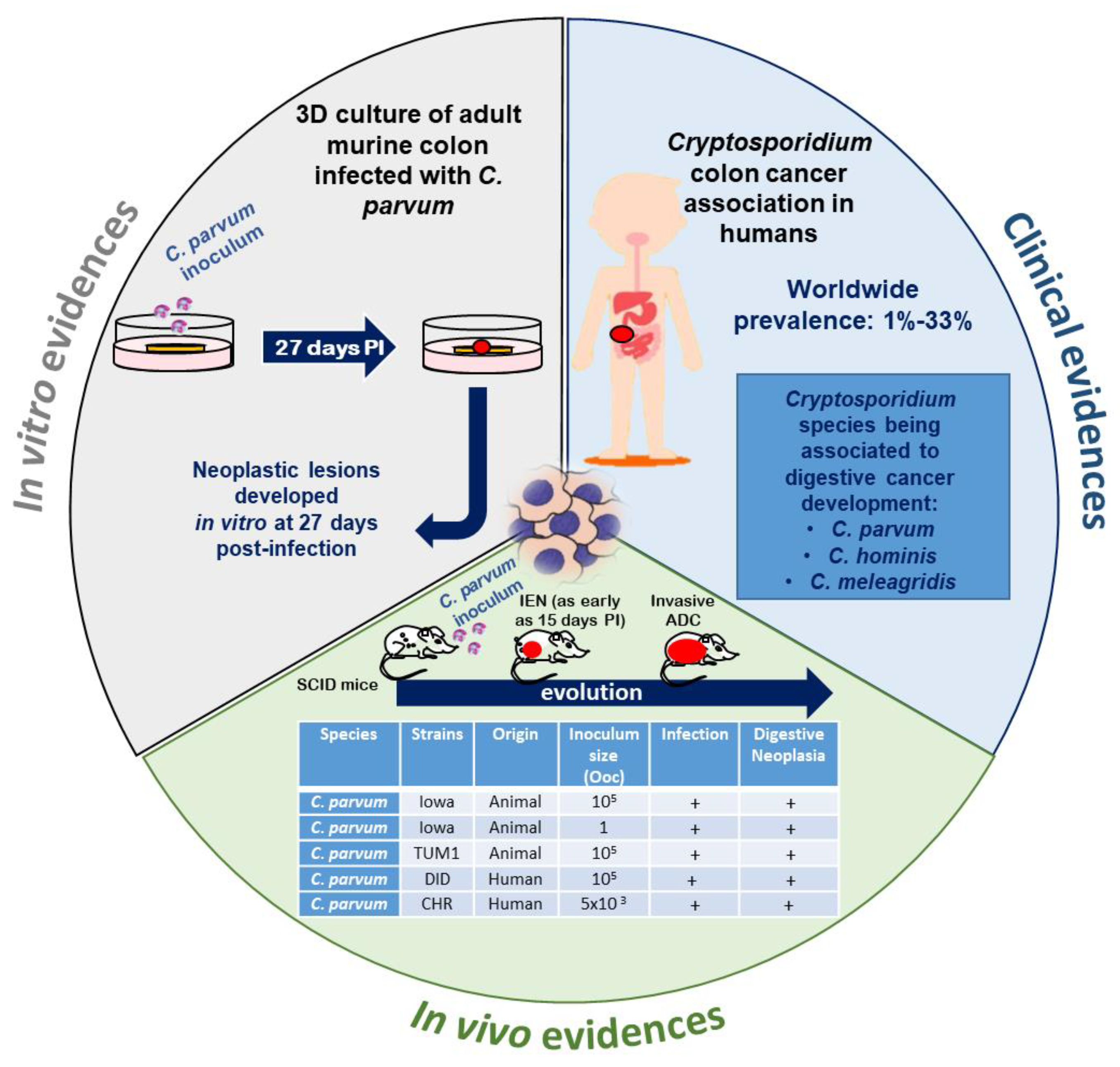
4. Cryptosporidium and Cancer: A Growing Body of Evidence

In various animal groups and in humans, epidemiological and experimental studies tend to reinforce the hypothesis of an association between Cryptosporidium infection and cancer (Figure 2).

4.1. Clinical Studies in Humans

Several growing pieces of clinical evidence about links between cryptosporidiosis and human digestive neoplasia in different populations strengthen the idea of a possible causal relation. The association of cryptosporidiosis and colonic adenocarcinoma was evoked in the case of a Spanish patient presenting both, who died quickly after the onset of clinical manifestations [33]. A cryptosporidiosis case of the biliary tract clinically mimicking a pancreatic cancer in an HIV/Acquired Immunodeficiency Syndrome (AIDS) patient was also described [34]. Other studies reported elevated colon carcinoma risk in AIDS patients with cryptosporidiosis [35] and bile duct carcinoma associated with Cryptosporidium infection in children with X-linked hyper-IgM syndrome and in immunodeficient mice [36,37,38]. Authors of the latter studies proposed that the mutation responsible for this defect might favor the colonization of the biliary epithelium by Cryptosporidium. A chronic infection by this parasite will follow and inflammation may be the cause of the malignant transformation [37,38]. Even if this condition is mainly diagnosed in children, a case of an adult patient with CD40L deficiency who suffered of cholangiocarcinoma arising from sclerosing cholangitis associated with chronic cryptosporidiosis was also reported [39]. In parallel, a clinical study conducted in Lebanon strongly suggested evidence for a link between cryptosporidiosis and colorectal cancer. Indeed, Cryptosporidium infection was identified by PCR in 21% (15/72) of biopsies from patients with recently diagnosed colon neoplasia (including low- or high-grade intraepithelial neoplasia and not invasive/invasive adenocarcinoma) before any treatment compared to 7% (9/146) of biopsies from patients without digestive neoplasia but with persistent gastrointestinal symptoms. The risk of Cryptosporidium infection was thus 4 times higher in the first group. Moreover, the molecular characterization of the corresponding Cryptosporidium isolates allowed for the identification of either C. parvum or C. hominis. In addition, the presence of Cryptosporidium developmental stages in the apical position within the epithelial cells of the intestinal glands was confirmed [40] (Figure 3).
Consistently, various clinical studies conducted in Poland confirm these observations [42,43,44]. On the other hand, in another cohort of immunocompetent patients from Poland with colorectal cancer, the presence of Cryptosporidium was found in one patient out of 145. After genotyping, the presence of C. meleagridis was identified, this being the first study reporting an association of this Cryptosporidium species with colon adenocarcinoma [45]. More recently, a Chinese case control study described a Cryptosporidium infection rate of 17.24% (20/116) in patients with colorectal cancer before chemotherapy. In addition, the same authors reported the presence of the parasite in liver, esophageal and small intestine cancers [46]. A summary of different reports about the link between Cryptosporidium infection and human digestive neoplasia in different populations is shown in Table 1.
Besides direct clinical evidence suggesting the association between Cryptosporidium and digestive cancer development, some studies conveyed that the risk of developing colon carcinoma is significantly elevated among AIDS patients, a group at risk of Cryptosporidium infection [47]. However, Cryptosporidium is an opportunistic agent that causes important morbidity and mortality in persons with immunodeficiency. Therefore, it is possible that immunocompromised people have a higher risk of developing malignancy induced by this parasite, especially when their immunosuppression is more severe. In fact, Cryptosporidium infection has been suggested to be associated with some malignancies such as leukemia [48]. Nevertheless, a meta-analysis recently reported that, even if a positive association was found between Cryptosporidium infection and cancer in general (OR = 3.3; 95 CI: 2.18–4.98), the occurrence of Cryptosporidium infection was mainly related to colorectal cancer (OR = 3.7; 95 CI: 2.10–6.50) but not to other types of malignancies, such as blood cancer [49], suggesting that the parasite is not a special risk to cancer patients. Even if there are several clinical pieces of evidence from different geographical areas, the majority of the articles in the literature are case reports. Further prospective studies should be conducted based on clinical trials using sensitive diagnostic tools for the identification of the parasite.

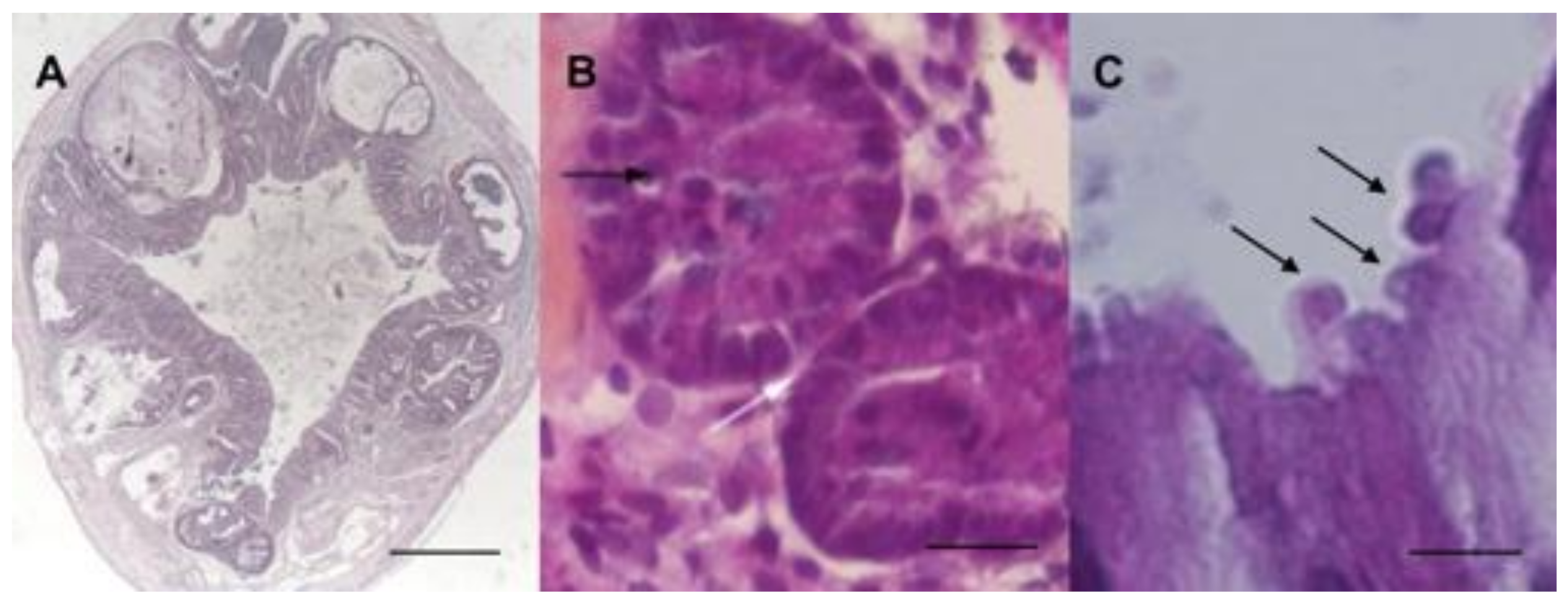
4.2. Natural or Experimental Infection in Animals

Links between non-malignant tumors or atypical histology and Cryptosporidium were already reported in naturally or experimentally infected animals. For example, two studies described an association between aural or aural-pharyngeal polyps and Cryptosporidium infection in iguanas [50,51]. An intestinal metaplasia of the proventriculus was also associated with Cryptosporidium baileyi in a snowy owl [52]. Moreover, histological analysis of twenty-three leopard geckos revealed a Cryptosporidium sp. infection associated with hyperplasia of small and large intestine [53]. To complete this overview, low grade dysplasia in bile ducts has also been reported in an experimental model of IFN-δ knockout mice infected with C. parvum [37]. Nevertheless, no studies, to our knowledge, described an association between Cryptosporidium sp. infection and malignant tumors, until 2007 [54]. Indeed, in order to explore the dynamics of Cryptosporidium infection, an animal model of cryptosporidiosis was developed using corticoid dexamethasone-treated or untreated adult Severe Combined Immunodeficiency (SCID) mice, orally infected with C. parvum or C. muris oocysts. Intriguingly, only C. parvum-infected animals developed ileo-caecal adenocarcinoma as soon as 45 days post-infection even when mice were infected with only one oocyst (Figure 3A) [54,55]. The inoculation of animals was performed with the C. parvum Iowa strain isolated from cattle and which is the reference strain for this Cryptosporidium species (first C. parvum strain to have an entire genome sequenced). However, additional C. parvum strains were tested in the same murine model including “TUM1” (isolated from a calf in the USA), and “Did” and “CHR” (isolated from patients in the Centre Hospitalier Universitaire de Lille, France). These three isolates were found to be more virulent than the Iowa strain [56,57,58]. Indeed, they induced a higher mortality rate, an earlier onset of neoplastic lesions (as soon as 15 days post-infection for the CHR strain), and a more rapid progression to invasive adenocarcinoma. Interestingly, the development of intestinal low- and high-grade dysplasia after only 30 days post-infection with C. parvum in a model of dexamethasone treated immunocompetent Swiss albino mice was reported by others [59]. However, adenocarcinoma development was not detected in this model, probably due to the early time of euthanasia. Similar observations were reported by others using the same mice model [60]. Moreover, the severity of the dysplastic lesions seemed to be correlated with the intensity of oocyst shedding [61].

4.3. In Vitro Models

Recently, a three-dimensional (3D) in-vivo-like culture model from adult murine colon was developed, allowing for biological investigations of Cryptosporidium infection and a better study of its pathophysiology. Indeed, the resulting system allowed for the maintaining of the infection but also the development of low-grade intra-epithelial neoplasia in vitro after only 27 days post-infection (Figure 3B) [41]. This model is currently adapted to human intestinal tissue in order to look for further evidence on the role of C. parvum and/or C. hominis in the induction of human colon cancer.

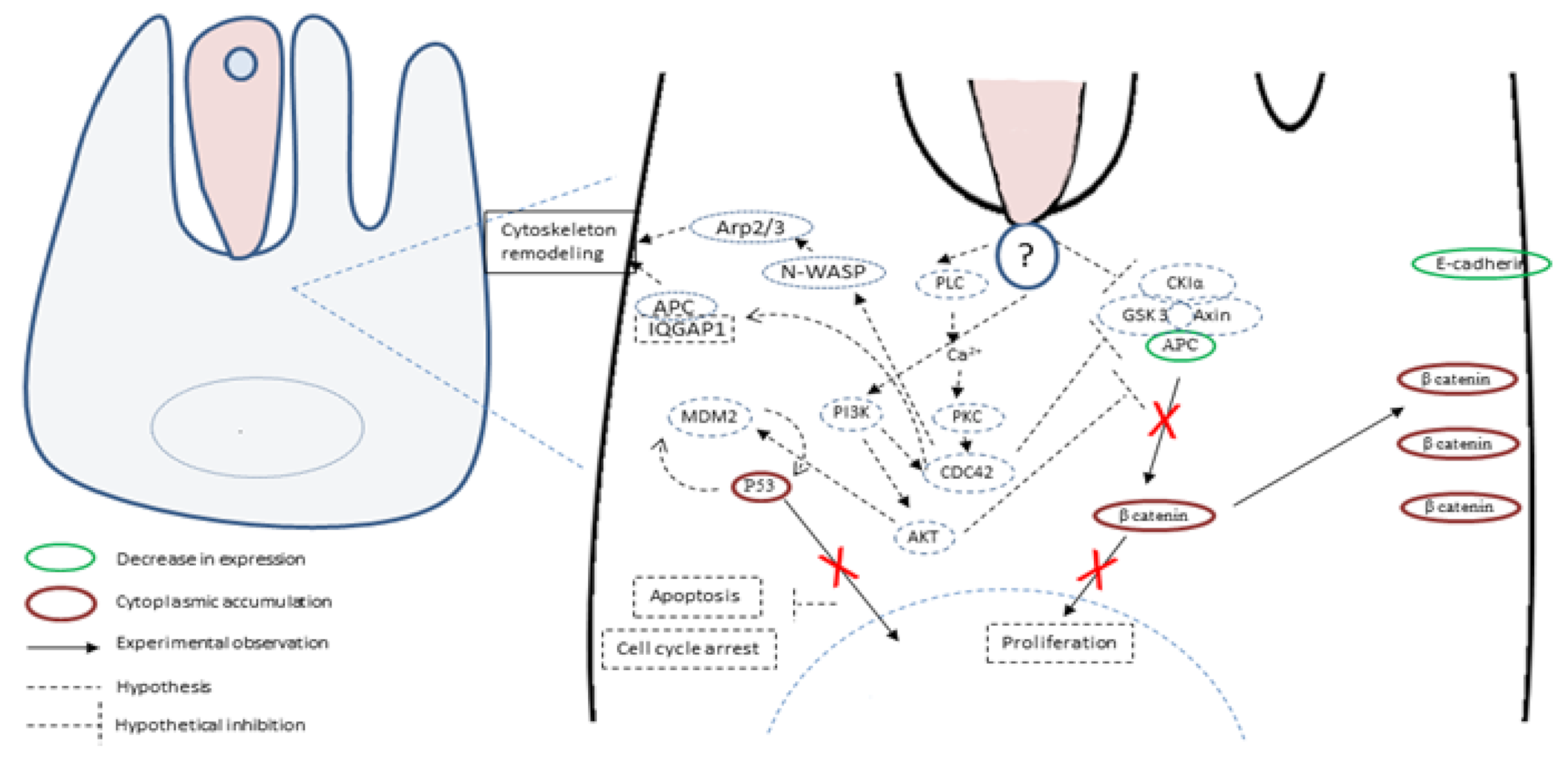
5. Hypotheses about Molecules and Mechanisms Involved in the Induction of Tumorigenesis by Cryptosporidium

The pathophysiological mechanisms of Cryptosporidium infection are multifactorial and not completely understood. Some advances were achieved recently and revealed that the infection by C. parvum induces cytoskeleton remodeling and actin reorganization through the implication of several intracellular signals involving, for example, PI3K, Src, Cdc42 and GTPase [62,63] (Figure 4).
Consistently, signal transduction pathways targeting cell proliferation, cellular junctions and adhesions have also been described in gastric cancer induced by H. pylori [64]. It was also reported that the infection by C. parvum leads to the activation of NF-κB [62], known to induce anti-apoptotic mechanisms and also to transmit oncogenic signals to epithelial cells [65,66]. In addition, microarray assays were recently performed on C. parvum IId Human Ileocaecal Adenocarcinoma (HCT-8)-infected cells. A differential profile of mRNAs was found between infected and non-infected cells. Indeed, mRNAs of the Wnt and hedgehog signaling pathways were significantly differentially expressed in infected cells compared to not infected ones [67]. Noticeably, these two pathways are also involved in the development and progression of colorectal cancer [68]. Despite the growing evidence about the hijacking of cellular pathways potentially being involved in cancer onset, this information has rarely been linked to the tumorigenic potential of the parasite. To our knowledge, only one study tried to decipher this process and highlighted the important role of the Wnt signaling pathway and the alteration of the cytoskeleton in the carcinogenic process induced by C. parvum experimental infection [69]. Indeed, the immunohistochemical analysis of ileocecal region sections embedded in paraffin from C. parvum infected versus non-infected mice showed alterations in APC, β-catenin, P53 and E-cadherin expression [69]. APC and E-cadherin labelings were decreased while those of β-catenin and P53 were increased in the cytoplasm of epithelial cells. In addition, the immunofluorescence analysis of these histological sections confirmed a membranous and juxtamembraneous localization of β-catenin without nucleus translocation, suggesting an involvement of the non-canonical Wnt pathway [69]. However, unlike Helicobacter pylori, for which bacterial virulence factors associated to the gastric cancer outcome were identified, such as cytotoxin associated gene A (CagA), vacuolating cytotoxin A (VacA) and outer inflammatory protein A (OipA) [70], virulence factors implied in the C. parvum-induced carcinogenic process remain unknown. The whole genome sequences of different species and isolates of Cryptosporidium are now available [71,72,73]. Hence, a comparative genomic analysis of different C. parvum strains (Did, TUM1, CHR and Iowa, the reference strain) with variable virulence was recently performed [58]. Overall, 125 common SNVs (single nucleotide variation) (corresponding to 90 CDSs (coding sequences) were found in the three more virulent strains (Did, TUM1 and CHR) compared to Iowa strain. The majority of these genes are over-expressed in the intracellular stages of the parasite. In addition, mucins, transporters (ABC and ATPase3) and cysteine proteases were also found, these genes being already described as virulence factors. This study also reported new potential factors involved in the virulence of C. parvum such as various phosphatases (PP2A, Cdc14) and a histone-lysine N-methyltransferase. Further investigations are needed to elucidate carcinogenic mechanisms induced by C. parvum. In particular, the biological function study of the potential virulent and/or carcinogenic factors identified could be facilitated by the use of genome-editing tools such as CRISPR/Cas9 [58].

6. Conclusions and Future Directions

Is colon cancer a cause or a consequence of Cyptosporidium infection? We presented an updated picture of the link between digestive cancer and infection by this parasite. Available experimental and clinical data synthesized herein suggest that the parasite is able to employ strategies to target cellular processes during its complex interactions with host cells, leading to a parasite-induced transformation. However, the fact that Cryptosporidium is an opportunistic agent is one of the main difficulties for proving its role in the induction of human digestive cancers. In addition, available experimental models with little or no immune response are not necessarily reflecting the human condition. Despite several pieces of evidence about associations between cryptosporidiosis and digestive neoplasia, it seems that not enough attention has been paid to the tumorigenic power of this protozoan parasite. Therefore, it is necessary to demonstrate a direct causal link, and to identify virulent factors and carcinogenic mechanisms responsible for the epithelial cell transformation. A potential solution would be to combine recent advances in 3D culture models, comparative genomic studies and transfection methods (CRISPR/Cas9 system) together with further clinical trials using sensitive diagnostic tools. If the causal link between Cryptosporidium and human cancer is clearly established, a great number of digestive cancers could be prevented using public health measures to reduce the risk of Cryptosporidium infection. In addition, this could entice researchers to explore new therapeutic targets and vaccines in order to clear or prevent the infection, thereby saving hundreds of thousand children from severe diarrhoea and mortality [24]. Research into this topic is urgently needed since the incidence of Cryptosporidium infection is increasing worldwide.

7. Search Strategy and Selection Criteria

We selected articles from PubMed and Google Scholar using the research terms “Cryptosporidium”, “cancer”, “oncogenesis”, “infection and cancer”, “colorectal cancer”, “cryptosporidiosis”, “parasites and cancer”, “Cryptosporidium and cancer”, “cancer causes”, “epigenetics and cancer”, “Cryptosporidium and virulence factors”, “digestive neoplasia”, “colon adenocarcinoma”, “cancer and mechanisms”. Reviews, clinical, epidemiological and experimental data were taking into account without restriction of date or language. Preference was given to the articles published within the past 20 years.

Author Contributions

Conceptualization, M.S., E.V., G.C., and S.B.-V.; Formal analysis, M.S., M.B., C.C., M.C., E.V., G.C., and S.B.-V.; Funding acquisition, E.V., G.C., and S.B.-V.; Resources, C.C., G.C. and S.B.-V.; Writing—original draft, M.S., E.V., G.C., and S.B.-V.; Writing—review and editing, M.S., M.B., C.C., M.C., E.V., G.C., and S.B.-V. All authors have read and agreed to the published version of the manuscript.

Funding

This research was supported by the Centre National de la Recherche Scientifique (CNRS), the Institut National de la Santé et de la Recherche Médicale, the Institute Pasteur de Lille, the Université de Lille, the Groupement des Hôpitaux de l’Institut Catholique de Lille, the Institut Catholique de Lille, the Centre hospitalier régional universitaire de Lille and the Plan Cancer/ Institut National de la Santé et de la Recherche Médicale (Inserm): “Epigenetics and cancer 2015” (PARACAN, EPIG2015069). M.S. was supported by a PhD fellowship from the Université de Lille.

Conflicts of Interest

The authors declare no conflict of interest.

References

  1. Bray, F.; Ferlay, J.; Soerjomataram, I.; Siegel, R.L.; Torre, L.A.; Jemal, A. Global cancer statistics 2018: GLOBOCAN estimates of incidence and mortality worldwide for 36 cancers in 185 countries. CA Cancer J. Clin. 2018, 68, 394–424. [Google Scholar] [CrossRef] [PubMed] [Green Version]
  2. Cheeseman, K.; Certad, G.; Weitzman, J.B. Parasites et cancer: Existe-t-il un lien? Méd./Sci. 2016, 32, 867–873. [Google Scholar] [CrossRef] [PubMed]
  3. Whiteman, D.C.; Wilson, L.F. The fractions of cancer attributable to modifiable factors: A global review. Cancer Epidemiol. 2016, 44, 203–221. [Google Scholar] [CrossRef] [PubMed]
  4. Benamrouz, S.; Conseil, V.; Creusy, C.; Calderon, E.; Dei-Cas, E.; Certad, G. Parasites and malignancies, a review, with emphasis on digestive cancer induced by Cryptosporidium parvum (Alveolata: Apicomplexa). Parasite 2012, 19, 101–115. [Google Scholar] [CrossRef] [Green Version]
  5. Bañuls, A.L.; Thomas, F.; Renaud, F. Of parasites and men. Infect. Genet. Evol. 2013, 20, 61–70. [Google Scholar] [CrossRef] [PubMed] [Green Version]
  6. Bouvard, V.; Baan, R.; Straif, K.; Grosse, Y.; Secretan, B.; El Ghissassi, F.; Benbrahim-Tallaa, L.; Guha, N.; Freeman, C.; Galichet, L.; et al. A review of human carcinogens—Part B: Biological agents. Lancet Oncol. 2009, 10, 321–322. [Google Scholar] [CrossRef]
  7. Zur Hausen, H. The search for infectious causes of human cancers: Where and why. Virology 2009, 392, 1–10. [Google Scholar] [CrossRef] [Green Version]
  8. Dalton-Griffin, L.; Kellam, P. Infectious causes of cancer and their detection. J. Biol. 2009, 8, 67. [Google Scholar] [CrossRef]
  9. O’Connor, S.M.; Taylor, C.E.; Hughes, J.M. Emerging Infectious Determinants of Chronic Diseases. Emerg. Infect. Dis. 2006, 12, 1051–1057. [Google Scholar] [CrossRef] [Green Version]
  10. IARC Working Group on the Evaluation of Carcinogenic Risks to Humans Biological Agents, Volume 100 B, a Review of Human Carcinogens Lyon, France. 2012. Available online: http://monographs.iarc.fr (accessed on 1 August 2020).
  11. Ewald, P.W.; Swain Ewald, H.A. Infection and cancer in multicellular organisms. Philos. Trans. R. Soc. B Biol. Sci. 2015, 370, 20140224. [Google Scholar] [CrossRef]
  12. Stark, J.R.; Judson, G.; Alderete, J.F.; Mundodi, V.; Kucknoor, A.S.; Giovannucci, E.L.; Platz, E.A.; Sutcliffe, S.; Fall, K.; Kurth, T.; et al. Prospective study of Trichomonas vaginalis infection and prostate cancer incidence and mortality: Physicians’ Health Study. J. Natl. Cancer Inst. 2009, 101, 1406–1411. [Google Scholar] [CrossRef] [PubMed]
  13. Yang, S.; Zhao, W.; Wang, H.; Wang, Y.; Li, J.; Wu, X. Trichomonas vaginalis infection-associated risk of cervical cancer: A meta-analysis. Eur. J. Obstet. Gynecol. Reprod. Biol. 2018, 228, 166–173. [Google Scholar] [CrossRef] [PubMed]
  14. Khurana, S.; Dubey, M.L.; Malla, N. Association of parasitic infections and cancers. Indian J. Med. Microbiol. 2005, 23, 74–79. [Google Scholar] [PubMed]
  15. Marsolier, J.; Perichon, M.; DeBarry, J.D.; Villoutreix, B.O.; Chluba, J.; Lopez, T.; Garrido, C.; Zhou, X.Z.; Lu, K.P.; Fritsch, L.; et al. Theileria parasites secrete a prolyl isomerase to maintain host leukocyte transformation. Nature 2015, 520, 378–382. [Google Scholar] [CrossRef] [Green Version]
  16. Robbiani, D.F.; Deroubaix, S.; Feldhahn, N.; Oliveira, T.Y.; Callen, E.; Wang, Q.; Jankovic, M.; Silva, I.T.; Rommel, P.C.; Bosque, D.; et al. Plasmodium infection promotes genomic instability and AID-dependent B cell lymphoma. Cell 2015, 162, 727–737. [Google Scholar] [CrossRef] [Green Version]
  17. Checkley, W.; White, A.C.; Jaganath, D.; Arrowood, M.J.; Chalmers, R.M.; Chen, X.M.; Fayer, R.; Griffiths, J.K.; Guerrant, R.L.; Hedstrom, L.; et al. A review of the global burden, novel diagnostics, therapeutics, and vaccine targets for Cryptosporidium. Lancet Infect. Dis. 2015, 15, 85–94. [Google Scholar] [CrossRef] [Green Version]
  18. Plummer, M.; de Martel, C.; Vignat, J.; Ferlay, J.; Bray, F.; Franceschi, S. Global burden of cancers attributable to infections in 2012: A synthetic analysis. Lancet Glob. Health 2016, 4, e609–e616. [Google Scholar] [CrossRef] [Green Version]
  19. Putignani, L.; Menichella, D. Global distribution, public health and clinical impact of the protozoan pathogen Cryptosporidium. Interdiscip. Perspect. Infect. Dis. 2010, 2010, 753512. [Google Scholar] [CrossRef] [Green Version]
  20. Ryan, U.; Fayer, R.; Xiao, L. Cryptosporidium species in humans and animals: Current understanding and research needs. Parasitology 2014, 141, 1667–1685. [Google Scholar] [CrossRef] [Green Version]
  21. Bhalchandra, S.; Cardenas, D.; Ward, H.D. Recent breakthroughs and ongoing limitations in Cryptosporidium research. F1000Research 2018, 7, 1380. [Google Scholar] [CrossRef] [Green Version]
  22. Lilja, M.; Widerström, M.; Lindh, J. Persisting post-infection symptoms 2 years after a large waterborne outbreak of Cryptosporidium hominis in northern Sweden. BMC Res. Notes 2018, 11, 625. [Google Scholar] [CrossRef] [PubMed] [Green Version]
  23. Kotloff, K.L.; Nataro, J.P.; Blackwelder, W.C.; Nasrin, D.; Farag, T.H.; Panchalingam, S.; Wu, Y.; Sow, S.; Sur, D.; Breiman, R.F.; et al. Burden and aetiology of diarrhoeal disease in infants and young children in developing countries (the Global Enteric Multicenter Study, GEMS): A prospective, case-control study. Lancet 2013, 382, 209–222. [Google Scholar] [CrossRef]
  24. Gibson, A.R.; Striepen, B. Cryptosporidium. Curr. Biol. 2018, 28, R193–R194. [Google Scholar] [CrossRef] [PubMed] [Green Version]
  25. Khalil, I.A.; Troeger, C.; Rao, P.C.; Blacker, B.F.; Brown, A.; Brewer, T.G.; Colombara, D.V.; De Hostos, E.L.; Engmann, C.; Guerrant, R.L.; et al. Morbidity, mortality, and long-term consequences associated with diarrhoea from Cryptosporidium infection in children younger than 5 years: A meta-analyses study. Lancet Glob. Health 2018, 6, e758–e768. [Google Scholar] [CrossRef] [Green Version]
  26. GBD 2016 Diarrhoeal Disease Collaborators. Estimates of the global, regional, and national morbidity, mortality, and aetiologies of diarrhoea in 195 countries: A systematic analysis for the Global Burden of Disease Study 2016. Lancet Infect. Dis. 2018, 18, 1211–1228. [Google Scholar] [CrossRef] [Green Version]
  27. Widerström, M.; Schönning, C.; Lilja, M.; Lebbad, M.; Ljung, T.; Allestam, G.; Ferm, M.; Björkholm, B.; Hansen, A.; Hiltula, J.; et al. Large outbreak of Cryptosporidium hominis infection transmitted through the public water supply, Sweden. Emerg. Infect. Dis. 2014, 20, 581–589. [Google Scholar]
  28. Corso, P.S.; Kramer, M.H.; Blair, K.A.; Addiss, D.G.; Davis, J.P.; Haddix, A.C. Cost of illness in the 1993 waterborne Cryptosporidium outbreak, Milwaukee, Wisconsin. Emerg. Infect. Dis. 2003, 9, 426–431. [Google Scholar] [CrossRef]
  29. Mac Kenzie, W.R.; Hoxie, N.J.; Proctor, M.E.; Gradus, M.S.; Blair, K.A.; Peterson, D.E.; Kazmierczak, J.J.; Addiss, D.G.; Fox, K.R.; Rose, J.B.; et al. A massive outbreak in Milwaukee of Cryptosporidium infection transmitted through the public water supply. N. Engl. J. Med. 1994, 331, 161–167. [Google Scholar] [CrossRef]
  30. Baldursson, S.; Karanis, P. Waterborne transmission of protozoan parasites: Review of worldwide outbreaks—An update 2004–2010. Water Res. 2011, 45, 6603–6614. [Google Scholar] [CrossRef]
  31. Efstratiou, A.; Ongerth, J.E.; Karanis, P. Waterborne transmission of protozoan parasites: Review of worldwide outbreaks—An update 2011–2016. Water Res. 2017, 114, 14–22. [Google Scholar] [CrossRef]
  32. CDC. Crypto Outbreaks Linked to Swimming Have Doubled since 2014. 2017. Available online: https://www.cdc.gov/media/releases/2017/p0518-Cryptosporidium-outbreaks.html (accessed on 1 August 2020).
  33. Izquierdo, J.; Antúnez, I.; Calderón, M.T.; Pérez Giraldo, C.; Muñoz Sanz, A. Diarrhea caused by Cryptosporidium and colonic neoplasia]. Rev. Clínica Española 1988, 182, 393–394. [Google Scholar]
  34. Souza, L.D.R.D.; Rodrigues, M.A.M.; Morceli, J.; Kemp, R.; Mendes, R.P. Cryptosporidiosis of the biliary tract mimicking pancreatic cancer in an AIDS patient. Rev. Soc. Bras. Med. Trop. 2004, 37, 182–185. [Google Scholar] [CrossRef] [PubMed] [Green Version]
  35. Shebl, F.M.; Engels, E.A.; Goedert, J.J. Opportunistic intestinal infections and risk of colorectal cancer among people with AIDS. AIDS Res. Hum. Retrovir. 2012, 28, 994–999. [Google Scholar] [CrossRef] [PubMed] [Green Version]
  36. Hayward, A.R.; Levy, J.; Facchetti, F.; Notarangelo, L.; Ochs, H.D.; Etzioni, A.; Bonnefoy, J.Y.; Cosyns, M.; Weinberg, A. Cholangiopathy and tumors of the pancreas, liver, and biliary tree in boys with X-linked immunodeficiency with hyper-IgM. J. Immunol. 1997, 158, 977–983. [Google Scholar]
  37. Stephens, J.; Cosyns, M.; Jones, M.; Hayward, A.R. Liver and bile duct pathology following Cryptosporidium parvum infection of immunodeficient mice. Hepatology 1999, 30, 27–35. [Google Scholar] [CrossRef]
  38. Leven, E.A.; Maffucci, P.; Ochs, H.D.; Scholl, P.R.; Buckley, R.H.; Fuleihan, R.L.; Geha, R.S.; Cunningham, C.K.; Bonilla, F.A.; Conley, M.E.; et al. Hyper IgM syndrome: A report from the USIDNET registry. J. Clin. Immunol. 2016, 36, 490–501. [Google Scholar] [CrossRef] [PubMed] [Green Version]
  39. Rahman, M.; Chapel, H.; Chapman, R.W.; Collier, J.D. Cholangiocarcinoma complicating secondary sclerosing cholangitis from cryptosporidiosis in an adult patient with CD40 ligand deficiency: Case report and review of the literature. Int. Arch. Allergy Immunol. 2012, 159, 204–208. [Google Scholar] [CrossRef]
  40. Osman, M.; Benamrouz, S.; Guyot, K.; Baydoun, M.; Frealle, E.; Chabe, M.; Gantois, N.; Delaire, B.; Goffard, A.; Aoun, A.; et al. High association of Cryptosporidium spp. infection with colon adenocarcinoma in Lebanese patients. PLoS ONE 2017, 12, e0189422. [Google Scholar] [CrossRef]
  41. Baydoun, M.; Vanneste, S.B.; Creusy, C.; Guyot, K.; Gantois, N.; Chabe, M.; Delaire, B.; Mouray, A.; Baydoun, A.; Forzy, G.; et al. Three-dimensional (3D) culture of adult murine colon as an in vitro model of cryptosporidiosis: Proof of concept. Sci. Rep. 2017, 7, 17288. [Google Scholar] [CrossRef] [Green Version]
  42. Sulzyc-Bielicka, V.; Kuźna-Grygiel, W.; Kołodziejczyk, L.; Bielicki, D.; Kładny, J.; Stepień-Korzonek, M.; Telatyńska-Smieszek, B. Cryptosporidiosis in patients with colorectal cancer. J. Parasitol. 2007, 93, 722–724. [Google Scholar] [CrossRef]
  43. Sulżyc-Bielicka, V.; Kołodziejczyk, L.; Jaczewska, S.; Bielicki, D.; Kładny, J.; Safranow, K. Prevalence of Cryptosporidium sp. in patients with colorectal cancer. Pol. Przegl. Chir. 2012, 84, 348–351. [Google Scholar] [CrossRef] [PubMed]
  44. Sulżyc-Bielicka, V.; Kołodziejczyk, L.; Jaczewska, S.; Bielicki, D.; Safranow, K.; Bielicki, P.; Kładny, J.; Rogowski, W. Colorectal cancer and Cryptosporidium spp. infection. PLoS ONE 2018, 13, e0195834. [Google Scholar] [CrossRef] [PubMed] [Green Version]
  45. Kopacz, Ż.; Kváč, M.; Karpiński, P.; Hendrich, A.B.; Sąsiadek, M.M.; Leszczyński, P.; Sak, B.; McEvoy, J.; Kicia, M. The First Evidence of Cryptosporidium meleagridis infection in a colon adenocarcinoma from an immunocompetent patient. Front. Cell. Infect. Microbiol. 2019, 9, 35. [Google Scholar] [CrossRef] [PubMed]
  46. Zhang, N.; Yu, X.; Zhang, H.; Cui, L.; Li, X.; Zhang, X.; Gong, P.; Li, J.; Li, Z.; Wang, X.; et al. Prevalence and genotyping of Cryptosporidium parvum in gastrointestinal cancer patients. J. Cancer 2020, 11, 3334–3339. [Google Scholar] [CrossRef] [PubMed] [Green Version]
  47. Patel, P.; Hanson, D.L.; Sullivan, P.S.; Novak, R.M.; Moorman, A.C.; Tong, T.C.; Holmberg, S.D.; Brooks, J.T. Adult and adolescent spectrum of disease project and HIV outpatient study investigators. Incidence of types of cancer among HIV-infected persons compared with the general population in the United States, 1992–2003. Ann. Intern. Med. 2008, 148, 728–736. [Google Scholar] [CrossRef] [PubMed]
  48. Hunter, P.R.; Nichols, G. Epidemiology and clinical features of Cryptosporidium infection in immunocompromised patients. Clin. Microbiol. Rev. 2002, 15, 145–154. [Google Scholar] [CrossRef] [PubMed] [Green Version]
  49. Kalantari, N.; Gorgani-Firouzjaee, T.; Ghaffari, S.; Bayani, M.; Ghaffari, T.; Chehrazi, M. Association between Cryptosporidium infection and cancer: A systematic review and meta-analysis. Parasitol. Int. 2020, 74, 101979. [Google Scholar] [CrossRef]
  50. Uhl, E.W.; Jacobson, E.; Bartick, T.E.; Micinilio, J.; Schimdt, R. Aural-pharyngeal polyps associated with Cryptosporidium infection in three iguanas (Iguana iguana). Vet. Pathol. 2001, 38, 239–242. [Google Scholar]
  51. Fitzgerald, S.D.; Moisan, P.G.; Bennett, R. Aural polyp associated with cryptosporidiosis in an iguana (Iguana Iguana ). J. Vet. Diagn. Investig. 1998, 10, 179–180. [Google Scholar] [CrossRef] [Green Version]
  52. Nakagun, S.; Horiuchi, N.; Sugimoto, M.; Tomikawa, S.; Watanabe, K.; Kobayashi, Y. Proventriculitis associated with Cryptosporidium baileyi in a snowy owl (Bubo scandiacus) and its epidemiological investigation. J. Parasitol. 2017, 103, 451–457. [Google Scholar] [CrossRef]
  53. Terrell, S.P.; Uhl, E.W.; Funk, R.S. Proliferative enteritis in leopard geckos (Eublepharis macularius) associated with Cryptosporidium sp. infection. J. Zoo Wildl. Med. 2003, 34, 69–75. [Google Scholar] [PubMed]
  54. Certad, G.; Ngouanesavanh, T.; Guyot, K.; Gantois, N.; Chassat, T.; Mouray, A.; Fleurisse, L.; Pinon, A.; Cailliez, J.C.; Dei-Cas, E.; et al. Cryptosporidium parvum, a potential cause of colic adenocarcinoma. Infect. Agent Cancer 2007, 2, 22. [Google Scholar] [CrossRef]
  55. Benamrouz, S.; Guyot, K.; Gazzola, S.; Mouray, A.; Chassat, T.; Delaire, B.; Chabé, M.; Gosset, P.; Viscogliosi, E.; Dei-Cas, E.; et al. Cryptosporidium parvum infection in SCID mice infected with only one oocyst: qPCR assessment of parasite replication in tissues and development of digestive cancer. PLoS ONE 2012, 7, e51232. [Google Scholar] [CrossRef]
  56. Certad, G.; Creusy, C.; Guyot, K.; Mouray, A.; Chassat, T.; Delaire, B.; Pinon, A.; Sitja-Bobadilla, A.; Alvarez-Pellitero, P.; Praet, M.; et al. Fulminant cryptosporidiosis associated with digestive adenocarcinoma in SCID mice infected with Cryptosporidium parvum TUM1 strain. Int. J. Parasitol. 2010, 40, 1469–1475. [Google Scholar] [CrossRef] [PubMed]
  57. Certad, G.; Benamrouz, S.; Guyot, K.; Mouray, A.; Chassat, T.; Flament, N.; Delhaes, L.; Coiteux, V.; Delaire, B.; Praet, M.; et al. Fulminant cryptosporidiosis after near-drowning: A human Cryptosporidium parvum strain implicated in invasive gastrointestinal adenocarcinoma and cholangiocarcinoma in an experimental model. Appl. Environ. Microbiol. 2012, 78, 1746–1751. [Google Scholar] [CrossRef] [PubMed] [Green Version]
  58. Audebert, C.; Bonardi, F.; Caboche, S.; Guyot, K.; Touzet, H.; Merlin, S.; Gantois, N.; Creusy, C.; Meloni, D.; Mouray, A.; et al. Genetic basis for virulence differences of various Cryptosporidium parvum carcinogenic isolates. Sci. Rep. 2020, 10, 7316. [Google Scholar] [CrossRef] [PubMed]
  59. Abdou, A.G.; Harba, N.M.; Afifi, A.F.; Elnaidany, N.F. Assessment of Cryptosporidium parvum infection in immunocompetent and immunocompromised mice and its role in triggering intestinal dysplasia. Int. J. Infect. Dis. 2013, 17, e593–e600. [Google Scholar] [CrossRef] [Green Version]
  60. Mostafa, N.E.; Abdel Hamed, E.F.; Fawzy, E.M.; Zalat, R.S.; Rashed, H.E.; Mohamed, S.Y. The new trend in the treatment of experimental cryptosporidiosis and the resulting intestinal dysplasia. Color. Cancer 2018, 7, CRC06. [Google Scholar] [CrossRef] [Green Version]
  61. Certad, G.; Creusy, C.; Ngouanesavanh, T.; Guyot, K.; Gantois, N.; Mouray, A.; Chassat, T.; Flament, N.; Fleurisse, L.; Pinon, A.; et al. Development of Cryptosporidium parvum-induced gastrointestinal neoplasia in severe combined immunodeficiency (SCID) mice: Severity of lesions is correlated with infection intensity. Am. J. Trop. Med. Hyg. 2010, 82, 257–265. [Google Scholar] [CrossRef] [Green Version]
  62. O’Hara, S.P.; Chen, X.M. The cell biology of Cryptosporidium infection. Microbes Infect. 2011, 13, 721–730. [Google Scholar] [CrossRef] [Green Version]
  63. Certad, G.; Viscogliosi, E.; Chabé, M.; Cacciò, S.M. Pathogenic Mechanisms of Cryptosporidium and Giardia. Trends Parasitol. 2017, 33, 561–576. [Google Scholar] [CrossRef] [PubMed]
  64. Schneider, S.; Weydig, C.; Wessler, S. Targeting focal adhesions: Helicobacter pylori-host communication in cell migration. Cell Commun. Signal. 2008, 6, 2. [Google Scholar] [CrossRef] [PubMed] [Green Version]
  65. Shostak, K.; Chariot, A. EGFR and NF-κB: Partners in cancer. Trends Mol. Med. 2015, 21, 385–393. [Google Scholar] [CrossRef] [PubMed]
  66. Ding, L.; Gu, H.; Lan, Z.; Lei, Q.; Wang, W.; Ruan, J.; Yu, M.; Lin, J.; Cui, Q. Downregulation of cyclooxygenase-1 stimulates mitochondrial apoptosis through the NF-κB signaling pathway in colorectal cancer cells. Oncol. Rep. 2018, 41, 559–569. [Google Scholar] [CrossRef] [PubMed] [Green Version]
  67. Liu, T.L.; Fan, X.C.; Li, Y.H.; Yuan, Y.J.; Yin, Y.L.; Wang, X.T.; Zhang, L.X.; Zhao, G.H. Expression profiles of mRNA and lncRNA in HCT-8 cells infected with Cryptosporidium parvum IId subtype. Front. Microbiol. 2018, 9, 1409. [Google Scholar] [CrossRef] [PubMed]
  68. Tiwari, A.; Saraf, S.; Verma, A.; Panda, K.; Jain, S.K. Novel targeting approaches and signaling pathways of colorectal cancer: An insight. World J. Gastroenterol. 2018, 24, 4428–4435. [Google Scholar] [CrossRef]
  69. Benamrouz, S.; Conseil, V.; Chabé, M.; Praet, M.; Audebert, C.; Blervaque, R.; Guyot, K.; Gazzola, S.; Mouray, A.; Chassat, T.; et al. Cryptosporidium parvum-induced ileo-caecal adenocarcinoma and Wnt signaling in a mouse model. Dis. Model. Mech. 2014, 7, 693–700. [Google Scholar] [CrossRef] [Green Version]
  70. Yamaoka, Y. Mechanisms of disease: Helicobacter pylori virulence factors. Nat. Rev. Gastroenterol. Hepatol. 2010, 7, 629–641. [Google Scholar] [CrossRef] [Green Version]
  71. Xu, P.; Widmer, G.; Wang, Y.; Ozaki, L.S.; Alves, J.M.; Serrano, M.G.; Puiu, D.; Manque, P.; Akiyoshi, D.; Mackey, A.J.; et al. The genome of Cryptosporidium hominis. Nature 2004, 431, 1107–1112. [Google Scholar] [CrossRef]
  72. Puiu, D.; Enomoto, S.; Buck, G.A.; Abrahamsen, M.S.; Kissinger, J.C. CryptoDB: The Cryptosporidium genome resource. Nucleic Acids Res. 2004, 32, D329–D331. [Google Scholar] [CrossRef]
  73. Hadfield, S.J.; Pachebat, J.A.; Swain, M.T.; Robinson, G.; Cameron, S.J.; Alexander, J.; Hegarty, M.J.; Elwin, K.; Chalmers, R.M. Generation of whole genome sequences of new Cryptosporidium hominis and Cryptosporidium parvum isolates directly from stool samples. BMC Genom. 2015, 16, 650. [Google Scholar] [CrossRef] [PubMed] [Green Version]
Figure 1. Infectious agents recognized by the International Agency for Research on Cancer (IARC) as major contributors to the global number of cancers.
Microorganisms 08 01665 g001
Figure 2. Recent experimental and clinical approaches contributing to expanding the understanding of the role of Cryptosporidium in the induction of digestive neoplasia. PI: post-infection, IEN: intraepithelial neoplasia, ADC: adenocarcinoma, Ooc: oocysts. Source of pictograms: https://fr.freepik.com/photos-vecteurs-libre/banner”>Banner vecteur créé par pch.vector—fr.freepik.com</a>.
Microorganisms 08 01665 g002
Figure 3. (A) Adenocarcinoma of the ileo-caecal region in a Dex-treated Severe Combined Immunodeficiency (SCID) mouse after 62 days of C. parvum infection. Scale bar, 1000 μm. (B) Low-grade intraepithelial neoplasia in a murine colonic explant after 27 days of C. parvum infection characterized by reduction of the interglandular space (white arrow), and loss of nuclear polarity with slight pseudostratification (black arrow). Scale bar, 25  µm. (C) Cryptosporidium developmental stages were observed in apical position (arrows) within the epithelial cells in a human colon adenocarcinoma of a Lebanese patient. Scale bar, 5  µm. (Hematoxyline and Eosine). Photomicrographs (B,C) were slightly modified from [41] and [40], respectively.
Microorganisms 08 01665 g003
Figure 4. Hypothetical host-cell signaling pathways linking Cryptosporidium parvum infection and tumorigenesis. Different studies have shown that C. parvum induces cytoskeleton remodeling and actin reorganization through the implication of several intracellular signals, including phosphatidylinositol 3-kinase (PI3K) and Cdc42. In addition, in an experimental model, Adenomatous Polyposis Coli (APC) and E-cadherin labelings were decreased while β-catenin and the P53 tumor suppressor labelings were increased in the cytoplasm of C. parvum-infected epithelial cells. β-catenin was found localized in a juxtamembraneous position, suggesting a role of the non-canonical Wnt pathway in this transformation process. P53 was not translocated into the nucleus, and its labeling increased in the cytoplasm where P53 could regulate key metabolic pathways associated to apoptosis and cell cycle arrest.
Microorganisms 08 01665 g004
Table 1. Associations between Cryptosporidium infection and human digestive neoplasia in different populations.
Type of CancerType of StudyGeographic LocalizationClinical Sample:Laboratory Methodn (%)p-ValueImmuno-SuppressionReference
Colonic adenocarcinomaCase reportSpainNot reported1NA aNo[33]
Pancreatic cancerCase reportBrazilTissues/Microscopical observation1NA aHIV/AIDS[34]
Colorectal cancer(Adenocarcinoma)Data matching between HIV/AIDS and cancer registry databases in 16 U.S. statesUnited StatesTissues/Microscopical observation3/269 (1%)0.70HIV/AIDS[35]
Colorectalsquamous cell carcinomaData matching between HIV/AIDS and cancer registry databases in 16 U.S. statesUnited StatesTissues/Microscopical observation1/8 (12.5%)0.02 bHIV/AIDS[35]
Uncommon colorectal cancers)Data matching between HIV/AIDS and cancer registry databases in 16 U.S. statesUnited StatesTissues/Microscopical observation3/43 (7%)0.04 bHIV/AIDS[35]
Bile duct carcinomaCase reportsUnited StatesTissues/Microscopical observationNot reportedNA aX linked immunodeficiency with hyper-lgM[36]
HepatomaAnalysis of the USIDNET RegistryUnited StatesNot reported1/145(1%)NA aX-linked hyper-IgMsyndrome in children[38]
CholangiocarcinomaCase reportUnited KingdomStool samples,Coprological analysis1NA aCD40L deficiency[39]
Colonic adenocarcinomaCase-controlLebanonDNA from biopsies, PCR15/72 (21%)0.003 bNo[42]
Colonic adenocarcinomaCasesPolandStool samples, coprology and ELISA4/55(18%)NA aNo[43]
Colonic adenocarcinomaCasesPolandStool samples,ELISA10/87(12%)NA aNo[44]
Colonic adenocarcinomaCase-controlPolandStool samples, coprology analysis and ELISA14/108 b(13%)0.015 bNo[45]
Colonic adenocarcinomaCasesPolandDNA from stools, PCR1/145(1%)NA aNo[46]
Colonic adenocarcinomaCase-controlChinaDNA from stools, PCR20/116 (17.24%)<0.001 bNo[46]
GastricCase-controlChinaDNA from stools, PCR2/51 (4%)0.121No[46]
EsophagealCase-controlChinaDNA from stools, PCR1/16 (6.25%)0.029 bNo[46]
LiverCase-controlChinaDNA from stools, PCR1/7 (14.29%)<0.001 bNo[46]
Small IntestineCase-controlChinaDNA from stools, PCR2/5 (40%)<0.001 bNO[46]
a NA, Not applicable. b The difference between the prevalence of the cases and controls is statistically significant.
Publisher’s Note: MDPI stays neutral with regard to jurisdictional claims in published maps and institutional affiliations.

