Article

Study on the Identification Methods for Effective Microorganisms in Commercially Available Organic Agriculture Materials

1
Department of Food Science and Technology, Yeungnam University, Gyeongsan, Gyeongsangbuk-do 38541, Korea
2
Division of Food and Nutrition and Cook, Taegu Science University, Daegu 41453, Korea
3
Experiment Research Institute, National Agricultural Products Quality Management Service, Gimcheon 39660, Korea
4
Gyeonggi Provincial Office, National Agricultural Products Quality Management Service, Anyang 14035, Korea
*
Author to whom correspondence should be addressed.
These authors contributed equally to this work.
Microorganisms 2020, 8(10), 1568; https://doi.org/10.3390/microorganisms8101568
Submission received: 9 September 2020 / Revised: 5 October 2020 / Accepted: 7 October 2020 / Published: 12 October 2020
(This article belongs to the Section Molecular Microbiology and Immunology)

Abstract

:
The identification of microorganisms in closely related groups is challenging. The present work focused on the different molecular methodology for the accurate microbial identification in the five commercially available organic agriculture materials enriched with effective microorganisms. From the tested five organic agricultural materials, a total of seven distinct bacterial colonies (A-1, B-1, C-1, D-1, E-1, E-2, and E-3) were isolated and processed for sequential identification utilizing HiCrome™ Bacillus agar, biochemical tests with API CHB50, 16S rRNA gene analysis, random amplified polymorphic DNA (RAPD), and species-specific PCR analysis. All the isolated microorganisms were Gram-positive rods and spore former belonging to Bacillus group and appeared as a differential characteristic feature on HiCrome™ Bacillus agar. All isolates showed high-percentage similarities with the different members of Bacillus species in biochemical testing and 16S rRNA gene analysis. The collective identification results revealed isolates, A-1, B-1, and C-1, close to B. velezensis. Further RAPD-PCR and species-specific PCR discriminated and provided confirmatory evidence for D-1 as B. thuringiensis and E-1, E-2, and E-3 as B. licheniformis, respectively. In addition, presence of B. thuringiensis was also confirmed by toxin crystal protein staining. In conclusion, the species-specific primers could be used as a rapid and accurate identification tool to discriminate closely related Bacillus species such as B. subtilis, B. licheniformis, and B. thuringiensis.

1. Introduction

In the past few decades, the frequent use of chemical fertilizers for agricultural purposes has increased drastically to meet the food demands of a rapidly increasing population. The excessive use of chemical fertilizers has an adverse impact on the agricultural ecosystem. Therefore, research has focused on alternatives to chemical fertilizers to improve crop productivity without affecting the soil health and agricultural ecosystem. Several microorganisms have proven their potential as suitable candidates for sustainable agricultural development. Such microorganisms have been intensely introduced into soil to improve fertility and plant growth [1,2] and are broadly known as beneficial soil microorganisms [3]. Recently, many studies have proposed microorganisms derived from organic fertilizers as safe and effective alternatives to chemical fertilizers [4].
As a pioneer, Higa (1996) [5] utilized the consortium of several natural microorganisms in a specific manner and proposed effective microorganisms for sustainable agricultural development. The effective microorganisms for agricultural purposes mainly belong to the genera Arthrobacter [6], Bacillus [7], and Pseudomonas [8]. Among them, several members of Bacillus species are attracting more attention owing to their spore-forming nature, which helps them to survive in fields for longer durations and favors their storage for commercial purposes [9]. Several Bacillus species are now commercially available, including RhizoVital® (Bacillus amyloliquefaciens FZB42; ABiTEP GmbH, Berlin, Germany), Amylo-X® WG (B. amyloliquefaciens subsp. plantarum D747; Certis Europe BV, Utrecht, Netherlands), RhizoPlus® (B. subtilis FZB24; ABiTEP GmbH, Berlin, Germany), Sonata® (B. pumilus QST2808; AgraQuest, Inc., Davis, California, USA), and Taegro® (B. subtilis var. amyloliquefaciens FZB24; Novozymes Biologicals Inc., Salem, Virginia, USA) [10,11]. These species and subspecies are closely related to each other; thus, proper differentiation of these species and subspecies is somewhat difficult.
Traditionally, biochemical tests, fatty acid profiling, and DNA–DNA hybridization have been used for microbial identification, which are very time-consuming and hence not applicable for rapid identification [12]. The next step is 16S rRNA gene sequencing, which is widely used for the molecular identification of bacteria up to the species level. However, it has the limitation of the inability to properly identify and distinguish between closely related groups, such as Bacillus species, where B. subtilis displays great sequence similarity with B. atrophaeus, B. amyloliquefaciens, and B. licheniformis at the 16S rRNA gene level [12,13]. For the proper and accurate identification of closely related species, the simultaneous involvement of several conventional and molecular techniques is required.
In Korea, numerous types of commercial organic agriculture materials enriched with effective microorganisms (AMEM) are available, which typically contain Bacillus species individually or in combination with other effective microbial species. Although most of the AMEM-producing companies revealed the identity of the used effective microorganism, few companies did not disclose the bacterial identity. Moreover, the identity of the effective microorganism in these commercial agricultural materials is based on the old traditional classification, which needs to be reexamined owing to the recent taxonomical changes in many bacterial species, including Bacillus species [11]. In this respect, the present study was performed to isolate and identify the bacterial species present in five frequently used commercially available AMEM in Korea, utilizing phenotypic, biochemical, and molecular methodologies.

2. Materials and Methods

2.1. Microorganisms and AMEM Products

The reference microbial strains used in the present study were collected from the American Type Culture Collection (ATCC), Korean Collection for Type Cultures (KCTC), and National Agricultural Products Quality Management Service (NAQS) (Table S1). Five distinct types of AMEM were purchased from different Korean companies and coded as A, B, C, D, and E. The basic constituents, supplemented microorganisms, pH, and physical state of different AMEM are listed in Table 1.

2.2. Isolation of Bacteria from AMEM

Microorganisms from different commercial AMEM were isolated by serially diluting up to 10−7 dilutions in 0.2% peptone water. Finally, 100 µL of each dilution was spread over the agar plates with different growth media, i.e., brain heart infusion agar (BD Biosciences, Franklin Lakes, NJ, USA), nutrient agar (BD Biosciences, Franklin Lakes, NJ, USA), and tryptic soy agar (BD Biosciences, Franklin Lakes, NJ, USA). Plates were incubated at 37 °C for 24 h to observe microbial growth. All bacterial isolates were repeatedly streaked on agar plates to obtain single-cell bacterial colonies and processed for identification utilizing different methodologies as depicted in Figure 1.

2.3. Morphological Characteristics of AMEM Isolates

All the bacterial isolates from AMEM and reference bacterial strains were stored in 25% glycerol stock at −20 °C for long-term use. Bacterial isolates were studied for their morphological characteristics, such as colony color, colony shape, colony elevation, and colony texture [14], as well as microscopic characteristics, such as Gram reaction, shape of bacterial cells, and spore staining.

2.4. Differential Properties and Biochemical Identification

The differential properties of all the bacterial isolates from different AMEM were examined in HiCrome™ Bacillus agar (HiMedia Laboratories, Mumbai, MS, India). For the biochemical analysis, the single purified colonies of the isolates were suspended in normal saline solution (0.85% NaCl) to achieve a turbidity of 2 McFarland. Later, bacterial suspension was added to the different wells of the API 50CHB, and a further process was performed according to the manufacturer’s instructions (bioMerieux, Marcy I’Etoile, France). Finally, the results were analyzed using the online software apiwebTM (https://apiweb.biomerieux.com) by submitting negative and positive responses according to the reference color reading table.

2.5. 16S rRNA Gene Sequencing Analysis
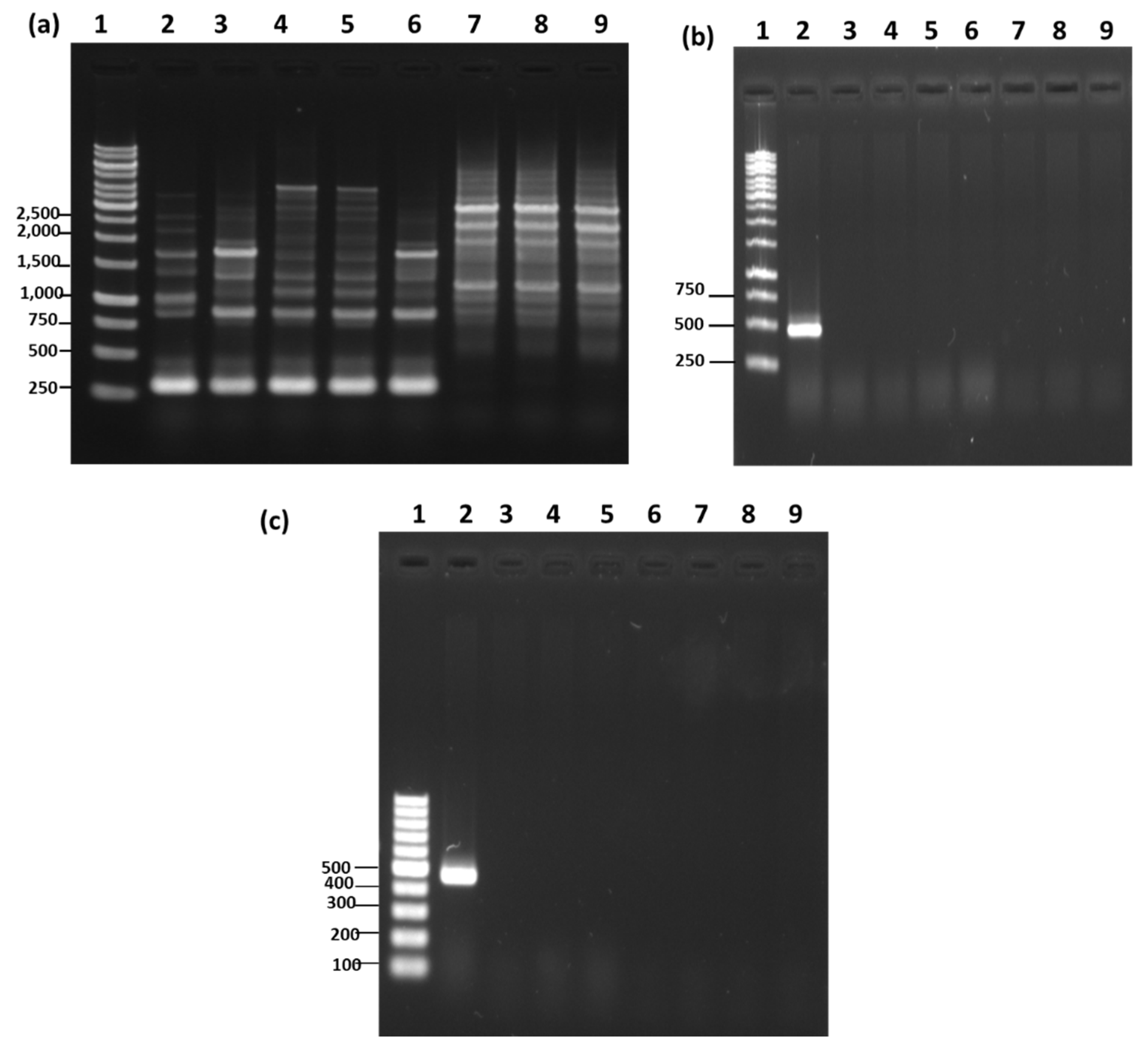
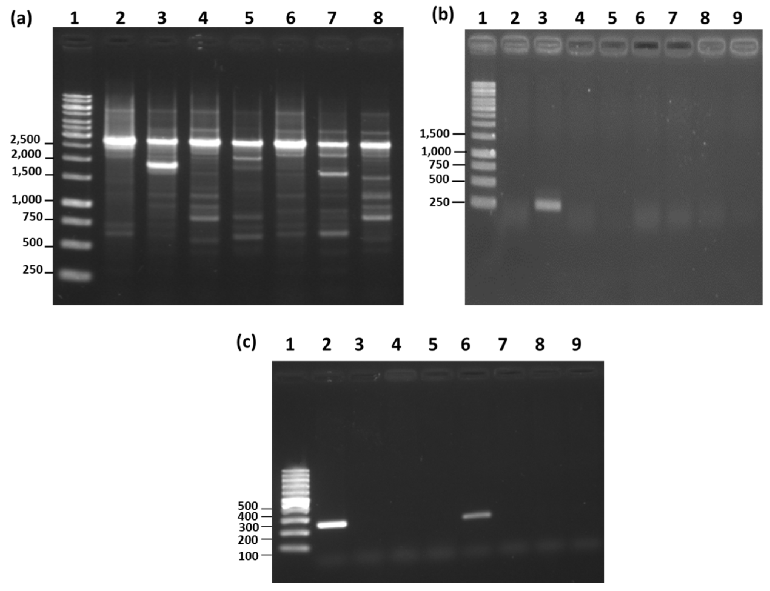
16S rRNA gene sequences of all the bacterial isolates were obtained by availing the commercial facility provided by SolGent Co., Ltd., Daejeon, Republic of Korea. The 16S rRNA gene region of DNA was amplified using 27F (5′-AGAGTTTGATCCTGGCTCAG-3′) and 1492R (5′-GTTTACCTTGTTACGACTT-3′) primers (SolGent Co., Ltd., Daejeon, Republic of Korea) using the PCR conditions: initial denaturation at 95 °C for 15 min, 30 cycles of denaturation at 95 °C for 20 s and annealing at 50 °C for 40 s, extension at 72 °C for 1 min 30 s, and final extension of 5 min at 72 °C. Finally, the amplified product was sequenced using BigDye® Terminator v3.1 cycle sequencing kit (Thermo Fisher Scientific, Waltham, MA, USA) and analyzed on ABI PRISM 3730 XL DNA analyzer (Applied Biosystems, Foster City, CA, USA). The homology of sequences was confirmed by comparing and analyzing the base sequence of 16S rRNA gene using BLASTn.

2.6. RAPD-PCR

RAPD-PCR was performed for all the bacterial isolates to confirm their identity up to the species level. Four types of 10-mer random primers were used for RAPD analysis. The primers were named as primer A (5′-GTGATCGCAG-3′), primer B (5′-CTTTCGCTCC-3′), primer C (5′-CGCAGACCTC-3′), and primer D (5′-GAACTGGAGT-3′). PCR was conducted in a reaction mixture of 25 μL, containing DNA template (1.0 μL, 50 ng), 12.5 μL GoTaq® G2 Green Master Mix (pH 8.3, 1.5 mM MgCl2, 200 μM of each dNTP, DNA polymerase) (Promega; Madison, WI, USA), primer (2.5 μL) (Bionics, Seoul, Republic of Korea), and nuclease-free distilled water (9.0 μL). PCR was carried out at initial denaturation for 5 min at 94 °C, followed by 40 cycles [95 °C for 15 s (denaturation), 36 °C for 15 s (primer A and primer B), 38.4 °C for 15 s (primer C), or 28.1 °C for 15 s (primer D) (annealing), and 72 °C for 2 min (extension)], and a final extension at 72 °C for 4 min. The amplified RAPD-PCR product (10.0 μL) was resolved on a 1.2% agarose gel at 50 V, followed by staining with a GelRed® fluorescent dye (Biotium, Fremont, CA, USA) for 10 min, and visualized using a gel documentation system (Vilber Lourmat, Marne-la-vallee, France).

2.7. Species-Specific PCR

In order to identify B. subtilis, B. thuringiensis, and B. licheniformis at the species level, species-specific PCR was performed. The species-specific primers were designed for the ytcP gene of B. subtilis (F 5′-CTTACGGGTTATCCCGC-3′ and R 5′-CCGACCCCATTTCAGACATATC-3′) [15], XRE gene of B. thuringiensis (F 5′-AAGATATTGCAAGCGGTAAGAT-3′ and R 5′-GTTTTGTTTCAGCATTCCAGTAC-3′) [16], and Blich gene of B. licheniformis (F 5′-AKACGGAAGTGACGGGAAC-3′ and R 5′-AGAAACTTTTCRAGCGCTT-3′) [12]. The total volume of the reaction mixture for PCR was 25 μL, which contained DNA template (1.0 μL, 50 ng), GoTaq® G2 Green Master Mix (12.5 μL, pH 8.3, 1.5 mM MgCl2, 200 μM of each dNTP, DNA polymerase), species-specific forward primer (1.0 μL), reverse primer (1.0 μL), and nuclease-free distilled water (9.5 μL). PCR was performed according to the conditions mentioned in Table S2. The species-specific PCR product was resolved on 1.2% agarose gel and visualized using a gel documentation system (Vilber Lourmat, Marne-la-vallee, France).

2.8. Crystalline Protein Staining

For the identification of B. thuringiensis, crystal protein staining was performed as the method suggested by USFDA [17]. Briefly, bacterial isolate was grown on nutrient agar medium and incubated at 30 °C for 24 h, followed by 5 days of incubation at room temperature. After incubation, bacterial smear was prepared, air-dried, and fixed by gentle heating. The bacterial smear was flooded with methanol for 30 s, followed by the addition of 0.5% basic fuchsin. Sides were heated gently until steam appeared. Finally, slides were rinsed with water and visualized under the microscope (SOMETECH, Seoul, Republic of Korea) for the presence of tetragonal (diamond-shaped) toxin crystals.

3. Results and Discussion

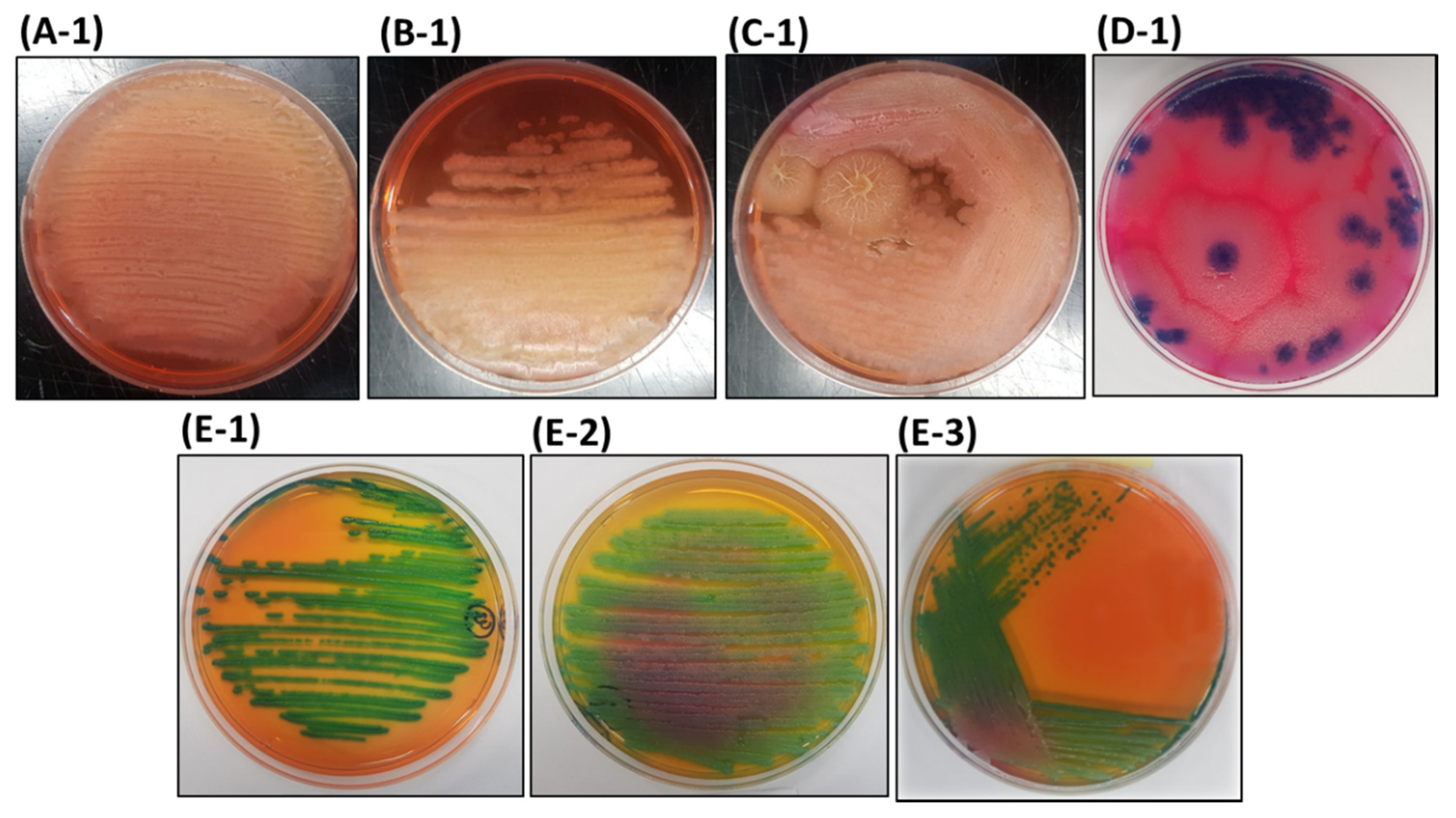
3.1. Isolation of Bacteria from AMEM

From five distinctive types of AMEM, different bacterial colonies were isolated using a standard isolation method. Microscopy examination and investigation of colony morphology in solid medium and appearance in liquid medium revealed the presence of single types of bacteria in products AMEM-A, AMEM-B, AMEM-C, and AMEM-D, and that of three distinct types of bacteria in AMEM-E, which were named as A-1, B-1, C-1, and D-1, and E-1, E-2, and E-3, respectively. Gram staining followed by microscopy examination suggested that all the isolates were Gram-positive, rod-shaped bacteria (Table S3).

3.2. Differential Properties of the Bacterial Isolates on HiCrome™ Bacillus agar

Most Bacillus spp. are known as the principal biological components used in various organic agriculture materials, owing to their stress management, disease prevention, and plant growth promotion properties [11,18,19]. The AMEM-producing company highlighted the presence of Bacillus species in their products, so as the first step of identification, all the isolates were grown on chromogenic HiCrome™ Bacillus agar, a differential media for Bacillus species [20]. The bacterial isolates A-1, B-1, and C-1 were white, irregular, and wrinkle shaped with yellow/pinkish-colored media pigmentation (Figure 2), which are the characteristic features of B. subtilis on HiCrome™ Bacillus agar medium [20]. However, B. amyloliquefaciens also displays similar culture characteristics [19]. These results provide the elementary evidence that isolates A-1, B-1, and C-1 may be B. subtilis, as supported by Alippi and Abrahamovich (2019) [20], who demonstrated the growth of 31 different strains of B. subtilis on HiCrome™ Bacillus agar and observed that 70.9% (22/31) of strains emerged as white, irregular, and wrinkle-shaped colonies with yellow-colored media pigmentation. In contrast, the bacterial isolate D-1 appeared as flat, large, and blue-colored colonies (Figure 2), which are the typical features of B. thuringiensis on HiCrome™ Bacillus agar [20], indicating that the isolate D-1 is B. thuringiensis. However, few closely related bacterial species such as B. cereus and B. mycoides also appear with similar characteristics, thus making the proper identification ambiguous.
The bacterial isolates E-1, E-2, and E-3 appeared as greenish and glistering with irregular colony characteristics with greenish-yellow media pigmentation (Figure 2), a typical characteristic of B. licheniformis on HiCrome™ Bacillus agar [20]. The culture-characteristic-based appearance of E-1, E-2, and E-3 on HiCrome™ Bacillus agar provided preliminary evidence that these isolates may be B. licheniformis. However, Brevibacillus brevis and Brevi. laterosporus also exhibit similar culture characteristics on HiCrome™ Bacillus agar [20], thereby necessitating the use of an additional robust methodology for accurate identification.
Colony characteristics on the HiCrome™ Bacillus agar provided an elementary idea about the identity of the bacterial isolates from AMEM; therefore, further detailed analysis is mandatory to provide correct confirmatory identification of the bacterial isolates.

3.3. Biochemical Characterization

The analytical profile index (API) is a method for the rapid identification of microbes up to the species level based on several biochemical tests and database searches (https://apiweb.biomerieux.com). The bacterial isolates A-1, B-1, C-1, D-1, E-1, E-2, and E-3 were tested on API CH50 CHB, which is a biochemical method conventionally used for the rapid identification of Bacillus and related genera based on biochemical examination and fermentation of 49 carbohydrates. API CH50 CHB examination identified isolates A-1, B-1, and C-1 as B. subtilis or B. amyloliquefaciens with a high percentage similarity of 99.4%, 99.7%, and 99.4% and substantial T-index of 0.85, 0.81, and 0.85, respectively (Table S4). The findings of API were similar to those of HiCrome™ Bacillus agar and failed to identify isolates A-1, B-1, and C-1 as a single species (B. subtilis or B. amyloliquefaciens). Furthermore, the API detection kit identified the isolate D-1 as a B. cereus with 96.0% similarity and 0.95 T index (Table S4). Although the API results suggest D-1 as a B. cereus, the literature suggests that B. cereus has colony morphology and biochemical characteristics similar to those of B. thuringiensis [20]. Hence, further examination is required to avoid ambiguities. Further, isolates E-1, E-2, and E-3 exhibited 99.9%, 99.7%, and 99.7%, respectively, similarity to B. liceniformis (Table S4) and showed consistency with the findings of HiCrome™ Bacillus agar, although molecular characterization is required for confirmatory identification.

3.4. Molecular Characterization Based on 16S rRNA Gene Analysis

16S rRNA gene sequencing is the most common method for bacterial identification [21] and is regarded as the most acceptable method for identifying genera and species for the isolates that do not fit properly in the biochemical profile of the existing system [21]. The strains with 97% sequence identity between the 16S rRNA genes are recognized as being the same species [12]. The isolated bacteria from AMEM were processed for 16S rRNA gene sequencing, followed by a gene bank database search (Table S5). More than 99% sequence similarity of A-1 (NCBI GenBank accession no. MW020274), B-1(NCBI GenBank accession no. MW020275), and C-1 (NCBI GenBank accession no. MW020276) isolates was observed between B. velezensis and B. subtilis (Table S5). Similarly, isolate D-1 (NCBI GenBank accession no. MW020277) displayed more than 99% sequence similarity with B. proteolytic and B. thuringiensis (Table S5). The isolate E-1 (NCBI GenBank accession no. MW020278) displayed more than 97% sequence similarity with B. hyanisis and B. licheniformis (Table S5), while isolates E-2 (NCBI GenBank accession no. MW020279) and E-3 (NCBI GenBank accession no. MW020280) exhibited more than 98% sequence similarity with B. hyanisis and B. licheniformis (Table S5). The results of 16S rRNA gene analysis were ambiguous and failed to distinguish the isolates as single species. The basic limitation of 16S rRNA gene analysis is its inability to discriminate closely related groups [22]. Unfortunately, a high percentage sequence homology occurred (98.1–99.8%) between the members of Bacillus species, thus resulting in the inability of 16S rRNA gene analysis to properly distinguish between species [12,15]. In other words, it is difficult to differentiate B. subtilis, B. amyloliquefaciens, and B. licheniformis using 16S rRNA gene sequencing, owing to the high sequence similarities. Therefore, another approach is required for the appropriate identification of isolates that are closely related to each other.

3.5. RAPD-PCR and Species-Specific PCR Analysis

RAPD-PCR analysis has been found to be effective in generating strain and species-specific DNA amplification profiles that help in species identification [15]. In some cases, RAPD-PCR amplifies a unique species and strain-specific fragment that is further used to develop a specific primer for rapid and accurate identification [15]. In the present study, four random primers were examined for their RAPD pattern against the eight different bacteria of Bacillus species (Figure S1). Based on the reproducibility and the amplification profile, primer B was selected for further RAPD analysis. The first isolates A-1, B-1, and C-1, which are supposed to be B. subtilis (as claimed by those companies and also suggested by the earlier results), were examined for RAPD-PCR using six reference strains of B. subtilis (Figure 3a). RAPD-PCR demonstrated that the two reference strains, B. subtilis KCTC 3104 (lane 3) and B. subtilis KCTC 3239 (lane 6), showed identical RAPD profiling, which is very close to the RAPD pattern of the reference strain B. subtilis KCTC 6633 (lane 2) (Figure 3a). The remaining two reference strains, B. subtilis KCTC 3135 (lane 4) and B. subtilis KCTC 2217 (lane 5), showed identical RAPD patterns, which were slightly different from the other three reference strains (lane 2, 3, and 6) (Figure 3a). In all the six reference strains, a strong amplification of ~275 bp and ~850 bp was observed and could be utilized as a dominant marker for RAPD identification of B. subtilis (Figure 3a). The RAPD patterns of isolates A-1, B-1, and C-1 did not match the RAPD profile of any reference strain, thus disproving the claim that these bacteria were B. subtilis. To further validate the RAPD-PCR results, B. subtilis-specific primers were used. First, the specificity of the species-specific primer was examined utilizing eight closely related Bacillus species (Figure 3b). The amplification of ~500 bp DNA fragment only in B. subtilis (lane 2) while no amplification was observed in any of the closely related Bacillus species was indicative of the high specificity of the selected primer for B. subtilis (Figure 3b). These results are in accordance with the findings of Kwon et al. (2009) [15], who tested the same primer (ytcP) in 14 different B. subtilis strains and observed the amplification of a ~500 bp DNA fragment. In addition, when the same specific primer was tested on four different strains of B. amyloliquefaciens, five different strains of B. licheniformis, and one strain of B. thuringiensis, it displayed no amplification, confirming the high specificity of the primer towards B. subtilis [15]. Knowing the high specificity of the primer (ytcP) towards B. subtilis, we tested the ytcP primer against the isolated microorganism from AMEM and observed no amplification of specific 500 bp DNA fragment (Figure 3c), confirming that these isolates are not B. subtilis, which contradicts the claims of AMEM-A, AMEM-B and AMEM-C producing companies. One of the major reasons for this mismatch is the reclassification of several Bacillus species as B. velezensis by recent taxonomy analysis [11]. Furthermore, 16S rRNA gene analysis also suggested that isolates A-1, B-1, and C-1 were the closest homologs of B. velezensis; therefore, we further performed RAPD-PCR for isolates A-1, B-1, and C-1 using B. velezensis as a reference strain. The results revealed that the reference strains, B. velezensis KCTC13012 (lane 2) and B. velezensis KCTC13417 (lane 3), and A-1, B-1, and C-1 have similar RAPD patterns (Figure 4). A dominant amplicon of ~750 bp, ~2000 bp, and ~2500 bp was observed in all the tested reference strains and isolates A-1, B-1, and C-1, indicating that these strains were B. velezensis. However, further confirmatory studies are required to establish these strains as B. velezensis. Many recent studies suggested a high similarity between B. subtilis, B. velezensis, and B. amyloliquefaciens [11]. Even some members of B. amyloliquefaciens has been reclassified as B. velezensis [11]. Additionally, our API CH50B identification results suggested isolates A-1, B-1, and C-1 either B. subtilis or B. amyloliquefaciens, therefore, to rule out any ambiguity, we performed RAPD-PCR for isolates A-1, B-1, and C-1 using B. amyloliquefaciens as a reference strains. The results revealed that the reference strains B. amyloliquefaciens KCTC1660 (lane 2), B. amyloliquefaciens KCTC1666 (lane 3), B. amyloliquefaciens KCTC3002 (lane 4), and A-1, B-1, and C-1 do not have much similarities in RAPD patterns (Figure S2), suggesting A-1, B-1, and C-1 do not belong to the strains of B. amyloliquefaciens.
Next, the isolated D-1 was examined, which was claimed as B. thuringiensis by the AMEM-D company. Our HiCrome™ Bacillus agar results also suggested the isolate D as B. thuringiensis, and similarly, the 16S rRNA gene findings also displayed a high percentage similarity with B. thuringiensis. However, the API results suggested that it was B. cereus. To address this discrepancy, RAPD-PCR of isolate D-1 was performed using six reference strains of B. thuringiensis (Figure 5a). The RAPD-PCR profile suggested that all the reference strains and isolate D-1 had a dominant amplicon of ~2500 bp, indicating isolate D-1 as B. thuringiensis (Figure 5a). As a final confirmatory test, B. thuringiensis-specific PCR was performed. First, the specificity of primer (XRE gene) towards B. thuringiensis was determined by performing PCR for different Bacillus species strains. The results revealed the specific amplification of the ~246 bp fragment in only B. thuringiensis, suggesting high specificity of the primer towards B. thuringiensis (Figure 5b). Furthermore, the primer was tested on the isolates from AMEM, and the results demonstrated the amplification of a ~246 bp fragment in isolate D-1 and confirmed it as B. thuringiensis (Figure 5c). The results are consistent with the findings of Wei et al. (2019) [16], showing the high specificity of XRE primers for identifying B. thuringiensis. Moreover, it has been well-documented that B. thuringiensis selectively produces insecticidal crystal proteins [16,23], which can be utilized as an appropriate marker for proper identification. Herein, we examined the presence of crystal proteins using crystal protein staining and observed the presence of these crystal proteins in isolate D-1 (Figure 6). Collectively, all the results (HiCrome™ Bacillus agar, RAPD-PCR, species-specific PCR, and crystal protein staining) affirmed isolate D-1 as B. thuringiensis.
Finally, the isolates E-1, E-2, and E-3, which seem to be B. licheniformis by the earlier finding of HiCrome™ Bacillus agar, API CHB50, and 16S rRNA gene analysis, were processed for RAPD-PCR against six reference strains of B. licheniformis (Figure 7a). The RAPD-PCR suggested that the isolate E-1 (lane 8) and E-3 (lane 10) had a similar RAPD profile to the reference strains, B. licheniformis KCTC3056 (lane 3) and B. licheniformis KCTC1029 (lane 4) (Figure 7a). In contrast, the RAPD profile of E-2 (lane 9) was similar to that of the reference strains of B. licheniformis ATCC21415 (lane 2), B. licheniformis KCTC1026 (lane 5), B. licheniformis KCTC1658 (lane 6), and B. licheniformis KCTC3559 (lane 7) (Figure 7a). Moreover, all the reference strains and E-1, E-2, and E-3 showed B. licheniformis-specific dominant amplification at ~1500 bp (Figure 7a). The RAPD profiles of isolates E-1, E-2, and E-3 identified it as B. licheniformis. Further, as a confirmatory examination, a species-specific PCR was performed, which amplified ~600 bp fragment only in B. licheniformis and not in the other closely related Bacillus species (Figure 7b). Consistent with this, isolates E-1, E-2, and E-3 amplified the B. licheniformis-specific ~600 bp fragment, confirming that the isolates are B. licheniformis (Figure 7c).

4. Conclusions

Based on the biochemical and molecular identification methodologies, B. thuringiensis and B. licheniformis were confirmed in AMEM-D and AMEM-E, respectively. The AMEM-A, AMEM-B, and AMEM-C were tentatively identified as B. velezensis based on the findings of 16S rRNA and RAPD-PCR. In conclusion, the findings revealed an accurate identification of B. thuringiensis in AMEM-D, which supports the claim of the company. Similarly, confirmatory evidence of B. licheniformis in AMEM-E was detected. However, for the isolates of A-1, B-1, and C-1, the results were slightly different from the claim of the respective companies and were inclined more towards B. velezensis rather than B. subtilis. Finally, we conclude that a combination of biochemical tests, staining, and molecular techniques (precisely, species-specific PCR) must be performed to enable the proper identification of closely related species.

Supplementary Materials

The following are available online at https://www.mdpi.com/2076-2607/8/10/1568/s1.

Author Contributions

Conceptualization, J.S.L., S.-Y.K., J.-Y.M., S.-K.C., H.C. and M.K.; methodology, A.B., V.K., A.-r.J., J.S.L., S.-Y.K., J.-Y.M., S.-K.C., H.C. and M.K.; data analysis, J.S.L. and M.K.; funding acquisition, M.K.; resources, M.K.; supervision, M.K.; writing—original draft, A.B. and V.K.; writing—review and editing, M.K. All authors have read and agreed to the published version of the manuscript.

Funding

This research was supported by a grant (2019-I-16) from National Agricultural Products Quality Management Service (NAQS), Republic of Korea, in 2019.

Acknowledgments

The authors thank the Core Research Support Center for Natural Products and Medical Materials (CRCNM) for technical support regarding the optical microscopy test.

Conflicts of Interest

The authors declare no conflict of interest.

References

  1. Brown, M.E. Seed and root bacterization. Annu. Rev. Phytopathol. 1974, 12, 181–197. [Google Scholar] [CrossRef]
  2. Schippers, B.; Scheffer, R.J.; Lugtenberg, B.J.J.; Weisbeck, P.J. Biocoating of seeds with plant growth-promoting rhizobacteria to improve plant establishment. Outlook Agric. 1995, 24, 179–185. [Google Scholar] [CrossRef] [Green Version]
  3. Pandey, A.; Tripathi, A.; Srivastava, P.; Choudhary, K.K.; Dikshit, A. Plant growth-promoting microorganisms in sustainable agriculture. In Role of Plant Growth Promoting Microorganisms in Sustainable Agriculture and Nanotechnology; Woodhead Publishing: Sawston, UK; Cambridge, UK, 2019; pp. 1–19. [Google Scholar]
  4. Wu, S.; Zhuang, G.; Bai, Z.; Cen, Y.; Xu, S.; Sun, H.; Zhuang, X. Mitigation of nitrous oxide emissions from acidic soils by Bacillus amyloliquefaciens, a plant growth-promoting bacterium. Glob. Chang. Biol. 2018, 24, 2352–2365. [Google Scholar] [CrossRef] [PubMed]
  5. Higa, T. An Earth Saving Revolution: A Means to Resolve Our World’s Problems through Effective Microorganisms (EM); Sunmark Publishing Inc.: Tokyo, Japan, 1996. [Google Scholar]
  6. Beall, F.; Tipping, B. Plant growth-promoting rhizobacteria in forestry. In Forest Research Marketing. Abstract 117; Ontario Forest Research Committee: Toronto, ON, Canada, 1989; p. 177. [Google Scholar]
  7. Holl, F.B.; Chaneway, C.P.; Turkingon, R.; Radley, R. Growth response of crested wheatgrass (Agropyron cristatum L.), white clover (Trifolium repens L.) to inoculation with Bacillus polymixa. Soil Biol. Biochem. 1988, 20, 19–24. [Google Scholar] [CrossRef]
  8. O’Neill, G.A.; Radley, R.A.; Chanway, C.P. Variable effects of emergence-promoting rhizobacteria on conifer seedling growth under nursery conditions. Biol. Fertil. Soils 1992, 13, 45–49. [Google Scholar] [CrossRef]
  9. Radhakrishnan, R.; Hashem, A.; AbdAllah, E.F. Bacillus: A biological tool for crop improvement through bio-molecular changes in adverse environments. Front. Physiol. 2017, 8, 667. [Google Scholar] [CrossRef] [PubMed]
  10. Pérez-García, A.; Romero, D.; de Vicente, A. Plant protection and growth stimulation by microorganisms: Biotechnological applications of Bacilli in agriculture. Curr. Opin. Biotechnol. 2011, 22, 187–193. [Google Scholar] [CrossRef] [PubMed]
  11. Rabbee, M.F.; Ali, S.M.; Choi, J.; Hwang, B.S.; Jeong, S.C.; Baek, K.H. Bacillus velezensis: A valuable member of bioactive molecules within plant microbiomes. Molecules 2019, 24, 1046. [Google Scholar] [CrossRef] [PubMed] [Green Version]
  12. Huang, C.H.; Chang, M.T.; Huang, L.; Chu, W.S. Development of a novel PCR assay based on the gyrase B gene for species identification of Bacillus licheniformis. Mol. Cell. Probes 2012, 26, 215–217. [Google Scholar] [CrossRef]
  13. Ashe, S.; Maji, U.J.; Sen, R.; Mohanty, S.; Maiti, N.K. Specific oligonucleotide primers for detection of endoglucanase positive Bacillus subtilis by PCR. 3 Biotech 2014, 4, 461–465. [Google Scholar] [CrossRef] [PubMed] [Green Version]
  14. Caroll, K.C.; Pfaller, M.A.; Landry, M.L.; McAdam, A.J.; Patel, R.; Richter, S.S.; Warnock, D.W. Manual of Clinical Microbiology, 12; ASM Publishing: Washington, DC, USA, 2019; p. 2668. [Google Scholar]
  15. Kwon, G.H.; Lee, H.A.; Park, J.Y.; Kim, J.S.; Lim, J.; Park, C.S.; Kwon, D.Y.; Kim, Y.S.; Kim, J.H. Development of a RAPD-PCR method for identification of Bacillus species isolated from Cheonggukjang. Int. J. Food Microbiol. 2009, 129, 282–287. [Google Scholar] [CrossRef] [PubMed]
  16. Wei, S.; Chelliah, R.; Park, B.J.; Kim, S.H.; Forghani, F.; Cho, M.S.; Park, D.S.; Jin, Y.G.; Oh, D.H. Differentiation of Bacillus thuringiensis from Bacillus cereus group using a unique marker based on real-time PCR. Front. Microbiol. 2019, 10, 883. [Google Scholar]
  17. Available online: https://www.fda.gov/food/laboratory-methods-food/bam-chapter-14-bacillus-cereus (accessed on 30 January 2012).
  18. Gotor-Vila, A.; Teixidó, N.; Usall, J.; Dashevskaya, S.; Torres, R. Development of a SCAR marker and a strain-specific genomic marker for the detection of the biocontrol agent strain CPA-8 Bacillus amyloliquefaciens (formerly B. subtilis). Ann. Appl. Biol. 2016, 169, 248–256. [Google Scholar] [CrossRef]
  19. Ji, Z.L.; Peng, S.; Zhu, W.; Dong, J.P.; Zhu, F. Induced resistance in nectarine fruit by Bacillus licheniformis W10 for the control of brown rot caused by Monilinia fructicola. Food Microbiol. 2020, 92, 103558. [Google Scholar] [CrossRef] [PubMed]
  20. Alippi, A.M.; Abrahamovich, E. HiCrome Bacillus agar for presumptive identification of Bacillus and related species isolated from honey samples. Int. J. Food Microbiol. 2019, 305, 108245. [Google Scholar] [CrossRef] [PubMed]
  21. Janda, J.M.; Abbott, S.L. 16S rRNA gene sequencing for bacterial identification in the diagnostic laboratory: Pluses, perils, and pitfalls. J. Clin. Microbiol. 2007, 45, 2761–2764. [Google Scholar] [CrossRef] [PubMed] [Green Version]
  22. Huang, C.H.; Huang, L.; Chang, M.T.; Wu, C.P. Use of novel specific primers targeted to pheS and tuf gene for species and subspecies identification and differentiation of the Bacillus subtilis subsp. subtilis, Bacillus amyloliquefaciens and Bacillus licheniformis. Afr. J. Microbiol. Res. 2017, 11, 264–270. [Google Scholar]
  23. Kumar, D.; Chaudhary, K.; Boora, K.S. Characterization of native Bacillus thuringiensis strains by PCR-RAPD based fingerprinting. Indian J. Microbiol. 2010, 50, 27–32. [Google Scholar] [CrossRef] [PubMed] [Green Version]
Figure 1. Systemic representation of bacterial identification in commercially available organic agriculture materials enriched with effective microorganisms.
Microorganisms 08 01568 g001
Figure 2. Morphology of bacterial colonies isolates from different commercially available organic agriculture materials enriched with effective microorganisms on HiCrome™ Bacillus agar. A-1, B-1, C-1, D-1, E-1, E-2, and E-3 are the bacterial isolates from AMEM-A, AMEM-B, AMEM-C, AMEM-D, and AMEM-E, respectively.
Microorganisms 08 01568 g002
Figure 3. (a) RAPD-PCR for various reference strains of Bacillus subtilis. Lane 1: 1 kb size ladder (unit: bp); Lane 2: Bacillus subtilis ATCC6633; Lane 3: Bacillus subtilis KCTC3014; Lane 4: Bacillus subtilis KCTC3135; Lane 5: Bacillus subtilis KCTC2217; Lane 6: Bacillus subtilis KCTC3239. (b) Bacillus subtilis species-specific PCR (ytcP gene). Lane 1: 1 kb size ladder (unit: bp); Lane 2: Bacillus subtilis ATCC6633; Lane 3: Bacillus thuringiensis KCTC3452; Lane 4: Bacillus megaterium KCTC1098; Lane 5: Bacillus velezensis KCTC13012; Lane 6: Bacillus licheniformis ATCC21415; Lane 7: Bacillus amyloliquefaciens KCTC1660; Lane 8: Bacillus pumilus ATCC7061; Lane 9: Bacillus mojavensis ATCC51516. (c) Species-specific PCR using ytcP gene primer on microorganisms isolated from commercially available organic agriculture materials enriched with effective microorganism. Lane 1: 0.1 kb size ladder (unit: bp); Lane 2: Bacillus subtilis ATCC6633; Lane 3: A-1; Lane 4: B-1; Lane 5: C-1; Lane 6: D-1; Lane 7: E-1; Lane 8: E-2; Lane 9: E-3.
Microorganisms 08 01568 g003
Figure 4. RAPD-PCR of various reference strains of Bacillus velezensis. Lane 1: 1 kb size ladder (unit: bp); Lane 2: Bacillus velezensis KCTC13012; Lane 3: Bacillus velezensis KCTC13417; Lane 4: A-1; Lane 5: B-1; Lane 6: C-1.
Microorganisms 08 01568 g004
Figure 5. (a) RAPD-PCR of various reference strains of Bacillus thuringiensis. Lane 1: 1 kb size ladder (unit: bp); Lane 2: Bacillus thuringiensis KCTC3452; Lane 3: Bacillus thuringiensis KCTC1508; Lane 4: Bacillus thuringiensis KCTC1510; Lane 5: Bacillus thuringiensis KCTC1511; Lane 6: Bacillus thuringiensis KCTC1524; Lane 7: Bacillus thuringiensis KCTC1525. (b) Species-specific PCR result of pure strains Bacillus thuringiensis (XRE gene primer). Lane 1: 1 kb size ladder (unit: bp); Lane 2: Bacillus subtilis ATCC6633; Lane 3: Bacillus thuringiensis KCTC3452; Lane 4: Bacillus megaterium KCTC1098; Lane 5: Bacillus velezensis KCTC13012; Lane 6: Bacillus licheniformis ATCC21415; Lane 7: Bacillus amyloliquefaciens KCTC1660; Lane 8: Bacillus pumilus ATCC7061; Lane 9: Bacillus mojavensis ATCC51516. (c) Species-specific PCR using XRE gene primer on microorganisms isolated from commercially available organic agriculture materials enriched with effective microorganism. Lane 1: 0.1 kb size ladder (unit: bp); Lane 2: Bacillus thuringiensis KCTC3452; Lane 3: A-1; Lane 4: B-1; Lane 5: C-1; Lane 6: D-1; Lane 7: E-1; Lane 8: E-2; Lane 9: E-3.
Microorganisms 08 01568 g005
Figure 6. Crystal protein staining for the detection of Bacillus thuringiensis. (a) Bacillus thuringiensis KCTC3452; (b) Microbial isolate D-1 (100× magnification). (c,d) A magnified view of (a,b). Arrows show the presence of tetragonal (diamond-shaped) toxin crystals.
Microorganisms 08 01568 g006
Figure 7. (a) RAPD-PCR of various reference strains of Bacillus licheniformis. Lane 1: 1 kb size ladder (unit: bp); Lane 2: Bacillus licheniformis ATCC21415; Lane 3: Bacillus licheniformis KCTC3056; Lane 4: Bacillus licheniformis KCTC1029; Lane 5: Bacillus licheniformis KCTC1026; Lane 6: Bacillus licheniformis KCTC1658; Lane 7: Bacillus licheniformis KCTC3559. (b) Species-specific PCR (Blich gene primer) of pure strains. Lane 1: 0.1 kb size ladder (unit: bp); Lane 2: Bacillus subtilis ATCC6633; Lane 3: Bacillus thuringiensis KCTC3452; Lane 4: Bacillus megaterium KCTC1098; Lane 5: Bacillus velezensis KCTC13012; Lane 6: Bacillus licheniformis ATCC21415; Lane 7: Bacillus amyloliquefaciens KCTC1660; Lane 8: Bacillus pumilus ATCC7061; Lane 9: Bacillus mojavensis ATCC51516. (c) Species-specific PCR using Blich gene primer on microorganisms isolated from commercially available organic agriculture materials enriched with effective microorganism. Lane 1: 0.1 kb size ladder (unit: bp); Lane 2: Bacillus licheniformis ATCC21415; Lane 3: A-1; Lane 4: B-1; Lane 5: C-1; Lane 6: D-1; Lane 7: E-1; Lane 8: E-2; Lane 9: E-3.
Microorganisms 08 01568 g007
Table 1. Specification of different commercially available organic agriculture materials enriched with effective microorganism.
Product CodeProduct TypeProduct CompositionLabelled MicroorganismspHAerobic Bacterial Count
AMEM-ALiquid-Bacillus subtilis 2%
-Extracts of dried ginseng 95%
Bacillus subtilis5.61 ± 0.337.92 ± 0.05 Log CFU/mL
AMEM-BLiquid-Bacillus subtilis 2%
-Extracts dried ginseng 60%
-Extracts of cinnamon 33%
Bacillus subtilis6.48 ± 0.307.80 ± 0.06 Log CFU/mL
AMEM-CSolid-Bacillus subtilis culture 55.6%
-Diatomite 33.4%
Bacillus subtilis6.67 ± 0.159.85 ± 0.08 Log CFU/g
AMEM-DSolid-Diatomite 58%
-Bacillus thuringiensis 32%
Bacillus thuringiensis5.52 ± 0.099.30 ± 0.02 Log CFU/g
AMEM-ELiquid-Fish products 65%
-Molasses 20%
-Microorganisms 15%
Not specified3.80 ± 0.163.26 ± 0.06 Log CFU/mL

