Transcriptome Analysis of Ice Plant Growth-Promoting Endophytic Bacterium Halomonas sp. Strain MC1 to Identify the Genes Involved in Salt Tolerance

Abstract
:
1. Introduction
2. Materials and Methods
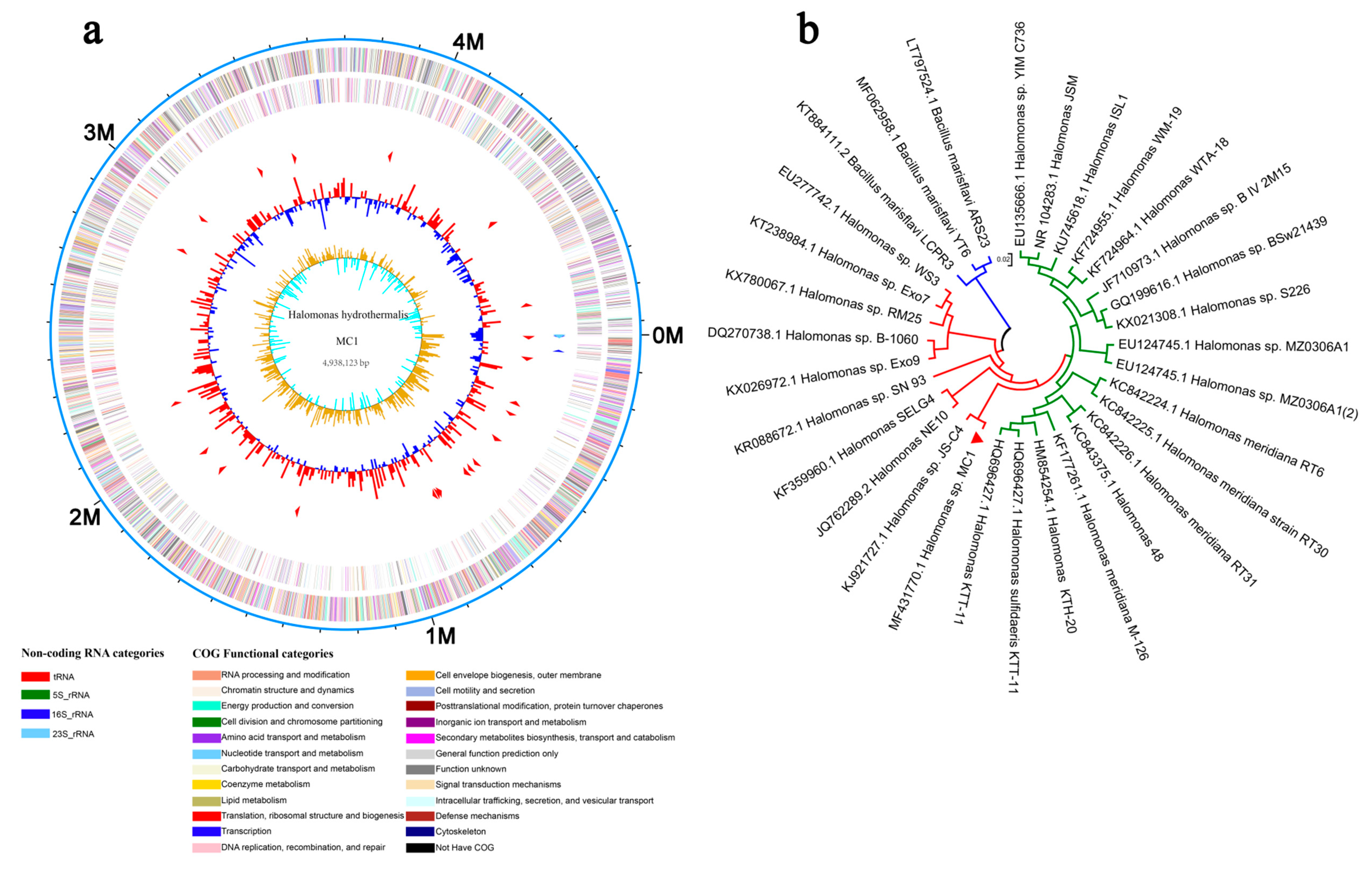
2.1. Strain and Culture Condition
2.2. Induction of Salt Tolerance and Bacterial Collection
2.3. RNA Extraction, Library Construction, and Sequencing
2.4. Quality Control and Mapping of Reads
2.5. Transcriptome De Novo Assembly and Annotation
2.6. Differential Expression Analysis and Functional Enrichment
2.7. Quantitative Real-Time Polymerase Chain Reaction (qRT-PCR) Validation
2.8. Statistical Analysis
3. Results
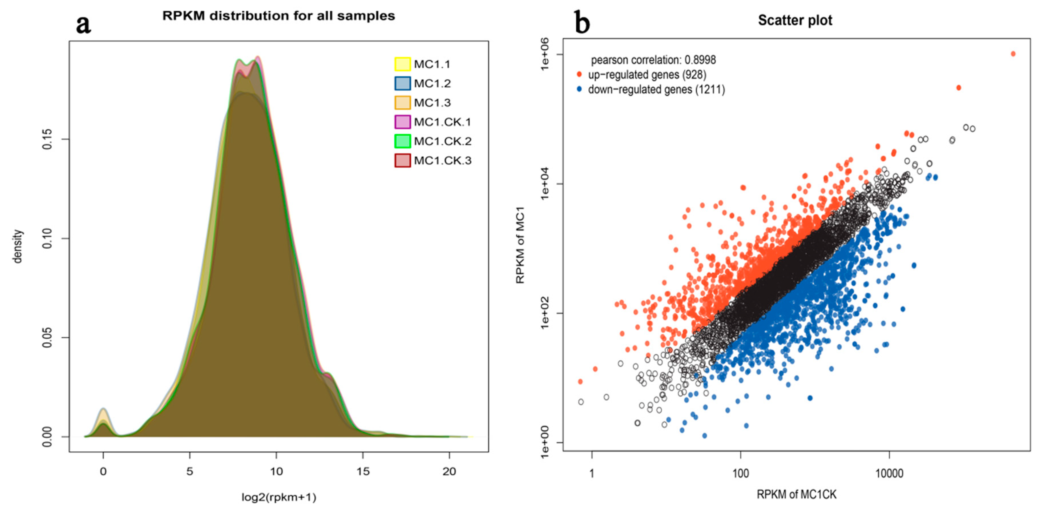
3.1. Data Processing and Analysis
3.2. Number of DEGs
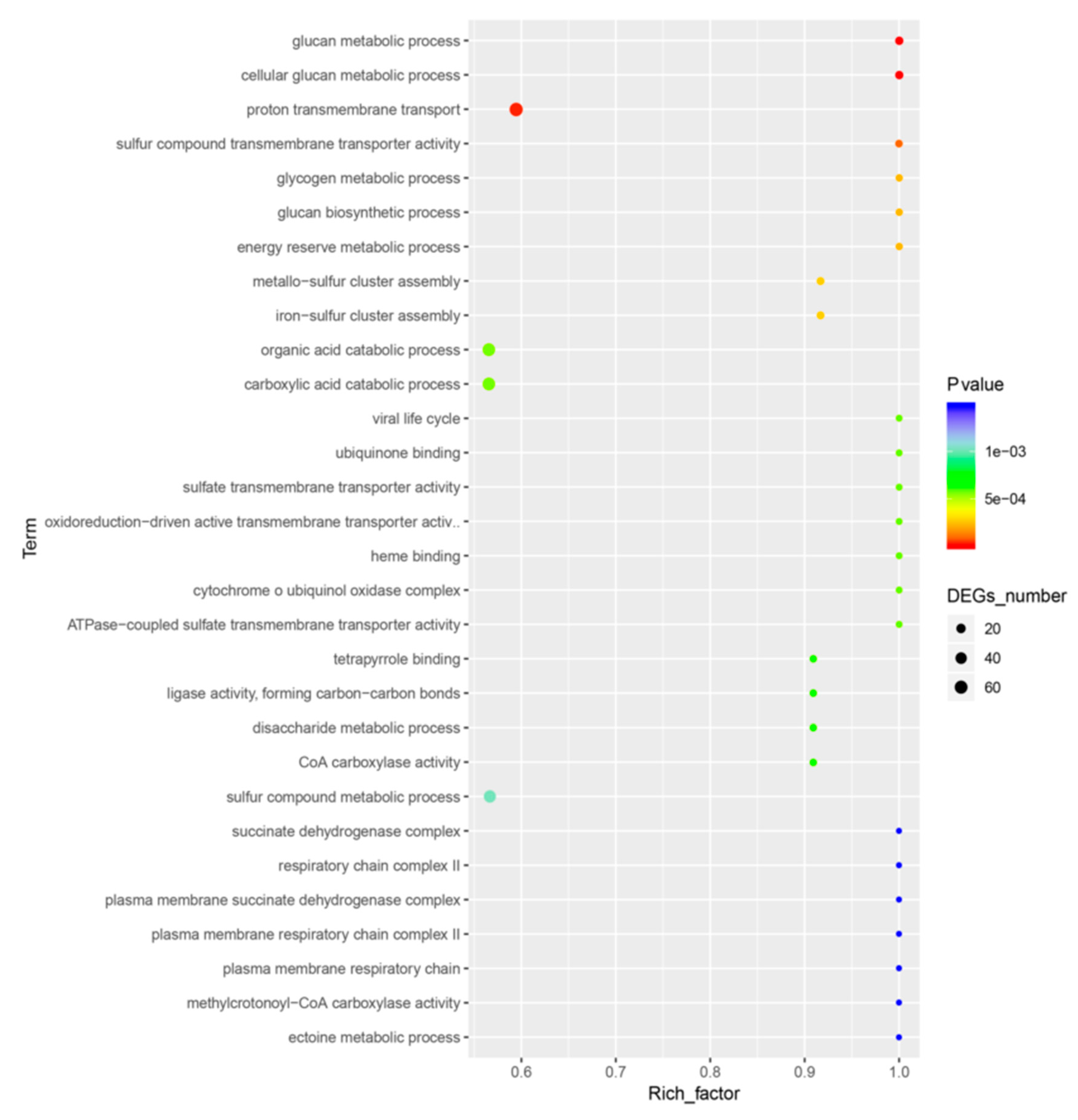
3.3. GO Analysis of DEGs
3.4. KEGG Analysis of DEGs
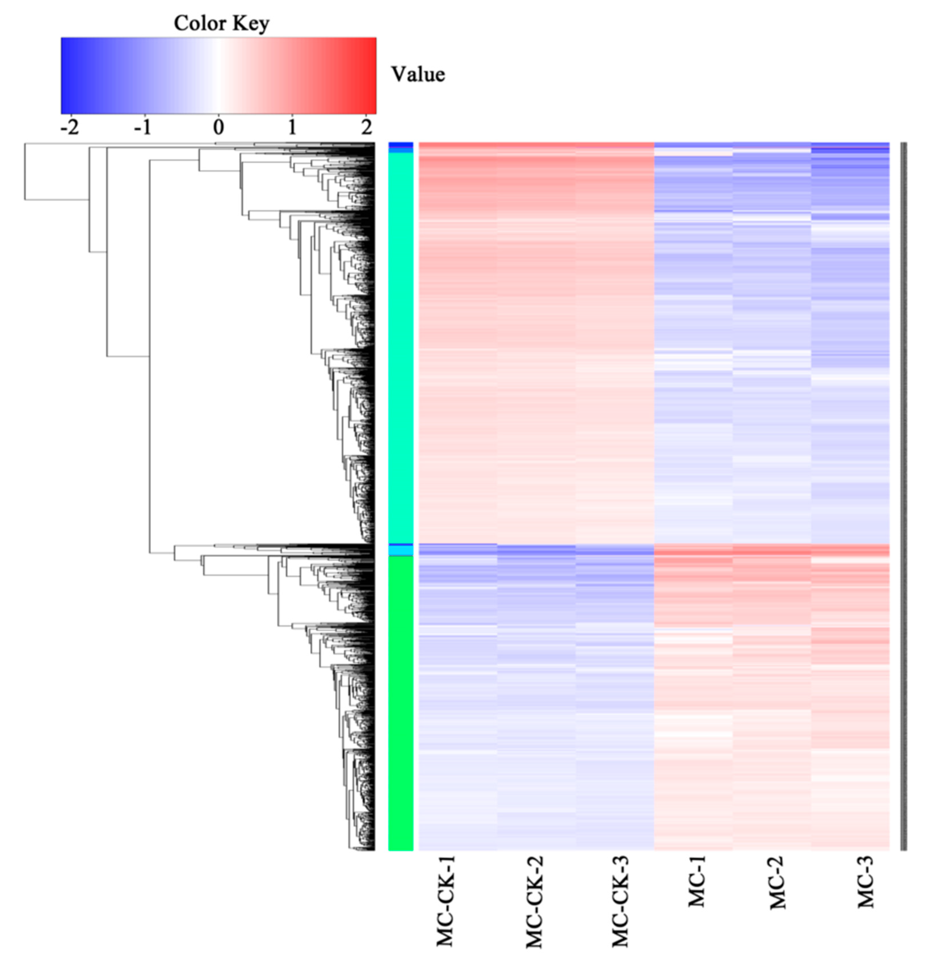
3.5. Clustering Analysis of DEGs
3.6. qRT-PCR Validation
4. Discussion
5. Conclusions
Supplementary Materials
Author Contributions
Funding
Acknowledgments
Conflicts of Interest
Abbreviations
| EB | endophytic bacteria |
| CFU | colony-forming units |
| KEGG | Kyoto Encyclopedia of Genes and Genomes |
| GO | Gene Ontology |
| DEGs | differential expression genes |
| FDR | false discovery rate |
References
- Foyer, C.H.; Rasool, B.; Davey, J.W.; Hancock, R.D. Cross-tolerance to biotic and abiotic stresses in plants: A focus on resistance to aphid infestation. J. Exp. Bot. 2016, 67, 2025–2037. [Google Scholar] [CrossRef]
- Ali, S.; Charles, T.C.; Glick, B.R. Amelioration of high salinity stress damage by plant growth-promoting bacterial endophytes that contain ACC deaminase. Plant Physiol. Biochem. 2014, 80, 160–167. [Google Scholar] [CrossRef] [PubMed]
- Krasensky, J.; Jonak, C. Drought, salt, and temperature stress-induced metabolic rearrangements and regulatory networks. J. Exp. Bot. 2012, 63, 1593–1608. [Google Scholar] [CrossRef] [PubMed] [Green Version]
- Shrivastava, P.; Kumar, R. Soil salinity: A serious environmental issue and plant growth promoting bacteria as one of the tools for its alleviation. Saudi J. Biol. Sci. 2015, 22, 123–131. [Google Scholar] [CrossRef] [Green Version]
- Khan, Z.; Rho, H.; Firrincieli, A.; Hung, S.H.; Luna, V.; Masciarelli, O.; Kim, S.-H.; Doty, S.L. Growth enhancement and drought tolerance of hybrid poplar upon inoculation with endophyte consortia. Curr. Plant Biol. 2016, 6, 38–47. [Google Scholar] [CrossRef] [Green Version]
- Dimkpa, C.; Weinand, T.; Asch, F. Plant–rhizobacteria interactions alleviate abiotic stress conditions. Plant Cell Environ. 2009, 32, 1682–1694. [Google Scholar] [CrossRef]
- Gouda, S.; Kerry, R.G.; Das, G.; Paramithiotis, S.; Shin, H.-S.; Patra, J.K. Revitalization of plant growth promoting rhizobacteria for sustainable development in agriculture. Microbiol. Res. 2018, 206, 131–140. [Google Scholar] [CrossRef]
- Palacios, O.A.; Bashan, Y.; de-Bashan, L.E. Proven and potential involvement of vitamins in interactions of plants with plant growth-promoting bacteria-an overview. Biol. Fertil. Soils 2014, 50, 415–432. [Google Scholar] [CrossRef]
- Zinniel, D.K.; Lambrecht, P.; Harris, N.B.; Feng, Z.; Kuczmarski, D.; Higley, P.; Ishimaru, C.A.; Arunakumari, A.; Barletta, R.G.; Vidaver, A.K. Isolation and characterization of endophytic colonizing bacteria from agronomic crops and prairie plants. Appl. Environ. Microbiol. 2002, 68, 2198. [Google Scholar] [CrossRef] [Green Version]
- Jha, P.; Panwar, J.; Jha, P.N. Mechanistic insights on plant root colonization by bacterial endophytes: A symbiotic relationship for sustainable agriculture. Environ. Sustain. 2018, 1, 25–38. [Google Scholar] [CrossRef]
- Rai, R.; Dash, P.K.; Prasanna, B.M.; Singh, A. Endophytic bacterial flora in the stem tissue of a tropical maize (Zea mays L.) genotype: Isolation, identification and enumeration. World J. Microbiol. Biotechnol. 2007, 23, 853–858. [Google Scholar] [CrossRef]
- Brader, G.; Compant, S.; Vescio, K.; Mitter, B.; Trognitz, F.; Ma, L.J.; Sessitsch, A. Ecology and genomic insights into plant-pathogenic and plant-nonpathogenic endophytes. Ann. Rev. Phytopathol. 2017, 55, 61–83. [Google Scholar] [CrossRef] [PubMed]
- Li, X.; Ma, L.; Li, Y.; Wang, L.; Zhang, L. Endophyte infection enhances accumulation of organic acids and minerals in rice under Pb2+ stress conditions. Ecotoxicol. Environ. Saf. 2019, 174, 255–262. [Google Scholar] [CrossRef]
- Khatabi, B.; Gharechahi, J.; Ghaffari, M.R.; Liu, D.; Haynes, P.A.; McKay, M.J.; Mirzaei, M.; Salekdeh, G.H. Plant-microbe symbiosis: What has proteomics taught us? Proteomics 2019. [Google Scholar] [CrossRef] [PubMed]
- Berg, G.; Rybakova, D.; Grube, M.; Köberl, M. The plant microbiome explored: Implications for experimental botany. J. Exp. Bot. 2015, 67, 995–1002. [Google Scholar] [CrossRef] [PubMed]
- Afzal, I.; Shinwari, Z.K.; Sikandar, S.; Shahzad, S. Plant beneficial endophytic bacteria: Mechanisms, diversity, host range and genetic determinants. Microbiol. Res. 2019, 221, 36–49. [Google Scholar] [CrossRef]
- Chebotar, V.K.; Malfanova, N.V.; Shcherbakov, A.V.; Ahtemova, G.A.; Borisov, A.Y.; Lugtenberg, B.; Tikhonovich, I.A. Endophytic bacteria in microbial preparations that improve plant development. Appl. Biochem. Microbiol. 2015, 51, 271–277. [Google Scholar] [CrossRef]
- Tran, D.Q.; Konishi, A.; Cushman, J.C.; Morokuma, M.; Toyota, M.; Agarie, S. Ion accumulation and expression of ion homeostasis-related genes associated with halophilism, NaCl-promoted growth in a halophyte Mesembryanthemum crystallinum L. Plant Prod. Sci. 2019, 1–12. [Google Scholar] [CrossRef] [Green Version]
- Dong, Z.-Y.; Narsing Rao, M.P.; Wang, H.-F.; Fang, B.-Z.; Liu, Y.-H.; Li, L.; Xiao, M.; Li, W.-J. Transcriptomic analysis of two endophytes involved in enhancing salt stress ability of Arabidopsis thaliana. Sci. Total Environ. 2019, 686, 107–117. [Google Scholar] [CrossRef]
- Wang, T.T.; Ding, P.; Chen, P.; Xing, K.; Bai, J.L.; Wan, W.; Jiang, J.H.; Qin, S. Complete genome sequence of endophyte Bacillus flexus KLBMP 4941 reveals its plant growth promotion mechanism and genetic basis for salt tolerance. J. Biotechnol. 2017, 260, 38–41. [Google Scholar] [CrossRef]
- Gond, S.K.; Torres, M.S.; Bergen, M.S.; Helsel, Z.; White, J.F., Jr. Induction of salt tolerance and up-regulation of aquaporin genes in tropical corn by rhizobacterium Pantoeaagglomerans. Lett. Appl. Microbiol. 2015, 60, 392–399. [Google Scholar] [CrossRef] [PubMed]
- Wani, Z.A.; Ashraf, N.; Mohiuddin, T.; Riyaz-Ul-Hassan, S. Plant-endophyte symbiosis, an ecological perspective. Appl. Microbiol. Biotechnol. 2015, 99, 2955–2965. [Google Scholar] [CrossRef] [PubMed]
- Vaishnav, A.; Shukla, A.K.; Sharma, A.; Kumar, R.; Choudhary, D.K. Endophytic bacteria in plant salt stress tolerance: Current and future prospects. J. Plant Growth Regul. 2019, 38, 650–668. [Google Scholar] [CrossRef]
- Nelson, D.E.; Koukoumanos, M.; Bohnert, H.J. Myo-inositol-dependent sodium uptake in ice plant. Plant Physiol. 1999, 119, 165–172. [Google Scholar] [CrossRef] [PubMed] [Green Version]
- Nanhapo, P.I.; Yamane, K.; Iijima, M. Mixed cropping with ice plant alleviates the damage and the growth of cowpea under consecutive NaCl treatment and after the recovery from high salinity. Plant Prod. Sci. 2017, 20, 111–125. [Google Scholar] [CrossRef] [Green Version]
- Zhang, J.; Wang, P.; Tian, H.; Jiang, H.; Wang, Y.; Yan, C. Identification of interior salt-tolerant bacteria from ice plant Mesembryanthemumcrystallinum and evaluation of their promoting effects. Symbiosis 2018, 76, 243–252. [Google Scholar] [CrossRef]
- Tjaden, B. De novo assembly of bacterial transcriptomes from RNA-seq data. Genome Biol. 2015, 16, 1. [Google Scholar] [CrossRef] [Green Version]
- Grabherr, M.G.; Haas, B.J.; Yassour, M.; Levin, J.Z.; Thompson, D.A.; Amit, I.; Adiconis, X.; Fan, L. Full-length transcriptome assembly from RNA-Seq data without a reference genome. Nat. Biotechnol. 2011, 29, 644–652. [Google Scholar] [CrossRef] [Green Version]
- Levin, J.Z.; Yassour, M.; Adiconis, X.; Nusbaum, C.; Thompson, D.A.; Friedman, N.; Gnirke, A.; Regev, A. Comprehensive comparative analysis of strand-specific RNA sequencing methods. Nat. Methods 2010, 7, 709. [Google Scholar] [CrossRef] [Green Version]
- Altermann, E.; Klaenhammer, T.R. PathwayVoyager: Pathway mapping using the Kyoto Encyclopedia of Genes and Genomes (KEGG) database. BMC Genom. 2005, 6, 60. [Google Scholar] [CrossRef] [Green Version]
- Pinard, D.; Fierro, A.C.; Marchal, K.; Myburg, A.A.; Mizrachi, E. Organellar carbon metabolism is coordinated with distinct developmental phases of secondary xylem. New Phytol. 2019, 222, 1832–1845. [Google Scholar] [CrossRef] [PubMed] [Green Version]
- Song, Y.; Shin, J.; Jin, S.; Lee, J.K.; Kim, D.R.; Kim, S.C.; Cho, S.; Cho, B.K. Genome-scale analysis of syngas fermenting acetogenic bacteria reveals the translational regulation for its autotrophic growth. BMC Genom. 2018, 19, 837. [Google Scholar] [CrossRef] [PubMed]
- Orozco-Mosqueda, M.d.C.; Duan, J.; DiBernardo, M.; Zetter, E.; Campos-García, J.; Glick, B.R.; Santoyo, G. The production of ACC deaminase and trehalose by the plant growth promoting bacterium Pseudomonas sp. UW4 synergistically protect tomato plants against salt stress. Front. Microbiol. 2019, 10, 1392. [Google Scholar] [CrossRef] [PubMed] [Green Version]
- Niu, S.-Q.; Li, H.-R.; Paré, P.W.; Aziz, M.; Wang, S.-M.; Shi, H.; Li, J.; Han, Q.-Q.; Guo, S.-Q.; Li, J.; et al. Induced growth promotion and higher salt tolerance in the halophyte grass Puccinelliatenuiflora by beneficial rhizobacteria. Plant Soil. 2016, 407, 217–230. [Google Scholar] [CrossRef]
- Kurt-Kızıldoğan, A.; Abanoz, B.; Okay, S. Global transcriptome analysis of Halolamina sp. to decipher the salt tolerance in extremely halophilic archaea. Gene 2017, 601, 56–64. [Google Scholar] [CrossRef]
- Zhu, M.; Chen, G.; Dong, T.; Wang, L.; Zhang, J.; Zhao, Z.; Hu, Z. SlDEAD31, a putative DEAD-Box RNA helicase gene, regulates salt and drought tolerance and stress-related genes in tomato. PLoS ONE 2015, 10, e0133849. [Google Scholar] [CrossRef] [Green Version]
- Fu, J.; Wang, Y.-F.; Liu, Z.-H.; Li, Z.-T.; Yang, K.-J. Trichodermaasperellum alleviates the effects of saline–alkaline stress on maize seedlings via the regulation of photosynthesis and nitrogen metabolism. Plant Growth Regul. 2018, 85, 363–374. [Google Scholar] [CrossRef]
- Yang, M.; Ye, J.; Qin, H.; Long, Y.; Li, Y. Influence of perfluorooctanoic acid on proteomic expression and cell membrane fatty acid of Escherichia coli. Environ. Pollut. 2017, 220, 532–539. [Google Scholar] [CrossRef]
- Zeng, J.; Yu, H.; Kjellberg, F. Transcriptome analysis of genes involved in the response of a pollinator fig wasp to volatile organic compounds from its host figs. ActaOecologica 2018, 90, 91–98. [Google Scholar] [CrossRef] [Green Version]
- Chatterjee, P.; Samaddar, S.; Niinemets, Ü.; Sa, T.-M. Brevibacterium linens RS16 confers salt tolerance to Oryza sativa genotypes by regulating antioxidant defense and H+ ATPase activity. Microbiol. Res. 2018, 215, 89–101. [Google Scholar] [CrossRef]
- Valabhoju, V.; Agrawal, S.; Sen, R. Molecular basis of NusG-mediated regulation of Rho-dependent transcription termination in bacteria. J. Biol. Chem. 2016, 291, 22386–22403. [Google Scholar] [CrossRef] [PubMed] [Green Version]
- Yang, H.; Meng, Y.; Song, Y.; Tan, Y.; Warren, A.; Li, J.; Lin, X. Salinity fluctuation influencing biological adaptation: Growth dynamics and Na+/K+-ATPase activity in a euryhaline bacterium. J. Basic Microbiol. 2017, 57, 617–624. [Google Scholar] [CrossRef] [PubMed]
- Afzal, M.; Yousaf, S.; Reichenauer, T.G.; Kuffner, M.; Sessitsch, A. Soil type affects plant colonization, activity and catabolic gene expression of inoculated bacterial strains during phytoremediation of diesel. J. Hazard. Mater. 2011, 186, 1568–1575. [Google Scholar] [CrossRef] [PubMed]
- Sarkar, P.; Roy, A.; Pal, S.; Mohapatra, B.; Kazy, S.K.; Maiti, M.K.; Sar, P. Enrichment and characterization of hydrocarbon-degrading bacteria from petroleum refinery waste as potent bioaugmentation agent for in situ bioremediation. Bioresour. Technol. 2017, 242, 15–27. [Google Scholar] [CrossRef]
- Yun, E.J.; Lee, S.; Kim, H.T.; Pelton, J.G.; Kim, S.; Ko, H.-J.; Choi, I.-G.; Kim, K.H. The novel catabolic pathway of 3,6-anhydro-L-galactose, the main component of red macroalgae, in a marine bacterium. Environ. Microbiol. 2015, 17, 1677–1688. [Google Scholar] [CrossRef]
- Douglass, J.F.; Radosevich, M.; Tuovinen, O.H. Microbial attenuation of atrazine in agricultural soils: Biometer assays, bacterial taxonomic diversity, and catabolic genes. Chemosphere 2017, 176, 352–360. [Google Scholar] [CrossRef] [Green Version]
- Rubiano-Labrador, C.; Bland, C.; Miotello, G.; Armengaud, J.; Baena, S. Salt Stress Induced Changes in the Exoproteome of the Halotolerant Bacterium Tistliaconsotensis Deciphered by Proteogenomics. PloS ONE 2015, 10, e0135065. [Google Scholar] [CrossRef] [Green Version]
- Kauffmann, A.; Huber, W. Microarray data quality control improves the detection of differentially expressed genes. Genomics 2010, 95, 138–142. [Google Scholar] [CrossRef] [Green Version]
- Locher, K.P. Mechanistic diversity in ATP-binding cassette (ABC) transporters. Nat. Struct. Mol. Biol. 2016, 23, 487. [Google Scholar] [CrossRef] [Green Version]
- Duarte, D.A.S.; Newbold, C.J.; Detmann, E.; Silva, F.F.; Freitas, P.H.F.; Veroneze, R.; Duarte, M.S. Genome-wide association studies pathway-based meta-analysis for residual feed intake in beef cattle. Anim. Genet. 2019, 50, 150–153. [Google Scholar] [CrossRef]
- Rutowski, J.; Zhong, F.; Xu, M.; Zhu, J. Metabolic shift of Staphylococcus aureus under sublethal dose of methicillin in the presence of glucose. J. Pharm. Biomed. Analy. 2019, 167, 140–148. [Google Scholar] [CrossRef] [PubMed]
- Wagatsuma, T. The membrane lipid bilayer as a regulated barrier to cope with detrimental ionic conditions: Making new tolerant plant lines with altered membrane lipid bilayer. Soil. Sci. Plant Nut. 2017, 63, 507–516. [Google Scholar] [CrossRef]
- Etesami, H. Bacterial mediated alleviation of heavy metal stress and decreased accumulation of metals in plant tissues: Mechanisms and future prospects. Ecotoxicol. Environ. Saf. 2018, 147, 175–191. [Google Scholar] [CrossRef] [PubMed]
- Kumar, A.; Verma, J.P. Does plant-microbe interaction confer stress tolerance in plants: A review? Microbiol. Res. 2018, 207, 41–52. [Google Scholar] [CrossRef]


| Sample | Raw Reads | Raw Bases | %≥Q30 | Clean Reads | Clean Bases | Mapped-Reads | Mapped Rate (%) | GC(%) | %≥Q30 | Error(%) |
|---|---|---|---|---|---|---|---|---|---|---|
| MC1-CK-1 | 13,471,728 | 2,020,759,200 | 91.21 | 11,691,054 | 1,726,157,998 | 10,887,922 | 93.13 | 56.93 | 93.90 | 0.0137 |
| MC1-CK-2 | 15,215,384 | 2,282,307,600 | 91.1 | 12,979,384 | 1,912,783,427 | 11,812,868 | 91.01 | 56.67 | 93.81 | 0.0138 |
| MC1-CK-3 | 16,388,692 | 2,458,303,800 | 91.5 | 14,177,492 | 2,094,489,027 | 13,124,998 | 92.58 | 56.44 | 94.01 | 0.0136 |
| MC1-1 | 13,820,672 | 2,073,100,800 | 94.9 | 12,093,922 | 1,783,589,346 | 10,993,370 | 90.90 | 55.94 | 96.86 | 0.0109 |
| MC1-2 | 16,604,690 | 2,490,703,500 | 93.72 | 14,554,380 | 2,145,906,752 | 12,859,768 | 88.36 | 56.12 | 96.07 | 0.0115 |
| MC1-3 | 16,857,140 | 2,528,571,000 | 93.64 | 14,246,340 | 2,090,108,492 | 12,558,154 | 88.15 | 55.82 | 96.07 | 0.0115 |
| Categories | Description | Ratio in Study * | Ratio in Total Genes | p-Value | GO ID |
|---|---|---|---|---|---|
| Cellular Component | outer membrane-bounded periplasmic space | 27 (1.26%) | 47 (0.87%) | 0.0156 | GO:0030288 |
| plasma membrane proton-transporting ATP synthase complex | 10 (0.47%) | 15 (0.28%) | 0.0363 | GO:0045260 | |
| proton-transporting ATP synthase complex | 10 (0.47%) | 15 (0.28%) | 0.0363 | GO:0045259 | |
| proton-transporting two-sector ATPase complex | 10 (0.47%) | 15 (0.28%) | 0.0363 | GO:0016469 | |
| cytochrome o ubiquinol oxidase complex | 8 (0.37%) | 8 (0.15%) | 0.0006 | GO:0009319 | |
| proton-transporting ATP synthase complex, catalytic core F(1) | 8 (0.37%) | 11 (0.20%) | 0.0307 | GO:0045261 | |
| plasma membrane proton-transporting ATP synthase complex, catalytic core F(1) | 8 (0.37%) | 11 (0.20%) | 0.0307 | GO:0045262 | |
| proton-transporting two-sector ATPase complex, catalytic domain | 8 (0.37%) | 11 (0.20%) | 0.0307 | GO:0033178 | |
| cytochrome complex | 8 (0.37%) | 10 (0.18%) | 0.0177 | GO:0070069 | |
| Molecular Function | ATPase activity, coupled to transmembrane movement of substances | 110 (5.14%) | 236 (4.36%) | 0.0245 | GO:0042626 |
| active transmembrane transporter activity | 73 (3.41%) | 154 (2.84%) | 0.0447 | GO:0022804 | |
| primary active transmembrane transporter activity | 65 (3.04%) | 132 (2.44%) | 0.0239 | GO:0015399 | |
| ion transmembrane transporter activity | 62 (2.90%) | 129 (2.38%) | 0.0452 | GO:0015075 | |
| ATPase coupled ion transmembrane transporter activity | 26 (1.22%) | 40 (0.74%) | 0.0017 | GO:0042625 | |
| active ion transmembrane transporter activity | 26 (1.22%) | 40 (0.74%) | 0.0016 | GO:0022853 | |
| proton transmembrane transporter activity | 25 (1.17%) | 45 (0.83%) | 0.0316 | GO:0015078 | |
| inorganic anion transmembrane transporter activity | 15 (0.70%) | 23 (0.42%) | 0.0169 | GO:0015103 | |
| ATPase-coupled anion transmembrane transporter activity | 15 (0.70%) | 20 (0.37%) | 0.0019 | GO:0043225 | |
| proton-transporting ATP synthase activity, rotational mechanism | 10 (0.47%) | 15 (0.28%) | 0.0363 | GO:0046933 | |
| Biological Process | catabolic process | 254 (11.87%) | 584 (10.78%) | 0.0392 | GO:0009056 |
| proton transmembrane transport | 66 (3.09%) | 111 (2.05%) | 0.0000 | GO:1902600 | |
| ion transmembrane transport | 64 (2.99%) | 134 (2.47%) | 0.0492 | GO:0034220 | |
| carboxylic acid catabolic process | 56 (2.62%) | 99 (1.83%) | 0.0005 | GO:0046395 | |
| organic acid catabolic process | 56 (2.62%) | 99 (1.83%) | 0.0005 | GO:0016054 | |
| sulfur compound metabolic process | 51 (2.38%) | 901.66%) | 0.0010 | GO:0006790 | |
| anion transport | 44 (2.06%) | 88 (1.62%) | 0.0475 | GO:0006820 | |
| cellular amino acid catabolic process | 27 (1.26%) | 50 (0.92%) | 0.0414 | GO:0009063 | |
| cellular response to chemical stimulus | 19 (0.89%) | 33 (0.61%) | 0.0476 | GO:0070887 | |
| response to oxygen-containing compound | 18 (0.84%) | 28 (0.52%) | 0.0105 | GO:1901700 |
| Pathway | Varied Number * | Marked Number ** | p-Value | Pathway ID |
|---|---|---|---|---|
| ABC transporters | 129 | 224 | 0.00001 | ko02010 |
| Valine, leucine and isoleucine degradation | 46 | 66 | 0.00050 | ko00280 |
| Oxidative phosphorylation | 33 | 52 | 0.00832 | ko00190 |
| Synthesis and degradation of ketone bodies | 14 | 21 | 0.05648 | ko00072 |
| Starch and sucrose metabolism | 20 | 34 | 0.05746 | ko00500 |
| Photosynthesis | 12 | 17 | 0.05826 | ko00195 |
© 2020 by the authors. Licensee MDPI, Basel, Switzerland. This article is an open access article distributed under the terms and conditions of the Creative Commons Attribution (CC BY) license (http://creativecommons.org/licenses/by/4.0/).
Share and Cite
Zhang, J.; Wang, P.; Tian, H.; Tao, Z.; Guo, T. Transcriptome Analysis of Ice Plant Growth-Promoting Endophytic Bacterium Halomonas sp. Strain MC1 to Identify the Genes Involved in Salt Tolerance. Microorganisms 2020, 8, 88. https://doi.org/10.3390/microorganisms8010088
Zhang J, Wang P, Tian H, Tao Z, Guo T. Transcriptome Analysis of Ice Plant Growth-Promoting Endophytic Bacterium Halomonas sp. Strain MC1 to Identify the Genes Involved in Salt Tolerance. Microorganisms. 2020; 8(1):88. https://doi.org/10.3390/microorganisms8010088
Chicago/Turabian StyleZhang, Jian, Pengcheng Wang, Hongmei Tian, Zhen Tao, and Tingting Guo. 2020. "Transcriptome Analysis of Ice Plant Growth-Promoting Endophytic Bacterium Halomonas sp. Strain MC1 to Identify the Genes Involved in Salt Tolerance" Microorganisms 8, no. 1: 88. https://doi.org/10.3390/microorganisms8010088
APA StyleZhang, J., Wang, P., Tian, H., Tao, Z., & Guo, T. (2020). Transcriptome Analysis of Ice Plant Growth-Promoting Endophytic Bacterium Halomonas sp. Strain MC1 to Identify the Genes Involved in Salt Tolerance. Microorganisms, 8(1), 88. https://doi.org/10.3390/microorganisms8010088