Influenza A Virus Detected in Native Bivalves in Waterfowl Habitat of the Delmarva Peninsula, USA


Abstract
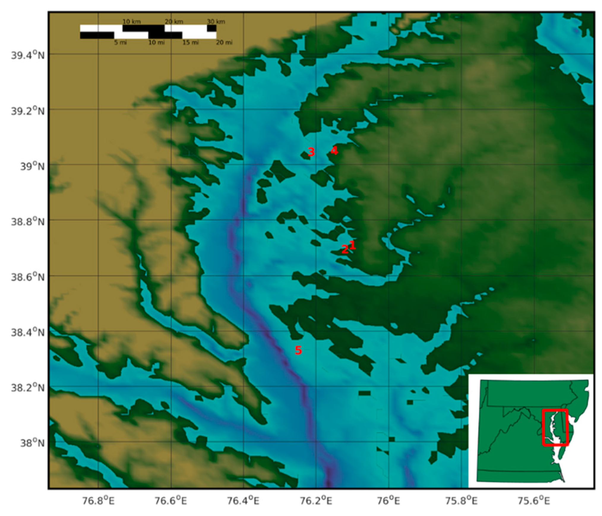
1. Introduction
2. Results
3. Discussion
4. Materials and Methods
Author Contributions
Funding
Acknowledgments
Conflicts of Interest
References
- Newell, R.I.E.; Kemp, W.M.; Hagy, J.D., III; Cerco, C.F.; Testa, J.M.; Boynton, W.R. Top-down control of phytoplankton by oysters in Chesapeake Bay, USA: Comment on Pomeroy et al. (2006). Mar. Ecol. Prog. Ser. 2007, 341, 293–298. [Google Scholar] [CrossRef][Green Version]
- Gerritson, J.; Holland, A.F.; Irvine, D.F. Suspension-feeding bivalves and the fate of primary production: An estuarine model applied to Chesapeake Bay. Estuaries 1994, 17, 403–416. [Google Scholar] [CrossRef]
- Newell, R.I. Ecological changes in Chesapeake Bay: Are they the result of overharvesting the American oyster, Crassostrea virginica. In Chesapeake Research Consortium Publication 129; Chesapeake Research Consortium: Edgewater, Maryland, USA, 1988; pp. 536–546. [Google Scholar]
- Cerco, C.F.; Noel, M.R. Monitoring, modeling, and management impacts of bivalve filter feeders in the oligohaline and tidal fresh regions of the Chesapeake Bay system. Ecol. Model. 2010, 221, 1054–1064. [Google Scholar] [CrossRef]
- Graczyk, T.K.; Farley, C.A.; Fayer, R.; Lewis, E.J.; Troutt, J.M. Detection of Cryptosporidium oocysts and Giardia cysts in the tissues of eastern oysters (Crassostrea virginica) carrying principal oyster infectious diseases. J. Parasitol. 1998, 84, 1039–1042. [Google Scholar] [CrossRef] [PubMed]
- Murphree, R.L.; Tamplin, M.L. Uptake and retention of Vibrio cholera O1 in the Eastern oyster, (Crassostrea virginica). Appl. Environ. Microbiol. 1995, 61, 3656–3660. [Google Scholar] [PubMed]
- Cromeans, T.L.; Nainan, O.; Margolis, H.S. Detection of hepatitis A virus RNA in oyster meat. Appl. Environ. Microbiol. 1997, 63, 2460–2463. [Google Scholar]
- Graczyk, T.K.; Conn, D.B.; Lucy, G.; Minchin, D.; Tamang, L.; Moura, L.N.S.; DaSilva, A.J. Human waterborne parasites in zebra mussels (Dreissena polymorpha) from the Shannon River drainage area, Ireland. Parasitol. Res. 2004, 93, 385–391. [Google Scholar] [CrossRef]
- Graczyk, T.K.; Thompson, R.C.; Fayer, R.; Adams, P.; Morgan, U.M.; Lewis, E.J. Giardia duoenalis cysts of genotype A recovered from clams in the Chesapeake Bay subestuary, Rhode River. Am. J. Trop. Med. Hyg. 1999, 61, 526–529. [Google Scholar] [CrossRef][Green Version]
- Lang, A.S.; Kelly, A.; Runstadler, J.A. Prevalence and diversity of avian influenza viruses in environmental reservoirs. J. Gen. Virol. 2008, 89, 509–519. [Google Scholar] [CrossRef]
- Densmore, C.L.; Iwanowicz, D.D.; Ottinger, C.A.; Hindman, L.J.; Bessler, A.M.; Iwanowicz, L.R.; Prosser, D.J.; Whitbeck, M.; Driscoll, C.P. Molecular detection of avian influenza virus from sediment samples in waterfowl habitat on the Delmarva Peninsula, United States. Avian. Dis. 2017, 61, 520–525. [Google Scholar] [CrossRef]
- Brown, J.D.; Goekjian, G.; Poulson, R.; Valeika, S.; Stallknecht, D.E. Avian influenza virus in water: Infectivity is dependent on pH, salinity and temperature. Vet. Microbiol. 2009, 136, 20–26. [Google Scholar] [CrossRef] [PubMed]
- Stumpf, P.; Failing, K.; Papp, T.; Nazir, J.; Böhm, R.; Marschang, R.E. Accumulation of a low pathogenic avian influenza virus in zebra mussels (Dreissena polymorpha). Avian. Dis. 2010, 54, 1183–1190. [Google Scholar] [CrossRef] [PubMed]
- Huyvaert, K.P.; Carlson, J.S.; Bentler, K.T.; Cobble, K.R.; Nolte, D.L.; Franklin, A.B. Freshwater clams as bioconcentrators of avian influenza virus in water. Vector Borne. Zoonotic. Dis. 2012, 12, 904–906. [Google Scholar] [CrossRef] [PubMed]
- Piffer, P.R.; Pintor de Aruda, E.; Passos, F.D. The biology and functional morphology of Macoma biota. Zoologia 2011, 28, 321–333. [Google Scholar] [CrossRef]
- Galimany, E.; Rose, J.M.; Dixon, M.S.; Wikfors, G.H. Quantifying feeding behavior of ribbed mussels (Guekensia demissa) in two urban sites (Long Island Sound, USA) with different seston characteristics. Estuaries Coast 2013, 36, 1265–1273. [Google Scholar] [CrossRef]
- Chesapeake Bay Mean Surface Salinity. Available online: https://www.chesapeakebay.net/what/maps/chesapeake_bay_mean_surface_salinity_spring_1985_2006 (accessed on 29 May 2019).
- Ladman, B.S.; Driscoll, C.P.; Pope, C.R.; Slemons, R.D.; Gelb, J. Potential of low pathogenicity avian influenza viruses of wild bird origin to establish experimental infections in turkeys and chickens. Avian. Dis. 2010, 5, 1091–1094. [Google Scholar] [CrossRef] [PubMed]
- Slemons, R.D.; Hansen, W.R.; Converse, K.A.; Senne, D.A. Type A influenza virus surveillance in free-flying, nonmigratory ducks residing on the Eastern Shore of Maryland. Avian. Dis. 2003, 47, 1107–1110. [Google Scholar] [CrossRef]
- Prosser, D.J.; Densmore, C.L.; Hindman, L.J.; Iwanowicz, D.D.; Ottinger, C.A.; Iwanowicz, L.R.; Driscoll, C.P.; Nagel, J. Low-pathogenic avian influenza viruses in wild migratory waterfowl in a region of high poultry production, Delmarva, Maryland. Avian. Dis. 2016, 61, 128–134. [Google Scholar] [CrossRef]
- Rohani, P.; Breban, R.; Stallknecht, D.E.; Drake, J.M. Environmental transmission of low pathogenicity avian influenza viruses and its implications for pathogen invasion. Proc. Natl. Acad. Sci. USA 2009, 106, 10365–10369. [Google Scholar] [CrossRef]
- Numberger, D.; Dreler, C.; Vulliod, C.; Gabriel, G.; Greenwood, A.D.; Grossart, H. Recovery of influenza A viruses from lake water and sediments by experimental inoculation. PLoS ONE 2019. [Google Scholar] [CrossRef]
- Spackman, E.; Senne, D.A.; Myers, T.J.; Bulaga, L.L.; Garber, L.P.; Perdue, M.L.; Lohman, K.; Daum, L.T.; Suarez, D.L. Development of a real-time reverse transcriptase PCR assay for type A influenza virus and the Avian H5 and H7 hemagglutinin subtypes. J. Clin. Microbiol. 2002, 40, 3256–3260. [Google Scholar] [CrossRef] [PubMed]


| Site Designation | Species | # Specimens Tested | # Positive Specimens | % Positive Specimens |
|---|---|---|---|---|
| Trippe Creek | Macoma balthica | 59 | 2 | 3.4 |
| Goldsborough Creek | Macoma phenax | 40 | 4 | 10.0 |
| Reed Creek | Guekensia demissa | 24 | 6 | 25.0 |
| Site Designation | Approx. Latitude | Approx. Longitude | Species (# Specimens) | Mean Weight (mg) | Mean Length (mm) |
|---|---|---|---|---|---|
| Trippe Creek | 38.710299 | −76.110816 | Macoma balthica (59) | 1180 | 19 |
| Macoma phenax (16) | 155 | 9 | |||
| Goldsborough Creek | 38.694905 | −76.131430 | Macoma balthica (10) | 148 | 10 |
| Macoma phenax (40) | 100 | 8 | |||
| Mulinia sp. (7) | 287 | 10 | |||
| Eastern Neck | 39.045982 | −76.223874 | Macoma balthica (48) | 1310 | 21 |
| Macoma phenax (2) | 130 | 10 | |||
| Rangia sp. (11) | 23,345 | 40 | |||
| Mya sp. (1) | 2800 | 32 | |||
| Mussel undet sp (1) | 160 | 11 | |||
| Reed Creek | 39.050508 | −76.160033 | Guekensia demissa (24) | 6691 | 42 |
| Rangia sp. (1) | 19,100 | 37 | |||
| Barren Island | 38.333101 | −76.259680 | Macoma balthica (36) | 210 | 11 |
| Mulinia sp. (8) | 213 | 9 |
| Description | Sequence |
|---|---|
| Forward primer (M+25) | AGATGAGTACTTCTAACCGAGGTCG |
| Reverse primer (M-124) | TGCAAAAACATCTTCAAGTCTCTG |
| Synthetic Matrix Standard | TGAAAGATGAGTCTTCTAACCGAGGTCGAAACGTACGTTCTCTCTATCGTCCCGTCAGGCCCCCTCAAAGCCGAGATCGCGCAGAGACTTGAAGATGTTTTTGCAGGGAAGAACACCGATCTCGAGGCACTCATGGAATGGCTAAAGACAAGACCAATCCTGTCACCTCTGACTAAGGGGATTTTAGGATTTGTGTTCACGCTCACCGTGCCCAGTGAGCGAGGACTGCAGCGTAGACGCTTTGTCCAGAA |
© 2019 by the authors. Licensee MDPI, Basel, Switzerland. This article is an open access article distributed under the terms and conditions of the Creative Commons Attribution (CC BY) license (http://creativecommons.org/licenses/by/4.0/).
Share and Cite
Densmore, C.L.; Iwanowicz, D.D.; McLaughlin, S.M.; Ottinger, C.A.; Spires, J.E.; Iwanowicz, L.R. Influenza A Virus Detected in Native Bivalves in Waterfowl Habitat of the Delmarva Peninsula, USA. Microorganisms 2019, 7, 334. https://doi.org/10.3390/microorganisms7090334
Densmore CL, Iwanowicz DD, McLaughlin SM, Ottinger CA, Spires JE, Iwanowicz LR. Influenza A Virus Detected in Native Bivalves in Waterfowl Habitat of the Delmarva Peninsula, USA. Microorganisms. 2019; 7(9):334. https://doi.org/10.3390/microorganisms7090334
Chicago/Turabian StyleDensmore, Christine L., Deborah D. Iwanowicz, Shawn M. McLaughlin, Christopher A. Ottinger, Jason E. Spires, and Luke R. Iwanowicz. 2019. "Influenza A Virus Detected in Native Bivalves in Waterfowl Habitat of the Delmarva Peninsula, USA" Microorganisms 7, no. 9: 334. https://doi.org/10.3390/microorganisms7090334
APA StyleDensmore, C. L., Iwanowicz, D. D., McLaughlin, S. M., Ottinger, C. A., Spires, J. E., & Iwanowicz, L. R. (2019). Influenza A Virus Detected in Native Bivalves in Waterfowl Habitat of the Delmarva Peninsula, USA. Microorganisms, 7(9), 334. https://doi.org/10.3390/microorganisms7090334