Article

Development and Validation of Shiga Toxin-Producing Escherichia coli Immunodiagnostic Assay

by
Miriam A. Silva
1,†,
Anna Raquel R. Santos
1,†,‡,
Leticia B. Rocha
1,†,
Bruna A. Caetano
1,
Thais Mitsunari
1,
Luanda I. Santos
1,
Juliana M. Polatto
1,
Denise S. P. Q. Horton
1,
Beatriz E. C. Guth
2,
Luís Fernando dos Santos
3 and
Roxane M. F. Piazza
1,*
1
Laboratório de Bacteriologia, Instituto Butantan, São Paulo 05503-900, Brazil
2
Departamento de Microbiologia, Imunologia, Parasitologia, Escola Paulista de Medicina Universidade Federal de São Paulo, São Paulo 04023-062, Brazil
3
Centro de Bacteriologia, Instituto Adolfo Lutz, São Paulo 01246-000, Brazil
*
Author to whom correspondence should be addressed.
These authors equally contributed to the present work.
Current address: Centro de Tecnologia em Vacinas Universidade Federal de Minas Gerais, Belo Horizonte, Minas Gerais, 31270-901, Brazil.
Microorganisms 2019, 7(9), 276; https://doi.org/10.3390/microorganisms7090276
Submission received: 1 July 2019 / Revised: 31 July 2019 / Accepted: 2 August 2019 / Published: 21 August 2019
(This article belongs to the Special Issue Shiga Toxin-Producing Escherichia coli)

Abstract

:
Shiga toxin (Stx)–producing Escherichia coli (STEC) and its subgroup enterohemorrhagic E. coli are important pathogens involved in diarrhea, which may be complicated by hemorrhagic colitis and hemolytic uremic syndrome, the leading cause of acute renal failure in children. Early diagnosis is essential for clinical management, as an antibiotic treatment in STEC infections is not recommended. Previously obtained antibodies against Stx1 and Stx2 toxins were employed to evaluate the sensitivity and specificity of the latex Agglutination test (LAT), lateral flow assay (LFA), and capture ELISA (cEIA) for STEC detection. The LAT (mAb Stx1 plus mAb stx2) showed 99% sensitivity and 97% specificity. Individually, Stx1 antibodies showed 95.5% and 94% sensitivity and a specificity of 97% and 99% in the cEIA and LFA assay, respectively. Stx2 antibodies showed a sensitivity of 92% in both assays and a specificity of 100% and 98% in the cEIA and LFA assay, respectively. These results allow us to conclude that we have robust tools for the diagnosis of STEC infections.

1. Introduction

Among the E. coli human pathogens, the Shiga toxin (Stx)–producing Escherichia coli (STEC) and its subgroup enterohemorrhagic E. coli (EHEC) have gained importance in the three last decades due to their involvement in diarrhea [1], that may be complicated by hemorrhagic colitis (HC) [2] and the hemolytic uremic syndrome (HUS) [3,4], the foremost cause of acute renal failure in children [1] due to action of the two major types of the phage-encoded Stxs, Stx1 and/or Stx2. HUS is associated more commonly with strains that produce Stx2 alone or in combination with Stx1 rather than those that produce Stx1 only [5,6]. Most cases of the STEC infection are acquired by consuming food of bovine origin; however, other foodstuffs, water, environmental contact, and person-to-person transmission are also important sources [7]. A large fraction of the reported STEC infections is due to E. coli O157:H7, the most involved serotype in complicated cases, which often evolve into HUS [8]. However, six serogroups (O26, O45, O103, O111, O121, and O145) account for many cases of non-O157 STEC infections; furthermore the non-O157 serotypes [9,10].
In the Latin American countries, human infections by STEC are endemic in Argentina and are mainly linked to O157 strains. In other Latin American countries STEC (O157 and non-O157) causes sporadic cases of diarrhea, bloody diarrhea, hemolytic anemia and HUS [11,12,13,14,15,16,17]. However, it is essential to point out that the distribution of STEC/EHEC in the gastrointestinal tract of a wide variety of animals indicates the zoonotic character of its infections. The role of different animal species as asymptomatic carriers of STEC/EHEC has been extensively studied in the last years in Brazil. In addition to cattle, which are their most common natural reservoir [18,19] the presence of these pathogens has been identified in the feces of dairy buffaloes [20], sheep [21,22], pigs [23,24], birds [25,26], and fishes [27]. It is noteworthy that some relevant serotypes linked to human infections such as O103:H2 and O157:H7 have been recovered from the feces of sheep and cattle [18,28].
Thus, early diagnosis certainly is fundamental for clinical management of the etiological agent involved in diarrhea; specifically for STEC infections, as the antibiotic treatment is not recommended, since its use may induce the Shiga toxins release, thus allowing its dissemination [29]. Moreover, the diagnosis may be indicative of a likely outbreak, followed by the required measures such as implementation of control and detection of emerging strains [30], thus a key point for therapeutic conduct and consequently to control the disease. The diagnosis of STEC in a routine laboratory is difficult, and only specific virulence factors such as the presence of the Shiga toxin, which is common to all STEC, allows differentiation from other E. coli [31].
Immunoserological methods have advantages for clinical laboratories because they significantly reduce the time of analysis, have excellent sensitivity and specificity, and are easy to perform [32]. Despite the availability of commercial immunoassays, such as the ELISA immunoassay (EIA): ProSpecT™ Shiga Toxin E. coli (Oxoid Ltd., Basingstoke, UK), Premier® EHEC (Meridian Bioscience, Inc., Cincinnati, OH, USA), Ridascreen® Verotoxin test (R-Biopharm AG, Darmstadt, Germany); Shiga toxin Check™ (TECHLAB, Inc., Blacksburg, VA, USA) and Shiga Toxins, EIA with Reflex to E coli O157, Culture (Quest Diagnostics, Inc., Saint Louis, MO, USA); lateral flow assay (LFA): Duopath®Verotoxins (Merck & Co., Inc. Palo Alto, CA, USA), ImmunoCard STAT!®EHEC (Meridian Bioscience, Inc., Cincinnati, OH, USA), Ridascreen® Quick Verotoxin/O157 (R-Biopharm AG, Darmstadt, Germany) and Shiga toxin Quick Check™ (TECHLAB, Inc., Blacksburg, VA, USA); immunomagnetic separation, such as the RapidCheck®Confirm™STEC (Romer Labs Holding, Tulln, Austria) and an optical immunoassay, such as the Biostar OIA Shigatoxin (Inverness Medical Professional Diagnostics, Waltham, MA, USA). These commercial available assays are not implemented in the routine of clinical laboratories of low and middle-income regions’ of developing countries (https://datahelpdesk.worldbank.org/knowledgebase/articles/906519), thus encouraging the present work, i.e., the desire of development of a screening test for the Shiga toxin detection for countries with high incidence, endemic or low information on this infection. Thus, the key of our study is affordability, i.e., to provide the health market, whether a private or public one, an option when analyzing the cost benefit issue (bureaucracy, quality, time to obtain the product and final value). The main project involves two steps: (a) Searching robust tools for the development of the test; (b) will focus on making the use of feces directly, calculating costs and price in the market.
In the present study we limited the search for robust tools, thus essaying the generated polyclonal (pAb) and monoclonal (mAb) antibodies against Stx1 and Stx2 [33,34,35] and the standardization of three platforms using these antibodies in order to verify their performance. Therefore, these antibodies were employed in an evaluation of the sensitivity and specificity of the immunoserological methods, LAT, cEIA and LFA for detection of Shiga toxin-producing Escherichia coli using a collection of bacterial isolates, of which 96 STEC presenting several serotypes and harboring different Stx subtypes and the achieved results indicated that we have robust tools for the the diagnosis of STEC infections.

2. Materials and Methods

2.1. Bacterial Isolates

We used in this study a collection of 96 Shiga toxin-producing E. coli (STEC) strains belonging to different serotypes and stx subtypes (Table 1). We also included for the ELISA (EIA) cut-off definition and specificity of the latex agglutination (LA) and lateral flow assay (LFA), 12 typical enteropathogenic E. coli (tEPEC) [36,37], 11 atypical enteropathogenic E. coli (aEPEC) [38], 45 enterotoxigenic E. coli (ETEC) [39,40], nine enteroaggregative E. coli (EAEC) [41], eight enteroinvasive E. coli (EIEC) [42], 14 diffusely-adherent E. coli (DAEC) [42], three fecal E. coli negative for DEC virulence factors (NVF E. coli), four microbiota E. coli isolates and 19 Enterobacteriaceae isolates (Citrobacter freundii, Edwardsiella tarda, Enterobacter cloacae, Klebsiella pneumoniae, K. oxitoca, Morganella morganii, Proteus mirabilis, Providencia spp., Salmonella Agona, S. Enteritidis, S. Infantis, S. Newport, S. Typhimurium, Serratia marcescens, Shigella boydii, S. flexneri, and S. sonnei) from our laboratory collection. The prototype EHEC EDL933 [43] was included in the assay as a positive control for the Stx1/Stx2 producing strain. To estimate the sample size for sensitivity and specificity of the diagnostic methods herein standardized nomogram determination was done according to Malhotra and Indrayan [44] and Hajian-Tilak [45] based on the studies of the diarrheagenic E. coli pathotypes infection prevalence [46,47].

2.2. Bacterial Supernatant Preparation

Bacterial supernatants were obtained by sequential bacterial culture in the LB medium for 18 h (1:100), followed by a further 4 h in the EC broth (1:10) containing ciprofloxacin (5 ng/mL) [33] and then lysed with 20% Triton X-100 for capture ELISA (cEIA) and latex agglutination test (LAT) [48] or 200 μg/mL polymyxin sulfate B for lateral flow assay (LFA).

2.3. Antibodies

The antibodies employed herein were produced in previous work from our group. Stx1 and Stx2 polyclonal antibodies (pAbs) were raised in rabbits and characterized elsewhere [33,34]. The generation and characterization of Stx1 and Stx2 monoclonal antibodies (mAbs) are also described elsewhere [35].

2.4. Capture ELISA Immunoassay (cEIA)

Microtiter plates (C96 Polysorp-NUNC) were incubated with 10 µg/mL of Stx1-pAb or 25 µg/mL of Stx2-pAb in carbonate-bicarbonate-buffered, pH 9.6 at 37 °C for 2 h and then further at 4 °C for 16 h. Phosphate buffered saline (PBS) with bovine serum albumin (BSA) 1% was added as a blocking agent and incubated for 1 h at 37 °C. The supernatant of bacterial cultures were incubated for 1 h at 37 °C. Toxin bound to Stx1-pAb or Stx2-pAb was then detected with 5 µg/mL of Stx1-mAb or Stx2-mAb followed by goat anti-mouse IgG peroxidase (Sigma-Aldrich, St Louis, MO, USA) diluted 1:5000 in the blocking solution. Reactions were developed with 0.5 mg/mL O-phenylenediamine (OPD; Sigma Aldrich Co, St Louis, MO, USA) plus 0.5-μL/mL hydrogen peroxide in 0.05 M citrate-phosphate buffer, pH 5.0, in the dark at room temperature. The reactions were interrupted after 15 min by the addition of 50 μL of 1 M HCl. The absorbance was measured at 492 nm in a Multiskan EX ELISA reader (Labsystems, Milford, MA, USA). At each step, the volume added was 100 μL/well, except in the washing and blocking steps, when the volume was 200 μL/well. Between incubations, the plates were washed three times with PBS-Tween 0.05%. All experiments were carried out in duplicate, and results correspond to three independent experiments.

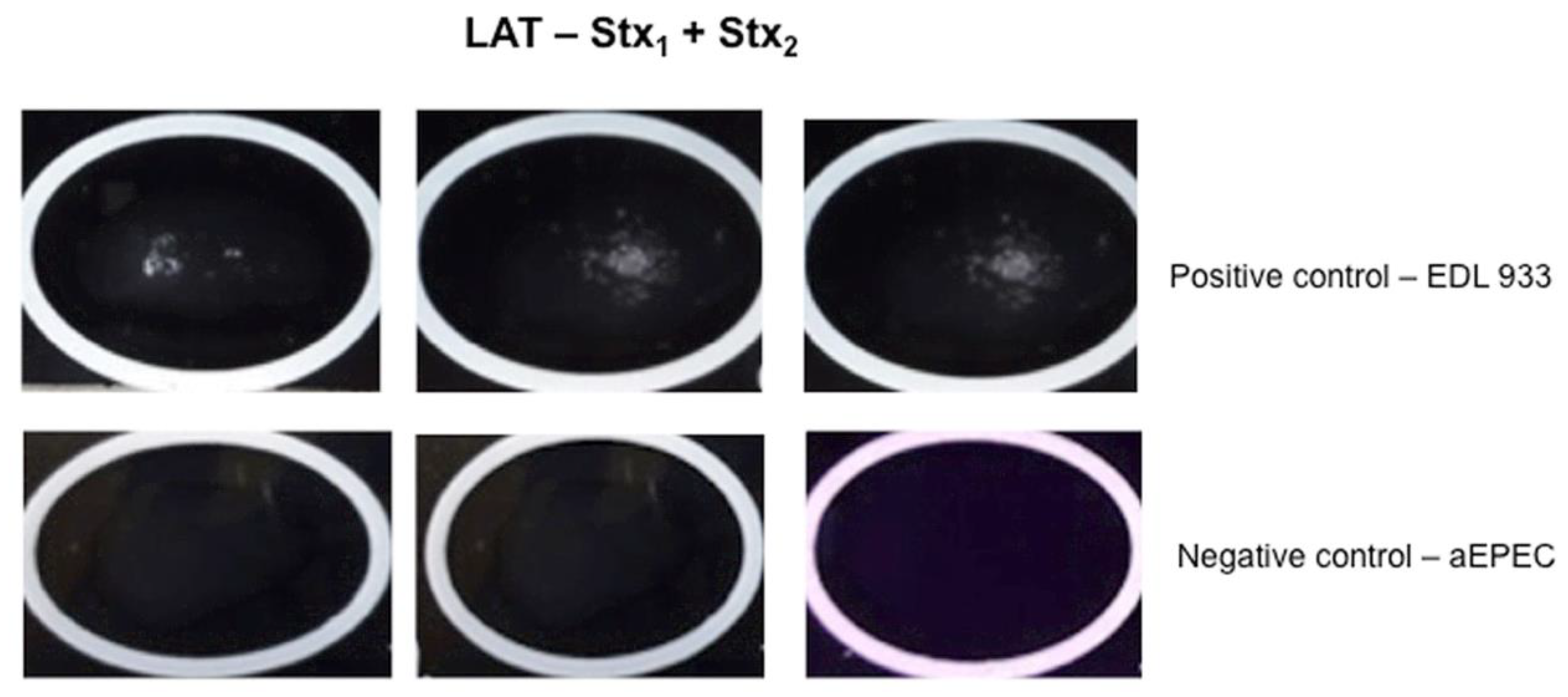
2.5. Latex Agglutination Test (LAT)

The beads were coupled with 100 µg of Stx1 mAb for LAT-Stx1 or 100 µg of Stx2 mAb for LAT-Stx2. For detection of Stx without subtype discrimination, the beads were coupled with Stx1 and Stx2 mAbs (1:1). The principle used was the nucleophilic addition to aldehyde group with amines. The glutaraldehyde was used as a spacer arm between the bead and mAbs. Briefly, the polybeads amino microsphere in a 2.5% aqueous suspension (1 µm diameter–Polyscience, Warrington, PA, USA) were washed three times with PBS and incubated with 8% glutaraldehyde in the PBS at room temperature for 4 h [48]. Next, 50 µg of Stx1 and 50 µg of Stx2 mAbs were added and the mixture incubated at room temperature for 16–18 h for coupling, followed by further incubation in the presence of 0.2 M ethanolamine and BSA. Both incubations were with gentle mixing at room temperature for 60 min. Between incubations, the coated beads were washed and centrifuged (7200× g) for 6 min. After the last washing procedure, the pellet was resuspended in the storage buffer (Polyscience, Warrington, PA, USA) and kept at 4 °C.
For LAT, bacterial lysate were obtained by sequential bacterial growth in the LB medium for 18 h (1:100), followed by a further 4 h in the EC broth (1:10) containing ciprofloxacin (5 ng/mL) and then, lysed with 20% Triton X-100 during 1 h and centrifuged (14,500× g) for 15 min. The assay was performed on a slide glass using 20 µL of bacterial lysate and 20 µL of latex beads coupled to Stx1 plus Stx2 mAbs, and checking for agglutination between 1–2 min of gentle mixing.

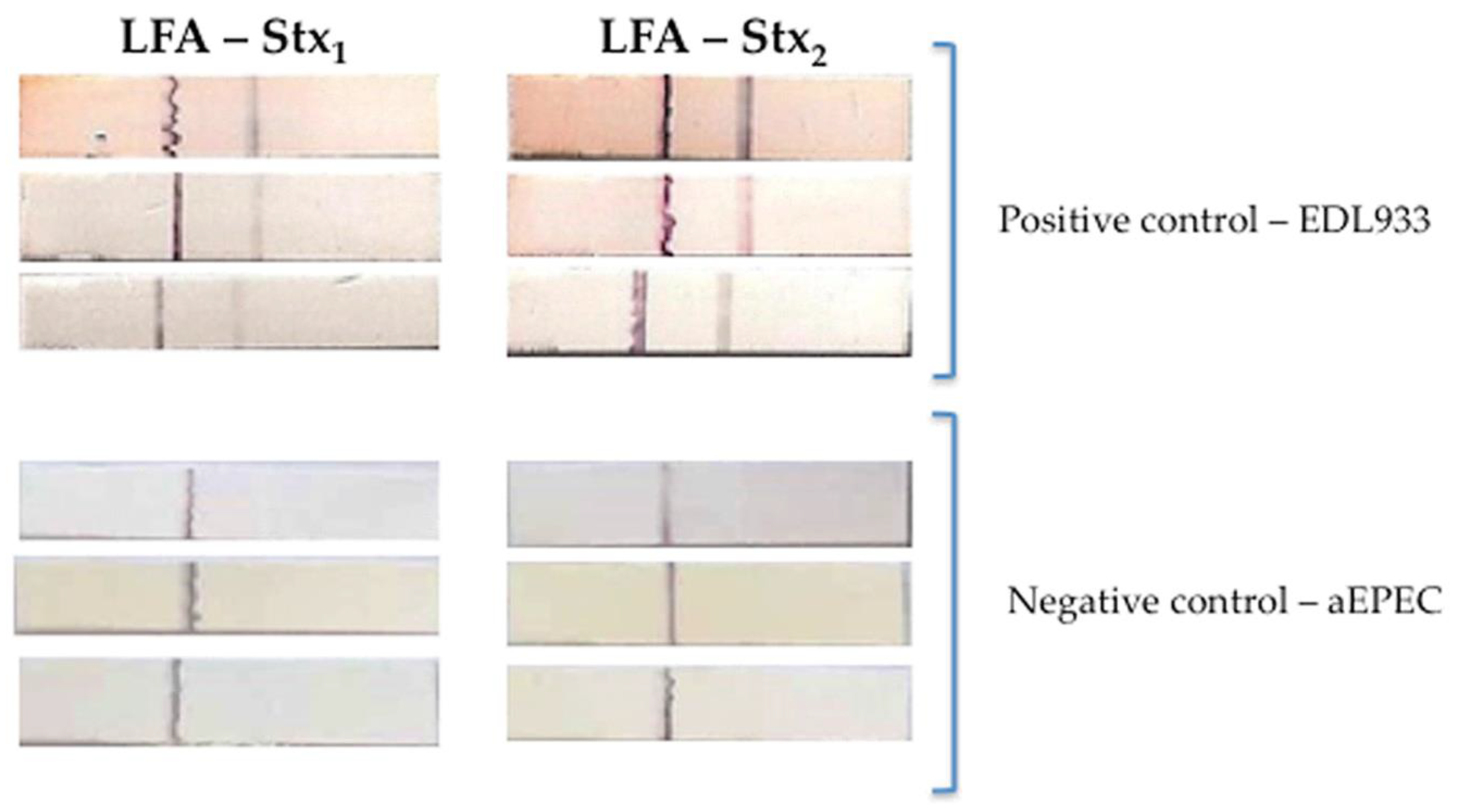
2.6. Lateral Flow Assay (LFA)

The Stx1 or Stx2 pAb rabbit sera were used as a capture antibody conjugated to colloidal gold particles. The protocol for conjugation of the pAbs with colloidal gold was performed according to Oliver (2010) [49], with some modifications. Briefly: 0.01% colloidal gold solution (20 nm diameter particles) (BBInternational, Cardiff, England) was previously adjusted to pH 9.0 with 0.1 M potassium carbonate solution (K2CO3). First, 1 mg of each of the pAb was resuspended in 1 mL of 0.2 M borate buffer (0.2 M sodium borate, 0.15 M NaCl, pH 9.0) and dialyzed against 2 mM borate buffer (2 mM sodium borate, 1.5 mM NaCl, pH 9.0) at room temperature for 2 h.
The amount of antibody required to stabilize colloidal gold was determined as follows: 100 μL of colloidal gold was added in microtubes containing 10 μL of serially diluted previously dialyzed pAb. After 10 min, 11 μL of 10% sodium chloride (NaCl) was added to each tube. The amount of antibody sufficient to stabilize the gold was the dilution in which the solution did not change color, i.e., an insufficient amount of antibody altered the coloration of the solution from red to blue [49].
After determination of the optimal ratio between the antibody and colloidal gold made in the previous step, a total volume of 10 mL of solution was obtained, which was kept under stirring at room temperature for 30 min. To block the reaction, a 10% BSA solution (10% BSA in 0.02 M borate buffer, pH 9.0) was added in sufficient volume to the final concentration of 1%. After incubation at room temperature for 30 min, the solution was centrifuged at 11,000× g for 20 min at room temperature and the supernatant discarded. The pellet was then resuspended in 2 mL of a 2% BSA solution (2% BSA in 0.01 M borate buffer) and centrifuged at 11,000× g for 10 min at room temperature. Finally, the pellet was resuspended in 1 mL of the storage buffer (3% BSA, 3% sucrose, 0.01 M sodium borate, and 0.05% sodium azide, pH 7.5) and the pAbs conjugated to colloidal gold (pAb-Au) were stored at 4 °C. Finally, 600 μL of pAb-Au was applied at 30 cm in the dried glass fiber in a desiccator whose relative humidity is 20% for 24 h.
The Stx1 (3 mg/mL) or Stx2 (4 mg/mL) mAbs were used for detection, and they were applied directly to the nitrocellulose membrane (Millipore HF180 NM) as the test line. As a control line of the LFA, the goat anti-rabbit IgG antibody was applied above the test line. The LFA tests were prepared on a large scale on a semi-automatic platform, consisting of a Matrix 1600 Reagent Dispensing Module (applies the antibodies in NM), Matrix 2210 Universal Laminator Module (mounts/overlaps all test membranes), and Matrix 2360 Programmable Shear (cuts LFA test strips), all obtained from Kinematic Automation, Inc. (Sonora, CA, USA). The strips were dried at room temperature in a desiccator whose UR is 20% for 24 h. The treatment of the sample pad portion was made by buffer containing 1% BSA, 0.25% Tween-20, and sodium azide.
For LFA, the bacterial supernatant were obtained by sequential bacterial growth in the LB medium for 18 h (1:100), followed by a further 4 h in the EC broth (1:10) containing ciprofloxacin (5 ng/mL) and then, lysed with 200 μg/mL polymyxin sulfate B during 1 h and centrifuged (14,500× g) for 15 min. Supernatant were kept at −20 °C until test analyzes.

2.7. Stx Subtyping

Stx subtyping was performed by PCR using the primers and amplification conditions as previously described [50].

2.8. Statistical Analyses

The Vero cell assay (VCA) was employed as the gold standard method for the Stx production [51]. Additionally, we differentiated the toxin subtype by PCR for stx1 and or stx2 in order to evaluate the sensitivity and specificity of Stx1 or Stx2 antibodies. The absorbance values from the duplicates of three independent experiments from Stx-positive and Stx-negative isolates after reaction with Stx1 or Stx2 antibodies were analyzed by GraphPrism 5.01, using the Student’s t-test and two-way ANOVA. The differences were considered statistically significant when p ≥ 0.05. The receiver operating characteristic (ROC) curve was employed for determining the calculation of the ELISA’s cut-off as well as providing the sensitivity and specificity report. Furthermore, the Cohen’s Kappa statistic was employed to test the interrater reliability [52].

3. Results

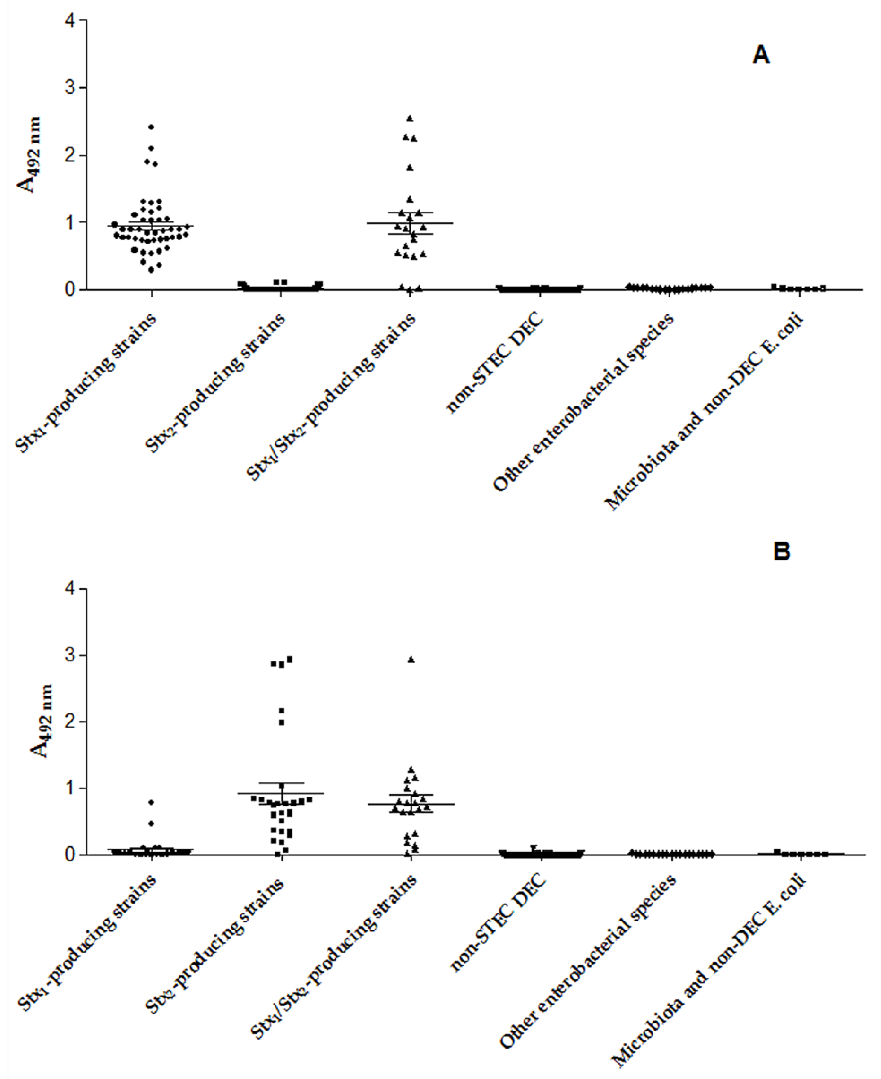
3.1. Presence and Production of Stx1 and Stx2

The production of Stx was analyzed in the bacterial collection (221 isolates) employing the gold standard Vero cell assay (VCA). The 96 STEC were confirmed as Stx producers, and the 125 other enterobacterial strains were Stx negative. In addition, the stx subtype was defined by PCR in the 96 STEC strains, in which 47 were stx1 and 28 were stx2 and 21 presented both genes (Table 1).

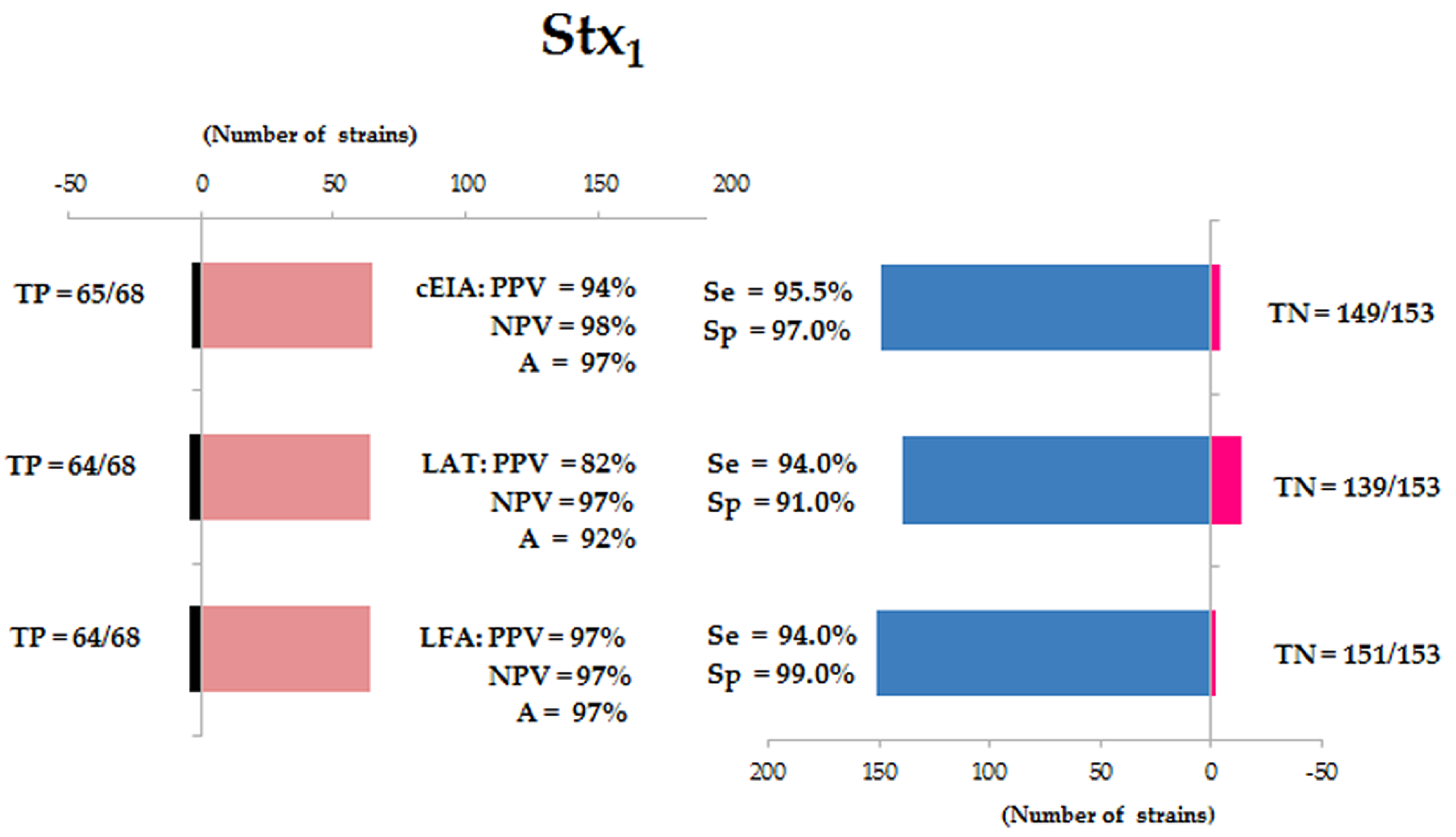
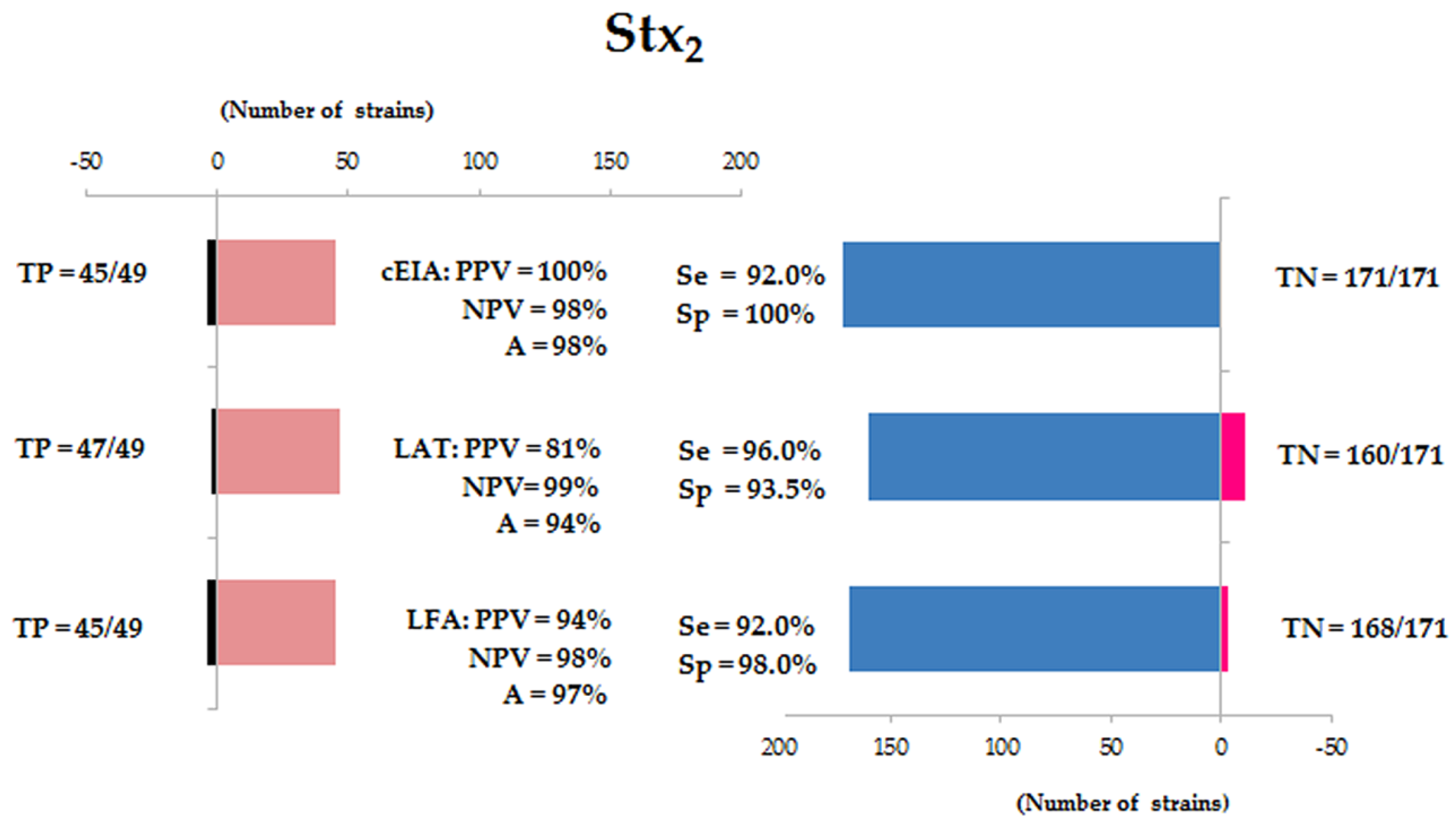
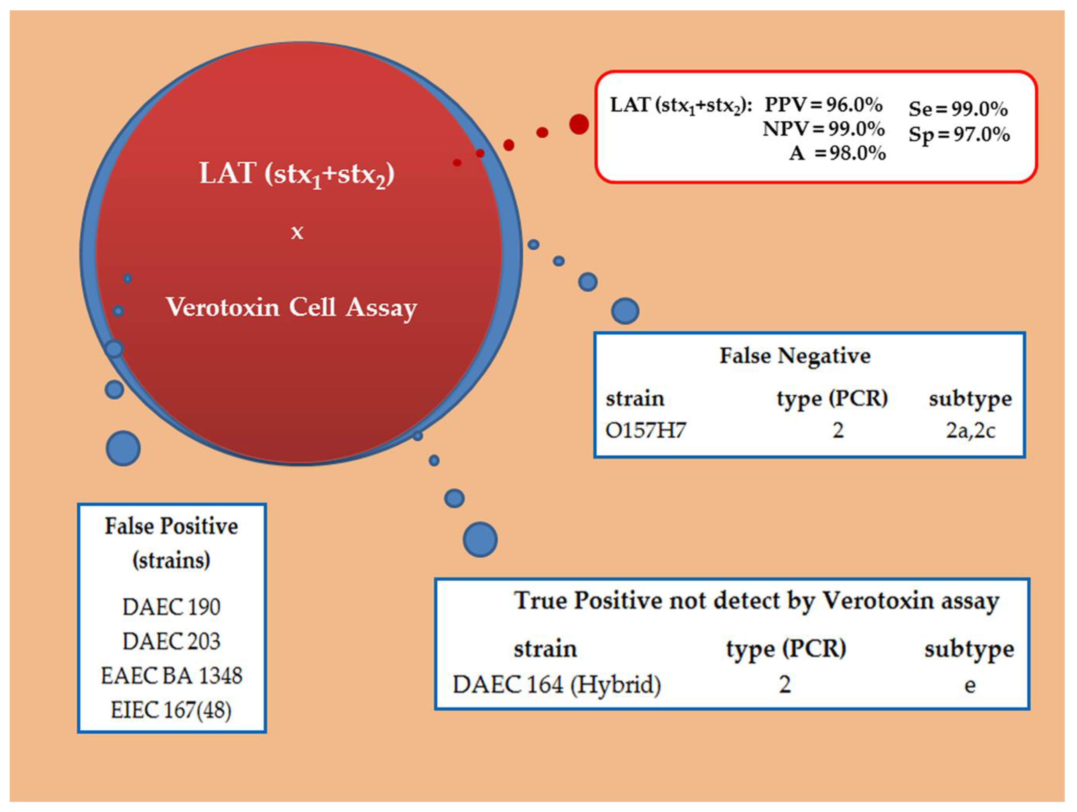
3.2. Validation of Diagnostic Immunoassays

In order to test the bacterial supernatants for each different assay, bacterial cultures were prepared differently. Thus, for capture ELISA (cEIA) and latex agglutination test (LAT) bacterial culture was lysed with Triton X-100 or polymyxin sulfate B for the lateral flow assay (LFA), since the detergent presence impaired the sample flow in LFA.
The bacterial collection mentioned above was analyzed using three different immunoassays employing Stx1 and Stx2 polyclonal and monoclonal antibodies raised in-house [33,34,35] in order to observe their performance in the screening assay (LAT and/or LFA) and/or confirmatory assay (cELISA). These analyses allowed calculating the assay parameters as predictive value (PV) for the positive (PPV) and negative (NPV) samples, the accuracy (A), the sensitivity (Se) and specificity (Sp). In addition, the kappa concordance index (κ) was evaluated using the p value < 0.001.
The analysis of the Stx1 detection in 68 strains (positive samples) and 153 strains (negative samples) by the three methods revealed for cEIA was PPV = 94%, NPV = 98%, A = 97%, Se = 95.5%, Sp = 97% and κ = 0.957. For LAT: PPV of 82%, NPV of 97% and A of 92%, thus the Se and Sp was 94% and 91%, respectively, and κ = 0.846. The parameters observed for LFA: 97% for PPV, 97% for NPV, 97% for A, 94% for Se, 99% for Sp and κ = 0.925 (Figure 1 and Figure 3).
Concerning the detection of Stx2 by 49 strains (positive samples) and 171 strains (negative samples) by the three methods revealed for cEIA: PPV = 100%, NPV = 98%, A = 98%, Se = 92%, Sp = 100% and κ = 0.933. These values for LAT: PPV of 81%, NPV of 99% and A of 94%, thus the Se and Sp was 96% and 93.5%, respectively, and κ = 0.829. The parameters observed for LFA: 94% for PPV, 98% for NPV, 97% for A, 92% for Se, 98% for Sp and κ = 0.883 (Figure 2 and Figure 3).
Comparing the values of the three immunoassays for Stx1 and Stx2, LAT employing mAbs individually coupled to latex particles always showed lower predictive values than LFA and cEIA. Thus, we decided to combine both monoclonal antibodies (Stx1 and Stx2) in order to detect Stx without distinction between the toxin types. We achieved the following values: PPV of 96%, NPV of 99% and A of 98%, thus the Se and Sp was 99% and 97%, respectively and κ = 0.945. Therefore, we observed five false positives ((three strains of DAEC (190 and 203), one EAEC (BA1348) and one EIEC (167(48)) and one false negative, the test was not able to detect one O157:H7 strain (Figure 4 and Figure 5).
In the cEIA, the Stx1 Abs (mAb and pAb) were able to recognize the Stx1 producers, showing 95.5% of sensitivity and 97% of specificity and an A492nm of 0.195 cut off (Figure 6A) and Stx2 Abs (mAb and pAb) were able to recognize the Stx2 producing strains, showing 92% of sensitivity and 100% of specificity and an A492nm of 0.1205 cut off (Figure 6B).

4. Discussion

Different protocols for detection of Shiga toxin-producing strains either in the feces of infected patients or contaminated food have already been described for routine diagnosis [53,54,55]. Currently, the PCR for stx has been employed in reference diagnosis centers and some laboratories of upper-middle-income regions of developing countries using bacterial confluent growth zones or sorbitol-fermenting and non-fermenting colonies taken from MacConkey sorbitol agar (SMAC plates) [47,53]. Nevertheless, the gold standard for Stx detection is the evaluation of the cytotoxicity of bacterial culture supernatants for eukaryotic cells (VCA) [3,51].
Thus encouraging the present work, i.e., the request of a development of a screening test for the Shiga toxin detection for countries with high incidence, endemic or low information on this infection. Thus, Stx1 and Stx2 pAbs and mAbs were generated in previous studies [33,34,35]; and different formats of immunoassays, employing these antibodies were developed and standardized. Herein, we evaluated the sensitivity and specificity of LAT, cEIA, and LFA employing VCA as gold standard.
An important point is which protocol should be used for toxin production and secretion. Usually, Stx1 is secreted into the medium [56], whereas Stx2 has been shown to be either periplasmatic [56] or liberated inside vesicles [57] therefore not secreted into the medium. In a former work of our group we have established a protocol for in vitro cultivation by sequential bacterial culture in LB medium for 18 h (1:100), followed by a further 4 h in the EC broth (1:100), and in order to enhance toxins secretion we added ciprofloxacin (5 ng/mL) [33]. Herein, we established a 1:10 dilution from the LB to the EC broth, in turn; to better stimulate toxin production, since in 1:100 dilutions the very low-producers strains were not detected. Another critical point we established is the different procedures for toxins secretion according to the assay requirements. Thus, bacterial supernatants were prepared differently; bacterial culture was lysed with Triton X-100 for cEIA and LAT or polymyxin sulfate B for LFA. Usually, the commercial available tests also employ different procedures for toxin secretion, except for the Ridascreen® Verotoxin test and Ridascreen® Quick Verotoxin/O157, that mention the use of mitomycin C as an inductor for the formation of Shiga toxins, the others refer generically to the diluent.
The sensitivity and specificity of each tested assay reached percentages as the commercial ones (pertaining the datasheet of each company), therefore encouraging results; concerning cEIA-Stx1 95.5 and 97%, respectively and cEIA-Stx2 92 and 100%, respectively, comparable to ProSpecT™ Shiga Toxin E. coli (92.3 and 99.6%); Premier® EHEC (100 and 97.9%); Ridascreen® Verotoxin test (93.9 and 96.1%) and Shiga toxin check™ (97.1 and 99.7%). For LFA-Stx1 (94 and 99%) and LFA-Stx2 (92 and 98%) for sensitivity and specificity, respectively; comparable to Duopath®Verotoxins (100 and 99.6%); ImmunoCard STAT!®EHEC (92.3 and 98.7%); Ridascreen® Quick Verotoxin/O157 (85 and 98.7%) and Shiga toxin Quick Check™ Stx1 (100 and 99.5%) and Stx2 (95.7 and 99.9%).
Regarding the sensitivity and specificity of the latex agglutination assay, LAT-Stx1 (94 and 91%); LAT-Stx2 (96 and 93.5%), respectively, but when both mAbs were combined these results reached to 99 and 97%, respectively. Our results cannot be compared to those of the reverse passive latex agglutination kits (VTEC-RPLA toxin detection and VTECRPLA “Seiken”, Japan) since they were no longer commercially accessible. Early diagnosis of diarrhea is the key to therapeutic behavior, accordingly, for a diagnostic assessment method we may employed RALT for EspB (97% sensitivity and 98% specificity) [48] and LAT-Stx (99% sensitivity and 97% specificity), so we may define in a short term if the diarrhea was due to EPEC/EHEC or STEC. Only one false negative occurred and five false-positives, in fact, among them, one strain initially described as the adherent diffuse E. coli (DAEC) by the daaC probe [42]. Herein it is a real positive, detected by PCR as Stx2e, but a non-producer Stx by VCA. Moreover, no cross-reaction was observed neither with E. coli negative for the DEC virulence factors nor with the microbiota strains and among the enterobacterial species.
Due to the feasibility of LFA, the LFA-Stx1 and LFA-Stx2 we consider that it can be used as a rapid test, such as described elsewhere [58,59], since it did not require expensive equipment or trained personnel to interpret the results. Unlike ELISA or PCR methods, colloidal gold technology can be used for point-of-care applications and screening as they require the only assessment of red colored lines for the end-point detection. Both standardized methods are reproducible, fast, easy to perform, showing high sensitivity in detecting Stx.
The same features were observed in cEIA, even detecting Stx in low-producing isolates, but, cEIA can be used as a diagnostic helper or confirmatory since a spectrophotometer is necessary for the absorbance’s reading. Currently, these assessment immunoassays are under technology transfer to a Brazilian start-up in order to validate those employing fecal samples and the commercial availability of them.

5. Conclusions

The standardized tests can be used not only in reference laboratories but also mainly in clinical laboratories and hospitals, given the importance of diagnosis for appropriate patient treatment and the prevention of outbreaks and contamination by STEC. The comparative analysis of cEIA, LAT and LFA allows the conclusion that we have robust tools for STEC diagnosis infections. Assessing all our data, including the rapidity of feedback to the patient, feasibility, and accuracy of the test, we can also conclude that when the Stx1 and Stx2 mAbs were coupled, better performance was observed for a screening test for the Shiga toxin detection.

Author Contributions

Conceptualization, M.A.S., A.R.R.S., L.B.R., D.S.P.Q.H., B.E.C.G., L.F.d.S. and R.M.F.P.; Methodology, M.A.S., A.R.R.S., L.B.R., B.A.C., T.M., L.I.S. and J.M.P.; Resources, L.F.d.S., B.E.C.G. and R.M.F.P.; Writing—original draft preparation, M.A.S., A.R.R.S., L.B.R. and R.M.F.P.; Writing—review and editing, M.A.S., A.R.R.S., L.B.R., B.A.C., T.M., D.S.P.Q.H., B.E.C.G., L.F.d.S. and R.M.F.P.; Project administration, R.M.F.P.; Funding acquisition, R.M.F.P.

Funding

This research was funded by São Paulo Research Foundation (FAPESP-2013/06589-6, 2013/50955-7). BECG and RMFP are research fellows of Conselho Nacional de Desenvolvimento Científico e Tecnológico. ARRS and LBR were FAPESP fellows (2012/02618-9, 2012/090906-2).

Conflicts of Interest

The authors declare no conflict of interest. The funders had no role in the design of the study; in the collection, analyses, or interpretation of data; in the writing of the manuscript, or in the decision to publish the results.

References

  1. Karmali, M.A.; Gannon, V.; Sargeant, J.M. Verocytotoxin-producing Escherichia coli (VTEC). Vet. Microbiol. 2010, 140, 360–370. [Google Scholar] [CrossRef] [PubMed]
  2. Riley, L.W.; Remis, R.S.; Helgerson, S.D.; McGee, H.B.; Wells, J.G.; Davis, B.R.; Hebert, R.J.; Olcott, E.S.; Johnson, L.M.; Hargrett, N.T.; et al. Hemorrhagic colitis associated with a rare Escherichia coli serotype. N. Engl. J. Med. 1983, 308, 681–685. [Google Scholar] [CrossRef] [PubMed]
  3. Karmali, M.A.; Steele, B.T.; Petric, M.; Lim, C. Sporadic cases of haemolytic-uraemic syndrome associated with faecal cytotoxin and cytotoxin-producing Escherichia coli in stools. Lancet 1983, 1, 619–620. [Google Scholar] [CrossRef]
  4. Karmali, M.A.; Petric, M.; Lim, C.; Fleming, P.C.; Arbus, G.S.; Lior, H. The association between idiopathic hemolytic uremic syndrome and infection by verotoxin-producing Escherichia coli. J. Infect. Dis. 1985, 151, 775–782. [Google Scholar] [CrossRef] [PubMed]
  5. Byrne, L.; Jenkins, C.; Launders, N.; Elson, R.; Adak, G.K. The epidemiology, microbiology and clinical impact of Shiga toxin-producing Escherichia coli in England, 2009–2012. Epidemiol. Infect. 2015, 143, 3475–3487. [Google Scholar] [CrossRef] [PubMed]
  6. Melton-Celsa, A.R. Shiga toxin (Stx) classification, structure, and function. Microbiol. Spectr. 2014, 2. [Google Scholar] [CrossRef] [PubMed]
  7. Heiman, K.E.; Mody, R.K.; Johnson, S.D.; Griffin, P.M.; Gould, L.H. Escherichia coli O157 outbreaks in the United States, 2003–2012. Emerg. Infect. Dis. 2015, 21, 1293–1301. [Google Scholar] [CrossRef] [PubMed]
  8. Tarr, P.I.; Gordon, C.A.; Chandler, W.L. Shiga-toxin-producing Escherichia coli and haemolytic uraemic syndrome. Lancet 2005, 365, 1073–1086. [Google Scholar] [CrossRef]
  9. Byrne, L.; Vanstone, G.L.; Perry, N.T.; Launders, N.; Adak, G.K.; Godbole, G.; Grant, K.A.; Smith, R.; Jenkins, C. Epidemiology and microbiology of Shiga toxin-producing Escherichia coli other than serogroup O157 in England, 2009–2013. J. Med. Microbiol. 2014, 63, 1181–1188. [Google Scholar] [CrossRef]
  10. Kuehne, A.; Bouwknegt, M.; Havelaar, A.; Gilsdorf, A.; Hoyer, P.; Stark, K.; Werber, D. HUS active surveillance network Germany. Estimating true incidence of O157 and non-O157 Shiga toxin-producing Escherichia coli illness in Germany based on notification data of haemolytic uraemic syndrome. Epidemiol. Infect. 2016, 114, 3305–3315. [Google Scholar] [CrossRef]
  11. Rivas, M.; Padola, N.L.; Lucchesi, P.M.A.; Masana, M. Diarrheogenic Escherichia coli in Argentina. In Pathogenic Escherichia coli in Latin America; Torres, A.G., Ed.; Bentham Books: Potomac, MD, USA, 2010; Volume 1, pp. 142–161. [Google Scholar]
  12. Vidal, R.M.; Oñate, A.; Salazar, J.C.; Prado, V. Shiga toxin producing Escherichia coli in Chile. In Pathogenic Escherichia coli in Latin America; Torres, A.G., Ed.; Bentham Books: Potomac, MD, USA, 2010; Volume 1, pp. 179–190. [Google Scholar]
  13. Guth, B.E.C.; Picheth, C.; Gomes, T.A.T. Escherichia coli situation in Brazil. In Pathogenic Escherichia coli in Latin America; Torres, A.G., Ed.; Bentham Books: Potomac, MD, USA, 2010; Volume 1, pp. 162–178. [Google Scholar]
  14. Varela, G.; Gómez-Garcia, O.; Ochoa, T. Diarrheogenic Escherichia coli in children from Uruguay, Colombia and Peru. In Pathogenic Escherichia coli in Latin America; Torres, A.G., Ed.; Bentham Books: Potomac, MD, USA, 2010; Volume 1, pp. 209–222. [Google Scholar]
  15. Navarro, A.; Estrada-Garcia, T. Epidemiology of diarrheogenic Escherichia coli Pathotypes in Mexico, past and present. In Pathogenic Escherichia coli in Latin America; Torres, A.G., Ed.; Bentham Books: Potomac, MD, USA, 2010; Volume 1, pp. 191–208. [Google Scholar]
  16. Rivas, M.; Chinen, I.; Guth, B.E.C. Enterohemorrhagic (Shiga Toxin-Producing) Escherichia coli. In Escherichia coli in the Americas; Torres, A.G., Ed.; Springer Nature: Basel, Switzerland, 2016; Volume 1, pp. 97–123. [Google Scholar]
  17. Torres, A.G.; Amaral, M.M.; Betancor, L.; Galli, L.; Goldstein, J.; Krüger, A.; Rojas-López, M. Recent Advances in Shiga Toxin-Producing Escherichia coli Research in Latin America. Microorganisms 2018, 6, 100. [Google Scholar] [CrossRef] [PubMed]
  18. Gonzalez, A.G.; Cerqueira, A.M.; Guth, B.E.; Coutinho, C.A.; Liberal, M.H.; Souza, R.M.; Andrade, J.R. Serotypes, virulence markers and cell invasion ability of Shiga toxin-producing Escherichia coli (STEC) strains isolated from healthy dairy cattle. J. Appl. Microbiol. 2016, 121, 1130–1143. [Google Scholar] [CrossRef] [PubMed]
  19. Lascowski, K.M.S.; Guth, B.E.C.; Martins, F.H.; Rocha, S.P.D.; Irino, K.; Pelayo, J.S. Shiga toxin-producing Escherichia coli in drinking water supplies of North Paraná State, Brazil. J. Appl. Microbiol. 2013, 114, 1230–1239. [Google Scholar] [CrossRef] [PubMed]
  20. Beraldo, L.G.; Borges, C.A.; Maluta, R.P.; Cardozo, M.V.; Rigobelo, E.C.; Ávila, F.A. Detection of Shiga toxigenic (STEC) and enteropathogenic (EPEC) Escherichia coli in dairy buffalo. Vet. Microbiol. 2014, 170, 162–166. [Google Scholar] [CrossRef] [PubMed]
  21. Maluta, R.P.; Fairbrother, J.M.; Stella, A.E.; Rigobelo, E.C.; Martinez, R.; Ávila, F.A. Potentially pathogenic Escherichia coli in healthy, pasture-raised sheep on farms and at the abattoir in Brazil. Vet. Microbiol. 2014, 169, 89–95. [Google Scholar] [CrossRef] [PubMed]
  22. Martins, F.H.; Guth, B.E.; Piazza, R.M.; Leão, S.C.; Ludovico, A.; Ludovico, M.S.; Dahbi, G.; Marzoa, J.; Mora, A.; Blanco, J.; et al. Diversity of Shiga toxin-producing Escherichia coli in sheep flocks of Paraná State, southern Brazil. Vet. Microbiol. 2015, 175, 150–156. [Google Scholar] [CrossRef] [PubMed]
  23. Borges, C.A.; Beraldo, L.G.; Maluta, R.P.; Cardozo, M.V.; Guth, B.E.; Rigobelo, E.C.; Ávila, F.A. Shiga toxigenic and atypical enteropathogenic Escherichia coli in the feces and carcasses of slaughtered pigs. Food Path. Dis. 2012, 10, 1–7. [Google Scholar]
  24. Martins, R.P.; Silva, M.C.; Dutra, V.; Nakazato, L.; Leite, D.S. Preliminary virulence genotyping and phylogeny of Escherichia coli from the gut of pigs at slaughtering stage in Brazil. Meat Sci. 2013, 93, 437–440. [Google Scholar] [CrossRef] [PubMed]
  25. Gioia-Di Chiacchio, R.M.C.; Cunha, M.P.; Sturn, R.M.; Moreno, L.Z.; Moreno, A.M.; Pereira, C.B.; Martins, F.H.; Franzolin, M.R.; Piazza, R.M.; Knöbl, T. Shiga toxin-producing Escherichia coli (STEC): Zoonotic risks associated with psittacine pet birds in home environments. Vet. Microbiol. 2016, 184, 27–30. [Google Scholar] [CrossRef]
  26. Gioia-Di Chiacchio, R.M.; Cunha, M.P.V.; Sá, L.R.M.; Davies, Y.M.; Pereira, C.B.P.; Martins, F.H.; Munhoz, D.D.; Abe, C.M.; Franzolin, M.R.; Santos, L.F.; et al. Novel Hybrid of Typical Enteropathogenic Escherichia coli and Shiga-Toxin-3 E. coli (tEPEC/STEC) Emerging From Pet Birds. Front. Microbiol. 2018, 9, 1–10. [Google Scholar] [CrossRef]
  27. Ribeiro, L.F.; Barbosa, M.M.C.; Pinto, F.R. Shiga toxigenic andenteropathogenic Escherichia coli in water and fishfrom pay-to-fish ponds. Lett. Appl. Microbiol. 2015, 62, 216–220. [Google Scholar] [CrossRef] [PubMed]
  28. Martins, F.H.; Guth, B.E.; Piazza, R.M.; Blanco, J.; Pelayo, J.S. First description of a Shiga toxin-producing Escherichia coli O103:H2 strain isolated from sheep in Brazil. J. Infect. Dev. Ctries. 2014, 8, 126–128. [Google Scholar] [CrossRef]
  29. Clements, A.; Young, J.C.; Constantinou, N.; Frankel, G. Infection strategies of enteric pathogenic Escherichia coli. Gut Microbes 2012, 3, 71–87. [Google Scholar] [CrossRef] [PubMed]
  30. Bettelheim, K.A.; Beutin, L. Rapid laboratory identification and characterization of verocytotoxigenic (Shiga toxin producing) Escherichia coli (VTEC/STEC). J. Appl. Microbiol. 2003, 95, 205–217. [Google Scholar] [CrossRef]
  31. Paton, J.C.; Paton, A.W. Pathogenesis and diagnosis of Shiga toxin-producing Escherichia coli infections. Clin. Microbiol. Rev. 1998, 11, 450–479. [Google Scholar] [CrossRef]
  32. Gould, L.H.; Bopp, C.; Strockbine, N.; Atkinson, R.; Baselski, V.; Body, B.; Carey, R.; Crandall, C.; Hurd, S.; Kaplan, R.; et al. Centers for Disease Control and Prevention. Recommendations for Diagnosis of Shiga Toxin. Morb. Mortal. Wkly. Rep. 2009, 58, 1–14. [Google Scholar]
  33. Rocha, L.B.; Piazza, R.M.F. Production of Shiga toxin by Shiga toxin-expressing Escherichia coli (STEC) in broth media: From divergence to definition. Lett. Appl. Microbiol. 2007, 45, 411–417. [Google Scholar] [CrossRef]
  34. Mendes-Ledesma, M.R.; Rocha, L.B.; Bueris, V.; Krause, G.; Beutin, L.; Franzolin, M.R.; Trabulsi, L.R.; Elias, W.P.; Piazza, R.M. Production and characterization of rabbit polyclonal sera against Shiga toxins Stx1 and Stx2 for detection of Shiga toxin-producing Escherichia coli. Microbiol. Immunol. 2008, 52, 484–491. [Google Scholar] [CrossRef] [PubMed]
  35. Rocha, L.B.; Luz, D.E.; Moraes, C.T.; Caravelli, A.; Fernandes, I.; Guth, B.E.; Horton, D.S.; Piazza, R.M. Interaction between Shiga toxin and monoclonal antibodies: Binding characteristics and in vitro neutralizing abilities. Toxins 2012, 4, 729–747. [Google Scholar] [CrossRef]
  36. Nara, J.M.; Cianciarullo, A.M.; Culler, H.F.; Bueris, V.; Horton, D.S.; Menezes, M.A.; Franzolin, M.R.; Elias, W.P.; Piazza, R.M. Differentiation of typical and atypical enteropathogenic Escherichia coli using colony immunoblot for detection of bundle-forming pilus expression. J. Appl. Microbiol. 2010, 109, 35–43. [Google Scholar]
  37. Menezes, M.A.; Rocha, L.B.; Koga, P.C.; Fernandes, I.; Nara, J.M.; Magalhães, C.A.; Abe, C.M.; Ayala, C.O.; Burgos, Y.K.; Elias, W.P.; et al. Identification of enteropathogenic and enterohaemorrhagic Escherichia coli strains by immunoserological detection of intimin. J. Appl. Microbiol. 2010, 108, 878–887. [Google Scholar] [CrossRef] [PubMed]
  38. Abe, C.M.; Trabulsi, L.R.; Blanco, J.; Blanco, M.; Dahbi, G.; Blanco, J.E.; Mora, A.; Franzolin, M.R.; Taddei, C.R.; Martinez, M.B.; et al. Virulence features of atypical enteropathogenic Escherichia coli identified by the eae(+) EAF-negative stx(-) genetic profile. Diagn. Microbiol. Infect. Dis. 2009, 64, 357–365. [Google Scholar] [CrossRef] [PubMed]
  39. Rocha, L.B.; Ozaki, C.Y.; Horton, D.S.; Menezes, C.A.; Silva, A.; Fernandes, I.; Magnoli, F.C.; Vaz, T.M.; Guth, B.E.; Piazza, R.M. Different assay conditions for detecting the production and release of heat-labile and heat-stable toxins in enterotoxigenic Escherichia coli isolates. Toxins 2013, 5, 2384–2402. [Google Scholar] [CrossRef]
  40. Ozaki, C.Y.; Silveira, C.R.; Andrade, F.B.; Nepomuceno, R.; Silva, A.; Munhoz, D.D.; Yamamoto, B.B.; Luz, D.; Abreu, P.A.; Horton, D.S.; et al. Single Chain Variable Fragments Produced in Escherichia coli against Heat-Labile and Heat-Stable Toxins from Enterotoxigenic, E. coli. PLoS ONE 2015, 10, 1–14. [Google Scholar] [CrossRef]
  41. Bueris, V.; Sircili, M.P.; Taddei, C.R.; Santos, M.F.; Franzolin, M.R.; Martinez, M.B.; Ferrer, S.R.; Barreto, M.L.; Trabulsi, L.R. Detection of diarrheagenic Escherichia coli from children with and without diarrhea in Salvador, Bahia, Brazil. Mem Inst. Oswaldo Cruz 2007, 102, 839–844. [Google Scholar] [CrossRef] [PubMed]
  42. Taddei, C.R.; Moreno, A.C.; Fernandes Filho, A.; Montemor, L.P.; Martinez, M.B. Prevalence of secreted autotransporter toxin gene among diffusely adhering Escherichia coli isolated from stools of children. FEMS Microbiol. Lett. 2003, 227, 249–253. [Google Scholar] [CrossRef]
  43. Perna, N.T.; Plunkett, G., 3rd; Burland, V.; Mau, B.; Glasner, J.D.; Rose, D.J.; Mayhew, G.F.; Evans, P.S.; Gregor, J.; Kirkpatrick, H.A.; et al. Genome sequence of enterohaemorrhagic Escherichia coli O157:H7. Nature 2001, 409, 529–533. [Google Scholar] [CrossRef] [PubMed]
  44. Malhotra, R.K.; Indrayan, A. A simple nomogram for sample size for estimating sensitivity and specificity of medical tests. Indian J. Ophthalmol. 2010, 58, 519–522. [Google Scholar] [CrossRef]
  45. Hajian-Tilaki, K. Sample size estimation in diagnostic test studies of biomedical informatics. J. Biomed. Inform. 2014, 49, 193–204. [Google Scholar] [CrossRef]
  46. Lanata, C.F.; Fischer-Walker, C.L.; Olascoaga, A.C.; Torres, C.X.; Aryee, M.J.; Black, R.E.; Child Health Epidemiology Reference Group of the World Health Organization and UNICEF. Global causes of diarrheal disease mortality in children <5 years of age: A systematic review. PLoS ONE 2013, 8, e72788. [Google Scholar] [CrossRef]
  47. Ori, E.L.; Takagi, E.H.; Andrade, T.S.; Miguel, B.T.; Cergole-Novella, M.C.; Guth, B.E.C.; Hernandes, R.T.; Dias, R.C.B.; Pinheiro, S.R.S.; Camargo, C.H.; et al. Diarrhoeagenic Escherichia coli and Escherichia albertii in Brazil: Pathotypes and serotypes over a 6-year period of surveillance. Epidemiol. Infect. 2018, 19, 1–9. [Google Scholar] [CrossRef]
  48. Rocha, L.B.; Santos, A.R.; Munhoz, D.D.; Cardoso, L.T.; Luz, D.E.; Andrade, F.B.; Horton, D.S.; Elias, W.P.; Piazza, R.M. Development of a rapid agglutination latex test for diagnosis of enteropathogenic and enterohemorrhagic Escherichia coli infection in developing world: Defining the biomarker, antibody and method. PLoS Negl. Trop. Dis. 2014, 8, 1–8. [Google Scholar] [CrossRef] [PubMed]
  49. Oliver, C. Immunocytochemical methods and protocols. In Methods in Molecular Biology, 3rd ed.; Walker, J.M., Ed.; Humana Press: New York, NY, USA, 2010; Volume 588, pp. 363–387. [Google Scholar]
  50. Scheutz, F.; Teel, L.D.; Beutin, L.; Piérard, D.; Buvens, G.; Karch, H.; Mellmann, A.; Caprioli, A.; Tozzoli, R.; Morabito, S.; et al. Multicenter evaluation of a sequence-based protocol for subtyping Shiga toxins and standardizing Stx nomenclature. Clin. Microbiol. 2012, 50, 2951–2963. [Google Scholar] [CrossRef] [PubMed]
  51. Konowalchuk, J.; Speirs, J.I.; Stravic, S. Vero response to a cytotoxin of Escherichia coli. Infect. Immun. 1977, 18, 775–779. [Google Scholar] [PubMed]
  52. Cohen, J.A. Coefficient of agreement for nominal scales. Ed. Psych. Meas 1960, 20, 37–46. [Google Scholar] [CrossRef]
  53. Piazza, R.M.F.; Abe, C.M.; Horton, D.S.P.Q.; Miliwebsky, E.; Chinen, I.; Vaz, T.M.I.; Irino, K. Detection and subtyping methods of diarrheogenic Escherichia coli strains. In Pathogenic Escherichia coli in Latin America; Torres, A.G., Ed.; Bentham Books: Potomac, MD, USA, 2010; Volume 1, pp. 95–115. [Google Scholar]
  54. Miliwebsky, E.; Schelotto, F.; Varela, G.; Luz, D.; Chinen, I.; Piazza, R.M.F. Human diarrheal infections: Diarrheagenic Escherichia coli pathotypes diagnosis. In Escherichia coli in the Americas; Torres, A.G., Ed.; Springer Nature: Basel, Switzerland, 2016; Volume 1, pp. 343–369. [Google Scholar]
  55. Gomes, T.A.; Elias, W.P.; Scaletsky, I.C.; Guth, B.E.; Rodrigues, J.F.; Piazza, R.M.; Ferreira, L.C.; Martinez, M.B. Diarrheagenic Escherichia coli. Braz. J. Microbiol. 2016, 47, 3–30. [Google Scholar]
  56. De Mena, M.F.; Tous, M.; Miliwebsky, E.; Chillemi, G.; Rivas, M. Differential kinetic patterns for Shiga toxin production by Escherichia coli. Rev. Argent. Microbiol. 1997, 29, 167–175. [Google Scholar]
  57. Kolling, G.L.; Matthews, K.R. Export of virulence genes and Shiga toxin by membrane vesicles of Escherichia coli O157:H7. Appl. Environ. Microbiol. 1999, 65, 1843–1848. [Google Scholar]
  58. Ching, K.H.; He, X.; Stanker, L.H.; Lin, A.V.; McGarvey, J.A.; Hnasko, R. Detection of Shiga Toxins by Lateral Flow Assay. Toxins 2015, 7, 1163–1173. [Google Scholar] [CrossRef] [PubMed] [Green Version]
  59. Wang, J.; Katani, R.; Li, L.; Hegde, N.; Roberts, E.L.; Kapur, V.; DebRoy, C. Rapid Detection of Escherichia coli O157 and Shiga Toxins by Lateral Flow Immunoassays. Toxins 2016, 8, 92. [Google Scholar] [CrossRef]
Figure 1. Analysis of Stx1 detection by capture ELISA (cEIA), latex agglutination test (LAT) and the lateral flow assay (LFA). TP = true positive strains; TN = true negative strains; PPV = positive predictive value; NPV = negative predictive value; A = accuracy; Se = sensitivity and Sp = specificity.
Microorganisms 07 00276 g001
Figure 2. Analysis of Stx2 detection by capture ELISA (cEIA), latex agglutination test (LAT) and lateral flow assay (LFA). TP = true positive strains; TN = true negative strains; PPV = predictive positive value; NPV = negative predictive value; A = accuracy; Se = sensitivity and Sp = specificity.
Microorganisms 07 00276 g002
Figure 3. Representative image of positive (EDL933 – O157:H7) and negative (atypical EPEC–aEPEC- O127:H7) controls strains in a lateral flow assay (LFA) employing Stx1 or Stx2 antibodies. Triplicates of each experiment.
Microorganisms 07 00276 g003
Figure 4. Venn diagram of latex agglutination test (LAT) employing Stx1 mAb combined with Stx2 mAb; PPV = positive predictive value; NPV = negative predictive value; A = accuracy; Se = sensitivity and Sp= specificity. Blue = VCA results; Red = LAT results.
Microorganisms 07 00276 g004
Figure 5. Representative image of positive (EDL 933 – O157:H7) and negative (atypical EPEC–aEPEC – O127:H7) controls strains in a latex agglutination test (LAT) employing Stx1 and Stx2 antibodies. Triplicates of each experiment.
Microorganisms 07 00276 g005
Figure 6. Capture ELISA (cEIA) analysis: (A) Employing Stx1 pAb and mAb; (B) Employing Stx2 pAb and mAb. The optical densities obtained for the isolates reacted with Stx1, or Stx2 pAb and mAb were analyzed by GraphPrism 5.01, using the Student’s t-test and two-way ANOVA. The differences were considered statistically significant when p ≤ 0.05.
Microorganisms 07 00276 g006
Table 1. Shiga toxin-producing E. coli (STEC) strains immunoassay results.
Strain IDSerotypeGene Presence
stx1/stx2
stx SubtypecEIA
(Stx1)
cEIA
(Stx2)
LAT
(Stx1 + Stx2)
LFA
(Stx1)
LFA
(Stx2)
IAL6189O24:H411a1.1490.000++
IAL6206O24:H411a0.7170.000+++
IAL6163O26:H1111a0.8100.000++
IAL6162O26:H1111a0.7770.000++
H30O26:H1111a1.2070.003++
H19O26:H1111a1.1090.000++
199O26:H1111a0.8920.000++
3529O26:H1111a0.7720.000++
EPM16O26:H1111a1.0190.006++
BA4123O26:H1111a0.8530.000++
D360-4-1O26:H1111a0.8320.000++
1557-77O26:H1111a2.4160.061++
CL5O26:H1211a0.7600.000++
EPM5O55:H1911a0.8960.008++
IAL6174O71:H811a0.7670.000++
IAL6290O76:H1911c0.5450.000++
IAL6173O91:H1411a0.2880.013++
IAL6186O103:H11a1.0270.016++
IAL6175O111:H+11a1.9000.100++
IAL6191O111:H+11a0.7390.000++
IAL6184O111:H811a0.8570.007++
IAL6177O111:H811a2.0930.088++
IAL6200O111:H811a0.7410.000++
EPM20O111:H811a0.5390.000++
IAL6183 O111:H811a0.8930.000++
EPM23O111:H811a0.8310.000++
IAL6187O111:H1111a0.7830.011++
IAL6178O111:HNM11a0.3540.000++
EPM26O111:HNM11a0.9310.000++
EPM27O111:HNM11a1.0530.000++
EPM017O112:H211c0.6100.000++
EPM11O118:H1611a0.8690.000++
IAL6196O118:H1611a0.8010.000++
IAL6188O118:H1611a0.5660.009++
IAL6171O123:H11a0.8970.000++
IAL6181O123:H211a1.8570.098++
IAL6180O123:H211a0.8880.010++
IAL6197O123:HNM11a0.7490.000++
IAL6192O153:H2111a0.7330.000++
82O157:H711a0.9630.000++
3299-85O157:H711a1.0320.078++
46240O157:H711NT1.1880.000++
3077-88O157:H711a1.2810.076++
C7-88O157:H711a1.3120.006++
EPM01ONT:H811c0.5840.000+
184332OR:H1911a0.4080.000++
BA597OR:NM11a1.3120.000++
IAL6176O8:H1922a + 2d0.0170.181+
01-9582-01O39:HR22f0.0182.929++
IALEc1054/05O91:H2122a + 2c0.0250.829++
IAL6201O100:H22e0.0280.602++
EPM82O112:H2122c0.0200.747++
IALEc226/04O113:H2122a0.1000.585++
IALEc678/04O113:H2122a0.0242.858++
IALEc603/04O141:H4922a0.0132.165++
IAL6182 O153:H2822NT0.0000.202++
IALEc1167/05O157:H22a + 2c + 2e0.7680.339+++
IALEc703/04O157:H22a + 2d0.0170.737++
IAL6193O157:H722a + 2c0.0001.022++
IAL6207O157:H722a + 2c0.0000.621++
IAL6179O157:H722a + 2c0.0000.268+
IAL6202O157:H722a + 2c0.0540.764++
EPM1O157:H722a + 2c0.0922.847++
EPM2O157:H722a + 2c0.0140.776++
EPM03O172:NM22a0.0000.637++
IAL6199O177:H22c0.0000.752++
IAL6172O178:H1922c0.1020.810+++
IALEc170/04ONT:H722a + 2f0.4630.000++
EPM59ONT:H1622d0.0000.349++
EPM022ONT:H1622b0.0120.058+
IALEc157/05ONT:H2322c + 2d0.0270.332++
IAL6195ONT:H4622a + 2d0.0000.489++
BA1132ONT:H4922a + 2c + 2d0.0100.809++
BA1189ONT:H4922a + 2d0.0160.792++
IAL6198OR:H22c0.0051.985++
EPM79O22:H161/21a + 2c + 2d0.0220.633++
IALEc515/05O43:H21/21NT + 2NT0.5580.176+++
BA3003O48:H71/21a + 2a0.6510.638+++
IALEc169/04O74:H251/21a + 2c2.2671.149+++
EPM036O75:H81/21c + 2b1.1530.005++
IAL6208O75:H141/21c + 2NT1.8190.141+++
IALEc617/04O84:HNM1/21NT + 2NT0.0490.794++
EPM50O87:H161/21NT + 2b0.0080.071++
EPM4O93:H191/21a + 2d2.5380.703+++
EPM44O98:H41/21a + 2NT0.9100.778+++
EPM53O98:H171/21a + 2a + 2c0.7470.781+++
EPM55O98:H171/21a + 2a + 2c0.8390.712+++
EPM9O103:H21/21a + 2c0.9400.912+++
EPM66O105:H181/21a + 2a + 2b1.0690.839+++
EPM055O146:H211/21a + 2a + 2b1.1440.674+++
3104-88O157:H71/21a + 2a0.5021.111+++
EDL933 O157:H71/21a + 2a1.3481.277+++
EPM45O181:H41/21a + 2a0.9410.274+++
IALEc161/04ONT:H181/21a + 2a + 2c2.2522.927+++
EPM81ONT:H381/21NT + 2a0.5170.990+++
IALEc630/04ONT:H461/21a + 2f0.5320.323+++

