Article

Investigation on Antibiotic-Resistance, Biofilm Formation and Virulence Factors in Multi Drug Resistant and Non Multi Drug Resistant Staphylococcus pseudintermedius

by
Gabriele Meroni
1,2,*,
Joel F. Soares Filipe
1,
Lorenzo Drago
2 and
Piera A. Martino
1
1
Department of Veterinary Medicine, Università degli Studi di Milano, 26900 Lodi, Italy
2
Department of Biomedical Sciences for Health, Università degli Studi di Milano, 20133 Milano, Italy
*
Author to whom correspondence should be addressed.
Microorganisms 2019, 7(12), 702; https://doi.org/10.3390/microorganisms7120702
Submission received: 20 November 2019 / Revised: 9 December 2019 / Accepted: 13 December 2019 / Published: 16 December 2019
(This article belongs to the Special Issue Biofilm Implant Related Infections)

Abstract

:
Staphylococcus pseudintermedius is a commensal bacterium frequently isolated from canine skin and recognized as a zoonotic agent especially for dog-owners. This study focused on (a) the antibiotic-resistance phenotypes; (b) the ability to produce biofilm (slime); and (c) the dissemination of virulence factors in S. pseudintermedius strains. Seventy-three S. pseudintermedius strains were screened for antibiotic-resistance against 22 different molecules by means of Kirby-Bauer assay. The ability to produce biofilm was investigated using the microtiter plate assay (MtP) and the amplification of icaA and icaD genes. Virulence factors such as cytotoxins (lukI), enterotoxins (seC), and exfoliative toxins (siet, expA, and expB) were evaluated. The antibiotic-resistance profiles revealed 42/73 (57%) multi-drug resistant (MDR) strains and 31/73 (43%) not-MDR. All the MDR strains and 8/31 (27%) of not-MDR resulted in biofilm producers. Leukotoxin LukI was found in 70/73 (96%) of the isolates. Moreover, the enterotoxin gene seC was detected in 47/73 (64%) of the strains. All the isolates carried the siet gene, whereas expA and expB were found in 3/73 (4%) and 5/73 (7%), respectively. In conclusion, S. pseudintermedius should be considered a potential zoonotic and human agent able to carry different virulence determinants and capable of producing biofilm which facilitates horizontal gene transfer.

1. Introduction

Staphylococcus pseudintermedius (SP) is one of the youngest members of the Staphylococcus genus, being described and recognized only in 2005 [1], with the first reported case in humans in 2006 [2] and the first molecular identification protocol in 2009 [3]. It is an opportunistic pathogen also known as one of the leading cause of skin, ear, and post-surgical infections in domestic animals, especially in dogs [4,5,6]. Even if infections in humans are less common than those reported in pets, the description of S. pseudintermedius as a human pathogen is being increasingly reported [7,8,9], however little is known about its pathogenesis and distribution, and in human medicine is still misdiagnosed as S. aureus [10]. A progressive expansion in resistance to commonly prescribed antimicrobial agents has been observed in the past years, in particular with the emergence and the global spread of multi drug resistant (MDR) bacteria with particular regards for the Methicillin Resistant S. pseudintermedius (MRSP) clones that dramatically complicate the treatment of these infections [7,11].
The ability to form biofilm is one of the major virulence determinants studied nowadays in bacteria because it facilitates the adherence to biotic and/or abiotic surfaces [12]. Biofilm-related infections are fastidiously faced because sessile bacteria are generally much more tolerant to antibiotics compared to the equivalent planktonic forms and can easily resist to host immune responses [13]. To form a biofilm, bacteria require at least two features: a) Adherence to a surface, and b) the possibility to accumulate it in order to form a multi-layered complex structure. The ability to form a biofilm resides in the formation of an extracellular matrix known as polysaccharide intercellular adhesion molecule (PIA), that is encoded by the ica operon, including four distinct genes (A, B, C, and D) [7,12,14]. The biofilm forming ability of S. pseudintermedius has been reported by different authors but nowadays is still not fully understood; in a recent study Stefanetti et al [11,15], up to 96% of canine SP strains was able to produce biofilm. Moreover, ica-independent biofilm formation has been reported in staphylococci [16,17,18].
The aim of the present study is to analyze the possible correlation between antibiotic-resistance, ability to form biofilm, and dissemination of virulence determinant in different isolated strains of SP.

2. Materials and Methods

2.1. Bacterial Isolation and Identification

The 73 S. pseudintermedius strains included in this study were collected between 2016 and 2018 at the Microbiology Laboratory of the Department of Veterinary Medicine (Università degli Studi di Milano). Clinical samples were cultivated on Trypticase Soy Agar (TSA) +5% defibrinated sheep blood agar (Microbiol, Uta, Sardinia, CA, Italy) and incubated aerobically at 37 °C for 24 h. Following morphological analysis, the suspected staphylococcal colonies were sub-cultured on Mannitol Salt Agar (MSA; Microbiol, Uta, Sardinia, CA, Italy) for genus identification and incubated at 37 °C for 24 h. To confirm the isolation of S. pseudintermedius, standard phenotypic techniques were used such as Gram stain, catalase test, and coagulase test. Finally, the amplification of the nuc gene (Table 1) described by Sasaki in 2010, was used to genetically confirm the isolates at species level [19]. All the strains were stored at −20 °C in 25% glycerol.

2.2. DNA Extraction

Pure cultures stocks in glycerol were thawed at room temperature and grown on blood agar plates at 37 °C for 24 h. A single colony was picked up and grown in Brain Heart Infusion Broth (BHI, Scharlau, Spain) at 37 °C for 8 h. One mL aliquot was used for DNA extraction using the previously described boiling method [20]. DNA was quantified and checked for its purity using the NanoDropTM 2000 Spectrophotometer (Thermo Fisher Scientific, Monza (MB), Lombardy, Italy).

2.3. Molecular Typing

In order to better characterize the S. pseudintermedius strains, two commonly used typing techniques were used: MultiLocus Sequence Typing (MLST) and SCCmec Typing.
Genetic diversity of the strains was determined by MLST of seven genes (tuf, cpn60, pta, purA, fdh, ack, sar); the primers used as well as the amplification conditions were the same as those previously described by Solyman et al. 2013 [21]. MLST sequences were aligned with sequences present in the NCBI nucleotide database in order to set out the allele number. Sequence types (STs) were assigned according to the literature [22] and using Staphylococcus pseudintermedius MLST database (https://pubmlst.org/spseudintermedius/).
SCCmec types I–VI were assigned, among MRSP strains only, using a specific set of multiplex PCR assays as reported by different authors, using the same set of primers and amplification conditions previously described [23,24].

2.4. Determination of Antibiotic-Resistance Profile

2.4.1. Kirby-Bauer Disk Diffusion Method

Susceptibility to a panel of 22 antimicrobial agents was determined by the Kirby-Bauer disk diffusion test according to the Guidelines of the Clinical Laboratory and Standards Institute (CLSI, 2015). Disks of 22 different antibiotics were used as reported below (in brackets the concentration in µg): Oxacillin (OX, 5), Amoxicillin + Clavulanic acid (AMX, 30 [20/10]), Amoxicillin (AML, 30), Carbenicillin (CAR, 100), Cephalexin (CL, 30), Cefovecin (CVN, 30), Ceftiofur (EFT, 30), Ceftriaxone (CRO, 30), Clindamycin (DA, 10), Lincomycin + Spectinomycin (MY, 15 [5/10]), Doxycycline (DO, 5), Enrofloxacin (ENR, 5), Marbofloxacin (MAR, 5), Pradofloxacin (5, 5), Amikacin (AK, 30), Gentamicin (CN, 30), Neomycin (N, 30), Tobramycin (TOB, 10), Kanamycin (K, 30), Rifampicin (RD, 30), Azithromycin (AZM, 15), Erythromycin (E, 30). The results were recorded as susceptible, intermediate, or resistant by the measurement of the inhibition halo diameter.

2.4.2. Amplification of Antibiotic-Resistance Genes (ARg)

The primers used for the detection of five different antibiotic-resistance genes (ARg) were taken from the literature [25,26,33], synthesized by Eurofins Genomics, and listed in Table 1. Predicted amplicon size and primers specificity were defined using BLAST search available through the National Center for Biotechnology Information website (www.ncbi.nlm.nih.gov) and coupled with BioEdit freeware software. To check specificity on DNA from genotypically defined isolates, single PCRs for each primer pair were performed before the Multiplex PCR assay.
For tetK, tetM, and aacA-aphD genes, the protocol described by Strommenger in 2003 was followed [26]. Multiplex PCR amplifications were carried out with AccuPrimeTM Taq DNA Polymerase system (Invitrogen, Italy) following the manufacturer′s instruction in a 25 µL volume comprising approximately 40 ng of DNA, 10 pmol of each of 6 primers, 2.5 µL of 10× AccuPrimeTM PCR Buffer II, 4 nM of MgCl2 (final concentration), 0.3 U of AccuPrimeTM Taq DNA Polymerase, and nuclease-free water (NFW) to reach the final volume.
For mecA and blaZ genes, the protocol used was described by Kang in 2014 [25]. Multiplex amplifications were carried out using the same kit previously described. The PCR products were resolved on a 1.5% agarose gel (GellyPhoreLE, EuroClone, Italy).

2.5. Biofilm Analysis

2.5.1. Identification of Biofilm-Forming Strains

The identification of biofilm-forming strains was carried out by the microtiter plate (MtP) assay, as previously described [34,35]. Briefly, after growing in BHI broth at 37 °C for 24 h, pure staphylococcal cultures were 1:100 diluted in fresh Trypticase broth (Oxoid, Italy) + 1% glucose (TSBg) and seeded in 96 well-plates (Corning, USA). After 24 h of incubation at 37 °C, planktonic bacteria were washed out and biofilm was stained with Crystal violet (Carlo Erba, Italy). Negative controls consist of TSBg only. Each strain was analyzed in triplicate on the same plate and three independent plates were used. The absorbance (570 nm) of negative controls was used to set the optical density cut-off (ODc) as three standard deviations above the mean OD of the negative control. Strains were classified as follows: Not adherent OD ≤ ODc; weakly adherent ODc < OD ≤ 2 × ODc; moderately adherent ODc < OD ≤ 4 × ODc; strongly adherent OD > 4 × ODc.

2.5.2. Amplification of Biofilm-Associated Genes and Agr-Typing

To confirm data from MtP assay, the detection of two pivotal genes of ica locus (icaA and icaD) was performed by conventional qualitative PCR (see Table 1) using primer according to literature [14]. Taking into account that the ability to produce biofilm could be associated with a specific antibiotic-resistance profile [7,36], the agr locus was analyzed with two duplex PCRs for the determination of agr type (I-IV), as previously reported [27]. For the two duplex PCRs, the already described AccuPrimeTM Taq DNA Polymerase system (Thermo Fisher Scientific, Monza (MB), Lombardy, Italy) was used.

2.6. Virulence Factors Carriage

To establish the pathogenicity of the isolated strains, specific virulence factors (lukS-F, seC, siet, expA and expB) were searched by qualitative PCR. Their specific thermal cycling conditions and primer pairs are listed in Table 1.

2.7. Statistical Analysis

Statistical significance was determined by GraphPad Prism v6 (GraphPad Software®, La Jolla CA, USA) using Fisher′s exact test. Data were analyzed by contingency tables (2-by-2 layout). A p-value < 0.05 was considered significant. The agreement between the MtP assay and PCR detection of icaA and icaD genes was calculated using Cohen’s Kappa values for dichotomous data in Microsoft Excel. The strength of the accordance was interpreted according to Landis and Koch (1977), who classified agreement in the following categories: 0–0.2 as poor; 0.21–0.4 as fair; 0.41–0.6 as moderate, 0.61–0.8 as good; 0.81–1 as very good [37].

3. Results

3.1. Molecular Identification of S. pseudintermedius, MLST, and SCCmec Typing

All the isolates were from dogs with clinical deep pyoderma, a single 926 bp fragment was derived from the amplification of themonuclease gene (nuc) confirming, at species level, the phenotypic isolation of 73 S. pseudintermedius strains.
The 73 isolates were assigned to three different STs with the following prevalences: ST 71 56/73 (77%), ST 258 12/73 (16.4%), and ST 106 5/73 (6.6%).
SCCmec types were assigned to 35/76 (48%) strains that resulted positive to mecA gene detection and classified as methicillin-resistant S. pseudintermedius (MRSP), resulting in two different types, as reported in Table 2.

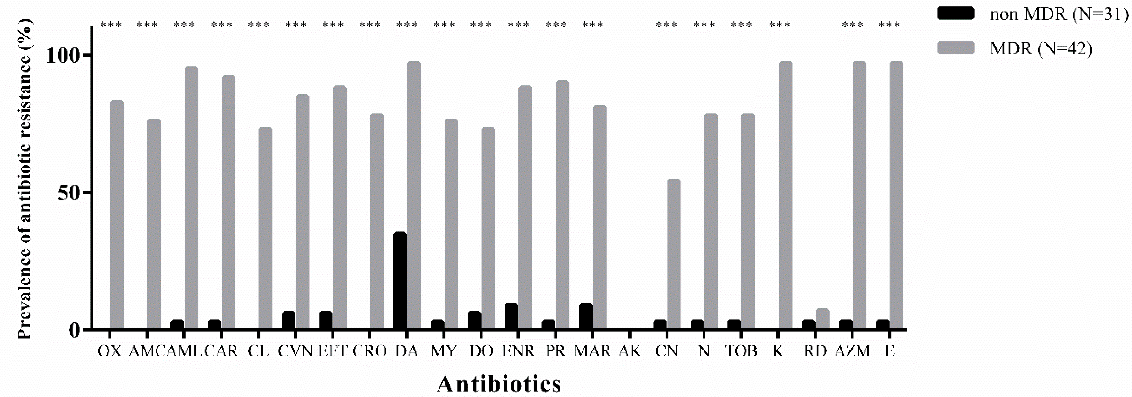
3.2. Overall Antibiotic-Resistance

The antibiotic-resistance of all 73 S. pseudintermedius isolates is shown in Figure 1. Kirby-Bauer assay demonstrated that 42/73 (57.5%) of SP isolates were MDR, exhibiting resistance against macrolides (97%), fluoroquinolones (86%), and β-lactams (72%). The most in-vitro effective molecules were amikacin (100% susceptible), rifampicin (93% susceptible), and partially gentamicin (45% susceptible). The remaining 31 not-MDR strains had a low prevalence of resistance for all the antibiotics with the exception of clindamycin which had a rate of resistance of 35% (11/31). Table 3 shows the prevalence of ARg by using Multiplex PCRs.

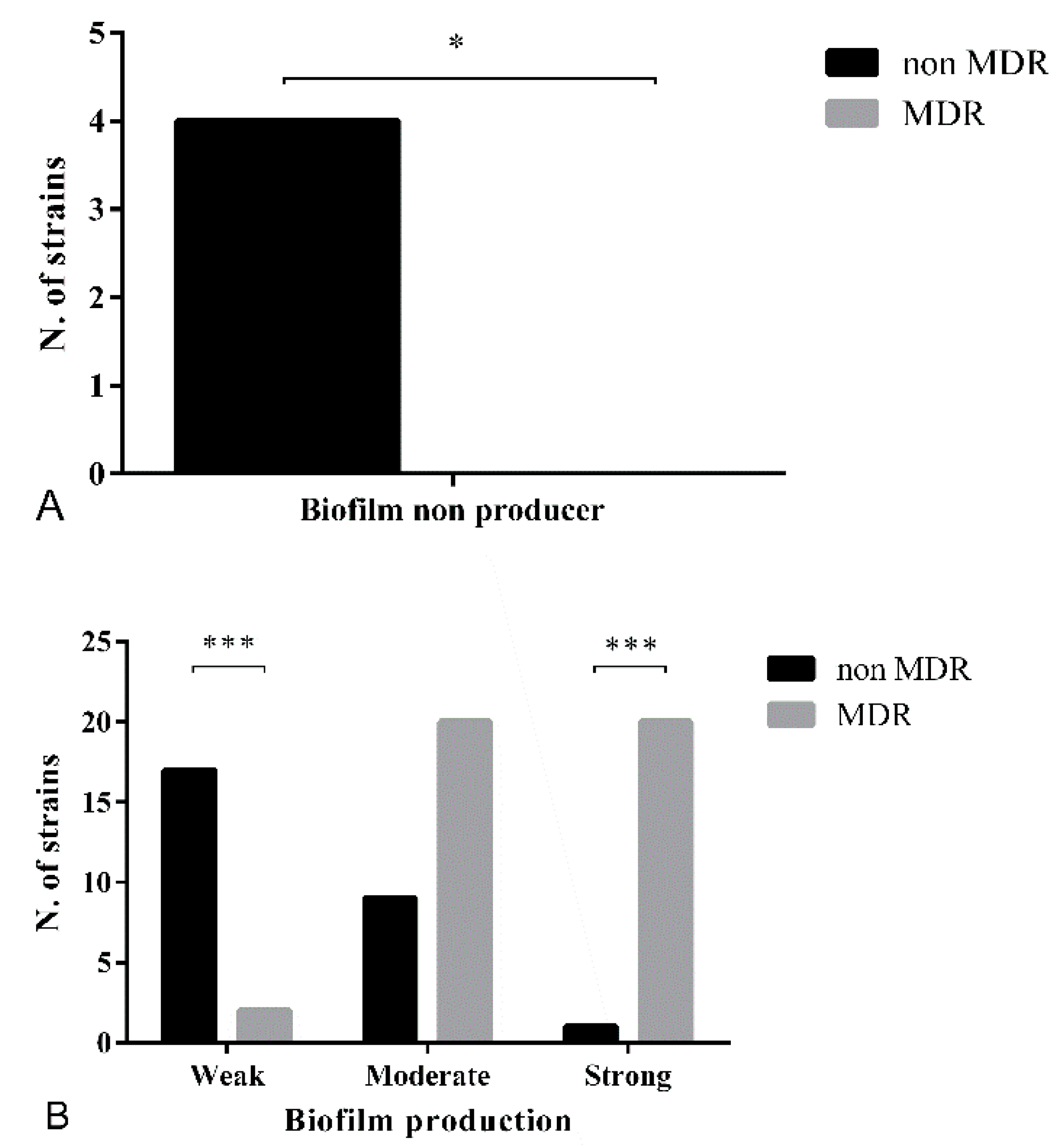
3.3. Biofilm Formation Assay

Figure 2A shows the difference in terms of number of non-biofilm producing strains among not-MDR and MDR strains. Between these two groups, there is a clear difference in terms of the ability to produce biofilm (Figure 2B). The MDR bacteria were all able to produce exopolysaccharide, resulting in 20/43 (46.5%) strong biofilm producers, 20/43 (46.5%) moderate, and 2/43 (4.6%) weak producers. In the not-MDR group, only 1/31 (3.22%) strains was strong producer, 9/31 (29%) strains were categorized as moderate producers, and 17/31 (55%) resulted in weak biofilm producers. The remaining 4/31 (13%) did not show slime production. At the molecular level, the presence of icaA and icaD genes was demonstrated by the amplification of the corresponding amplicons. Both the icaA and icaD genes were detected in 30/31 (97%) not-MDR strains, while 28/42 (66.6%) of MDR bacteria had both the targeted genes. Among the remaining 14 strains, one was negative for both these genes (but still able to produce biofilm) and the remaining 13 were all positive for icaD only. Neither one of these genes was detected in 2/73 (2.7%) isolates (details are reported in Supplementary Table S1). The agreement between the microplate assay and the amplification of ica locus genes was not significant (k < 0.01). The determination of agr-typing showed that all the 73 SP strains belong to the agr type I.

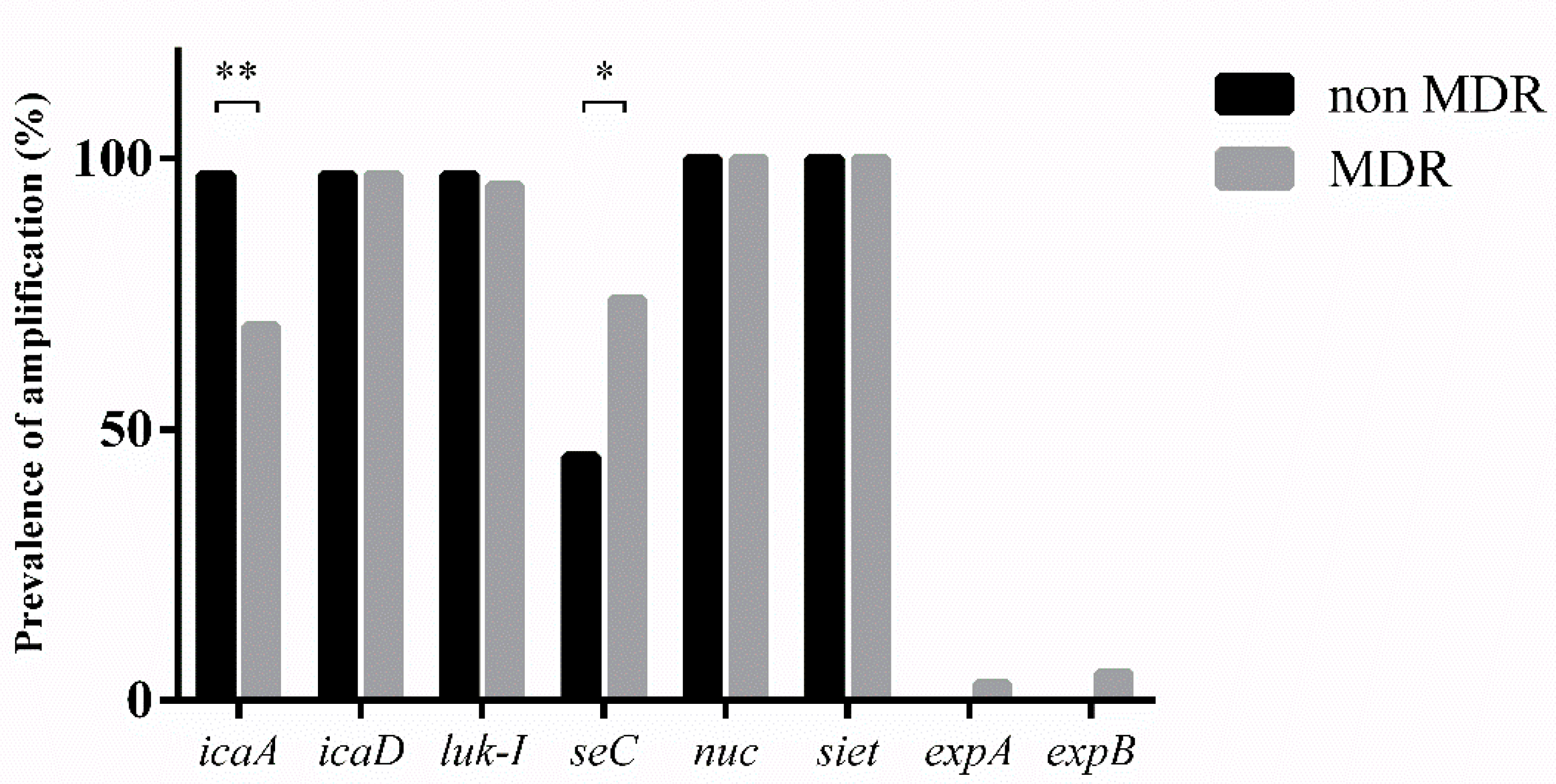
3.4. Virulence Factors

Table 2 shows the distribution of all the genes analyzed in this study. Among the virulence factors, Chi-squared test showed differences in icaA and seC genes between MDR and not-MDR bacteria, for all the other virulence determinants, no differences were found (Figure 3).

4. Discussion

Antibiotic-resistance remains one of the most important problems to face within treatment and control of S. pseudintermedius related infection in human and in veterinary medicine. SP is nowadays considered a potential zoonotic agent able to colonize also humans and is regarded as one of the increasing skin and soft tissue-related pathogens [9,38,39]. MDR strains are defined when “resistant to at least 1 agent in 3 or more antimicrobial categories” [40]. In this study, 57.5% (42/73) of the strains had MDR phenotype, also confirmed by the detection of AR genes. The majority of the resistant strains 35/42 (83%) resulted positive for mecA gene, associated with oxacillin resistance; this result is in concordance with a previous study which revealed a 100% of mecA gene amplification in MRSP strains [41]. A large portion of isolates showed a pattern of resistance against β-lactams, macrolides, and fluoroquinolones.
A total of three STs (ST 71, ST 258 and ST 106) were detected among the 73 SP strains. This result is not totally concordant with the literature in which different authors found a higher genetic diversity in the analyzed population (in terms of STs assigned) [42,43]. ST 71 is predominant and was assigned to 56/73 (77%) strains; this particular sequence type remains the most abundant in Europe, while ST 68 is predominant in United States, and ST 45/ST 112 in Asia [21,22,23,24,25,26,27,28,29,30,31,32,33,34,35,36,37,38,39,40,41,42,43,44]. In Europe, between 2012–2013 and 2015–2016, the prevalence of ST 71 rapidly decreased from 65.3% to 55.2%, whereas the emergence of ST 258, originally derived from Northern Europe, was described (from 1.1% to 5.78%) [43]. ST 71 was highly resistant to antibiotics, showing multiple resistances against commonly veterinary-licensed antibiotics (e.g., tetracyclines). On the other hand, ST 258 was reported to be more frequently susceptible to antibiotics (e.g., enrofloxacin, gentamicin) [43,44].
In this study, all the MRSP strains (35/73; 48%) resulted in MDR, and the majority (68.5%) were classified as ST 71. SCCmec typing showed two chromosomal cassettes types: II-III present in all ST 71, and IV detected in the other MRSP strains belonging to STs 258/106 (Table 2).
A clear correlation was found between antibiotic-resistance and the ability to produce biofilm, suggesting that MDR staphylococci are more prone to produce large quantities of slime. This particular result is in accordance with other studies and could be reasonably explained by the presence of transposons which are the mobile element that easily can be exchanged between strains [11,12,14].
The majority (94.5%) of the 73 isolates were biofilm producers, and this result is in agreement with the current literature on biofilm in Staphylococcus clinical isolates [14,45]. Moreover, a genetic approach revealed the concomitant presence of either icaA or icaD in 79.5% (58/73) of the strains studied, while 18% (13/73) of the isolates presented the amplification of icaD and only two strains were negative for both these two genes but still able to produce biofilm. Authors suggest that the presence of icaD only (found in 42/43 MDR strains) is sufficient to produce exopolysaccharide, as demonstrated for S. epidermidis [12]. These findings are discordant with a previous study [14] in which the author suggested that biofilm formation occurred only when both icaA and icaD are expressed, but however concordant with another study [15] which reported the absence of correlation between the presence of both ica genes and biofilm formation (ica-independent biofilm producers strains). These results suggest the importance of combined, phenotypic, and genetic methods for checking biofilm formation in S. pseudintermedius.
The prevalence of agr groups in our clinical isolates is very different to that described by Little et al. [7], with agrI being the unique group found in our collection, while any of the strains were assigned to one of the other three (agr II, III and IV). Following the suggestions of the author, in this contest, it is not possible to correlate the ability to produce biofilm with the corresponding agr group. Little et al. (2019) stated that strains harboring agrI were more prone to produce biofilm and to be MDR [7]. Our findings indicate a major ability for MDR bacteria to be strong biofilm producers.
Among the virulence determinants analyzed in this study, only the prevalence of specific enterotoxin seC was found statistically significant between MDR and not-MDR strains (p-value: 0.016). The prevalence of individual genes was similar to the literature [28,29].

Supplementary Materials

The following are available online at https://www.mdpi.com/2076-2607/7/12/702/s1. Table S1: Phenotypic and genetic determination of biofilm-forming ability.

Author Contributions

Conceptualization, G.M. and P.A.M.; Data Curation, G.M. and J.F.S.F.; Writing-Original Draft Preparation, G.M.; Writing-Review & Editing, G.M. and L.D.; Supervision, P.A.M. and L.D.

Funding

This research received no external funding.

Conflicts of Interest

The authors declare no conflict of interest.

References

  1. Devriese, L.A.; Vancanneyt, M.; Baele, M.; Vaneechoutte, M.; De Graef, E.; Snauwaert, C.; Haesebrouck, F. Staphylococcus pseudintermedius sp. nov., a coagulase-positive species from animals. Int. J. Syst. Evol. Microbiol. 2005, 55, 1569–1573. [Google Scholar] [CrossRef] [PubMed] [Green Version]
  2. Van Hoovels, L.; Vankeerberghen, A.; Boel, A.; Van Vaerenbergh, K.; De Beenhouwer, H. First case of Staphylococcus pseudintermedius infection in a human. J. Clin. Microbiol. 2006, 44, 4609–4612. [Google Scholar] [CrossRef] [PubMed] [Green Version]
  3. Bannoehr, J.; Franco, A.; Iurescia, M.; Battisti, A.; Fitzgerald, J.R. Molecular diagnostic identification of Staphylococcus pseudintermedius. J. Clin. Microbiol. 2009, 47, 469–471. [Google Scholar] [CrossRef] [PubMed] [Green Version]
  4. Guardabassi, L.; Loeber, M.E.; Jacobson, A. Transmission of multiple antimicrobial-resistant Staphylococcus intermedius between dogs affected by deep pyoderma and their owners. Vet. Microbiol. 2004, 98, 23–27. [Google Scholar] [CrossRef]
  5. Bannoehr, J.; Guardabassi, L. Staphylococcus pseudintermedius in the dog: Taxonomy, diagnostics, ecology, epidemiology and pathogenicity. Vet. Dermatol. 2012, 23, 253-e52. [Google Scholar] [CrossRef]
  6. Bartlett, S.J.; Rosenkrantz, W.S.; Sanchez, S. Bacterial contamination of commercial ear cleaners following routine home use. Vet. Dermatol. 2011, 22, 546–553. [Google Scholar] [CrossRef]
  7. Little, S.V.; Bryan, L.K.; Hillhouse, A.E.; Cohen, N.D.; Lawhon, S.D. Characterization of agr Groups of Staphylococcus pseudintermedius Isolates from Dogs in Texas. mSphere 2019, 4, e00033-19. [Google Scholar] [CrossRef] [Green Version]
  8. Starlander, G.; Börjesson, S.; Grönlund-Andersson, U.; Tellgren-Roth, C.; Melhus, Å. Cluster of Infections Caused by Methicillin-Resistant Staphylococcus pseudintermedius in Humans in a Tertiary Hospital. J. Clin. Microbiol. 2014, 52, 3118–3120. [Google Scholar] [CrossRef] [Green Version]
  9. Somayaji, R.; Priyantha, M.A.R.; Rubin, J.E.; Church, D. Human infections due to Staphylococcus pseudintermedius, an emerging zoonosis of canine origin: Report of 24 cases. Diagn. Microbiol. Infect. Dis. 2016, 85, 471–476. [Google Scholar] [CrossRef]
  10. Börjesson, S.; Gómez-Sanz, E.; Ekström, K.; Torres, C.; Grönlund, U. Staphylococcus pseudintermedius can be misdiagnosed as Staphylococcus aureus in humans with dog bite wounds. Eur. J. Clin. Microbiol. Infect. Dis. 2015, 34, 839–844. [Google Scholar] [CrossRef]
  11. Stefanetti, V.; Bietta, A.; Pascucci, L.; Marenzoni, M.L.; Coletti, M.; Franciosini, M.P.; Passmonti, F.; Casagrandde, P.P. Investigation of the antibiotic resistance and biofilm formation of Staphylococcus pseudintermedius strains isolated from canine pyoderma. Vet. Ital. 2017, 53, 289–296. [Google Scholar]
  12. Arciola, C.R.; Campoccia, D.; Gamberini, S.; Donati, M.E.; Pirini, V.; Visai, L.; Speziale, P.; Montanaro, L. Antibiotic resistance in exopolysaccharide-forming Staphylococcus epidermidis clinical isolates from orthopaedic implant infections. Biomaterials 2005, 26, 6530–6535. [Google Scholar] [CrossRef]
  13. Resch, A.; Rosenstein, R.; Nerz, C.; Götz, F. Differential gene expression profiling of Staphylococcus aureus cultivated under biofilm and planktonic conditions. Appl. Environ. Microbiol. 2005, 71, 2663–2676. [Google Scholar] [CrossRef] [Green Version]
  14. Proietti, P.C.; Stefanetti, V.; Hyatt, D.R.; Marenzoni, M.L.; Capomaccio, S.; Coletti, M.; Bietta, A.; Franciosini, M.P.; Passmonti, F. Phenotypic and genotypic characterization of canine pyoderma isolates of Staphylococcus pseudintermedius for biofilm formation. J. Vet. Med. Sci. 2015, 77, 945–951. [Google Scholar]
  15. Singh, A.; Walker, M.; Rousseau, J.; Weese, J.S. Characterization of the biofilm forming ability of Staphylococcus pseudintermedius from dogs. BMC Vet. Res. 2013, 9, 93. [Google Scholar] [CrossRef] [Green Version]
  16. O’Gara, J.P. Ica and beyond: Biofilm mechanisms and regulation in Staphylococcus epidermidis and Staphylococcus aureus. FEMS Microbiol. Lett. 2007, 270, 179–188. [Google Scholar] [CrossRef] [Green Version]
  17. Panda, S.; Singh, D.V. Biofilm formation by ica-negative ocular isolates of staphylococcus haemolyticus. Front. Microbiol. 2018, 9, 1–9. [Google Scholar] [CrossRef]
  18. Hennig, S.; Wai, S.N.; Ziebuhr, W. Spontaneous switch to PIA-independent biofilm formation in an ica-positive Staphylococcus epidermidis isolate. Int. J. Med. Microbiol. 2007, 297, 117–122. [Google Scholar] [CrossRef]
  19. Sasaki, T.; Tsubakishita, S.; Tanaka, Y.; Sakusabe, A.; Ohtsuka, M.; Hirotaki, S.; Kawakami, T.; Fukata, T.; Hiramatsu, K. Multiplex-PCR method for species identification of coagulase-positive staphylococci. J. Clin. Microbiol. 2010, 48, 765–769. [Google Scholar] [CrossRef] [Green Version]
  20. Adwan, K. Fast DNA isolation and PCR protocols for detection of methicillin-resistant staphylococci. Folia Microbiol. 2014, 59, 5–8. [Google Scholar] [CrossRef]
  21. Solyman, S.M.; Black, C.C.; Duim, B.; Perreten, V.; Duijkeren, E.V.; Wagenaar, J.A.; Eberlein, L.C.; Sadeghi, L.N.; Videla, R. Multilocus Sequence Typing for Characterization of Staphylococcus pseudintermedius. J. Clin. Microbiol. 2013, 51, 306–310. [Google Scholar] [CrossRef] [Green Version]
  22. Bannoehr, J.; Zakour, N.L.B.; Waller, A.S.; Guardabassi, L.; Thoday, K.L.; Van Den Broek, A.H.; Fitzgerald, J.R. Population genetic structure of the Staphylococcus intermedius group: Insights into agr diversification and the emergence of methicillin-resistant strains. J. Bacteriol. 2007, 189, 8685–8692. [Google Scholar] [CrossRef] [Green Version]
  23. Kadlec, K.; Schwarz, S.; Perreten, V.; Grönlund Andersson, U.; Finn, M.; Greko, C.; Moodley, A.; Kania, S.A.; Frank, L.A.; Bemis, D.A. Molecular analysis of methicillin-resistant Staphylococcus pseudintermedius of feline origin from different European countries and North America. J. Antimicrob Chemother. 2010, 65, 1826–1828. [Google Scholar] [CrossRef]
  24. Perreten, V.; Kadlec, K.; Schwarz, S.; Andersson, U.G.; Finn, M.; Greko, C.; Moodley, A.; Kania, S.A.; Frank, L.A.; Bemis, D.A. Clonal spread of methicillin-resistant Staphylococcus pseudintermedius in Europe and North America: An international multicentre study. J. Antimicrob. Chemother. 2010, 65, 1145–1154. [Google Scholar] [CrossRef] [Green Version]
  25. Kang, M.H.; Chae, M.J.; Yoon, J.W.; Kim, S.G.; Lee, S.Y.; Yoo, J.H.; Park, H.M. Antibiotic resistance and molecular characterization of ophthalmic Staphylococcus pseudintermedius isolates from dogs. J. Vet. Sci. 2014, 15, 409–415. [Google Scholar] [CrossRef] [Green Version]
  26. Strommenger, B.; Kettlitz, C.; Werner, G. Multiplex PCR assay for simultaneous detection of nine clinically relevant antibiotic resistance genes in Staphylococcus aureus. J. Clin. Microbiol. 2003, 41, 4089–4094. [Google Scholar] [CrossRef] [Green Version]
  27. Shopsin, B.; Mathema, B.; Alcabes, P.; Said-Salim, B.; Lina, G.; Matsuka, A. Prevalence of agr specificity groups among Staphylococcus aureus strains colonizing children and their guardians. J. Clin. Microbiol. 2003, 41, 456–459. [Google Scholar] [CrossRef] [Green Version]
  28. Futagawa-Saito, K.; Sugiyama, T.; Karube, S.; Sakurai, N.; Ba-Thein, W.; Fukuyasu, T. Prevalence and characterization of leukotoxin-producing Staphylococcus intermedius in isolates from dogs and pigeons. J. Clin. Microbiol. 2004, 42, 5324–5326. [Google Scholar] [CrossRef] [Green Version]
  29. Yoon, J.W.; Lee, G.J.; Lee, S.Y.; Park, C.; Yoo, J.H.; Park, H.M. Prevalence of genes for enterotoxins, toxic shock syndrome toxin 1 and exfoliative toxin among clinical isolates of Staphylococcus pseudintermedius from canine origin. Vet. Dermatol. 2010, 21, 484–489. [Google Scholar] [CrossRef]
  30. Lautz, S.; Kanbar, T.; Alber, J.; Lämmler, C.; Weiss, R.; Prenger-Berninghoff, E.; Zschock, M. Dissemination of the gene encoding exfoliative toxin of Staphylococcus intermedius among strains isolated from dogs during routine microbiological diagnostics. J. Vet. Med. Ser. B Infect. Dis. Vet. Public Health 2006, 53, 434–438. [Google Scholar] [CrossRef]
  31. Futagawa-Saito, K.; Makino, S.; Sunaga, F.; Kato, Y.; Sakurai-Komada, N.; Ba-Thein, W.; Fukuyasu, T. Identification of first exfoliative toxin in Staphylococcus pseudintermedius. FEMS Microbiol. Lett. 2009, 301, 176–180. [Google Scholar] [CrossRef] [PubMed] [Green Version]
  32. Iyori, K.; Hisatsune, J.; Kawakami, T.; Shibata, S.; Murayama, N.; Ide, K.; Nagata, M.; Fukata, T.; Iwasaki, T.; Oshima, K. Identification of a novel Staphylococcus pseudintermedius exfoliative toxin gene and its prevalence in isolates from canines with pyoderma and healthy dogs. FEMS Microbiol. Lett. 2010, 312, 169–175. [Google Scholar] [CrossRef] [PubMed] [Green Version]
  33. El Zubeir, I.E.M.; Kanbar, T.; Alber, J.; Lämmler, C.; Akineden, Ö.; Weiss, R.; Zschock, M. Phenotypic and genotypic characteristics of methicillin/oxacillin-resistant Staphylococcus intermedius isolated from clinical specimens during routine veterinary microbiological examinations. Vet. Microbiol. 2007, 121, 170–176. [Google Scholar] [CrossRef] [PubMed]
  34. Stepanovic, S.; Vukovic, D.; Dakic, I.; Savic, B.; Svabic-Vlahovic, M. A modified microtiter-plate test for quantification of staphylococcal biofilm formation. J. Microbiol. Methods 2000, 40, 175–179. [Google Scholar] [CrossRef]
  35. Stepanović, S.; Vuković, D.; Hola, V.; Di Bonaventura, G.; Djukić, S.; Cirković, I.; Ruzicka, F. Quantification of biofilm in microtiter plates: Overview of testing conditions and practical recommendations for assessment of biofilm production by staphylococci. Apmis 2007, 115, 891–899. [Google Scholar] [CrossRef]
  36. Melchior, M.B.; Van Osch, M.H.J.; Graat, R.M.; Van Duijkeren, E.; Mevius, D.J.; Nielen, M.; Gaastra, W.; Fink-Gremme, J. Biofilm formation and genotyping of Staphylococcus aureus bovine mastitis isolates: Evidence for lack of penicillin-resistance in Agr-type II strains. Vet. Microbiol. 2009, 137, 83–89. [Google Scholar] [CrossRef]
  37. Landis, J.R.; Koch, G.G. The Measurement of Observer Agreement for Categorical Data. Biometrics 1977, 33, 159–174. [Google Scholar] [CrossRef] [Green Version]
  38. Kmieciak, W.; Szewczyk, E.M. Are zoonotic Staphylococcus pseudintermedius strains a growing threat for humans? Folia Microbiol. (Praha) 2018, 63, 743–747. [Google Scholar] [CrossRef] [Green Version]
  39. Somayaji, R.; Rubin, J.E.; Priyantha, M.A.; Church, D. Exploring Staphylococcus pseudintermedius: An emerging zoonotic pathogen? Future Microbiol. 2016, 11, 1371–1374. [Google Scholar] [CrossRef] [Green Version]
  40. Magiorakos, A.P.; Srinivasan, A.; Carey, R.B.; Carmeli, Y.; Falagas, M.E.; Giske, C.G.; Harbarth, S.; Hindler, J.F.; Kahlmeter, G.; Olsson-Liljequist, B. Multidrug-resistant, extensively drug-resistant and pandrug-resistant bacteria: An international expert proposal for interim standard definitions for acquired resistance. Clin. Microbiol. Infect. 2012, 18, 268–281. [Google Scholar] [CrossRef] [Green Version]
  41. Moyaert, H.; de Jong, A.; Simjee, S.; Rose, M.; Youala, M.; Garch, F.E.; Vila, T.; Klein, U.; Rzewuska, M.; Morrissey, I. Survey of antimicrobial susceptibility of bacterial pathogens isolated from dogs and cats with respiratory tract infections in Europe: ComPath results. J. Appl. Microbiol. 2019, 127, 29–46. [Google Scholar] [CrossRef] [PubMed]
  42. Damborg, P.; Moodley, A.; Aalbæk, B.; Ventrella, G.; dos Santos, T.P.; Guardabassi, L. High genotypic diversity among methicillin-resistant Staphylococcus pseudintermedius isolated from canine infections in Denmark. BMC Vet. Res. 2016, 12, 131–136. [Google Scholar] [CrossRef] [PubMed] [Green Version]
  43. Bergot, M.; Martins-Simoes, P.; Kilian, H.; Châtre, P.; Worthing, K.A.; Norris, J.M.; Madec, J.-Y.; Laurent, F.; Haenni, M. Evolution of the Population Structure of Staphylococcus pseudintermedius in France. Front. Microbil. 2018, 9, 3055. [Google Scholar] [CrossRef] [PubMed]
  44. Dos Santos, T.P.; Damborg, P.; Moodley, A.; Guardabassi, L. Systematic review on global epidemiology of methicillin-resistant staphylococcus pseudintermedius: Inference of population structure from multilocus sequence typing data. Front. Microbiol. 2016, 7, 1599. [Google Scholar] [CrossRef] [Green Version]
  45. Rinsky, J.L.; Nadimpalli, M.; Wing, S.; Hall, D.; Baron, D.; Price, L.B.; Larsen, J.; Stegger, M.; Stewart, J.; Heaney, C.D. Livestock-Associated Methicillin and Multidrug Resistant Staphylococcus aureus Is Present among Industrial, Not Antibiotic-Free Livestock Operation Workers in North Carolina. PLoS ONE 2013, 8, e67641. [Google Scholar] [CrossRef] [Green Version]
Figure 1. Overall prevalence of antibiotic-resistance among multi-drug resistant (MDR) and not-MDR isolates. The Chi-squared test showed statistical differences among the two groups for the majority of antibiotic molecules tested except for amikacin and rifampicin. (***: p-value < 0.001).
Microorganisms 07 00702 g001
Figure 2. Biofilm forming ability in not-MDR and MDR Staphylococcus pseudintermedius (SP) strains. (A) All the MDR strains are able to produce biofilm and these data are statistically significant compared to the not MDR group in which 4 strains resulted in non-biofilm producers. (B) The majority of MDR strains were strongly biofilm producers (20/42), whereas not-MDR strains were mostly categorized as weak slime producers. (*: p-value between 0.05 and 0.01; ***: p-value < 0.001).
Microorganisms 07 00702 g002
Figure 3. Dissemination of virulence factors among MDR and not-MDR strains. (*: p-value between 0.05 and 0.01; **: p-value < 0.01).
Microorganisms 07 00702 g003
Table 1. Primers, amplicon size, and amplification conditions.
Genes Sequence (5’-3’)Amplicon Size (bp)PCR ConditionsReferences
Antibiotic-resistance genesmecAFAAAATCGATGGTAAAGGTTGGC53295 °C × 4 min, 30 × (95 °C × 1 min, 58 °C × 1 min, 72 °C × 1 min) 72 °C × 7 min, 4 °C[25]
RAGTTCTGCAGTACCGGATTTGC
blaZFTGACCACTTTTATCAGCAACC750
RGCCATTTCAACACCTTCTTTC
tetKFGTAGCGACAATAGGTAATAGT36094 °C × 3 min, 30 × (94 °C × 30 s, 55 °C × 30 s, 72 °C × 30 s) 72 °C × 7 min, 4 °C[26]
RGTAGTGACAATAAACCTCCTA
tetMFAGTGGAGCGATTACAGAA158
RCATATGTCCTGGCGTGTCTA
aacA-aphDFTAATCCAAGAGCAATAAGGGC227
RGCCACACTATCATAACCACTA
Biofilm genesicaAFACTGTTTCGGGGACAAGCAT13494 °C × 3 min, 35 × (94 °C × 15 s, 60 °C × 20 s, 72 °C × 20 s) 72 °C × 7 min, 4 °C[14]
RATTGAGGCTGTAGGGCGTTG
icaDFCGTTAATGCCTTCTTTCTTATTGCG16694 °C × 3 min, 35 × (94 °C × 15 s, 56 °C × 20 s, 72 °C × 20 s) 72 °C × 7 min, 4 °C
RATTAGCGCACATTCGGTGTT
Quorum-sensing genespan-agrFATGCACATGGTGCACATGC 94 °C × 3 min, 35 × (94 °C × 15 s, 56 °C × 20 s, 72 °C × 20 s) 72 °C × 7 min, 4 °C[27]
agrIRGTCACAAGTACTATAAGCTGCGAT
agrIIRGTATTACTAATTGAAAAGTGCCATAGC
agrIIIRCTGTTGAAAAAGTCAACTAAAAGCTC
agrIVRCGATAATGCCGTAATACCCG
Virulence factorsluk-FFCCTGTCTATGCCGCTAATCCA57294 °C × 3 min, 35 × (94 °C × 1 min, 57 °C × 1 min, 72 °C × 1 min) 72 °C × 7 min, 4 °C[28]
RAGGTCATGGAAGCTATCTCGA
luk-SFTGTAAGCAGCAGAAAATGGGG503
RGCCCGATAGGACTTCTTACAA
seCFGGCGGCAATATTGGCGCTCG27195 °C × 2 min, 30 × (95 °C × 1 min, 55 °C × 1 min, 72 °C × 2 min) 72 °C × 5 min, 4 °C[29]
RTTACTGTCAATGCTCTGACC
nucFTRGGCAGTAGGATTCGTTAA92695 °C × 2 min, 30 × (95 °C × 30 s, 52 °C × 30 s, 72 °C × 30 s) 72 °C × 2 min, 4 °C[19]
RCTTTTGTGCTYCMTTTTGG
sietFATGGAAAATTTAGCGGCATCTGG35994 °C × 3 min, 30 × (94 °C × 30 s, 56 °C × 30 s, 72 °C × 1 min) 72 °C × 5 min, 4 °C[30]
RCCATTACTTTTCGCTTGTTGTGC
expAFGTKTTAATTGGWAAAAATACA41394 °C × 3 min, 30 × (94 °C × 1 min, 42 °C × 1 min, 72 °C × 1 min) 72 °C × 4 min, 4°C[31]
RATNCCWGAKCCTGAATTWCC
expBFGGGCATGCACATATGATGAAGCC82095 °C × 3 min, 30 × (95 °C × 1 min, 53 °C × 1 min, 72 °C × 1 min) 72 °C × 4 min, 4 °C[32]
RCCAGATCTATCTTCTGATTCAGC
Table 2. Molecular characterization of the 35 methicillin-resistant S. pseudintermedius (MRSP) isolates.
MLSTSCCmec TypesNo. of Isolates (%)
ST 71II-III24 (68.5%)
ST 258IV9 (25.7%)
ST 106IV2 (5.7%)
Table 3. Distribution of antibiotic-resistance and virulence factors genes.
Genesnot MDR StrainsMDR Strainsp-Value
Antibiotic-resistance genesmecA035/42 (83%)<0.0001
blaZ7/31 (23%)42/42 (100%)<0.0001
tetK2/31 (6.4%)13/42 (31%)0.0171
tetM022/42 (50%)<0.0001
aacA-aphD5/31 (16.6%)32/42 (76%)<0.0001
Biofilm genesicaA30/31 (97%)29/42 (69%)0.0026
icaD30/31 (97%)41/42 (97%)>0.05
Virulence factorsluk-I30/31 (97%)40/42 (95%)>0.05
seC14/31 (45%)31/42 (74%)0.016
nuc31/31 (100%)42/42 (100%)>0.05
siet31/31 (100%)42/42 (100%)>0.05
expA03/42 (7%)>0.05
expB05/42 (12%)>0.05

