Article

Diversity of Rickettsia in Ticks Collected in Abruzzi and Molise Regions (Central Italy)

Istituto Zooprofilattico Sperimentale dell’Abruzzo e del Molise “G. Caporale”, 64100 Teramo, Italy
*
Author to whom correspondence should be addressed.
Microorganisms 2019, 7(12), 696; https://doi.org/10.3390/microorganisms7120696
Submission received: 31 October 2019 / Revised: 6 December 2019 / Accepted: 6 December 2019 / Published: 13 December 2019
(This article belongs to the Special Issue Epidemiology of Zoonotic Diseases)

Abstract

:
Rickettsiae have worldwide occurrence and rickettsiosis are widely recognized as emerging infections in several parts of the world. For decades, it was thought that a single pathogenic tick-borne spotted fever group (SFG), Rickettsia, occurred in each continent. Nowadays, thanks to molecular biology, new species of Rickettsia responsible for disease in humans are continuously identified worldwide. In a framework of diagnostic activities of the Istituto Zooprofilattico Sperimentale dell’Abruzzo e del Molise “G. Gaporale” and considering some reports of suspected human clinical cases of rickettsiosis, a survey on ticks collected form animals and humans was carried out with the aim to identify the Rickettsia species circulating in Abruzzi and Molise regions. A total of 603 ticks, previously identified at species level by morphology, pooled into 178 tick samples, were tested by pan-Rickettsia RealTime PCR. DNA from specimens positive for Rickettsia spp. was then sequenced in order to identify the Rickettsia species involved. The highest infection rate was detected in Dermacentor marginatus followed by Ixodes ricinus. The selected targets for this purpose were OmpA and gltA. Rickettsia slovaca, Rickettsia monacensis, Rickettsia massiliae, Rickettsia conorii, Rickettsia aeschlimannii, Rickettsia helvetica, Rickettsia raoultii, and Rickettsia felis – like organisms were identified in this study. These are the first data available in the literature for the circulation of SFG Rickettsia species in the selected geographical area. Results made evidence of high rate of infection in ticks. All Rickettsia species detected have been previously involved in human infection. The diversity of Rickettsia detected, and tick species collected reflects the biodiversity in term of wildlife and environment of the area. An association between Rickettsia species, ticks, and the relationships with vertebrate host species are discussed. Due to the peculiar eco-biology of each Rickettsia species, the use of diagnostic tools able to identify Rickettsia at the species level is thus recommended in order to assess the risk for humans and to elucidate more precise etiological diagnosis in clinical cases.

1. Introduction

Rickettsiae (family Rickettsiaceae, order Rickettsiales) are obligate intracellular bacteria transmitted by ticks, fleas, lice, and mites. Members of the genus Rickettsia can be classified into the spotted fever group (SFG) Rickettsiae, typhus group Rickettsiae, the Rickettsia bellii group, and the Rickettsia canadensis group [1]. Rickettsiae have worldwide occurrence and rickettsiosis are widely recognized as emerging infections in several parts of the globe, amongst these, spotted fever has a prominent role (2). For decades, it was thought that a single pathogenic tick-borne SFG Rickettsia occurred in each continent. Moreover, different Rickettsia species isolated from ticks were not considered pathogenic for humans for years or decades until a definitive association with human disease was established, such as the emblematic case of Rickettsia massiliae in Europe [2]. Hard ticks are not only the main vectors of SFG Rickettsiae, but also their main reservoir, even if an amplifier role of vertebrate species is suspected or to be clarified for some SFG Rickettsia species [1]. Vertical transmission and mainly an effective transovaric transmission (TOT) with a high level of filial infection rate (FIR), as described in ticks for the associated Rickettsia species, are the main ways of maintaining an infection in tick populations [1,3,4]. For example, for R. massiliae in Rhipicephalus turanicus ticks, a TOT of 100% and FIR of 98.5% have been reported [5]. For Rickettsia africae a TOT 100% and a FIR of 93.4% was also described in Amblyomma variegatum ticks [4]. Rickettsia conorii subspecies (sbsp) conorii the agent of Mediterranean spotted fever (MSF) is considered endemic mainly in the central and southern Regions of the Mediterranean [2]. Countries of the Mediterranean basin have noticed an increased incidence of MSF over the past 10 years. In Portugal, an annual incidence rate of 9.8 cases per 100,000 persons, the highest of the rates of all Mediterranean countries, has been reported [6,7]. In Italy, according to the Italian National Institute of Health [8], around 10,000 cases have been reported in the period 1996–2009. These cases are mainly described in warmer areas of the country, namely in central southern Italy and in the main islands. Four regions, Sicily, Sardinia, Latium, and Calabria contained more the 85% of total cases, generally reported during the long hot season from July to October, thus according with the seasonal dynamic of the main vector Rhipicepahlus sanguineus that behaves as an exophilic tick at those latitudes [8,9]. More recently, an average incidence of 21.17–176.88 cases per million persons of Rickettsiosis has been reported from 2009 to 2013 [10]. This rate likely underestimates the true incidence, and some authors suggest that there are seven times more cases than officially reported [6] since a great part of the diagnosis is made by clinical symptoms and serology without the identification of the pathogen involved. For many years, it was assumed that R. conorii sbsp conorii was the only agent of SFG Rickettsia circulating in Italy, and that the classical clinical manifestation of MSF was the only one developed after the infection in humans. In the last decades, thanks to the improved diagnostic skills and, mainly, to the use of molecular tools, different SFG Rickettsia have been identified in the Italian territory including Rickettsia slovaca, Rickettsia aeschlimanni, Rickettsia massilliae, Rickettsia monacensis, Rickettsia conorii subspecies. Israelensis, Rickettsia conorii subsp. Indica, Rickettsia raoulti, Rickettsia helvetica, and Rickettsia hoogstraalii, [1,2,11,12,13,14].
In Abruzzi and Molise according the Ministry of Health in the period from 1996–2009, 81 and 43 human cases respectively, were notified. However, the presence of subclinical forms that may be undetected and the possible circulation of Rickettsia species that can cause symptoms different from the classical MSF could have led to an underreporting. Moreover, cases of hospitalized human Rickettsiosis have been reported during the time 2001–2015 [8,15] and human cases are still presently diagnosed at the local hospitals. Likewise, previous preliminary studies on tick borne diseases carried out in the area showed the circulation of different Rickettsia species such as R. slovaca [16] and R. monacensis [17]. The present study aims to investigate the possible circulation of Rickettsia species, and to identify species of interest for human health in Abruzzi and Molise Regions.

2. Materials and Methods

The Istituto Zooprofilattico Sperimentale dell’Abruzzo e del Molise “G. Caporale” is a public health institute which operates in the Abruzzi and Molise Regions, performing analytical work for public veterinary services, and providing the technical and scientific collaboration necessary to enable them to carry out their functions in the field of veterinary public health. The diagnostic department receives carcasses of domestic and wild animals for diagnostic purposes and in the framework of specific surveillance and monitoring programs such as the one on wildlife health status. Working under the same ministry, it receives samples from hospitals as well in cases of suspected infection with a zoonotic agent. Considering reports of suspected human clinical cases of Rickettsiosis and a lack of detailed information for the area, a passive survey on Rickettsia in ticks collected from domestic and wild animals and humans received at the diagnostic department from 2014 to 2106 was carried out.

2.1. Study Area

With an altitude from sea level to over 2900 m. above sea level. the study area covers different environments and it is characterized by high biodiversity in terms wildlife with 36% of the territory protected in three national parks and many natural reserves. At least 2 endemic endangered mammals species are present in the region, such as the Marsican brown bear (Ursus arctos marsicanus) and Apennine chamois (Rupicapra pyrenaica ornata), but the environmental features of the area are able to support a high density of wild ungulates (roe deer—Capreolus capreolus, red deer—Cervus elaphus, and wild boar—Sus scrofa) and wild carnivorous (wolf—Canis lupus, European badger—Meles meles, and red fox—Vulpes vulpes). Meanwhile, rural farming is common in the area, especially for small ruminants.

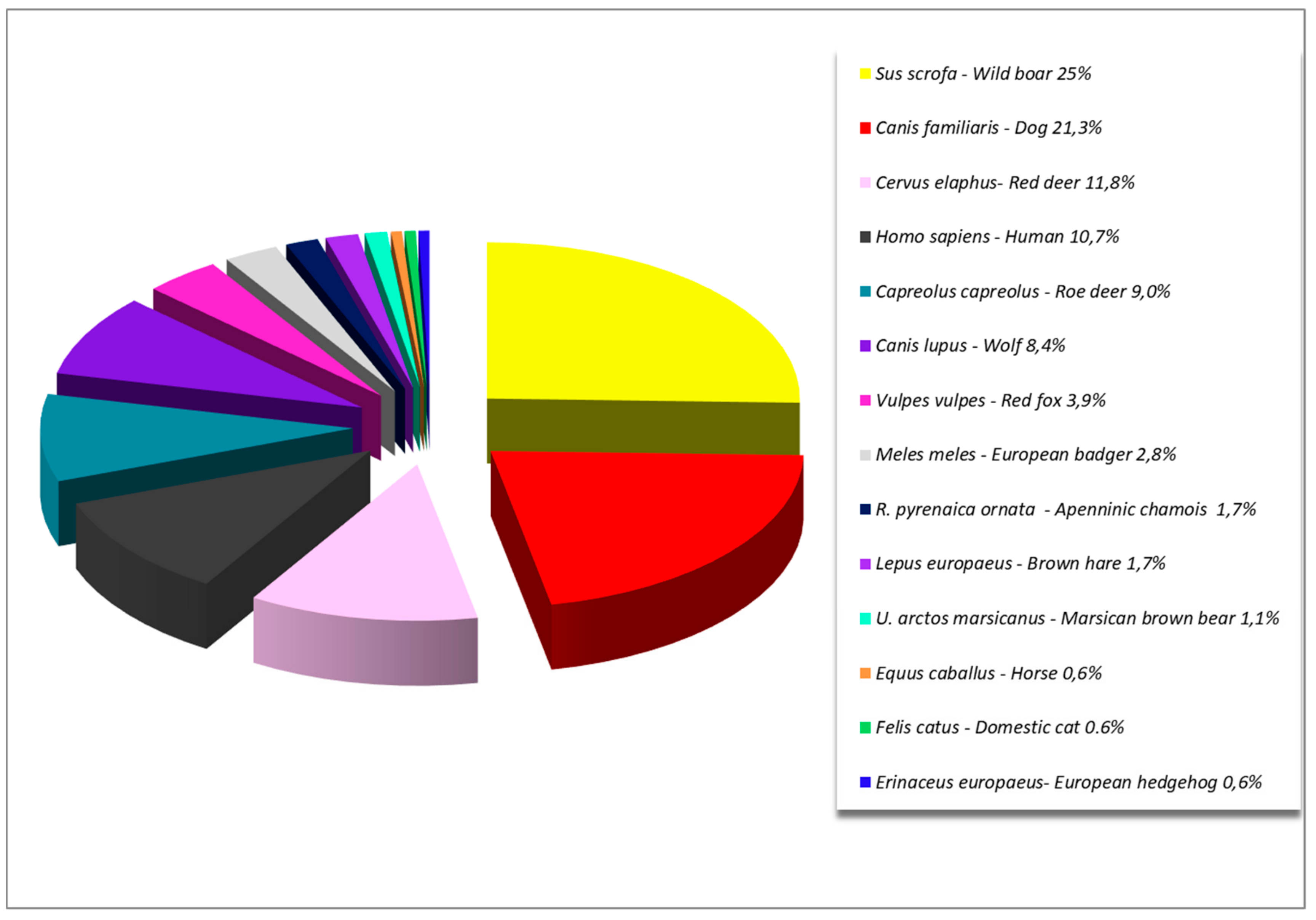
2.2. Tick Collection and Identification

From January 2014 to the end of December 2016, ticks were collected during post mortem examination, placed in a vial, and identified with an identification number. In the case of ticks collected from animals, the ID number was the same as the carcass to which they belong. Collection data (host species, date, municipality) were registered in the laboratory informative system during the acceptance of the carcass or tick. Ticks were preserved in 70% ethanol and subsequently identified at the species level and developmental stage by morphology using the dichotomous key described by Manilla [9].

2.3. Rickettsia DNA Detection and Identification

DNA from all from samples (pools) was extracted by the Maxwell 16 Tissue DNA Purification Kit (Promega) and analyzed by pan-Rickettsia Real-Time PCR [18] targeting 23S rRna gene. The reaction mixture consisted of 20 µL containing 2X GoTaq Probe qPCR Master Mix (Promega, Madison, US), 250 nM of TaqMan probe (PanR8_P), 600 nM of each primer (PanR8_F and PanR8_R), 5 µL DNA and nuclease-free water up to final volume. The reaction protocol was used in fast mode: 20 s at 95 °C followed by 40 cycles of 1 s at 95 °C and 20 s at 60°C. The reaction was performed on the 7900HT Fast RealTime PCR System (Applied Biosystems, Foster City, US) and analysed by the sequence detection system software (SDS) v2.4 (Applied Biosystems). The RealTime PCR assay was always performed including a plasmid containing a portion of the Rickettsia 23S rRNA gene as positive control and No Template Control (NTC). Rickettsia spp. positive samples were further investigated by sequencing a portion of OmpA gene and gltA gene [19]. Five µl of purified DNA were amplified in a 50 µLreaction mixture containing 300 nM of each forward and reverse primer, 200 µM dNTPs (Promega), 2.5 mM MgCl2 Solution (Applied Biosystem), 0.125 U/µL AmpliTaq Gold (Applied Biosystem) and 1 × PCR Buffer II (Applied Biosystem). Amplifications were carried out in a GeneAmp PCR System 9700 (Applied Biosystems) under the following conditions: initial denaturation 10 min at 94 °C, followed by 45 cycles of denaturation (30 s at 94 °C), annealing (30 s at 56 °C), and extension (30 s at 72 °C). The final extension was completed at 72 °C for 7 min. PCR products were purified using the GeneAll Expin PCR Kit (GeneAll, Seoul, Korea) and sequenced by the BigDye Terminator v.3.1 (Applied Biosystems). Analyses were performed on the 3130 XL Genetic Analyzer (Applied Biosystems). Raw sequence data were assembled using Vector NTI suite 9.1 (Invitrogen, Waltham, US)

3. Results

Ticks were collected from 15 different host species including humans while 584 ticks from different wild and domestic species and 19 ticks biting humans were included, for a total of 603 adult ticks. Pools were created for each single host, according tick species and developmental stage. Totally 178 adult tick pools of 1–5 individuals each, were tested. Wild boars (Sus scrofa) followed by cervids (Capreolus capreolus and Cervus elaphus) and dogs (Canis familiaris) were the greatest contributors to the collection (Figure 1).
Tick species: seven tick species were detected and the most frequent in the collection was I. Ricinus, followed by D. marginatus and R. sanguineus sensu lato. In Table 1, results of the identification of ticks are shown. The most common tick on humans was I. ricinus, but R. turanicus, D. marginatus, and Ixodes hexagonus were also detected.
Infection with Rickettsia spp.: the percentage of positivity for Rickettsia spp. was expressed as minimum infection rate (MIR) assuming that one tick was positive in each positive pool. The highest MIR was detected in D. marginatus (81.1%) followed by I. ricinus (53.5%).
Positive tick–host association: the most important contribution of D. marginatus positive tick pools was provided by the wild boar (35/43) with a lesser extent by red deer (6/43), while for I. ricinus positive pools were more homogeneously distributed amongst the different hosts including humans. Eight out of the 19 ticks collected from human beings, tested positive (42%). Figure 2 shows the details of RealTime Rickettsia spp. positive tick–host association.
Rickettsia–tick species association: eight species of Rickettsia have been detected in 93 positive pools. R. slovaca and R. monacensis were 85% of the positives. In term of ticks, the only species found infected with R. slovaca was D. marginatus, while R. monacensis was mainly identified in I. ricinus with one positive pool of D. marginatus, R. sanguineus and Haemaphysalis punctata respectively.
Rickettsia–tick species-host association: in term of vertebrate host, D. marginatus ticks infected by R. slovaca were mainly carried by wild boar (S. scrofa) in 34 of 41 cases, with the contribution of red deer (C. elaphus) at 6 in 34, and of a sample from humans, while a wider host range including five samples from humans contributed to the amount of I. ricinus infected with R. monacensis, which was the most frequent species detected in ticks collected from humans (5 out of 19), where R. massiliae and R. slovaca were also identified.
In Table 2, data on Rickettsia species in relation to ticks and host species are shown.

4. Discussion

To our knowledge, these are the first comprehensive data that investigate Rickettsia circulation in the environment in the study area and even if significant bias affected the passive feature of the monitoring (mainly wild host and according seasonality and hunting), they provide important information to assess the possible risk for humans. Results demonstrated evidence of a high rate of infection (52.2%) for Rickettsia spp in ticks (93 positive pools out of 178). The infection rate was higher than what has been reported in a similar study carried out on 77 pools of three ticks by Ebani and collaborators [20] in Tuscany (20.78%) and higher than what has been reported by Scarpulla and collaborators [12] on 113 ticks collected from host and environment in Latium and Tuscany regions (12.4%) and tested singularly (38), or in a pool of 10 ticks (18). Likewise, our infection rate was higher than what has been recently reported in a study carried out testing free living ticks in a sub urban area in Latium 25.8% where however 255 ticks had been selected randomly out of 518 ticks, and singularly tested [21]. The wide range of tick species collected reflects the biodiversity in term of wildlife and environments. I. ricinus has been confirmed as a generalist ticks with the broadest host range (10 species out of 15) and as the most frequent on humans (15 out of 19) confirming the literature data reporting several hundred species of animals included in the host range [9,22]. Another Ixodes tick frequent in our collection was I. hexagonus, a species associated with the European hedgehog (Erinaceus europeus) and its predators [9,23] that seldom bites humans. As a confirmation of the literature, it has been found in our survey in hedgehog and in different carnivorous species, such as the European badger (Meles meles), red fox (Vulpes vulpes), wolf (Canis lupus), and domestic dogs. The ticks R. sanguigneus has been found mainly on dogs as known in literature [9], but also on other canids. Samples of this ticks were not further investigated in order to identify different lineages that, according Zemtsova and collaborators [24], are circulating in the temperate zone. The closely related species R. turanicus was also collected from a wider host-range, including human. Furthermore, our findings confirm the strict association between wild boar and D. marginatus just as described in other studies in Southern Europe. A strong host–parasite association was observed between wild boar and adult D. marginatus by Selmi and collaborators in a study performed in the North West Mediterranean region [25] wherein 99.7% of adult D. marginatus ticks obtained from wild ungulates were collected on wild boars. Similarly, in our study, 83% of D. marginatus ticks were found on wild boar. Selmi and collaborators [25] hypothesize that wild boar plays a role in D. marginatus breeding aggregations. The only other species found on wild boar in our survey was Hyalomma marginatum, a xerophilic tick that usually as adult feeds on large ungulates such as bovine and equids but also on red deer and seldom on wild boar [9]. The small number of H. marginatum likely depends to both, the scarcity in our collection of big ungulates and the environmental features of the collection areas (mainly humid biotopes). Furthermore, the detection of Hae. punctata on the endemic species, Apennine chamois, is a common finding in the area. The reason for this association between the wild ungulate and tick species is probably to be sought in the sharing of habitat type while both, in fact, are commonly found in high rocky pastures and not in the forested area of the Mediterranean [9].
In our study, eight SFG Rickettsia species have been identified in ticks: R. slovaca, R. monacensis, R. massiliae, R. conorii, R. aeschlimannii, R. helvetica, R. raoultii, and R. felis. The findings on Rickettsia species of our study not completely overlap with the findings of similar studies carried out by Scarpulla and collaborators [12] in Tuscany, Maioli and collaborators [11] in Emilia Romagna, and Chisu and collaborators [26] in Sardinia. R. monacensis the second most representative species in our study was the most frequent species in Tuscany [12], and previously in Emilia Romagna [11], while it was not detected in Sardinia [26]. The reasons for the difference are mainly found in the tick composition of the collection that is affected by the host, and also by the environmental features of the study area, that indirectly affect the Rickettsia species composition. R. monacensis and R. helvetica in fact are both mainly transmitted by I. ricinus that is scarcely present in Sardinia [27]. In our study, R. helvetica was identified exclusively in I. ricinus pools (two) which are the main vector and natural reservoir [1], while R. monacensis was found mainly in I. ricinus pools (35 out of 38), with one positive in D. marginatus, R. sanguineus and Hae.punctata—the latter is similar to the finding of Scarpulla and collaborators [12]. These findings suggest that, even if the circulation in the environment is mainly maintained by I. ricinus, there may be other species implicated in the transmission or R. monacensis as suggested by Madeddu and collaborators [28]. Both Rickettsia species have been implicated in human cases of spotted fever in Italy [2,28].
The most frequent Rickettsia identified was R. slovaca (44.1%—41 out of 93), with R. raoultii also detected alone and in co-infection in our study. R. slovaca is associated in humans with a syndrome characterized by scalp eschars and neck lymphadenopathy following tick bite. Initially, this syndrome was named TIBOLA (tick borne lymphadenopathy) or DEBONEL (Dermacentor-borne necrotic erythema and lymphadenopathy) and after 2010, SENLAT (scalp eschar and neck lymphadenopathy after a tick bite) [1] because of its strict but not exclusive association with Dermacentor ticks described also in our study. The positive percentage of R. slovaca in D. marginatus pools is notably high (79.5 %) compared with a similar study carried out in Emilia Romagna testing ticks singularly [11] where it was 22.7%. Dermancentor positive ticks were mainly collected from hunted wild boars (35 out of 53), however, positive results were also obtained in ticks from red deer and humans. Our results, as well as the data reported in the literature, [11,13] suggest a possible role of the wild boar in the transmission of these Rickettsia, but, according to different authors this remains to be demonstrated. To support the latter statement, a preliminary survey was previously performed in L’Aquila province to assess the presence of emerging tick-borne bacteria in wild boar, testing by RealTime PCR for Rickettsia spp., Francisella tularensis, Coxiella burnetii, Anaplasma phagocytophilum ticks, and spleen samples of hunted wild boars. In this study, out of 33 pools of D. marginatus, 27 tested positive for Rickettsia slovaca. Conversely, none of the 104 spleen samples gave positive results for Rickettsia spp. [16]. Considering that in Italy, as for all around Europe, an increased density of wild boars has been reported during the past few decades [29,30], and that hunting pressure probably induced an adaptive behavior of the wild boar, making it able to live in close proximity to human activities [25] and enhancing the risk of contact between humans and R. slovaca [13], the level of awareness of this pathogen should be raised.
Furthermore, SFG Rickettsiae, long ago recognized as agents of human diseases also in Italy [2], were detected in our study such as R. conorii and R. massiliae both only in Rhipicephalus ticks confirming literature data that describe R. massiliae presence all over R. turanicus and R. sanguineus geographic distribution [1]. Our results corroborate moreover the strict vector/ pathogen association, being the two Rickettsiae detected only in Rhipicephalus ticks: R. conorii only in R. turanicus, while R. massiliae in both Rhipicepahlus tick species. It is noteworthy that out of nine pools of R. turanicus tested, two were positive for R. massiliae. This is not surprising considering that a study on naturally infected ticks showed 98.5% FIR of Rickettsia massiliae in R. turanicus ticks [5]. Similarly, the relatively low general MIR for R. conorii (0.6%) and its absence in R. sanguineus is in accordance with experimental results and field data that demonstrated the excellent fitness of a R. conorii-infected tick laboratory colony that diverges from the low prevalence of infected R. sanguineus ticks collected in the wild (–1%), and thus even in endemic areas [1,31]. These observations suggest a possible involvement of undetermined vertebrate host in the life cycle of R. conorii. The detection of R. raoultii confirms literature data. R. raoultii has just been implicated in cases of DEBONEL/TIBOLA [32]. In Italy it has just been reported in Liguria in D. marginatus ticks [13] and in Sardinia in R. sanguineus and D. marginatus ticks. R. raoultii is mainly associated with both European Dermacentor ticks, D. marginatus and D. reticulatus that are able to transmit it to the progeny. However, R. raoultii prevalence in ticks is extremely variable according to the predominant species in the area and the concomitant circulation of R. slovaca. Generally, R. raoultii is mainly associated to D. reticulatus, absent in a great part of Italian territory, and where this tick is not present it usually circulates in D. marginatus with a lower infection rate than R. slovaca [13]. The latter condition will suit our findings.
Our results on R. aeschilmanni are in line with literature data that describe R. aeschilmanni in Hyalomma ticks as a common finding all around Southern Europe [1], including Italy [ 12]. Literature data suggest that this Rickettsia may be spread through migratory birds carrying immature Hylomma rufipes ticks from Africa or Hylomma marginatum through the Mediterranean [33,34,35]. Since its detection in Morocco in 1997, H. marginatum [36] has indeed been implicated in human spotted fever, demonstrating a primary role among the emerging pathogens also in our country [37].
The detection of R. felis in I. hexagonus ticks is an original finding, but it needs further investigation: R. felis is commonly recognized as an emergent pathogen common cause of febrile illness in humans, mainly in sub-Saharan Africa. Nevertheless, its taxonomy is tricky and the mechanism of genetic evolution remains under study. At the beginning, according to an ompA gene sequence, it was included in the SFG. Afterwards, more detailed phylogenetic analysis placed R. felis together with Rickettsia akari and R. hoogstraalii in a separate ‘transitional’ group that has features of both SFG and the typhus group (TG) of Rickettsiae [38], the identification of the bacteria belonging to this transitional group is still ongoing. According to Angelakis and collaborators [38], a great part of the worldwide reports of ‘R. felis’ are probably not given by the type strain of R. felis, but rather by other genetically related species. All of these strains or genetic variants were reported as R. felis, but they probably represent a not yet isolated species within the R. felis cluster for these defined R. felis-like organisms (RFLOs) that, in contrast to the classical R. felis, are widely distributed in arthropods and are not able to cause the disease in humans [38]. To date, it has been assigned to the cat flea (Ctenocephalides felis), the role of the only confirmed biological vector of R. felis, while RFLOs have been identified in more than 20 different hematophagous species of flea, mosquito, as well as soft and hard ticks all around the world [38].
In Europe, RFLO infection was initially detected in Hae. sulcata in Croatia [39], but successively identified as R. hoogstraalii infection [40], and recently in southern Italy [41] in R. turanicus.
Conversely, in our study, RFLOs were only detected in I. hexagonus. The reason for this association may be found in the host preference of ticks, but further investigations are needed considering that R. felis strains isolated from different arthropods shows host-specific genomic variation [42].
The detection of R. monacensis, R. helvetica, R. massiliae, and R. slovaca in the 19 samples from humans is in accordance with the above description for tick–Rickettsia species association. Accordingly, public awareness should be increased, demonstrating how easy it is for humans in the area to come in contact with these pathogens.
The wide range of tick species collected reflects the biodiversity in terms of wildlife and environments of the area, and subsequently the variety of tick species directly affects the diversity of Rickettsia species identified in our survey. Rickettsiae have, in fact, developed many strategies to adapt to different environmental conditions, including those within their arthropod vectors and vertebrate hosts [3]. Due to the peculiar bio-ecological features of each Rickettsia species detected, the use of diagnostic tools able to identify Rickettsia at the species level, it is recommended to assess the risk for humans and to elucidate more precise etiological diagnosis in clinical cases
According Merhej and collaborators [43], molecular studies have revolutionized the systematic study of Rickettsia and unveiled the great diversity of Rickettsia spp. with respect to host ranges, effects on hosts, and geographical distribution.

5. Conclusions

Our results are the first comprehensive data available for Rickettsia spp. circulation in the area and can be considered a baseline study to assess the possible risk for humans. This type of information is even more necessary in geographical areas, such as the study area here, characterized by human activities that take place in a rural context in a conserved natural environment rich in wildlife, wherein humans, domestic and wild animals, vectors, and transmitted pathogens interact easily.

Author Contributions

Conceptualization, I.P. and M.D.D.; Methodology, M.D.D., I.P. and C.C.; Investigation M.D.D., I.P., V.C., A.C. and D.A.; Resources, N.D. and C.C.; Data Curation, M.D.D., V.C. and I.P.; Writing–Original Draft Preparation, I.P., M.D.D.; Writing–Review & Editing I.P., M.D.D., C.C., V.C., A.C. and D.A.; Supervision C.C., M.D.D. and I.P.; Fund Acquisition N.D. and C.C.

Conflicts of Interest

The authors declare no conflict of interest.

References

  1. Parola, P.; Paddock, C.D.; Socolovschi, C.; Labruna, M.B.; Mediannikov, O.; Kernif, T.; Abdad, M.Y.; Stenos, J.; Bitam, I.; Fournier, P.-E.; et al. Update on tick borne rickettsioses around the world: A geographic approach. Clin. Microbiol. Rev. 2013, 26, 657–702. [Google Scholar] [CrossRef] [PubMed] [Green Version]
  2. Parola, P.; Paddock, C.D.; Raoult, D. Tick-borne rickettsioses around the world: Emerging diseases challenging old concepts. Clin. Microbiol. Rev. 2005, 18, 719–756. [Google Scholar] [CrossRef] [PubMed] [Green Version]
  3. Socolovschi, C.; Mediannikov, O.; Raoult, D.; Parola, P. The relationship between spotted fever group Rickettsiae and ixodid ticks. Vet. Res. 2009, 40, 34. [Google Scholar] [CrossRef] [PubMed] [Green Version]
  4. Socolovschi, C.; Huynh, T.P.; Davoust, B.; Gomez, J.; Raoult, D.; Parola, P. Transovarial and trans-stadial transmission of Rickettsiae africae in Amblyomma variegatum ticks. Clin. Microbiol. Infect. 2009, 15, 317–318. [Google Scholar] [CrossRef] [Green Version]
  5. Matsumoto, K.; Brouqui, P.; Raoult, D.; Parola, P. Experimental infection models of ticks of the Rhipicephalus sanguineus group with Rickettsia conorii. Vector Borne Zoonotic Dis. 2005, 5, 363–372. [Google Scholar] [CrossRef]
  6. de Sousa, R.; Nobrega, S.D.; Bacellar, F.; Torgal, J. Mediterranean spotted fever in Portugal: Risk factors for fatal outcome in 105 hospitalized patients. Ann. N. Y. Acad. Sci. 2003, 990, 285–294. [Google Scholar] [CrossRef]
  7. Duque, V.; Ventura, C.; Seixas, D.; Barai, A.; Mendonça, N.; Martins, J.; da Cunha, S.; Meliço-Silvestre, A. Mediterranean spotted fever and encephalitis: A case report and review of the literature. J. Infect. Chemother. 2012, 18, 105–108. [Google Scholar] [CrossRef]
  8. Mancini, F.; Rezza, G.; Ciervo, A. Le rickettsiosi in Italia: Diagnosi e sorveglianza. Not. Ist. Super Sanità 2015, 28, 3–8. [Google Scholar]
  9. Manilla, G. Acari Ixodida. In Fauna d’Italia, 1st ed.; Edizioni Calderini: Bologna, Italy, 1998. [Google Scholar]
  10. Graziani, C.; Duranti, A.; Morelli, A.; Busani, L.; Pezzoti, P. Zoonosi in Italia Nel Periodo 2009–2013; Rapporti ISTISAN; Istituto Superiore di Sanità: Rome, Italy, 2016; 72p. [Google Scholar]
  11. Maioli, G.; Pistone, D.; Bonilauri, P.; Pajoro, M.; Barbieri, I.; Mulatti, P.; Vicari, N.; Dottori, M. Etiological agents of rickettsiosis and anaplasmosis in ticks collected in Emilia-Romagna region (Italy) during 2008 and 2009. Exp. Appl. Acarol. 2012, 57, 199–208. [Google Scholar] [CrossRef]
  12. Scarpulla, M.; Barlozzari, G.; Marcario, A.; Salvato, L.; Blanda, V.; De Liberato, C.; D’Agostini, C.; Torina, A.; Macrì, G. Molecular detection and characterization of spotted fever group rickettsiae in ticks from Central Italy. Ticks Tick Borne Dis. 2016, 7, 1052–1056. [Google Scholar] [CrossRef] [Green Version]
  13. Selmi, M.; Ballardini, M.; Salvato, L.; Ricci, E. Rickettsia spp. in Dermacentor marginatus ticks: Analysis of the host–vector–pathogen interactions in a northern Mediterranean area. Exp. Appl. Acarol. 2017, 72, 79–91. [Google Scholar] [CrossRef] [PubMed]
  14. Chisu, V.; Leulmi, H.; Masala, G.; Piredda, M.; Foxi, C.; Parola, P. Detection of Rickettsia hoogstraalii, Rickettsia helvetica, Rickettsia massiliae, Rickettsia slovaca and Rickettsia aeschlimannii in ticks from Sardinia, Italy. Ticks Tick Borne Dis. 2017, 8, 347–352. [Google Scholar] [CrossRef] [PubMed]
  15. Gomez-Barroso, D.; Vescio, M.F.; Bella, A.; Ciervo, A.; Busani, L.; Rizzo, C.; Rezza, G.; Pezzotti, P. Mediterranean spotted fever rickettsiosis in Italy, 2001–2015: Spatio-temporal distribution based on hospitalization records. Ticks Tick Borne Dis. 2019, 10, 43–50. [Google Scholar] [CrossRef] [PubMed]
  16. Di Domenico, M.; Cammà, C.; Curini, V.; Dall’Acqua, F.; Di Sabatino, D.; Pascucci, I. Molecular survey of tick-borne pathogens in wild boars from Central Italy. In Proceedings of the 15th Biodefence Conference, Munich, Germany, 26–29 April 2016. [Google Scholar]
  17. Pascucci, I.; Di Domenico, M.; Angelico, G.; Curini, V.; Cammà, C.; Sozio, G. Ecology of tick-borne pathogens: Investigation on rodents and ticks in protected areas in Central Italy. In Proceedings of the 20th International Conference of European Society for Vector Ecology E Sove, Lisbon, Portugal, 2–7 October 2016. [Google Scholar]
  18. Kato, C.Y.; Chung, I.H.; Robinson, L.K.; Austin, A.L.; Dasch, G.A.; Massung, R.F. Assessment of real-time PCR assay for detection of Rickettsia spp. and Rickettsia rickettsii in banked clinical samples. J. Clin. Microbiol. 2013, 51, 314–317. [Google Scholar] [CrossRef] [PubMed] [Green Version]
  19. Santibáñez, S.; Portillo, A.; Santibáñez, P.; Palomar, A.M.; Oteo, J.A. Usefulness of rickettsial PCR assays for the molecular diagnosis of human rickettsioses. Enferm. Infecc. Microbiol. Clin. 2013, 31, 283–288. [Google Scholar] [CrossRef] [PubMed]
  20. Ebani, V.V.; Bertelloni, F.; Turchi, B.; Filogari, D.; Cerri, D. Molecular survey of tick-borne pathogens in Ixodid ticks collected from hunted wild animals in Tuscany, Italy. Asian Pac. J. Trop. Med. 2015, 8, 714–717. [Google Scholar] [CrossRef] [PubMed]
  21. Mancini, F.; Vescio, M.F.; Toma, L.; Di Luca, M.; Severini, F.; Cacciò, S.M.; Mariano, C.; Nicolai, G.; Laghezza Masci, V.; Fausto, A.M.; et al. Detection of tick-borne pathogens in ticks collected in the suburban area of Monte Romano, Lazio Region, Central Italy. Ann. Ist Super Sanita 2019, 55, 143–150. [Google Scholar] [CrossRef]
  22. Kiewra, D.; Lonc, E. Epidemiological consequences of host specificity of ticks (Ixodida). Ann. Parasitol. 2012, 58, 181–187. [Google Scholar]
  23. Arthur, D.R. The host relationships of Ixodes hexagonus leach in Britain. Parasitology 1953, 43, 227–238. [Google Scholar] [CrossRef]
  24. Zemtsova, G.E.; Apanaskevich, D.A.; Reeves, W.K.; Hahn, M.; Snellgrove, A.; Levin, M.L. Phylogeography of Rhipicephalus sanguineus sensu lato and its relationships with climatic factors. Exp. Appl. Acarol. 2016, 69, 191–203. [Google Scholar] [CrossRef] [Green Version]
  25. Selmi, M.; Tomassone, L.; Ceballos, L.A.; Crisci, A.; Ragagli, C.; Pintore, M.D.; Mignone, W.; Pautasso, A.; Ballardini, M.; Casalone, C.; et al. Analysis of the environmental and host-related factors affecting the distribution of the tick Dermacentor marginatus. Exp. Appl. Acarol. 2018, 75, 209–225. [Google Scholar] [CrossRef] [PubMed]
  26. Chisu, V.; Foxi, C.; Masala, G. First molecular detection of the human pathogen Rickettsia raoultii and other spotted fever group Rickettsiae in Ixodid ticks from wild and domestic mammals. Parasitol. Res. 2018, 117, 3421–3429. [Google Scholar] [CrossRef] [PubMed]
  27. Satta, G.; Chisu, V.; Cabras, P.; Fois, F.; Masala, G. Pathogens and symbionts in ticks: A survey on tick species distribution and presence of tick-transmitted micro-organisms in Sardinia, Italy. J. Med. Microbiol. 2011, 60, 63–68. [Google Scholar] [CrossRef] [PubMed] [Green Version]
  28. Madeddu, G.; Mancini, F.; Caddeo, A.; Ciervo, A.; Babudieri, S.; Maida, I.; Fiori, M.L.; Rezza, G.; Mura, M.S. Rickettsia monacensis as cause of Mediterranean spotted fever-like illness, Italy. Emerg. Infect. Dis. 2012, 18, 702–704. [Google Scholar] [CrossRef] [PubMed]
  29. Neet, C.R. Population dynamics and management of Sus scrofa in western Switzerland: A statistical modeling approach. Ibex JME 1995, 3, 188–191. [Google Scholar]
  30. Apollonio, M.; Andersen, R.; Putman, R. European Ungulates and Their Management in the 21st Century; Cambridge University Press: Cambridge, UK, 2010; p. 604. [Google Scholar]
  31. Socolovschi, C.; Gaudart, J.; Bitam, I.; Huynh, T.P.; Raoult, D.; Parola, P. Why are there so few Rickettsia conorii conorii-infected Rhipicephalus sanguineus ticks in the wild? PLoS Negl. Trop. Dis. 2012, 6, e1697. [Google Scholar] [CrossRef]
  32. Mediannikov, O.; Matsumoto, K.; Samoylenko, I.; Drancourt, M.; Roux, V.; Rydkina, E.; Davoust, B.; Tarasevich, I.; Brouqui, P.; Fournier, P.E. Rickettsia raoultii sp. nov., a spotted fever group Rickettsia associated with Dermacentor ticks in Europe and Russia. Int. J. Syst. Evol. Microbiol. 2008, 58, 1635–1639. [Google Scholar] [CrossRef] [Green Version]
  33. Toma, L.; Mancini, F.; Di Luca, M.; Cecere, J.G.; Bianchi, R.; Khoury, C.; Quarchioni, E.; Manzia, F.; Rezza, G.; Ciervo, A. Detection of Microbial Agents in Ticks Collected from Migratory Birds in Central Italy. Vector-Borne Zoonotic Dis. 2014, 14, 199–205. [Google Scholar] [CrossRef] [Green Version]
  34. Wallménius, K.; Barboutis, C.; Fransson, T.; Jaenson, T.G.; Lindgren, P.E.; Nyström, F.; Olsen, B.; Salaneck, E.; Nilsson, K. Spotted fever Rickettsia species in Hyalomma and Ixodes ticks infesting migratory birds in the European Mediterranean area. Parasit. Vectors 2014, 10, 318. [Google Scholar] [CrossRef] [Green Version]
  35. Pascucci, I.; Di Domenico, M.; Capobianco Dondona, G.; Di Gennaro, A.; Polci, A.; Capobianco Dondona, A.; Mancuso, E.; Cammà, C.; Savini, G.; Cecere, J.; et al. Assessing the role of migratory birds in the introduction of ticks and tick-borne pathogens from African countries: An Italian experience. Ticks Tick Borne Dis. 2019, 10, 101272. [Google Scholar] [CrossRef]
  36. Beati, L.; Meskini, M.; Thiers, B.; Raoult, D. Rickettsia aeschlimannii sp. nov., a new spotted fever group rickettsia associated with Hyalomma marginatum ticks. Int. J. Syst. Bacteriol. 1997, 47, 548–554. [Google Scholar] [CrossRef] [PubMed] [Green Version]
  37. Tosoni, A.; Mirijello, A.; Ciervo, A.; Mancini, F.; Rezza, G.; Damiano, F.; Cauda, R.; Gasbarrini, A.; Addolorato, G. Human Rickettsia aeschlimannii infection: First case with acute hepatitis and review of the literature. Eur. Rev. Med. Pharmacol. Sci. 2016, 20, 2630–2633. [Google Scholar] [PubMed]
  38. Angelakis, E.; Mediannikov, O.; Parola, P.; Raoult, D. Rickettsia felis: The complex journey of an emergent human pathogen. Trends Parasitol. 2016, 32, 554–564. [Google Scholar] [CrossRef] [PubMed]
  39. Duh, D.; Punda-Polić, V.; Trilar, T.; Petrovec, M.; Bradarić, N.; Avsic-Zupanc, T. Molecular identification of Rickettsia felis-like bacteria in Haemaphysalis sulcata ticks collected from domestic animals in southern Croatia. Ann. N. Y. Acad. Sci. 2006, 1078, 347–351. [Google Scholar] [CrossRef] [PubMed]
  40. Duh, D.; Punda-Polic, V.; Avsic-Zupanc, T.; Bouyer, D.; Walker, D.H.; Popov, V.L.; Jelovsek, M.; Gracner, M.; Trilar, T.; Bradaric, N.; et al. Rickettsia hoogstraalii sp. nov., isolated from hard- and soft-bodied ticks. Int. J. Syst. Evol. Microbiol. 2010, 60, 977–984. [Google Scholar] [CrossRef] [PubMed] [Green Version]
  41. Raele, D.A.; Galante, D.; Pugliese, N.; Salandra, G.; Cafiero, M.A. Spotted fever group rickettsiae associated with ixodid ticks in wild environment in Southern Italy. Microbiologyopen 2018, 7. [Google Scholar] [CrossRef] [PubMed] [Green Version]
  42. Radzijevskaja, J.; Kaminskienė, E.; Lipatova, I.; Mardosaitė-Busaitienė, D.; Balčiauskas, L.; Stanko, M.; Paulauskas, A. Prevalence and diversity of Rickettsia species in ectoparasites collected from small rodents in Lithuania. Parasites Vectors 2018, 28, 375. [Google Scholar] [CrossRef]
  43. Merhej, V.; Angelakis, E.; Socolovschi, C.; Raoult, D. Genotyping, evolution and epidemiological findings of Rickettsia species. Infect. Genet. Evol. 2014, 25, 122–137. [Google Scholar] [CrossRef]
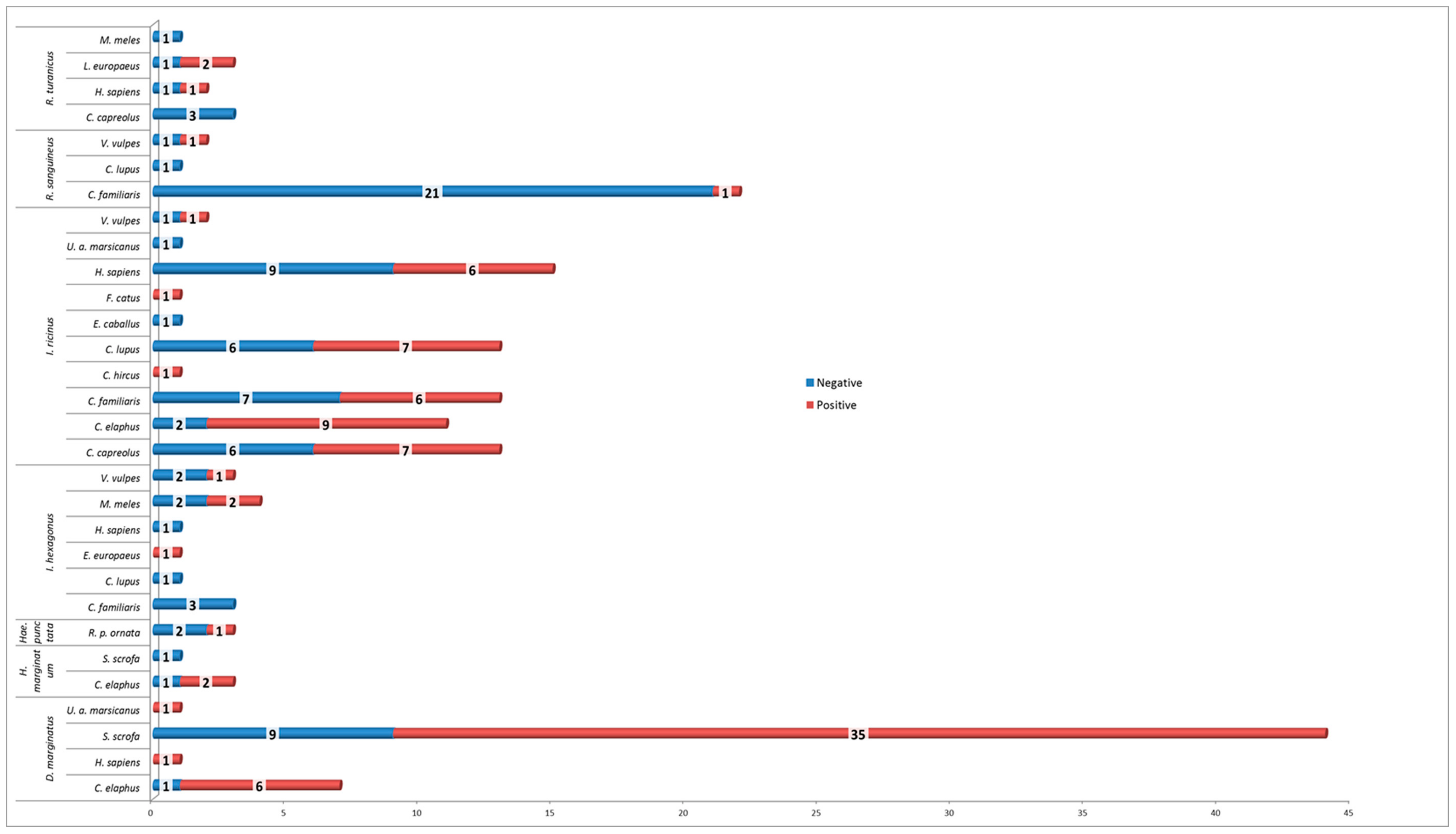
Figure 1. Distribution of tick-pools according host species.
Microorganisms 07 00696 g001
Figure 2. Details of RealTime Rickettsia spp. positive tick-pools/host association.
Microorganisms 07 00696 g002
Table 1. details of the results of RealTime PCR for Rickettsia spp. in each tick species.
TableNegativePositiveTotalTick Species FrequencyMinimum Infection Rate
I. ricinus33387139.9%53.5%
D. marginatus10435329.8%81.1%
R. sanguineus2322514.0%8.0%
I. hexagonus94137.3%30.8%
R. turanicus6395.1%33.3%
H. marginatum2242.2%50.0%
Hae. punctata2131.7%33.3%
Total8593178 52.25%
Table 2. Rickettsia species in relation to tick and host species.
Rickettsia speciesHostTicks
D. marginatusH. marginatumHae. punctataI. hexagonusI. ricinusR. sanguineus s.l.R. turanicusTotal%Total MIR
R. conoriiL. europaeus//////1///
R. conorii Total //////111.1%0.6%
R. felisE. europaeus///1//////
V. vulpes///1//////
R. felis Total ///2///22.2%1.1%
R. haeschlimanniC. elaphus/2////////
R. haeschlimanni Total //2/////22.2%1.1%
R.helveticaH. sapiens////1/////
V. vulpes////1/////
R. helvetica Total/////2//22.2%1.1%
R. massiliaeH. sapiens//////1///
L. europaeus//////1///
V. vulpes/////1////
R. massiliae Total //////1233.2%1.7%
R. monacensisC. capreolus////7/////
C. elaphus////8/////
C. familiaris////61////
C. lupus////7/////
F. catus////1/////
H. sapiens////5/////
R. p. ornata//1///////
U. a. marsicanus1/////////
C. hircus////1/////
R. monacensis Total /1/1/351/3840.9%21.3%
R. raoultii/R. slovacaC. elaphus////1/////
M. meles///1//////
S. scrofa1/////////
R. raoultii/R. slovaca Total /1//11//33.2%1.7%
R. slovacaC. elaphus6/////////
H. sapiens1/////////
S. scrofa34/////////
R. slovaca Total /41//////4144.1%23.0%
R. raoultiiM. meles///1//////
R. raoultii Total / //1///11,1%0.6%
Total/43214382393100%52.2%
Quantitative data on Rickettsia species are shown in relation to tick and host species.

