Introducing Murine Microbiome Database (MMDB): A Curated Database with Taxonomic Profiling of the Healthy Mouse Gastrointestinal Microbiome


Abstract
1. Introduction
2. Materials and Methods
2.1. Collection of Metagenome Sequences
2.2. Construction of Microbiome Taxonomic Profile (MTP) Database
2.3. Statistics
2.4. Operating System and Programming Languages
3. Results and Discussion
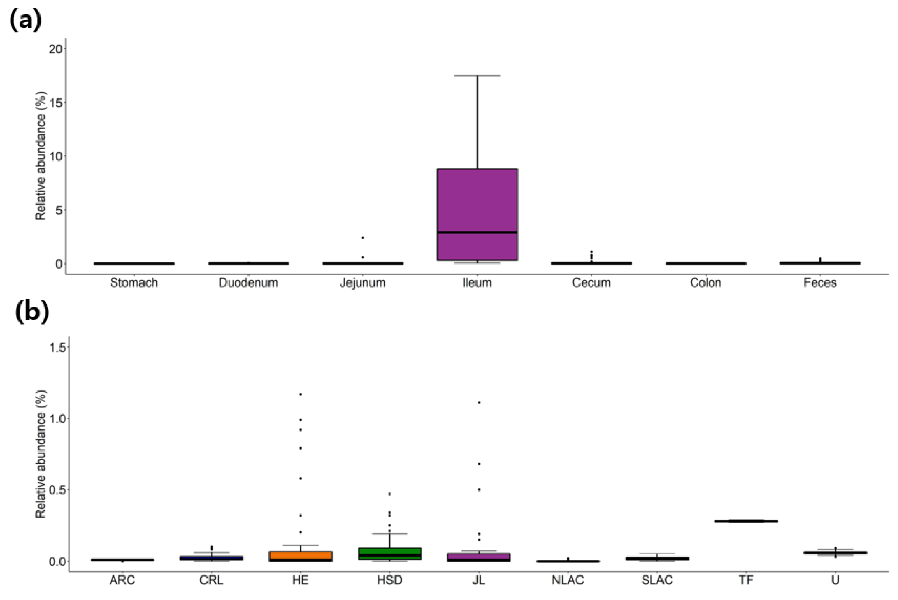
3.1. Taxonomic Profiles of Heathy Mouse Gastrointestinal Microbiome
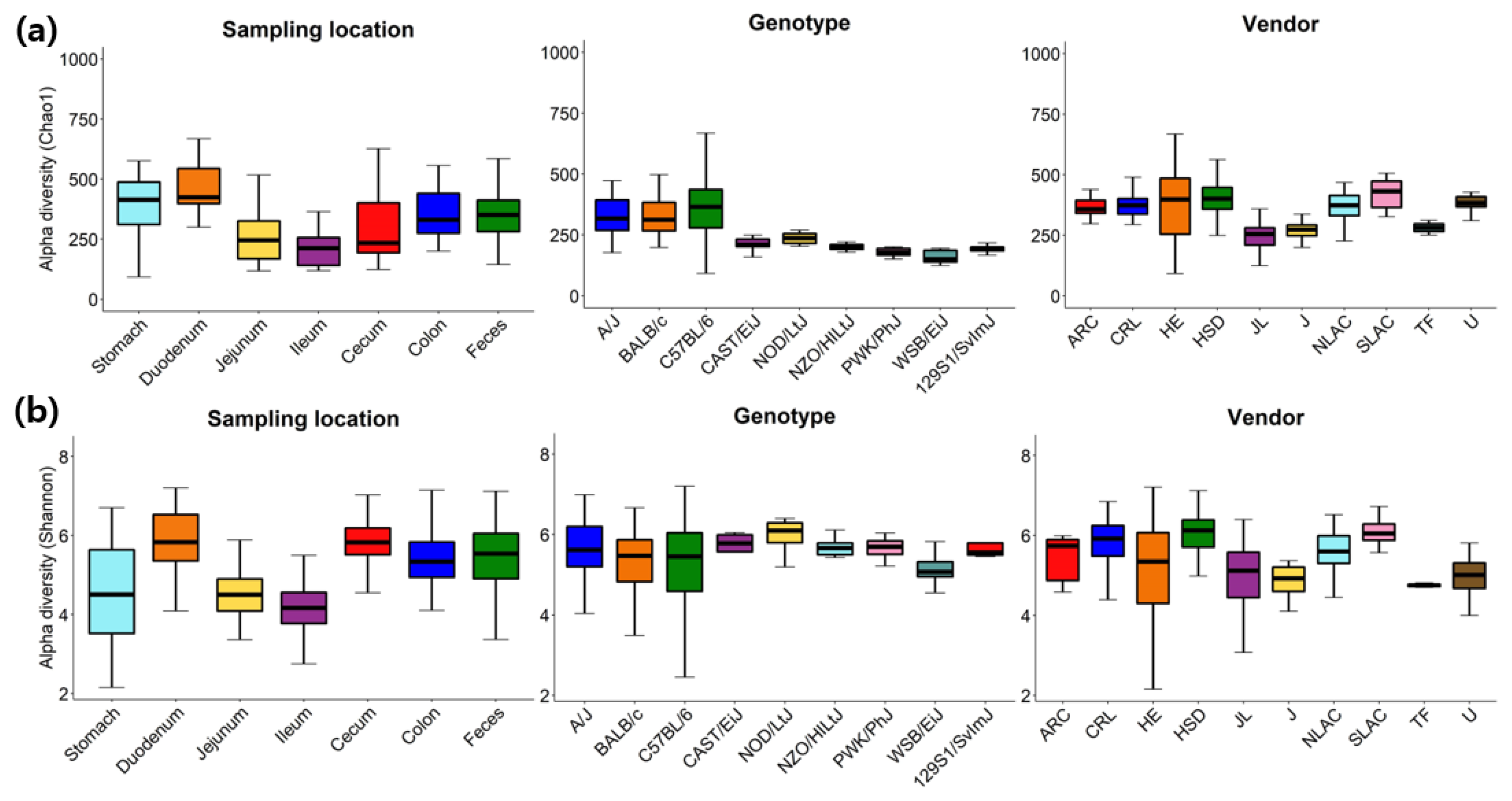
3.2. Alpha-Diversity Variations in the Healthy Mice
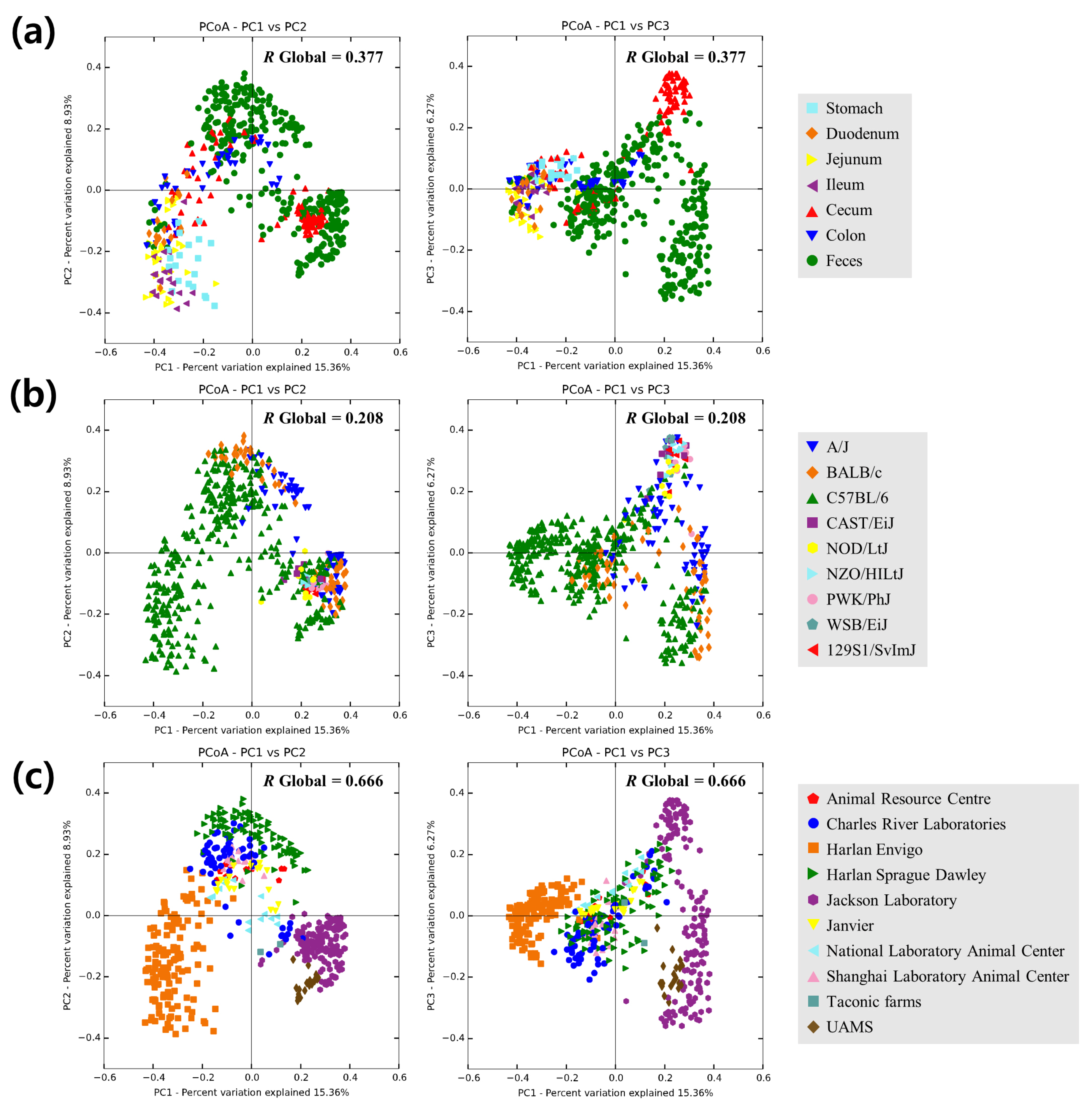
3.3. Beta-Diversity
4. Conclusions
Supplementary Materials
Author Contributions
Funding
Conflicts of Interest
References
- Carter, A.M. Animal models of human placentation--a review. Placenta 2007, 28, S41–S47. [Google Scholar] [CrossRef] [PubMed]
- Parker, K.D.; Albeke, S.E.; Gigley, J.P.; Goldstein, A.M.; Ward, N.L. Microbiome Composition in Both Wild-Type and Disease Model Mice Is Heavily Influenced by Mouse Facility. Front. Microbiol. 2018, 9, 1598. [Google Scholar] [CrossRef] [PubMed]
- Beck, J.A.; Lloyd, S.; Hafezparast, M.; Lennon-Pierce, M.; Eppig, J.T.; Festing, M.F.; Fisher, E.M. Genealogies of mouse inbred strains. Nat. Genet. 2000, 24, 23–25. [Google Scholar] [CrossRef] [PubMed]
- Bonder, M.J.; Kurilshikov, A.; Tigchelaar, E.F.; Mujagic, Z.; Imhann, F.; Vila, A.V.; Deelen, P.; Vatanen, T.; Schirmer, M.; Smeekens, S.P.; et al. The effect of host genetics on the gut microbiome. Nat. Genet. 2016, 48, 1407–1412. [Google Scholar] [CrossRef] [PubMed]
- Colombo, B.M.; Scalvenzi, T.; Benlamara, S.; Pollet, N. Microbiota and mucosal immunity in amphibians. Front. Immunol. 2015, 6. [Google Scholar] [CrossRef] [PubMed]
- Colston, T.J.; Jackson, C.R. Microbiome evolution along divergent branches of the vertebrate tree of life: What is known and unknown. Mol. Ecol. 2016, 25, 3776–3800. [Google Scholar] [CrossRef]
- Fraune, S.; Bosch, T.C.G. Why bacteria matter in animal development and evolution. Bioessays 2010, 32, 571–580. [Google Scholar] [CrossRef]
- Berer, K.; Mues, M.; Koutrolos, M.; Rasbi, Z.A.; Boziki, M.; Johner, C.; Wekerle, H.; Krishnamoorthy, G. Commensal microbiota and myelin autoantigen cooperate to trigger autoimmune demyelination. Nature 2011, 479, 538–541. [Google Scholar] [CrossRef]
- Bohn, E.; Bechtold, O.; Zahir, N.; Frick, J.S.; Reimann, J.; Jilge, B.; Autenrieth, I.B. Host gene expression in the colon of gnotobiotic interleukin-2-deficient mice colonized with commensal colitogenic or noncolitogenic bacterial strains: common patterns and bacteria strain specific signatures. Inflamm. Bowel. Dis. 2006, 12, 853–862. [Google Scholar] [CrossRef]
- Dianda, L.; Hanby, A.M.; Wright, N.A.; Sebesteny, A.; Hayday, A.C.; Owen, M.J. T cell receptor-alpha beta-deficient mice fail to develop colitis in the absence of a microbial environment. Am. J. Pathol. 1997, 150, 91–97. [Google Scholar]
- Garrett, W.S.; Gallini, C.A.; Yatsunenko, T.; Michaud, M.; DuBois, A.; Delaney, M.L.; Punit, S.; Karlsson, M.; Bry, L.; Glickman, J.N.; et al. Enterobacteriaceae act in concert with the gut microbiota to induce spontaneous and maternally transmitted colitis. Cell Host Microbe. 2010, 8, 292–300. [Google Scholar] [CrossRef] [PubMed]
- Lee, Y.K.; Menezes, J.S.; Umesaki, Y.; Mazmanian, S.K. Proinflammatory T-cell responses to gut microbiota promote experimental autoimmune encephalomyelitis. Proc. Natl. Acad. Sci. USA 2011, 108, 4615–4622. [Google Scholar] [CrossRef] [PubMed]
- Rehakova, Z.; Capkova, J.; Stepankova, R.; Sinkora, J.; Louzecka, A.; Ivanyi, P.; Weinreich, S. Germ-free mice do not develop ankylosing enthesopathy, a spontaneous joint disease. Hum. Immunol. 2000, 61, 555–558. [Google Scholar] [CrossRef]
- Rooks, M.G.; Garrett, W.S. Gut microbiota, metabolites and host immunity. Nat. Rev. Immunol. 2016, 16, 341–352. [Google Scholar] [CrossRef] [PubMed]
- Sellon, R.K.; Tonkonogy, S.; Schultz, M.; Dieleman, L.A.; Grenther, W.; Balish, E.; Rennick, D.M.; Sartor, R.B. Resident enteric bacteria are necessary for development of spontaneous colitis and immune system activation in interleukin-10-deficient mice. Infect. Immun. 1998, 66, 5224–5231. [Google Scholar] [PubMed]
- Sudo, N.; Chida, Y.; Aiba, Y.; Sonoda, J.; Oyama, N.; Yu, X.N.; Kubo, C.; Koga, Y. Postnatal microbial colonization programs the hypothalamic-pituitary-adrenal system for stress response in mice. J. Physiol. 2004, 558, 263–275. [Google Scholar] [CrossRef] [PubMed]
- Wen, L.; Ley, R.E.; Volchkov, P.Y.; Stranges, P.B.; Avanesyan, L.; Stonebraker, A.C.; Hu, C.; Wong, F.S.; Szot, G.L.; Bluestone, J.A.; et al. Innate immunity and intestinal microbiota in the development of Type 1 diabetes. Nature 2008, 455, 1109–1113. [Google Scholar] [CrossRef]
- Wu, H.J.; Ivanov, I.I.; Darce, J.; Hattori, K.; Shima, T.; Umesaki, Y.; Littman, D.R.; Benoist, C.; Mathis, D. Gut-residing segmented filamentous bacteria drive autoimmune arthritis via T helper 17 cells. Immunity 2010, 32, 815–827. [Google Scholar] [CrossRef]
- Sivan, A.; Corrales, L.; Hubert, N.; Williams, J.B.; Aquino-Michaels, K.; Earley, Z.M.; Benyamin, F.W.; Lei, Y.M.; Jabri, B.; Alegre, M.L.; et al. Commensal Bifidobacterium promotes antitumor immunity and facilitates anti-PD-L1 efficacy. Science 2015, 350, 1084–1089. [Google Scholar] [CrossRef]
- Campbell, J.H.; Foster, C.M.; Vishnivetskaya, T.; Campbell, A.G.; Yang, Z.K.; Wymore, A.; Palumbo, A.V.; Chesler, E.J.; Podar, M. Host genetic and environmental effects on mouse intestinal microbiota. ISME J. 2012, 6, 2033–2044. [Google Scholar] [CrossRef]
- Ericsson, A.C.; Davis, J.W.; Spollen, W.; Bivens, N.; Givan, S.; Hagan, C.E.; McIntosh, M.; Franklin, C.L. Effects of vendor and genetic background on the composition of the fecal microbiota of inbred mice. PLoS ONE 2015, 10, e0116704. [Google Scholar] [CrossRef] [PubMed]
- Friswell, M.K.; Gika, H.; Stratford, I.J.; Theodoridis, G.; Telfer, B.; Wilson, I.D.; McBain, A.J. Site and strain-specific variation in gut microbiota profiles and metabolism in experimental mice. PLoS ONE 2010, 5, e8584. [Google Scholar] [CrossRef] [PubMed]
- Hufeldt, M.R.; Nielsen, D.S.; Vogensen, F.K.; Midtvedt, T.; Hansen, A.K. Variation in the gut microbiota of laboratory mice is related to both genetic and environmental factors. Comp. Med. 2010, 60, 336–347. [Google Scholar] [PubMed]
- Zmora, N.; Zilberman-Schapira, G.; Suez, J.; Mor, U.; Dori-Bachash, M.; Bashiardes, S.; Kotler, E.; Zur, M.; Regev-Lehavi, D.; Brik, R.B.; et al. Personalized Gut Mucosal Colonization Resistance to Empiric Probiotics Is Associated with Unique Host and Microbiome Features. Cell 2018, 174, 1388–1405. [Google Scholar] [CrossRef]
- Yoon, S.H.; Ha, S.M.; Kwon, S.; Lim, J.; Kim, Y.; Seo, H.; Chun, J. Introducing EzBioCloud: a taxonomically united database of 16S rRNA gene sequences and whole-genome assemblies. Int. J. Syst. Evol. Microbiol. 2017, 67, 1613–1617. [Google Scholar] [CrossRef]
- Edgar, R.C. Search and clustering orders of magnitude faster than BLAST. Bioinformatics 2010, 26, 2460–2461. [Google Scholar] [CrossRef]
- Rognes, T.; Flouri, T.; Nichols, B.; Quince, C.; Mahe, F. VSEARCH: a versatile open source tool for metagenomics. Peer J. 2016, 4, e2584. [Google Scholar] [CrossRef]
- Yarza, P.; Yilmaz, P.; Pruesse, E.; Glockner, F.O.; Ludwig, W.; Schleifer, K.H.; Whitman, W.B.; Euzeby, J.; Amann, R.; Rossello-Mora, R. Uniting the classification of cultured and uncultured bacteria and archaea using 16S rRNA gene sequences. Nat. Rev. Microbiol. 2014, 12, 635–645. [Google Scholar] [CrossRef]
- Edgar, R.C.; Haas, B.J.; Clemente, J.C.; Quince, C.; Knight, R. UCHIME improves sensitivity and speed of chimera detection. Bioinformatics 2011, 27, 2194–2200. [Google Scholar] [CrossRef]
- Ondov, B.D.; Bergman, N.H.; Phillippy, A.M. Interactive metagenomic visualization in a Web browser. BMC Bioinform. 2011, 12, 385. [Google Scholar] [CrossRef]
- Kassambara, A. ggpubr:“ggplot2” based publication ready plots. R package version 0.1 2017, 6. [Google Scholar]
- Caporaso, J.G.; Kuczynski, J.; Stombaugh, J.; Bittinger, K.; Bushman, F.D.; Costello, E.K.; Fierer, N.; Pena, A.G.; Goodrich, J.K.; Gordon, J.I.; et al. QIIME allows analysis of high-throughput community sequencing data. Nat. Methods 2010, 7, 335–336. [Google Scholar] [CrossRef] [PubMed]
- Wilcoxon, F.; Katti, S.; Wilcox, R.A. Critical values and probability levels for the Wilcoxon rank sum test and the Wilcoxon signed rank test. In Selected Tables in Mathematical Statistics; Harter, H., Owen, D.B., Eds.; Markham Publishing Company: Markham, ON, Canada, 1970; Volume 1, pp. 171–259. [Google Scholar]
- Bolyen, E.; Rideout, J.R.; Dillon, M.R.; Bokulich, N.A.; Abnet, C.C.; Al-Ghalith, G.A.; Alexander, H.; Alm, E.J.; Arumugam, M.; Asnicar, F.; et al. Reproducible, interactive, scalable and extensible microbiome data science using QIIME 2. Nat. Biotechnol. 2019, 37, 852–857. [Google Scholar] [CrossRef] [PubMed]
- Lagkouvardos, I.; Lesker, T.R.; Hitch, T.C.A.; Galvez, E.J.C.; Smit, N.; Neuhaus, K.; Wang, J.; Baines, J.F.; Abt, B.; Stecher, B.; et al. Sequence and cultivation study of Muribaculaceae reveals novel species, host preference, and functional potential of this yet undescribed family. Microbiome. 2019, 7, 28. [Google Scholar] [CrossRef]
- Wang, L.; Chen, C.; Cui, S.; Lee, Y.K.; Wang, G.; Zhao, J.; Zhang, H.; Chen, W. Adhesive Bifidobacterium Induced Changes in Cecal Microbiome Alleviated Constipation in Mice. Front. Microbiol. 2019, 10, 1721. [Google Scholar] [CrossRef]
- Moreira Junior, R.E.; de Carvalho, L.M.; Pedersen, A.S.B.; Damasceno, S.; Maioli, T.U.; de Faria, A.M.C.; Godard, A.L.B. Interaction between high-fat diet and ethanol intake leads to changes on the fecal microbiome. J. Nutr. Biochem. 2019, 72, 108215. [Google Scholar] [CrossRef]
- Guo, B.; Yang, B.; Pang, X.; Chen, T.; Chen, F.; Cheng, K.W. Fucoxanthin modulates cecal and fecal microbiota differently based on diet. Food Funct. 2019. [Google Scholar] [CrossRef]
- Fan, J.; Wang, Y.; You, Y.; Ai, Z.; Dai, W.; Piao, C.; Liu, J.; Wang, Y. Fermented ginseng improved alcohol liver injury in association with changes in the gut microbiota of mice. Food Funct. 2019. [Google Scholar] [CrossRef]
- Ivanov, I.I.; Atarashi, K.; Manel, N.; Brodie, E.L.; Shima, T.; Karaoz, U.; Wei, D.; Goldfarb, K.C.; Santee, C.A.; Lynch, S.V.; et al. Induction of intestinal Th17 cells by segmented filamentous bacteria. Cell 2009, 139, 485–498. [Google Scholar] [CrossRef]
- Bluemel, S.; Wang, L.; Kuelbs, C.; Moncera, K.; Torralba, M.; Singh, H.; Fouts, D.E.; Schnabl, B. Intestinal and hepatic microbiota changes associated with chronic ethanol administration in mice. Gut Microbes. 2019, 14, 1–11. [Google Scholar] [CrossRef]
- Allen, J.M.; Berg Miller, M.E.; Pence, B.D.; Whitlock, K.; Nehra, V.; Gaskins, H.R.; White, B.A.; Fryer, J.D.; Woods, J.A. Voluntary and forced exercise differentially alters the gut microbiome in C57BL/6J mice. J. Appl. Physiol. (1985) 2015, 118, 1059–1066. [Google Scholar] [CrossRef] [PubMed]
- Fransen, F.; Zagato, E.; Mazzini, E.; Fosso, B.; Manzari, C.; El Aidy, S.; Chiavelli, A.; D’Erchia, A.M.; Sethi, M.K.; Pabst, O.; et al. BALB/c and C57BL/6 Mice Differ in Polyreactive IgA Abundance, which Impacts the Generation of Antigen-Specific IgA and Microbiota Diversity. Immunity 2015, 43, 527–540. [Google Scholar] [CrossRef] [PubMed]
- Gu, S.; Chen, D.; Zhang, J.N.; Lv, X.; Wang, K.; Duan, L.P.; Nie, Y.; Wu, X.L. Bacterial community mapping of the mouse gastrointestinal tract. PLoS ONE 2013, 8, e74957. [Google Scholar] [CrossRef] [PubMed]


| Counts | |
|---|---|
| Total numbers of projects | 14 |
| Total numbers of samples | 554 |
| Mean OTUs/sample ± SD | 657 ± 288 |
| Mean valid reads/sample ± SD | 61,280 ± 27,893 |
| Number of sampling locations in gastrointestinal (GI) tract | 7 |
| Number of strains/genotypes | 9 |
| Number of vendors | 10 |
| Total number of phyla found in all samples | 58 |
| Total number of classes found in all samples | 138 |
| Total number of orders found in all samples | 286 |
| Total number of families found in all samples | 585 |
| Total number of genera found in all samples | 1732 |
| Total number of species found in all samples | 4703 |
| Sampling location | Name | Taxonomy | Proportions of samples (%) | Max (%) | Median (%) |
|---|---|---|---|---|---|
| Stomach | Lactobacillus gasseri group | Firmicutes; Bacilli; Lactobacillales; Lactobacillaceae; Lactobacillus; Lactobacillus gasseri group; | 95.00 | 40.86 | 13.89 |
| (n = 20) | Lactobacillus reuteri group | Firmicutes; Bacilli; Lactobacillales; Lactobacillaceae; Lactobacillus; Lactobacillus reuteri group; | 95.00 | 55.55 | 11.64 |
| Lactobacillus intestinalis | Firmicutes; Bacilli; Lactobacillales; Lactobacillaceae; Lactobacillus; Lactobacillus intestinalis; | 90.00 | 28.95 | 7.70 | |
| PAC000185_s | Bacteria; Proteobacteria; Alphaproteobacteria; Rhodospirillales; Rhodospirillaceae; LARJ_g; PAC000185_s; | 55.00 | 4.47 | 1.86 | |
| PAC001472_s | Bacteroidetes; Bacteroidia; Bacteroidales; Muribaculaceae; PAC001472_g; PAC001472_s; | 55.00 | 3.50 | 1.27 | |
| Dudenum | PAC001075_s | Bacteroidetes; Bacteroidia; Bacteroidales; Muribaculaceae; PAC000198_g; PAC001075_s; | 93.75 | 6.30 | 2.07 |
| (n = 16) | Akkermansia muciniphila | Verrucomicrobia; Verrucomicrobiae; Verrucomicrobiales; Akkermansiaceae; Akkermansia; Akkermansia muciniphila; | 87.50 | 18.95 | 4.30 |
| Lactobacillus gasseri group | Firmicutes; Bacilli; Lactobacillales; Lactobacillaceae; Lactobacillus; Lactobacillus gasseri group; | 81.25 | 36.58 | 5.20 | |
| PAC001065_s group | Bacteroidetes; Bacteroidia; Bacteroidales; Muribaculaceae; PAC000186_g; PAC001065_s group; | 81.25 | 7.48 | 4.06 | |
| PAC001472_s | Bacteroidetes; Bacteroidia; Bacteroidales; Muribaculaceae; PAC001472_g; PAC001472_s; | 81.25 | 9.17 | 3.75 | |
| Jejunum | PAC001065_s group | Bacteroidetes; Bacteroidia; Bacteroidales; Muribaculaceae; PAC000186_g; PAC001065_s group; | 88.24 | 10.44 | 4.27 |
| (n = 34) | PAC001075_s | Bacteroidetes; Bacteroidia; Bacteroidales; Muribaculaceae; PAC000198_g; PAC001075_s; | 88.24 | 11.83 | 3.12 |
| Lactobacillus gasseri group | Firmicutes; Bacilli; Lactobacillales; Lactobacillaceae; Lactobacillus; Lactobacillus gasseri group; | 85.29 | 39.67 | 6.85 | |
| Lactobacillus reuteri group | Firmicutes; Bacilli; Lactobacillales; Lactobacillaceae; Lactobacillus; Lactobacillus reuteri group; | 82.35 | 31.43 | 3.04 | |
| Lactobacillus intestinalis | Firmicutes; Bacilli; Lactobacillales; Lactobacillaceae; Lactobacillus; Lactobacillus intestinalis; | 79.41 | 37.06 | 3.55 | |
| Ileum | Lactobacillus gasseri group | Firmicutes; Bacilli; Lactobacillales; Lactobacillaceae; Lactobacillus; Lactobacillus gasseri group; | 100.00 | 47.00 | 12.02 |
| (n = 20) | Lactobacillus reuteri group | Firmicutes; Bacilli; Lactobacillales; Lactobacillaceae; Lactobacillus; Lactobacillus reuteri group; | 95.00 | 39.17 | 5.93 |
| Ileibacterium valens | Firmicutes; Erysipelotrichi; Erysipelotrichales; Erysipelotrichaceae; Ileibacterium; Ileibacterium valens; | 95.00 | 39.23 | 4.88 | |
| PAC001472_s | Bacteroidetes; Bacteroidia; Bacteroidales; Muribaculaceae; PAC001472_g; PAC001472_s; | 80.00 | 10.97 | 2.46 | |
| PAC001065_s group | Bacteroidetes; Bacteroidia; Bacteroidales; Muribaculaceae; PAC000186_g; PAC001065_s group; | 80.00 | 8.53 | 2.20 | |
| Cecum | PAC001188_s | Firmicutes; Clostridia; Clostridiales; Ruminococcaceae; Oscillibacter; PAC001188_s; | 66.67 | 5.99 | 2.13 |
| (n = 96) | KE159538_s | Firmicutes; Clostridia; Clostridiales; Lachnospiraceae; KE159538_g; KE159538_s; | 45.83 | 20.38 | 4.38 |
| KE159714_s group | Firmicutes; Clostridia; Clostridiales; Ruminococcaceae; Oscillibacter; KE159714_s group; | 41.67 | 16.71 | 2.03 | |
| KE159628_s | Bacteria; Firmicutes; Clostridia; Clostridiales; Lachnospiraceae; KE159628_g; KE159628_s; | 41.67 | 8.30 | 1.75 | |
| PAC001080_s | Tenericutes; Mollicutes; Acholeplasmatales; Acholeplasmataceae; Acholeplasma_g2; PAC001080_s; | 40.63 | 16.87 | 3.44 | |
| Colon | PAC001061_s | Bacteroidetes; Bacteroidia; Bacteroidales; Rikenellaceae; Alistipes; PAC001061_s; | 70.00 | 10.61 | 3.47 |
| (n = 40) | PAC001074_s | Bacteroidetes; Bacteroidia; Bacteroidales; Muribaculaceae; PAC001074_g; PAC001074_s; | 67.50 | 5.83 | 2.11 |
| PAC001471_s | Bacteroidetes; Bacteroidia; Bacteroidales; Rikenellaceae; Alistipes; PAC001471_s; | 52.50 | 12.00 | 5.04 | |
| Akkermansia muciniphila | Verrucomicrobia; Verrucomicrobiae; Verrucomicrobiales; Akkermansiaceae; Akkermansia; Akkermansia muciniphila; | 50.00 | 27.47 | 10.49 | |
| PAC002478_s | Bacteria; Proteobacteria; Deltaproteobacteria; Desulfovibrionales; Desulfovibrionaceae; LT706945_g; PAC002478_s; | 50.00 | 42.00 | 10.27 | |
| Feces | PAC001071_s | Bacteroidetes; Bacteroidia; Bacteroidales; Muribaculaceae; PAC001068_g; PAC001071_s; | 77.13 | 58.21 | 4.01 |
| (n = 328) | PAC001060_s | Bacteroidetes; Bacteroidia; Bacteroidales; Rikenellaceae; Alistipes; PAC001060_s; | 55.79 | 52.82 | 6.78 |
| PAC001188_s | Bacteria; Firmicutes; Clostridia; Clostridiales; Ruminococcaceae; Oscillibacter; PAC001188_s; | 49.70 | 18.59 | 2.18 | |
| PAC001369_s group | Bacteria; Firmicutes; Clostridia; Clostridiales; Ruminococcaceae; Oscillibacter; PAC001369_s group; | 39.63 | 12.95 | 1.99 | |
| PAC001065_s group | Bacteroidetes; Bacteroidia; Bacteroidales; Muribaculaceae; PAC000186_g; PAC001065_s group; | 37.50 | 13.89 | 2.23 |
© 2019 by the authors. Licensee MDPI, Basel, Switzerland. This article is an open access article distributed under the terms and conditions of the Creative Commons Attribution (CC BY) license (http://creativecommons.org/licenses/by/4.0/).
Share and Cite
Yang, J.; Park, J.; Park, S.; Baek, I.; Chun, J. Introducing Murine Microbiome Database (MMDB): A Curated Database with Taxonomic Profiling of the Healthy Mouse Gastrointestinal Microbiome. Microorganisms 2019, 7, 480. https://doi.org/10.3390/microorganisms7110480
Yang J, Park J, Park S, Baek I, Chun J. Introducing Murine Microbiome Database (MMDB): A Curated Database with Taxonomic Profiling of the Healthy Mouse Gastrointestinal Microbiome. Microorganisms. 2019; 7(11):480. https://doi.org/10.3390/microorganisms7110480
Chicago/Turabian StyleYang, Junwon, Jonghyun Park, Sein Park, Inwoo Baek, and Jongsik Chun. 2019. "Introducing Murine Microbiome Database (MMDB): A Curated Database with Taxonomic Profiling of the Healthy Mouse Gastrointestinal Microbiome" Microorganisms 7, no. 11: 480. https://doi.org/10.3390/microorganisms7110480
APA StyleYang, J., Park, J., Park, S., Baek, I., & Chun, J. (2019). Introducing Murine Microbiome Database (MMDB): A Curated Database with Taxonomic Profiling of the Healthy Mouse Gastrointestinal Microbiome. Microorganisms, 7(11), 480. https://doi.org/10.3390/microorganisms7110480