Article

Molecular Epidemiological Characterization of Staphylococcus argenteus Clinical Isolates in Japan: Identification of Three Clones (ST1223, ST2198, and ST2550) and a Novel Staphylocoagulase Genotype XV

1
Department of Hygiene, Sapporo Medical University School of Medicine, Hokkaido, 060-8556 Sapporo, Japan
2
Sapporo Clinical Laboratory, Inc., Hokkaido, 060-0005 Sapporo, Japan
*
Author to whom correspondence should be addressed.
Microorganisms 2019, 7(10), 389; https://doi.org/10.3390/microorganisms7100389
Submission received: 9 August 2019 / Revised: 7 September 2019 / Accepted: 23 September 2019 / Published: 24 September 2019
(This article belongs to the Section Medical Microbiology)

Abstract

:
Staphylococcus argenteus, a novel emerging species within Staphylococcus aureus complex (SAC), has been increasingly reported worldwide. In this study, prevalence of S. argenteus among human clinical isolates, and their clonal diversity and genetic characteristics of virulence factors were investigated in Hokkaido, the northern main island of Japan. During a four-month period starting from March 2019, twenty-four S. argenteus and 4330 S. aureus isolates were recovered from clinical specimens (the ratio of S. argenteus to S. aureus :0.0055). Half of S. argenteus isolates (n = 12) belonged to MLST sequence type (ST) 2250 and its single-locus variant, with staphylocoagulase genotype (coa-) XId, while the remaining isolates were assigned to ST2198/coa-XIV (n = 6), and ST1223 with a novel coa-XV identified in this study (n = 6). All the isolates were mecA-negative, and susceptible to all the antimicrobials tested, except for an ST2198 isolate with blaZ and an ST2250 isolate with tet(L) showing resistance to ampicillin and tetracyclines, respectively. Common virulence factors in the S. argenteus isolates were staphylococcal enterotoxin (-like) genes sey, selz, sel26, and sel27 in ST2250, selx in ST2198, and enterotoxin gene cluster (egc-1: seg-sei-sem-sen-seo) in ST1223 isolates, in addition to hemolysin genes (hla, hlb, and hld) distributed universally. Elastin binding protein gene (ebpS) and MSCRAMM family adhesin SdrE gene (sdrE) detected in all the isolates showed high sequence identity among them (> 97%), while relatively lower identity to those of S. aureus (78–92%). Phylogenetically, ebpS, sdrE, selx, sey, selw, sel26, and sel27 of S. argenteus formed clusters distinct from those of S. aureus, unlike sec, selz, tst-1, and staphylokinase gene (sak). The present study revealed the prevalence of S. argenteus among clinical isolates, and presence of three distinct S. argenteus clones (ST2250; ST2198 and ST1223) harboring different virulence factors in northern Japan. ST2198 S. argenteus, a minor clone (strain BN75-like) that had been rarely reported, was first identified in Japan as human isolates.

1. Introduction

Staphylococcus argenteus is a novel species of coagulase-positive staphylococci previously described as a divergent lineage of S. aureus [1], and has been increasingly reported worldwide as an emerging pathogen affecting both humans [2,3,4,5,6,7,8,9,10,11,12,13,14,15,16,17,18,19,20,21,22,23,24,25,26] and animals [1,25,27,28,29]. The major characteristic of this lineage is non-pigmented (white) colonies on blood agar plates due to lack of the crtOPQMN gene operon required for staphyloxanthin pigment production [1,2]. S. argenteus and S. aureus are unable to be discriminated by routine diagnostic microbiological testing, and also by analysis of 16S rRNA gene, because these species have identical sequence of this gene [1]. However, S. argenteus shows relatively low genome sequence identity (87%) to S. aureus, and only 4% genes were shared between the two species at 100% homology level [1,3]. Accordingly, evident sequence difference was documented in early reports for the thermonuclease gene (nuc) and the nonribosomal peptide synthetase (NRPS) gene [1,2,4], which are available as markers for identification of these species. Thereafter, sequence diversity and genetic characteristics between these species were revealed for genes encoding major virulence factors, i.e., staphylococagulase, protein A, and alpha-hemolysin [5].
The type strain of S. argenteus MSHR1132T belongs to ST1850 grouped into clonal complex 75 (CC75) [1]. After the first report of genetically divergent ST75 in Australia in 2002 [6,7], S. argenteus belonging to CC75 and other CC represented by CC1223, CC2198, CC2250, and CC2596 were increasingly reported in other Oceanian countries (New Zealand [8] and Fiji [8,9]), Asia (Thailand [10,11], Lao PDR [12], Cambodia [13], Myanmar [5,14], China [4], Japan [15,16,17] and Taiwan [18]), Europe (Belgium [19], France [20], the UK [10], Denmark [3] and Sweden [21]) and South America (French Guiana [22] and Trinidad and Tobago [23]). There are at least three geographical ‘hot spots’ of S. argenteus, such as Southeast Asia, remote human populations in Australia and the Amazon [24,25]. S. argenteus infections in humans have been commonly community-onset [11,18], occasionally associated with high mortality [18], and some isolates were revealed to harbor Panton-Valentine leucocidin that may be related to severe diseases in humans [14,20]. However, information on S. argenteus reported to date are still insufficient to delineate its epidemiology, clinical significance and nosocomial impact, containing partly contradictory views [25]. In Japan, only limited information of S. argenteus in humans are available; a case report of purulent lymphadenitis [26], two reports of food poisoning outbreaks [15,16], and two cases of bacteremia through retrospective study [17], while the prevalence of S. argenteus among presumptive S. aureus from general clinical specimens has not yet clearly been understood.
In the present study, we investigated the prevalence of S. argenteus from various clinical specimens from patients in Hokkaido, the northern main island of Japan, and analyzed their genotypes and virulence factors including toxins and adhesins. The results indicated the presence of three clones with different toxin gene profiles including newer enterotoxin-like genes (selz, sel26, and sel27), novel staphylocoagulase genotype XV, and S. argenteus-specific genetic groups in the primary virulence factors (SdrE, elastin-binding protein).

2. Materials and Methods

2.1. Bacterial Isolates, Species Identification

S. aureus and S. argenteus were isolated from various clinical specimens that were brought to the Sapporo Clinical Laboratory Inc., Sapporo, Japan, from both hospitalized patients and outpatients in medical facilities in Hokkaido, during a four-month period starting on 7th March 2019. The clinical specimens were inoculated onto blood agar plates and incubated at 37 °C for 24 h aerobically. Gram-positive, coagulase-positive isolates were collected for further bacteriological identification. Initial screening of S. argenteus was performed by MALDI-TOF mass spectrometry using MALDI Biotyper (BRUKER). Isolates assigned as S.argenteus were confirmed genetically by three methods: (1) PCR targeting crtOPQMN gene operon-deficient region, (2) PCR targeting NRPS [4], and (3) sequence analysis of the partial NRPS gene [4] and nuc gene [1]. The method (1) was designed in the present study and performed by multiplex PCR to amplify crtOPQMN-deficient region (S. argenteus) or crtP (S. aureus) using primers listed in Table S1. In the method (2), non-S. aureus SAC (S. argenteus/S. schweitzeri)-specific amplicon that is 180-bp longer than that of S. aureus was detected. In method (3), full-length nucleotide sequence of nuc and partial NRPS gene were determined by PCR and direct sequencing, followed by alignment with those of S. aureus and S. argenteus using Clustal Omega program (https://www.ebi.ac.uk/Tools/msa/clustalo/).

2.2. Antimicrobial Susceptibility Testing

Minimum inhibitory concentrations (MICs) within limited ranges were measured by broth microdilution test against 18 antimicrobial agents (oxacillin, OXA; ampicillin, AMP; cefazolin, CFZ; cefmetazole, CMZ; flomoxef, FMX; imipenem, IPM; gentamicin, GEN; arbekacin, ABK; erythromycin, ERY; clindamycin, CLI; vancomycin, VAN; teicoplanin, TEC; linezolid, LZD; minocycline, MIN; Fosfomycin, FOF; levofloxacin, LVX; cefoxitin, FOX; trimethoprim/sulfamethoxazole, SXT) by using the Dry Plate ‘Eiken’ DP32 (Eiken Chemical, Tokyo, Japan), and also tetracycline and doxycycline manually. Resistance or susceptibility was judged according to the breakpoints defined in the Clinical Laboratory Standards Institute (CLSI) guidelines [30] for most of the antimicrobial drugs examined. For fosfomycin and arbekacin, whose break points are not defined by CLSI guidelines, the European Committee on Antimicrobial Susceptibility Testing (EUCAST) breakpoint (FOF, 32 μg/mL, Staphylococcus spp.) [31] and a unique breakpoint (ABK, 4μg/mL, which is higher than the 2 μg/mL defined by the Japanese Society of Chemotherapy for respiratory infection) were used [32]. A breakpoint of flomoxef (16 μg/mL) defined by the Japanese Society of Chemotherapy for urinary tract infection was also applied [32].

2.3. Genetic Typing, Detection of Virulence Factors and Drug Resistance Genes

For all the isolates, presence of staphylococcal 16S rRNA, nuc, mecA, PVL genes and ACME-arcA (arginine deiminase gene) was examined by multiplex PCR assay as described by Zhang et al. [33]. Sequence type (ST) was determined according to the scheme of multilocus sequencence typing (MLST) (https://pubmlst.org/) [34]. spa type based on sequence of protein A gene X-region was determined by PCR and sequencing as described previously [35] using Ridom SpaServer (http://spa.ridom.de/index.shtml). Staphylocoagulase genotype (coa-type) was determined by multiplex PCR assay as described previously [36]. For the isolates of which coa-type was not classified by the multiplex PCR, partial staphylocoagulase gene sequences (D1, D2, and the central regions) were determined and their highly similar staphylocoagulase sequences were searched by Basic Local Alignment Search Tool (BLAST: https://blast.ncbi.nlm.nih.gov/Blast.cgi) to assign their coa-types. For untypeable isolates, nucleotide sequences of whole staphylocoagulase gene was determined as described previously [37], subsequently sequence identities of D1 region, D2 and the central (C) region to those of established coa-types were analyzed by using Clustal Omega program.
The presence of 28 staphylococcal enterotoxin (SE) (-like) genes (sea-see, seg-selu, selx, sey, selw, selz, sel26 and sel27), the TSST-1 gene (tst-1) and exfoliative toxin genes (eta, etb and etd), leukocidins (lukDE and lukM), haemolysins (hla, hlb, hld and hlg), adhesin genes (eno, cna, sdrC, sdrD, sdrE, fib, clfA, clfB, fnbA, fnbB, icaA, icaD, ednA, ednB, bap and vWbp), modulators of host defense (sak, chp and scn) were analyzed by multiplex or uniplex PCRs [38,39,40]. For detection of selz, sel26, and sel27 by PCR, primers shown in Table S1 were newly designed based on selz sequence reported for bovine S. aureus strain RF122 (GenBank accession no. NC_007622, locus tag SAB_RS00140) [41] and sel26/sel27 sequences reported for S. aureus strain 50 and RF14 (GenBank accession no. MF370874 and CP011528, respectively) [42]. Terms of “sel27” and “sel28” assigned to strain 50 in the annotations in GenBank database were identical to “sel26” and “sel27”, respectively, described by Zhang and coworkers [42].
Genes conferring resistance to penicillin (blaZ), tetracycline (tet(K), tet(L), and tet(M)), macrolide (ermA, ermB, ermC, and msrA), aminoglycoside (aac(6′)-Im, aac(6′)-Ie-aph(2”)-Ia, ant(3”)-Ia, ant(4′)-Ia, ant(6)-Ia, ant(9)-Ia, ant(9)-Ib, aph(2”)-Ib, aph(2”)-Ic, aph(2”)-Id, and aph(3′)-IIIa) were detected by uniplex or multiplex PCR using the primers previously reported [38,39,40].

2.4. Phylogenetic Analysis of Virulence Factors

Nucleotide sequences of full-length ORF were determined for staphylocoagulase genes belonging to a novel type, microbial surface component recognizing adhesive matrix molecule (MSCRAMM) family adhesin SdrE gene (sdrE), elastin-binding protein gene (ebpS), SE(-like) genes (selw, selx, sey, selz, sel26, sel27), and staphylokinase gene (sak) by PCR with primers listed in Table S1, followed by Sanger sequencing using the BigDye Terminator v3.1 Cycle Sequencing kit (Applied Biosystems, Foster City, CA, USA) on an automated DNA sequencer (ABI PRISM 3100). Similarly, full length sequences of enterotoxin gene sec and tst-1 gene were also determined. Phylogenetic dendrograms of these virulence factors were constructed by the maximum likelihood method using the MEGA7 software, together with sequence data of staphylococcal strains available in GenBank database. The dendrograms were statistically supported by bootstrapping with 1000 replicates. Clustal Omega program was used for multiple alignments of amino acid sequences of EbpS. Prediction of transmembrane/hydrophobic region in EbpS was performed by TMpred program (https://embnet.vital-it.ch/software/TMPRED_form.html). All the sequence data of S. argenteus genes (nuc, nrps, sec, selx, sey, selw, selz, sel26, sel27, tst-1, sdrE, ebpS and sak) determined in the present study were deposited in GenBank database under accession numbers shown in Table S2.

3. Results

3.1. Identification and Prevalence of S. argenteus

A total of twenty-four S. argenteus isolates derived from twenty-three patients (15 outpatients and 8 inpatients) were isolated as listed in Table 1. From an isolate of a single patient (ear discharge), two clones showing hemolysis on blood agar plate (SG05-1) and no hemolysis (SG05-2) were isolated. From an inpatient, two isolates (SG22, SG23) were recovered from different specimens (pharynx and blood, respectively). S. argenteus isolates were derived from various specimens, with those from respiratory system being dominant (sputum (n = 6), pharynx (n = 2), nasal discharge (n = 2)), followed by stool (n = 4), skin and abscess (n = 3), urine (n = 2), vaginal discharge (n = 2), ear discharge (n = 1), blood (n = 1), and subdural abscess (n = 1). Specimens were brought from eleven cities/towns, and ratio of outpatients to inpatient was 5:3. Age range of the patient was 14–94 years, with equal sex ratio. During the same study period, number of non-duplicate S. aureus isolates was 4330, that comprised 2817 methicillin-susceptible S. aureus (MSSA) and 1513 methicillin-resistant S. aureus (MRSA). Isolation ratios of S. argenteus to S. aureus, and to MSSA were 0.0055 (0.55%, 24/4330) and 0.0085 (0.85%, 24/2817), respectively.
Thermostable nuclease gene (nuc) of all the 24 isolates comprised 669 nucleotides that was shorter than that of S. aureus by 18 bp, and showed 98.8–100% identity to those of S. argenteus strains (MSHR1132T, BN75, XNO62, XNO106, 51183) (Table S3), while 82% to S. aureus nuc gene. Similarly, partial NRPS gene sequences (264 bp) of the 24 isolates had 98.1–100% identity among them and to those of the reported S. argenteus strains, in contrast they were 180-bp longer than that of S. aureus strain NCTC8325 showing 60–61% identity.

3.2. Genotypes (ST, spa Type, coa-Type)

Among the twenty-four S. argenteus isolates, half of them (n = 12) was classified into ST2250 (11 isolates) and its single-locus variant (ST3951, one isolate), and remainings were assigned to ST2198 (25%, n = 6), and ST1223 (25%, n = 6) (Table 1). ST2250/ST3951 isolates were classified into spa type t5078 (6 isolates) and other spa types (t5787, t7960, t17928, and unassigned types) having similar repeat profile to t5078. ST2198 isolates were assigned to t7959 or other types with t7959-like profile, while ST1223 isolates were typed as t5142 or t5142-related types (Table 1).
Because coa-type of S. argenteus isolates could not be assigned to I through X by the multiplex PCR method, sequence identities of D1 region and D2-C region to those of established coa-types Ia-XIV were analyzed. All the ST2250 isolates were assigned to staphylocoagulase genotype XId, while ST2198 isolates to coa-XIV.
According to the criteria to define coa-type proposed previously [43], staphylocoagulase genes showing > 90% sequence identity in D1 region and D2-C regions are assigned to identical coa-type and subtype within the coa-type, respectively. D1 and D2-C regions of six isolates (SG03, SG06, SG07, SG15, SG17 and SG18) showed less than 88.3% and 82.9% identities to the 14 established coa-types, respectively, while 100% identities to S. argenteus strain SJTU F20214, D7903 and M051_MSHR of which coa-type has not yet been described (Table 2). Therefore, a novel staphylocoagulase genotype XV was created and assigned to all the six ST1223 S. argenteus isolates.

3.3. Prevalence of Virulence Factors, Drug Resistance Genes and Antimicrobial Susceptibility

None of the isolates had PVL genes (lukS-PV-lukF-PV) or ACME-arcA genes. Alpha-, beta-, and delta-hemolysin genes (hla, hlb, hld) were detected in all the isolates (Table 1). SE-like genes sey, selz, sel26 and sel27 were detected in mainly ST2250 isolates, while selz was harbored by some ST2198 and ST2113 isolates. selx was found in all the ST2198 isolates and one isolate each of ST2250 and ST1223. All the ST1223 isolates harbored seg-sei-sem-sen-seo (egc-1) and selw. sec and tst-1 genes were detected in three and two isolates of ST2250, respectively. However, all other remaining SE genes and exfoliative toxin genes examined were not detected. Staphylokinase gene (sak) was detected in seven isolates (six ST2250 and one ST2198 isolates). Nine adhesin genes (fib, clfA, clfB, ebpS, eno, fnbA, fnbB, sdrE, icaA) were identified in all the isolates, while most isolates had sdrC and sdrD. Two hemolysis-positive and negative clones (SG05-1 and SG05-2, respectively) from a patient, were classified into the same genotypes (ST2250/coa-XId) and showed identical profiles of virulence factors.
All the isolates were methicillin-sensitive without having mecA, and susceptible to 20 antimicrobials tested, except for two isolates; an ST2198 isolate SG20 with blaZ was resistant to ampicillin (MIC, 4 μg/mL) and an ST2250 isolate SG23 with tet(L) showed resistance to tetracycline (MIC, 64 μg/mL) and intermediate resistance to doxycycline (MIC, 8 μg/mL). None of the isolates harbored other resistance genes tested.

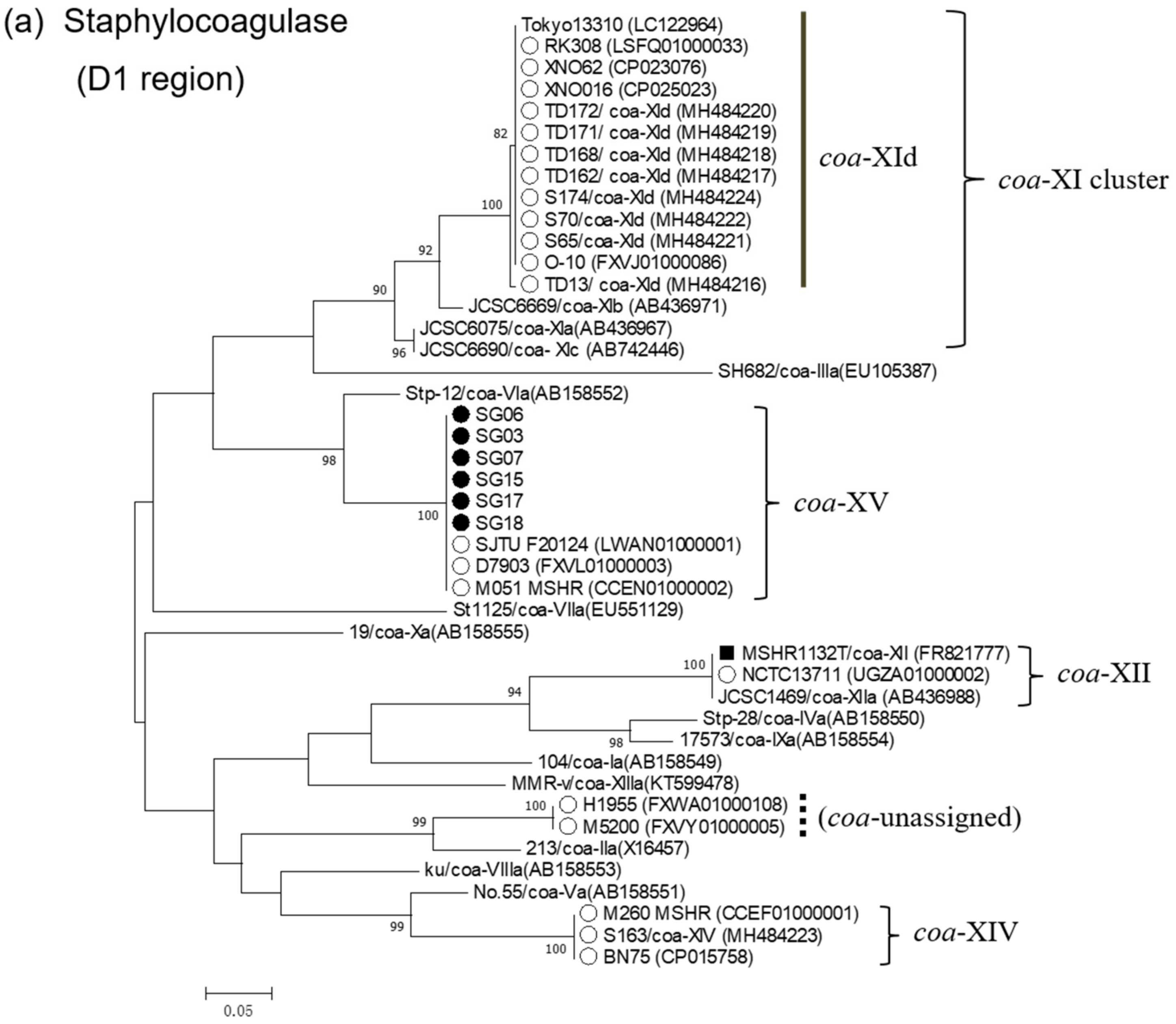
3.4. Phylogenetic Analysis of Virulence Factors

To clarify phylogenetic relatedness of staphylocoagulase genes of the novel coa-type XV to those of other types, dendrograms of D1 and D2-C regions of staphylocoagulase genes were constructed (Figure 1a,b, respectively). In D1 region, S. argenteus strains were discriminated into five clusters representing coa-XId, -XII, -XIV, -XV, and an unassigned type. coa-XV D1 region was revealed to be of the same lineage as that of coa-VIa, although sequence identity was 88.3% (Table 2). In contrast, D2-C regions of all the S. argenteus strains including coa-XV clustered in a single phylogenetic group, which was distinctive from those of S. aureus.
SE-like toxin genes selx, selw, and sel27 in S argenteus isolates, were phylogenetically distinct from those of S. aureus with 94.4–98.4% sequence identity, forming isolated clusters (Figure S1). Similarly, sey and sel26 of S. argenteus formed clusters evidently distinct from S. aureus, while showing 98–99% identity to those of S. aureus. In contrast, selz of S. argenteus was not distinctive from that of S. aureus, likewise sec, tst-1 and sak having 98–100% sequence identity to S. aureus. (Table S4)
Elastin binding protein gene (ebpS) was phylogenetically distinct between S. argenteus and S. aureus with only 78–89% identity, while higher sequence identity (>98.9%) was noted among S. argenteus isolates. Despite the lower identity and difference in amino acid length, S. argenteus EbpS contained three hydrophobic domains (H1, H2, and H3) that were located at almost the same positions as in the S. aureus EbpS [44], among which H3 domain was the most conserved between the two species (Figure S2).
SdrE genes (sdrE) was revealed to have two major lineages I and II, both of which contained S. argenteus clusters (cluster 1 and 2, respectively). ST2250 and ST1223 isolates were grouped into cluster 1, which was closely related to an S. schweitzeri strain, while cluster 2 comprised ST2198 S. argenteus including strains MSHR1132T and BN75. Within a same cluster, S. argenteus sdrE showed >97% identity, while 88.6–90.2% identity between cluster 1 and 2. Within a same lineage, identity of sdrE sequence between S. aureus and S. argenteus was 90–92.7%. Amino acid sequence alignment of SdrE revealed that TYTFTDYVD motif, which is conserved within MSCRAMMs (ClfA, ClfB, SdrC, SdrD, SdrE) of S. aureus [45], are also found in S. argenteus isolates, although only lineage II (ST2198) strains had a modified motif (TYKFTDYVD, single amino acid difference is underlined) that was reported for Bbp (bone sialoprotein-binding protein, an allelic variant of SdrE) [46] (Figure S3). In addition, CnaBE3 domain, which is implicated in immunological protection of bacterial cells in mouse model [47] was also highly conserved, except for an ST1223 isolate SG06.

4. Discussion

Several clonal complexes including more than 60 STs of S. argenteus have been described so far [1,3], and some CCs (CC75 and CC2250) show a wide geographical distribution indicating international spread [48]. The prevalence of S. argenteus reported to date have varied depending on geographical areas, study populations and infection sites. In northern Australia, CC75 S. argenteus accounted for 8% of MSSA and 69% of MRSA isolated from skin infections of indigenous children [1].
Two separate studies in Thailand revealed an S. argenteus prevalence of 19% for community acquired MRSA causing sepsis [11] and 4.1% among invasive staphylococcal infections [10]. In Lao PDR, 6% of presumptive S. aureus isolates from skin and soft tissue infections were identified as S. argenteus [12]. In Myanmar, prevalence of S. argenteus was reported as 0.9% (5/563) from healthy nasal carriers [14] and 2.9% (4/137) [5] in clinical isolates. In contrast, the prevalence seems to be low in European countries (< 1%, Belgium (0.16%) [19], Sweden (0.3%) [21] and Denmark (0.35%) [3], and eastern China (0.7%) [4]. In our present study, the prevalence (ratio of S. argenteus to S. aureus) in the northern Japan was revealed to be 0.55% (0.85% to MSSA), that was lower than Southeast Asian countries but comparable to those of European countries and eastern China. The 24 S. argenteus isolates in our study were derived from 11 cities/towns, and various specimens from inpatients and outpatients of all age groups, suggesting that this species may be widely distributed to general population endemically, despite considerably lower prevalence than S. aureus.
Among S. argenteus, ST2250 has been described as the most widely spread genotype [45], and found to be a dominant clone in Thailand [10,11,24], Denmark [3], and Myanmar [5,14]. In contrast, ST1223 (CC1223) and ST2198 (CC2198) are recognized as minor lineages [3,45], and reported in Thailand [10,11] and Lao PDR [12] at low detection rate. In Japan, food poisoning outbreaks were caused by ST1223 S. argenteus strains [15,16], while a purulent lymphadenitis and bacteremia cases by ST2250 strain [17,26]. Despite the dominance of ST2550, our present surveillance suggested that ST1223 S. argenteus may not be a rare clone in Japan. All the six ST1223 isolates in our study had enterotoxin gene cluster (egc-1), as reported for isolates from the food poisoning [15], and three and one isolates were derived from stools and subdural abscess, respectively. Thus, it is suggested that ST1223 may be related to gastrointestinal and/or invasive infections. Meanwhile, S. argenteus of ST2198 was first reported in Japan in our study. Genetically, most well-characterized ST2198 strain is BN75 which was isolated from feces of a western lowland gorilla in Gabon [27]. This strain has less virulence factor genes and no drug resistance genes, showing susceptibility to all the antimicrobials tested, which was similar characteristics to our present ST2198 isolates.
Along with coa-XII that was assigned to strain MSHR1132T, S. argenteus strains reported to date belong to at least four coa-types, XId, XII, XIV, and XV, which are scarcely detected in S. aureus. The coa-type is defined by sequence diversity in the D1 and D2 regions, which contain antigenic regions and are associated with prothrombin-binding of staphylocoagulase [49]. Therefore, the presence of the coa-types in S. argenteus distinctive from S. aureus suggests that S. argenteus staphylocoagulase might have evolved through selective pressure with antibodies and/or prothrombin in animal species that is different from humans. This assumption is supported by the finding of S. argenteus isolates in Thailand which are genetically related to livestock-associated S. aureus [24].
In this study, prevalence of SE (-like) genes in S. argenteus isolates was revealed to be generally different depending on the clone. A report in Thailand described that S. argenteus has less virulence factors compared with S. aureus, suggesting its less virulence to humans [11]. In contrast, higher risk of mortality was reported for S. argenteus bacteremia than that of S. aureus [18]. Although it is not certain whether toxins are sole determinants of virulence, it is evident that prevalence of virulence factors is different depending on S. argenteus clone. Thus, virulence of S. argenteus is suggested to be variable by individual clones. Because S. argenteus genome has 87% identity to that of S. aureus [1], and as high as 38% of S. argenteus genes have only 85% homology to S. aureus [3], it is conceivable that S. argenteus may have any unidentified virulence factors, or variants of virulence factors commonly shared with S. aureus.
Phylogenetic analysis revealed that SE (-like) genes, selw, selx, sey, sel26 and sel27 of S. argenteus were genetically distinguished from those in S. aureus, despite showing high sequence identity to S. aureus, suggesting that these genes might have been shared among the two species or present in the most recent common ancestor since long time ago, followed by evolving within individual species. Similar findings were reported for protein A and alpha-hemolysin genes in our previous study [5]. In contrast, sec, selz, tst-1 and sak detected in S. argenteus isolates were almost identical to those of S. aureus, indicating that dissemination of these genes might have occurred more recently.
It was notable in the present study that ebpS and sdrE genes were genetically distinct from those of S. aureus with lower identity for ebpS (78–89%) and slightly high identity for sdrE (90–92%). However, elastin binding protein (EbpS) of S. argenteus was presumed to have three hydrophobic domains at the same positions as S. aureus [44], suggesting that it has the same topology and function as S. aureus EbpS. Similarly, TYTFTDYVD motif and CnaBE3 domain of SdrE were conserved among S. argenteus and S. aureus, suggesting that SdrE of these two species may have the same function.
In summary, the present study revealed the prevalence and clonal diversity of S. argenteus in northern Japan, together with presence of SE(-like) genes and other virulence factors. A novel staphylocoagulase genotype XV was identified for ST1223 clones. S. argenteus isolates harbored ebpS, sdrE, selw, selx, sey, sel26, and sel27 that were phylogenetically distinct from those of S. aureus. This is the first comprehensive report on molecular epidemiological study of S. argenteus clinical isolates in Japan. Identification of emerging S. argenteus strains with three different clones with different profiles of virulence factors highlights the need for continuous surveillance of S. argenteus to understand its clinical significance.

Supplementary Materials

The supplementary materials are available online at https://www.mdpi.com/2076-2607/7/10/389/s1.

Author Contributions

Conceptualization, M.S.A. and N.K.; methodology, M.S.A., N.K. and M.I. (Masahiko Ito).; investigation, M.S.A., N.U., M.K.; resources, S.T., M.I. (Masahiko Ito), M.I. (Miyo Ike), and S.H.; data curation, A.S.; writing—original draft preparation, M.S.A.; N.K.; writing—review and editing, M.S.A.; supervision, N.K.; funding acquisition, M.S.A.

Funding

This research was supported by JSPS (Japan Society for the Promotion of Science) KAKENHI Grant No. 18K10054.

Conflicts of Interest

The authors declare no conflict of interest. We declare that we have no financial and personal relationships with other people or organizations that can inappropriately influence our work.

References

  1. Tong, S.Y.; Schaumburg, F.; Ellington, M.J.; Corander, J.; Pichon, B.; Leendertz, F.; Bentley, S.D.; Parkhill, J.; Holt, D.C.; Peters, G.; et al. Novel staphylococcal species that form part of a Staphylococcus aureus-related complex: The non-pigmented Staphylococcus argenteus sp. nov. and the non-human primate-associated Staphylococcus schweitzeri sp. nov. Int. J. Syst. Evol. Microbiol. 2015, 65, 15–22. [Google Scholar] [CrossRef] [PubMed]
  2. Holt, D.C.; Holden, M.T.; Tong, S.Y.; Castillo-Ramirez, S.; Clarke, L.; Quail, M.A.; Currie, B.J.; Parkhill, J.; Bentley, S.D.; Feil, E.J.; et al. A very early-branching Staphylococcus aureus lineage lacking the carotenoid pigment staphyloxanthin. Genome Biol. Evol. 2011, 3, 881–895. [Google Scholar] [CrossRef]
  3. Hansen, T.A.; Bartels, M.D.; Høgh, S.V.; Dons, L.E.; Pedersen, M.; Jensen, T.G.; Kemp, M.; Skov, M.N.; Gumpert, H.; Worning, P.; et al. Whole genome sequencing of Danish Staphylococcus argenteus reveals a genetically diverse collection with clear separation from Staphylococcus aureus. Front. Microbiol. 2017, 8. [Google Scholar] [CrossRef]
  4. Zhang, D.F.; Xu, X.; Song, Q.; Bai, Y.; Zhang, Y.; Song, M.; Shi, C.; Shi, X. Identification of Staphylococcus argenteus in Eastern China based on a nonribosomal peptide synthetase (NRPS) gene. Future Microbiol. 2016, 11, 1113–1121. [Google Scholar] [CrossRef] [PubMed]
  5. Aung, M.S.; San, T.; San, N.; Oo, W.M.; Ko, P.M.; Thet, K.T.; Urushibara, N.; Kawaguchiya, M.; Sumi, A.; Kobayashi, N. Molecular characterization of Staphylococcus argenteus in Myanmar: Identification of novel genotypes/clusters in staphylocoagulase, protein Aalpha-haemolysin and other virulence factors. J. Med. Microbiol. 2019, 68, 95–104. [Google Scholar]
  6. McDonald, M.; Dougall, A.; Holt, D.; Huygens, F.; Oppedisano, F.; Giffard, P.M.; Inman-Bamber, J.; Stephens, A.J.; Towers, R.; Carapetis, J.R.; et al. Use of a single-nucleotide polymorphism genotyping system to demonstrate the unique epidemiology of methicillin-resistant Staphylococcus aureus in remote aboriginal communities. J. Clin. Microbiol. 2006, 44, 3720–3727. [Google Scholar] [CrossRef] [PubMed]
  7. Okuma, K.; Iwakawa, K.; Turnidge, J.D.; Grubb, W.B.; Bell, J.M.; O’Brien, F.G.; Coombs, G.W.; Pearman, J.W.; Tenover, F.C.; Kapi, M.; et al. Dissemination of new methicillin-resistant Staphylococcus aureus clones in the community. J. Clin. Microbiol. 2002, 40, 4289–4294. [Google Scholar] [CrossRef]
  8. Ritchie, S.R.; Thomas, M.G.; Rainey, P.B. The genetic structure of Staphylococcus aureus populations from the Southwest Pacific. PLoS ONE 2014, 9. [Google Scholar] [CrossRef]
  9. Jenney, A.; Holt, D.; Ritika, R.; Southwell, P.; Pravin, S.; Buadromo, E.; Carapetis, J.; Tong, S.; Steer, A. The clinical and molecular epidemiology of Staphylococcus aureus infections in Fiji. BMC Infect. Dis. 2014, 14. [Google Scholar] [CrossRef]
  10. Thaipadungpanit, J.; Amornchai, P.; Nickerson, E.K.; Wongsuvan, G.; Wuthiekanun, V.; Limmathurotsakul, D.; Peacock, S.J. Clinical and molecular epidemiology Staphylococcus argenteus infections in Thailand. J. Clin. Microbiol. 2015, 53, 1005–1008. [Google Scholar] [CrossRef]
  11. Chantratita, N.; Wikraiphat, C.; Tandhavanant, S.; Wongsuvan, G.; Ariyaprasert, P.; Suntornsut, P.; Thaipadungpanit, J.; Teerawattanasook, N.; Jutrakul, Y.; Srisurat, N.; et al. Comparison of community-onset Staphylococcus argenteus and Staphylococcus aureus sepsis in Thailand: A prospective multicentre observational study. Clin. Microbiol. Infect. 2016, 22. [Google Scholar] [CrossRef]
  12. Yeap, A.D.; Woods, K.; Dance, D.A.B.; Pichon, B.; Rattanavong, S.; Davong, V.; Phetsouvanh, R.; Newton, P.N.; Shetty, N.; Kearns, A.M. Molecular epidemiology of Staphylococcus aureus skin and soft tissue infections in the Lao People’s Democratic Republic. Am. J. Trop. Med. Hyg. 2017, 97, 423–428. [Google Scholar] [CrossRef] [PubMed]
  13. Ruimy, R.; Armand-Lefevre, L.; Barbier, F.; Ruppe, E.; Cocojaru, R.; Mesli, Y.; Maiga, A.; Benkalfat., M.; Benchouk, S.; Hassaine, H.; et al. Comparisons between geographically diverse samples of carried Staphylococcus aureus. J. Bacteriol. 2009, 191, 5577–5583. [Google Scholar] [CrossRef] [PubMed]
  14. Aung, M.S.; San, T.; Aye, M.M.; Mya, S.; Maw, W.W.; Zan, K.N.; Htut, W.H.W.; Kawaguchiya, M.; Urushibara, N.; Kobayashi, N. Prevalence and genetic characteristics of Staphylococcus aureus and Staphylococcus argenteus isolates harboring Panton-Valentine leukocidin, enterotoxins, and TSST-1 genes from food handlers in Myanmar. Toxins 2017, 9, 241. [Google Scholar] [CrossRef] [PubMed]
  15. Wakabayashi, Y.; Umeda, K.; Yonog, I.S.; Nakamura, H.; Yamamoto, K.; Kumeda, Y.; Kawatsu, K. Staphylococcal food poisoning caused by Staphylococcus argenteus harboring staphylococcal enterotoxin genes. Int. J. Food Microbiol. 2018, 265, 23–29. [Google Scholar] [CrossRef] [PubMed]
  16. Suzuki, Y.; Kubota, H.; Ono, H.K.; Kobayashi, M.; Murauchi, K.; Kato, R.; Hirai, A.; Sadamas, K. Food poisoning outbreak in Tokyo, Japan caused by Staphylococcus argenteus. Int. J. Food Microbiol. 2017, 262, 31–37. [Google Scholar] [CrossRef] [PubMed]
  17. Kitagawa, H.; Ohge., H.; Hisatsune, J.; Masuda, K.; Aziz, F.; Hara, T.; Kuroo, Y.; Sugai, M. Low incidence of Staphylococcus argenteus bacteremia in Hiroshima, Japan. J. Infect. Chemother. 2019. [Google Scholar] [CrossRef]
  18. Chen, S.Y.; Lee, H.; Wang, X.M.; Lee, T.F.; Liao, C.H.; Teng., L.J.; Hsueh, P.R. High mortality impact of Staphylococcus argenteus on patients with community-onset staphylococcal bacteraemia. Int. J. Antimicrob. Agents 2018, 52, 747–753. [Google Scholar] [CrossRef] [PubMed]
  19. Argudín, M.A.; Dodémont, M.; Vandendriessche, S.; Rottiers, S.; Tribes, C.; Roisin, S.; de Mendonça, R.; Nonhoff, C.; Deplano, A.; Denis, O. Low occurrence of the new species Staphylococcus argenteus in a Staphylococcus aureus collection of human isolates from Belgium. Eur. J. Clin. Microbiol. Infect. Dis. 2016, 35, 1017–1022. [Google Scholar] [CrossRef]
  20. Dupieux, C.; Blonde, R.; Bouchiat, C.; Meugnier, H.; Bes, M.; Laurent, S.; Vandenesch, F.; Laurent, F.; Tristan, A. Community-acquired infections due to Staphylococcus argenteus lineage isolates harboring the Panton-Valentine leucocidin, France, 2014. Euro. Surveill. 2015, 20. [Google Scholar] [CrossRef]
  21. Tang Hallbäck, E.; Karami, N.; Adlerberth, I.; Cardew, S.; Ohlén, M. Methicillin-resistant Staphylococcus argenteus misidentified as methicillin-resistant Staphylococcus aureus emerging in western Sweden. J. Med. Microbiol. 2018, 67, 968–971. [Google Scholar] [CrossRef]
  22. Ruimy, R.; Angebault, C.; Djossou, F.; Dupont, C.; Epelboin, L.; Jarraud, S.; Lefevre, L.A.; Bes, M.; Lixandru, B.E.; Bertine, M.; et al. Are host genetics the predominant determinant of persistent nasal Staphylococcus aureus carriage in humans? J. Infect. Dis. 2010, 202, 924–934. [Google Scholar] [CrossRef] [PubMed]
  23. Monecke, S.; Stieber, B.; Roberts, R.; Akpaka, P.E.; Slickers, P.; Ehricht, R. Population structure of Staphylococcus aureus from Trinidad & Tobago. PLoS ONE 2014, 9. [Google Scholar] [CrossRef]
  24. Moradigaravand, D.; Jamrozy, D.; Mostowy, R.; Anderson, A.; Nickerson, E.K.; Thaipadungpanit, J.; Wuthiekanun, V.; Limmathurotsakul, D.; Tandhavanant, S.; Wikraiphat, C.; et al. Evolution of the Staphylococcus argenteus ST2250 clone in Northeastern Thailand is linked with the acquisition of livestock-associated staphylococcal genes. MBIO 2017, 8. [Google Scholar] [CrossRef] [PubMed]
  25. Becker, K.; Schaumburg, F.; Kearns, A.; Larsen, A.R.; Lindsay, J.A.; Skov, R.L.; Westh, H. Implications of identifying the recently defined members of the Staphylococcus aureus complex S. argenteus and S. schweitzeri: A position paper of members of the ESCMID Study Group for Staphylococci and Staphylococcal Diseases (ESGS). Clin. Microbiol. Infect. 2019, 25, 1064–1070. [Google Scholar] [CrossRef] [PubMed]
  26. Ohnishi, T.; Shinjoh, M.; Ohara, H.; Kawai, T.; Kamimaki, I.; Mizushima, R.; Kamada, K.; Itakura, Y.; Iguchi, S.; Uzawa, Y.; et al. Purulent lymphadenitis caused by Staphylococcus argenteus, representing the first Japanese case of Staphylococcus argenteus (multilocus sequence type 2250) infection in a 12-year-old boy. J. Infect. Chemother. 2018, 24, 925–927. [Google Scholar] [CrossRef]
  27. Schuster, D.; Rickmeyer, J.; Gajdiss, M.; Thye, T.; Lorenzen, S.; Reif, M.; Josten, M.; Szekat, C.; Melo, L.D.R.; Schmithausen, R.M.; et al. Differentiation of Staphylococcus argenteus (formerly: Staphylococcus aureus clonal complex 75) by mass spectrometry from S. aureus using the first strain isolated from a wild African great ape. Int. J. Med. Microbiol. 2017, 307, 57–63. [Google Scholar] [CrossRef]
  28. Indrawattana, N.; Pumipuntu, N.; Suriyakhun, N.; Jangsangthong, A.; Kulpeanprasit, S.; Chantratita, N.; Sookrung, N.; Chaicumpa, W.; Buranasinsup, S. Staphylococcus argenteus from rabbits in Thailand. Microbiologyopen 2019, 8. [Google Scholar] [CrossRef]
  29. Pumipuntu, N.; Tunyong, W.; Chantratita, N.; Diraphat, P.; Pumirat, P.; Sookrung, N.; Chaicumpa, W.; Indrawattana, N. Staphylococcus spp. associated with subclinical bovine mastitis in central and northeast provinces of Thailand. Peer J. 2019, 14. [Google Scholar] [CrossRef]
  30. Clinical and Laboratory Standards Institute (CLSI). Performance Standards for Antimicrobial Susceptibility Testing, Wayne, PA, USA, 2007. Twenty-Seven Informational Supplement, M100-S27. Available online: https//clisi.org>media>catalog2017_web (accessed on 9 August 2019).
  31. The European Committee on Antimicrobial Susceptibility Testing (EUCAST). Breakpoint Tables for Interpretation of MICs and Zone Diameters, Version 7.1. 2017, pp. 21–25. Available online: www.eucast.org>clinical_breakpoints (accessed on 9 August 2019).
  32. Watanabe, A.; Yanagihara, K.; Matsumoto, T.; Kohno, S.; Aoki, N.; Oguri, T.; Sato, J.; Muratani, T.; Yagisawa, M.; Ogasawara, K.; et al. Nationwide surveillance of bacterial respiratory pathogens conducted by the Surveillance Committee of Japanese Society of Chemotherapy, Japanese Association for Infectious Diseases, and Japanese Society for Clinical Microbiology in 2009: General view of the pathogens’ antibacterial susceptibility. J. Infect. Chemother. 2012, 18, 609–620. [Google Scholar]
  33. Zhang, K.; McClure, J.A.; Elsayed, S.; Louie, T.; Conly, J.M. Novel multiplex PCR assay for simultaneous identification of community-associated methicillin-resistant Staphylococcus aureus strains USA300 and USA400 and detection of mecA and panton-valentine leukocidin genes, with discrimination of Staphylococcus aureus from coagulase-negative staphylococci. J. Clin. Microbiol. 2018, 46, 1118–1122. [Google Scholar]
  34. Enright, M.C.; Day, N.P.; Davies, C.E.; Peacock, S.J.; Spratt, B.G. Multilocus sequence typing for characterization of methicillin-resistant and methicillin susceptible clones of Staphylococcus aureus. J. Clin. Microbiol. 2000, 38, 1008–1015. [Google Scholar] [PubMed]
  35. Shopsin, B.; Gomez, M.; Montgomery, S.O.; Smith, D.H.; Waddington, M.; Dodge, D.E.; Bost, D.A.; Riehman, M.; Naidich, S.; Kreiswirth, B.N. Evaluation of protein A gene polymorphic region DNA sequencing for typing of Staphylococcus aureus strains. J. Clin. Microbiol. 1999, 37, 3556–3563. [Google Scholar] [PubMed]
  36. Hirose, M.; Kobayashi., N.; Ghosh, S.; Paul, S.K.; Shen, T.; Urushibara, N.; Kawaguchiya, M.; Shinagawa, M.; Watanabe, N. Identification of staphylocoagulase genotypes I-X and discrimination of type IV and V subtypes by multiplex PCR assay for clinical isolates of Staphylococcus Aureus. Jpn. J. Infect. Dis. 2010, 63, 257–263. [Google Scholar] [PubMed]
  37. Kinoshita, M.; Kobayashi, N.; Nagashima, S.; Ishino, M.; Otokozawa, S.; Mise, K.; Sumi, A.; Tsutsumi, H.; Uehara, N.; Watanabe, N.; et al. Diversity of staphylocoagulase and identification of novel variants of staphylocoagulase gene in Staphylococcus Aureus. Microbiol. Immunol. 2008, 52, 334–348. [Google Scholar] [CrossRef] [PubMed]
  38. Aung, M.S.; Urushibara, N.; Kawaguchiya, M.; Aung, T.S.; Mya, S.; San, T.; Nwe, K.M.; Kobayashi, N. Virulence factors and genetic characteristics of methicillin-resistant and -susceptible Staphylococcus aureus isolates in Myanmar. Microb. Drug Resist. 2011, 17, 525–535. [Google Scholar] [CrossRef] [PubMed]
  39. Aung, M.S.; Urushibara, N.; Kawaguchiya, M.; Sumi, A.; Shinagawa, M.; Takahashi, S.; Kobayashi, N. Clonal Diversity and Genetic Characteristics of Methicillin-Resistant Staphylococcus aureus Isolates from a Tertiary Care Hospital in Japan. Microb. Drug Resist. 2019. [Google Scholar] [CrossRef]
  40. Aung, M.S.; Kawaguchiya, M.; Urushibara, N.; Sumi, A.; Ito, M.; Kudo, K.; Morimoto, S.; Hosoya, S.; Kobayashi, N. Molecular Characterization of Methicillin-Resistant Staphylococcus aureus from Outpatients in Northern Japan: Increasing Tendency of ST5/ST764 MRSA-IIa with Arginine Catabolic Mobile Element. Microb. Drug Resist. 2017, 23, 616–625. [Google Scholar] [CrossRef]
  41. Wilson, G.J.; Tuffs, S.W.; Wee, B.A.; Seo, K.S.; Park, N.; Connelley, T.; Guinane, C.M.; Morrison, W.I.; Fitzgerald, J.R. Bovine Staphylococcus aureus Superantigens Stimulate the Entire T Cell Repertoire of Cattle. Infect. Immun. 2018, 86. [Google Scholar] [CrossRef]
  42. Zhang, D.F.; Yang, X.Y.; Zhang, J.; Qin, X.; Huang, X.; Cui, Y.; Zhou, M.; Shi, C.; French, N.P.; Shi, X. Identification and characterization of two novel superantigens among Staphylococcus aureus complex. Int. J. Med. Microbiol. 2018, 308, 438–446. [Google Scholar] [CrossRef]
  43. Watanabe, S.; Ito, T.; Sasaki, T.; Li, S.; Uchiyama, I.; Kishii, K.; Kikuchi, K.; Skov, R.L.; Hiramatsu, K. Genetic diversity of staphylocoagulase genes (coa): Insight into the evolution of variable chromosomal virulence factors in Staphylococcus aureus. PLoS ONE 2009, 4. [Google Scholar] [CrossRef]
  44. Downer, R.; Roche, F.; Park, P.W.; Mecham, R.P.; Foster, T.J. The elastin-binding protein of Staphylococcus aureus (EbpS) is expressed at the cell surface as an integral membrane protein and not as a cell wall-associated protein. J. Biol. Chem. 2002, 277, 243–250. [Google Scholar] [CrossRef] [PubMed]
  45. Josefsson, E.; McCrea, K.W.; Ní Eidhin, D.; O’Connell, D.; Cox, J.; Höök, M.; Foster, T.J. Three new members of the serine-aspartate repeat protein multigene family of Staphylococcus aureus. Microbiology 1998, 144, 3387–3395. [Google Scholar] [CrossRef] [PubMed]
  46. Zhang, X.; Wu., M.; Zhuo, W.; Gu, J.; Zhang, S.; Ge, J.; Yang, M. Crystal structures of Bbp from Staphylococcus aureus reveal the ligand binding mechanism with Fibrinogen α. Protein Cell 2015, 6, 757–766. [Google Scholar] [CrossRef] [PubMed]
  47. Becherelli, M.; Prachi, P.; Viciani, E.; Biagini, M.; Fiaschi, L.; Chiarot, E.; Nosari, S.; Brettoni, C.; Marchi, S.; Biancucci, M.; et al. Protective activity of the CnaBE3 domain conserved among Staphylococcus aureus Sdr proteins. PLoS ONE 2013, 8. [Google Scholar] [CrossRef] [PubMed]
  48. Zhang, D.F.; Zhi, X.Y.; Zhang, J.; Paoli, G.C.; Cui, Y.; Shi, C.; Shi, X. Preliminary comparative genomics revealed pathogenic potential and international spread of Staphylococcus argenteus. BMC Genom. 2017, 18, 808. [Google Scholar] [CrossRef] [PubMed]
  49. Watanabe, S.; Ito, T.; Takeuchi, F.; Endo, M.; Okuno, E.; Hiramatsu, K. Structural comparison of ten serotypes of staphylocoagulases in Staphylococcus aureus. J. Bacteriol. 2005, 187, 3698–3707. [Google Scholar] [CrossRef] [PubMed]
Figure 1. Phylogenetic dendrograms of staphylocoagulase gene encoding D1 region (a) and D2-C region (b) of S. argenteus and S. aureus strains constructed by maximum-likelihood method with MEGA.7 program. Trees were statistically supported by bootstrapping with 1000 replicates, and genetic distances were calculated by Kimura two-parameter model. Variation scale is described at the bottom. Percent bootstrap support is indicated by the values at each node (the values < 80 are omitted). Filled circle and open circle indicate S. argenteus isolates isolated in the present study and those reported elsewhere previously, respectively. A square indicates strain MSHR1132T. Others are S. aureus strains representing individual coagulase genotypes. Clusters of coa-XI, -XII, -XIV, and -XV (a) and S. argenteus cluster (b) are shown on the right.
Microorganisms 07 00389 g001aMicroorganisms 07 00389 g001b
Table 1. Genotypes, virulence factors and drug resistance profile of S. argenteus isolates of Hokkaido, Japan (March to July 2019).
Isolate IDAge/SexSpecimenCity/Town aInpatient/Outpatientcoa GenotypeSTspa Type bspa Repeat ProfileLeukocidins, Haemolysins, Enterotoxins, TSST-1 c,dAdhesins, Modulators of Host Defense c,dDrug Resistance Genes eAntimicrobial Resistance Profile f
SG0181/FsputumAInpatientXI-dST2250t5078299-31-25-17-17-16-16-16-16sec2, sey, selz, sel26, sel27sdrC, sdrD
SG0430/FurineBOutpatientXI-dST2250NT299-31-25-17-16-16-16sec3, sei, sel, sem, sey, selz, sel26, sel27, tst-1sdrC, sak
SG05-146/Fear dischargeAOutpatientXI-dST2250NT299-31-25-17-17-16sey, selz, sel26, sel27sdrC, sdrD, sak
SG05-246/Fear dischargeAOutpatientXI-dST2250NT299-31-25-17-17-16sey, selz, sel26, sel27sdrC, sdrD, sak
SG0914/Mnasal dischargeBOutpatientXI-dST2250NTN-17-17-16-16-16-16sey, selz, sel26, sel27sdrC, sdrD
SG1170/Fvaginal dischargeAOutpatientXI-dST2250t17928299-31-25-17-17-16-16-16sey, selz, sel26, sel27sdrC, sdrD
SG16N g/FstoolCOutpatientXI-dST2250t5078299-31-25-17-17-16-16-16-16selx, sey, sel26, sel27sdrC, sdrD, sak
SG1968/FsputumAInpatientXI-dST2250t5787299-31-31-25-17-17-16-16-16-16sec3, sey, sel26, sel27, tst-1sdrD
SG2181/MsputumAOutpatientXI-dST2250t7960299-25-17-17-16-16-16-16seysdrC, sdrD
SG2283/FpharynxAInpatientXI-dST2250t5078299-31-25-17-17-16-16-16-16seysak, sdrC, sdrD
SG2383/FbloodAInpatientXI-dST2250t5078299-31-25-17-17-16-16-16-16seysak, sdrC, sdrDtet(L)TET, DOX
SG2594/FsputumAInpatientXI-dST2250t17928299-31-25-17-17-16-16-16seysdrC, sdrD
SG1388/MsputumDInpatientXI-dST3951 (ST2250 SLV)t5078299-31-25-17-17-16-16-16-16sey, selz, sel26, sel27sdrD
SG0275/FurineEOutpatientXIVST2198NT259-23-23-23-23-17-17-16selxsdrC, sdrD
SG0873/MskinFOutpatientXIVST2198t7959259-23-23-17-16selx, selzsdrC
SG1081/MsputumAInpatientXIVST2198t9385259-366-23-17-17-17-23-23-368-17-16selx, selzsdrC, sak
SG1282/MpharynxGInpatientXIVST2198NT259-17-16selx, selzsdrC
SG1475/MskinAOutpatientXIVST2198NT259-23-23-23-23-17-16selxsdrC
SG2074/MpusHOutpatientXIVST2198NT259-23-23-23-17-16selxsdrC, sdrDblaZAMP
SG0365/Msubdural abscessDInpatientXVST1223t5142259-25-17-17-16-16-16-17-16-16-16-16-16seg, sei, sem, sen, seo, selw, selzsdrC, sdrD
SG0673/Mnasal dischargeIOutpatientXVST1223NT259-25-16-16-16-16-16seg, sei, sem, sen, seo, selw, selzsdrC, sdrD
SG0717/MstoolJOutpatientXVST1223NT259-25seg, sei, sem, sen, seo, selw, selzsdrC
SG15N/FstoolAOutpatientXVST1223t9791259-25-17-17-16-16-17-16-16-16-16-16seg, sei, sem, sen, seo, selx, selwsdrC, sdrD
SG1732/MstoolKOutpatientXVST1223t7463259-25-17-17-16-16-16-17-16-16-16-16seg, sei, sem, sen, seo, selwsdrC, sdrD
SG18N/Fvaginal dischargeAOutpatientXVST1223t5142259-25-17-17-16-16-16-17-16-16-16-16-16seg, sei, sem, sen, seo, selwsdrC, sdrD
a City/Town are shown by symbols A-K; b NT, non-typable (new spa allele); c The following genes were detected in all strains: hla, hlb, hld, ebpS, eno, sdrE, fib, clfA, clfB, fnbA, fnbB, icaA; d The following genes were not detected in any strain: hlg, lukM, lukDE, lukS-PV-lukF-PV, sea, seb, sed, see, seh, sep, seq, ser, ses, set, seu, eta, etb, etd, icaD, cna, chp, bap, edn-A, edn-B, vWbp; e The following genes were undetectable in any strain: mecA, erm(A), erm(B), erm(C), msrA, tet(M), tet(K), aac(6′)-Ie-aph(2″)-Ia, aac(6′)-Im, ant(4′)-Ia, ant(9)-Ia, ant(9)-Ib, ant(3″)-Ia, aph(3′)-IIIa, aph(2″)-Ib, aph(2″)-Ic and aph(2″)-Id; f Antimicrobials tested: ABK, Arbekacin; CFZ, Cefazolin; CLI, Clindamycin; CMZ, Cefmetazole; ERY, Erythormycin; FMX, Flomoxef: FOF, Fosfomycin; FOX, Cefoxitin; GEN, Gentamicin; IPM, Imipenem; LVX, Levofloxacin; LZD, Linezolid; MIN, Minocycline; TET, tetracycline; DOX, doxycycline; OXA, Oxacillin; SXT, Sulfamethoxazole-Trimethoprim; TEC, Teicoplanin; VAN, Vancomycin. g N, no information of patient’s age was available.
Table 2. Nucleotide sequence identities of staphylocoagulase genes (D1 and D2C regions) of S. argenteus isolates (SG03, SG06, SG17, SG18) to those of established coa genotypes of S. arueus and S. argenteus strains.
StrainSpeciesGenBank Accession No.coa Type aSG03SG06SG17SG18
D1D2CD1D2CD1D2CD1D2C
104S. aureusAB158549Ia68.869.168.868.868.869.168.869.2
213S. aureusX16457IIa66.570.666.570.366.570.666.570.7
SH682S. aureusEU105387IIIa66.071.466.071.166.071.466.071.5
Stp-28S. aureusAB158550IVa64.971.864.971.564.971.864.971.9
No.55S. aureusAB158551Va66.565.666.565.366.565.666.565.7
Stp-12S. aureusAB158552VIa88.376.888.376.588.376.888.376.9
St-1125S. aureusEU551129VIIa67.067.767.067.467.067.767.067.8
KuS. aureusAB158553VIIIa69.368.869.368.569.368.869.368.9
17573S. aureusAB158554IXa66.170.666.170.366.170.666.170.7
19S. aureusAB158555Xa65.866.065.865.765.866.065.866.1
JCSC6075S. aureusAB436967XIa71.970.371.970.071.970.371.970.4
JCSC6669S. aureusAB436971XIb71.669.071.668.771.669.071.669.1
JCSC6990S. aureusAB742446XIc72.165.872.165.572.165.872.165.9
Tokyo 13310S. aureusLC122964XI-variant70.782.970.782.670.782.770.782.8
JCSC1469S. aureusAB436988XIIa67.778.767.778.467.778.767.778.8
mmr-vS. aureusKT599478XIIIa64.968.364.968.064.968.364.968.4
MSHR1132TS. argenteusFR821777XII67.778.767.778.467.778.767.778.8
S174S. argenteusMH484224XId70.782.970.782.670.782.770.782.8
RK308S. argenteusLSFQ01000033XId70.782.970.782.670.782.770.782.8
O-10S. argenteusFXVJ01000086XId70.782.970.782.670.782.770.782.8
XNO016S. argenteusCP025023XId a70.782.970.782.670.782.770.782.8
XNO62S. argenteusCP023076XId a70.782.970.782.670.782.770.782.8
BN75S. argenteusCP015758XIV67.082.867.082.567.082.667.082.7
S163S. argenteusMH484223XIV67.082.867.082.567.082.667.082.7
M260_MSHRS. argenteusCCEF01000001XIV67.082.867.082.567.082.567.082.6
SJTU F20124S. argenteusLWAN01000001XV10010010099.710099.810099.9
D7903S. argenteusFXVL01000003XV10010010099.710099.810099.9
M051_MSHRS. argenteusCCEN01000002XV10010010099.710099.810099.9
SG03S. argenteusMN166536XV10010010099.710099.810099.9
SG06S. argenteusMN166537XV10099.710010010099.410099.5
SG17S. argenteusMN166540XV10099.810099.410010010099.9
SG18S. argenteusMN166541XV10099.910099.510099.9100100
NCTC13711S. argenteusUGZA01000002NA67.778.767.778.467.778.767.778.8
H1955S. argenteusFXWA01000108NA68.986.568.986.268.986.568.986.6
M5200S. argenteusFXVY01000005NA67.486.367.486.067.467.367.486.6
acoa genotype of these isolates were assigned to XId in our previous study [5]. coa-XV is a novel type identified in this study. NA, not assigned; presumptive coa type XVI was assigned in our previous study [5]; Nucleotide sequences of SG07 and SG15 were not included to this Table because they were 100% identical to that of SG03.

