Combined High Hydrostatic Pressure and Additive Chemical Treatment Enhances Decontamination Efficiency in Bone Tissue Infected with Staphylococcal Biofilms

 , , ,
, , ,
Abstract
1. Introduction
2. Materials and Methods
2.1. Preparation of Bacterial Suspensions
2.2. Culture-Based Assessment of Viable Bacteria Numbers
2.3. Evaluation of Effects of Chemical Dispersal Agents in Bacterial Suspensions
2.3.1. Checkerboard Assay
2.3.2. Time–Kill Assay
2.4. Evaluation of Effects of HHP Plus SDS and EDTA in Planktonic Bacteria
2.5. Evaluation of Effects of HHP Plus SDS and EDTA in Biofilm-Infected Bone Samples
2.6. Scanning Electron Microscopy (SEM)
2.7. Graphical Illustration and Statistical Analyses
3. Results
3.1. Additive Dose Determination
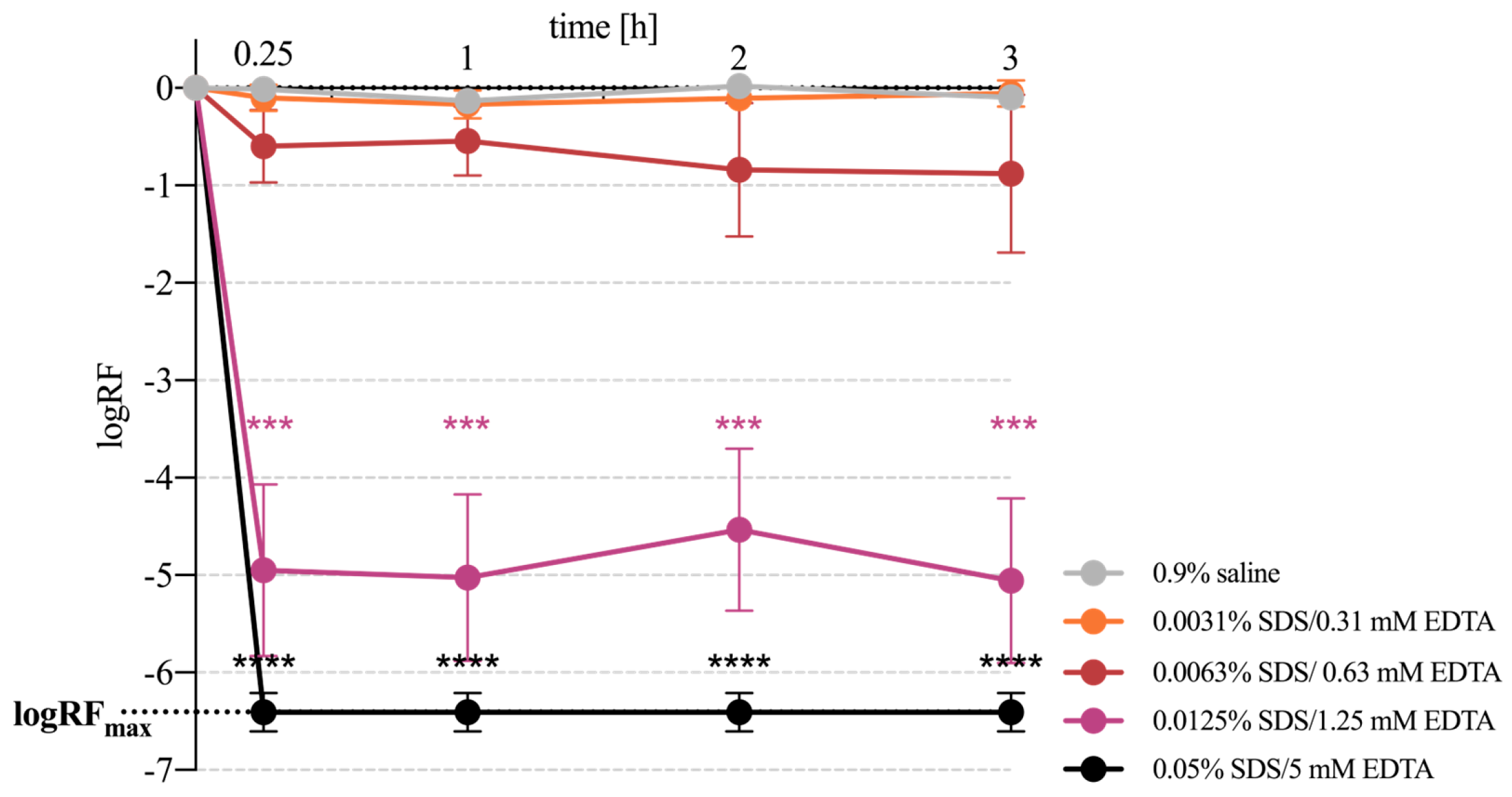
3.2. Time–Kill Kinetics of Bacterial Killing by SDS and EDTA
3.3. Combined Effect of HHP and Additive Medium in Planktonic Staphylococci
3.4. Combined Effect of HHP and Additive Medium in Biofilm-Associated Staphylococci
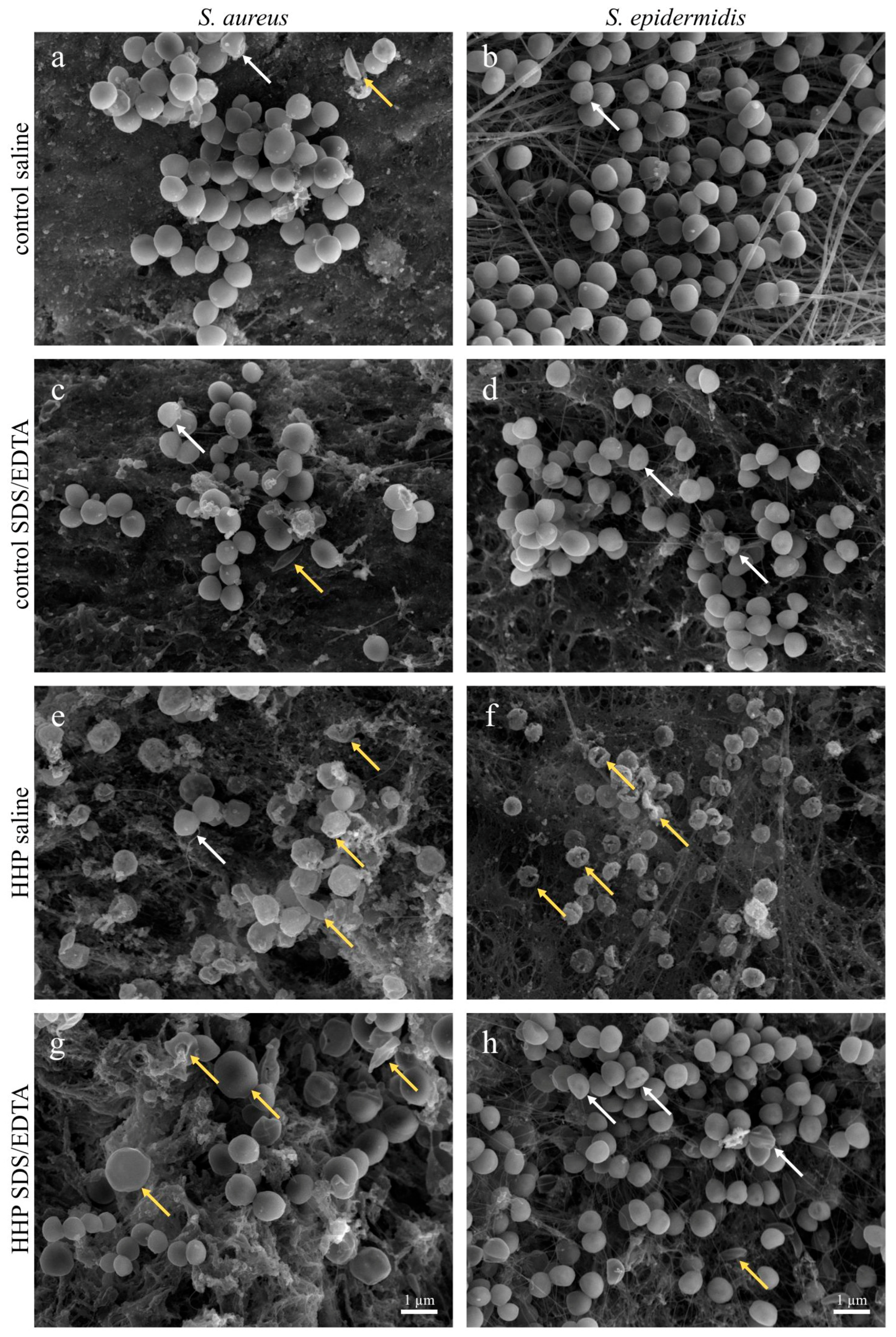
3.5. Morphological Evaluation of Bone-Associated Bacterial Biofilms After HHP Treatment Combined with Additives
4. Discussion
5. Conclusions
Author Contributions
Funding
Institutional Review Board Statement
Informed Consent Statement
Data Availability Statement
Acknowledgments
Conflicts of Interest
Abbreviations
| ATCC | American Type Culture Collection |
| CFU | colony forming units |
| CRC | checkerboard reference concentration |
| EPS | extracellular polymeric substances |
| EDTA | ethylenediaminetetraacetic acid |
| FIC | fractional inhibitory concentration |
| HHP | high hydrostatic pressure |
| logRF | logarithmic reduction factor |
| logRFmax | maximum achievable log reduction |
| MIC | minimum inhibitory concentration |
| MICcomb | minimum inhibitory concentration of the additive combination |
| MPa | Megapascal |
| PBS | phosphate-buffered saline |
| SD | standard deviation |
| SDS | sodium dodecyl sulfate |
| SEM | scanning electron microscopy |
| TSB | tryptic soy broth |
Appendix A

| Pressure-Transmitting Medium | Time [h] | Viable Count ± SD [CFU/mL] × 103 | Min to Max Range [CFU/mL] × 103 | Number of Culture-Positive Results per Total Number of Experiments |
|---|---|---|---|---|
| 0.9% saline control | 0 | 154,940 ± 63,313 | 87,100–224,000 | 5/5 |
| 0.9% saline | 0.25 | 149,080 ± 59,951 | 84,000–210,000 | 5/5 |
| 1 | 111,880 ± 37,286 | 56,000–152,000 | 5/5 | |
| 2 | 167,960 ± 80,493 | 79,000–252,000 | 5/5 | |
| 3 | 127,640 ± 54,510 | 44,800–172,000 | 5/5 | |
| 0.0031% SDS/0.31 mM EDTA | 0.25 | 118,560 ± 33,848 | 74,800–167,000 | 5/5 |
| 1 | 101,200 ± 28,560 | 56,000–134,000 | 5/5 | |
| 2 | 120,120 ± 43,663 | 64,400–154,000 | 5/5 | |
| 3 | 140,980 ± 81,186 | 86,800–274,000 | 5/5 | |
| 0.0063% SDS/0.63 mM EDTA | 0.25 | 44,360 ± 32,674 | 17,100–98,500 | 5/5 |
| 1 | 53,560 ± 46,991 | 14,600–135,000 | 5/5 | |
| 2 | 43,376 ± 56,105 | 3040–141,000 | 5/5 | |
| 3 | 39,030 ± 39,687 | 9520–106,000 | 5/5 | |
| 0.0125% SDS/1.25 mM EDTA | 0.25 | 3.07 ± 1.92 | 0–5.10 | 4/5 |
| 1 | 2.52 ± 1.70 | 0–4.41 | 4/5 | |
| 2 | 14.8 ± 22.7 | 0.31–54.6 | 5/5 | |
| 3 | 2.72 ± 2.53 | 0–6.36 | 4/5 | |
| 0.05% SDS/5 mM EDTA | 0.25 | 0.00 ± 0.00 | – | 0/5 |
| 1 | 0.00 ± 0.00 | – | 0/5 | |
| 2 | 0.00 ± 0.00 | – | 0/5 | |
| 3 | 0.00 ± 0.00 | – | 0/5 |
| Sample Organism (ATCC) | Pressure-Transmitting Medium | Pressure Level [MPa] | Viable Count ± SD [CFU/mL] × 103 | Min to Max Range [CFU/mL] × 103 | Number of Culture-Positive Results per Total Number of Experiments |
|---|---|---|---|---|---|
| Planktonic S. aureus (35556) | 0.9% saline | 0 (control) | 334,143 ± 139,933 | 174,000–552,000 | 7/7 |
| 250 | 351,571 ± 163,531 | 137,000–568,000 | 7/7 | ||
| 350 | 156,400 ± 109,667 | 72,200–372,000 | 7/7 | ||
| 0.0031% SDS/ 0.31 mM EDTA | 0 (control) | 230,000 ± 107,793 | 134,000–440,000 | 7/7 | |
| 250 | 79,577 ± 11,028 | 560–282,000 | 7/7 | ||
| 350 | 41,475 ± 59,474 | 84–158,000 | 7/7 | ||
| 0.0063% SDS/ 0.63 mM EDTA | 0 (control) | 10,008 ± 8994 | 28–23,500 | 6/6 | |
| 250 | 13.2 ± 13.0 | 0–27.3 | 6/6 | ||
| 350 | 34.7 ± 58.3 | 0–140 | 6/6 | ||
| 0.0125% SDS/ 1.25 mM EDTA | 0 (control) | 0.48 ± 1.27 | 0–3.36 | 1/7 | |
| 250 | 0.03 ± 0.08 | 0–0.22 | 1/7 | ||
| 350 | 2.01 ± 5.33 | 0–14.1 | 1/7 | ||
| 0.05% SDS/ 5 mM EDTA | 0 (control) | 0.00 ± 0.00 | – | 0/7 | |
| 250 | 0.00 ± 0.00 | – | 0/7 | ||
| 350 | 0.00 ± 0.00 | – | 0/7 | ||
| Planktonic S. epidermidis (35984) | 0.9% saline | 0 (control) | 32,990 ± 29,199 | 7560–70,000 | 4/4 |
| 250 | 1.34 ± 2.23 | 0–3.92 | 2/3 | ||
| 350 | 0.00 ± 0.00 | – | 0/3 | ||
| 0.0031% SDS/ 0.31 mM EDTA | 0 (control) | 30,310 ± 41,841 | 4480–92,400 | 4/4 | |
| 250 | 14.0 ± 28.0 | 0–56 | 1/4 | ||
| 350 | 0.00 ± 0.00 | – | 0/3 | ||
| 0.0063% SDS/ 0.63 mM EDTA | 0 (control) | 2655 ± 3693 | 273–6910 | 3/3 | |
| 250 | 0.53 ± 0.92 | 0–1.60 | 1/3 | ||
| 350 | 0.00 ± 0.00 | – | 0/3 | ||
| 0.0125% SDS/ 1.25 mM EDTA | 0 (control) | 38.6 ± 62.4 | 1.34–131 | 3/4 | |
| 250 | 0.00 ± 0.00 | – | 0/3 | ||
| 350 | 0.00 ± 0.00 | – | 0/3 | ||
| 0.05% SDS/ 5 mM EDTA | 0 (control) | 0.00 ± 0.00 | – | 0/3 | |
| 250 | 0.00 ± 0.00 | – | 0/3 | ||
| 350 | 0.00 ± 0.00 | – | 0/3 | ||
| Planktonic S. epidermidis (12228) | 0.9% saline | 0 (control) | 291,667 ± 197,130 | 168,000–519,000 | 3/3 |
| 250 | 115 ± 147 | 17.9–284 | 3/3 | ||
| 350 | 0.00 ± 0.00 | – | 0/3 | ||
| 0.0031% SDS/ 0.31 mM EDTA | 0 (control) | 142,833 ± 64,634 | 93,500–216,000 | 3/3 | |
| 250 | 0.63 ± 0.54 | 0–0.98 | 2/3 | ||
| 350 | 0.00 ± 0.00 | – | 0/3 | ||
| 0.0063% SDS/ 0.63 mM EDTA | 0 (control) | 24,253 ± 39,098 | 1620–69,400 | 3/3 | |
| 250 | 0.00 ± 0.00 | – | 0/3 | ||
| 350 | 0.00 ± 0.00 | – | 0/3 | ||
| 0.0125% SDS/ 1.25 mM EDTA | 0 (control) | 20.6 ± 35.3 | 0.17–61.3 | 3/3 | |
| 250 | 0.00 ± 0.00 | – | 0/3 | ||
| 350 | 0.00 ± 0.00 | – | 0/3 | ||
| 0.05% SDS/ 5 mM EDTA | 0 (control) | 0.00 ± 0.00 | – | 0/3 | |
| 250 | 0.00 ± 0.00 | – | 0/3 | ||
| 350 | 0.00 ± 0.00 | – | 0/3 |
| Sample Organism (ATCC) | Pressure-Transmitting Medium | Pressure Level [MPa] | Viable Count ± SD [CFU/mL] × 103 | Min to Max Range [CFU/mL] × 103 | Number of Culture-Positive Results per Total Number of Experiments |
|---|---|---|---|---|---|
| Biofilm-associated S. aureus (35556) | 0.9% saline | 0 (control) | 615,500 ± 416,999 | 241,000–1,210,000 | 4/4 |
| 250 | 557,250 ± 297,474 | 199,000–899,000 | 4/4 | ||
| 350 | 124,100 ± 53,494 | 68,000–185,000 | 4/4 | ||
| 0.0125% SDS/ 1.25 mM EDTA | 0 (control) | 150,500 ± 25,173 | 126,000–185,000 | 4/4 | |
| 250 | 81,500 ± 61,997 | 23,500–165,000 | 4/4 | ||
| 350 | 7277 ± 4413 | 2270–10,600 | 3/3 | ||
| 0.05% SDS/ 5 mM EDTA | 0 (control) | 104 ± 121 | 0–280 | 2/5 | |
| 250 | 7.95 ± 15.9 | 0–31.8 | 1/4 | ||
| 350 | 0.00 ± 0.00 | – | 0/4 | ||
| Biofilm-associated S. epidermidis (35984) | 0.9% saline | 0 (control) | 193,967 ± 114,063 | 80,900–309,000 | 3/3 |
| 250 | 25,773 ± 34,137 | 2800–65,000 | 3/3 | ||
| 350 | 16,228 ± 22,871 | 56–32,400 | 2/2 | ||
| 0.0125% SDS/ 1.25 mM EDTA | 0 (control) | 35,860 ± 35,717 | 7280–75,900 | 3/3 | |
| 250 | 3074 ± 2898 | 2230–6920 | 3/3 | ||
| 350 | 2.05 ± 1.48 | 0.84–3.7 | 3/3 | ||
| 0.05% SDS/ 5 mM EDTA | 0 (control) | 15.4 ± 26.6 | 0–46.1 | 1/3 | |
| 250 | 0.00 ± 0.00 | – | 0/3 | ||
| 350 | 0.00 ± 0.00 | – | 0/3 |
References
- Urish, K.L.; Cassat, J.E. Staphylococcus aureus osteomyelitis: Bone, bugs, and surgery. Infect. Immun. 2020, 88, e00932-19. [Google Scholar] [CrossRef] [PubMed]
- Lew, D.P.; Waldvogel, F.A. Osteomyelitis. Lancet 2004, 364, 369–379. [Google Scholar] [CrossRef] [PubMed]
- Guarch-Pérez, C.; Riool, M.; Zaat, S.A. Current osteomyelitis mouse models, a systematic review. Eur. Cells Mater. 2021, 42, 334–374. [Google Scholar] [CrossRef] [PubMed]
- Yalikun, A.; Yushan, M.; Li, W.; Abulaiti, A.; Yusufu, A. Risk factors associated with infection recurrence of posttraumatic osteomyelitis treated with Ilizarov bone transport technique—A retrospective study of 149 cases. BMC Musculoskelet. Disord. 2021, 22, 573. [Google Scholar] [CrossRef] [PubMed]
- Walter, N.; Baertl, S.; Alt, V.; Rupp, M. What is the burden of osteomyelitis in Germany? An analysis of inpatient data from 2008 through 2018. BMC Infect. Dis. 2021, 21, 550. [Google Scholar] [CrossRef] [PubMed]
- Kremers, H.M.; Nwojo, M.E.; Ransom, J.E.; Wood-Wentz, C.M.; Melton, L.J.; Huddleston, P.M. Trends in the epidemiology of osteomyelitis: A population-based study, 1969 to 2009. J. Bone Jt. Surg. 2015, 97, 837–845. [Google Scholar] [CrossRef] [PubMed]
- Son, W.S. Salvage articular reconstruction of the distal radius using a 3D simulation-guided distal clavicle osteochondral autograft after fracture-related infection: A case report. Trauma Case Rep. 2026, 61, 101314. [Google Scholar] [CrossRef] [PubMed]
- Subramanyam, K.N.; Mundargi, A.V.; Prabhu, M.V.; Gopakumar, K.; Gowda, D.A.; Reddy, D.R. Surgical management of chronic osteomyelitis: Organisms, recurrence and treatment outcome. Chin. J. Traumatol. 2023, 26, 228–235. [Google Scholar] [CrossRef] [PubMed]
- Lichte, P. Osteitis/Osteomyelitis. In Orthopädie und Unfallchirurgie; Engelhardt, M., Raschke, M.J., Eds.; Springer: Berlin/Heidelberg, Germany, 2019; pp. 1–9. [Google Scholar]
- Kavanagh, N.; Ryan, E.J.; Widaa, A.; Sexton, G.; Fennell, J.; O’Rourke, S.; Cahill, K.C.; Kearney, C.J.; O’Brien, F.J.; Kerrigan, S.W. Staphylococcal osteomyelitis: Disease progression, treatment challenges, and future directions. Clin. Microbiol. Rev. 2018, 31, e00084-17. [Google Scholar] [CrossRef] [PubMed]
- Hatzenbuehler, J.; Pulling, T. Diagnosis and management of osteomyelitis. Am. Fam. Physician 2011, 84, 1027–1033. [Google Scholar] [PubMed]
- Senneville, E.; Melliez, H.; Beltrand, E.; Legout, L.; Valette, M.; Cazaubie, M.; Cordonnier, M.; Caillaux, M.; Yazdanpanah, Y.; Mouton, Y. Culture of percutaneous bone biopsy specimens for diagnosis of diabetic foot osteomyelitis: Concordance with ulcer swab cultures. Clin. Infect. Dis. 2006, 42, 57–62. [Google Scholar] [CrossRef] [PubMed]
- Vemu, L.; Sudhaharan, S.; Mamidi, N.; Chavali, P. Need for appropriate specimen for microbiology diagnosis of chronic osteomyelitis. J. Lab. Physicians 2018, 10, 21–25. [Google Scholar] [CrossRef] [PubMed]
- Liò, P.; Paoletti, N.; Moni, M.A.; Atwell, K.; Merelli, E.; Viceconti, M. Modelling osteomyelitis. BMC Bioinform. 2012, 13, S12. [Google Scholar] [CrossRef] [PubMed]
- Schilcher, K.; Horswill, A.R. Staphylococcal biofilm development: Structure, regulation, and treatment strategies. Microbiol. Mol. Biol. Rev. 2020, 84, e00026-19. [Google Scholar] [CrossRef] [PubMed]
- Beenken, K.E.; Campbell, M.J.; Reyes-Pardo, H.; O’BRien, C.A.; Smeltzer, M.S. Mechanistic insights into the pathogenesis and therapeutic recalcitrance of Staphylococcus aureus osteomyelitis. Clin. Microbiol. Rev. 2026, 39, e0025925. [Google Scholar] [CrossRef] [PubMed]
- Nauth, A.; McKee, M.D.; Einhorn, T.A.; Watson, J.T.; Li, R.; Schemitsch, E.H. Managing bone defects. J. Orthop. Trauma 2011, 25, 462–466. [Google Scholar] [CrossRef] [PubMed]
- Sohn, H.-S.; Oh, J.-K. Review of bone graft and bone substitutes with an emphasis on fracture surgeries. Biomater. Res. 2019, 23, 9. [Google Scholar] [CrossRef] [PubMed]
- Zhao, R.; Yang, R.; Cooper, P.R.; Khurshid, Z.; Shavandi, A.; Ratnayake, J. Bone grafts and substitutes in dentistry: A review of current trends and developments. Molecules 2021, 26, 3007. [Google Scholar] [CrossRef] [PubMed]
- Mansor, A.; Ariffin, A.F.; Yusof, N.; Mohd, S.; Ramalingam, S.; Saad, A.P.M.; Baharin, R.; Min, N.W. Effects of processing and gamma radiation on mechanical properties and organic composition of frozen, freeze-dried and demineralised human cortical bone allograft. Cell Tissue Bank. 2023, 24, 25–35. [Google Scholar] [CrossRef] [PubMed]
- Harrell, C.R.; Djonov, V.; Fellabaum, C.; Volarevic, V. Risks of using sterilization by gamma radiation: The other side of the coin. Int. J. Med. Sci. 2018, 15, 274–279. [Google Scholar] [CrossRef] [PubMed]
- Rauh, J.; Despang, F.; Baas, J.; Liebers, C.; Pruss, A.; Gelinsky, M.; Günther, K.-P.; Stiehler, M. Comparative biomechanical and microstructural analysis of native versus peracetic acid-ethanol treated cancellous bone graft. BioMed Res. Int. 2014, 2014, 784702. [Google Scholar] [CrossRef] [PubMed]
- Vuotto, C.; Donelli, G. Novel Treatment Strategies for Biofilm-Based Infections. Drugs 2019, 79, 1635–1655. [Google Scholar] [CrossRef] [PubMed]
- Cavaliere, R.; Ball, J.L.; Turnbull, L.; Whitchurch, C.B. The biofilm matrix destabilizers, EDTA and DNaseI, enhance the susceptibility of nontypeable Hemophilus influenzae biofilms to treatment with ampicillin and ciprofloxacin. Microbiologyopen 2014, 3, 557–567. [Google Scholar] [CrossRef] [PubMed]
- Finnegan, S.; Percival, S.L. EDTA: An Antimicrobial and Antibiofilm Agent for Use in Wound Care. Adv. Wound Care 2015, 4, 415–421. [Google Scholar] [CrossRef] [PubMed]
- Zhu, J.; Huang, J.J.; Tham, C.; Zhou, W.; Li, D. Synergy of sodium dodecyl sulfate and citric acid in removing bacterial biofilms in hydroponic farming facilities. Lett. Appl. Microbiol. 2024, 77, ovae133. [Google Scholar] [CrossRef] [PubMed]
- Yan, X.; Xu, Y.; Shen, C.; Chen, D. Inactivation of Staphylococcus aureus by levulinic acid plus sodium dodecyl sulfate and their antibacterial mechanisms on S. aureus biofilms by transcriptomic analysis. J. Food Prot. 2023, 86, 100050. [Google Scholar] [CrossRef] [PubMed]
- Banin, E.; Brady, K.M.; Greenberg, E.P. Chelator-induced dispersal and killing of Pseudomonas aeruginosa cells in a biofilm. Appl. Environ. Microbiol. 2006, 72, 2064–2069. [Google Scholar] [CrossRef] [PubMed]
- Chen, X.; Stewart, P.S. Biofilm removal caused by chemical treatments. Water Res. 2000, 34, 4229–4233. [Google Scholar] [CrossRef]
- Rivalain, N.; Roquain, J.; Demazeau, G. Development of high hydrostatic pressure in biosciences: Pressure effect on biological structures and potential applications in biotechnologies. Biotechnol. Adv. 2010, 28, 659–672. [Google Scholar] [CrossRef] [PubMed]
- Huang, H.-W.; Lung, H.-M.; Yang, B.B.; Wang, C.-Y. Responses of microorganisms to high hydrostatic pressure processing. Food Control 2014, 40, 250–259. [Google Scholar] [CrossRef]
- Loeffler, H.; Sass, J.-O.; Muelders, L.; Bauer, J.; Friedrich, O.; Bader, R.; Klinder, A.; Waletzko-Hellwig, J. Comprehensive characterization of cell and tissue responses toward high hydrostatic pressure treatment: Molecular feedback and structural integrity in bone graft processing. J. Tissue Eng. 2025, 16, 20417314251337193. [Google Scholar] [CrossRef] [PubMed]
- Waletzko-Hellwig, J.; Dau, M.; Krebs, V.; Bader, R. Osteogenic differentiation of mesenchymal stem cells cultured on allogenic trabecular bone grafts treated with high hydrostatic pressure. J. Biomed. Mater. Res. Part B Appl. Biomater. 2023, 111, 1741–1750. [Google Scholar] [CrossRef] [PubMed]
- Diehl, P.; Schmitt, M.; Schauwecker, J.; Eichelberg, K.; Gollwitzer, H.; Gradinger, R.; Goebel, M.; Preissner, K.T.; Mittelmeier, W.; Magdolen, U. Effect of high hydrostatic pressure on biological properties of extracellular bone matrix proteins. Int. J. Mol. Med. 2005, 16, 285–289. [Google Scholar] [CrossRef]
- Loeffler, H.; Waletzko-Hellwig, J.; Fischer, R.-J.; Basen, M.; Frank, M.; Jonitz-Heincke, A.; Bader, R.; Klinder, A. Systematic enhancement of microbial decontamination efficiency in bone graft processing by means of high hydrostatic pressure using Escherichia coli as a model organism. J. Biomed. Mater. Res. Part B Appl. Biomater. 2024, 112, e35383, Correction in J. Biomed. Mater. Res. Part B Appl. Biomater. 2024, 112, e35505. https://doi.org/10.1002/jbm.b.35505. [Google Scholar] [CrossRef] [PubMed]
- Wuytack, E.Y.; Diels, A.M.; Michiels, C.W. Bacterial inactivation by high-pressure homogenisation and high hydrostatic pressure. Int. J. Food Microbiol. 2002, 77, 205–212. [Google Scholar] [CrossRef] [PubMed]
- Gill, S.R.; Fouts, D.E.; Archer, G.L.; Mongodin, E.F.; DeBoy, R.T.; Ravel, J.; Paulsen, I.T.; Kolonay, J.F.; Brinkac, L.; Beanan, M.; et al. Insights on evolution of virulence and resistance from the complete genome analysis of an early methicillin-resistant Staphylococcus aureus strain and a biofilm-producing methicillin-resistant Staphylococcus epidermidis strain. J. Bacteriol. 2005, 187, 2426–2438. [Google Scholar] [CrossRef] [PubMed]
- Zhang, Y.-Q.; Ren, S.-X.; Li, H.-L.; Wang, Y.-X.; Fu, G.; Yang, J.; Qin, Z.-Q.; Miao, Y.-G.; Wang, W.-Y.; Chen, R.-S.; et al. Genome-based analysis of virulence genes in a non-biofilm-forming Staphylococcus epidermidis strain (ATCC 12228). Mol. Microbiol. 2003, 49, 1577–1593. [Google Scholar] [CrossRef] [PubMed]
- André, M.C.; Gille, C.; Glemser, P.; Woiterski, J.; Hsu, H.-Y.; Spring, B.; Keppeler, H.; Kramer, B.W.; Handgretinger, R.; Poets, C.F.; et al. Bacterial reprogramming of PBMCs impairs monocyte phagocytosis and modulates adaptive T cell responses. J. Leukoc. Biol. 2012, 91, 977–989. [Google Scholar] [CrossRef] [PubMed]
- Penduka, D.; Mosa, R.; Simelane, M.; Basson, A.; Okoh, A.; Opoku, A. Evaluation of the anti-Listeria potentials of some plant-derived triterpenes. Ann. Clin. Microbiol. Antimicrob. 2014, 13, 37. [Google Scholar] [CrossRef] [PubMed]
- Claro, T.; Widaa, A.; O’Seaghdha, M.; Miajlovic, H.; Foster, T.J.; O’Brien, F.J.; Kerrigan, S.W. Staphylococcus aureus protein A binds to osteoblasts and triggers signals that weaken bone in osteomyelitis. PLoS ONE 2011, 6, e18748. [Google Scholar] [CrossRef] [PubMed]
- de Souza, A.R.; Da Costa Demonte, A.L.; de Araujo Costa, K.; Faria, M.A.C.; Durães-Carvalho, R.; Lancellotti, M.; Bonafe, C.F.S. Potentiation of high hydrostatic pressure inactivation of Mycobacterium by combination with physical and chemical conditions. Appl. Microbiol. Biotechnol. 2013, 97, 7417–7425. [Google Scholar] [CrossRef] [PubMed]
- Yamin, M.; de Souza, A.R.; Castelucci, B.G.; Mattoso, J.G.; Bonafe, C.F.S. Synergism between high hydrostatic pressure and glutaraldehyde for the inactivation of Staphylococcus aureus at moderate temperature. Appl. Microbiol. Biotechnol. 2018, 102, 8341–8350. [Google Scholar] [CrossRef] [PubMed]
- Sheen, S.; Huang, C.-Y.; Chuang, S. Synergistic effect of high hydrostatic pressure, allyl isothiocyanate, and acetic acid on the inactivation and survival of pathogenic Escherichia coli in ground chicken. J. Food Sci. 2022, 87, 5042–5053. [Google Scholar] [CrossRef] [PubMed]
- Díaz De Rienzo, M.A.; Stevenson, P.S.; Marchant, R.; Banat, I. Pseudomonas aeruginosa biofilm disruption using microbial surfactants. J. Appl. Microbiol. 2016, 120, 868–876. [Google Scholar] [CrossRef] [PubMed]
- Liu, Z.; Lin, Y.; Lu, Q.; Li, F.; Yu, J.; Wang, Z.; He, Y.; Song, C. In vitro and in vivo activity of EDTA and antibacterial agents against the biofilm of mucoid Pseudomonas aeruginosa. Infection 2017, 45, 23–31. [Google Scholar] [CrossRef] [PubMed]
- de Almeida, J.; Hoogenkamp, M.; Felippe, W.T.; Crielaard, W.; van der Waal, S.V. Effectiveness of EDTA and modified salt solution to detach and kill cells from Enterococcus faecalis biofilm. J. Endod. 2016, 42, 320–323. [Google Scholar] [CrossRef] [PubMed]
- Emami, A.; Talaei-Khozani, T.; Vojdani, Z.; Fard, N.Z. Comparative assessment of the efficiency of various decellularization agents for bone tissue engineering. J. Biomed. Mater. Res. Part B Appl. Biomater. 2021, 109, 19–32. [Google Scholar] [CrossRef] [PubMed]
- Hamoud, R.; Reichling, J.; Wink, M. Synergistic antibacterial activity of the combination of the alkaloid sanguinarine with EDTA and the antibiotic streptomycin against multidrug resistant bacteria. J. Pharm. Pharmacol. 2015, 67, 264–273. [Google Scholar] [CrossRef] [PubMed]
- Liu, F.; Hansra, S.; Crockford, G.; Köster, W.; Allan, B.J.; Blondeau, J.M.; Lainesse, C.; White, A.P. Tetrasodium EDTA is effective at eradicating biofilms formed by clinically relevant microorganisms from patients’ central venous catheters. mSphere 2018, 3, e00525-18. [Google Scholar] [CrossRef] [PubMed]
- Jang, H.; Choi, S.Y.; Mitchell, R.J. Staphylococcus aureus sensitivity to membrane disrupting antibacterials is increased under microgravity. Cells 2023, 12, 1907. [Google Scholar] [CrossRef] [PubMed]
- Rauch, C.; Cherkaoui, M.; Egan, S.; Leigh, J. The bio-physics of condensation of divalent cations into the bacterial wall has implications for growth of Gram-positive bacteria. Biochim. Biophys. Acta Biomembr. 2017, 1859, 282–288. [Google Scholar] [CrossRef] [PubMed]
- Hauben, K.J.; Wuytack, E.Y.; Soontjens, C.C.; Michiels, C.W. High-pressure transient sensitization of Escherichia coli to lysozyme and nisin by disruption of outer-membrane permeability. J. Food Prot. 1996, 59, 350–355. [Google Scholar] [CrossRef] [PubMed]
- Rivalain, N.; Roquain, J.; Boiron, J.-M.; Maurel, J.-P.; Largeteau, A.; Ivanovic, Z.; Demazeau, G. High hydrostatic pressure treatment for the inactivation of Staphylococcus aureus in human blood plasma. New Biotechnol. 2012, 29, 409–414. [Google Scholar] [CrossRef] [PubMed]
- Wang, L.; Pan, J.; Xie, H.; Yang, Y.; Lin, C. Inactivation of Staphylococcus aureus and Escherichia coli by the synergistic action of high hydrostatic pressure and dissolved CO2. Int. J. Food Microbiol. 2010, 144, 118–125. [Google Scholar] [CrossRef] [PubMed]
- Pagán, R.; Mackey, B. Relationship between membrane damage and cell death in pressure-treated Escherichia coli cells: Differences between exponential- and stationary-phase cells and variation among strains. Appl. Environ. Microbiol. 2000, 66, 2829–2834. [Google Scholar] [CrossRef] [PubMed]
- Cheung, G.Y.; Bae, J.S.; Otto, M. Pathogenicity and virulence of Staphylococcus aureus. Virulence 2021, 12, 547–569. [Google Scholar] [CrossRef] [PubMed]
- Wang, M.; Buist, G.; van Dijl, J.M. Staphylococcus aureus cell wall maintenance—The multifaceted roles of peptidoglycan hydrolases in bacterial growth, fitness, and virulence. FEMS Microbiol. Rev. 2022, 46, fuac025. [Google Scholar] [CrossRef] [PubMed]
- Gayán, E.; Govers, S.K.; Aertsen, A. Impact of high hydrostatic pressure on bacterial proteostasis. Biophys. Chem. 2017, 231, 3–9. [Google Scholar] [CrossRef] [PubMed]
- Abe, F. Chapter 18: Effects of high hydrostatic pressure on microbial cell membranes: Structural and functional perspectives. In Subcellular Biochemistry; Akasaka, K., Matsuki, H., Eds.; Springer Science & Business Media: Dordrecht, The Netherlands, 2015; pp. 371–381. [Google Scholar]
- Moriarty, T.F.; Grainger, D.W.; Richards, R.G. Challenges in linking preclinical anti-microbial research strategies with clinical outcomes for device-associated infections. Eur. Cells Mater. 2014, 28, 112–128. [Google Scholar] [CrossRef] [PubMed]
- DIN Deutsches Institut für Normung e.V. Chemische Desinfektionsmittel und Antiseptika—Quantitativer Suspensionsversuch der Bakteriziden Wirkung (Basistest) Chemischer Desinfektionsmittel und Antiseptika; DIN EN 1040:2005-03; Beuth Verlag GmbH: Berlin, Germany, 2006. [Google Scholar]
- European Committee on Organ Transplantation. Chapter 21: Musculoskeletal tissue. In Guide to the Quality and Safety of Cells and Tissues for Human Application, 4th ed.; Keitel, S., Ed.; European Directorate for the Quality of Medicines & HealthCare: Strasbourg, France, 2019; pp. 257–265. [Google Scholar]
- Wang, W.; Yang, P.; Rao, L.; Zhao, L.; Wu, X.; Wang, Y.; Liao, X. Effect of high hydrostatic pressure processing on the structure, functionality, and nutritional properties of food proteins: A review. Compr. Rev. Food Sci. Food Saf. 2022, 21, 4640–4682. [Google Scholar] [CrossRef] [PubMed]
- Prieto-Calvo, M.; Prieto, M.; López, M.; Alvarez-Ordóñez, A. Effects of high hydrostatic pressure on Escherichia coli ultrastructure, membrane integrity and molecular composition as assessed by FTIR spectroscopy and microscopic imaging techniques. Molecules 2014, 19, 21310–21323. [Google Scholar] [CrossRef] [PubMed]
- Liao, Q.; Tao, H.; Li, Y.; Xu, Y.; Wang, H.-L. Evaluation of structural changes and molecular mechanism induced by high hydrostatic pressure in Enterobacter sakazakii. Front. Nutr. 2021, 8, 739863. [Google Scholar] [CrossRef] [PubMed]
- Kaya, E.; Grassi, L.; Benedetti, A.; Maisetta, G.; Pileggi, C.; Di Luca, M.; Batoni, G.; Esin, S. In vitro interaction of Pseudomonas aeruginosa biofilms with human peripheral blood mononuclear cells. Front. Cell. Infect. Microbiol. 2020, 10, 187. [Google Scholar] [CrossRef] [PubMed]
- Aaron, S.D.; Ferris, W.; Ramotar, K.; Vandemheen, K.; Chan, F.; Saginur, R. Single and combination antibiotic susceptibilities of planktonic, adherent, and biofilm-grown Pseudomonas aeruginosa isolates cultured from sputa of adults with cystic fibrosis. J. Clin. Microbiol. 2002, 40, 4172–4179. [Google Scholar] [CrossRef] [PubMed]
- Cerca, N.; Martins, S.; Cerca, F.; Jefferson, K.K.; Pier, G.B.; Oliveira, R.; Azeredo, J. Comparative assessment of antibiotic susceptibility of coagulasenegative staphylococci in biofilm versus planktonic culture as assessed by bacterial enumeration or rapid XTT colorimetry. J. Antimicrob. Chemother. 2005, 56, 331–336. [Google Scholar] [CrossRef] [PubMed]
- Saginur, R.; StDenis, M.; Ferris, W.; Aaron, S.D.; Chan, F.; Lee, C.; Ramotar, K. Multiple combination bactericidal testing of staphylococcal biofilms from implant-associated infections. Antimicrob. Agents Chemother. 2006, 50, 55–61. [Google Scholar] [CrossRef] [PubMed]
- Jenul, C.; Horswill, A.R. Regulation of Staphylococcus aureus virulence. Microbiol. Spectr. 2019, 7, 10–1128. [Google Scholar] [CrossRef] [PubMed]
- Chua, S.L.; Liu, Y.; Yam, J.K.; Chen, Y.; Vejborg, R.M.; Tan, B.G.C.; Kjelleberg, S.; Tolker-Nielsen, T.; Givskov, M.; Yang, L. Dispersed cells represent a distinct stage in the transition from bacterial biofilm to planktonic lifestyles. Nat. Commun. 2014, 5, 4462. [Google Scholar] [CrossRef] [PubMed]
- Gollwitzer, H.; Mittelmeier, W.; Brendle, M.; Weber, P.; Miethke, T.; O Hofmann, G.; Gerdesmeyer, L.; Schauwecker, J.; Diehl, P. High hydrostatic pressure for disinfection of bone grafts and biomaterials: An experimental study. Open Orthop. J. 2009, 3, 1–7. [Google Scholar] [CrossRef] [PubMed]
- Enz, A.; Müller, S.; Mittelmeier, W.; Klinder, A. Severe polymicrobial and fungal periprosthetic osteomyelitis persisting after hip disarticulations treated with caspofungin in risk patients: A case series. Ann. Clin. Microbiol. Antimicrob. 2021, 20, 86. [Google Scholar] [CrossRef] [PubMed]
- Hutchinson, K.; Banks, J.; Arnold, A.; Lohn, J.; Trompeter, A. Orthoplastic management of delayed sternal osteomyelitis and non-union: A retrospective case series applying fracture-related infection principles. Int. Orthop. 2026. [Google Scholar] [CrossRef] [PubMed]
- Yang, L.; Feng, J.; Liu, J.; Yu, L.; Zhao, C.; Liang, R.; He, W.; Peng, J. Pathogen identification in 84 patients with post-traumatic osteomyelitis after limb fractures. Ann. Palliat. Med. 2020, 9, 451–458, Erratum in Ann. Palliat. Med. 2020, 9, 1351. https://doi.org/10.21037/apm-2020-04. [Google Scholar] [CrossRef] [PubMed]
- MacCain, W.J.; Tuomanen, E.I. Mini-review: Bioactivities of bacterial cell envelopes in the central nervous system. Front. Cell. Infect. Microbiol. 2020, 10, 588378. [Google Scholar] [CrossRef] [PubMed]
- Wolf, A.J.; Underhill, D.M. Peptidoglycan recognition by the innate immune system. Nat. Rev. Immunol. 2018, 18, 243–254. [Google Scholar] [CrossRef] [PubMed]
- Schottroff, F.; Fröhling, A.; Zunabovic-Pichler, M.; Krottenthaler, A.; Schlüter, O.; Jäger, H. Sublethal injury and viable but non-culturable (VBNC) state in microorganisms during preservation of food and biological materials by non-thermal processes. Front. Microbiol. 2018, 9, 2773. [Google Scholar] [CrossRef] [PubMed]
- Li, Y.; Huang, T.-Y.; Mao, Y.; Chen, Y.; Shi, F.; Peng, R.; Chen, J.; Yuan, L.; Bai, C.; Chen, L.; et al. Study on the viable but non-culturable (VBNC) state formation of Staphylococcus aureus and its control in food system. Front. Microbiol. 2020, 11, 599739. [Google Scholar] [CrossRef] [PubMed]
- Lin-Gibson, S.; Lin, N.J.; Jackson, S.; Viswanathan, S.; Zylberberg, C.; Wolfrum, J.; Basu, S.; Roy, K.; Marshall, D.; McFarland, R.; et al. Standards efforts and landscape for rapid microbial testing methodologies in regenerative medicine. Cytotherapy 2021, 23, 390–398. [Google Scholar] [CrossRef] [PubMed]
- Charnwichai, P.; Kitkumthorn, N.; Ruangvejvorachai, P.; Wongphoom, J.; Meesakul, T.; Tammachote, N.; Tammachote, R. Rapid decalcification of articular cartilage and subchondral bone using an ultrasonic cleaner with EDTA. Acta Histochem. 2023, 125, 152009. [Google Scholar] [CrossRef] [PubMed]
- Salih, M.M. Comparison between conventional decalcification and a microwave-assisted method in bone tissue affected with mycetoma. Biochem. Res. Int. 2020, 2020, 6561980. [Google Scholar] [CrossRef] [PubMed]
- Sangeetha, R.; Uma, K.; Chandavarkar, V. Comparison of routine decalcification methods with microwave decalcification of bone and teeth. J. Oral Maxillofac. Pathol. 2013, 17, 386–391. [Google Scholar] [CrossRef] [PubMed]
- Lee, D.J.; Diachina, S.; Lee, Y.T.; Zhao, L.; Zou, R.; Tang, N.; Han, H.; Chen, X.; Ko, C.-C. Decellularized bone matrix grafts for calvaria regeneration. J. Tissue Eng. 2016, 7, 2041731416680306. [Google Scholar] [CrossRef] [PubMed]
- Lanigan, R.S.; Yamarik, T.A. Final Report on the Safety Assessment of EDTA, Calcium Disodium EDTA, Diammonium EDTA, Dipotassium EDTA, Disodium EDTA, TEA-EDTA, Tetrasodium EDTA, Tripotassium EDTA, Trisodium EDTA, HEDTA, and Trisodium HEDTA. Int. J. Toxicol. 2002, 21, 95–142. [Google Scholar] [CrossRef] [PubMed]
- Marins, J.S.; Sassone, L.M.; Fidel, S.R.; Ribeiro, D.A. In Vitro Genotoxicity and Cytotoxicity in Murine Fibroblasts Exposed to EDTA, NaOCl, MTAD and Citric Acid. Braz. Dent. J. 2012, 23, 527–533. [Google Scholar] [CrossRef] [PubMed]
- Zhang, Y.; Newton, B.; Lewis, E.; Fu, P.P.; Kafoury, R.; Ray, P.C.; Yu, H. Cytotoxicity of organic surface coating agents used for nanoparticles synthesis and stability. Toxicol. In Vitro 2015, 29, 762–768. [Google Scholar] [CrossRef] [PubMed]
- Zvarova, B.; Uhl, F.E.; Uriarte, J.J.; Borg, Z.D.; Coffey, A.L.; Bonenfant, N.R.; Weiss, D.J.; Wagner, D.E. Residual Detergent Detection Method for Nondestructive Cytocompatibility Evaluation of Decellularized Whole Lung Scaffolds, Tissue Eng. Part C Methods 2016, 22, 418–428. [Google Scholar] [CrossRef] [PubMed]
- Kasi, S.R.; Roffel, S.; Özcan, M.; Gibbs, S.; Feilzer, A.J. In vitro cytotoxicity (irritant potency) of toothpaste ingredients. PLoS ONE 2025, 20, e0318565. [Google Scholar] [CrossRef] [PubMed]
- Gratzer, P.F.; Harrison, R.D.; Woods, T. Matrix Alteration and Not Residual Sodium Dodecyl Sulfate Cytotoxicity Affects the Cellular Repopulation of a Decellularized Matrix. Tissue Eng. 2006, 12, 2975–2983. [Google Scholar] [CrossRef] [PubMed]
- Arnold, S.R.; Giordano, V.; Obremskey, W.T.; Schwarz, E.M.; Smeltzer, M.S.; Veis, D.J.; Cassat, J.E. Osteomyelitis. Nat. Rev. Dis. Prim. 2026, 12, 33. [Google Scholar] [CrossRef] [PubMed]


| Medium | SDS [%] | EDTA [mM] |
|---|---|---|
| Control (0.9% saline) | 0 | 0 |
| ½× CRC | 0.0031 | 0.31 |
| 1× CRC | 0.0063 | 0.63 |
| 2× CRC | 0.0125 | 1.25 |
| 8× CRC | 0.05 | 5 |
| Replicate | MIC SDS [%] | MIC EDTA [mM] | MICcomb SDS [%] | MICcomb EDTA [mM] | FIC Index |
|---|---|---|---|---|---|
| 1 | 0.0125 | 2.5 | 0.0063 | 0.16 | 0.57 |
| 2 | 0.0125 | 2.5 | 0.0063 | 0.63 | 0.76 |
| 3 | 0.0125 | 2.5 | 0.0063 | 0.63 | 0.76 |
| mean ± SD | 0.0125 | 2.5 | 0.0063 | 0.47 ± 0.22 | 0.70 ± 0.09 |
| Sample Organism (ATCC) | Pressure-Transmitting Medium | Pressure Level [MPa] | log10 Reduction (Mean ± SD) | Number of Culture-Positive Results per Total Number of Experiments | p vs. 0 MPa |
|---|---|---|---|---|---|
| Planktonic S. aureus (35556) | 0.9% saline | 0 (control) | 0 | 7/7 | - |
| 250 | −0.007 ± 0.140 | 7/7 | 0.9997 | ||
| 350 | 0.377 ± 0.205 | 7/7 | 0.5378 | ||
| 0.0031% SDS/0.31 mM EDTA | 0 (control) | 0.163 ± 0.091 | 7/7 | - | |
| 250 | 1.407 ± 0.982 | 7/7 | 0.0046 | ||
| 350 | 1.579 ± 1.032 | 7/7 | 0.0012 | ||
| 0.0063% SDS/0.63 mM EDTA | 0 (control) | 1.987 ± 0.985 | 6/6 | - | |
| 250 | 5.043 ± 1.338 | 6/6 | <0.0001 | ||
| 350 | 5.620 ± 1.671 | 6/6 | <0.0001 | ||
| 0.0125% SDS/1.25 mM EDTA | 0 (control) | 6.487 ± 0.690 | 1/7 | - | |
| 250 | 6.656 ± 0.191 | 1/7 | 0.8777 | ||
| 350 | 6.399 ± 0.812 | 1/7 | 0.9642 | ||
| 0.05% SDS/5 mM EDTA | 0 (control) | 6.741 ± 0.187 | 0/7 | - | |
| 250 | 6.741 ± 0.187 | 0/7 | >0.999 | ||
| 350 | 6.741 ± 0.187 | 0/7 | >0.999 | ||
| Planktonic S. epidermidis (35984) | 0.9% saline | 0 (control) | 0 | 4/4 | - |
| 250 | 4.730 ± 0.600 | 2/3 | <0.0001 | ||
| 350 | 5.443 ± 0.390 | 0/3 | <0.0001 | ||
| 0.0031% SDS/0.31 mM EDTA | 0 (control) | 0.190 ± 0.253 | 4/4 | - | |
| 250 | 4.858 ±1.214 | 1/4 | <0.0001 | ||
| 350 | 5.443 ± 0.390 | 0/3 | <0.0001 | ||
| 0.0063% SDS/0.63 mM EDTA | 0 (control) | 1.440 ± 0.290 | 3/3 | - | |
| 250 | 5.217 ± 0.625 | 1/3 | <0.0001 | ||
| 350 | 5.553 ± 0.384 | 0/3 | <0.0001 | ||
| 0.0125% SDS/1.25 mM EDTA | 0 (control) | 3.770 ± 1.033 | 3/4 | - | |
| 250 | 5.553 ± 0.384 | 0/3 | <0.0001 | ||
| 350 | 5.553 ± 0.384 | 0/3 | <0.0001 | ||
| 0.05% SDS/ 5 mM EDTA | 0 (control) | 5.553 ± 0.384 | 0/3 | - | |
| 250 | 5.553 ± 0.384 | 0/3 | >0.9999 | ||
| 350 | 5.553 ± 0.384 | 0/3 | >0.9999 | ||
| Planktonic S. epidermidis (12228) | 0.9% saline | 0 (control) | 0 | 3/3 | - |
| 250 | 3.623 ± 0.355 | 3/3 | <0.0001 | ||
| 350 | 6.660 ± 0.270 | 0/3 | <0.0001 | ||
| 0.0031% SDS/ 0.31 mM EDTA | 0 (control) | 0.277 ± 0.430 | 3/3 | - | |
| 250 | 5.840 ± 0.589 | 2/3 | <0.0001 | ||
| 350 | 6.660 ± 0.270 | 0/3 | <0.0001 | ||
| 0.0063% SDS/ 0.63 mM EDTA | 0 (control) | 1.643 ± 1.082 | 3/3 | - | |
| 250 | 6.660 ± 0.270 | 0/3 | <0.0001 | ||
| 350 | 6.660 ± 0.270 | 0/3 | <0.0001 | ||
| 0.0125% SDS/ 1.25 mM EDTA | 0 (control) | 5.287 ± 1.586 | 3/3 | - | |
| 250 | 6.660 ± 0.270 | 0/3 | 0.0126 | ||
| 350 | 6.660 ± 0.270 | 0/3 | 0.0126 | ||
| 0.05% SDS/ 5 mM EDTA | 0 (control) | 6.660 ± 0.270 | 0/3 | - | |
| 250 | 6.660 ± 0.270 | 0/3 | >0.9999 | ||
| 350 | 6.660 ± 0.270 | 0/3 | >0.9999 |
| Sample Organism (ATCC) | Pressure-Transmitting Medium | Pressure Level [MPa] | log10 Reduction (Mean ± SD) | Number of Culture-Positive Results per Total Number of Experiments | p vs. 0 MPa |
|---|---|---|---|---|---|
| Biofilm-associated S. aureus (35556) | 0.9% saline | 0 (control) | 0 | 4/4 | - |
| 250 | 0.030 ± 0.129 | 4/4 | 0.9985 | ||
| 350 | 0.655 ± 0.189 | 4/4 | 0.5010 | ||
| 0.0125% SDS/1.25 mM EDTA | 0 (control) | 0.543 ± 0.332 | 4/4 | - | |
| 250 | 0.910 ± 0.509 | 4/4 | 0.7990 | ||
| 350 | 2.050 ± 0.589 | 3/3 | 0.0667 | ||
| 0.05% SDS/5 mM EDTA | 0 (control) | 5.374 ± 1.373 | 2/5 | - | |
| 250 | 6.110 ± 1.770 | 1/4 | 0.9203 | ||
| 350 | 6.798 ± 0.469 | 0/4 | 0.3471 | ||
| Biofilm-associated S. epidermidis (35984) | 0.9% saline | 0 (control) | 0 | 3/3 | - |
| 250 | 1.150 ± 0.610 | 3/3 | 0.0761 | ||
| 350 | 2.255 ± 1.803 | 2/2 | 0.0014 | ||
| 0.0125% SDS/1.25 mM EDTA | 0 (control) | 0.850 ± 0.496 | 3/3 | - | |
| 250 | 1.897 ± 0.771 | 3/3 | 0.1106 | ||
| 350 | 5.000 ± 0.494 | 3/3 | <0.0001 | ||
| 0.05% SDS/5 mM EDTA | 0 (control) | 5.507 ± 1.464 | 1/3 | - | |
| 250 | 6.477 ± 0.294 | 0/3 | 0.1455 | ||
| 350 | 6.477 ± 0.294 | 0/3 | 0.1455 |
Disclaimer/Publisher’s Note: The statements, opinions and data contained in all publications are solely those of the individual author(s) and contributor(s) and not of MDPI and/or the editor(s). MDPI and/or the editor(s) disclaim responsibility for any injury to people or property resulting from any ideas, methods, instructions or products referred to in the content. |
© 2026 by the authors. Licensee MDPI, Basel, Switzerland. This article is an open access article distributed under the terms and conditions of the Creative Commons Attribution (CC BY) license.
Share and Cite
Loeffler, H.; Bieda, A.; Sombetzki, M.; Fischer, R.-J.; Basen, M.; Schulz, K.; Waletzko, J.; Bader, R.; Klinder, A. Combined High Hydrostatic Pressure and Additive Chemical Treatment Enhances Decontamination Efficiency in Bone Tissue Infected with Staphylococcal Biofilms. Microorganisms 2026, 14, 1502. https://doi.org/10.3390/microorganisms14071502
Loeffler H, Bieda A, Sombetzki M, Fischer R-J, Basen M, Schulz K, Waletzko J, Bader R, Klinder A. Combined High Hydrostatic Pressure and Additive Chemical Treatment Enhances Decontamination Efficiency in Bone Tissue Infected with Staphylococcal Biofilms. Microorganisms. 2026; 14(7):1502. https://doi.org/10.3390/microorganisms14071502
Chicago/Turabian StyleLoeffler, Henrike, Adam Bieda, Martina Sombetzki, Ralf-Joerg Fischer, Mirko Basen, Karoline Schulz, Janine Waletzko, Rainer Bader, and Annett Klinder. 2026. "Combined High Hydrostatic Pressure and Additive Chemical Treatment Enhances Decontamination Efficiency in Bone Tissue Infected with Staphylococcal Biofilms" Microorganisms 14, no. 7: 1502. https://doi.org/10.3390/microorganisms14071502
APA StyleLoeffler, H., Bieda, A., Sombetzki, M., Fischer, R.-J., Basen, M., Schulz, K., Waletzko, J., Bader, R., & Klinder, A. (2026). Combined High Hydrostatic Pressure and Additive Chemical Treatment Enhances Decontamination Efficiency in Bone Tissue Infected with Staphylococcal Biofilms. Microorganisms, 14(7), 1502. https://doi.org/10.3390/microorganisms14071502