Article

Diversity of RNA Viruses in Declining Mediterranean Forests

by
Sergio Diez-Hermano
1,
Alba Diez-Galán
1,2,*,
Pedro Luis Pérez-Alonso
1,
Wilson Acosta Morel
1,
Jonatan Niño-Sanchez
1,
Marcos de la Peña
3 and
Julio Javier Díez
1,2,*
1
Department of Plant Production and Forest Resources, Sustainable Forest Management Research Institute (iuFOR), Higher Technical School of Agricultural Engineering (ETSIIAA), University of Valladolid, 34071 Palencia, Spain
2
One Health Chair On Forest and Human Health, University of Valladolid, 34071 Valladolid, Spain
3
Institute for Plant Molecular and Cellular Biology (CSIC-UPV), 46022 Valencia, Spain
*
Authors to whom correspondence should be addressed.
Microorganisms 2026, 14(7), 1445; https://doi.org/10.3390/microorganisms14071445
Submission received: 12 May 2026 / Revised: 25 June 2026 / Accepted: 26 June 2026 / Published: 30 June 2026
(This article belongs to the Section Virology)

Abstract

Global change alters forestry habitats and facilitates the entry of new pathogens that do not share a co-evolution history with the forest, leading them into a spiral of decline. As a result, relationships between forest organisms become imbalanced. RNA viruses are of particular concern given their capacity to infect hosts across different kingdoms of life, yet the viromes of Mediterranean forests remain largely unexplored. For this reason, the study of RNA viruses is essential for understanding how viral flow across different hosts might occur, and to prevent possible outbreaks of diseases in the future. In this work, the RNA virus diversity found in trees, arthropods, and fungi from declining Mediterranean forests is described. To this extent, three habitats (Quercus ilex, Castanea sativa and Pinus radiata) were sampled and RNAseq was performed on tree tissues, arthropods, and fungi. In total, 146 viral sequences were detected by searching for matches with conserved motifs of the RNA-dependent RNA polymerase (RdRP) using Palmscan. Up to 15 viral families were identified, with Botourmiaviridae (28.7%) and Partitiviridae (9.6%) being the most abundant. In terms of genome type, ssRNA(+) viruses were the most represented (83.5%), followed by dsRNA (15%) and two ssRNA(−) representatives. Notably, families with documented cross-kingdom capabilities such as Hypoviridae (1), Mitoviridae (6), and Narnaviridae (5) were detected across multiple host types, with one Mitovirus sequence recovered unexpectedly from pine tree tissue. Distribution of viruses across ecosystems included Q. ilex (57.5%), P. radiata (26.7%), and C. sativa (15.8%). Interestingly, two RdRP sequences showed no similarity to any entry in current viral databases, representing potentially novel viruses warranting further characterization. These findings reveal a rich and partially uncharacterized RNA virosphere in declining Mediterranean forests and underscore the importance of multi-host virome surveys for understanding viral flow across kingdoms in threatened ecosystems.

1. Introduction

In recent decades, it has become increasingly evident that all biotic components of forest ecosystems (bacteria, protists, fungi, vertebrate, and invertebrate animals) host their own viral communities (viromes), which together form the metavirome or virosphere of the forest ecosystem. Recent advances in metagenomics and artificial intelligence have greatly expanded the understanding of this hidden diversity. A deep learning model has identified over 160,000 potential RNA virus species grouped into approximately 180 supergroups globally [1]. Large-scale environmental studies have also uncovered thousands of new RNA viruses in terrestrial and sediment environments, including Mediterranean-like soils [2]. Despite this progress, the viromes of forest ecosystems remain among the least characterized, particularly in the context of ecosystems under anthropogenic and climatic stress.
Currently, around 64 virus species are known to affect 19 forest plant species in temperate and urban forests [3], although this figure is likely to be an underestimate given the rapid expansion of virome studies. Most belong to the (+)ssRNA genome group, including families such as Betaviridae, Secoviridae, Bromoviridae, Tombusviridae, Virgaviridae, and Mayoviridae. The (−)ssRNA component of forest plant viromes has proven more diverse than initially recognized, with members of Fimoviridae (which replaced the former Emaraviridae and includes the genus Emaravirus), Rhabdoviridae, Phenuiviridae, Tospoviridae, and Aspiviridae all reported infecting woody and forest plant species [4,5]. A few dsRNA viruses have also been described in pine and ash, as well as some dsDNA viruses with reverse transcriptase activity in Betula sp., Castanea sativa, and Fraxinus americana. Among the pathogenic genera, emaraviruses and badnaviruses are considered causative agents of disease in trees. Most viruses affecting forest fungi and oomycetes have a dsRNA genome and belong to the Partitiviridae, Totiviridae, Curvulaviridae, and Reoviridae families. (+)ssRNA viruses are classified within Hypoviridae, Endornaviridae, Botourmiaviridae, Fusariviridae, and Mitoviridae. Several of these have been shown to modulate the pathogenicity of important forest pathogens, such as Cryphonectria parasitica, Ophiostoma novo-ulmi, Gremmeniella abietina, Fusarium circinatum, Heterobasidion annosum, and Hymenoscyphus fraxineus [6].
Recent studies have expanded this knowledge, revealing novel dsRNA and ambisense viruses in pathogens such as Armillaria spp. and C. parasitica [7]. A comprehensive virome survey of Armillaria isolates from different regions uncovered unexpected (+)ssRNA and circular ambisense viruses [8]. Furthermore, analysis of public sequencing data identified 12 novel fungal RNA virus sequences, revealing the existence of an unexplored viral dark matter [9].
Cross-kingdom viruses play a significant role in forest ecosystems, particularly between trees, fungi, and insects [10]. Some Toti-, Partitiviridae, Endornaviridae, and Chrysoviridae members replicate in meristematic cells in plant and can infect fungi in vitro [11]. Natural cross-kingdom infection has been experimentally demonstrated, such as in Rhizoctonia solani, which acquired and transmitted cucumber mosaic virus [12]. Functional cooperation between plant and fungal viruses has also been documented, facilitating transmission across vegetatively incompatible fungal strains [13]. Notably, a negative-sense RNA virus (Valsa mali negative-strand RNA virus 1, VmNSRV1) was recently shown to naturally infect both a plant host (apple) and its fungal pathogen (Valsa mali), providing new evidence of interkingdom infection [14].
Despite their importance as vectors, insect viromes in forest ecosystems remain poorly characterized. Once inside the insect, plant viruses can develop forms of non-persistent transmission (in external structures of the insect) or circulative transmission (using internal compartments as reservoirs). In some cases, circular transmission may involve replication of the plant virus within the cells of the insect host [15]. Recent research on three wheat aphid species identified 12 new RNA viruses and 90 endogenous viral elements (EVEs), suggesting complex viral interactions involving potential fungal associations [16]. Viruses have also been found in lepidopterans (Lymantria dispar, Thaumetopoea pityocampa, Leucoma salicis), coleopterans (Ips typographus), and hymenopterans (Neodiprion sertifer) [17,18]. Importantly, arthropods may act as bridges for viral transmission between plant and fungal hosts, yet this role has rarely been investigated in forest virome studies through simultaneous sampling of multiple host types.
Mediterranean forests are among the terrestrial ecosystems most threatened by globalization and climate change [19]. Predicted reductions in precipitation and rising temperatures will expose these ecosystems to prolonged droughts and more frequent wildfires [20]. These stresses decrease the resilience of Mediterranean tree species against pests and diseases, facilitating the invasion of novel pathogens. Recent surveys of Rosellinia necatrix, a soilborne fungus widespread in Mediterranean areas, reported a virosis incidence of ~14% in isolates from Spain, Israel, and Italy, revealing viruses from Partitiviridae, Hypoviridae, Megabirnaviridae, and Fusagraviridae families, and new Megatotiviridae members [21].
RNA viruses are known agents of emerging infectious diseases and may be released following forest disturbances [22,23], increasing the risk of unexpected pandemics. In this context, Quercus ilex, Castanea sativa, and Pinus radiata represent three of the most ecologically and economically relevant tree species in the Iberian Peninsula, all of which are currently experiencing significant decline associated with drought stress, fungal pathogens and insect pests, making them priority targets for virome characterization.
Given the ability of RNA viruses to cross barriers and alter pathogenicity, it is essential to better understand viral flows in endangered ecosystems. Here, the metavirome of three declining Mediterranean forests is described using high-throughput sequencing, analyzing viral diversity across plants, fungi, and arthropods.

2. Materials and Methods

2.1. Sampling Sites and Procedure

Bark, wood, and leaf samples were extracted from trees of Castanea sativa (chestnut), Quercus ilex (holm oak), and Pinus radiata (Monterrey pine) from Castile and Leon (Spain) forests (Table 1 and Figure 1). Each sampling site corresponded to a single tree species. Sampling was conducted for all four sites in June and July 2021. Trees samples were selected based on symptoms associated with forest decline and disease. P. radiata individuals exhibited resinous cankers and dieback symptoms, Q. ilex trees showed decline symptoms, and C. sativa individuals presented chestnut cankers. Material was sampled from sixteen trees per plot and pooled. After removing the external bark, one sample was taken per tree from the main trunk at a height of 50 cm over the collar, to a depth of 2–3 cm. Only xylem and the internal bark layer (phloem) were considered in the analysis. Living individuals of common arthropods (ants; Formicidae, grasshoppers; Caelifera, and beetles; Coleoptera) were also collected (8–10 specimens each). Samples were stored at 4 °C for one day prior to processing and at −80 °C afterwards. Arthropods were placed in collection tubes and transported to the laboratory, where they were stored in ethanol until RNA extraction.

2.2. Sample Processing and Sequencing

Fungi were cultured from the tree and arthropod samples. Bark, wood, and leaves were cut into small pieces and placed on Petri dishes with PDA+ampicillin (100 μg/mL) medium for 3 days at 25 °C. Arthropods were washed in a Ringer solution and 50 μL of the solution were poured and spread on Petri dishes with PDA+ampicillin medium for 3 days at 25 °C, which is likely to have favored fast-growing fungi and may have led to an underestimate of total fungal diversity. Visually different fungal colonies were picked, isolated, and stored at −80 °C.
Frozen samples of bark, wood, leaves, and arthropods were pulverized and pooled per sampling site prior to RNA extraction. Isolated fungi were pooled likewise. Total RNA was purified from trees, arthropods, and fungi using a Spectrum Plant kit (Sigma Aldrich, St. Louis, MO, USA) following the manufacturer’s protocol. Quality control with Qubit yielded RIN values > 7 for all extractions. Samples were sent to Macrogen for sequencing (Illumina Miseq with TruSeq libraries and ribosomal RNA depletion, 150 bp paired-end reads (Seoul, Republic of Korea).

2.3. Bioinformatic Analysis

Raw reads were cleaned and trimmed using Cutadapt v.3.5 (−q 20) [24]. Contigs were assembled with SPAdes v.3.15.5 (rnaviralSPAdes.py) [25] and scanned for the presence of RNA-dependent RNA polymerase (RdRP) sequences using Palmscan v1.0 [26], a software that searches sequencing data for matches with the conserved motifs A, B, and C (the “palmprint”) from the primary protein sequence of RdRP. RdRP sequences that matched any non-viral organism in the nr NCBI database were discarded. Viral identities were assigned to the remaining RdRPs by aligning the found sequences against PALMdb [26] and RdRp-scan [27] databases using DIAMOND v2.1.8 [28]. Taxonomy was assigned using percent identity thresholds established for RNA viruses (species at >90%, genus at >70% and family at >30%), following criteria previously applied in environmental virome studies [26,27]. Multiple sequence alignment was performed with MAFFT (E-INS iterative refinement method) [29] and used as input for phylogenetic tree building in IQ-TREE (default parameters) [30] with ultrafast bootstrap [31] and automatic model selection [32].
Viral diversity analyses were performed and visualized in R v.4.2.1 [33] using the packages ggtree v.4.0.5 [34], ggmsa v.1.16.0 [35], and seqinr v.4.2-36 [36]. Sequence logos were plotted with Skylign [37] (https://skylign.org; accessed on 19 January 2026).

3. Results

3.1. Taxonomy and Distribution of RdRPs

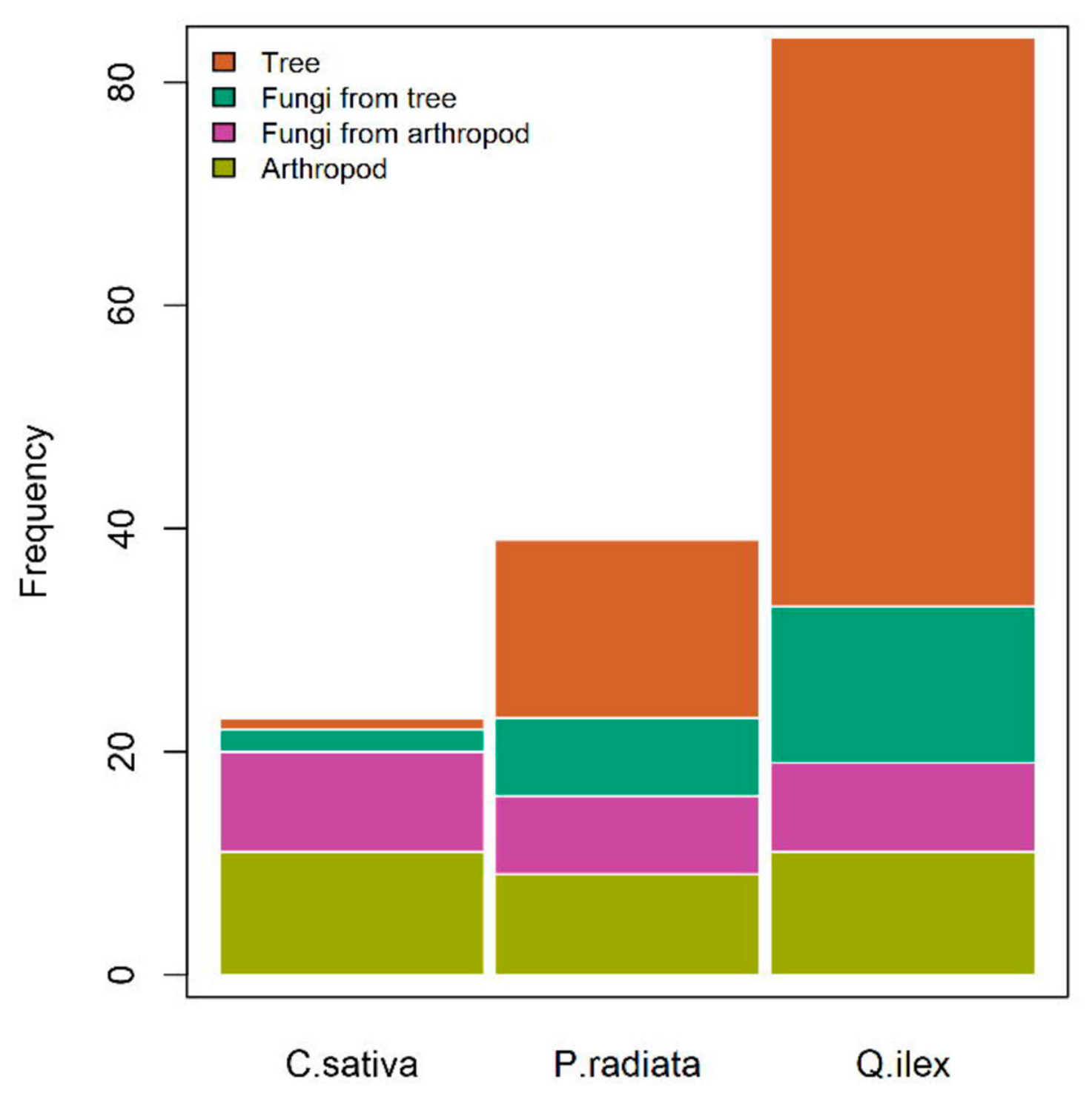
A total of 146 RdRPs were identified across all analyzed samples, from an equivalent sampling effort across the three habitats (sixteen trees and associated arthropods and fungi per plot). Of these, 122 (83%) were assigned to existing viral groups with different levels of taxonomic resolution, 22 (15%) had matches in current databases but lacked taxonomic assignment, and 2 were absent from any database (Table 2 and Table 3). Most of the RdRPs were found in the habitat of Q. ilex (57.5%), followed by P. radiata (26.7%) and C. sativa (15.8%) (Figure 2 and Table 4), suggesting that holm oak forests harbor a substantially richer RNA virosphere than the other two ecosystems sampled. Regarding sample type, nearly half of the RdRPs were found in tree samples (46.5%), followed by arthropods (21.2%), fungi from arthropods (16.4%), and fungi from trees (15.7%). Notably, one RdRP sequence assigned to the genus Mitovirus (family Mitoviridae) was recovered from pine tree tissue, a host type outside the known fungal host range of this genus, representing a potentially anomalous host association warranting further investigation.
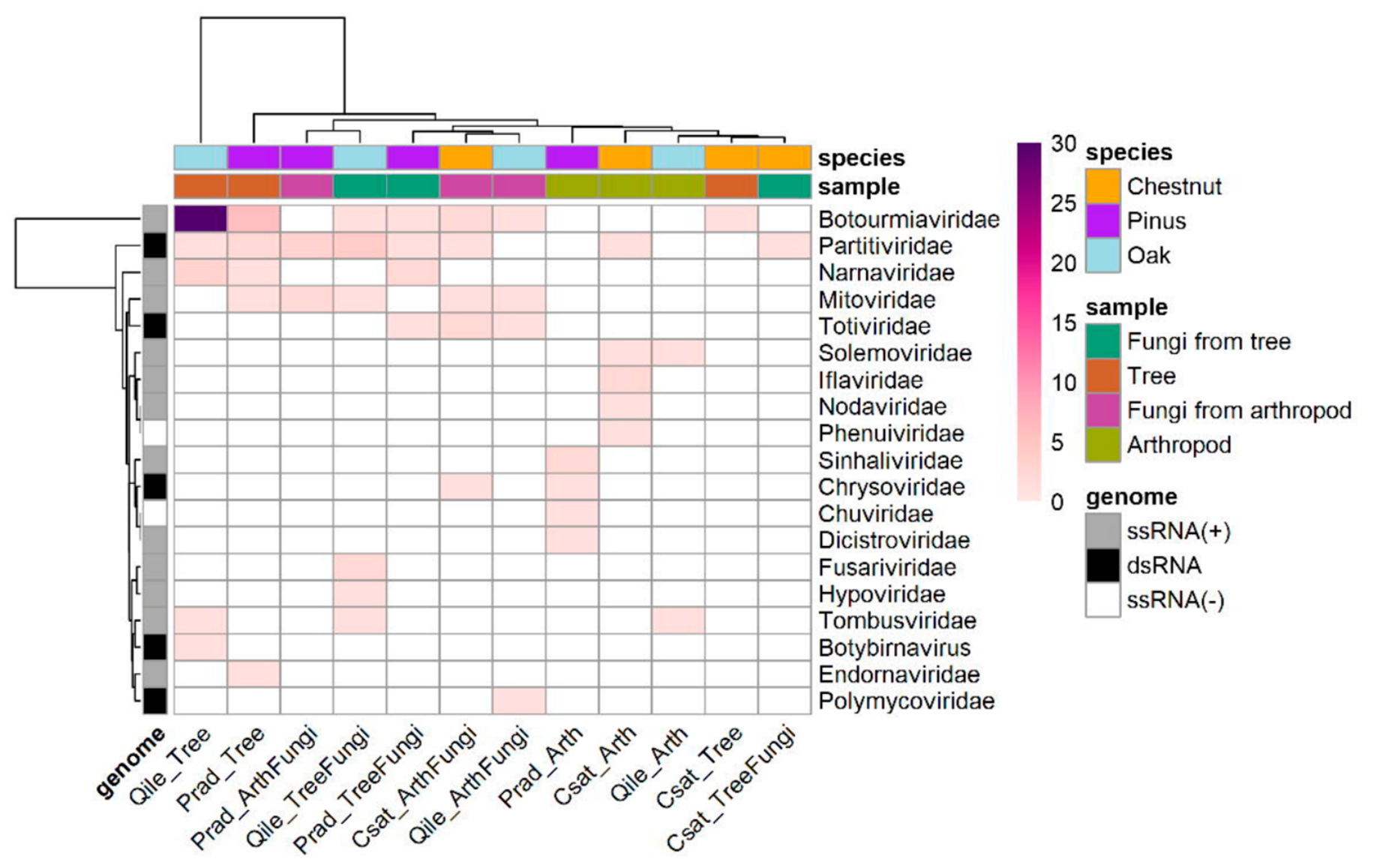
The most numerous viral families were Botourmiaviridae (28.7%), Partitiviridae (9.6%), Mitoviridae (4.1%), and Narnaviridae (4.1%) (Figure 3). In terms of genome type, ssRNA(+) viruses were the most represented (83.5%), followed by dsRNA (15%) and two ssRNA(−) representatives, Chuviridae and Phenuiviridae, found in arthropods from pine and chestnut habitats, respectively.
The distribution of RdRPs revealed that sample types have different viral profiles (Figure 3). Some families were exclusive to fungi (Totiviridae, Fusariviridae, Hypoviridae and Polymycoviridae) or arthropods (only in chestnut habitat: Iflaviridae, Nodaviridae, Phenuiviridae; and only in pine habitat: Sinhaliviridae, Chuviridae, and Dicistroviridae). One family was found to be exclusive to holm oak habitat (Tombusviridae), regardless of sample type. Endornaviridae family was found only in pines, whereas Botybirnavirus was exclusive to holm oaks.

3.2. Phylogeny of RdRPs

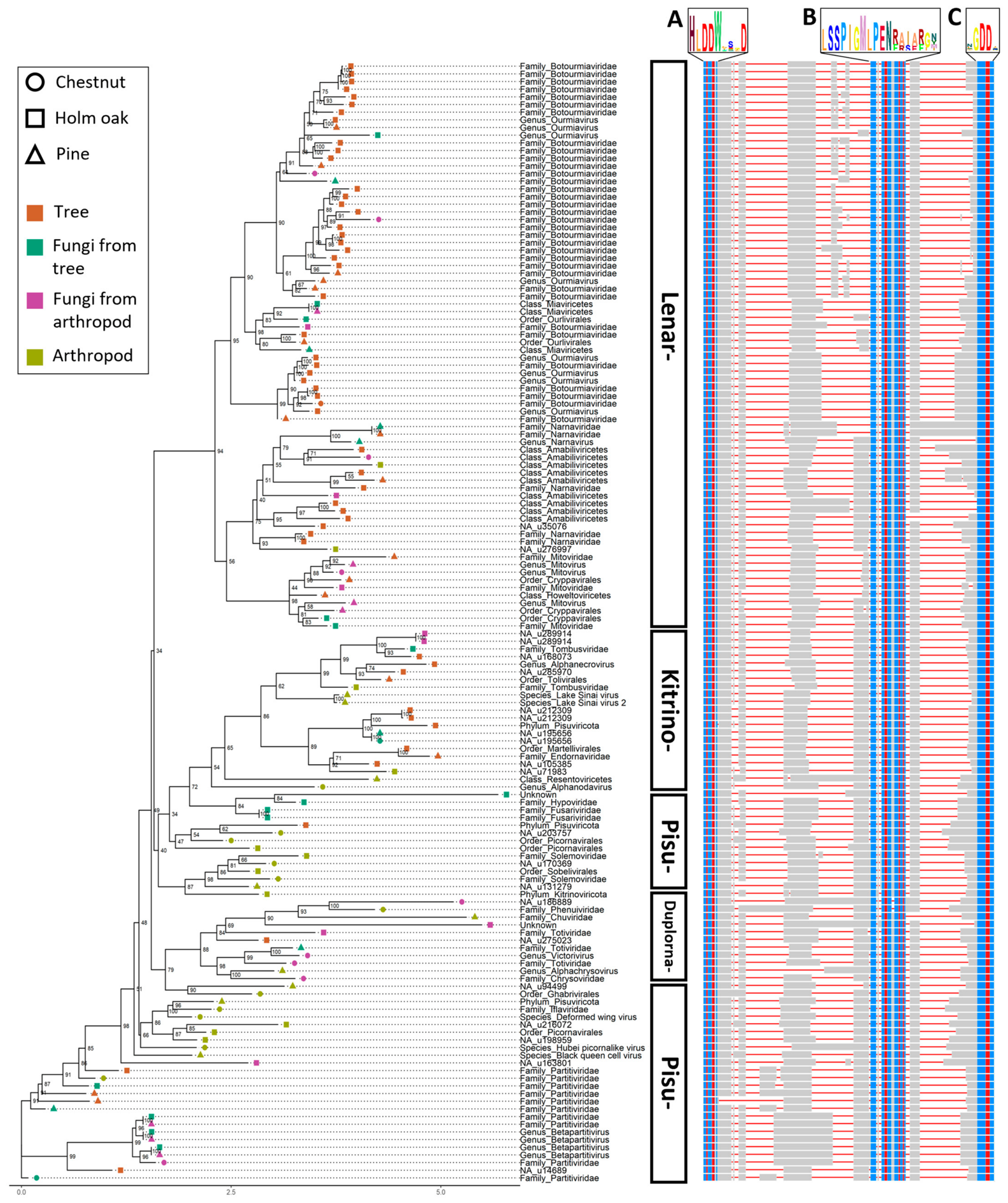
MSA of the RdRPs resulted in the dendrogram shown in Figure 4. RdRPs from trees and arthropod samples clustered in two distinct groups, while RdRPs from isolated fungi were interspersed, forming heterogeneous subgroups. No grouping by habitat was observed except for most RdRPs from holm oak samples clustering together within the Lenarviricota supernode. All RdRPs identified to species level belonged to arthropod samples: Lake Sinai virus 1 and 2, deformed wing virus, black queen cell virus, and Hubei picorna-like virus 15, in the Kitrinoviricota and Pisuviricota nodes. In nodes where RdRPs from tree fungi and arthropod fungi were paired, as was the case for some Betapartitivirus and Narnaviridae (bottom part of the tree), their sequences shared 100% identity. In all other cases, the homology in the conserved RdRP motifs A, B, and C was at least greater than 50%.
RdRPs existing in current databases but lacking taxonomy assignment (NA in Figure 4) were classified as Kitrinoviricota, Duplornaviricota, or Pisuviricota. As for the unknown RdRPs (with bona fide A, B, and C motifs but absent from databases), one shared high sequence similarity with the Hypoviridae representative and was found in the same sample type (fungi from holm oaks), whereas the other was located in the vicinity of ssRNA(−) viruses (Chuviridae and Phenuiviridae) and belonged to Negarnaviricota, sharing no common origin with the former.

4. Discussion

The study of environmental viromes has grown in popularity thanks to recent major advances in massive sequencing technologies. The scale of these analyses is highly variable, with some studies focusing on a particular species [38,39], complete ecosystems [40], or different environments [41,42]. However, most of the advances in plant virology focus on crops or fruit trees of economic interest, which contrasts with the scarcity of such studies in forestry [3]. Reducing the scope even further to Mediterranean forests, with the Mediterranean basin being considered a biodiversity hotspot [43], hardly any information is available. In the present work, focused on three Mediterranean forest habitats in north-western Spain, 146 RdRPs of viral origin have been found, of which 22 have no clear taxonomy yet and 2 are completely unknown. These findings highlight the great lack of knowledge about viral diversity that exists in Mediterranean forest ecosystems.
Most of the reported RdRPs were identified as ssRNA(+) viruses, which are accepted to be the evolutionary origin of the other two identified groups, ssRNA(−) and dsRNA [44,45]. The holm oak habitat showed the highest viral richness (57.5% of all the RdRPs). Holm oak samples were taken in a “dehesa”, a unique ecosystem in the Iberian Peninsula that has been modified by man to simultaneously obtain livestock and non-timber forest resources. This makes dehesas ecosystems with particularly high biodiversity [46,47], so it is not surprising that there is also a high viral diversity that has never been investigated.
Some of the viral families were found to be ubiquitous across sample types and habitats, which could be interpreted as a first indication of their cross-kingdom potential. This is the case for Botourmiaviridae, Partitiviridae, and Narnaviridae. Many genera in Botourmiaviridae family are known to infect fungi, and some of them have been identified on grapevine leaves affected by the oomycete Plasmopara viticola [48]. Mycoviruses of this family can persist in their fungal host without the need for a capsid and are thought to require only the RdRP to replicate [49]. Only the genus Ourmiavirus infects plants exclusively [50]. The RdRP of this genus is most similar to that of the genera Mitovirus and Narnavirus (family Narnaviridae) and its movement protein (MP) is similar to that of the family Tombusviridae. These viruses are very easily transmitted mechanically, and no vector has been identified, so horizontal transmission is thought to be possible [51].
The Narnaviridae family has the simplest genome of all RNA viruses, encoding only one polypeptide in which RdRP is found. Within this family, the genus Narnavirus replicates in the cytoplasm, while Mitovirus replicate in mitochondria and can cause hypovirulence in pathogenic fungi [52,53]. Although no Mitovirus have been found outside fungi, one RdRP identified as a Mitovirus was found in a pine tree sample in the present study. Nonetheless, Mitovirus sequences have previously been found in plant mitochondrial sequences [54,55]. As for the Partitiviridae family, many of its genera have characteristic hosts, which can be plants (Deltapartitivirus), fungi (Gammapartitivirus), or protozoa (Cryspovirus). Many sequences of this family have also been identified in arthropods [56]. One genus, Betapartitivirus, whose hosts can be plants or fungi, was identified. Regarding the family Tombusviridae, this has been described in animals [57], plants [58], and fungi [59], consistent with the detection of this family in all samples types, although restricted to holm oak habitat. Tombusviridae infections are frequently confined to the root system, but they can infect the whole plant thanks to their MPs [60]. The Solemoviridae family infects plants, but it is known that one of its modes of transmission is also the use of insects as vectors [61], which may explain the detection of this family exclusively in arthropods.
For some families, only one RdRP was detected in the samples analyzed, but evidence of their ability to infect several kingdoms has been described. This is the case for Totiviridae, Chuviridae, Phenuiviridae, Chrysoviridae, Polymycoviridae, Endornaviridae, and two riboviruses with incomplete classification: the genus Botybirnavirus and the species Hubei picorna-like virus 15 (unclassified ribovirus). The family Totiviridae is associated with latent infections in fungi and protozoa. The families Chuviridae and Phenuiviridae were the only ones detected in this study possessing a ssRNA(−) genome. Many endogenous viral elements inserted within the host genome have been related to Chuviridae infections [62]. The Chrysoviridae family infects fungi, plants, and insects [63]. The Polymycoviridae family affects fungi, in which it causes hypovirulence or hypersensitivity to antifungals or bacteria, and is even capable of modulating carbon, nitrogen, and iron metabolism [63]. Endornaviridae are ssRNA(+) viruses that do not have a true capsid; the genome is encapsulated together with a viral replicase and are able to infect plants and fungi. The genus Botybirnavirus (Orthornavirae) has fungi as natural hosts [64]. However, a Botybirnavirus RdRP sequence was detected in a holm oak tree sample. This finding most probably reflects the presence of fungal material associated with the sampled tissue, although other explanations, such as fungal colonization or the presence of fungal mycelium within the sample, cannot be excluded. Further studies, including transmission assays and experimental inoculation of both fungal and plant hosts, are required to determine whether cross-kingdom transmission occurs.
Finally, some limitations of the present study should be noted. First, fungal isolation relied on a single culture medium and incubation condition, which may have introduced a selection bias toward fast-growing culturable fungi and excluded part of the naturally associated fungal community. Additionally, many sequences in viral databases have no or incomplete taxonomic assignment, and the high genetic variation of viruses makes it difficult to obtain an accurate assignment at genus or species level. Recurrent updating of the RdRP databases is necessary to facilitate the assignment of new ones. Secondly, although some of the virus families described may be “cross-kingdom” because they contain members that infect several kingdoms, it is more common for individual viruses to specialize in hosts of a single kingdom, as is the case, for example, with the Totiviridae family, where some genera infect fungi and others protozoa. Similarly, the in silico analysis performed did not allow verification of whether the detection of RdRPs in organisms from different kingdoms than expected implies that the host is susceptible to the virus, or that it only acts as a vector or asymptomatic carrier.

5. Conclusions

This study provides the first comprehensive description of the RNA virosphere of declining Mediterranean forest ecosystems across trees, fungi, and arthropods. A total of 146 RdRP sequences spanning 15 viral families were identified, with Botourmiaviridae and Partitiviridae being the most abundant and ssRNA(+) viruses the most represented genome type. Viral richness was highest in the Q. ilex habitat, reflecting the exceptional biodiversity of the dehesa ecosystem. Several families showed distribution patterns consistent with cross-kingdom potential, and anomalous host associations (including a Mitovirus sequence in pine tree tissue) suggest that viral flow across kingdom boundaries may be more frequent than currently recognized. Two RdRP sequences absent from all known databases represent potentially novel viral lineages warranting further investigation. These findings underscore the importance of multi-host metagenomic approaches for understanding viral diversity and dynamics in threatened forest ecosystems facing increasing pressure from climate change and emerging pathogens.

Author Contributions

Conceptualization, J.J.D. and S.D.-H.; methodology, J.N.-S. and W.A.M.; software, S.D.-H., A.D.-G. and M.d.l.P.; validation, S.D.-H. and J.J.D.; formal analysis, S.D.-H., M.d.l.P. and A.D.-G.; investigation, S.D.-H. and A.D.-G.; resources, J.N.-S., P.L.P.-A. and W.A.M.; data curation, M.d.l.P. and A.D.-G.; writing—original draft preparation, S.D.-H.; writing—review and editing, J.J.D. and A.D.-G.; visualization, A.D.-G.; supervision, J.J.D.; project administration, J.J.D.; funding acquisition, J.J.D. All authors have read and agreed to the published version of the manuscript.

Funding

This research was funded by Junta de Castilla Y León (Spain) through projects VA208P20 and VA178P23, co-financed by the European Regional Development Fund (ERDF/FEDER, European Union).

Institutional Review Board Statement

Not applicable.

Informed Consent Statement

Not applicable.

Data Availability Statement

Data and scripts used in this study are available in the following GitHub repository: https://github.com/serbiodh/2023_Virome_MedForests accessed on 15 June 2023. Raw sequencing data are available at NCBI SRA under BioProject PRJNA1032577.

Conflicts of Interest

The authors declare no conflicts of interest.

Abbreviations

The following abbreviations are used in this manuscript:
MPsMovement proteins
PDAPotato dextrose agar
RdRPRNA-dependent RNA polymerase
RNARibonucleic acid
ssRNASingle-stranded ribonucleic acid

References

  1. Hou, X.; He, Y.; Fang, P.; Mei, S.-Q.; Xu, Z.; Wu, W.-C.; Tian, J.-H.; Zhang, S.; Zeng, Z.-Y.; Gou, Q.-Y.; et al. Using Artificial Intelligence to Document the Hidden RNA Virosphere. Cell 2024, 187, 6929–6942.e16. [Google Scholar] [CrossRef] [PubMed]
  2. Neri, U.; Wolf, Y.I.; Roux, S.; Camargo, A.P.; Lee, B.; Kazlauskas, D.; Chen, I.M.; Ivanova, N.; Zeigler Allen, L.; Paez-Espino, D.; et al. Expansion of the Global RNA Virome Reveals Diverse Clades of Bacteriophages. Cell 2022, 185, 4023–4037.e18. [Google Scholar] [CrossRef] [PubMed]
  3. Rumbou, A.; Vainio, E.J.; Büttner, C. Towards the Forest Virome: High-Throughput Sequencing Drastically Expands Our Understanding on Virosphere in Temperate Forest Ecosystems. Microorganisms 2021, 9, 1730. [Google Scholar] [CrossRef] [PubMed]
  4. Bejerman, N.; Debat, H.; Dietzgen, R.G. The Plant Negative-Sense RNA Virosphere: Virus Discovery Through New Eyes. Front. Microbiol. 2020, 11, 588427. [Google Scholar] [CrossRef] [PubMed]
  5. Rehanek, M.; Karlin, D.G.; Bandte, M.; Al Kubrusli, R.; Nourinejhad Zarghani, S.; Candresse, T.; Büttner, C.; von Bargen, S. The Complex World of Emaraviruses—Challenges, Insights, and Prospects. Forests 2022, 13, 1868. [Google Scholar] [CrossRef]
  6. van Diepeningen, A.D. Biocontrol via Mycoviruses: A Neglected Option for Bioprotection? In Microbial Bioprotectants for Plant Disease Managemen; Köhl, J., Ravensberg, W., Eds.; Burleigh Dodds Science Publishing: Cambridge, UK, 2021; pp. 541–584. [Google Scholar] [CrossRef]
  7. Linnakoski, R.; Sutela, S.; Coetzee, M.P.A.; Duong, T.A.; Pavlov, I.N.; Litovka, Y.A.; Hantula, J.; Wingfield, B.D.; Vainio, E.J. Armillaria Root Rot Fungi Host Single-Stranded RNA Viruses. Sci. Rep. 2021, 11, 7336. [Google Scholar] [CrossRef] [PubMed]
  8. Shamsi, W.; Heinzelmann, R.; Ulrich, S.; Kondo, H.; Cornejo, C. Decoding the RNA Virome of the Tree Parasite Armillaria Provides New Insights into the Viral Community of Soil-borne Fungi. Environ. Microbiol. 2024, 26, e16583. [Google Scholar] [CrossRef] [PubMed]
  9. Lu, X.; Dai, Z.; Xue, J.; Li, W.; Ni, P.; Xu, J.; Zhou, C.; Zhang, W. Discovery of Novel RNA Viruses through Analysis of Fungi-Associated next-Generation Sequencing Data. BMC Genom. 2024, 25, 517. [Google Scholar] [CrossRef] [PubMed]
  10. Lefeuvre, P.; Martin, D.P.; Elena, S.F.; Shepherd, D.N.; Roumagnac, P.; Varsani, A. Evolution and Ecology of Plant Viruses. Nat. Rev. Microbiol. 2019, 17, 632–644. [Google Scholar] [CrossRef] [PubMed]
  11. Nerva, L.; Varese, G.C.; Falk, B.W.; Turina, M. Mycoviruses of an Endophytic Fungus Can Replicate in Plant Cells: Evolutionary Implications. Sci. Rep. 2017, 7, 1908. [Google Scholar] [CrossRef] [PubMed]
  12. Andika, I.B.; Wei, S.; Cao, C.; Salaipeth, L.; Kondo, H.; Sun, L. Phytopathogenic Fungus Hosts a Plant Virus: A Naturally Occurring Cross-Kingdom Viral Infection. Proc. Natl. Acad. Sci. USA 2017, 114, 12267–12272. [Google Scholar] [CrossRef] [PubMed]
  13. Bian, R.; Andika, I.B.; Pang, T.; Lian, Z.; Wei, S.; Niu, E.; Wu, Y.; Kondo, H.; Liu, X.; Sun, L. Facilitative and Synergistic Interactions between Fungal and Plant Viruses. Proc. Natl. Acad. Sci. USA 2020, 117, 3779–3788. [Google Scholar] [CrossRef] [PubMed]
  14. Dai, R.; Yang, S.; Pang, T.; Tian, M.; Wang, H.; Zhang, D.; Wu, Y.; Kondo, H.; Andika, I.B.; Kang, Z.; et al. Identification of a Negative-Strand RNA Virus with Natural Plant and Fungal Hosts. Proc. Natl. Acad. Sci. USA 2024, 121, e2319582121. [Google Scholar] [CrossRef] [PubMed]
  15. Whitfield, A.E.; Falk, B.W.; Rotenberg, D. Insect Vector-Mediated Transmission of Plant Viruses. Virology 2015, 479–480, 278–289. [Google Scholar] [CrossRef] [PubMed]
  16. Feng, K.-H.; Qi, Y.-H.; Ye, Z.-X.; Li, T.; Jiao, G.-Y.; Zhang, C.-X.; Chen, J.-P.; Lu, G.; Li, J.-M. Diversity and evolution analysis of RNA viruses in three wheat aphid species. BMC Genom. 2025, 26, 353. [Google Scholar] [CrossRef] [PubMed]
  17. Jakubowska, A.; Nalcacioglu, R.; Millán-Leiva, A.; Sanz-Carbonell, A.; Muratoglu, H.; Herrero, S.; Demirbag, Z. In Search of Pathogens: Transcriptome-Based Identification of Viral Sequences from the Pine Processionary Moth (Thaumetopoea pityocampa). Viruses 2015, 7, 456–479. [Google Scholar] [CrossRef] [PubMed]
  18. Paraskevopoulou, S.; Käfer, S.; Zirkel, F.; Donath, A.; Petersen, M.; Liu, S.; Zhou, X.; Drosten, C.; Misof, B.; Junglen, S. Viromics of Extant Insect Orders Unveil the Evolution of the Flavi-like Superfamily. Virus Evol. 2021, 7, veab030. [Google Scholar] [CrossRef] [PubMed]
  19. Newbold, T.; Oppenheimer, P.; Etard, A.; Williams, J.J. Tropical and Mediterranean Biodiversity Is Disproportionately Sensitive to Land-Use and Climate Change. Nat. Ecol. Evol. 2020, 4, 1630–1638. [Google Scholar] [CrossRef] [PubMed]
  20. Nunes, L.J.R.; Meireles, C.I.R.; Gomes, C.J.P.; Ribeiro, N.M.C.A. The Impact of Climate Change on Forest Development: A Sustainable Approach to Management Models Applied to Mediterranean-Type Climate Regions. Plants 2021, 11, 69. [Google Scholar] [CrossRef] [PubMed]
  21. Arjona-Lopez, J.M.; Telengech, P.; Jamal, A.; Hisano, S.; Kondo, H.; Yelin, M.D.; Arjona-Girona, I.; Kanematsu, S.; Lopez-Herrera, C.J.; Suzuki, N. Novel, Diverse RNA Viruses from Mediterranean Isolates of the Phytopathogenic Fungus, Rosellinia necatrix: Insights into Evolutionary Biology of Fungal Viruses. Environ. Microbiol. 2018, 20, 1464–1483. [Google Scholar] [CrossRef] [PubMed]
  22. Wilcox, B.A.; Ellis, B. Forests and Emerging Infectious Diseases of Humans. Unasylva 2006, 57, 11–18. [Google Scholar]
  23. Mackay, I.M.; Arden, K.E. Mayaro Virus: A Forest Virus Primed for a Trip to the City? Microbes Infect. 2016, 18, 724–734. [Google Scholar] [CrossRef] [PubMed]
  24. Martin, M. Cutadapt Removes Adapter Sequences from High-Throughput Sequencing Reads. EMBnet. J. 2011, 17, 10. [Google Scholar] [CrossRef]
  25. Prjibelski, A.; Antipov, D.; Meleshko, D.; Lapidus, A.; Korobeynikov, A. Using SPAdes De Novo Assembler. Curr. Protoc. Bioinform. 2020, 70, e102. [Google Scholar] [CrossRef] [PubMed]
  26. Babaian, A.; Edgar, R. Ribovirus Classification by a Polymerase Barcode Sequence. PeerJ 2022, 10, e14055. [Google Scholar] [CrossRef] [PubMed]
  27. Charon, J.; Buchmann, J.P.; Sadiq, S.; Holmes, E.C. RdRp-Scan: A Bioinformatic Resource to Identify and Annotate Divergent RNA Viruses in Metagenomic Sequence Data. Virus Evol. 2022, 8, veac082. [Google Scholar] [CrossRef] [PubMed]
  28. Buchfink, B.; Reuter, K.; Drost, H.-G. Sensitive Protein Alignments at Tree-of-Life Scale Using DIAMOND. Nat. Methods 2021, 18, 366–368. [Google Scholar] [CrossRef] [PubMed]
  29. Katoh, K.; Rozewicki, J.; Yamada, K.D. MAFFT Online Service: Multiple Sequence Alignment, Interactive Sequence Choice and Visualization. Brief. Bioinform. 2019, 20, 1160–1166. [Google Scholar] [CrossRef] [PubMed]
  30. Nguyen, L.-T.; Schmidt, H.A.; von Haeseler, A.; Minh, B.Q. IQ-TREE: A Fast and Effective Stochastic Algorithm for Estimating Maximum-Likelihood Phylogenies. Mol. Biol. Evol. 2015, 32, 268–274. [Google Scholar] [CrossRef] [PubMed]
  31. Hoang, D.T.; Chernomor, O.; von Haeseler, A.; Minh, B.Q.; Vinh, L.S. UFBoot2: Improving the Ultrafast Bootstrap Approximation. Mol. Biol. Evol. 2018, 35, 518–522. [Google Scholar] [CrossRef] [PubMed]
  32. Kalyaanamoorthy, S.; Minh, B.Q.; Wong, T.K.F.; von Haeseler, A.; Jermiin, L.S. ModelFinder: Fast Model Selection for Accurate Phylogenetic Estimates. Nat. Methods 2017, 14, 587–589. [Google Scholar] [CrossRef] [PubMed]
  33. R Core Team. R: A Language and Environment for Statistical Computing, Vienna, Austria. Available online: https://www.R-project.org/ (accessed on 11 May 2026).
  34. Yu, G. Using Ggtree to Visualize Data on Tree-Like Structures. Curr. Protoc. Bioinform. 2020, 69, e96. [Google Scholar] [CrossRef] [PubMed]
  35. Zhou, L.; Feng, T.; Xu, S.; Gao, F.; Lam, T.T.; Wang, Q.; Wu, T.; Huang, H.; Zhan, L.; Li, L.; et al. Ggmsa: A Visual Exploration Tool for Multiple Sequence Alignment and Associated Data. Brief. Bioinform. 2022, 23, bbac222. [Google Scholar] [CrossRef] [PubMed]
  36. Charif, D.; Lobry, J.R.; Necsulea, A.; Palmeira, L.; Penel, S.; Perriere, G.; Penel, M.S. Seqinr: Biological Sequences Retrieval and Analysis. Available online: https://seqinr.r-forge.r-project.org/ (accessed on 11 May 2026).
  37. Wheeler, T.J.; Clements, J.; Finn, R.D. Skylign: A Tool for Creating Informative, Interactive Logos Representing Sequence Alignments and Profile Hidden Markov Models. BMC Bioinform. 2014, 15, 7. [Google Scholar] [CrossRef] [PubMed]
  38. Faizah, A.N.; Kobayashi, D.; Isawa, H.; Amoa-Bosompem, M.; Murota, K.; Higa, Y.; Futami, K.; Shimada, S.; Kim, K.S.; Itokawa, K.; et al. Deciphering the Virome of Culex Vishnui Subgroup Mosquitoes, the Major Vectors of Japanese Encephalitis, in Japan. Viruses 2020, 12, 264. [Google Scholar] [CrossRef] [PubMed]
  39. Wu, Z.; Yang, L.; Ren, X.; He, G.; Zhang, J.; Yang, J.; Qian, Z.; Dong, J.; Sun, L.; Zhu, Y.; et al. Deciphering the Bat Virome Catalog to Better Understand the Ecological Diversity of Bat Viruses and the Bat Origin of Emerging Infectious Diseases. ISME J. 2016, 10, 609–620. [Google Scholar] [CrossRef] [PubMed]
  40. Hurwitz, B.L.; Sullivan, M.B. The Pacific Ocean Virome (POV): A Marine Viral Metagenomic Dataset and Associated Protein Clusters for Quantitative Viral Ecology. PLoS ONE 2013, 8, e57355. [Google Scholar] [CrossRef] [PubMed]
  41. Daugrois, J.H.; Filloux, D.; Julian, C.; Claude, L.; Ferdinand, R.; Fernandez, E.; Fontes, H.; Rott, P.C.; Roumagnac, P. Comparison of the Virome of Quarantined Sugarcane Varieties and the Virome of Grasses Growing near the Quarantine Station. Viruses 2021, 13, 922. [Google Scholar] [CrossRef] [PubMed]
  42. Ma, Y.; Fort, T.; Marais, A.; Lefebvre, M.; Theil, S.; Vacher, C.; Candresse, T. Leaf-associated Fungal and Viral Communities of Wild Plant Populations Differ between Cultivated and Natural Ecosystems. Plant-Environ. Interact. 2021, 2, 87–99. [Google Scholar] [CrossRef] [PubMed]
  43. Médail, F.; Monnet, A.-C.; Pavon, D.; Nikolic, T.; Dimopoulos, P.; Bacchetta, G.; Arroyo, J.; Barina, Z.; Albassatneh, M.C.; Domina, G.; et al. What Is a Tree in the Mediterranean Basin Hotspot? A Critical Analysis. For. Ecosyst. 2019, 6, 17. [Google Scholar] [CrossRef]
  44. Koonin, E.V.; Dolja, V.V.; Krupovic, M. Origins and Evolution of Viruses of Eukaryotes: The Ultimate Modularity. Virology 2015, 479–480, 2–25. [Google Scholar] [CrossRef] [PubMed]
  45. Wolf, Y.I.; Kazlauskas, D.; Iranzo, J.; Lucía-Sanz, A.; Kuhn, J.H.; Krupovic, M.; Dolja, V.V.; Koonin, E.V. Origins and Evolution of the Global RNA Virome. mBio 2018, 9, 10–1128. [Google Scholar] [CrossRef] [PubMed]
  46. Moreno, G.; Gonzalez-Bornay, G.; Pulido, F.; Lopez-Diaz, M.L.; Bertomeu, M.; Juárez, E.; Diaz, M. Exploring the Causes of High Biodiversity of Iberian Dehesas: The Importance of Wood Pastures and Marginal Habitats. Agrofor. Syst. 2016, 90, 87–105. [Google Scholar] [CrossRef]
  47. Rodríguez-Rojo, M.; Roig, S.; López-Carrasco, C.; Redondo García, M.; Sánchez-Mata, D. Which Factors Favour Biodiversity in Iberian Dehesas? Sustainability 2022, 14, 2345. [Google Scholar] [CrossRef]
  48. Chiapello, M.; Rodríguez-Romero, J.; Ayllón, M.A.; Turina, M. Analysis of the Virome Associated to Grapevine Downy Mildew Lesions Reveals New Mycovirus Lineages. Virus Evol. 2020, 6, veaa058. [Google Scholar] [CrossRef] [PubMed]
  49. Wang, Q.; Mu, F.; Xie, J.; Cheng, J.; Fu, Y.; Jiang, D. A Single SsRNA Segment Encoding RdRp Is Sufficient for Replication, Infection, and Transmission of Ourmia-Like Virus in Fungi. Front. Microbiol. 2020, 11, 379. [Google Scholar] [CrossRef] [PubMed]
  50. Rastgou, M.; Habibi, M.K.; Izadpanah, K.; Masenga, V.; Milne, R.G.; Wolf, Y.I.; Koonin, E.V.; Turina, M. Molecular Characterization of the Plant Virus Genus Ourmiavirus and Evidence of Inter-Kingdom Reassortment of Viral Genome Segments as Its Possible Route of Origin. J. Gen. General Virol. 2009, 90, 2525–2535. [Google Scholar] [CrossRef] [PubMed]
  51. Ayllón, M.A.; Turina, M.; Xie, J.; Nerva, L.; Marzano, S.-Y.L.; Donaire, L.; Jiang, D.; Consortium, I.R. ICTV Virus Taxonomy Profile: Botourmiaviridae. J. Gen. General Virol. 2020, 101, 454–455. [Google Scholar] [CrossRef] [PubMed]
  52. de Rezende, R.R.; de Oliveira Souza, F.; Leal, L.L.; Morgan, T.; Bermudez, J.M.M.; Cascardo, R.d.S.; de Barros, D.R.; Alfenas-Zerbini, P. Characterization of a New Mitovirus Infecting the Phytopathogenic Fungus Microdochium albescens. Arch. Virol. 2021, 166, 3455–3459. [Google Scholar] [CrossRef] [PubMed]
  53. Hillman, B.I.; Cai, G. The Family Narnaviridae. In Advances in Virus Research; Ghabrial, S.A., Ed.; Academic Press: Cambridge, MA, USA, 2013; Volume 86, pp. 149–176. [Google Scholar] [CrossRef] [PubMed]
  54. Hong, Y.; Cole, T.E.; Brasier, C.M.; Buck, K.W. Evolutionary Relationships among Putative RNA-Dependent RNA Polymerases Encoded by a Mitochondrial Virus-like RNA in the Dutch Elm Disease Fungus, Ophiostoma novo-ulmi, by Other Viruses and Virus-like RNAs and by The Arabidopsis Mitochondrial Genome. Virology 1998, 246, 158–169. [Google Scholar] [CrossRef] [PubMed]
  55. Marienfeld, J.; Unseld, M.; Brennicke, A. The Mitochondrial Genome of Arabidopsis Is Composed of Both Native and Immigrant Information. Trends Plant Sci. 1999, 4, 495–502. [Google Scholar] [CrossRef] [PubMed]
  56. Cross, S.T.; Brehm, A.L.; Dunham, T.J.; Rodgers, C.P.; Keene, A.H.; Borlee, G.I.; Stenglein, M.D. Galbut Virus Infection Minimally Influences Drosophila melanogaster Fitness Traits in a Strain and Sex-Dependent Manner. Viruses 2023, 15, 539. [Google Scholar] [CrossRef] [PubMed]
  57. Yin, H.; Wan, D.; Chen, H. Metagenomic Analysis of Viral Diversity and a Novel Astroviruse of Forest Rodent. Virol. J. 2022, 19, 138. [Google Scholar] [CrossRef] [PubMed]
  58. Lappe, R.R.; Elmore, M.G.; Lozier, Z.R.; Jander, G.; Miller, W.A.; Whitham, S.A. Metagenomic Identification of Novel Viruses of Maize and Teosinte in North America. BMC Genom. 2022, 23, 767. [Google Scholar] [CrossRef] [PubMed]
  59. Botella, L.; Jung, M.H.; Rost, M.; Jung, T. Natural Populations from the Phytophthora Palustris Complex Show a High Diversity and Abundance of SsRNA and DsRNA Viruses. J. Fungi 2022, 8, 1118. [Google Scholar] [CrossRef] [PubMed]
  60. Erickson, A.; Falk, B.W. Dissecting Dynamic Plant Virus Synergism in Mixed Infections of Poleroviruses, Umbraviruses, and Tombusvirus-like Associated RNAs. Front. Microbiol. 2023, 14, 1223265. [Google Scholar] [CrossRef] [PubMed]
  61. Sõmera, M.; Fargette, D.; Hébrard, E.; Sarmiento, C. ICTV Virus Taxonomy Profile: Solemoviridae 2021. J. Gen. Virol. 2021, 102, 001707. [Google Scholar] [CrossRef] [PubMed]
  62. Dezordi, F.Z.; Vasconcelos, C.R.d.S.; Rezende, A.M.; Wallau, G.L. In and Outs of Chuviridae Endogenous Viral Elements: Origin of a Potentially New Retrovirus and Signature of Ancient and Ongoing Arms Race in Mosquito Genomes. Front. Genet. 2020, 11, 542437. [Google Scholar] [CrossRef] [PubMed]
  63. Kotta-Loizou, I.; Coutts, R.H.A. ICTV Virus Taxonomy Profile: Polymycoviridae 2022. J. Gen. Virol. 2022, 103, 001747. [Google Scholar] [CrossRef] [PubMed]
  64. Hough, B.; Steenkamp, E.; Wingfield, B.; Read, D. Fungal Viruses Unveiled: A Comprehensive Review of Mycoviruses. Viruses 2023, 15, 1202, Correction in Viruses 2024, 16, 632. [Google Scholar] [CrossRef] [PubMed]
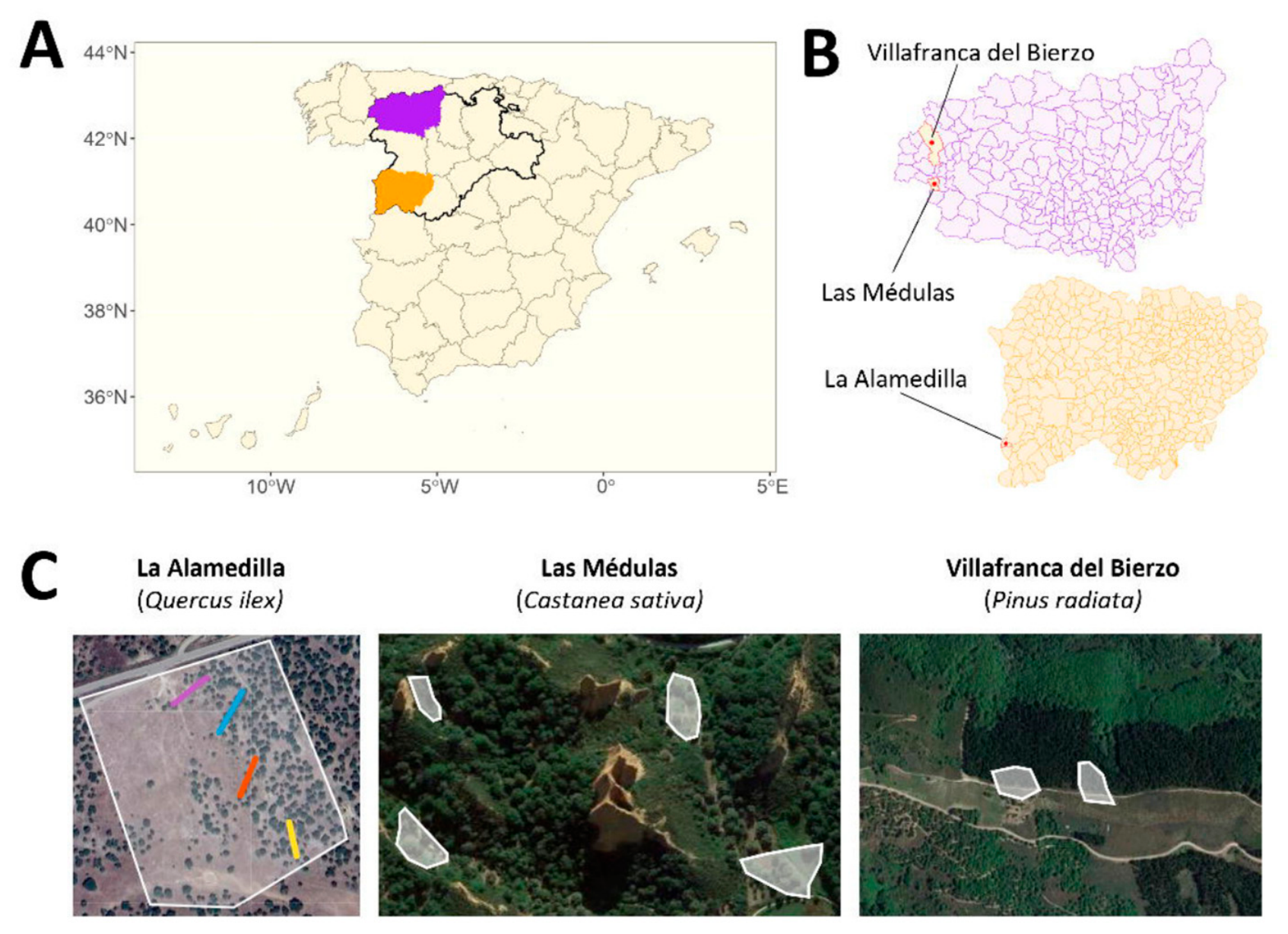
Figure 1. Map of sampling sites. (A)—Map of Spain. Castile and Leon region is highlighted with a black and thick contour. Orange and purple areas correspond to Salamanca and León provinces, respectively. (B)—Municipalities from where samples were taken (top: León, bottom: Salamanca). (C)—Sampling areas (white polygons) overlayed on top of physical maps. Colored lines in La Alamedilla correspond to four sampling transects.
Microorganisms 14 01445 g001
Figure 2. RdRP counts by habitat and sample type.
Microorganisms 14 01445 g002
Figure 3. Distribution of viral families per habitat, sample type, and genome type. Color gradient from white to purple indicates abundance.
Microorganisms 14 01445 g003
Figure 4. Phylogenetic tree of RdRPs. Left: consensus tree with bootstrap confidence values. Each branch is labelled with the taxonomic level followed by the identity assigned to that particular RdRP. Vertical rectangles with black outline indicate viral phyla (−viricota). Right: multiple sequence alignment with A, B, and C motifs and their corresponding logos. Blue and red regions indicate well-conserved residues.
Microorganisms 14 01445 g004
Table 1. Description of sampling sites.
LocationProvinceCharacteristicsSampled Species
La AlamedillaSalamancaElevation: 756 m
Rainfall: 600–854 mm
Quercus ilex
Las MédulasLeónElevation: 511 m
Rainfall: 400–500 mm
Castanea sativa
Villafranca del BierzoLeónElevation: 505 m
Rainfall: 400–500 mm
Pinus radiata
Table 2. Taxonomic distribution of RdRPs.
PhylumClassOrderFamilyGenusSpeciesNot Assigned 1Unknown 2
4141267205222
1 RdRPs found in databases that had no taxonomy assigned yet. 2 RdRPs that could not be found in any database.
Table 3. Taxonomic assignment of RdRPs per genome type.
ssRNA(+)ssRNA(−)dsRNA
Family
(-viridae)
Botourmia-
Dicistro-
Endorna-
Fusari-
Hypo-
Ifla-
Mito-
Narna-
Sinhali-
Solemo-
Tombus-
Chu-
Phenui-
Chryso-
Partiti-
Polymyco-
Toti-
Genus
(-virus)
Alphanoda-
Alphanecro-
Mito-
Narna-
Ourmia-
Alphacryso-
Betapartiti-
Botybirna-
Victori-
Virus detectedBlack queen cell virus
Deformed wing virus
Lake Sinai virus
Lake Sinai virus 2
Hubei picorna-like virus 15
Table 4. Count of RdRPs per habitat and sample type.
TreeFungi from TreeArthropodFungi from Arthropod
Chestnut12119
Pine16797
Holm oak5114118
Disclaimer/Publisher’s Note: The statements, opinions and data contained in all publications are solely those of the individual author(s) and contributor(s) and not of MDPI and/or the editor(s). MDPI and/or the editor(s) disclaim responsibility for any injury to people or property resulting from any ideas, methods, instructions or products referred to in the content.