Review

Inclusion Membrane Proteins of Chlamydia trachomatis: A Review with Emphasis on Biological Functions and Host Cell Modulation

Institute of Pathogenic Biology, Hengyang Medical College, University of South China, Hengyang 421001, China
*
Author to whom correspondence should be addressed.
These authors contributed equally to this work.
Microorganisms 2026, 14(7), 1443; https://doi.org/10.3390/microorganisms14071443
Submission received: 5 May 2026 / Revised: 22 June 2026 / Accepted: 29 June 2026 / Published: 30 June 2026
(This article belongs to the Special Issue Chlamydiae and Chlamydia-Like Infections)

Abstract

Chlamydia trachomatis is an obligate intracellular prokaryote. During the process of infecting host cells, C. trachomatis forms a unique inclusion structure, which serves as a critical niche for the survival and replication of C. trachomatis within host cells. The inclusion membrane is composed of various inclusion membrane proteins (Inc proteins), which play a pivotal role in the interaction between C. trachomatis and host cells. This review illustrates functional research progress of C. trachomatis Inc proteins and their molecular interactions with host cells, facilitating deeper understanding of chlamydial pathogenic mechanisms.

1. Introduction

Chlamydia trachomatis is an obligate intracellular human pathogen and one of the most prevalent etiological agents of bacterial sexually transmitted infections (STIs) worldwide, with an estimated 130 million new cases annually [1,2,3]. Coinfection of Chlamydia with other microbes may result in more severe inflammation and even tumor development [4,5].
C. trachomatis exhibits a unique biphasic life cycle. Extracellular infectious elementary bodies (EBs) invade host cells and are subsequently enclosed in membrane-bound vacuoles termed inclusions. Inside the inclusions, EBs differentiate into metabolically active reticulate bodies (RBs), the replicative form responsible for proliferation. Following proliferation, mature progeny EBs are released to initiate a new infection cycle [6].
The inclusion is a crucial site for the replication and biosynthesis of Chlamydia, providing a protected environment for the pathogen that enables it to evade the host’s immune clearance. In addition, C. trachomatis obtains nutrients from the host cell through the inclusion and expels metabolic waste into the host cell [7]. Inc proteins are a class of proteins localized to the inclusion membrane of Chlamydia. As structurally conserved proteins, Inc proteins carry an N-terminal type III secretion signal required for secretion from chlamydiae, plus a conserved hydrophobic domain with at least two transmembrane α-helices that anchors them to the inclusion membrane [8]. Inc proteins are exposed to the host cytoplasm and accumulate in kinase-rich microdomains [9,10]. These proteins also engage in intermolecular structural interactions within the family to achieve synergistic functions. Due to their essential involvement in chlamydial virulence, Inc proteins serve as ideal targets for drug development. Elucidating their functions and regulatory pathways facilitates the creation of innovative anti-chlamydial interventions and supports the prevention of sexually transmitted diseases [11,12]. Considering their critical roles in C. trachomatis pathogenesis, the present review systematically outlines recent advances in Inc protein studies, highlighting widespread molecular interactions between Inc proteins and host cells throughout C. trachomatis infection.

2. Mediate the Structural Stability of Inclusions and Reshape the Host Cytoskeleton

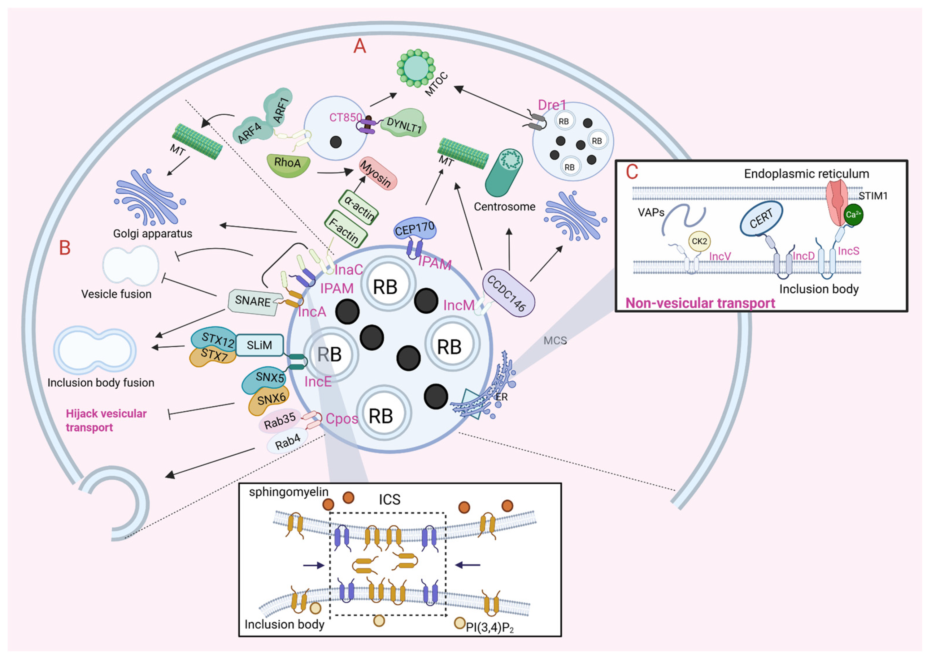
As the inclusion continues to expand, C. trachomatis ingeniously utilizes four cytoskeletal components that function to sustain inclusion stability and architecture: microtubules (MTs), actin, intermediate filaments (IFs), and septin proteins [13,14]. IFs provide stability to the expanding inclusion, while MTs are progressively organized on the surface of the inclusion and assemble into interconnected scaffolds and nest-like superstructures. Septin proteins are cytoskeletal proteins that directly interact with membranes, acting as scaffolds to recruit proteins to cellular locations and as structural diffusion barriers [15]. Actin is closely related to the invasion and release processes of C. trachomatis [16]. At various developmental stages of C. trachomatis, Inc proteins reshape microfilaments and MTs of the host cell cytoskeleton [17]. Hijacking the host MT cytoskeleton must be an important component of C. trachomatis infection (Figure 1A). InaC (CT813), IPAM (CT223), IncM (CT288), and CT850 function sequentially to modulate microtubule and actin networks, mediate inclusion transport to the microtubule-organizing center (MTOC), and maintain inclusion morphology [18,19,20]. IncA serves as the central fusogen that drives homotypic fusion by forming trans-interactions at specialized inclusion contact sites (ICSs), which is tightly regulated by PI(3,4)P2 and sphingomyelin [21]. Dre1 (CT192/CTL0444) further enhances C. trachomatis fitness by targeting host dynactin to reposition key organelles around the inclusion and stabilize its structure [22].
InaC is mainly responsible for regulating the stability of the cytoskeleton during C. trachomatis infection [19]. By comparing the characteristics of InaC mutant and wild-type strains, Kokes et al. found that InaC is involved in the accumulation of F-actin around inclusions [23]. InaC also recruits ADP-ribosylation factor 1 (ARF1) and ARF4 to the inclusion membrane and modulates their activation; the activated ARF1 governs the formation of post-translationally modified MTs, coordinates the assembly of microfilaments around the inclusion, and thereby finely regulates the distribution of the Golgi apparatus around the inclusion [24,25]. In addition to stabilizing MTs, InaC regulates the stability of the cytoskeleton by controlling the actin scaffold to support the integrity of the envelope during infection. InaC recruits RhoA to promote the formation of the actin scaffold around the inclusion, and activated RhoA further drives actin polymerization at the inclusion periphery [13,24]. C. trachomatis also hijacks α-actin to cross-link these scaffolds and stabilize the inclusion structure. α-actin is recruited to the inclusion membrane in an InaC-dependent manner, which reinforces the expanding survival niche for C. trachomatis and enables tight binding to the actin scaffold of the inclusion [26]. In summary, InaC guides the dynamics of actin and post-translationally modified MT scaffolds by mediating the crosstalk between cytoskeleton elements and GTPases of RhoA and ARF1.
The Inc protein IPAM and the host centrosome protein CEP170 are involved in the assembly of MTs around the C. trachomatis inclusion [18]. IPAM is located in the inclusion microdomain near the centrosome. It interacts with the pericentriolar material (PCM) on the centrosome by virtue of its primary structure similar to centrosome and MT-associated proteins, and thus interferes with MT assembly. IPAM locally hijacks the host MT-organizing activity by recruiting and stimulating CEP170. Knocking down CEP170 in C. trachomatis-infected cells results in the reduction in the MT scaffold and disappearance of the MT nest, leading to a characteristic rounded cell morphology. Meanwhile, CEP170 depletion triggers defects in MT organization during MT regrowth and causes aberrant inclusion morphology [17]. Therefore, CEP170 is critical for C. trachomatis to govern host MT assembly, drive inclusion morphogenesis, and maintain bacterial infectivity [17,27].
IncM localizes to inclusion microdomains. It interferes with the division process of host cells [28], affects the localization of the centrosome [29], and changes the distribution of the Golgi apparatus around the inclusion [20], thus affecting the morphology and stability of the inclusion. Almeida F. et al. reported that IncM targets the centrosome, modulates centrosome positioning and remodels the MT cytoskeleton. A novel interaction between IncM and host cell protein CCDC146 has been identified. CCDC146 is recruited to the periphery of the inclusion membrane, suggesting a potential key role during the host cell infection by C. trachomatis [30]. Collectively, the molecular mechanism underlying IncM-mediated cytoskeleton regulation remains poorly understood. Importantly, the direct functional relationship between IncM and CCDC146 has not been clarified in existing studies.
It is worth noting that the C. trachomatis inclusion membrane protein CT850 interacts with dynein light chain 1 (DYNLT1), mediating the transport process of the inclusion to the microtubule-organizing center. Research shows that the deletion of DYNLT1 disrupts the characteristic connection between the inclusion membrane and the centrosome, reducing the association between the C. trachomatis inclusion and the MTOC. On the C. trachomatis inclusion membrane, DYNLT1, which is known to interact with dynein and MTs, aggregates to form concentrated foci mediated by CT850 [31]. In summary, the interaction between CT850 and DYNLT1 contributes to the correct positioning of inclusions at the MTOC. IncA acts as the core fusogenic effector that mediates inclusion homotypic fusion by trans-interacting and clustering at specialized ICSs; this process is spatially controlled by PI(3,4)P2 and sphingomyelin, and further supported by IPAM to maintain inclusion structural stability [21]. Recently, the Inc protein Dre1 has been shown to target host dynactin at MTOCs, thereby repositioning the centrosome, Golgi, and other organelles around the inclusion to stabilize inclusion structure and promote C. trachomatis fitness [22]. Overall, the precise molecular mechanisms underlying effector–target binding and pathway crosstalk remain elusive, and relevant clinical validations are pending further investigation.

3. Hijack the Host Cell’s Vesicular Transport

When interacting with other pathways within host cells, the inclusions of C. trachomatis deviate from the normal lysosomal pathway and obtain the nutrients required for growth and replication from the host cells. C. trachomatis exploits its own Inc proteins to hijack host vesicular and non-vesicular transport machineries. By modulating intracellular trafficking and membrane fusion, the pathogen sequesters essential nutrients from the host cytoplasm to sustain its survival and replication, and supports inclusion maturation [32,33,34] (Figure 1B). Functional differentiation among distinct Inc proteins drives extensive remodeling of host intracellular trafficking networks, inhibition of host innate restriction against infection, and sustained survival and proliferation of C. trachomatis inclusions. Distinct members of the C. trachomatis Inc protein repertoire, such as IncA, CpoS and IncE (CT116), mediate disparate regulatory effects on the host vesicular trafficking machinery. IncA structurally mimics host soluble N-ethylmaleimide-sensitive factor (NSF) attachment protein receptor (SNARE) proteins to hijack host membrane fusion machinery [35,36]; CpoS remodels RAB GTPase-dependent vesicular trafficking to reroute nutrient-containing host vesicles toward inclusions [33,37]; and IncE engages sorting nexin 5 (SNX5), sorting nexin 6 (SNX6), and STX7 and STX12 through distinct short linear interaction motifs (SLiMs) to subvert retrograde vesicular transport and facilitate inclusion fusion [38].
C. trachomatis is capable of obtaining nutrients by hijacking the vesicular transport of host cells. SNARE proteins, together with small guanosine triphosphate (GTP)-binding proteins including ADP-ribosylation factors (ARFs) and RAB GTPases, are key regulators of membrane vesicle trafficking and fusion in eukaryotic cells [33]. SNAREs are key components of the intracellular fusion machinery, where vesicular SNAREs bind to their corresponding target membrane SNAREs to drive membrane fusion. ARFs are small GTPases that play a crucial role in regulating vesicular transport by recruiting the coat proteins required for vesicle formation [39]. RAB GTPases are localized to different organelles, are the main regulators of vesicular transport in eukaryotes, and regulate vesicle budding, transport, docking, and fusion [33,34,40]. Some of them (RAB 1, 4, 6, 8, 10, 11, 14, 34, 35, 39a, 39b) have been reported to be localized on or near the inclusion membrane of C. trachomatis [33,41,42,43]. In C. trachomatis, several inclusion membrane proteins such as IncA, IPAM and InaC possess SNARE motifs that mimic SNARE proteins and inhibit harmful vesicular fusion [35,36]. For instance, IncA possess two SNARE-like coiled-coil domains, SLD-1 and SLD-2, on the inclusion membrane, which mediate homotypic inclusion fusion and block SNARE-dependent fusion, with each domain alone exerting this inhibition [44,45].
The recruitment of Rab proteins is considered to reflect the ability of bacteria to capture host vesicles and thus obtain lipids. Inc proteins play an important role in the recruitment of RAB GTPases [46,47]. Among them, CpoS functions as one of the key regulators of host cell vesicular transport in C. trachomatis-infected cells. It targets various RAB GTPases and their corresponding effector proteins to the inclusions, intercepts host vesicles from the recycling pathway, and redirects Rab-containing vesicles to the inclusions, thereby regulating the transport of transferrin and mannose-6-phosphate receptors [33]. Furthermore, the latest genome-wide screening evidence reveals that CpoS preserves the stability of C. trachomatis inclusions by regulating Rab-dependent vesicular transport of Golgi-originated sphingolipids, while the ceramide transfer protein (CERT)-mediated non-vesicular ceramide transport pathway remains unperturbed [37]. Meier K. et al. also contend that loss of CpoS diminishes ceramide acquisition by C. trachomatis inclusions [46].
Beyond modulating Rab GTPase-dependent vesicular trafficking, Inc proteins also hijack host retrograde transport machineries to facilitate intracellular survival. SNXs are important components of the retrograde transport complex. It contains a PX (phox homology) domain, which mediates the recycling of endosomal substances to the plasma membrane or the trans-Golgi network (TGN) [27]. It is worth noting that various sorting nexins will accumulate in the specific cellular structure of the inclusion. Among them, the inclusion membrane protein IncE binds to the PX domain of the sorting nexin SNX5/6 and recruit these components of the retrograde transport complex to the inclusions [48]. This inhibits host retrograde vesicular trafficking and attenuates host cellular restriction to C. trachomatis infection, thereby promoting pathogen survival and replication inside host cells and elevating infectious progeny production [49]. For example, IncE competes with the cation-independent mannose-6-phosphate receptor (CI-M6PR) for binding to SNX5, inhibits the interaction between SNX5 and CI-M6PR, and further affects the binding of CI-M6PR cargo to the endosomal subdomain containing retromer, resulting in disorders in the transport and recycling processes of lysosomal enzymes [34,50,51]. IncE possesses two distinct SLiMs at its cytosolic C terminus. The proximal SLiM mimics an R-SNARE fragment to recruit STX7/STX12-bearing vesicles, while the distal SLiM recognizes the cargo-binding site of SNX5/SNX6 and recruits SNX6-associated vesicles to the inclusion [38]. This action underscores the sophisticated manipulation of host vesicular transport systems by Inc proteins.

4. Regulate Host Cell’s Non-Vesicular Transport

The inclusion of C. trachomatis not only manipulates the vesicular transport between host organelles but also establishes direct membrane contact sites (MCSs) with organelles such as the endoplasmic reticulum (ER) [52]. The formation of MCSs is regarded as a mechanism by which intracellular pathogens hijack cellular resources and establish their own replication microenvironment (Figure 1C). Pathogens can utilize MCSs [53]. Inc proteins including IncD, IncV and IncS exert pivotal functions in MCSs, sustaining MCS stability and modulating organelle functions and interorganellar interactions.
The ER integral proteins VAPA and VAPB (collectively referred to as VAPs) are common components of the MCSs formed between the ER and various organelles [54]. The ER-resident CERT drives non-vesicular transport of sphingomyelin precursor ceramide toward the trans-Golgi network. At ER–inclusion MCSs enriched with ER-localized VAPA/B, C. trachomatis secretes effector IncD (CT115) to bind CERT, assemble the IncD/CERT/VAP complex, and recruit CERT to such MCSs. By hijacking the CERT-VAP machinery, the pathogen reroutes ceramide away from canonical ER-to-Golgi trafficking and mediates non-vesicular ceramide shuttling from the ER into inclusions to secure sphingomyelin for its intracellular survival [34,54,55,56]. At the MCS formed between the C. trachomatis inclusion membrane and VAPA/B-containing ER tubules, IncD binds CERT, recruits the protein to this contact site, and facilitates non-vesicular ceramide delivery from the ER to inclusions for sphingomyelin synthesis [53,57,58]. IncD may interact with as-yet-unidentified host or bacterial factors that localize to the inclusion membrane. Further structural and functional characterization of IncD is required to address these questions. IncV (CT005) plays a structural role in mediating the formation of MCS. IncV inserted into the inclusion membrane is one of the main molecular bridges promoting the formation of ER–inclusion MCSs. The cytoplasmic C-terminal tail of IncV contains two FFAT motifs, which synergistically mediate the interaction between IncV and VAPA/B, thereby promoting the formation of ER–inclusion MCSs [54]. Overexpression of IncV is sufficient to bring the ER closer to the membrane containing IncV. Through its C-terminal region containing three CK2 phosphorylation motifs, IncV recruits CK2 to the inclusions, and the assembly of the IncV-VAP complex is regulated by the post-translational phosphorylation of the host kinase CK2 [59]. However, the deletion of IncV only partially reduces the association between VAP and the inclusions and does not inhibit the formation of ER–inclusion MCSs, indicating that IncV may have functional redundancy with other factors involved in establishing MCS [54].
In addition to CERT, stromal interaction molecule 1 (STIM1) is also recruited to the MCS. The ER calcium (Ca2+) sensor STIM1 can maintain Ca2+ homeostasis through store-operated calcium entry (SOCE) [60]. The ER–Golgi MCS and the ER–inclusion MCS are, respectively, related to the non-vesicular lipid transport from the ER to the Golgi apparatus and Ca2+ homeostasis [52]. Therefore, C. trachomatis encodes an Inc protein similar to Orai1 to participate in the recruitment of STIM1, namely IncS. IncS is a new component of the ER–inclusion MCS of C. trachomatis. It directly interacts with the cytoplasmic domain of the previously identified host component STIM1 at the ER–inclusion MCS [61]. This specific interaction recruits STIM1 to the ER–inclusion MCS and promotes their co-localization [62].

5. Modulate Host Cell Survival

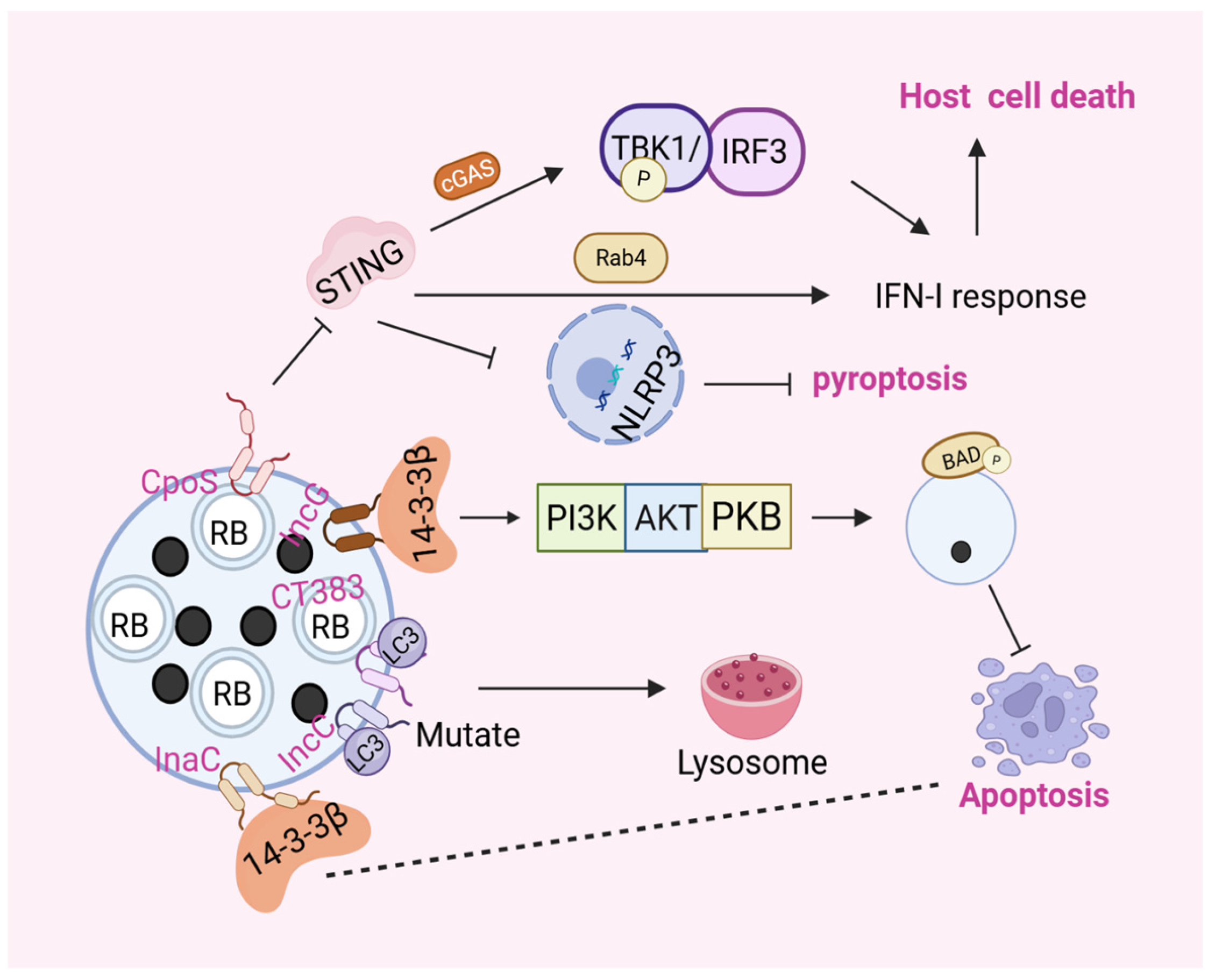
The completion of the replication cycle of C. trachomatis entirely depends on the host environment [63,64]. To maintain this process, it promotes the survival of host cells by inhibiting apoptosis [65,66]. Studies have demonstrated that the absence of specific Inc proteins including CpoS, IncC and CT383 causes premature lysis of C. trachomatis inclusions, triggering autolysosomal recognition, activation of endogenous apoptosis, and premature arrest of the C. trachomatis developmental cycle [67]. In addition to apoptosis regulation, Inc proteins broadly remodel multiple host cell death programs, including pyroptosis, apoptosis, and autophagy. These functionally distinct Inc proteins work in concert to govern the central signaling axis reliant on stimulator of interferon genes (STING), the key cytosolic DNA sensor adaptor. Via stage-specific tuning of host cell death programs, these proteins promote sustained intracellular colonization of C. trachomatis (Figure 2).
To avoid excessive premature pyroptosis in early infection, CpoS serves as a critical anti-pyroptotic effector. CpoS interacts with the GTPase Rab4 to target ER-localized STING, thereby suppressing type I interferon responses and pro-death signaling to block early host cell death [68]. CpoS loss-of-function mutations trigger STING activation and its translocation from the ER to perinuclear vesicles, which activates the cGAS/STING/TBK1/IRF3 pathway to induce robust type I interferon responses. As a central signaling hub, STING bridges interferon signaling and inflammasome activation, and the cGAS–STING pathway tightly restrains NLRP3 inflammasome assembly. Upon full activation, the NLRP3 inflammasome triggers caspase-1 maturation; activated caspase-1 subsequently cleaves gasdermin D (GSDMD), which oligomerizes to form plasma membrane pores and ultimately executes host pyroptosis [67,68,69]. Notably, direct evidence verifying that CpoS restrains pyroptosis specifically through the cGAS-STING axis remains insufficient [68]. Independently of inflammasome signaling, STING binds the ER calcium pump SERCA2 to disrupt ER calcium homeostasis and trigger cell death, and pharmacological SERCA inhibition alleviates the cytotoxicity caused by CpoS-deficient C. trachomatis infection [68,70]. Overall, STING integrates multiple cell fate regulatory networks and is differentially manipulated by distinct Inc proteins throughout infection.
In contrast to the early pro-survival function of CpoS, the Inc protein GarD mediates late-stage STING-dependent host cell lysis to support chlamydial proliferation. During the late developmental stage of C. trachomatis inclusions, GarD facilitates STING translocation from the ER to the Golgi apparatus, which initiates host cell lysis [71,72]. GarD overexpression further disrupts inclusion membrane integrity and aggravates host cell death, highlighting its pro-lytic role in late infection [71].
Multiple Inc proteins synergistically inhibit host apoptosis to stabilize intracellular C. trachomatis survival. IncC and CT383 preserve inclusion membrane stability to prevent premature inclusion lysis and host cell death. Defective IncC or CT383 function results in LC3 labeling of impaired inclusions, followed by autolysosomal degradation and endogenous host apoptosis [67,68]. This dynamic interplay between IncC/CT383 and host autophagy is pivotal for maintaining intracellular infection homeostasis, although the detailed mechanism linking their anti-apoptotic function to STING signaling remains uncharacterized. In addition to membrane-stabilizing anti-apoptotic mechanisms, IncG (CT118) regulates host apoptosis by binding the host adaptor protein 14-3-3β [67,73]. The IncG-14-3-3β interaction activates the PI3K/AKT signaling cascade, which maintains phosphorylation of the pro-apoptotic factor BAD and suppresses its pro-death activity. Phosphorylated BAD is sequestered at C. trachomatis inclusions, preventing mitochondrial cytochrome c release and inhibiting host intrinsic apoptosis [73,74]. Studies related to immunoprecipitation reactions and mass spectrometry analysis have revealed the binding of the ε, η, ζ, γ, θ, and β subtypes of 14-3-3β to InaC [23]. To date, no available evidence has functionally linked the interaction between InaC and 14-3-3β to host apoptotic modulation; nevertheless, this physical association provides a promising research direction, and subsequent investigations may uncover unrecognized roles of InaC in C. trachomatis-governed apoptotic pathways.
Some studies predict that factors such as CT227, CT058, CrpA, and CT449 may be related to the regulation of apoptosis [27]. However, currently, there is no specific literature elaborating in detail. Regardless of whether C. trachomatis promotes or inhibits host cell apoptosis, these mechanisms all represent evolutionary adaptations that enable its survival within host cells.

6. Orchestrate the Host Cell Immune Response

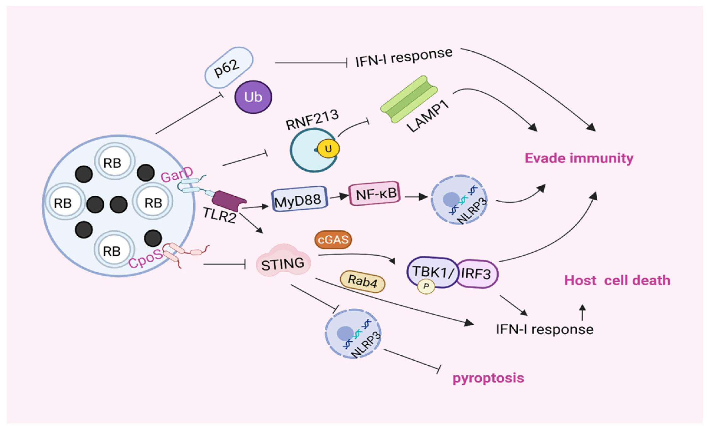
Inc proteins actively mediate immune evasion of C. trachomatis through distinct molecular mechanisms to counteract host immune surveillance and support intracellular bacterial replication and persistent infection (Figure 3). GarD interferes with IFN-γ-triggered cell-autonomous immunity and neutrophil-mediated defense, while CpoS represses STING-dependent type I interferon signaling. Distinct molecular mechanisms empower C. trachomatis to evade host immune attack and develop persistent infection.
The γ-resistance determinant GarD (CT135) of C. trachomatis Inc protects the inclusion from the attack of cellular autonomous immunity and is a genuine immune evasion factor [75]. A key ubiquitin E3 ligase, RNF213, effectively labels the inclusion and mediates the downstream clearance process. GarD prevents RNF213 from targeting the inclusion, thereby blocking the IFN-γ-dependent ubiquitin-mediated inclusion labeling process, preventing the binding of lysosome-associated membrane protein 1 (LAMP1) to the inclusion, protecting the inclusion from ubiquitin-dependent destruction, and enabling it to evade the cellular autonomous immunity activated by IFN-γ [75,76,77]. As a crucial Inc protein, GarD acts through the above mechanism to inhibit the recruitment of ubiquitin and p62/SQSTM to the inclusion surface. This immune evasion strategy is conserved across human and nonhuman primate hosts, and a loss of GarD severely attenuates C. trachomatis growth and survival both in vitro and in vivo [78]. These findings highlight GarD as an essential virulence determinant that allows C. trachomatis inclusions to evade host degradation and establish productive infection.
Moreover, GarD specifically activates the NLRP3 inflammasome in neutrophils during the infection of C. trachomatis, thus evading the host defense mediated by neutrophils [71,79]. CpoS (CT229) is a critical suppressor of host cellular immune surveillance and regulates the IFN signaling pathway through interaction with Rab proteins. The CpoS-Rab interaction effectively suppresses the type I interferon response mediated by STING, and weakens the host’s defense mechanism by deeply participating in blocking the host immune signaling pathway [46,77,80]. Overall, these mechanisms reflect the sophisticated strategy adopted by C. trachomatis to manipulate host immune responses for intracellular survival and proliferation.

7. Coordinate the Production and Release of EBs

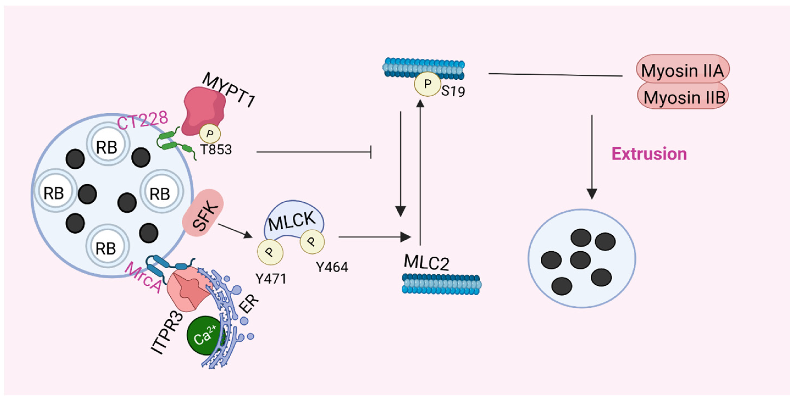
After completion of the C. trachomatis replication cycle, EBs are released from host cells via two routes: cell lysis (rupture of both the inclusions and host cells) or exocytosis (inclusions enveloped by the host plasma membrane are expelled from the host cells) [27,62]. Extrusion represents an active release process dependent on actin cytoskeleton remodeling and functional Inc proteins [81], and vesicles formed during extrusion protect extracellular bacteria to improve the survival of C. trachomatis [82]. CT228 and MrcA, two representative Inc proteins, orchestrate inclusion extrusion and egress of C. trachomatis. By interacting with host molecules, they regulate myosin light chain 2 (MLC2) phosphorylation, calcium homeostasis and cytoskeleton dynamics (Figure 4). Notably, GarD mediates bacterial release via the STING-dependent pathway.
CT228 recruits myosin phosphatase targeting subunit 1 (MYPT1) to the microdomains around the inclusion and regulates the extrusion and release of EBs [83]. Research by Lutter E.I. et al. indicates that in the early stage of C. trachomatis infection, the interaction between CT228 and MYPT1 prevents the phosphorylation of MYPT1, and MLC2 is dephosphorylated and inactivated, thus preventing the premature extrusion of inclusions. In the later stage of the developmental cycle, phosphorylated but inactive MYPT1 enriches in the microdomains, which is conducive to maintaining phosphorylated and active myosin light chain 2 (MLC2) and myosin light chain kinase (MLCK). MLC2 interacts with myosin IIA and myosin IIB to form an active myosin motor complex, which promotes the expulsion of inclusions and initiates a new round of the infection cycle [83,84].
Ca2+ signaling plays a pivotal role in the regulation of extrusion. The combined effect of increased intracellular Ca2+ leads to the activation of MLCK and the inhibition of antagonistic myosin phosphatase, favoring phosphorylation, which promotes extrusion [84]. Inositol 1,4,5-trisphosphate receptor type 3 (ITPR3) is an ER cation channel that conducts Ca2+ and is involved in calcium signaling [85]. STIM1 controls the intracellular Ca2+ level and is crucial for regulating store-operated calcium entry (SOCE) [85,86]. Therefore, both the calcium channel ITPR3 and the Ca2+ sensor STIM1 are of great importance during the process of exocytosis formation. The Inc protein MrcA binds to ITPR3 to regulate the calcium level, thereby promoting the extrusion and release [87]. MrcA recruits ITPR3 to the microdomain, where it regulates the release of Ca2+ from the reservoir by interacting with STIM1 and ITPR3, increasing the concentration of Ca2+ in the cytoplasm. The increase in the Ca2+ concentration not only promotes the phosphorylation of MLC2 but also activates Rho kinase, which in turn phosphorylates MYPT1, thus maintaining the activity of the myosin motor complex to facilitate the exocytosis process of C. trachomatis. Loss of MrcA function reduces the recruitment of ITPR3, which consequently restricts inclusion extrusion and egress [84]. IncS also specifically interacts with and recruits STIM1, which contributes to maintaining Ca2+ homeostasis [56].
Furthermore, InaC-dependent recruitment of F-actin to the inclusion membrane may also contribute to optimal extrusion [81], and IPAM is also considered a foci of extrusion [84]. GarD exits the bacterium via lysis during late stages of the developmental cycle in a STING-dependent manner [71].

8. Summary and Prospect

Benefiting from synergistic advances in bacterial genetics, structural biology and cell biology, substantial progress has been made in the functional characterization of Inc proteins. Table 1 summarizes known Inc proteins of C. trachomatis, highlighting distinct differences in their expression cycles, target host molecules of action, and main regulatory or functional roles in host cells during the bacterial intracellular developmental cycle.
However, several unresolved bottlenecks impede precise dissection of molecular cascades within Inc-centered regulatory networks and restrict comprehensive understanding of Inc-mediated host–pathogen interaction mechanisms. Firstly, most existing studies concentrate on the independent functions of individual Inc family proteins, while synergistic and antagonistic crosstalk among distinct Inc proteins remains barely explored. Second, the scarcity of in vivo experimental data on the Inc protein family severely limits the translational potential of fundamental mechanistic research on C. trachomatis, as most relevant observations are derived from simplified in vitro culture models that cannot recapitulate authentic tissue microenvironments of human infection. Finally, well-documented chlamydia–host metabolic crosstalk, key molecules governing pathogen developmental shifts, intracellular signal sensing and host immune responses remain unknown, and Inc proteins acting as central linkers of these processes are essential to decipher such complex pathogenic mechanisms.
In terms of technical methodologies, conventional biochemical strategies, including co-immunoprecipitation, affinity purification and site-directed mutagenesis, have been widely applied to verify interactions among C. trachomatis Inc proteins, bacterial effectors and host cellular factors. However, these traditional approaches fail to resolve the precise binding interfaces, interaction affinity and dynamic conformational remodeling of protein complexes during C. trachomatis infection. Cryo-electron microscopy (cryo-EM) overcomes these technical barriers and enables high-resolution structural dissection of Inc-centered supramolecular assemblies. Future cryo-EM-based investigations will facilitate the delineation of the spatial architecture of inclusion membrane–effector–host complexes, the identification of key binding residues and interfacial domains, and the comprehensive characterization of dynamic interaction landscapes throughout the entire C. trachomatis infection cycle.
Future research frameworks can integrate cryo-EM-based structural biology with state-of-the-art technologies, including single-cell RNA sequencing, quantitative proteomics and metagenomic next-generation sequencing (mNGS), together with optimized genetic manipulation systems and physiologically relevant infection models based on primary cells and organoids [101,102]. This multi-dimensional analytical platform will substantially advance the mechanistic understanding of the intricate pathogenic networks of C. trachomatis. Integrated structural and functional profiling of diverse Inc proteins will further elucidate their synergistic roles in regulating host cytoskeleton organization and centrosome homeostasis.
Collectively, while current studies have mapped basic functions of Inc proteins, integrated structural, multi-omics and physiologically authentic infection models are urgently required to address outstanding research gaps and fully decode the sophisticated Inc-dependent pathogenic mechanisms of C. trachomatis.

Author Contributions

Y.G. and J.X.: writing—original draft preparation, and writing—review and editing. B.S.: visualization. Y.X.: supervision. L.Z.: project administration, funding acquisition. All authors have read and agreed to the published version of the manuscript.

Funding

This work was supported by the National Natural Science Foundation of China (No. 82402137), This work was supported by Educational Committee Foundation of Hunan Province (23A0328). Undergraduate Training Programs for Innovation and Entrepreneurship of Hunan Province (210XCX498).

Institutional Review Board Statement

Not applicable.

Informed Consent Statement

Not applicable.

Data Availability Statement

No new data were created or analyzed in this study. Data sharing is not applicable to this article.

Conflicts of Interest

The authors declare no conflicts of interest.

References

  1. Dzakah, E.E.; Huang, L.; Xue, Y.; Wei, S.; Wang, X.; Chen, H.; Shui, J.; Kyei, F.; Rashid, F.; Zheng, H.; et al. Host cell response and distinct gene expression profiles at different stages of Chlamydia trachomatis infection reveals stage-specific biomarkers of infection. BMC Microbiol. 2021, 21, 3. [Google Scholar] [CrossRef] [Scilit] [PubMed]
  2. Geisler, W.M.; Hocking, J.S.; Darville, T.; Batteiger, B.E.; Brunham, R.C. Diagnosis and Management of Uncomplicated Chlamydia trachomatis Infections in Adolescents and Adults: Summary of Evidence Reviewed for the 2021 Centers for Disease Control and Prevention Sexually Transmitted Infections Treatment Guidelines. Clin. Infect. Dis. 2022, 74, S112–S126. [Google Scholar] [CrossRef] [Scilit] [PubMed]
  3. Gauliard, E.; Ouellette, S.P.; Rueden, K.J.; Ladant, D. Characterization of interactions between inclusion membrane proteins from Chlamydia trachomatis. Front. Cell. Infect. Microbiol. 2015, 5, 13. [Google Scholar] [CrossRef] [Scilit] [PubMed]
  4. Chen, H.; Wang, L.; Zhao, L.; Luo, L.; Min, S.; Wen, Y.; Lei, W.; Shu, M.; Li, Z. Alterations of Vaginal Microbiota in Women with Infertility and Chlamydia trachomatis Infection. Front. Cell. Infect. Microbiol. 2021, 11, 698840. [Google Scholar] [CrossRef] [Scilit] [PubMed]
  5. Zhang, Q.; Peng, L.; Yuan, Y.; Hu, Z.; Zeng, Y.; Zeng, W.; Chen, J.; Chen, W.; Liu, P. High rates of Treponema pallidum, Neisseria gonorrhoeae, Chlamydia trachomatis, or Trichomonas vaginalis co-infection in people with HIV: A systematic review and meta-analysis. Eur. J. Clin. Microbiol. Infect. Dis. 2025, 44, 1–15. [Google Scholar] [CrossRef] [Scilit] [PubMed]
  6. Chen, H.; Wen, Y.; Li, Z. Clear Victory for Chlamydia: The Subversion of Host Innate Immunity. Front. Microbiol. 2019, 10, 1412. [Google Scholar] [CrossRef] [Scilit] [PubMed]
  7. Weber, M.M.; Bauler, L.D.; Lam, J.; Hackstadt, T. Expression and localization of predicted inclusion membrane proteins in Chlamydia trachomatis. Infect. Immun. 2015, 83, 4710–4718. [Google Scholar] [CrossRef] [Scilit] [PubMed]
  8. Dehoux, P.; Flores, R.; Dauga, C.; Zhong, G.; Subtil, A. Multi-genome identification and characterization of chlamydiae-specific type III secretion substrates: The Inc proteins. BMC Genom. 2011, 12, 109. [Google Scholar] [CrossRef] [Scilit] [PubMed]
  9. Mital, J.; Miller, N.J.; Fischer, E.R.; Hackstadt, T. Specific chlamydial inclusion membrane proteins associate with active Src family kinases in microdomains that interact with the host microtubule network. Cell Microbiol. 2010, 12, 1235–1249. [Google Scholar] [CrossRef] [Scilit] [PubMed]
  10. Mital, J.; Hackstadt, T. Diverse requirements for SRC-family tyrosine kinases distinguish chlamydial species. mBio 2011, 2, e00031-11. [Google Scholar] [CrossRef] [Scilit] [PubMed]
  11. Hou, C.; Jin, Y.; Wu, H.; Li, P.; Liu, L.; Zheng, K.; Wang, C. Alternative strategies for Chlamydia treatment: Promising non-antibiotic approaches. Front. Microbiol. 2022, 13, 987662. [Google Scholar] [CrossRef] [Scilit] [PubMed]
  12. Chen, Q.; Li, Y.; Yan, X.; Sun, Z.; Wang, C.; Liu, S.; Xiao, J.; Lu, C.; Wu, Y. Chlamydia psittaci Plasmid-Encoded CPSIT_P7 Elicits Inflammatory Response in Human Monocytes via TLR4/Mal/MyD88/NF-κB Signaling Pathway. Front. Microbiol. 2020, 11, 578009. [Google Scholar] [CrossRef] [Scilit] [PubMed]
  13. Kumar, Y.; Valdivia, R.H. Actin and intermediate filaments stabilize the Chlamydia trachomatis vacuole by forming dynamic structural scaffolds. Cell Host Microbe 2008, 4, 159–169. [Google Scholar] [CrossRef] [Scilit] [PubMed]
  14. Al-Zeer, M.A.; Al-Younes, H.M.; Kerr, M.; Abu-Lubad, M.; Gonzalez, E.; Brinkmann, V.; Meyer, T.F. Chlamydia trachomatis remodels stable microtubules to coordinate Golgi stack recruitment to the chlamydial inclusion surface. Mol. Microbiol. 2014, 94, 1285–1297. [Google Scholar] [CrossRef] [Scilit] [PubMed]
  15. El Alaoui, F.; Al-Akiki, I.; Ibanes, S.; Lyonnais, S.; Sanchez-Fuentes, D.; Desgarceaux, R.; Cazevieille, C.; Blanchard, M.P.; Parmeggiani, A.; Carretero-Genevrier, A.; et al. Septin assemblies promote the lipid organization of membranes. Structure 2025, 33, 451–464.e5. [Google Scholar] [CrossRef] [Scilit] [PubMed]
  16. Andersen, S.E.; Bulman, L.M.; Steiert, B.; Faris, R.; Weber, M.M. Got mutants? How advances in chlamydial genetics have furthered the study of effector proteins. Pathog. Dis. 2021, 79, ftaa078. [Google Scholar] [CrossRef] [Scilit] [PubMed]
  17. Dumoux, M.; Menny, A.; Delacour, D.; Hayward, R.D. A Chlamydia effector recruits CEP170 to reprogram host microtubule organization. J. Cell Sci. 2015, 128, 3420–3434. [Google Scholar] [CrossRef] [Scilit] [PubMed]
  18. Alzhanov, D.T.; Weeks, S.K.; Burnett, J.R.; Rockey, D.D. Cytokinesis is blocked in mammalian cells transfected with Chlamydia trachomatis gene CT223. BMC Microbiol. 2009, 9, 2. [Google Scholar] [CrossRef] [Scilit] [PubMed]
  19. Chen, C.; Chen, D.; Sharma, J.; Cheng, W.; Zhong, Y.; Liu, K.; Jensen, J.; Shain, R.; Arulanandam, B.; Zhong, G. The hypothetical protein CT813 is localized in the Chlamydia trachomatis inclusion membrane and is immunogenic in women urogenitally infected with C. trachomatis. Infect. Immun. 2006, 74, 4826–4840. [Google Scholar] [CrossRef] [Scilit] [PubMed]
  20. Heuer, D.; Rejman Lipinski, A.; Machuy, N.; Karlas, A.; Wehrens, A.; Siedler, F.; Brinkmann, V.; Meyer, T.F. Chlamydia causes fragmentation of the Golgi compartment to ensure reproduction. Nature 2009, 457, 731–735. [Google Scholar] [CrossRef] [Scilit] [PubMed]
  21. Linton, C.; Wesolowski, J.; Lobley, A.; Yamaji, T.; Hanada, K.; Paumet, F. Specialized contact sites regulate the fusion of chlamydial inclusion membranes. Nat. Commun. 2024, 15, 9250, Erratum in Nat. Commun. 2025, 16, 1454. https://doi.org/10.1038/s41467-025-56777-y. [Google Scholar] [CrossRef] [Scilit] [PubMed] [PubMed Central]
  22. Sherry, J.; Pawar, K.I.; Dolat, L.; Smith, E.; Chang, I.C.; Pha, K.; Kaake, R.; Swaney, D.L.; Herrera, C.; McMahon, E.; et al. The Chlamydia effector Dre1 binds dynactin to reposition host organelles during infection. Cell Rep. 2025, 44, 115509. [Google Scholar] [CrossRef] [Scilit] [PubMed]
  23. Kokes, M.; Dunn, J.D.; Granek, J.A.; Nguyen, B.D.; Barker, J.R.; Valdivia, R.H.; Bastidas, R.J. Integrating chemical mutagenesis and whole-genome sequencing as a platform for forward and reverse genetic analysis of Chlamydia. Cell Host Microbe 2015, 17, 716–725. [Google Scholar] [CrossRef] [Scilit] [PubMed]
  24. Haines, A.; Wesolowski, J.; Ryan, N.M.; Monteiro-Brás, T.; Paumet, F. Cross Talk between ARF1 and RhoA Coordinates the Formation of Cytoskeletal Scaffolds during Chlamydia Infection. mBio 2021, 12, e0239721. [Google Scholar] [CrossRef] [Scilit] [PubMed]
  25. Wesolowski, J.; Weber, M.M.; Nawrotek, A.; Dooley, C.A.; Calderon, M.; St Croix, C.M.; Hackstadt, T.; Cherfils, J.; Paumet, F. Chlamydia Hijacks ARF GTPases to Coordinate Microtubule Posttranslational Modifications and Golgi Complex Positioning. mBio 2017, 8, e02280-16. [Google Scholar] [CrossRef] [Scilit] [PubMed]
  26. Haines, A.; Wesolowski, J.; Paumet, F. Chlamydia trachomatis Subverts Alpha-Actinins to Stabilize Its Inclusion. Microbiol. Spectr. 2023, 11, e0261422. [Google Scholar] [CrossRef] [Scilit] [PubMed]
  27. Bugalhão, J.N.; Mota, L.J. The multiple functions of the numerous Chlamydia trachomatis secreted proteins: The tip of the iceberg. Microb. Cell 2019, 6, 414–449. [Google Scholar] [CrossRef] [Scilit] [PubMed]
  28. Greene, W.; Zhong, G. Inhibition of host cell cytokinesis by Chlamydia trachomatis infection. J. Infect. 2003, 47, 45–51. [Google Scholar] [CrossRef] [Scilit] [PubMed]
  29. Knowlton, A.E.; Brown, H.M.; Richards, T.S.; Andreolas, L.A.; Patel, R.K.; Grieshaber, S.S. Chlamydia trachomatis infection causes mitotic spindle pole defects independently from its effects on centrosome amplification. Traffic 2011, 12, 854–866. [Google Scholar] [CrossRef] [Scilit] [PubMed]
  30. Almeida, F.; Luís, M.P.; Pereira, I.S.; Pais, S.V.; Mota, L.J. The Human Centrosomal Protein CCDC146 Binds Chlamydia trachomatis Inclusion Membrane Protein CT288 and Is Recruited to the Periphery of the Chlamydia-Containing Vacuole. Front. Cell. Infect. Microbiol. 2018, 8, 254. [Google Scholar] [CrossRef] [Scilit] [PubMed]
  31. Mital, J.; Lutter, E.I.; Barger, A.C.; Dooley, C.A.; Hackstadt, T. Chlamydia trachomatis inclusion membrane protein CT850 interacts with the dynein light chain DYNLT1 (Tctex1). Biochem. Biophys. Res. Commun. 2015, 462, 165–170. [Google Scholar] [CrossRef] [Scilit] [PubMed]
  32. Bugalhão, J.N.; Luís, M.P.; Pereira, I.S.; da Cunha, M.; Pais, S.V.; Mota, L.J. The Chlamydia trachomatis inclusion membrane protein CT006 associates with lipid droplets in eukaryotic cells. PLoS ONE 2022, 17, e0264292. [Google Scholar] [CrossRef] [Scilit] [PubMed]
  33. Faris, R.; Merling, M.; Andersen, S.E.; Dooley, C.A.; Hackstadt, T.; Weber, M.M. Chlamydia trachomatis CT229 Subverts Rab GTPase-Dependent CCV Trafficking Pathways to Promote Chlamydial Infection. Cell Rep. 2019, 26, 3380–3390.e5. [Google Scholar] [CrossRef] [Scilit] [PubMed]
  34. Wenbo, L.; Yewei, Y.; Hui, Z.; Zhongyu, L. Hijacking host cell vesicular transport: New insights into the nutrient acquisition mechanism of Chlamydia. Virulence 2024, 15, 2351234. [Google Scholar] [CrossRef] [Scilit] [PubMed]
  35. Cingolani, G.; McCauley, M.; Lobley, A.; Bryer, A.J.; Wesolowski, J.; Greco, D.L.; Lokareddy, R.K.; Ronzone, E.; Perilla, J.R.; Paumet, F. Structural basis for the homotypic fusion of chlamydial inclusions by the SNARE-like protein IncA. Nat. Commun. 2019, 10, 2747. [Google Scholar] [CrossRef] [Scilit] [PubMed]
  36. Delevoye, C.; Nilges, M.; Dehoux, P.; Paumet, F.; Perrinet, S.; Dautry-Varsat, A.; Subtil, A. SNARE protein mimicry by an intracellular bacterium. PLoS Pathog. 2008, 4, e1000022. [Google Scholar] [CrossRef] [Scilit] [PubMed]
  37. Babu Sait, M.R.; Jachmann, L.H.; Türköz, G.; Milivojevic, M.; Llorente-Sáez, C.; Dhanjal, S.; Schumacher, F.; Henriksson, S.; Gayathri Vegesna, N.V.; Seddik, N.; et al. Genome-wide identification of modulators of Chlamydia trachomatis parasitophorous vacuole stability highlights an important role for sphingolipid supply. PLoS Biol. 2025, 23, e3003297. [Google Scholar] [CrossRef] [Scilit] [PubMed]
  38. Pha, K.; Mirrashidi, K.; Sherry, J.; Tran, C.J.; Herrera, C.M.; McMahon, E.; Elwell, C.A.; Engel, J.N. The Chlamydia effector IncE employs two short linear motifs to reprogram host vesicle trafficking. Cell Rep. 2024, 43, 114624. [Google Scholar] [CrossRef] [Scilit] [PubMed]
  39. Donaldson, J.G.; Jackson, C.L. ARF family G proteins and their regulators: Roles in membrane transport, development and disease. Nat. Rev. Mol. Cell Biol. 2011, 12, 362–375, Erratum in Nat. Rev. Mol. Cell Biol. 2011, 12, 533. [Google Scholar] [CrossRef] [Scilit] [PubMed] [PubMed Central]
  40. Stenmark, H. Rab GTPases as coordinators of vesicle traffic. Nat. Rev. Mol. Cell Biol. 2009, 10, 513–525. [Google Scholar] [CrossRef] [Scilit] [PubMed]
  41. Rzomp, K.A.; Scholtes, L.D.; Briggs, B.J.; Whittaker, G.R.; Scidmore, M.A. Rab GTPases are recruited to chlamydial inclusions in both a species-dependent and species-independent manner. Infect. Immun. 2003, 71, 5855–5870. [Google Scholar] [CrossRef] [Scilit] [PubMed]
  42. Capmany, A.; Damiani, M.T. Chlamydia trachomatis intercepts Golgi-derived sphingolipids through a Rab14-mediated transport required for bacterial development and replication. PLoS ONE 2010, 5, e14084. [Google Scholar] [CrossRef] [Scilit] [PubMed]
  43. Gambarte Tudela, J.; Buonfigli, J.; Luján, A.; Alonso Bivou, M.; Cebrián, I.; Capmany, A.; Damiani, M.T. Rab39a and Rab39b Display Different Intracellular Distribution and Function in Sphingolipids and Phospholipids Transport. Int. J. Mol. Sci. 2019, 20, 1688. [Google Scholar] [CrossRef] [Scilit] [PubMed]
  44. Weber, M.M.; Noriea, N.F.; Bauler, L.D.; Lam, J.L.; Sager, J.; Wesolowski, J.; Paumet, F.; Hackstadt, T. A Functional Core of IncA Is Required for Chlamydia trachomatis Inclusion Fusion. J. Bacteriol. 2016, 198, 1347–1355. [Google Scholar] [CrossRef] [Scilit] [PubMed]
  45. Stelzner, K.; Vollmuth, N.; Rudel, T. Intracellular lifestyle of Chlamydia trachomatis and host-pathogen interactions. Nat. Rev. Microbiol. 2023, 21, 448–462. [Google Scholar] [CrossRef] [Scilit] [PubMed]
  46. Meier, K.; Jachmann, L.H.; Türköz, G.; Babu Sait, M.R.; Pérez, L.; Kepp, O.; Valdivia, R.H.; Kroemer, G.; Sixt, B.S. The Chlamydia effector CpoS modulates the inclusion microenvironment and restricts the interferon response by acting on Rab35. mBio 2023, 14, e0319022. [Google Scholar] [CrossRef] [Scilit] [PubMed]
  47. Rzomp, K.A.; Moorhead, A.R.; Scidmore, M.A. The GTPase Rab4 interacts with Chlamydia trachomatis inclusion membrane protein CT229. Infect. Immun. 2006, 74, 5362–5373. [Google Scholar] [CrossRef] [Scilit] [PubMed]
  48. Mirrashidi, K.M.; Elwell, C.A.; Verschueren, E.; Johnson, J.R.; Frando, A.; Von Dollen, J.; Rosenberg, O.; Gulbahce, N.; Jang, G.; Johnson, T.; et al. Global Mapping of the Inc-Human Interactome Reveals that Retromer Restricts Chlamydia Infection. Cell Host Microbe 2015, 18, 109–121. [Google Scholar] [CrossRef] [Scilit] [PubMed]
  49. Aeberhard, L.; Banhart, S.; Fischer, M.; Jehmlich, N.; Rose, L.; Koch, S.; Laue, M.; Renard, B.Y.; Schmidt, F.; Heuer, D. The Proteome of the Isolated Chlamydia trachomatis Containing Vacuole Reveals a Complex Trafficking Platform Enriched for Retromer Components. PLoS Pathog. 2015, 11, e1004883. [Google Scholar] [CrossRef] [Scilit] [PubMed]
  50. Sun, Q.; Yong, X.; Sun, X.; Yang, F.; Dai, Z.; Gong, Y.; Zhou, L.; Zhang, X.; Niu, D.; Dai, L.; et al. Structural and functional insights into sorting nexin 5/6 interaction with bacterial effector IncE. Signal Transduct. Target. Ther. 2017, 2, 17030. [Google Scholar] [CrossRef] [Scilit] [PubMed]
  51. Pfeffer, S.R. Multiple routes of protein transport from endosomes to the trans Golgi network. FEBS Lett. 2009, 583, 3811–3816. [Google Scholar] [CrossRef] [Scilit] [PubMed]
  52. Wen, Y.; Lei, W.; Liu, Y.; Sheng, Y.; Li, R.; Li, Z. Endoplasmic reticulum: The target of chlamydial manipulation. Arch. Microbiol. 2025, 207, 219. [Google Scholar] [CrossRef] [Scilit] [PubMed]
  53. Derré, I.; Swiss, R.; Agaisse, H. The lipid transfer protein CERT interacts with the Chlamydia inclusion protein IncD and participates to ER-Chlamydia inclusion membrane contact sites. PLoS Pathog. 2011, 7, e1002092. [Google Scholar] [CrossRef] [Scilit] [PubMed]
  54. Murray, R.; Flora, E.; Bayne, C.; Derré, I. IncV, a FFAT motif-containing Chlamydia protein, tethers the endoplasmic reticulum to the pathogen-containing vacuole. Proc. Natl. Acad. Sci. USA 2017, 114, 12039–12044. [Google Scholar] [CrossRef] [Scilit] [PubMed]
  55. Elwell, C.A.; Jiang, S.; Kim, J.H.; Lee, A.; Wittmann, T.; Hanada, K.; Melancon, P.; Engel, J.N. Chlamydia trachomatis co-opts GBF1 and CERT to acquire host sphingomyelin for distinct roles during intracellular development. PLoS Pathog. 2011, 7, e1002198, Erratum in PLoS Pathog. 2013, 9. https://doi.org/10.1371/annotation/f8e7c7e3-c347-4243-9146-db77900cb90c. [Google Scholar] [CrossRef] [Scilit] [PubMed]
  56. Mital, J.; Miller, N.J.; Dorward, D.W.; Dooley, C.A.; Hackstadt, T. Role for chlamydial inclusion membrane proteins in inclusion membrane structure and biogenesis. PLoS ONE 2013, 8, e63426. [Google Scholar] [CrossRef] [Scilit] [PubMed]
  57. Banhart, S.; Schäfer, E.K.; Gensch, J.M.; Heuer, D. Sphingolipid Metabolism and Transport in Chlamydia trachomatis and Chlamydia psittaci Infections. Front. Cell Dev. Biol. 2019, 7, 223. [Google Scholar] [CrossRef] [Scilit] [PubMed]
  58. Kumagai, K.; Elwell, C.A.; Ando, S.; Engel, J.N.; Hanada, K. Both the N- and C- terminal regions of the Chlamydial inclusion protein D (IncD) are required for interaction with the pleckstrin homology domain of the ceramide transport protein CERT. Biochem. Biophys. Res. Commun. 2018, 505, 1070–1076. [Google Scholar] [CrossRef] [Scilit] [PubMed]
  59. Ende, R.J.; Murray, R.L.; D’Spain, S.K.; Coppens, I.; Derré, I. Phosphoregulation accommodates Type III secretion and assembly of a tether of ER-Chlamydia inclusion membrane contact sites. eLife 2022, 11, e74535. [Google Scholar] [CrossRef] [Scilit] [PubMed]
  60. Agaisse, H.; Derré, I. STIM1 Is a Novel Component of ER-Chlamydia trachomatis Inclusion Membrane Contact Sites. PLoS ONE 2015, 10, e0125671. [Google Scholar] [CrossRef] [Scilit] [PubMed]
  61. Cortina, M.E.; Bishop, R.C.; DeVasure, B.A.; Coppens, I.; Derré, I. The inclusion membrane protein IncS is critical for initiation of the Chlamydia intracellular developmental cycle. PLoS Pathog. 2022, 18, e1010818. [Google Scholar] [CrossRef] [Scilit] [PubMed]
  62. Cortina, M.E.; Derré, I. Homologues of the Chlamydia trachomatis and Chlamydia muridarum Inclusion Membrane Protein IncS Are Interchangeable for Early Development but Not for Inclusion Stability in the Late Developmental Cycle. mSphere 2023, 8, e0000323. [Google Scholar] [CrossRef] [Scilit] [PubMed]
  63. Yang, Y.; Lei, W.; Zhao, L.; Wen, Y.; Li, Z. Insights into Mitochondrial Dynamics in Chlamydial Infection. Front. Cell. Infect. Microbiol. 2022, 12, 835181. [Google Scholar] [CrossRef] [Scilit] [PubMed]
  64. Wen, Y.; Luo, F.; Zhao, L.; Su, S.; Lei, W.; Liu, Y.; Shi, K.; Li, Z. Long Non-Coding RNA FGD5-AS1 Induced by Chlamydia trachomatis Infection Inhibits Apoptosis via Wnt/β-Catenin Signaling Pathway. Front. Cell. Infect. Microbiol. 2021, 11, 701352. [Google Scholar] [CrossRef] [Scilit] [PubMed]
  65. Wen, Y.; Chen, H.; Luo, F.; Zhao, L.; Shu, M.; Su, S.; Zhao, Y.; Huang, Q.; Li, Z. Chlamydia trachomatis Plasmid Protein pORF5 Up-Regulates ZFAS1 to Promote Host Cell Survival via MAPK/p38 Pathway. Front. Microbiol. 2020, 11, 593295. [Google Scholar] [CrossRef] [Scilit] [PubMed]
  66. Tang, T.; Wu, H.; Chen, X.; Chen, L.; Liu, L.; Li, Z.; Bai, Q.; Chen, Y.; Chen, L. The Hypothetical Inclusion Membrane Protein CPSIT_0846 Regulates Mitochondrial-Mediated Host Cell Apoptosis via the ERK/JNK Signaling Pathway. Front. Cell Infect. Microbiol. 2021, 11, 607422. [Google Scholar] [CrossRef] [Scilit] [PubMed]
  67. Weber, M.M.; Lam, J.L.; Dooley, C.A.; Noriea, N.F.; Hansen, B.T.; Hoyt, F.H.; Carmody, A.B.; Sturdevant, G.L.; Hackstadt, T. Absence of Specific Chlamydia trachomatis Inclusion Membrane Proteins Triggers Premature Inclusion Membrane Lysis and Host Cell Death. Cell Rep. 2017, 19, 1406–1417. [Google Scholar] [CrossRef] [Scilit] [PubMed]
  68. Sixt, B.S.; Bastidas, R.J.; Finethy, R.; Baxter, R.M.; Carpenter, V.K.; Kroemer, G.; Coers, J.; Valdivia, R.H. The Chlamydia trachomatis Inclusion Membrane Protein CpoS Counteracts STING-Mediated Cellular Surveillance and Suicide Programs. Cell Host Microbe 2017, 21, 113–121. [Google Scholar] [CrossRef] [Scilit] [PubMed]
  69. Liu, J.; Zhou, J.; Luan, Y.; Li, X.; Meng, X.; Liao, W.; Tang, J.; Wang, Z. cGAS-STING, inflammasomes and pyroptosis: An overview of crosstalk mechanism of activation and regulation. Cell Commun. Signal. 2024, 22, 22. [Google Scholar] [CrossRef] [Scilit] [PubMed]
  70. Orrenius, S.; Gogvadze, V.; Zhivotovsky, B. Calcium and mitochondria in the regulation of cell death. Biochem. Biophys. Res. Commun. 2015, 460, 72–81. [Google Scholar] [CrossRef] [Scilit] [PubMed]
  71. Bishop, R.C.; Derré, I. The Chlamydia trachomatis Inclusion Membrane Protein CTL0390 Mediates Host Cell Exit via Lysis through STING Activation. Infect. Immun. 2022, 90, e0019022. [Google Scholar] [CrossRef] [Scilit] [PubMed]
  72. Dobbs, N.; Burnaevskiy, N.; Chen, D.; Gonugunta, V.K.; Alto, N.M.; Yan, N. STING Activation by Translocation from the ER Is Associated with Infection and Autoinflammatory Disease. Cell Host Microbe 2015, 18, 157–168. [Google Scholar] [CrossRef] [Scilit] [PubMed]
  73. Scidmore, M.A.; Hackstadt, T. Mammalian 14-3-3beta associates with the Chlamydia trachomatis inclusion membrane via its interaction with IncG. Mol. Microbiol. 2001, 39, 1638–1650. [Google Scholar] [CrossRef] [Scilit] [PubMed]
  74. Verbeke, P.; Welter-Stahl, L.; Ying, S.; Hansen, J.; Häcker, G.; Darville, T.; Ojcius, D.M. Recruitment of BAD by the Chlamydia trachomatis vacuole correlates with host-cell survival. PLoS Pathog. 2006, 2, e45. [Google Scholar] [CrossRef] [Scilit] [PubMed]
  75. Walsh, S.C.; Reitano, J.R.; Dickinson, M.S.; Kutsch, M.; Hernandez, D.; Barnes, A.B.; Schott, B.H.; Wang, L.; Ko, D.C.; Kim, S.Y.; et al. The bacterial effector GarD shields Chlamydia trachomatis inclusions from RNF213-mediated ubiquitylation and destruction. Cell Host Microbe 2022, 30, 1671–1684.e9. [Google Scholar] [CrossRef] [Scilit] [PubMed]
  76. Gilliland, H.N.; Olive, A.J. GarD-ing the pathogen-containing vacuole from destruction. Cell Host Microbe 2022, 30, 1655–1657. [Google Scholar] [CrossRef] [Scilit] [PubMed]
  77. Chen, X.; Yang, W.; Hu, Y.; Zhou, Y.; Zhou, Z. Role of IFN-γ from Different Immune Cells in Chlamydia Infection. Microorganisms 2025, 13, 2374. [Google Scholar] [CrossRef] [Scilit] [PubMed]
  78. Fernandez, M.C.; Sweeney, Y.C.; Suchland, R.J.; Carrell, S.J.; Soge, O.O.; Phan, I.Q.; Rockey, D.D.; Patton, D.L.; Hybiske, K. CT135 mediates the resistance of Chlamydia trachomatis to primate interferon gamma stimulated immune defenses. iScience 2024, 27, 110143. [Google Scholar] [CrossRef] [Scilit] [PubMed]
  79. Yang, C.; Lei, L.; Collins, J.W.M.; Briones, M.; Ma, L.; Sturdevant, G.L.; Su, H.; Kashyap, A.K.; Dorward, D.; Bock, K.W.; et al. Chlamydia evasion of neutrophil host defense results in NLRP3 dependent myeloid-mediated sterile inflammation through the purinergic P2X7 receptor. Nat. Commun. 2021, 12, 5454. [Google Scholar] [CrossRef] [Scilit] [PubMed]
  80. Sixt, B.S.; Valdivia, R.H.; Kroemer, G. Chlamydia trachomatis’ struggle to keep its host alive. Microb. Cell 2017, 4, 101–104. [Google Scholar] [CrossRef] [Scilit] [PubMed]
  81. Chin, E.; Kirker, K.; Zuck, M.; James, G.; Hybiske, K. Actin recruitment to the Chlamydia inclusion is spatiotemporally regulated by a mechanism that requires host and bacterial factors. PLoS ONE 2012, 7, e46949. [Google Scholar] [CrossRef] [Scilit] [PubMed]
  82. Zuck, M.; Sherrid, A.; Suchland, R.; Ellis, T.; Hybiske, K. Conservation of extrusion as an exit mechanism for Chlamydia. Pathog. Dis. 2016, 74, ftw093. [Google Scholar] [CrossRef] [Scilit] [PubMed]
  83. Lutter, E.I.; Barger, A.C.; Nair, V.; Hackstadt, T. Chlamydia trachomatis inclusion membrane protein CT228 recruits elements of the myosin phosphatase pathway to regulate release mechanisms. Cell Rep. 2013, 3, 1921–1931. [Google Scholar] [CrossRef] [Scilit] [PubMed]
  84. Nguyen, P.H.; Lutter, E.I.; Hackstadt, T. Chlamydia trachomatis inclusion membrane protein MrcA interacts with the inositol 1,4,5-trisphosphate receptor type 3 (ITPR3) to regulate extrusion formation. PLoS Pathog. 2018, 14, e1006911. [Google Scholar] [CrossRef] [Scilit] [PubMed]
  85. Chamberlain, N.B.; Dimond, Z.; Hackstadt, T. Chlamydia trachomatis suppresses host cell store-operated Ca(2+) entry and inhibits NFAT/calcineurin signaling. Sci. Rep. 2022, 12, 21406. [Google Scholar] [CrossRef] [Scilit] [PubMed]
  86. Soboloff, J.; Rothberg, B.S.; Madesh, M.; Gill, D.L. STIM proteins: Dynamic calcium signal transducers. Nat. Rev. Mol. Cell Biol. 2012, 13, 549–565. [Google Scholar] [CrossRef] [Scilit] [PubMed]
  87. Prole, D.L.; Taylor, C.W. Inositol 1,4,5-trisphosphate receptors and their protein partners as signalling hubs. J. Physiol. 2016, 594, 2849–2866. [Google Scholar] [CrossRef] [Scilit] [PubMed]
  88. Shaw, E.I.; Dooley, C.A.; Fischer, E.R.; Scidmore, M.A.; Fields, K.A.; Hackstadt, T. Three temporal classes of gene expression during the Chlamydia trachomatis developmental cycle. Mol. Microbiol. 2000, 37, 913–925. [Google Scholar] [CrossRef] [Scilit] [PubMed]
  89. Olson-Wood, M.G.; Jorgenson, L.M.; Ouellette, S.P.; Rucks, E.A. Inclusion Membrane Growth and Composition Are Altered by Overexpression of Specific Inclusion Membrane Proteins in Chlamydia trachomatis L2. Infect. Immun. 2021, 89, e0009421. [Google Scholar] [CrossRef] [Scilit] [PubMed]
  90. Agaisse, H.; Derré, I. Expression of the effector protein IncD in Chlamydia trachomatis mediates recruitment of the lipid transfer protein CERT and the endoplasmic reticulum-resident protein VAPB to the inclusion membrane. Infect. Immun. 2014, 82, 2037–2047. [Google Scholar] [CrossRef] [Scilit] [PubMed]
  91. Bui, D.C.; Jorgenson, L.M.; Ouellette, S.P.; Rucks, E.A. Eukaryotic SNARE VAMP3 Dynamically Interacts with Multiple Chlamydial Inclusion Membrane Proteins. Infect. Immun. 2021, 89, 22. [Google Scholar] [CrossRef] [Scilit] [PubMed]
  92. Suchland, R.J.; Rockey, D.D.; Bannantine, J.P.; Stamm, W.E. Isolates of Chlamydia trachomatis that occupy nonfusogenic inclusions lack IncA, a protein localized to the inclusion membrane. Infect. Immun. 2000, 68, 360–367. [Google Scholar] [CrossRef] [Scilit] [PubMed]
  93. Sturd, N.A.; Knight, L.A.; Wood, M.G.; Durham, L.; Ouellette, S.P.; Rucks, E.A. Chlamydia trachomatis Inc Ct226 is vital for FLI1 and LRRF1 recruitment to the chlamydial inclusion. mSphere 2024, 9, e0047324. [Google Scholar] [CrossRef] [Scilit] [PubMed]
  94. Shaw, J.H.; Key, C.E.; Snider, T.A.; Sah, P.; Shaw, E.I.; Fisher, D.J.; Lutter, E.I. Genetic Inactivation of Chlamydia trachomatis Inclusion Membrane Protein CT228 Alters MYPT1 Recruitment, Extrusion Production, and Longevity of Infection. Front. Cell. Infect. Microbiol. 2018, 8, 415, Erratum in Front. Cell Infect. Microbiol. 2023, 12, 1075768. https://doi.org/10.3389/fcimb.2022.1075768. [Google Scholar] [CrossRef] [Scilit] [PubMed] [PubMed Central]
  95. Kostrjukova, E.S.; Lazarev, V.N.; Titova, G.A.; Akopian, T.A.; Levitskii, S.A.; Govorun, V.M. Expression of Chlamydia trachomatis inclusion membrane protein genes IncB and IncC in Escherichia coli. Biochemistry 2006, 71, 262–269. [Google Scholar] [CrossRef] [Scilit] [PubMed]
  96. Luís, M.P.; Pereira, I.S.; Bugalhão, J.N.; Simões, C.N.; Mota, C.; Romão, M.J.; Mota, L.J. The Chlamydia trachomatis IncM Protein Interferes with Host Cell Cytokinesis, Centrosome Positioning, and Golgi Distribution and Contributes to the Stability of the Pathogen-Containing Vacuole. Infect. Immun. 2023, 91, e0040522. [Google Scholar] [CrossRef] [Scilit] [PubMed]
  97. Li, Z.; Huang, Q.; Su, S.; Zhou, Z.; Chen, C.; Zhong, G.; Wu, Y. Localization and characterization of the hypothetical protein CT440 in Chlamydia trachomatis-infected cells. Sci. China Life Sci. 2011, 54, 1048–1054. [Google Scholar] [CrossRef] [Scilit] [PubMed]
  98. Starnbach, M.N.; Loomis, W.P.; Ovendale, P.; Regan, D.; Hess, B.; Alderson, M.R.; Fling, S.P. An inclusion membrane protein from Chlamydia trachomatis enters the MHC class I pathway and stimulates a CD8+ T cell response. J. Immunol. 2003, 171, 4742–4749. [Google Scholar] [CrossRef] [Scilit] [PubMed]
  99. Fling, S.P.; Sutherland, R.A.; Steele, L.N.; Hess, B.; D’Orazio, S.E.; Maisonneuve, J.; Lampe, M.F.; Probst, P.; Starnbach, M.N. CD8+ T cells recognize an inclusion membrane-associated protein from the vacuolar pathogen Chlamydia trachomatis. Proc. Natl. Acad. Sci. USA 2001, 98, 1160–1165. [Google Scholar] [CrossRef] [Scilit] [PubMed]
  100. Spaeth, K.E.; Chen, Y.S.; Valdivia, R.H. The Chlamydia type III secretion system C-ring engages a chaperone-effector protein complex. PLoS Pathog. 2009, 5, e1000579, Erratum in PLoS Pathog. 2009, 5. https://doi.org/10.1371/annotation/7e073adb-7483-43be-88ec-e318399e76c2. [Google Scholar] [CrossRef] [Scilit] [PubMed]
  101. Wang, Y.; Arthur, E.W.; Liu, N.; Li, X.; Xiang, W.; Maxwell, A.; Li, Z.; Zhou, Z. iTRAQ-Based Quantitative Proteomics Analysis of HeLa Cells Infected with Chlamydia muridarum TC0668 Mutant and Wild-Type Strains. Front. Microbiol. 2019, 10, 2553. [Google Scholar] [CrossRef] [Scilit] [PubMed]
  102. Chen, L.; Peng, W.; Lan, G.; Long, G.; Yang, H.; Xu, Y.; Fu, A.; Yi, H.; Wan, Q. The application of metagenomic next-generation sequencing in patients with infection or colonization caused by Lichtheimia species. Front. Cell Infect. Microbiol. 2023, 13, 1103626. [Google Scholar] [CrossRef] [Scilit] [PubMed]
Figure 1. (A) Inc proteins drive host cytoskeletal remodeling for inclusion development and integrity. IPAM-CEP170 interaction stabilizes microtubules (MTs) at the inclusion surface. CT850 engages DYNLT1 to mediate inclusion trafficking toward the microtubule-organizing center (MTOC). InaC activates ARF1/ARF4 and RhoA signaling to regulate actin polymerization and myosin activity. IncM recruits CCDC146 to centrosomes and other host structures to reinforce inclusion stability. Sphingomyelin and PI(3,4)P2 accumulate at the inclusion contact site (ICS) (dashed box) to recruit IncA; trans IncA-IncA interactions amplify clustering for fusion initiation, while IPAM maintains inclusion structural integrity. Dre1 targets dynactin to reposition organelles around the inclusion and further enhance its structural integrity. RB, reticulate body. (B) Inc proteins hijack host vesicular trafficking and fusion machineries to support inclusion development and nutrient acquisition. IncA interacts with host soluble attachment protein receptor for N-ethylmaleimide-sensitive factor (SNARE) machinery via SNARE-like domains to mediate homotypic inclusion fusion and block harmful heterotypic vesicle fusion. IPAM and InaC also possess SNARE motifs to antagonize host vesicle fusion, while InaC additionally regulates Golgi architecture and actin cytoskeleton remodeling. CpoS recruits host Rab4 and Rab35 GTPases to intercept recycling vesicles, maintaining inclusion stability and supporting lipid acquisition from the Golgi apparatus. IncE binds the retromer components sorting nexin 5 (SNX5) and sorting nexin 6 (SNX6), and recruits STX7/STX12 via distinct short linear interaction motifs (SLiMs), hijacking retrograde vesicular transport to disrupt host trafficking and facilitate nutrient uptake. (C) Inc proteins mediate endoplasmic reticulum (ER)–inclusion membrane contact sites (MCSs) for non-vesicular transport and host organelle manipulation. IncV interacts with ER-resident VAP proteins to stabilize the MCS. IncD recruits the ceramide transfer protein (CERT) to the MCS, enabling non-vesicular ceramide transport from the ER to the inclusion. IncS engages ER-localized stromal interaction molecule 1 (STIM1) to modulate calcium (Ca2+) signaling at the contact site.
Microorganisms 14 01443 g001
Figure 2. Inc proteins manipulate multiple host cell death pathways to sustain intracellular infection. CpoS interacts with Rab4 to target ER-localized stimulator of interferon genes (STING), thereby suppressing the cGAS-STING-TBK1/IRF3 axis and NLRP3 inflammasome activation to block premature host pyroptosis and type I interferon responses during early infection. IncG binds host 14-3-3β and activates the PI3K/AKT signaling cascade, which maintains BAD phosphorylation to inhibit the intrinsic apoptotic pathway. IncC and CT383 preserve inclusion membrane integrity to avoid LC3-mediated autolysosomal degradation and prevent premature inclusion lysis and apoptosis. The interaction between InaC and 14-3-3β represents a potential uncharacterized link to apoptotic modulation during infection.
Microorganisms 14 01443 g002
Figure 3. Inc proteins mediate immune evasion and host cell fate control through multiple signaling axes. GarD blocks RNF213-dependent ubiquitination of the inclusion to evade IFN-γ-mediated autophagic clearance and lysosome-associated membrane protein 1 (LAMP1) targeting. GarD activates TLR2-MyD88-NF-κB signaling to trigger NLRP3 inflammasome activation, counteracting neutrophil defense. CpoS inhibits STING-dependent type I IFN responses and pyroptosis by interacting with Rab4, limiting early host immune surveillance.
Microorganisms 14 01443 g003
Figure 4. A model of C. trachomatis inclusion extrusion. CT228 recruits myosin phosphatase targeting subunit 1 (MYPT1) to inclusion-associated microdomains, preventing premature MYPT1 phosphorylation and myosin light chain 2 (MLC2) activation to block early inclusion extrusion. MrcA binds and recruits the ER-resident Ca2+ channel inositol 1,4,5-trisphosphate receptor type 3 (ITPR3) to the inclusion–ER interface, triggering Ca2+ release to elevate cytoplasmic Ca2+ levels. Elevated Ca2+ activates myosin light chain kinase (MLCK) and promotes inhibitory phosphorylation of MYPT1, sustaining MLC2 phosphorylation and myosin motor activity. Phosphorylated MLC2 engages myosin IIA and myosin IIB to form the contractile motor complex that drives inclusion extrusion and elementary body (EB) release.
Microorganisms 14 01443 g004
Table 1. Known Inc proteins of Chlamydia trachomatis, their expression cycles, target molecules of action, and main functions in host cells.
Inc ProteinExpression StageTarget Molecule in Host CellKnown Functions
Strain D/UW3Strain L2/434General
CT005 [88]CTL0260IncVEarlyVAPA/Bcontribute to the formation of MCSs; mediate non-vesicular lipid uptake; tether the ER to the pathogen-containing vacuole [54,89]
CT006 [32]CTL0261GFP, lipid droplets (LDs)
isoforms of 14-3-3β
correlate with the formation, function and organelle interaction of LDs; inhibit apoptosis [32]
CT101 [84]CTL0356MrcAMetaphase, 8-12 hpiITPR3, STIMIregulate extrusion [9,85]
CT115 [53]CTL0370IncDEarlyCERTcontribute to the formation of MCSs; mediate non-vesicular lipid uptake [53,58,90]
CT116 [50]CTL0371IncEEarlySNXs 7suppress retromer-mediated transport; reprogram vesicle trafficking [38,68]
CT117 [91]CTL0372IncFEarlyAPEX2, VAMP3mediate Inc-Inc interactions [3,91]
CT118 [73]CTL0373IncGEarly, 2hpi14-3-3β, VAMP3associate with LDs; inhibit apoptosis [73,91]
CT119 [35] CTL0374IncAMetaphase, Transcribed 10–12 hpiVAMP3/7/8
SNARE
promote homotypic fusion of inclusions; interfere with host cell vesicular trafficking; inhibit endocytic membrane fusion [36,91,92]
CT135 [75]CTL0390GarD8hpiRNF213
STING
shield inclusions from cellular autophagy; mediate host cell lysis and bacterial exit; block ubiquitin/p62 recruitment to evade IFN-γ-triggered immunity; facilitate chlamydial growth in primates in vitro and in vivo [71,76,78]
CT147 [61]CTL0402IncS1 hpi,
Entire developmental cycle expression
STIM1form MCSs; maintain calcium homeostasis; modulate extrusion [61]
CT192 [22]CTL0444Dre1Mid-stageDynactin complexrecruit dynactin to inclusion; reposition host centrosome and Golgi; facilitate inclusion homotypic fusion; improve chlamydial progeny production [22]
CT222 [9]CTL0475Metaphase, 8-12 hpimediate Inc-Inc interactions [9]
CT223 [17]CTL0476IPAMMetaphase, 8 hpiCEP170manipulate host cell MTs; inhibit vesicle fusion and host cell transport; block host cell cytokinesis [17]
CT224 [88]CTL0477MetaphaseTRAF7 (Tumor necrosis factor (TNF) receptor-related factors)inhibit host cell cytokinesis
CT225 [18]CTL0477AMetaphaseinhibit host cell cytokinesis [18]
CT226 [93]CTL0478leucine rich repeat Flightless-1 (LRRF1)govern the localization of FLI1 and LRRF1 to the inclusion
CT228 [83]CTL0480MYP1prevent C. trachomatis compression; modulate MYPT1 recruitment, bacterial excretion and infection persistence [30,94]
CT229 [47]CTL0481CpoSEarly,
1 hpi
RABs Rab4, 35, STINGmaintain envelope stability and control host cell death; modulate host vesicle transport and non-vesicular trafficking; inhibit host cell death and immune signaling pathways [33,46,67,68]
CT232 [95]CTL0484IncB2 hpiSnapin (SNARE-associated protein)establish protective Th1 response to eliminate C. trachomatis-infected host cells [9]
CT233 [95]CTL0485IncC2 hpiARF1,4regulate envelope stability; establish a protective Th1 cell response to eliminate C. trachomatis-infected host cells [67]
CT288 [30]CTL0540IncMCCDC146interfere with host cell cytokinesis, centrosome positioning and Golgi distribution; maintain the stability of pathogen-containing vacuole [30,96]
CT383 [7]CTL0639regulation of envelope stability, inhibit apoptosis
CT440 [97]CTL069912 hpiRet finger protein (RFP)may participate in the C. trachomatis interactions with host cells [97]
CT442 [98]CTL0701CrpAMHC-I, VAMP3activate the adaptive immune response [91]
CT529 [99]Cap1MHC-Iactivate the adaptive immune response [99,100]
CT618 [100]CTL0882associate with LDs
CT813 [19]CTL0184InaCMetaphase, Earlyto 12 hpi14-3-3 proteins, ARF1/4, CREB3
VAMP3/7/8,
RhoA
stabilize actin scaffolds and MT scaffolds; act as cytoskeletal core stabilizer; manipulate MTs and induces actin assembly and Golgi redistribution around the inclusion; elicit the host immune response; inhibit apoptosis [19,25,91]
CT850 [31]CTL0223Metaphase, 2 hpiDYNLT1promote appropriate positioning of the inclusion at the MTOC [9]
Disclaimer/Publisher’s Note: The statements, opinions and data contained in all publications are solely those of the individual author(s) and contributor(s) and not of MDPI and/or the editor(s). MDPI and/or the editor(s) disclaim responsibility for any injury to people or property resulting from any ideas, methods, instructions or products referred to in the content.