Systematic Review

Leptospira santarosai: A Systematic Review on Its Serological Diversity, Geographical Distribution, Natural Sources of Infection, and Human Leptospirosis

by
Ronald Guillermo Peláez Sánchez
1,*,
Jorge Emilio Salazar Flórez
1,
Luz Estella Giraldo Cardona
1,
Lina Paola Cifuentes
1,
Daniela Sánchez Mejía
1,
Santiago Pineda
2,
Mariana Ossa-Yepes
3,
Marco Torres-Castro
4,
Alejandro Suarez-Galaz
4,
Rodrigo Urrego
5,
Luis Ernesto López-Rojas
6,
Sergio Agudelo-Pérez
7 and
Fernando P. Monroy
8
1
Infectious and Chronic Diseases Study Group (GEINCRO), School of Health Sciences, San Martin University, Sabaneta 055457, Colombia
2
Department of Ecological Science, Vrije Universiteit Amsterdam, 1081 HV Amsterdam, The Netherlands
3
Research Group in Biology CES, Directorate of Science, Technology, and Innovation, Universidad CES, Antioquia 050021, Colombia
4
Dr. Hideyo Noguchi Regional Research Center, Laboratory of Zoonoses and Other Vector-Borne Diseases, Autonomous University of Yucatan, Yucatan 97000, Mexico
5
INCA-CES Group, Faculty of Agricultural and Natural Sciences, Universidad CES, Medellín 050021, Colombia
6
Colombian Institute of Tropical Medicine (ICMT), Sabaneta 055450, Colombia
7
School of Medicine, Universidad de La Sabana, Chía 025001, Colombia
8
Department of Biological Sciences, Northern Arizona University, Flagstaff, AZ 86011, USA
*
Author to whom correspondence should be addressed.
Microorganisms 2026, 14(6), 1364; https://doi.org/10.3390/microorganisms14061364
Submission received: 16 March 2026 / Revised: 5 June 2026 / Accepted: 8 June 2026 / Published: 18 June 2026
(This article belongs to the Section Medical Microbiology)

Abstract

Leptospirosis is a globally distributed zoonotic disease caused by pathogenic bacteria of the Leptospira genus. Currently, 77 genomic species have been described. Leptospira interrogans is the most extensively studied species due to its high prevalence worldwide and the severity of the disease it causes in humans and animals. However, Leptospira santarosai is an important pathogenic species in the Americas, the Caribbean islands, and Taiwan. This species has a high serological diversity: it can infect domestic, wild, and agricultural production animals, causing reproductive problems and substantial economic losses. Additionally, Leptospira santarosai has been detected in water sources and wet soils. In humans, infection with this species can lead to a wide range of clinical manifestations and severe complications. Therefore, this study aimed to synthesize available information on the serological diversity, geographical distribution, natural sources of infection, and human leptospirosis caused by Leptospira santarosai to better understand their role in the leptospirosis transmission cycle. Methods: A systematic review of the literature was conducted, following the criteria established by the PRISMA-2020 guide, the search for scientific articles was conducted in five specialized and multidisciplinary databases (PubMed, Scopus, Web of Science, SciELO, and LILACS), and a search engine (Google Scholar). Two different search strategies (Leptospira santarosai OR L. santarosai) were used. Result: Once the search was carried out in the databases, 2989 scientific articles were identified. These articles underwent a process of identification, screening, eligibility, and inclusion, resulting in 84 articles that met all established inclusion criteria. These articles were included in the qualitative synthesis and elaboration of the systematic review. Conclusions: Leptospira santarosai shows a high serological diversity, with 14 serogroups and 59 serovars. The species has a wide geographic distribution, having been reported on five continents and in 26 countries, and has been described as an infectious agent in at least 24 host animals. It has also been detected in environmental sources such as water and wet soils; 24 serovars have been identified as the causative agents of human leptospirosis, causing clinical manifestations that range from mild to severe forms of the disease and clinical complications such as myocarditis, uveitis, and neuroleptospirosis. Although L. santarosai is considered native to the Americas, it shows an expansion pattern to other continents and countries. Therefore, this pathogenic species of the Leptospira genus represents an important public health problem worldwide.

1. Introduction

Leptospirosis is a zoonotic disease distributed worldwide, which is caused by pathogenic bacteria belonging to the Leptospira genus [1]. Previous epidemiological studies have estimated that 1.03 million cases and 58,900 deaths occur annually worldwide due to human leptospirosis [2]. Leptospirosis is considered a neglected disease, and its highest incidence can be found in tropical regions of developing countries [3]. In addition, leptospirosis is recognized as an emerging infectious disease due to large outbreaks reported in different regions of the world, often associated with environmental disasters and aquatic sporting events [4]. Severe clinical presentations, such as Weil’s syndrome and severe pulmonary hemorrhage syndrome (SPHS), have also been increasingly reported and are major causes of death in regions where the disease is endemic [5,6]. Currently, 77 genomic species of the Leptospira genus have been described, and their genomes are available in the NCBI-Genome database (https://www.ncbi.nlm.nih.gov/genome, accessed on 14 January 2026). These species have been classified into four main clades, pathogenic (P1), intermediate/pathogenic (P2), saprophytic (S1), and newly described saprophytic (S2), based on the phylogenetic analysis of 1371 orthologous genes representing the core genome shared by all species in the genus [7]. Leptospira santarosai belongs to clade P1 (L. santarosai, L. adleri, L. ainazelensis, L. ainlahdjerensis, L. alexanderi, L. alstonii, L. barantonii, L. borgpetersenii, L. ellisii, L. gomenensis, L. gorisiae, L. interrogans, L. kirschneri, L. kmetyi, L. mayottensis, L. noguchii, L. sanjuanensis, L. stimsonii, L. tipperaryensis, L. weilii, and L. yasudae). This clade includes the most virulent and pathogenic species, which are responsible for severe cases of leptospirosis in humans and animals. Historically, Leptospira santarosai was first isolated from the kidney of a spiny rat captured in the Panama Canal zone and was proposed as a new species in 1987 by Yasuda et al. [8]. This species was named in honor of the Brazilian scientist Carlos Santarosa, a veterinary microbiologist and pioneer in leptospirosis research in Brazil [8]. In parallel, a serological classification system for the Leptospira genus is also used and has been maintained due to the selectivity of some serovars to infect specific hosts in some geographic regions. The identification of Leptospira isolates at the serovar and serogroup levels is performed using the Cross-Agglutination Absorption Test (CAAT) and the Microscopic Agglutination Test (MAT). However, these techniques have several limitations. The MAT has low sensitivity during the acute phase of the disease, its interpretation is complex in endemic areas, it exhibits cross-reactivity between serogroups, vaccination can affect the results, the technique is complex to perform, and it requires at least two paired samples. Meanwhile, the CAAT requires a lengthy procedure, is technically complex and expensive, and its use is limited to the identification of isolates at the serovar level in a few specialized laboratories worldwide. Using this methodology, more than 268 serovars have been described (approximately 230 of which are recognized as pathogenic), which are grouped into at least 30 antigenically related serogroups [9]. Leptospira santarosai has a high serological diversity with 59 serovars, which are grouped into 14 serogroups. Knowing which Leptospira species and serovars circulate in a country is essential for understanding the epidemiology of the disease, designing more specific and sensitive diagnostic and typing tools, developing vaccines with improved protective efficacy, and linking serovars to their respective animal hosts. This information is also important for identifying infection sources and establishing more effective prevention and control measures. Leptospira interrogans is the most studied species of the Leptospira genus due to its high prevalence worldwide and the severity of the disease it causes in humans and animals [10]. However, other pathogenic species, such as Leptospira santarosai, have been described as important causative agents of leptospirosis worldwide [11]. This species is considered native to the American continent; however, cases have also been reported in Asia, Oceania, Europe, and Africa, suggesting a pattern of global dispersal [12,13,14,15]. Leptospira santarosai exhibits high serological diversity [14] and infects both domestic [16] and wild animals [17]. In farm animals such as cattle and horses, it can cause reproductive disorders that lead to significant economic losses [18,19]. Additionally, the presence of this species has been reported in environmental water sources and wet soils [20]. Leptospira santarosai can cause a wide range of clinical presentations including benign anicteric leptospirosis [21], Weil’s syndrome [22], and severe pulmonary hemorrhagic syndrome (SPHS) [23]. Furthermore, serious clinical complications such as myocarditis [24], uveitis [25], and neuroleptospirosis [26] have been reported. Although significant advances have been made in the study of Leptospira santarosai, the available information remains scarce and scattered. Therefore, it is important to synthesize the existing evidence through a systematic review to consolidate current knowledge of the species and identify gaps that require further investigation. Accordingly, the objective of this systematic review was to synthesize information on the serological diversity, geographical distribution, natural sources of infection, and human leptospirosis caused by Leptospira santarosai.

2. Materials and Methods

2.1. Study Type

This study consisted of a systematic literature review aimed at synthesizing the available information on the serological diversity, geographic distribution, environmental sources, and human leptospirosis caused by Leptospira santarosai. The search and selection strategy for scientific articles followed the phases of the PRISMA 2020 guidelines (Preferred reporting items for systematic reviews and metanalyses), which include identification, screening, eligibility, and inclusion [27].

2.1.1. Identification

Two researchers independently performed the literature search, article selection, and data extraction. Discrepancies between the searches were resolved by a third researcher. Additionally, the remaining authors reviewed the consolidated results and participated in the writing of the systematic review. To identify relevant publications, the Leptospira santarosai term was used to find synonymous words using the controlled vocabulary systems DeCS (Descriptors in Health Sciences) and MeSH (Medical Subject Headings), which are used in health sciences to organize, index, and retrieve scientific information. The search for the terms “Leptospira santarosai” identified “L. santarosai” as the only synonymous term. Therefore, the algorithm “Leptospira santarosai” OR “L. santarosai” were used in the search strategy to ensure specificity. Scientific articles were searched in five specialized and multidisciplinary databases (PubMed, Scopus, Web of Science, SciELO, and LILACS) and in the search engine Google Scholar (See, Supplementary Table S1).

2.1.2. Screening

Articles obtained through a search using the algorithm “Leptospira santarosai” OR “L. santarosai” (using the Boolean operator “OR”) were imported into a database created in Microsoft Excel®. Duplicate publications were removed and all records were registered. The following initial screening criteria were applied: the search terms had to appear in the title, abstract, or in the full text. Articles were not restricted by language, geographic area, or year of publication.

2.1.3. Eligibility

  • Inclusion criteria
Studies reporting the molecular identification of Leptospira santarosai using techniques such as DNA/DNA hybridization and phylogenetic analysis were included. Additionally, studies that performed serological identification at the serogroup or serovar level using techniques such as the Microscopic Agglutination Test (MAT), Cross-Agglutinin Absorption Test (CAAT), Pulsed-Field Gel Electrophoresis (PFGE), and Multilocus Sequence Typing (MLST) were also included. Studies addressing one or more of the following topics were considered eligible: serological diversity, geographic distribution, natural sources of infection, and human leptospirosis caused by L. santarosai. Only original research articles published in indexed, peer-reviewed journals were included.
  • Exclusion criteria
Articles that were withdrawn, unavailable in databases, without full-text access, opinion pieces or editorials, drafts, conference abstracts, posters, and preprints were excluded. During the full-text screening, manuscripts were excluded if they did not mention Leptospira santarosai or L. santarosai, addressed the study topics only marginally, incorrectly identified the species at the molecular level, incorrectly identified the serogroup or serovar at the serological level, or lacked information on serological diversity, geographic distribution, environmental sources, or human leptospirosis caused by Leptospira santarosai.

2.1.4. Included Studies

The literature search ended on 14 January 2026. The selected articles were downloaded in PDF format and stored in a database for subsequent review. Information related to the four variables analyzed (serological diversity, geographic distribution, environmental sources of infection, and human leptospirosis) was extracted from each article and recorded in a database created in Microsoft Word® (Supplementary Tables S2–S7). The extracted information was organized, tabulated, and graphically summarized to facilitate analysis and synthesis of the available evidence. The protocol used for the systematic review was registered in the International Prospective Register of Ongoing Systematic Reviews (PROSPERO) under the code CRD420251058799 and is available at the following link: (https://www.crd.york.ac.uk/PROSPERO/view/CRD420251058799, accessed on 14 January 2026).

2.1.5. Assessment of Methodological Quality and Risk of Bias

The type of study was determined for the 84 scientific articles selected for the systematic review. Depending on the study type of each article, an appropriate guide was selected for evaluating methodological quality and risk of bias. After applying the guide, each article was classified as high-quality, moderate-quality, or low-quality. Subsequently, the risk of bias for each scientific publication was determined, and the articles were classified as having high risk of bias, medium risk of bias, or low risk of bias. Finally, the strengths and weaknesses of each scientific article were highlighted. The methodological quality and risk of bias of the included studies were evaluated using the STROBE and ARRIVE guide. STROBE guide (Strengthening the Reporting of Observational Studies in Epidemiology) provides guidance for reporting observational studies such as cohort, case–control, and cross-sectional designs; and ARRIVE guide (Animal Research: Reporting of In Vivo Experiments) provides guidelines for reporting research involving animal experiments. The detailed procedure for the quality assessment process and risk of bias is described in detail in the (Supplementary Table S2).

3. Results

3.1. Search and Selection of Information

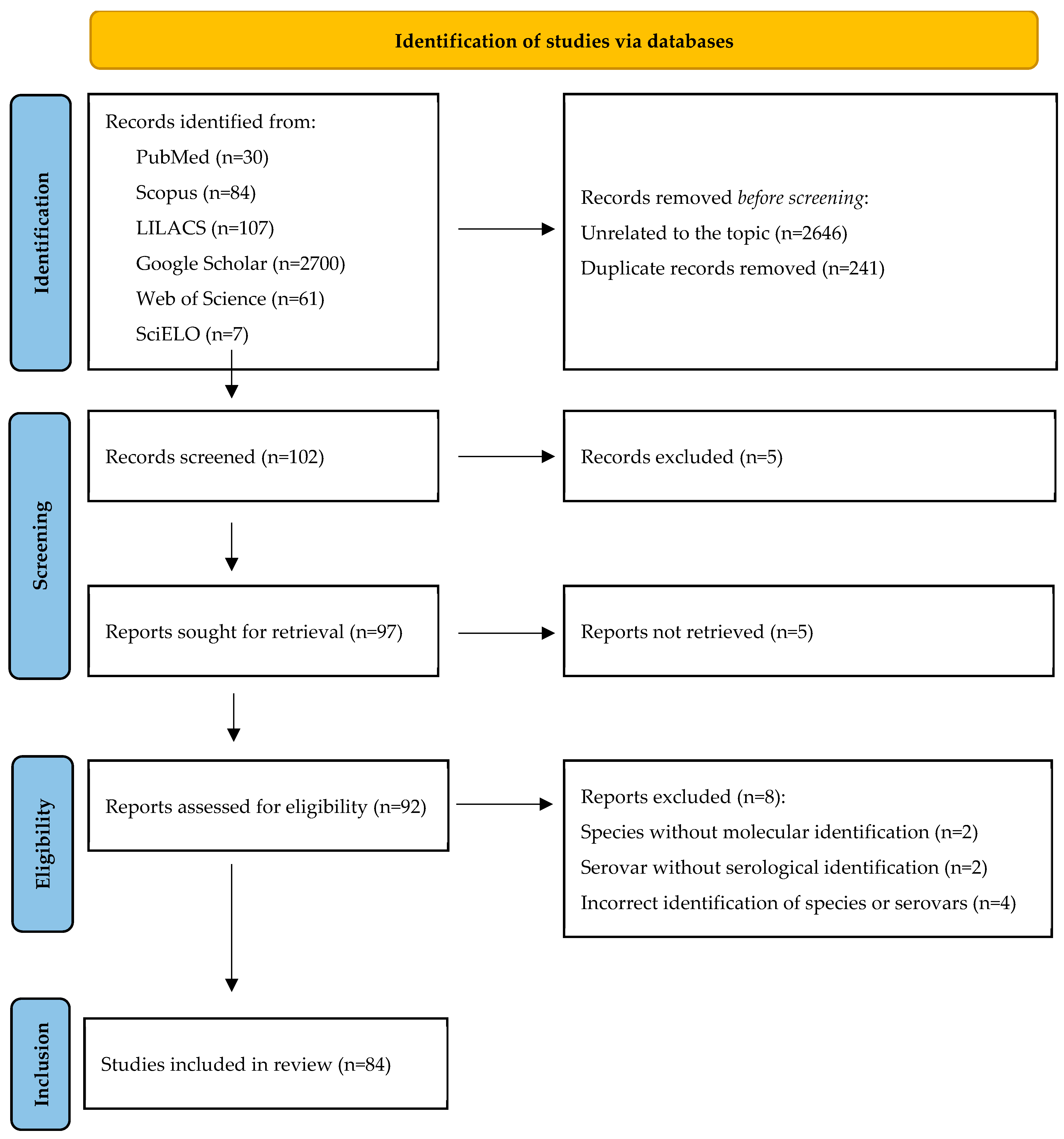
The search and selection process identified 2989 records containing the terms “Leptospira santarosai” OR “L. santarosai” (PubMed = 30, Scopus = 84, LILACS = 107, Google Scholar = 2700, Web of Science = 61, and SciELO = 7). During the identification stage, records unrelated to the topic (n = 2646) and duplicate records (n = 241) were removed, leaving 102 records for screening. In the screening stage, the titles and abstracts of these records were evaluated, and 5 records were excluded (n = 5), leaving 97 records sought for retrieval. Of these, 5 reports could not be retrieved (n = 5), leaving 92 reports assessed for eligibility. During the eligibility stage, 8 articles were excluded due to species without molecular identification (n = 2), serovars without serological identification (n = 2), and incorrect identification of species or serovars (n = 4). Finally, 84 articles met the inclusion criteria and were included in the qualitative synthesis (Figure 1).

3.2. Evaluation of Methodological Quality and Risk of Bias

The included studies were classified according to their purpose as descriptive or analytical, according to the temporality as cross-sectional, according to the control of factors as experimental or observational, and according to the chronology as retrospective or prospective (Supplementary Table S2). The publications had a compliance range of the STROBE guide between 78–91%. In terms of the level of methodological quality, the distribution was as follows: 3 articles (very high quality—3.57%), 73 articles (high quality—86.9%), 7 articles (moderate to high quality—8.33%), and 1 article (moderate quality—1.19%). In terms of the risk of bias, the distribution was as follows: 4 articles (high risk of bias—4.76%), 7 articles (medium to high risk of bias—8.33%), 68 articles (medium risk of bias—80.95%), and 5 articles (medium to low risk of bias—5.95%) (Table 1 and Supplementary Table S2).

3.3. Serological Diversity of Leptospira santarosai

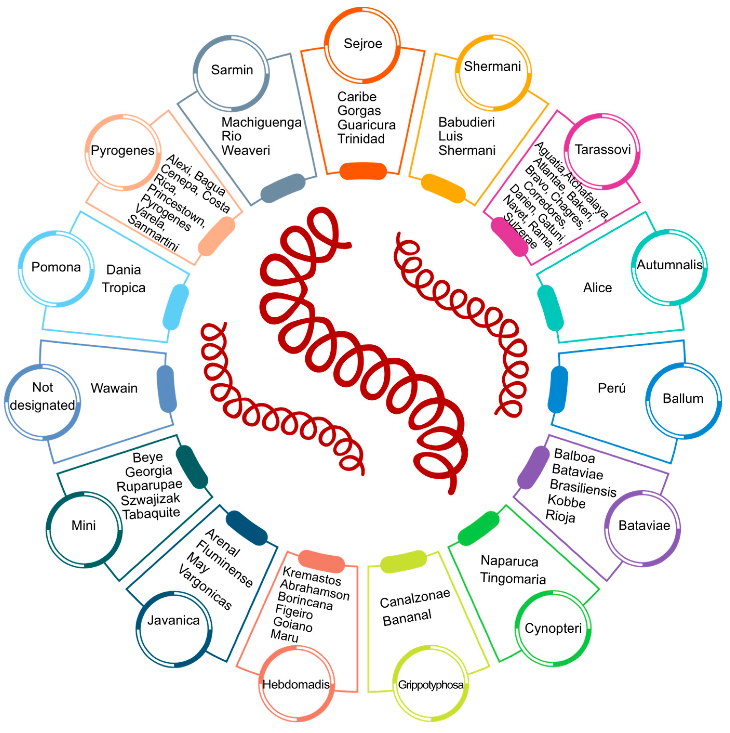
The first specific objective of this systematic review was to describe serological diversity of Leptospira santarosai. According to the information available in the Medical Reference Center for Leptospirosis (https://leptospira.amsterdamumc.org/filters/species/l-santarosai/, accessed on 14 January 2026), described by Brenner et al. in 1999 [14], NCBI-Taxonomy (https://www.ncbi.nlm.nih.gov/Taxonomy/, accessed on 14 January 2026), and subsequent publications [38,42,43,44,52,68,73,74], a total of 59 serovars have been described. As a result of this systematic review, these 59 serovars were systematically organized into 14 serogroups, providing a synthesis of the current knowledge on the serological diversity of this species (Table 2 and Figure 2).

3.4. Geographical Distribution of Leptospira santarosai

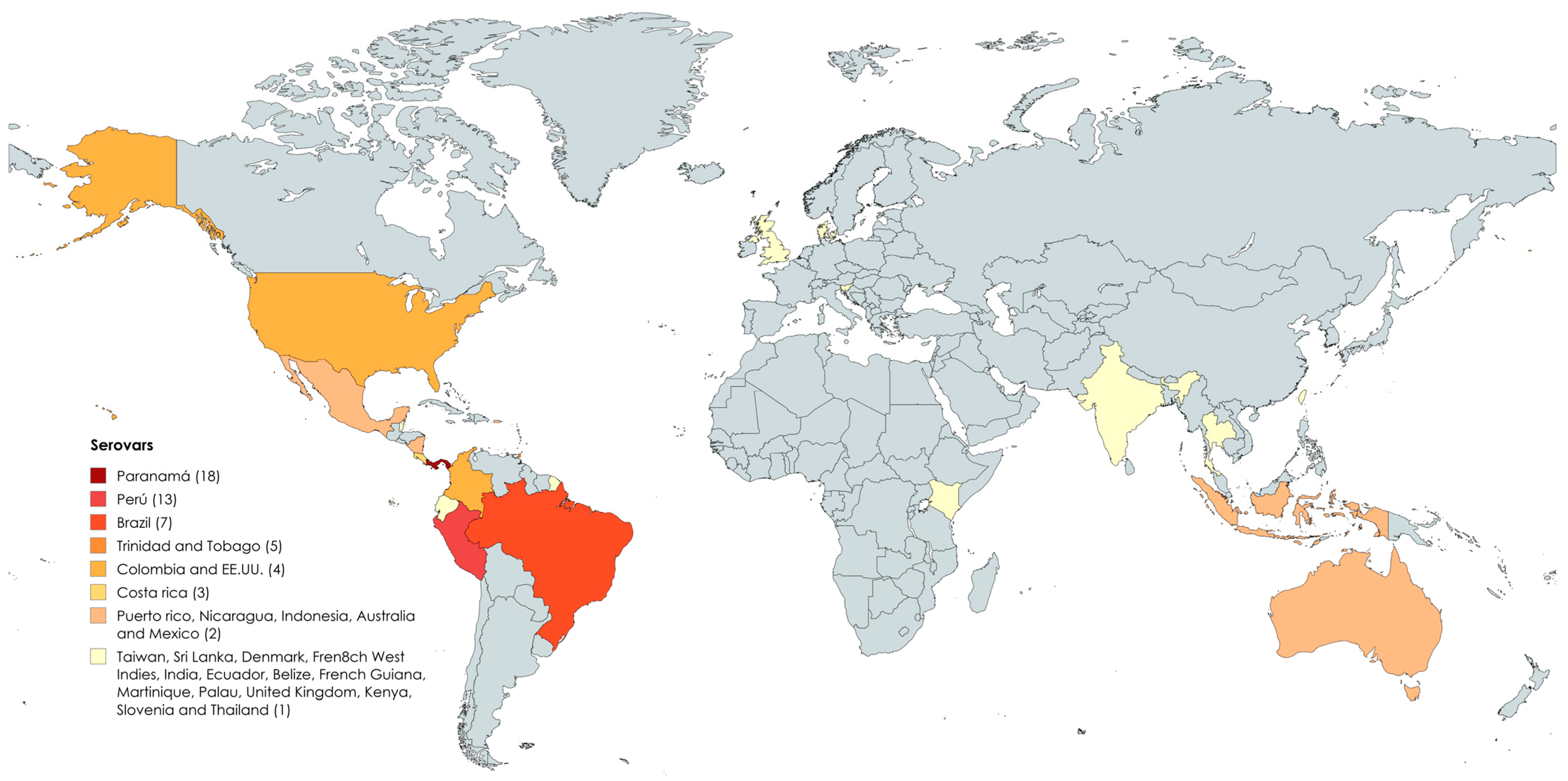
As a second objective, the geographic distribution of Leptospira santarosai was described, revealing its presence on five continents (excluding Antarctica) [12,13,14,51,79]. The continent with the highest number of reports of Leptospira santarosai was America, followed by Asia, Europe, Oceanica, and Africa. In America, the species has been reported in Panama, Peru, Brazil, Trinidad and Tobago, Colombia, USA, Costa Rica, Puerto Rico, Nicaragua, French West Indies, Mexico, Ecuador, Belize, French Guiana, and Martinique. In Asia, it has been reported in Indonesia, Taiwan, Sri Lanka, Thailand, and India. In Europe, the species has been reported in Denmark, Slovenia, and the United Kingdom. In Oceania, it has been reported in Australia and Palau, while, in Africa, it has been reported in Kenya. The geographic distribution of serovars associated with Leptospira santarosai showed that 59 serovars have been identified across 26 countries and territories. However, in seven countries, isolates were identified only at the species level. Panama reported the highest number of serovars (18), followed by Peru (13) and Brazil (7) (Figure 3 and Supplementary Table S4).

3.5. Animals Infected by Leptospira santarosai

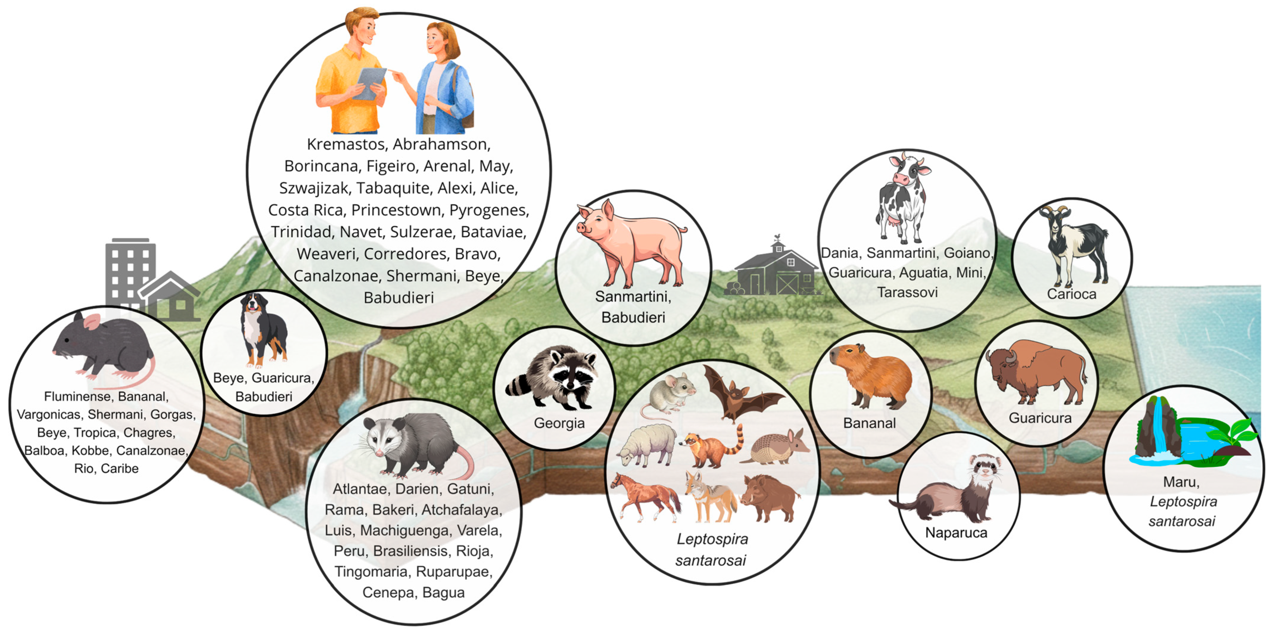
The third objective was to relate Leptospira santarosai serovars to their animal hosts. A total of 45 serovars were reported infecting 24 hosts, including the following: Galictis cuja, pigs (Sus scrofa domesticus) [52], rodents (Rodentia) [52], raccoons (Procyon lotor) [13], cattle (Bos taurus) [59], opossums (Didelphimorphia) [13], capybaras (Hydrochoerus hydrochaeris) [38], buffalo (Bubalus bubalis) [73], dogs (Canis lupus familiaris) [30,31,50,76,89], goats (Capra aegagrus hircus) [74], coatis (Nasua) [16], coyotes (Canis latrans) [75], equids (Equidae) [18], sheep (Ovis aries) [62], Marmosops ocellatus [49,55], Marmosops paulensis [49,55], Metachirus myosuros [49,55], Metachirus nudicaudatus [49,55], Monodelphis glirina [49,55], Monodelphis peruviana [49,55], nine-banded armadillos (Dasypus novemcinctus) [16], Nectomys squamipes [52], wild boar (Sus scrofa) [35], and bats [78]. Among these hosts, opossums (Didelphimorphia) were associated with the highest number of serovars (16), followed by rodents (13) and cattle (7). Additionally, 13 isolates were identified only at the species level (Figure 4 and Supplementary Table S5). According to the results obtained, three transmission cycles of Leptospira santarosai can be identified: a transmission cycle involving wild animals, including Galictis cuja, raccoons (Procyon lotor), opossums (Didelphimorphia), capybaras (Hydrochoerus hydrochaeris), coatis (Nasua), coyotes (Canis latrans), Marmosops ocellatus, Marmosops paulencis, Metachirus myosuros, Metachirus nudicaudatus, Monodelphis glirine, Monodelphis peruviana, nine-banded armadillos (Dasypus novemcinctus), Nectomys squamipes, wild boar (Sus scrofa), and bats; a transmission cycle involving domestic animals, including dogs (Canis lupus familiaris), pigs (Sus scrofa domesticus), cattle (Bos taurus), buffalo (Bubalus bubalis), sheep (Ovis aries), equids (Equidae), and goats (Capra aegagrus hircus); and a transmission cycle involving synanthropic animals, represented by rodents (Rodentia). Regarding potential transmission routes, infections of different animal species with the same serovar have been reported. For example, the serovar Bananal has been identified in rodents and capybaras, Shermani in rodents and humans, Beye in rodents, dogs, and humans, Canalzonae in rodents and humans, Sanmartini in cattle and pigs, Babudieri in pigs, dogs, and humans, and Guaricura in cattle, buffalo, and dogs. These findings reflect the ability of some serovars to infect multiple animal hosts. Additionally, rodents, dogs, and pigs may act as sources of infection for humans because they share the same serovars.

3.6. Leptospira santarosai and Natural Sources of Infection

The fourth specific objective of the systematic review was to evaluate the association of Leptospira santarosai with environmental sources. The species has been reported in wet soils, lakes, and waterfalls. However, only one environmental isolate has been identified at the serovar level (Leptospira santarosai serovar Maru), which was detected in water sources in the Panama Canal area (Figure 4 and Supplementary Table S5) [12,19,45,54,82,84,88,94].

3.7. Leptospira santarosai and Human Leptospirosis

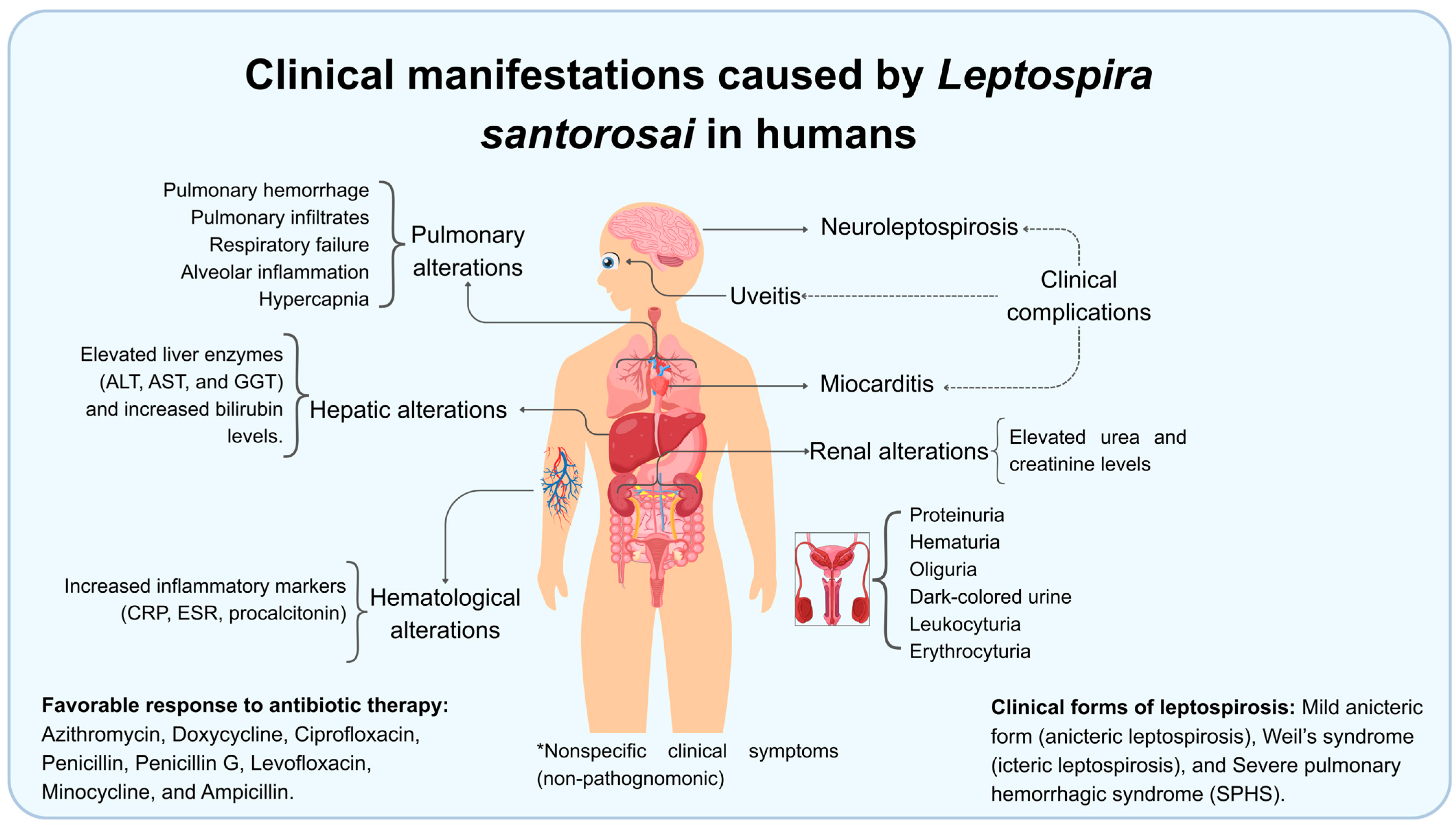
The fifth objective was to identify Leptospira santarosai as a causative agent of human leptospirosis and to describe the clinical manifestations associated with infection. This species has been reported as an infectious agent in humans, with 24 isolates identified at the serovar level (Kremastos, Abrahamson, Borincana, Figeiro, Arenal, May, Szwajizak, Tabaquite, Alexi, Alice, Costa Rica, Princestown, Pyrogenes, Trinidad, Navet, Sulzerae, Bataviae, Weaveri, Corredores, Bravo, Canalzonae, Shermani, Beye, and Babudieri) [13,14,20,21,22,23,24,25,28,29,32,33,40,41,44,47,51,60,67,68,69,72,79,81,82,83,85,90,96]. The infection has been associated with clinical presentations such as benign anicteric leptospirosis, Weil’s syndrome, and severe pulmonary hemorrhagic syndrome (SPHS) [21,22]. Additionally, complications include myocarditis [23], uveitis [24], and neuroleptospirosis [25,96] have been reported (Figure 5 and Supplementary Table S6). Laboratory findings included urinary abnormalities such as proteinuria, hematuria, leukocyturia, dark-colored urine, leukocyturia, erythruria, and oliguria. Renal alterations include increased urea and creatinine levels. Hematological findings include elevated C-reactive protein (CRP), erythrocyte sedimentation rate (ESR), and procalcitonin levels. Hepatic involvement has been associated with increased liver enzyme levels, including alanine aminotransferase (ALT), aspartate aminotransferase (AST), gamma-glutamil transferasa (GGT), and bilirubin. Pulmonary alterations such as pulmonary hemorrhage, pulmonary infiltrates, respiratory failure, alveolar inflammation, and hypercapnia have also been described. However, no single clinical sign has been pathognomonic for the disease [11,20,21,22,23,30,31,33,44,50,51,59,69,72,76,79,81,83,89,96]. Additionally, Leptospira santarosai has been reported to be susceptible to several antibiotics, including azithromycin, doxycycline, ciprofloxacin, penicillin, levofloxacin, minocycline, and ampicillin, resulting in rapid recovery of the patients [40,83,90] (Figure 5 and Supplementary Table S7). Regarding the frequency of clinical presentations, Leptospira santarosai was most frequently associated with the benign anicteric form of leptospirosis (8 cases), followed by Weil’s syndrome (3 cases), and Severe Pulmonary Hemorrhagic Syndrome (SPHS) (3 cases). These findings suggest that this species may be less virulent than other species such as Leptospira interrogans, and may be better adapted to the human host, since at least 24 serovars with the potential to infect humans have been described. Regarding the frequency of clinical complications, neuroleptospirosis (3 cases), uveitis (2 cases), and myocarditis (1 case) were the most frequently reported. However, these complications were more frequently associated with immunocompromised patients. Regarding the diagnostic relevance of the clinical signs and symptoms, no pathognomonic feature specific to the diagnosis of leptospirosis was identified. Fever and myalgia were the only symptoms reported in most patients, although both were nonspecific. Laboratory findings were highly variable and lacked diagnostic specificity, highlighting the need for serological and molecular tests to confirm infection. It is worth mentioning that marked alterations in laboratory parameters may be associated with the development of severe clinical complication. Overall additional research is needed to better understand the pathophysiology of Leptospira santarosai infection.

4. Discussion

According to the results of this systematic review, Leptospira santarosai is a pathogenic species that exhibits wide serological diversity, with at least 59 serovars grouped into 14 serogroups. This broad intraspecific serological diversity underscores the epidemiological importance of this species and likely reflects its capacity to adapt to a wide range of environments. Indeed, Leptospira santarosai has been reported in at least 24 animal hosts, environmental sources such as water bodies, lakes, waterfalls, and wet soils, and humans, for whom 24 infective serovars have been described. Understanding the serological diversity of Leptospira santarosai is crucial and presents several challenges in public and veterinary health. From a diagnostic perspective, serological tests, such as the Microscopic Agglutination Test (MAT) depend on including local serovars to be accurate. If the serovars present in a region are unknown, false-negative results may occur. Epidemiologically, identifying circulating serovars allows for a better understanding of transmission patterns, animal reservoirs, and environmental risk factors. Regarding prevention and control, vaccines against Leptospira generate serovar-specific immunity only against the serovar included in the vaccine formulation. Therefore, knowledge of serological diversity is essential for the development of effective vaccines and control strategies adapted to local conditions. Additionally, the broad serological diversity of Leptospira santarosai has important public health implications. Different serovars can persist in the environmental for prolonged periods and be transmitted through water, wet soils, or animal hosts, increasing the risk of outbreaks. From an evolutionary perspective, the presence of numerous serovars likely reflects a long history of adaptation to diverse environments and hosts, resulting in the diversification of the genetic content and, consequently, the serological phenotype of the species. These findings also highlight important knowledge gaps regarding the ecological determinants of Leptospira santarosai. Future studies should investigate the environmental factors that influence the presence, distribution, and dynamics of this species across different ecosystems. According to the list of serovars reported by Brenner et al. in 1999 [14], the pathogenic species with the highest number of serovars in the genus Leptospira are L. interrogans (91 serovars), L. santarosai (59 serovars), L. borgpetersenii (49 serovars), L. kirschneri (39 serovars), L. noguchii (18 serovars), and L. weilii (15 serovars). Therefore, Leptospira santarosai is the second most serologically diverse species currently recognized within the genus and is consequently of considerable epidemiological, environmental, and clinical importance.
Leptospira santarosai has a wide geographic distribution and has been reported on five continents and 26 countries [12,13,14,51,70,79,91,92,93]. Reports of this species are most frequent in the American continent, particularly in Central America, South America, and the Caribbean islands. Panama reported the highest number of serovars (18), followed by Peru (13) and Brazil (7). However, the greater number of serovars reported in these countries likely reflects a higher sampling effort and a larger number of studies rather than the true serological diversity of the species. Therefore, it is necessary to carry out further research in countries where information is currently unavailable to determine the actual serological diversity and distribution of Leptospira santarosai. Additionally, in Taiwan, Leptospira santarosai serovar Shermani has been reported as one of the most prevalent serovars associated with human infection. These observations suggest that this species represents an important etiological agent of leptospirosis in various regions of the world [12]. Furthermore, 45 serovars were reported exclusively in a single country, whereas eight serovars were detected in more than one country: Canalzonae (Panama and Colombia), Beye (Panama and Colombia), Pyrogenes (Panama and Indonesia), Babudieri (Peru and Colombia), Tabaquite (Trinidad and Tobago, and the French Wes Indies), Alice (Sri Lanka and Colombia), Shermani (Panama and Taiwan), and Princestown (Trinidad and Tobago, and the USA). These findings indicate that, while many Leptospira santarosai serovars appear to have restricted geographic distributions, some are detected across multiple countries, showing a pattern of dispersion among the countries. When comparing geographical regions, the Americas reported the highest number of countries with the presence of Leptospira santarosai (15 countries and 57 serovars), followed by Asia (5 countries and 6 serovars), Europe (3 countries and 1 serovar), Oceania (2 countries and 2 serovars), and Africa (1 country, without identification of serovars). Regarding possible distribution routes, dispersion patterns can be observed among countries that share the same serovars. One group includes Panama, Colombia, Peru, Sri Lanka and Indonesia, whereas another includes Trinidad and Tobago, the United States, and the French West Indies. However, clonal dispersal studies among the serovars are required to verify these proposed distribution patterns. It is also important to acknowledge the potential for selection bias, as many countries lack taxonomic studies capable of identifying Leptospira santarosai and its serovars. Consequently, the current distribution patterns may reflect differences in research efforts and taxonomic resolution rather than the true geographic distribution of the species.
The evidence indicates that 39 serovars of Leptospira santarosai infect 24 hosts [13,16,18,30,31,34,35,38,46,48,49,50,52,53,55,56,57,58,59,61,62,63,64,65,66,73,74,75,76,77,80,86,87,89]. This serological diversity suggests that the species is associated with multiple hosts. Several serovars were reported infecting the same animal species (which is consistent with the theory that some serovars selectively infect certain animal hosts), while other serovars were identified infecting several animal hosts. For example, the serovar Georgia has been reported in Procyon lotor, whereas the serovar Carioca has been reported in Capra aegagrus hircus. In contrast, several serovars have been detected in multiple animal species, including Sanmartini in pigs and cattle, Babudieri in dogs and pigs, Bananal in capybaras and rodents, Beye in rodents and dogs, and Guaricura in cattle, buffalo, and dogs. These observations demonstrate that certain serovars may circulate among multiple host species, suggesting the possibility of interspecies transmission. The presence of Leptospira santarosai in agriculturally important hosts such as cattle, buffalo, goats, and pigs highlight the potential role of livestock in the epidemiology of leptospirosis caused by this species. These observations suggest that animal hosts other than rodents may contribute to the transmission cycle of the disease. Furthermore, it is evident that agricultural activities may also represent a risk factor for leptospirosis caused by this species, given its high capacity to infect various agriculturally important hosts such as cattle, buffalo, goats, and pigs. These observations suggest that livestock may contribute to transmission in both rural and per-urban settings. Reports of infection in domestic animals such as dogs indicate that these animals may also represent potential sources of exposure for humans. In addition, infections detected in wild animals suggest the existence of a sylvatic cycle that may represent another potential source of human infection when people enter these habitats. Several serovars have been reported in both animals and humans, including Canalzonae (rodents and humans), Babudieri (pigs, canines, and humans), Shermani (rodents and humans), Beye (rodents, canines, and humans). This overlap suggests that these hosts may act as a potential risk factor for human infection. Overall, these findings indicate that Leptospira santarosai may circulate across wildlife, livestock, and domestic animal populations. Human activities such as agriculture and environmental disturbance may increase contact with infected hosts, thereby increasing the risk of infection.
Furthermore, at least seven studies have reported the presence of Leptospira santarosai in various environmental sources such as soils, lakes, and waterfalls [12,19,45,54,84,88,94]. However, only one study identified the isolates at serovar level (Leptospira santarosai serovar Maru in water sources from the Panama Canal Zone). This finding demonstrates the ability of this species to persist in the environment. These observations highlight the need for additional studies detecting the presence of pathogenic Leptospira species in environmental sources to determine their role in the environmental persistence of the bacteria, evaluate whether environmental replication occurs, and determine whether water and soil act as effective vehicles for disease transmission. Moreover, the detection of Leptospira santarosai in water sources could be associated with the infection in non-traditional exposure contexts, such as environmental disasters, recreational water activities, and aquatic events. These scenarios have previously been identified as risk factors for infection by other Leptospira species [36,37,39,71,95,97,98,99,100]. The limited number of reports of Leptospira santarosai from environmental sources likely reflects both the small number of studies conducted and the methodological challenges associated with the detection and isolation of Leptospira from environmental samples. These challenges include the complexity of environmental matrices, which contain DNA from multiple organisms; the presence of substances that inhibit molecular assays, such as humic compounds, heavy metals, and organic matter; the low concentration of bacteria in environmental samples, resulting in reduced detection rates; and the high microbial diversity present in these environments, which requires the use of highly specific molecular markers. Additional limitations include the presence of degraded or fragmented DNA, which can lead to false-negative results, and the lack of standardized protocols for DNA extraction from different environmental matrices. Furthermore, the detection of bacterial DNA does not necessarily indicate the presence of viable or metabolically active organisms, which may lead to inaccurate conclusions regarding environmental transmission risks.
At least 24 serovars belonging to Leptospira santarosai have been identified as causative agents of human leptospirosis. The wide variety of serovars reported in human infections demonstrates the presence of multiple animal reservoirs and environmental sources contaminated with the bacterium, which may act as sources of infection for humans. According to the World Health Organization (WHO) and the International Leptospirosis Society (ILS), the disease presents in four broad clinical categories: a mild influenza-like illness; Weil’s syndrome, characterized by jaundice, renal failure, hemorrhage and myocarditis with arrhythmias; meningitis or meningoencephalitis; and pulmonary hemorrhage with respiratory failure (https://iris.paho.org/items/23e5a770-23d2-47eb-87c4-e30ac9ff4e13, accessed on 14 January 2026). Reports of infections caused by Leptospira santarosai include cases of benign anicteric leptospirosis, Weil’s syndrome, severe pulmonary hemorrhage syndrome (SPHS), and meningitis or meningoencephalitis (neuroleptospirosis), indicating that this species has been associated with all major clinical presentations of leptospirosis [13,14,20,21,22,23,24,25,28,29,32,33,40,41,44,47,51,60,67,68,69,72,79,81,82,83,85,90,96]. Late sequelae of leptospirosis may include chronic fatigue, neuropsychiatric symptoms such as headache, paresis, paralysis, and mood changes and depression, as well as uveitis. Leptospira santarosai has been described as a causative agent of leptospirosis with late ocular complications such as uveitis in humans, suggesting the capacity of this species to invade immune-privileged organs [24]. According to the World Health Organization (WHO) and the International Leptospirosis Society (ILS) in “Human leptospirosis: a guide for diagnosis, surveillance and control”, laboratory studies of specimens from hospitalized patients frequently show several non-diagnostic abnormalities, including elevated erythrocyte sedimentation rate, thrombocytopenia, leukocytosis, hyperbilirubinemia, increased serum creatinine, elevated creatinine kinase, and elevated serum amylase, and, after crossing host barriers, Leptospira disseminates through the bloodstream and can spread to multiple organs. In patients infected with Leptospira santarosai, clinical studies have reported alterations in several hematological and inflammatory markers, including increased C-reactive protein (CRP), erythrocyte sedimentation rate (ESR), and procalcitonin levels. These findings indicate a systemic inflammatory response during infection. The kidneys appear to be the main target organ in Leptospira infection, as reflected by the frequent renal alterations reported in infected patients, including elevated urea and creatinine levels, proteinuria, hematuria, leukocyturia, dark-colored urine, erythruria, and oliguria. These findings are consistent with renal involvement during infection. Alterations in liver function have also been described, including increased levels of alanine aminotransferase (ALT), aspartate aminotransferase (AST), gamma-glutamil transferasa (GGT), and bilirubin. Elevated bilirubin levels may lead to jaundice, one of the characteristic clinical signs of leptospirosis. Pulmonary complications have also been reported, including pulmonary hemorrhage, pulmonary infiltrates, respiratory failure, alveolar inflammation, and hypercapnia, indicating that infection may involve the respiratory system [11,20,21,22,23,30,31,33,44,50,51,59,69,72,76,79,81,83,89,96]. Overall, the alterations observed in inflammatory markers and organ function tests suggest that Leptospira santarosai infection may involve multiple organs, particularly the kidneys, liver, and lungs. However, no single biomarker has been identified as pathognomonic for the disease. Additionally, Leptospira santarosai isolates reported in the literature have shown susceptibility to several antibiotics, including azithromycin, doxycycline, ciprofloxacin, penicillin, levofloxacin, minocycline, and ampicillin, and patients generally recover with appropriate treatment [40,83,90]. These findings demonstrate the importance of timely diagnosis and treatment in preventing severe forms of the disease.
Regarding the limitations of this systematic review, the methodological quality of the 84 included studies was evaluated, resulting in 27 articles classified as having high methodological quality (32.14%), 44 as moderate-to-high methodological quality (52.38%), 12 as moderate methodological quality (14.28%), and 1 as low-to-moderate methodological quality (1.19%). One of the main limitations of the evidence included in the systematic review is that only 27 studies were classified as having high methodological quality according to the quality and risk-of-bias assessment. Additional limitations include the use of only five databases for the literature search, the inclusion of articles published exclusively in English and Spanish, the heterogeneity of the study designs, and the inability to perform a meta-analysis because of the heterogeneity of the available data. Regarding serological diversity, although 59 serovars of Leptospira santarosai have been identified, this number likely represents only a fraction of the true diversity of the species. Therefore, additional sampling is needed in countries where studies have not yet been conducted. This sampling should focus on detecting and identifying new serovars associated with Leptospira santarosai in human, animal, and environmental samples. With respect to geographic distribution, the main limitation is that the available data originate from only 26 countries, representing 13.33% of the world’s countries (26/195). This finding suggests that the current distribution data remain incomplete and that many regions have yet to be investigated. Similarly, Leptospira santarosai has been reported in only 24 animal hosts, indicating that its host range may be substantially underestimated. Environmental sources of infection remain among the least explored aspects of the ecology of Leptospira santarosai. To date, the species has been reported in wet soils, lakes, waterfalls, and other water sources. Additional environmental studies are therefore needed to better understand its distribution and persistence in natural ecosystems. Regarding human leptospirosis, 24 serovars associated with Leptospira santarosai have been reported to infect humans and produce a broad spectrum of clinical manifestations ranging from mild to severe disease. However, further experimental studies are needed to investigate in greater depth the pathophysiology, pathogenicity, virulence factors, and specific clinical manifestations associated with these serovars in humans.

5. Conclusions

Leptospira santarosai is a pathogenic species characterized by a high serological diversity, a wide geographic distribution, and the ability to infect multiple domestic, synanthropic, and wild animals. Additionally, it can persist in the environment and infect humans, causing clinical manifestations ranging from mild to severe disease, as well as complications such as uveitis, myocarditis, and neuroleptospirosis. The species has been reported on five continents; however, the vast majority of reports originate from the American continent. Therefore, additional epidemiological studies are needed in other regions of the world. These findings highlight the importance of Leptospira santarosai as a causative agent of human and animal leptospirosis worldwide and support its inclusion in serological and molecular diagnostic tests to improve diagnostic sensitivity and specificity. Furthermore, this species should be considered in the development of vaccines for humans and animals in regions where it is prevalent. Future studies should examine the ecological determinants that influence the presence, persistence, distribution, and dynamics of Leptospira santarosai across different ecosystems. Evolutionary studies based on genome sequencing are also needed to determine the geographic origin, migration patterns, and dispersal routes of the species. Additionally, studies involving different animal hosts and the identification of the associated serovars are necessary to determine whether individual serovars exhibit host specificity or ecological flexibility. Computational biology and bioinformatics should be used to identify pathogenicity and virulence factors that contribute to the pathophysiology of the disease. Comparative genomic analyses with other pathogenic species of the genus Leptospira may also help identify the genetic determinants responsible for differences in virulence. Furthermore, additional studies of environmental sources are needed to better understand the ecological behavior of the species and to support the development of more effective prevention and control strategies. Despite recent advances, important knowledge gaps remain. These include the discovery of new serovars, determination of the true serological diversity of the species, identification of its presence in countries where studies have not yet been conducted, evaluation of its ability to survive or multiply in environmental sources, identification of all animal hosts involved in its transmission cycle, and a better understanding of how the species infects, multiplies, and causes disease in humans and animals.

Supplementary Materials

The following supporting information can be downloaded at: https://www.mdpi.com/article/10.3390/microorganisms14061364/s1. Supplementary Table S1. Search algorithms used in the PubMed, Scopus, Lilacs, Google Scholar, Web of Science, and SciELO databases to identify publications that investigated: serological diversity, geographic distribution, reservoirs, natural sources of infection, human leptospirosis, and genomes of Leptospira santarosai. Supplementary Table S2. Summary of the study type of the 84 scientific articles included in the systematic review and the assessment of methodological quality and risk of bias using an appropriate evaluation guide. Supplementary Table S3. Summary of the 84 scientific articles included in the systematic review and the extracted information on serological diversity, geographical distribution, reservoirs, natural sources of infection, and reported cases of human leptospirosis caused by Leptospira santarosai. Supplementary Table S4. Summary of geographic distributions of serovars associated with Leptospira santarosai. The table lists the countries where the species has been reported, the corresponding serovars, and the total number of serovars identified per country. Supplementary Table S5. Summary of the representative host taxa, environmental sources, and accidental hosts (humans) reported to be infected with serovars of Leptospira santarosai. Supplementary Table S6. Summary of clinical presentations and complications described in patients infected with Leptospira santarosai. The table summarizes the country, the infecting species or serovar, the reported clinical presentations of the disease, including benign anicteric leptospirosis, Weil’s syndrome, and severe pulmonary hemorrhagic syndrome (SPHS), as well as complications that may occur after infection such as myocarditis, uveitis, and neuroleptospirosis. It also lists the bibliographic sources from which the data were extracted. Supplementary Table S7. Summary of the clinical manifestations and laboratory findings (blood cell count, kidney function, liver function, urine chemistry, lung function, inflammatory markers, and antibiotic response) reported in patients with leptospirosis caused by Leptospira santarosai. The table also lists the bibliographic sources from which the information was extracted.

Author Contributions

Conceptualization, R.G.P.S., J.E.S.F., L.P.C., D.S.M., and F.P.M.; methodology, R.G.P.S., J.E.S.F., and F.P.M.; software, R.G.P.S. and M.O.-Y.; validation, R.G.P.S. and M.O.-Y.; formal analysis, R.G.P.S., S.P., M.O.-Y., M.T.-C., A.S.-G., R.U., L.E.L.-R., J.E.S.F., S.A.-P., and F.P.M.; investigation, R.G.P.S., S.P., M.O.-Y., M.T.-C., A.S.-G., R.U., L.E.L.-R., J.E.S.F., S.A.-P., and F.P.M.; resources, R.G.P.S., J.E.S.F., and F.P.M.; data curation, R.G.P.S., S.P., M.O.-Y., M.T.-C., A.S.-G., R.U., L.E.L.-R., J.E.S.F., S.A.-P., L.E.G.C., and F.P.M.; writing—original draft preparation, R.G.P.S., S.P., M.O.-Y., M.T.-C., A.S.-G., R.U., L.E.L.-R., J.E.S.F., S.A.-P., L.E.G.C., L.P.C., D.S.M., and F.P.M.; writing—review and editing, R.G.P.S., S.P., M.O.-Y., M.T.-C., A.S.-G., R.U., L.E.L.-R., J.E.S.F., S.A.-P., L.E.G.C., L.P.C., D.S.M., and F.P.M.; visualization, R.G.P.S., S.P., M.O.-Y., M.T.-C., A.S.-G., R.U., L.E.L.-R., J.E.S.F., S.A.-P., L.E.G.C., and F.P.M.; supervision, R.G.P.S.; project administration, R.G.P.S. and L.E.G.C.; funding acquisition, R.G.P.S., S.P., M.O.-Y., M.T.-C., A.S.-G., R.U., L.E.L.-R., J.E.S.F., S.A.-P., L.E.G.C., and F.P.M. All authors have read and agreed to the published version of the manuscript.

Funding

This project was funded by the Universidad de la Sabana. Grant number MED-20-2024, under the Project “Detección de genes relacionados con la Resistencia antibiótica en Leptospira”.

Institutional Review Board Statement

Not applicable.

Informed Consent Statement

Not applicable.

Data Availability Statement

No new data were created or analyzed in this study. Data sharing is not applicable to this article.

Acknowledgments

The authors would like to thank the support of the San Martin University, Vrije Universiteit Amsterdam, Autonomous University of Yucatan, CES University, Colombian Institute of Tropical Medicine (ICMT), Universidad de la Sabana, and Northern Arizona University.

Conflicts of Interest

The authors declare no conflict of interest.

References

  1. Adler, B.; de la Peña Moctezuma, A. Leptospira and leptospirosis. Vet. Microbiol. 2010, 140, 287–296. [Google Scholar] [CrossRef] [PubMed]
  2. Costa, F.; Hagan, J.E.; Calcagno, J.; Kane, M.; Torgerson, P.; Martinez-Silveira, M.S.; Stein, C.; Abela-Ridder, B.; Ko, A.I. Global Morbidity and Mortality of Leptospirosis: A Systematic Review. PLoS Neglected Trop. Dis. 2015, 9, e0003898. [Google Scholar] [CrossRef] [PubMed] [PubMed Central]
  3. Levett, P.N. Leptospirosis. Clin. Microbiol. Rev. 2001, 14, 296–326. [Google Scholar] [CrossRef] [PubMed] [PubMed Central]
  4. Wasiński, B.; Dutkiewicz, J. Leptospirosis—Current risk factors connected with human activity and the environment. Ann. Agric. Environ. Med. 2013, 20, 239–244. [Google Scholar] [PubMed]
  5. Haake, D.A.; Levett, P.N. Leptospirosis in humans. Curr. Top. Microbiol. Immunol. 2015, 387, 65–97. [Google Scholar] [CrossRef] [PubMed] [PubMed Central]
  6. Trevejo, R.T.; Rigau-Pérez, J.G.; Ashford, D.A.; McClure, E.M.; Jarquín-González, C.; Amador, J.J.; Reyes, J.O.d.L.; Gonzalez, A.; Zaki, S.R.; Shieh, W.; et al. Epidemic leptospirosis associated with pulmonary hemorrhage-Nicaragua, 1995. J. Infect. Dis. 1998, 178, 1457–1463. [Google Scholar] [CrossRef] [PubMed]
  7. Vincent, A.T.; Schiettekatte, O.; Goarant, C.; Neela, V.K.; Bernet, E.; Thibeaux, R.; Ismail, N.; Khalid, M.K.N.M.; Amran, F.; Masuzawa, T.; et al. Revisiting the taxonomy and evolution of pathogenicity of the genus Leptospira through the prism of genomics. PLoS Neglected Trop. Dis. 2019, 13, e0007270. [Google Scholar] [CrossRef] [PubMed] [PubMed Central]
  8. Yasuda, P.H.; Steigerwalt, A.G.; Sulzer, K.R.; Kaufmann, A.F.; Rogers, F.; Brenner, D.J. Deoxyribonucleic Acid Relatedness between Serogroups and Serovars in the Family Leptospiraceae with Proposals for Seven New Leptospira Species. Int. J. Syst. Evol. Microbiol. 1987, 37, 407–415. [Google Scholar] [CrossRef]
  9. Cerqueira, G.M.; Picardeau, M. A century of Leptospira strain typing. Infect. Genet. Evol. 2009, 9, 760–768. [Google Scholar] [CrossRef] [PubMed]
  10. Guglielmini, J.; Bourhy, P.; Schiettekatte, O.; Zinini, F.; Brisse, S.; Picardeau, M. Genus-wide Leptospira core genome multilocus sequence typing for strain taxonomy and global surveillance. PLoS Neglected Trop. Dis. 2020, 14, e0008673, Correction in: PLoS Neglected Trop. Dis. 2019, 13, e0007374. https://doi.org/10.1371/journal.pntd.0007374.. [Google Scholar] [CrossRef]
  11. Chinchilla, D.; Nieves, C.; Gutiérrez, R.; Sordoillet, V.; Veyrier, F.J.; Picardeau, M. Phylogenomics of Leptospira santarosai, a prevalent pathogenic species in the Americas. PLoS Neglected Trop. Dis. 2023, 17, e0011733. [Google Scholar] [CrossRef] [PubMed]
  12. Yang, C.W. Leptospirosis in Taiwan—An underestimated infectious disease. Chang Gung Med. J. 2007, 30, 109–115. [Google Scholar] [PubMed]
  13. Sato, Y.; Tsurui-Sato, K.; Uchima, Y.; Udui, C.A.; Lorin, O.; Rengulbai, K.; Toma, C.; Suzuki, R. A systematic survey of environmental DNA in Palau’s lakes and waterfalls reveals an increase in Leptospira levels after flooding. One Health 2024, 19, 100898. [Google Scholar] [CrossRef] [PubMed] [PubMed Central]
  14. Brenner, D.J.; Kaufmann, A.F.; Sulzer, K.R.; Steigerwalt, A.G.; Rogers, F.C.; Weyant, R.S. Further determination of DNA relatedness between serogroups and serovars in the family Leptospiraceae with a proposal for Leptospira alexanderi sp. nov. and four new Leptospira genomospecies. Int. J. Syst. Bacteriol. 1999, 49, 839–858. [Google Scholar] [CrossRef] [PubMed]
  15. Allan, K.J.; Biggs, H.M.; Halliday, J.E.B.; Kazwala, R.R.; Maro, V.P.; Cleaveland, S.; Crump, J.A. Epidemiology of Leptospirosis in Africa: A Systematic Review of a Neglected Zoonosis and a Paradigm for ‘One Health’ in Africa. PLoS Neglected Trop. Dis. 2016, 10, e0004552, Correction in: PLoS Neglected Trop. Dis. 2015, 9, e0003899. https://doi.org/10.1371/journal.pntd.0003899.. [Google Scholar]
  16. Miotto, B.A.; Moreno, L.Z.; Guilloux, A.G.A.; Sousa, G.O.D.; Loureiro, A.P.; Moreno, A.M.; Lilenbaum, W.; Vasconcellos, S.A.; Heinemann, M.B.; Hagiwara, M.K. Molecular and serological characterization of the first Leptospira santarosai strain isolated from a dog. Acta Trop. 2016, 162, 1–4. [Google Scholar] [CrossRef] [PubMed]
  17. Fornazari, F.; Langoni, H.; Marson, P.M.; Nóbrega, D.B.; Teixeira, C.R. Leptospira reservoirs among wildlife in Brazil: Beyond rodents. Acta Trop. 2018, 178, 205–212. [Google Scholar] [CrossRef] [PubMed]
  18. Carmona-Gasca, C.A.; Lara, L.L.; Castillo-Sánchez, L.O.; Ramírez-Ortega, J.M.; Palomera, C.L.; de la Peña-Moctezuma, A. Detection of Leptospira santarosai and L. kirschneri in cattle: New isolates with potential impact in bovine production and public health. Vet. México 2011, 42, 277–288. [Google Scholar]
  19. Ramos-Vázquez, J.R.; Sánchez-Montes, S.; Esparza-González, S.C.; Romero-Salas, D.; Pardio-Sedas, V.; Estrada-Coates, A.T.; Alva-Trujillo, M.; Ballados-González, G.G.; Grostieta, E.; Becker, I.; et al. Isolation and molecular identification of Leptospira santarosai and Leptospira interrogans in equines from eastern Mexico. Acta Trop. 2024, 256, 107242. [Google Scholar] [CrossRef] [PubMed]
  20. Gorman, M.; Xu, R.; Prakoso, D.; Salvador, L.C.M.; Rajeev, S. Leptospira enrichment culture followed by ONT metagenomic sequencing allows better detection of Leptospira presence and diversity in water and soil samples. PLoS Neglected Trop. Dis. 2022, 16, e0010589. [Google Scholar] [CrossRef] [PubMed]
  21. Nguyen-Tran, H.; Erdem, G.; Laufer, P.M.; Patterson, L.; Ahmed, A.A.; Bower, W.A.; Galloway, R.M.; Saporta-Keating, S. Use of Advanced Diagnostics for Timely Identification of Travel-associated Leptospira santarosai Infection in Four Adolescents Through Plasma Microbial Cell-free DNA Sequencing with the Karius Test. Pediatr. Infect. Dis. J. 2024, 43, e169–e174. [Google Scholar] [CrossRef] [PubMed]
  22. Peláez Sanchez, R.G.; Lopez, J.Á.; Pereira, M.M.; Arboleda Naranjo, M.; Agudelo-Flórez, P. Genetic diversity of Leptospira in northwestern Colombia: First report of Leptospira santarosai as a recognised leptospirosis agent. Memórias Inst. Oswaldo Cruz 2016, 111, 737–744. [Google Scholar] [CrossRef] [PubMed]
  23. Liao, C.Y.; Ben, R.J.; Wu, H.M.; Chang, S.K.; Liu, M.Y.; Chin, H.K.; Yeh, Y.C. Acute Respiratory Distress Syndrome Manifested by Leptospirosis Successfully Teated by Extracorporeal Membrane Oxygenation (ECMO). Intern. Med. 2015, 54, 2943–2946. [Google Scholar] [CrossRef] [PubMed]
  24. Wang, H.K.; Lee, M.H.; Chen, Y.C.; Hsueh, P.R.; Chang, S.C. Factors associated with severity and mortality in patients with confirmed leptospirosis at a regional hospital in northern Taiwan. J. Microbiol. Immunol. Infect. 2020, 53, 307–314. [Google Scholar] [CrossRef] [PubMed]
  25. Gonzales, J.A.; Sundararajan, M.; Qian, Y.; Hwang, T.; Doan, T. Detection of Leptospirosis Genome from the Aqueous Humor of a Patient with Bilateral Uveitis. Ocul. Immunol. Inflamm. 2023, 31, 1076–1078. [Google Scholar] [CrossRef] [PubMed]
  26. Wilson, M.R.; Naccache, S.N.; Samayoa, E.; Biagtan, M.; Bashir, H.; Yu, G.; Salamat, S.M.; Somasekar, S.; Federman, S.; Miller, S.; et al. Actionable diagnosis of neuroleptospirosis by next-generation sequencing. N. Engl. J. Med. 2014, 370, 2408–2417. [Google Scholar] [CrossRef] [PubMed] [PubMed Central]
  27. Page, M.J.; Moher, D.; Bossuyt, P.M.; Boutron, I.; Hoffmann, T.C.; Mulrow, C.D.; Shamseer, L.; Tetzlaff, J.M.; Akl, E.A.; Brennan, S.E.; et al. PRISMA 2020 explanation and elaboration: Updated guidance and exemplars for reporting systematic reviews. BMJ 2021, 372, n160. [Google Scholar] [CrossRef] [PubMed]
  28. Restrepo-López, N.; Silva-Ramos, C.R.; Rodas, J.D.; Arboleda, M.; Fernández, D.; Uribe-Restrepo, P.; Agudelo-Flórez, P.; Tobón-Castaño, A.; Hidalgo, M.; Melby, P.C.; et al. Malaria, Dengue Fever, and Leptospirosis in the Urabá Antioqueño Region, Colombia: Etiological and Molecular Characterization among Patients with Acute Undifferentiated Febrile Illness. Am. J. Trop. Med. Hyg. 2025, 112, 403–413. [Google Scholar] [CrossRef] [PubMed]
  29. Silva-Ramos, C.R.; Matiz-González, J.M.; Gil-Mora, J.; Martínez Díaz, H.C.; Faccini-Martínez, Á.A.; Cuervo, C.; Melby, P.C.; Aguilar, P.V.; Cabada, M.M.; Rodas, J.D.; et al. Molecular Characterization of Leptospira Species among Patients with Acute Undifferentiated Febrile Illness from the Municipality of Villeta, Colombia. TropicalMed 2024, 9, 168. [Google Scholar] [CrossRef] [PubMed]
  30. Perez-Garcia, J.; Monroy, F.P.; Agudelo-Florez, P. Canine Leptospirosis in a Northwestern Region of Colombia: Serological, Molecular and Epidemiological Factors. Pathogens 2022, 11, 1040. [Google Scholar] [CrossRef] [PubMed]
  31. Silva-Ramos, C.R.; Lemaitre, G.P.; Mejorano-Fonseca, J.A.; Matiz-González, J.M.; Aricapa-Giraldo, H.J.; Agudelo, J.C.; Cárdenas, J.E.P.; Hidalgo, M. Molecular Evidence of Leptospira spp. Infection Among Household Dogs from 15 Municipalities of the Department of Caldas, Colombia. Zoonoses Public Health 2025, 72, 215–222. [Google Scholar] [CrossRef] [PubMed]
  32. Agudelo-Flórez, P.; Durango, H.; Aranzazu, D.; Rodas, J.D.; Travi, B. Genotyping and evaluation of infection dynamics in a Colombian isolate of Leptospira santarosai in hamster as an experimental model. Biomedica 2014, 34, 460–472. [Google Scholar] [CrossRef] [PubMed]
  33. Uribe-Restrepo, P.; Perez-Garcia, J.; Arboleda, M.; Munoz-Zanzi, C.; Agudelo-Florez, P. Clinical presentation of human leptospirosis in febrile patients: Urabá, Colombia. PLoS Neglected Trop. Dis. 2024, 18, e0012449. [Google Scholar] [CrossRef] [PubMed]
  34. Hamond, C.; Dirsmith, K.L.; LeCount, K.; Soltero, F.V.; Rivera-Garcia, S.; Camp, P.; Anderson, T.; Hicks, J.A.; Galloway, R.; Sutherland, G.; et al. Leptospira borgpetersenii serovar Hardjo and Leptospira santarosai serogroup Pyrogenes isolated from bovine dairy herds in Puerto Rico. Front. Vet. Sci. 2022, 9, 1025282. [Google Scholar] [CrossRef] [PubMed]
  35. Diaz, E.A.; Luna, L.; Burgos-Mayorga, A.; Donoso, G.; Guzman, D.A.; Baquero, M.I.; Pearson, T.; Barragan, V.A. First detection of Leptospira santarosai in the reproductive track of a boar: A potential threat to swine production and public health. PLoS ONE 2022, 17, e0274362. [Google Scholar] [CrossRef] [PubMed]
  36. Chou, L.F.; Chen, Y.T.; Lu, C.W.; Ko, Y.C.; Tang, C.Y.; Pan, M.J.; Tian, Y.C.; Chiu, C.H.; Hung, C.C.; Yang, C.W. Sequence of Leptospira santarosai serovar Shermani genome and prediction of virulence-associated genes. Gene 2012, 511, 364–370. [Google Scholar] [CrossRef] [PubMed]
  37. Di Azevedo, M.I.N.; Kremer, F.; Ezepha, C.; Kraychete, G.B.; Bourhy, P.; Lilenbaum, W. Draft-genome sequences of Leptospira santarosai strains isolated from urogenital tract of cows. Microbiol. Resour. Announc. 2023, 12, e00686-23. [Google Scholar] [CrossRef] [PubMed]
  38. Moreno, L.Z.; Miraglia, F.; Marvulo, M.F.V.; Silva, J.C.R.; Paula, C.D.; Costa, B.L.P.; Morais, Z.M.; Ferreira, F.; Neto, J.S.F.; Dellagostin, O.A.; et al. Characterization of Leptospira santarosai Serogroup Grippotyphosa Serovar Bananal Isolated from Capybara (Hydrochaeris hydrochaeris) in Brazil. J. Wildl. Dis. 2016, 52, 688–693. [Google Scholar] [CrossRef] [PubMed]
  39. Hamond, C.; Pinna, M.; Medeiros, M.A.; Bourhy, P.; Lilenbaum, W.; Picardeau, M. A multilocus variable number tandem repeat analysis assay provides high discrimination for genotyping Leptospira santarosai strains. J. Med. Microbiol. 2015, 64, 507–512. [Google Scholar] [CrossRef] [PubMed]
  40. Chou, L.F.; Chen, T.W.; Ko, Y.C.; Pan, M.J.; Tian, Y.C.; Chiu, C.H.; Tang, P.; Hung, C.C.; Yang, C.W. Potential impact on kidney infection: A whole-genome analysis of Leptospira santarosai serovar Shermani. Emerg. Microbes Infect. 2014, 3, 1–11. [Google Scholar] [CrossRef] [PubMed]
  41. Delgado, M.A.; Cáceres, O.A.; Calderón, J.E.; Balda, L.; Sotil, G.; Céspedes, M.J. New Genetic Variants of Leptospira spp Characterized by MLST from Peruvian Isolates. J. Trop. Med. 2022, 2022, 4184326. [Google Scholar] [CrossRef] [PubMed]
  42. Pinto, P.S.; Pestana, C.; Medeiros, M.A.; Lilenbaum, W. Plurality of Leptospira strains on slaughtered animals suggest a broader concept of adaptability of leptospires to cattle. Acta Trop. 2017, 172, 156–159. [Google Scholar] [CrossRef] [PubMed]
  43. Loureiro, A.P.; Hamond, C.; Pinto, P.; Bremont, S.; Bourhy, P.; Lilenbaum, W. Molecular analysis of leptospires from serogroup Sejroe obtained from asymptomatic cattle in Rio de Janeiro—Brazil reveals genetic proximity to serovar Guaricura. Res. Vet. Sci. 2016, 105, 249–253. [Google Scholar] [CrossRef] [PubMed]
  44. Valverde, M.a.D.L.A.; Goris, M.G.A.; González, V.; Anchia, M.E.; Díaz, P.; Ahmed, A.; Hartskeerl, R.A. New serovars of Leptospira isolated from patients in Costa Rica: Implications for public health. J. Med. Microbiol. 2013, 62, 1263–1271. [Google Scholar] [CrossRef] [PubMed]
  45. Rivera, P.; Ticlla, M.; Balda, L.; Gonzalez, D.; Céspedes, M. Diversidad genética de aislamientos peruanos de Leptospira spp. mediante electroforesis en gel de campo pulsado. Rev. Peru. Med. Exp. Salud Publica 2012, 29, 469–476. [Google Scholar] [CrossRef] [PubMed]
  46. Hamond, C.; Pestana, C.P.; Medeiros, M.A.; Lilenbaum, W. Genotyping of Leptospira directly in urine samples of cattle demonstrates a diversity of species and strains in Brazil. Epidemiol. Infect. 2016, 144, 72–75. [Google Scholar] [CrossRef] [PubMed]
  47. Bourhy, P.; Herrmann Storck, C.; Theodose, R.; Olive, C.; Nicolas, M.; Hochedez, P.; Lamaury, I.; Zinini, F.; Brémont, S.; Landier, A.; et al. Serovar Diversity of Pathogenic Leptospira Circulating in the French West Indies. PLoS Neglected Trop. Dis. 2013, 7, e2114. [Google Scholar] [CrossRef] [PubMed]
  48. Guedes, I.B.; Araújo, S.A.D.A.; De Souza, G.O.; De Souza Silva, S.O.; Taniwaki, S.A.; Cortez, A.; Brandão, P.E.; Heinemann, M.B. Circulating Leptospira species identified in cattle of the Brazilian Amazon. Acta Trop. 2019, 191, 212–216. [Google Scholar] [CrossRef] [PubMed]
  49. Vieira, A.S.; D’Andrea, P.S.; Vilela, R.D.V.; Loretto, D.; Jaeger, L.H.; Carvalho-Costa, F.A.; Lilenbaum, W. Pathogenic Leptospira species are widely disseminated among small mammals in Atlantic Forest biome. Transbound. Emerg. Dis. 2019, 66, 1195–1201. [Google Scholar] [CrossRef] [PubMed]
  50. Miotto, B.A.; Guilloux, A.G.A.; Tozzi, B.F.; Moreno, L.Z.; Da Hora, A.S.; Dias, R.A.; Heinemann, M.B.; Moreno, A.M.; Filho, A.F.D.S.; Lilenbaum, W.; et al. Prospective study of canine leptospirosis in shelter and stray dog populations: Identification of chronic carriers and different Leptospira species infecting dogs. PLoS ONE 2018, 13, e0200384. [Google Scholar] [CrossRef] [PubMed]
  51. Weiss, S.; Menezes, A.; Woods, K.; Chanthongthip, A.; Dittrich, S.; Opoku-Boateng, A.; Kimuli, M.; Chalker, V. An Extended Multilocus Sequence Typing (MLST) Scheme for Rapid Direct Typing of Leptospira from Clinical Samples. PLoS Neglected Trop. Dis. 2016, 10, e0004996. [Google Scholar] [CrossRef] [PubMed]
  52. Barragan, V.; Chiriboga, J.; Miller, E.; Olivas, S.; Birdsell, D.; Hepp, C.; Hornstra, H.; Schupp, J.M.; Morales, M.; Gonzalez, M.; et al. High Leptospira Diversity in Animals and Humans Complicates the Search for Common Reservoirs of Human Disease in Rural Ecuador. PLoS Neglected Trop. Dis. 2016, 10, e0004990. [Google Scholar] [CrossRef] [PubMed]
  53. Jaeger, L.; Pestana, C.; Correia, L.; Carvalho-Costa, F.; Medeiros, M.; Lilenbaum, W. Novel MLST sequence types of pathogenic Leptospira spp.: Opening the black box of animal leptospirosis in Brazil. Acta Trop. 2019, 196, 135–141. [Google Scholar] [CrossRef] [PubMed]
  54. Chaiwattanarungruengpaisan, S.; Thepapichaikul, W.; Paungpin, W.; Ketchim, K.; Suwanpakdee, S.; Thongdee, M. Potentially Pathogenic Leptospira in the Environment of an Elephant Camp in Thailand. TropicalMed 2020, 5, 183. [Google Scholar] [CrossRef] [PubMed]
  55. Medeiros, L.D.S.; Braga Domingos, S.C.; Azevedo, M.I.N.D.; Peruquetti, R.C.; De Albuquerque, N.F.; D’Andrea, P.S.; Botelho, A.L.d.M.; Crisóstomo, C.F.; Vieira, A.S.; Martins, G.; et al. Small Mammals as Carriers/Hosts of Leptospira spp. in the Western Amazon Forest. Front. Vet. Sci. 2020, 7, 569004. [Google Scholar] [CrossRef] [PubMed]
  56. Guedes, I.B.; Souza, G.O.D.; Rocha, K.D.S.; Cavalini, M.B.; Damasceno Neto, M.S.; Castro, J.F.D.P.; Filho, A.F.d.S.; Negrão, M.P.; Cortez, A.; de Moraes, C.C.G.; et al. Leptospira strains isolated from cattle in the Amazon region, Brazil, evidence of a variety of species and serogroups with a high frequency of the Sejroe serogroup. Comp. Immunol. Microbiol. Infect. Dis. 2021, 74, 101579. [Google Scholar] [CrossRef] [PubMed]
  57. Di Azevedo, M.I.N.; Pires, B.C.; Barbosa, L.F.C.; Carvalho-Costa, F.A.; Lilenbaum, W. Characterization of leptospiral DNA in the follicular fluid of non-pregnant cows. Vet. Rec. 2021, 188, e143. [Google Scholar] [CrossRef] [PubMed]
  58. Santos, J.C.D.A.; De Vasconcelos, I.F.F.; Nogueira, D.B.; Junior, J.P.A.; Malossi, C.D.; Ullmann, L.S.; Santos, C.d.S.A.B.; Alves, C.J.; Silva, M.L.C.R.; de Azevedo, S.S. New insights on Leptospira sp. infection in ewes maintained in field semiarid conditions. Acta Trop. 2022, 234, 106610. [Google Scholar] [CrossRef] [PubMed]
  59. Aymée, L.; Di Azevedo, M.I.N.; Borges, A.L.D.S.B.; Carvalho-Costa, F.A.; Lilenbaum, W. Leptospira spp. strains associated with Bovine Genital Leptospirosis (BGL). Microb. Pathog. 2022, 173, 105841. [Google Scholar] [CrossRef] [PubMed]
  60. Ružić-Sabljić, E.; Podgoršek, D.; Strašek Smrdel, K.; Celar Šturm, A.; Logar, M.; Pavlović, A.; Remec, T.; Baklan, Z.; Pal, E.; Kišek, T.C. First Report on Leptospira Species Isolated from Patients in Slovenia. Microorganisms 2023, 11, 2739. [Google Scholar] [CrossRef] [PubMed]
  61. Aymée, L.; Nogueira Di Azevedo, M.I.; De Souza Pedrosa, J.; Loria De Melo, J.D.S.; Carvalho-Costa, F.A.; Lilenbaum, W. The role of Leptospira santarosai serovar Guaricura as agent of Bovine Genital Leptospirosis. Vet. Microbiol. 2022, 268, 109413. [Google Scholar] [CrossRef] [PubMed]
  62. De Araujo Santos, J.C.; De Vasconcelos, I.F.F.; Nogueira, D.B.; Júnior, J.P.A.; Malossi, C.D.; Santos, C.D.S.A.B.; Alves, C.J.; Silva, M.L.C.R.; de Azevedo, S.S. Follow-up investigation revealed that sheep may play an important role in the transmission of Leptospira spp. infection in Caatinga biome field conditions. Small Rumin. Res. 2024, 239, 107344. [Google Scholar] [CrossRef]
  63. Mosquera, P.; Mejia, L.; Ortiz, G.; Pazmino, G.; Pearson, T.; Barragán, V.; Trueba, G. Mixed Leptospira infections in domestic animals from a rural community with high leptospirosis endemicity. PLoS ONE 2024, 19, e0312556. [Google Scholar] [CrossRef] [PubMed]
  64. Di Azevedo, M.I.N.; Lilenbaum, W. Genetic Diversity and Clonal Expansion of Pathogenic Leptospira in Brazil: A Multi-Host and Multi-Regional Panorama. Microorganisms 2025, 13, 2512. [Google Scholar] [CrossRef] [PubMed]
  65. Di Azevedo, M.I.N.; Lilenbaum, W. Ecological range and host–biome associations of pathogenic Leptospira in Brazil: A One Health perspective from a tropical area. Acta Trop. 2025, 271, 107895. [Google Scholar] [CrossRef] [PubMed]
  66. Jaeger, L.H.; Loureiro, A.P.; Lilenbaum, W. VNTR analysis demonstrates new patterns and high genetic diversity of Leptospira sp. of animal origin in Brazil. Lett. Appl. Microbiol. 2018, 67, 183–189. [Google Scholar] [CrossRef] [PubMed]
  67. Chinchilla, D.; Sánchez, I.; Montero, D.; Picardeau, M.; Gutiérrez, R. In-house isolation protocol from human serum samples demonstrates the circulating of a broad diversity of Leptospira serogroups in Costa Rica. Sci. Rep. 2025, 15, 9614. [Google Scholar] [CrossRef] [PubMed]
  68. Valverde, M.a.D.L.A.; Ramírez, J.M.; Oca, L.G.M.D.; Goris, M.G.A.; Ahmed, N.; Hartskeerl, R.A. Arenal, a new Leptospira serovar of serogroup Javanica, isolated from a patient in Costa Rica. Infect. Genet. Evol. 2008, 8, 529–533. [Google Scholar] [CrossRef] [PubMed]
  69. Kallel, H.; Bourhy, P.; Mayence, C.; Houcke, S.; Hommel, D.; Picardeau, M.; Caro, V.; Matheus, S. First report of human Leptospira santarosai infection in French Guiana. J. Infect. Public Health 2020, 13, 1181–1183. [Google Scholar] [CrossRef] [PubMed]
  70. Lata, K.S.; Vaghasia, V.; Bhairappanavar, S.B.; Kumar, S.; Ayachit, G.; Patel, S.; Das, J. Whole genome sequencing and de novo assembly of three virulent Indian isolates of Leptospira. Infect. Genet. Evol. 2020, 85, 104579. [Google Scholar] [CrossRef] [PubMed]
  71. Kremer, F.S.; Eslabão, M.R.; Provisor, M.; Woloski, R.D.S.; Ramires, O.V.; Moreno, L.Z.; Moreno, A.M.; Hamond, C.; Lilenbaum, W.; Dellagostin, O.A. Draft Genome Sequences of Leptospira santarosai Strains U160, U164, and U233, Isolated from Asymptomatic Cattle. Genome Announc. 2015, 3, e00910-15. [Google Scholar] [CrossRef] [PubMed]
  72. Premarathne, S.S.; Gamage, C.; Chandrajith, R.; Ratnatunge, N.V.; Wijetunge, S.; Wazil, A.; Chou, L.-F.; Ko, Y.-C.; Huang, C.-T.; Yang, H.-Y.; et al. Leptospirosis: A Potential Culprit for Chronic Kidney Disease of Uncertain Etiology. Nephron 2023, 147, 510–520. [Google Scholar] [CrossRef] [PubMed]
  73. Vasconcellos, S.A.; Oliveira, J.C.F.; Morais, Z.M.; Baruselli, P.S.; Amaral, R.; Pinheiro, S.R.; Ferreira, F.; Ferreira-Neto, J.S.; Schönberg, A.; Hartskeerl, R.A. Isolation of Leptospira santarosai, serovar guaricura from buffaloes (Bubalus bubalis) in Vale do Ribeira, São Paulo, Brazil. Braz. J. Microbiol. 2001, 32, 298–300. [Google Scholar] [CrossRef]
  74. Lilenbaum, W.; Kremer, F.; Ristow, P.; Dellagostin, O.; Bourhy, P.; Hartskeerl, R.; Vasconcellos, S. Molecular characterization of the first leptospires isolated from goats in Brazil. Braz. J. Microbiol. 2014, 45, 1527–1530. [Google Scholar] [CrossRef] [PubMed][Green Version]
  75. Baker, E.; Dennis, M.M.; Miller, D.; Rajeev, S.; Abouelkhair, M.A.; Jensen, A.; Black, C.; Gerhold, R. Survey of coyotes Canis latrans for vector-borne and bacterial pathogens in South Carolina and Tennessee, USA. J. Wildl. Dis. 2025, 61, 901–913. [Google Scholar] [CrossRef] [PubMed]
  76. Guzmán, D.A.; Diaz, E.; Sáenz, C.; Álvarez, H.; Cueva, R.; Zapata-Ríos, G.; Prado-Vivar, B.; Falconí, M.; Pearson, T.; Barragan, V. Domestic dogs in indigenous Amazonian communities: Key players in Leptospira cycling and transmission? PLoS Neglected Trop. Dis. 2024, 18, e0011671. [Google Scholar] [CrossRef] [PubMed]
  77. Di Azevedo, M.I.N.; Kremer, F.; Ezepha, C.; Greco, J.P.G.; Da Silva, I.C.V.; Bourhy, P.; Lilenbaum, W. Comparative genomics of Leptospira santarosai reveals genomic adaptations in bovine genital strains. Front. Microbiol. 2025, 15, 1517151. [Google Scholar] [CrossRef] [PubMed]
  78. Torres-Castro, M.; Febles-Solís, V.; Hernández-Betancourt, S.; Noh-Pech, H.; Estrella, E.; Peláez-Sánchez, R.; Panti-May, A.; Herrera-Flores, B.; Reyes-Hernández, B.; Sosa-Escalante, J. Leptospira patógena en murciélagos de Campeche y Yucatán, México. Rev. MVZ Córdoba 2020, 25, 17–26. [Google Scholar] [CrossRef]
  79. Naotunna, C.; Agampodi, S.B.; Agampodi, T.C. Etiological agents causing leptospirosis in Sri Lanka: A review. Asian Pac. J. Trop. Med. 2016, 9, 390–394. [Google Scholar] [CrossRef] [PubMed]
  80. Loureiro, A.P.; Martins, G.; Pinto, P.; Narduche, L.; Teixeira, R.C.; Lilenbaum, W. Usage of a selective media (EMJH-STAFF) in primary culturing of pathogenic leptospires from bovine clinical samples. Lett. Appl. Microbiol. 2015, 61, 603–606. [Google Scholar] [CrossRef] [PubMed]
  81. Hochedez, P.; Theodose, R.; Olive, C.; Bourhy, P.; Hurtrel, G.; Vignier, N.; Mehdaoui, H.; Valentino, R.; Martinez, R.; Delord, J.-M.; et al. Factors Associated with Severe Leptospirosis, Martinique, 2010–2013. Emerg. Infect. Dis. 2015, 21, 2221–2224. [Google Scholar] [CrossRef] [PubMed]
  82. Hochedez, P.; Escher, M.; Decoussy, H.; Pasgrimaud, L.; Martinez, R.; Rosine, J.; Théodose, R.; Bourhy, P.; Picardeau, M.; Olive, C.; et al. Outbreak of leptospirosis among canyoning participants, Martinique, 2011. Eurosurveillance 2013, 18, 20472. [Google Scholar] [CrossRef] [PubMed]
  83. Yang, H.Y.; Hung, C.C.; Liu, S.H.; Guo, Y.G.; Chen, Y.C.; Ko, Y.C.; Huang, C.T.; Chou, L.F.; Tian, Y.C.; Chang, M.Y.; et al. Overlooked Risk for Chronic Kidney Disease after Leptospiral Infection: A Population-Based Survey and Epidemiological Cohort Evidence. PLoS Neglected Trop. Dis. 2015, 9, e0004105. [Google Scholar] [CrossRef] [PubMed]
  84. Stone, N.E.; Hall, C.M.; Ortiz, M.; Hutton, S.M.; Santana-Propper, E.; Celona, K.R.; Williamson, C.H.D.; Bratsch, N.; Fernandes, L.G.V.; Busch, J.D.; et al. Diverse lineages of pathogenic Leptospira species are widespread in the environment in Puerto Rico, USA. PLoS Neglected Trop. Dis. 2022, 16, e0009959. [Google Scholar] [CrossRef] [PubMed]
  85. Séverine, M.; Baliere, C.; Hourdel, V.; Thiberge, J.; Cheny, O.; Demar, M.; Houcke, S.; Hommel, D.; Manuguerra, J.; Caro, V.; et al. Microbiological investigations of severe tropical infections in French Amazonia: A prospective pilot study of first-line tests and metagenomics. J. Microbiol. Infect. Dis. 2024, 14, 178–185. [Google Scholar] [CrossRef]
  86. Román, F.; Chavez-Chica, K.; Chávez-Valdivieso, R. Molecular identification of Leptospira spp., present in the dairy of Loja-Ecuador. Rev. Cent. Biotecnol. 2016. [Google Scholar]
  87. Loureiro, A.P.; Pestana, C.; Medeiros, M.A.; Lilenbaum, W. High frequency of leptospiral vaginal carriers among slaughtered cows. Anim. Reprod. Sci. 2017, 178, 50–54. [Google Scholar] [CrossRef] [PubMed]
  88. Ganoza, C.A.; Matthias, M.A.; Collins-Richards, D.; Brouwer, K.C.; Cunningham, C.B.; Segura, E.R.; Gilman, R.H.; Gotuzzo, E.; Vinetz, J.M. Determining Risk for Severe Leptospirosis by Molecular Analysis of Environmental Surface Waters for Pathogenic Leptospira. PLoS Med. 2006, 3, e308. [Google Scholar] [CrossRef]
  89. Di Azevedo, M.I.N.; Aymée, L.; Borges, A.L.D.S.B.; Lilenbaum, W. Molecular Epidemiology of Pathogenic Leptospira spp. Infecting Dogs in Latin America. Animals 2023, 13, 2422. [Google Scholar] [CrossRef] [PubMed]
  90. Yang, C.W. Leptospirosis renal disease: Understanding the initiation by Toll-like receptors. Kidney Int. 2007, 72, 918–925. [Google Scholar] [CrossRef] [PubMed]
  91. Hu, W.; Lin, X.; Yan, J. Leptospira and leptospirosis in China. Curr. Opin. Infect. Dis. 2014, 27, 432–436. [Google Scholar] [CrossRef] [PubMed]
  92. de Geus, A.; Wolff, J.W.; Timmer, V.E. Clinical leptospirosis in Kenya (II): A field study in Nyanza Province. East Afr. Med. J. 1977, 54, 125–132. [Google Scholar] [PubMed]
  93. Geus Ade Wolff, J.W.; Timmer, V.R. Clinical leptospirosis in Kenya (1): A clinical study in Kwale District, Cost Province. East Afr. Med. J. 1977, 54, 115–124. [Google Scholar]
  94. Calvopiña, M.; Romero-Alvarez, D.; Vasconez, E.; Valverde-Muñoz, G.; Trueba, G.; Garcia-Bereguiain, M.A.; Orlando, S.A. Leptospirosis in Ecuador: Current Status and Future Prospects. TropicalMed 2023, 8, 202. [Google Scholar] [CrossRef] [PubMed]
  95. Barragan, V.; Sahl, J.W.; Wiggins, K.; Chiriboga, J.; Salinas, A.; Cantos, N.E.; Loor, M.N.; Intriago, B.I.; Morales, M.; Trueba, G.; et al. Draft Genome Sequence of the First Pathogenic Leptospira Isolates from Ecuador. Genome Announc. 2016, 4, e00271-16. [Google Scholar] [CrossRef] [PubMed]
  96. Wang, N.; Han, Y.H.; Sung, J.Y.; Lee, W.S.; Ou, T.Y. Atypical leptospirosis: An overlooked cause of aseptic meningitis. BMC Res. Notes 2016, 9, 154. [Google Scholar] [CrossRef] [PubMed]
  97. van Crevel, R.; Speelman, P.; Gravekamp, C.; Terpstra, W.J. Leptospirosis in travelers. Clin. Infect. Dis. 1994, 19, 132–134. [Google Scholar] [CrossRef] [PubMed]
  98. From the Centers for Disease Control Prevention Outbreak of acute febrile illness pulmonary hemorrhage—Nicaragua 1995. JAMA 1995, 274, 1668. [CrossRef] [PubMed]
  99. Morgan, J.; Bornstein, S.L.; Karpati, A.M.; Bruce, M.; Bolin, C.A.; Austin, C.C.; Woods, C.W.; Lingappa, J.; Langkop, C.; Davis, B.; et al. Outbreak of leptospirosis among triathlon participants and community residents in Springfield, Illinois, 1998. Clin. Infect. Dis. 2002, 34, 1593–1599. [Google Scholar] [CrossRef] [PubMed]
  100. Stern, E.J.; Galloway, R.; Shadomy, S.V.; Wannemuehler, K.; Atrubin, D.; Blackmore, C.; Wofford, T.; Wilkins, P.P.; Ari, M.D.; Harris, L.; et al. Outbreak of leptospirosis among Adventure Race participants in Florida, 2005. Clin. Infect. Dis. 2010, 50, 843–849. [Google Scholar] [CrossRef] [PubMed]
Figure 1. The figure shows the flow diagram used for the search of scientific articles related to Leptospira santarosai, following the identification, screening, eligibility, and inclusion stages recommended by the PRISMA 2020 guide for the preparation of systematic reviews.
Microorganisms 14 01364 g001
Figure 2. Serological classification of Leptospira santarosai, including 59 serovars grouped into 14 serogroups. Serogroup names are represented by circles and serovar names by squares. The serovar Wawain has not been assigned to a serogroup.
Microorganisms 14 01364 g002
Figure 3. Geographic distribution of Leptospira santarosai. The map highlights in color the countries where the species has been reported. The range of red shades represents the number of reported serovars per country, with darker shades indicating higher numbers of serovars and lighter shades indicating lower numbers. Gray indicates countries where L. santarosai has not been reported. Overall, the species has been reported on five continents and in 26 countries. The map was created using the web server (https://mapchart.net/world.html, accessed on 14 January 2026).
Microorganisms 14 01364 g003
Figure 4. Hosts, accidental hosts, and environmental sources in which Leptospira santarosai has been detected (circles). Associated serovars and their host associations are also shown. The background depicts an urban environment with humans, dogs, and rodents, and a jungle environment with wild animals. Rodents are present in both environments. The figure was generated using Google Gemini® (https://gemini.google.com/app?hl=es, accessed on 14 January 2026) and edited in Canva® (https://www.canva.com/es_419/, accessed on 14 January 2026).
Microorganisms 14 01364 g004
Figure 5. Clinical manifestations of Leptospira santarosai infection in humans, including pulmonary, hepatic, renal, and hematological alterations. Clinical forms of the disease, associated complications, and antibiotics reported for successful treatment of the patients are also shown.
Microorganisms 14 01364 g005
Table 1. Evaluation of methodological quality and risk of bias. The table shows the numerical code of the scientific publications included in the systematic review, the first author of each publication, the percentage of compliance with the STROBE guide, the quality level, and the risk of bias of the 84 articles evaluated.
NumberPublicationSTROBE
Compliance %
Quality LevelRisk of Bias
1Peláez et al. [22].86%High Microorganisms 14 01364 i001Medium Microorganisms 14 01364 i002
2Restrepo-Lopez et al. [28].86%High Microorganisms 14 01364 i001Medium Microorganisms 14 01364 i002
3Silva-Ramos et al. [29].86%High Microorganisms 14 01364 i001Medium Microorganisms 14 01364 i002
4Perez-Garcia et al. [30].86%High Microorganisms 14 01364 i001Medium Microorganisms 14 01364 i002
5Silva-Ramos et al. [31].86%High Microorganisms 14 01364 i001Medium Microorganisms 14 01364 i002
6Agudelo-Florez et al. [32].86%High Microorganisms 14 01364 i001Medium Microorganisms 14 01364 i002
7Uribe-Restrepo et al. [33].86%High Microorganisms 14 01364 i001Medium Microorganisms 14 01364 i002
8Hamond et al. [34]. 86%High Microorganisms 14 01364 i001Medium Microorganisms 14 01364 i002
9Miotto et al. [16]. 86%High Microorganisms 14 01364 i001Medium–High Microorganisms 14 01364 i003
10Diaz et al. [35]. 86%High Microorganisms 14 01364 i001Medium–High Microorganisms 14 01364 i003
11Li-Fang et al. [36]. 84%High Microorganisms 14 01364 i001Medium Microorganisms 14 01364 i002
12Nogueira et al. [37]. 84%High Microorganisms 14 01364 i001Medium Microorganisms 14 01364 i002
13Moreno et al. [38]. 86%High Microorganisms 14 01364 i001Medium–High Microorganisms 14 01364 i003
14Hamond et al. [39]. 84%High Microorganisms 14 01364 i001Medium Microorganisms 14 01364 i002
15Li-Fang et al. [40]. 84%High Microorganisms 14 01364 i001Medium Microorganisms 14 01364 i002
16Hai Nguyen-Tran et al. [21]. 80%Moderate to High Microorganisms 14 01364 i002High Microorganisms 14 01364 i004
17Delgado et al. [41].84%High Microorganisms 14 01364 i001Medium Microorganisms 14 01364 i002
18Pinto et al. [42]. 86%High Microorganisms 14 01364 i001Medium Microorganisms 14 01364 i002
19Loureiro et al. [43]. 86%High Microorganisms 14 01364 i001Medium Microorganisms 14 01364 i002
20Valverde et al. [44]. 86%High Microorganisms 14 01364 i001Medium Microorganisms 14 01364 i002
21Carmon-Gasca et al. [18]. 86%High Microorganisms 14 01364 i001Medium Microorganisms 14 01364 i002
22Rivera et al. [45]. 84%High Microorganisms 14 01364 i001Medium Microorganisms 14 01364 i002
23Hamond et al. [46]. 86%High Microorganisms 14 01364 i001Medium Microorganisms 14 01364 i002
24Pascal Bourhy et al. [47]86%High Microorganisms 14 01364 i001Medium Microorganisms 14 01364 i002
25Barbosa-Guedes et al. [48]. 86%High Microorganisms 14 01364 i001Medium Microorganisms 14 01364 i002
26Vieira et al. [49]. 86%High Microorganisms 14 01364 i001Medium Microorganisms 14 01364 i002
27Miotto et al. [50]. 86%High Microorganisms 14 01364 i001Medium Microorganisms 14 01364 i002
28Fornazari et al. [17]. 86%High Microorganisms 14 01364 i001Medium Microorganisms 14 01364 i002
29Weiss et al. [51].86%High Microorganisms 14 01364 i001Medium Microorganisms 14 01364 i002
30Barragan et al. [52]. 86%High Microorganisms 14 01364 i001Medium Microorganisms 14 01364 i002
31Jaeger et al. [53].86%High Microorganisms 14 01364 i001Medium Microorganisms 14 01364 i002
32Somjit C. et al. [54].84%High Microorganisms 14 01364 i001Medium Microorganisms 14 01364 i002
33Dos santos Madeiros et al. [55].86%High Microorganisms 14 01364 i001Medium Microorganisms 14 01364 i002
34Barbosa Guedes et al. [56].86%High Microorganisms 14 01364 i001Medium Microorganisms 14 01364 i002
35Nogueira di Azevedo et al. [57].86%High Microorganisms 14 01364 i001Medium Microorganisms 14 01364 i002
36Araujo Santos et al. [58].86%High Microorganisms 14 01364 i001Medium Microorganisms 14 01364 i002
37Luiza Aymée et al. [59].86%High Microorganisms 14 01364 i001Medium Microorganisms 14 01364 i002
38Ruzie-Sabljie et al. [60].86%High Microorganisms 14 01364 i001Medium Microorganisms 14 01364 i002
39Aymée et al. [61].86%High Microorganisms 14 01364 i001Medium Microorganisms 14 01364 i002
40Chinchilla et al. [11].85%High Microorganisms 14 01364 i001Medium Microorganisms 14 01364 i002
41De Araujo Santos et al. [62].86%High Microorganisms 14 01364 i001Medium Microorganisms 14 01364 i002
42Mosquera et al. [63].86%High Microorganisms 14 01364 i001Medium Microorganisms 14 01364 i002
43Nogueira di Azevedo et al. [64].86%High Microorganisms 14 01364 i001Medium Microorganisms 14 01364 i002
44Nogueira di Azevedo et al. [65].86%High Microorganisms 14 01364 i001Medium Microorganisms 14 01364 i002
45Chih-Wei et al. [12].86%High Microorganisms 14 01364 i001Medium Microorganisms 14 01364 i002
46Jaeger et al. [66].85%High Microorganisms 14 01364 i001Medium Microorganisms 14 01364 i002
47Chinchilla et al. [67].86%High Microorganisms 14 01364 i001Medium Microorganisms 14 01364 i002
48Valverde et al. [68].81%Moderate to High Microorganisms 14 01364 i002Medium–High Microorganisms 14 01364 i003
49Hua-Kung Wang et al. [24].89%High Microorganisms 14 01364 i001Low–Medium Microorganisms 14 01364 i001
50Michael R. Wilson et al. [26].81%Moderate to High Microorganisms 14 01364 i002Medium–High Microorganisms 14 01364 i003
51Hatem Kallel et al. [69].81%Moderate to High Microorganisms 14 01364 i002Medium–High Microorganisms 14 01364 i003
52Ramos-Vasquez et al. [19]86%High Microorganisms 14 01364 i001Medium Microorganisms 14 01364 i002
53Kumari Snehkant Lata et al. [70].85%High Microorganisms 14 01364 i001Medium Microorganisms 14 01364 i002
54Federico S. Kremer et al. [71].85%High Microorganisms 14 01364 i001Medium Microorganisms 14 01364 i002
55Shakila Sudarsshani et al. [72].89%High Microorganisms 14 01364 i001Low–Medium Microorganisms 14 01364 i001
56Vasconcellos et al. [73].86%High Microorganisms 14 01364 i001Medium Microorganisms 14 01364 i002
57Lilenbaum et al. [74].86%High Microorganisms 14 01364 i001Medium Microorganisms 14 01364 i002
58Baker et al. [75].86%High Microorganisms 14 01364 i001Medium Microorganisms 14 01364 i002
59Guzman et al. [76].86%High Microorganisms 14 01364 i001Medium Microorganisms 14 01364 i002
60Nogueira Di Azevedo et al. [77].85%High Microorganisms 14 01364 i001Medium Microorganisms 14 01364 i002
61Torres-Castro et al. [78].86%High Microorganisms 14 01364 i001Medium Microorganisms 14 01364 i002
62Chamidri Naotunna et al. [79].78%Moderate Microorganisms 14 01364 i003Medium–High Microorganisms 14 01364 i003
63Loureiro et al. [80].86%High Microorganisms 14 01364 i001Medium Microorganisms 14 01364 i002
64Gorman et al. [20].85%High Microorganisms 14 01364 i001Medium Microorganisms 14 01364 i002
65Patrick Hochedez et al. [81].91%Very High Microorganisms 14 01364 i001Low–Medium Microorganisms 14 01364 i001
66Hochedez et al. [82].91%Very High Microorganisms 14 01364 i001Low–Medium Microorganisms 14 01364 i001
67Huang-Yu Yang et al. [83].91%Very High Microorganisms 14 01364 i001Low–Medium Microorganisms 14 01364 i001
68Ston et al. [84].85%High Microorganisms 14 01364 i001Medium Microorganisms 14 01364 i002
69Severine Matheus et al. [85].89%High Microorganisms 14 01364 i001Medium Microorganisms 14 01364 i002
70Sato et al. [13].85%High Microorganisms 14 01364 i001Medium Microorganisms 14 01364 i002
71Chen-Yi Liao et al. [23]80%Moderate to High Microorganisms 14 01364 i002High Microorganisms 14 01364 i004
72Roman-Cardenas et al. [86].86%High Microorganisms 14 01364 i001Medium Microorganisms 14 01364 i002
73Loureiro et al. [87].86%High Microorganisms 14 01364 i001Medium Microorganisms 14 01364 i002
74Gonzales et al. [25].80%Moderate to High Microorganisms 14 01364 i002High Microorganisms 14 01364 i004
75Ganosa et al. [88].85%High Microorganisms 14 01364 i001Medium Microorganisms 14 01364 i002
76Nogueira di azevedo et al. [89].86%High Microorganisms 14 01364 i001Medium Microorganisms 14 01364 i002
77Yang et al. [90].82%High Microorganisms 14 01364 i001Medium Microorganisms 14 01364 i002
78Weilin Hu et al. [91].83%High Microorganisms 14 01364 i001Medium Microorganisms 14 01364 i002
79Allan et al. [15].83%High Microorganisms 14 01364 i001Medium Microorganisms 14 01364 i002
80De Geus A et al. [92].86%High Microorganisms 14 01364 i001Medium Microorganisms 14 01364 i002
81De Geus A et al. [93].86%High Microorganisms 14 01364 i001Medium Microorganisms 14 01364 i002
82Calvopiña et al. [94].83%High Microorganisms 14 01364 i001Medium Microorganisms 14 01364 i002
83Barragan et al. [95].83%High Microorganisms 14 01364 i001Medium Microorganisms 14 01364 i002
84Ning Wang et al. [96].80%Moderate to High Microorganisms 14 01364 i002High Microorganisms 14 01364 i004
Table 2. Serovars associated with Leptospira santarosai, including their serogroups, year of isolation, reservoir, country of isolation, author who described the serovar, and the reported reference strain. Dashes indicate unavailable information.
SerogroupsSerovarsYearReservoirCountryDescribedReference Strain
AutumnalisAlice1966HumanSri LankaChernukhaAlice
BallumPerú OpossumPerú-MW 10
BataviaeBalboa1966Echimyidae -Spiny RatPanamáSulzer735 U
Bataviae1925HumanIndonesiaWalch y SoesiloSchoolby
Brasiliensis1961OpossumBrazilSanta RosaAn 776
Kobbe1962Echimyidae -Spiny RatPanamáGaleCZ 320
Rioja1970OpossumPerúLiceras de HidalgoMR 12
CynopteriNaparuca Galictis cujaPerú-NN-1
Tingomaria1970OpossumPerúLiceras de HidalgoM 13
GrippotyphosaCanalzonae1964Echimyidae -Spiny RatPanamáGaleCZ 188
Bananal Apodemus sylvaticusBrazil-Aa 14
HebdomadisKremastos1952HumanAustraliaWolff y BohlanderKremastos
Abrahamson HumanPanamá-Abrahamson
Borincana1951HumanPuerto RicoAlexanderHS 622
Figeiro HumanPanamá-Figeiro
Goiano1962Bos taurus indicusBrazilSanta Rosa Bovino 131
Maru1962WaterPanamáGale CZ 285
Sanmartini1971Sus scrofa domesticusPerúAgirre y ChernukhaCT 63
JavanicaArenal HumanCosta Rica MAVJ 401
Fluminense1970Apodemus sylvaticusBrazilCordeiro Aa 3
May HumanPanamá-May
Vargonicas RodentPerú-24
MiniBeye1960Echimyidae -Spiny RatPanamáKmety1537 U
Georgia1952ProcyonUSAGalton LT 117
Ruparupae1970OpossumPerúLiceras de HidalgoM 3
Szwajizak1952HumanAustraliaBabudieriSzwajizak
Tabaquite1965HumanTrinidad and TobagoKmetyTRVL 3214
PomonaDania Bos taurusDenmark-K 1
Trópica1962Echimyidae -Spiny RatPanamáGale CZ 299
PyrogenesAlexi1951HumanPuerto RicoAlexander HS 616
Bagua OpossumPerú-MW-12
Cenepa OpossumPerú-MW-2
Costa Rica HumanCosta Rica-INCIENSA 04
Princestown1971HumanTrinidad and TobagoGreen TRVL 112499
Pyrogenes1924HumanPanamá-Northrup
Varela1964OpossumNicaraguaSulzer 1019
SarminMachiguenga1970OpossumPerúLiceras de HidalgoMMD 3
Rio1973Rattus rattusBrazilCordeiro Rr 5
Weaveri1961HumanPanamáGale CZ 390
SejroeCaribe1965Rattus norvegicusTrinidad and TobagoGreen TRVL 61866
Gorgas1967Echimyidae -Spiny RatPanamáSulzer1413 U
Guaricura1962–1968Bos taurus indicusBrazilSanta RosaBov. G
Trinidad1972HumanTrinidad and TobagoKmetyTRVL 34056
ShermaniBabudieri Sus scrofa domesticusPerú-CI 40
Luis1970OpossumPerúLiceras de HidalgoM 6
Shermani1967Echimyidae -Spiny RatPanamáSulzer 1342 K
TarassoviAguatia Bos taurusPerú-45-74
Atchafalaya OpossumUSARoth LSU 1013
Atlantae1955OpossumUSAWolff y BohlanderLT 81
Bakeri1955OpossumUSAGaltonLT 79
Bravo1961HumanPanamáGale Bravo
Chagres1967Echimyidae -Spiny RatPanamáSulzer 1913 K
Corredores HumanCosta Rica-JICH 05
Darien OpossumPanamáSulzer 637 K
Gatuni1967OpossumPanamáSulzer 1473 K
Navet1971HumanTrinidad and TobagoGreenTRVL 109873
Rama1962OpossumNicaraguaClark 316
Sulzerae1970HumanChinaZhang Fang-zhengMengpeng A 82
Not designatedWawain *1999OpossumPerúBrennerMW6
* Serovar without designated serogroup.
Disclaimer/Publisher’s Note: The statements, opinions and data contained in all publications are solely those of the individual author(s) and contributor(s) and not of MDPI and/or the editor(s). MDPI and/or the editor(s) disclaim responsibility for any injury to people or property resulting from any ideas, methods, instructions or products referred to in the content.