Communication

Differences in RNA Binding Between Segmented and Non-Segmented Negative-Strand Virus Nucleocapsids

by
Rob W. H. Ruigrok
1,*,
Allison Ballandras-Colas
1,
Thibaut Crépin
1,
Hélène Malet
1 and
Dan Kolakofsky
2,*
1
University Grenoble Alpes, CNRS, CEA, IBS, F-38000 Grenoble, France
2
Department of Microbiology and Molecular Medicine, Faculty of Medicine, Medical School, University of Geneva, 1211 Geneva, Switzerland
*
Authors to whom correspondence should be addressed.
Microorganisms 2026, 14(6), 1194; https://doi.org/10.3390/microorganisms14061194
Submission received: 27 April 2026 / Revised: 20 May 2026 / Accepted: 20 May 2026 / Published: 26 May 2026
(This article belongs to the Special Issue Structural Studies of RNA Virus Replication)

Abstract

Segmented and non-segmented negative-strand RNA viruses share the same general pathway for genome transcription, which generates messenger RNA, and genome replication which duplicates the viral RNA. These processes are performed by the viral polymerase and necessitate the viral RNA to be coated by a non-covalent polymer of nucleoproteins known as nucleocapsid. The non-segmented negative-strand RNA viruses (nsNSVs) have rigid nucleocapsids covering the entire tightly bound genome and require a phosphoprotein cofactor for proper replication and transcription by the polymerase, while the segmented negative-strand RNA viruses (sNSVs) have very flexible nucleocapsids with only few nucleotides tightly bound to each nucleoprotein, and their viral RNA genome ends are directly bound to the polymerase. We discuss here how the differences in RNA binding are likely to be crucial for proper replication and transcription in both nsNSVs and sNSVs.

1. Introduction

Upon entering a cell, negative-strand RNA virus (NSV; Negarnaviricota) replication obligatorily begins with the synthesis of viral mRNA from the viral genomic RNA (vRNA). These viral genomes are never present as free RNAs; they are always associated with multiple copies of the viral nucleoprotein (N/NP). N/NP specifically binds single-stranded RNA and oligomerizes into a non-covalent helical polymer that encapsidates the genomic and antigenomic RNAs. NSVs do not require RNA helicases, as the N/NP-RNA nucleocapsid prevents the formation of double-stranded RNA between viral mRNAs and the negative-sense genomic RNA. The N/NP shell also forms a protective barrier against antiviral host proteins, a function that is particularly important early in infection before newly synthesized viral proteins assemble viral replication factories.
NSVs are broadly divided into two groups. The first comprises viruses with a single, continuous RNA genome in which 5–10 genes are arranged sequentially, each flanked by transcriptional start and stop signals. These viruses are referred to as non-segmented NSVs (nsNSV) and belong primarily to the order Mononegavirales, which includes viruses such as measles virus, rabies virus, human respiratory syncytial virus (hRSV), Ebolavirus, and Borna disease virus. The second group comprises segmented NSVs (sNSV), whose genomes are divided into multiple RNA segments, each encoding one or more viral proteins. These viruses are classified in two classes named Insthoviricetes and Bunyaviricetes. Insthoviricetes contains the order Articulavirales that includes viruses such as influenza virus. Bunyaviricetes includes the Elliovirales and Hareavirales orders and encompasses emergent viruses such as Lassa virus, Crimean-Congo hemorrhagic fever virus, Rift Valley fever virus, and Hantaan virus. There are many reviews on the structures on NSV nucleocapsids [1,2,3,4,5,6] that mainly describe the nucleoproteins and their assemblages. This review, in contrast, treats how the viral RNA binds to the N/NP within the nucleocapsids.

2. RNA in Non-Segmented Viruses

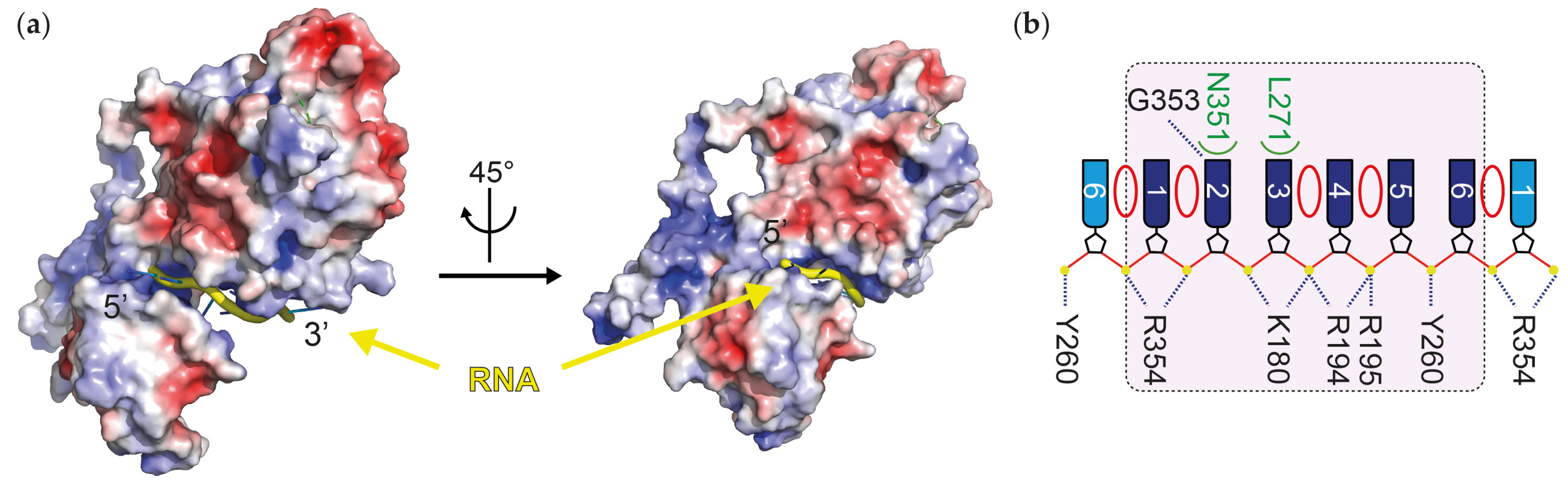
The RNA of non-segmented negative-strand RNA virus (nsNSVs) cannot easily be cut by RNases [7,8,9] because all phosphates of the backbone are bound to the nucleoprotein (Figure 1b). The nucleocapsids are rather rigid structures. Their protomers have masses between 41 and 83 kDa (Table 1), and the nucleoprotein weight per nucleotide is between 5 and 14 kDa (the measles virus nucleoprotein has a mass of 58,131 Da and is associated with six nucleotides per protomer, therefore, 9688 protein Da per nucleotide; see Table 1). nsNSV nucleoproteins have two domains, with a narrow and positive-charged cleft between them in which the genome RNA is bound (Figure 1a). nsNSV nucleoproteins bind a fixed number of nucleotides per protomer, between six to nine nts among different families. The RNA cleft can bind six nucleotides per protomer for paramyxoviruses, like measles [10], mumps [11], or murine respirovirus [12,13], and for filoviruses, like Ebola [14,15,16] or Marburg [17]. The nucleoprotein of pneumovirus (hRSV and hMPV) binds seven nucleotides [18,19], the protomer of Borna disease virus 1 binds eight nucleotides [20], and the nucleoprotein for rabies and vesicular stomatitis virus (VSV) binds nine nucleotides [21,22]. The nucleotide bases can point either toward or away from the protein core and either stack with each other or with amino acid residues [23] (Figure 1b). The nucleotide stacks make A-form structures, and their direction changes every three or four nucleobases. Therefore, unless the viral polymerase is actively engaged in RNA synthesis, the nucleotides bound within the nucleoprotein are as structurally constrained as the nucleocapsid itself in Mononegavirales [6].

3. RNA in Segmented Viruses

The nucleocapsids of segmented negative-strand RNA virus (sNSVs) are very different and very flexible [3,6]. Their vRNA is not entirely bound to nucleoproteins, as the viral polymerases bind both ~10 terminal nucleotides of the 5′ and 3′ termini into two separate and specific single-stranded RNA binding sites, preventing hybridization of these nucleotides despite their complete complementarity. Nucleotides ~10 to 20 of the 5′ and 3′ end form a distal duplex that is essential for the subsequent initiation of replication and/or transcription. The remaining genome RNA, which is associated with nucleoproteins, forms the nucleocapsid that can be easily cut by RNAse treatment in vitro [36,37,38].
A notable shared property of Bunyaviruses and Articulaviruses nucleocapsids is their pronounced flexibility, which prevents their high-resolution (∼3 Å resolution) X-ray or cryo-EM structures’ determination and therefore impairs the understanding of the molecular interaction between long viral RNA and nucleoproteins. Instead, only high-resolution structures of N/NP-RNA rings with a defined number of N/NP proteins and limited-length RNA have been obtained. These in vitro assemblies, while informative for protein-RNA interactions, do not fully represent nucleocapsids due to their restricted size. To obtain molecular insight into binding of longer RNA to sNSV nucleocapsids, likely to reflect native nucleocapsid organization, N/NP-RNA nucleocapsid-like complexes have been reconstituted for influenza A and Hantaan virus [26,34], which are more likely to reflect native nucleocapsid organization. In addition, a mini-RNP containing nine NPs and one polymerase, both bound to RNA, has been structurally determined for influenza [35]. Many of these cryo-EM or X-ray structures were generated in vitro using defined RNA sequences, such as poly-U for Rift Valley fever virus or poly-UC for influenza structures. The Hantaan nucleocapsid-like structure was formed during the nucleoprotein expression in insect cells and is therefore bound to endogenous RNA in an aspecific manner.
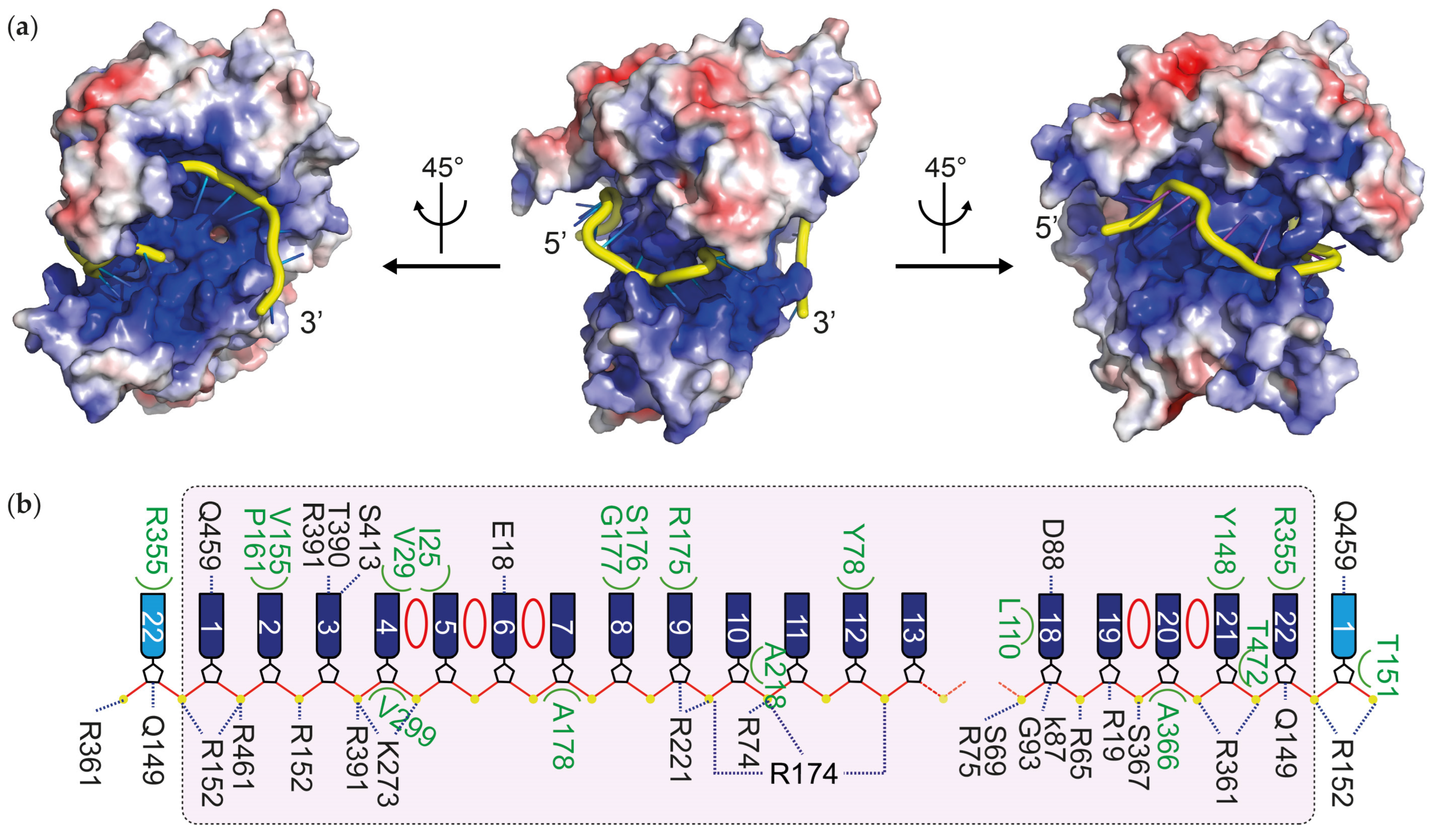
Except influenza A nucleoprotein, which is comparable in size, most sNSV nucleoproteins are smaller than those of nsNSVs. However, they bind lower protein mass per bound nucleotide, ranging from 2.4 to 4 kDa per nucleotide (Table 1). In addition, their RNA-binding cleft is significantly larger (see Figure 1a versus Figure 2a), indicating a greater capacity for RNA interaction compared to the narrower cleft observed in nsNSVs.
Presently, there are 11 high-resolution N/NP-RNA structures showing that sNSV nucleoproteins can accommodate a broader range of nucleotides per protomer (Table 1). From the class Bunyaviricetes, the two Phenuiviruses (Order Hareavirales), Rift Valley fever virus (RVFV) and Toscana virus, accommodate between six and seven bases per N protomer [31,32], and the four peribunyavirus nucleocapsid structures (Order Elliovirales) have 11 bases per protomer [23,27,28,29]. With the four latter, nucleotides 9, 10, and 11 make minimal contact with the nucleoprotein. The large nucleoprotein of Lassa virus (Order Hareavirales) comprises 569 residues, featuring an N-terminal domain (residues 1–340) similar to those of other bunyavirus nucleoproteins and a C-terminal ExoN domain that degrades double-stranded RNA [39]. The N-terminal domain binds eight RNA bases, and the structure of the N-RNA shows a narrow RNA path only for residues 2–4 [30], but residues 5–8 are more flexible. Similarly, the cryo-EM structure of the RNA-bound nucleocapsid of Hantaan virus (Order Elliovirales) reveals that the nucleoproteins form a long, tightly wound nucleocapsid helix containing a continuous positively charged groove likely to be able to accommodate long RNA [26]. Only three bases are resolved per protomer; the remaining nucleotides are likely flexible and not stably associated with the nucleoprotein, which would explain why they are not visible in the cryo-EM structure.
Within the order Articulavirales, the nucleoprotein of tilapia lake virus (family Amnoonviridae) binds 12 nucleotides [33]. In the N-RNA structure of the pseudo-C5 oligomer, each protomer accommodates 12 nucleotides; however, nucleotides C2 to A4 do not interact with the protein. Instead, their bases stack together and remain flexible. There are several nucleocapsid-like structures of influenza A virus (family Orthomyxoviridae) with up to 18 bases, but there is place for more nucleotides, up to 24 bases per protomer [34,40,41]. More recently, the structure of the influenza A virus mini-RNP [35], originally designed by the group of Juan Ortín [42], showed that influenza nucleoprotein can bind 24 nucleotides.
In influenza-like nucleocapsids, four structures have been determined using an N-terminally truncated nucleoprotein (residues 15–498) in complex with poly-UC RNAs of varying lengths (12, 14, and 18 nucleotides; Figure 2). In all cases, the 5′ end of the RNA is positioned between two NP protomers, and the first seven nucleotides adopt a highly conserved conformation, reminiscent of the RNA binding observed in orthobunyavirus N-RNA complexes. Beyond this conserved region, the RNA-binding groove becomes wider, allowing downstream nucleotides to adopt multiple conformations. In the double antiparallel assembly containing an 18-nucleotide RNA, two distinct trajectories are observed: a relaxed strand (strand 1) and a stretched strand (strand 2) ([34]; Figure 2b). In strand 2, nucleotides 5–8 do not interact with the protein and instead form a stacked arrangement of bases, similar to nucleotides 2–4 in the tilapia lake virus nucleoprotein-RNA structure [33]. Notably, the NP-RNA complex within the mini-RNP adopts a particularly open conformation, with only 10 nucleotides resolved in contact with the protein [35]. This observation suggests that the remaining nucleotides are flexible and likely not stably associated with the nucleoprotein.
From the structures of the segmented viruses mentioned here, one could see that the RNA phosphate–ribose backbone is often not bound to the nucleoprotein and, therefore, is sensitive to RNases.

4. Hypothesis: The Flexible Nucleotides of the Segmented Viruses Can Slide on the Nucleoprotein Inside the Viral Capsids

Nucleocapsids of segmented NSV need to protect the viral RNA while supporting the complex processes of vRNA replication/transcription and the formation of a nascent nucleocapsid. We hypothesize four criteria to be essential for these processes: (i) the flexibility of the nucleocapsid, (ii) the tight binding of only few nucleotides per nucleoprotein, (iii) the strong interaction between adjacent nucleoproteins, and (iv) the loose interactions between more distant nucleoproteins. Indeed, during replication and transcription, the polymerase needs to transiently access to the RNA without disrupting the overall architecture of the nucleocapsid. For this, a working model hypothesis is that the viral RNA that enters the polymerase would detach from few nucleoproteins of the nucleocapsid without perturbing the interactions between adjacent nucleoproteins that transiently become RNA-free. After being copied by the polymerase, the viral RNA would then bind again to the same nucleoproteins. For this to happen easily, a flexible nucleocapsid with few nucleotides tightly bound and strong interactions between adjacent nucleoproteins appears ideal to favor transient RNA release from the nucleocapsid during copying, which would bind again when exiting the polymerase.
The same logic could be applied for the proper relative positioning of the eight RNPs during influenza virion formation. This step follows the exit of influenza virus RNP from the nucleus to form the complete genome that buds from the cell. This remarkable eight-part assembly [43,44] is thought to result from stem-loop RNA structures that extrude from each of the various nucleocapsid segments and interact with similar structures from other RNPs to form the complete eight RNA-segment genome [45,46]. It is unlikely that these extruded RNAs are present all the time because they could be sensitive to RNases or bind cell proteins that induce an immune response. It is more likely that these extruded RNAs would be in dynamic equilibrium with alternate structures on each nucleocapsid that form and change during the life cycle of each segment. This interconversion of alternate RNA:nucleoprotein interactions on each segment would be greatly aided by the ability of the RNA to detach and reattach easily to the nucleoprotein. Here again, having only few nucleotides tightly bound would ensure an easy transient extrusion of the RNA.
Having few nucleotides tightly bound, strong adjacent nucleoprotein interactions, and loose interactions between more distant nucleoproteins is also likely to be essential for nascent RNP formation in influenza. One could indeed hypothesize that the addition of a new nucleoprotein onto a nascent, right-handed-growing, and anti-parallel nucleocapsid in influenza is required to modify the relative positioning of the non-adjacent nucleoproteins, while keeping intact the interactions between adjacent nucleoproteins. Tight binding of a few nucleotides per nucleoprotein would prevent sliding of the vRNA and might be essential to preserve nucleocapsid integrity. The loose binding of the other nucleotides to the nucleoprotein would be necessary to enhance the necessary flexibility of this highly dynamic process.
Non-segmented NSV strongly differ from segmented NSV, as the entire RNA backbone of the nsNSV nucleocapsids is bound to the nucleoprotein polymer. Moreover, given the changes in direction of the A-form structures (whose bases point as a group toward or away from the nucleoprotein core), the nucleotides cannot be detached from the protein without extra energy. This tight binding of the RNA allows the replication of paramyxoviruses to be governed by “the rule of six”; only genomes whose total lengths are precisely a multiple of six nucleotides are viable. The ability of the polymerase to access the RNA in this tight configuration would not be possible without the action of the phosphoprotein P that appears as a central actor for disruption of nucleoprotein interactions and exposure of the RNA for its replication.
Overall, the flexible and dynamic nature of sNSV nucleocapsids, characterized by partial RNA binding, strong local protein interactions, and the ability of nucleoproteins to slide along the RNA, appears to be a key evolutionary adaptation. This structural plasticity supports efficient replication, precise genome assembly, and regulated RNA accessibility, distinguishing segmented viruses from their more rigid non-segmented counterparts.

Author Contributions

Writing—original draft preparation, R.W.H.R. and D.K.; writing—review and editing, R.W.H.R., A.B.-C., T.C., H.M. and D.K. All authors have read and agreed to the published version of the manuscript.

Funding

This research was funded by Agence Nationale pour la Recherche, grant Bavarian (ANR-21-CE15-0026).

Institutional Review Board Statement

Not applicable.

Informed Consent Statement

Not applicable.

Data Availability Statement

The original contributions presented in this study are included in the article. Further inquiries can be directed to the corresponding authors.

Conflicts of Interest

The authors declare no conflicts of interest.

Abbreviations

The following abbreviations are used in this manuscript:
vRNAviral genomic RNA
nsNSVnon-segmented negative-strand RNA virus
sNSVsegmented negative-strand RNA virus
N/NPnucleoprotein

References

  1. Ruigrok, R.W.; Crepin, T.; Kolakofsky, D. Nucleoproteins and nucleocapsids of negative-strand RNA viruses. Curr. Opin. Microbiol. 2011, 14, 504–510. [Google Scholar] [CrossRef]
  2. Green, T.J.; Cox, R.; Tsao, J.; Rowse, M.; Qiu, S.; Luo, M. Common mechanism for RNA encapsidation by negative-strand RNA viruses. J. Virol. 2014, 88, 3766–3775. [Google Scholar] [CrossRef][Green Version]
  3. Reguera, J.; Cusack, S.; Kolakofsky, D. Segmented negative strand RNA virus nucleoprotein structure. Curr. Opin. Virol. 2014, 5, 7–15. [Google Scholar] [CrossRef] [PubMed]
  4. Luo, M.; Terrell, J.R.; McManus, S.A. Nucleocapsid Structure of Negative Strand RNA Virus. Viruses 2020, 12, 835. [Google Scholar] [CrossRef]
  5. Modrego, A.; Carlero, D.; Arranz, R.; Martin-Benito, J. CryoEM of Viral Ribonucleoproteins and Nucleocapsids of Single-Stranded RNA Viruses. Viruses 2023, 15, 653. [Google Scholar] [CrossRef] [PubMed]
  6. Sabsay, K.R.; Te Velthuis, A.J.W. Negative and ambisense RNA virus ribonucleocapsids: More than protective armor. Microbiol. Mol. Biol. Rev. 2023, 87, e0008223. [Google Scholar] [CrossRef] [PubMed]
  7. Hefti, E.; Bishop, D.H. The 5′ sequence of VSV viral RNA and its in vitro transcription product RNA. Biochem. Biophys. Res. Commun. 1975, 66, 785–792. [Google Scholar] [CrossRef]
  8. Lynch, S.; Kolakofsky, D. Ends of the RNA within Sendai virus defective interfering nucleocapsids are not free. J. Virol. 1978, 28, 584–589. [Google Scholar] [CrossRef]
  9. Green, T.J.; Rowse, M.; Tsao, J.; Kang, J.; Ge, P.; Zhou, Z.H.; Luo, M. Access to RNA encapsidated in the nucleocapsid of vesicular stomatitis virus. J. Virol. 2011, 85, 2714–2722. [Google Scholar] [CrossRef]
  10. Gutsche, I.; Desfosses, A.; Effantin, G.; Ling, W.L.; Haupt, M.; Ruigrok, R.W.; Sachse, C.; Schoehn, G. Structural virology. Near-atomic cryo-EM structure of the helical measles virus nucleocapsid. Science 2015, 348, 704–707. [Google Scholar] [CrossRef]
  11. Shan, H.; Su, X.; Li, T.; Qin, Y.; Zhang, N.; Yang, L.; Ma, L.; Bai, Y.; Qi, L.; Liu, Y.; et al. Structural plasticity of mumps virus nucleocapsids with cryo-EM structures. Commun. Biol. 2021, 4, 833. [Google Scholar] [CrossRef]
  12. Zhang, N.; Shan, H.; Liu, M.; Li, T.; Luo, R.; Yang, L.; Qi, L.; Chu, X.; Su, X.; Wang, R.; et al. Structure and assembly of double-headed Sendai virus nucleocapsids. Commun. Biol. 2021, 4, 494. [Google Scholar] [CrossRef]
  13. Calain, P.; Roux, L. The rule of six, a basic feature for efficient replication of Sendai virus defective interfering RNA. J. Virol. 1993, 67, 4822–4830. [Google Scholar] [CrossRef]
  14. Wan, W.; Kolesnikova, L.; Clarke, M.; Koehler, A.; Noda, T.; Becker, S.; Briggs, J.A.G. Structure and assembly of the Ebola virus nucleocapsid. Nature 2017, 551, 394–397. [Google Scholar] [CrossRef] [PubMed]
  15. Su, Z.; Wu, C.; Shi, L.; Luthra, P.; Pintilie, G.D.; Johnson, B.; Porter, J.R.; Ge, P.; Chen, M.; Liu, G.; et al. Electron Cryo-microscopy Structure of Ebola Virus Nucleoprotein Reveals a Mechanism for Nucleocapsid-like Assembly. Cell 2018, 172, 966–978 e912. [Google Scholar] [CrossRef] [PubMed]
  16. Sugita, Y.; Matsunami, H.; Kawaoka, Y.; Noda, T.; Wolf, M. Cryo-EM structure of the Ebola virus nucleoprotein-RNA complex at 3.6 A resolution. Nature 2018, 563, 137–140, Erratum in Nature 2022, 601, E11. https://doi.org/10.1038/s41586-021-04304-6. [Google Scholar] [CrossRef] [PubMed]
  17. Fujita-Fujiharu, Y.; Sugita, Y.; Takamatsu, Y.; Houri, K.; Igarashi, M.; Muramoto, Y.; Nakano, M.; Tsunoda, Y.; Taniguchi, I.; Becker, S.; et al. Structural insight into Marburg virus nucleoprotein-RNA complex formation. Nat. Commun. 2022, 13, 1191. [Google Scholar] [CrossRef]
  18. Tawar, R.G.; Duquerroy, S.; Vonrhein, C.; Varela, P.F.; Damier-Piolle, L.; Castagne, N.; MacLellan, K.; Bedouelle, H.; Bricogne, G.; Bhella, D.; et al. Crystal structure of a nucleocapsid-like nucleoprotein-RNA complex of respiratory syncytial virus. Science 2009, 326, 1279–1283. [Google Scholar] [CrossRef]
  19. Whitehead, J.D.; Decool, H.; Leyrat, C.; Carrique, L.; Fix, J.; Eleouet, J.F.; Galloux, M.; Renner, M. Structure of the N-RNA/P interface indicates mode of L/P recruitment to the nucleocapsid of human metapneumovirus. Nat. Commun. 2023, 14, 7627. [Google Scholar] [CrossRef]
  20. Sugita, Y.; Hirai, Y.; Goto, S.H.; Fujiwara, T.; Tomonaga, K.; Noda, T.; Horie, M. Structure and assembly of Borna disease virus 1 nucleoprotein-RNA complexes. Sci. Adv. 2026, 12, eaeb0835. [Google Scholar] [CrossRef]
  21. Albertini, A.A.; Wernimont, A.K.; Muziol, T.; Ravelli, R.B.; Clapier, C.R.; Schoehn, G.; Weissenhorn, W.; Ruigrok, R.W. Crystal structure of the rabies virus nucleoprotein-RNA complex. Science 2006, 313, 360–363. [Google Scholar] [CrossRef]
  22. Green, T.J.; Zhang, X.; Wertz, G.W.; Luo, M. Structure of the vesicular stomatitis virus nucleoprotein-RNA complex. Science 2006, 313, 357–360. [Google Scholar] [CrossRef]
  23. Dong, H.; Li, P.; Bottcher, B.; Elliott, R.M.; Dong, C. Crystal structure of Schmallenberg orthobunyavirus nucleoprotein-RNA complex reveals a novel RNA sequestration mechanism. RNA 2013, 19, 1129–1136. [Google Scholar] [CrossRef] [PubMed]
  24. Song, X.; Shan, H.; Zhu, Y.; Hu, S.; Xue, L.; Chen, Y.; Ding, W.; Niu, T.; Gu, J.; Ouyang, S.; et al. Self-capping of nucleoprotein filaments protects the Newcastle disease virus genome. Elife 2019, 8, e45057. [Google Scholar] [CrossRef] [PubMed]
  25. Ker, D.S.; Jenkins, H.T.; Greive, S.J.; Antson, A.A. CryoEM structure of the Nipah virus nucleocapsid assembly. PLoS Pathog. 2021, 17, e1009740. [Google Scholar] [CrossRef]
  26. Arragain, B.; Reguera, J.; Desfosses, A.; Gutsche, I.; Schoehn, G.; Malet, H. High resolution cryo-EM structure of the helical RNA-bound Hantaan virus nucleocapsid reveals its assembly mechanisms. Elife 2019, 8, e43075. [Google Scholar] [CrossRef] [PubMed]
  27. Niu, F.; Shaw, N.; Wang, Y.E.; Jiao, L.; Ding, W.; Li, X.; Zhu, P.; Upur, H.; Ouyang, S.; Cheng, G.; et al. Structure of the Leanyer orthobunyavirus nucleoprotein-RNA complex reveals unique architecture for RNA encapsidation. Proc. Natl. Acad. Sci. USA 2013, 110, 9054–9059. [Google Scholar] [CrossRef]
  28. Ariza, A.; Tanner, S.J.; Walter, C.T.; Dent, K.C.; Shepherd, D.A.; Wu, W.; Matthews, S.V.; Hiscox, J.A.; Green, T.J.; Luo, M.; et al. Nucleocapsid protein structures from orthobunyaviruses reveal insight into ribonucleoprotein architecture and RNA polymerization. Nucleic Acids Res. 2013, 41, 5912–5926. [Google Scholar] [CrossRef]
  29. Reguera, J.; Malet, H.; Weber, F.; Cusack, S. Structural basis for encapsidation of genomic RNA by La Crosse Orthobunyavirus nucleoprotein. Proc. Natl. Acad. Sci. USA 2013, 110, 7246–7251. [Google Scholar] [CrossRef]
  30. Hastie, K.M.; Liu, T.; Li, S.; King, L.B.; Ngo, N.; Zandonatti, M.A.; Woods, V.L., Jr.; de la Torre, J.C.; Saphire, E.O. Crystal structure of the Lassa virus nucleoprotein-RNA complex reveals a gating mechanism for RNA binding. Proc. Natl. Acad. Sci. USA 2011, 108, 19365–19370. [Google Scholar] [CrossRef]
  31. Raymond, D.D.; Piper, M.E.; Gerrard, S.R.; Skiniotis, G.; Smith, J.L. Phleboviruses encapsidate their genomes by sequestering RNA bases. Proc. Natl. Acad. Sci. USA 2012, 109, 19208–19213. [Google Scholar] [CrossRef] [PubMed]
  32. Olal, D.; Dick, A.; Woods, V.L., Jr.; Liu, T.; Li, S.; Devignot, S.; Weber, F.; Saphire, E.O.; Daumke, O. Structural insights into RNA encapsidation and helical assembly of the Toscana virus nucleoprotein. Nucleic Acids Res. 2014, 42, 6025–6037. [Google Scholar] [CrossRef] [PubMed]
  33. Arragain, B.; Pelosse, M.; Huard, K.; Cusack, S. Structure of the tilapia lake virus nucleoprotein bound to RNA. Nucleic Acids Res. 2025, 53, gkaf112. [Google Scholar] [CrossRef] [PubMed]
  34. Chenavier, F.; Zarkadas, E.; Freslon, L.L.; Stelfox, A.J.; Schoehn, G.; Ruigrok, R.W.H.; Ballandras-Colas, A.; Crepin, T. Influenza a virus antiparallel helical nucleocapsid-like pseudo-atomic structure. Nucleic Acids Res. 2025, 53, gkae1211. [Google Scholar] [CrossRef] [PubMed]
  35. Kang, H.; Yang, Y.; Liu, Y.; Li, M.; Zhang, L.; Lin, Y.; Witte, L.; Chen, K.Y.; Song, W.; Xu, Z.; et al. Coupling of polymerase-nucleoprotein-RNA in an influenza virus mini ribonucleoprotein complex. Nat. Commun. 2025, 16, 9741. [Google Scholar] [CrossRef]
  36. Pons, M.W.; Schulze, I.T.; Hirst, G.K.; Hauser, R. Isolation and characterization of the ribonucleoprotein of influenza virus. Virology 1969, 39, 250–259. [Google Scholar] [CrossRef]
  37. Obijeski, J.F.; Bishop, D.H.; Palmer, E.L.; Murphy, F.A. Segmented genome and nucleocapsid of La Crosse virus. J. Virol. 1976, 20, 664–675. [Google Scholar] [CrossRef]
  38. Klumpp, K.; Ruigrok, R.W.; Baudin, F. Roles of the influenza virus polymerase and nucleoprotein in forming a functional RNP structure. EMBO J. 1997, 16, 1248–1257. [Google Scholar] [CrossRef]
  39. Papageorgiou, N.; Spiliopoulou, M.; Nguyen, T.V.; Vaitsopoulou, A.; Laban, E.Y.; Alvarez, K.; Margiolaki, I.; Canard, B.; Ferron, F. Brothers in Arms: Structure, Assembly and Function of Arenaviridae Nucleoprotein. Viruses 2020, 12, 772. [Google Scholar] [CrossRef]
  40. Compans, R.W.; Content, J.; Duesberg, P.H. Structure of the ribonucleoprotein of influenza virus. J. Virol. 1972, 10, 795–800. [Google Scholar] [CrossRef]
  41. Peng, R.; Xu, X.; Nepal, B.; Gong, Y.; Li, F.; Ferretti, M.B.; Zhou, M.; Lynch, K.W.; Burslem, G.M.; Kortagere, S.; et al. Molecular basis of influenza ribonucleoprotein complex assembly and processive RNA synthesis. Science 2025, 388, eadq7597. [Google Scholar] [CrossRef]
  42. Ortega, J.; Martin-Benito, J.; Zurcher, T.; Valpuesta, J.M.; Carrascosa, J.L.; Ortin, J. Ultrastructural and functional analyses of recombinant influenza virus ribonucleoproteins suggest dimerization of nucleoprotein during virus amplification. J. Virol. 2000, 74, 156–163. [Google Scholar] [CrossRef] [PubMed]
  43. Noda, T.; Sagara, H.; Yen, A.; Takada, A.; Kida, H.; Cheng, R.H.; Kawaoka, Y. Architecture of ribonucleoprotein complexes in influenza A virus particles. Nature 2006, 439, 490–492. [Google Scholar] [CrossRef]
  44. Noda, T.; Sugita, Y.; Aoyama, K.; Hirase, A.; Kawakami, E.; Miyazawa, A.; Sagara, H.; Kawaoka, Y. Three-dimensional analysis of ribonucleoprotein complexes in influenza A virus. Nat. Commun. 2012, 3, 639. [Google Scholar] [CrossRef]
  45. Dadonaite, B.; Gilbertson, B.; Knight, M.L.; Trifkovic, S.; Rockman, S.; Laederach, A.; Brown, L.E.; Fodor, E.; Bauer, D.L.V. The structure of the influenza A virus genome. Nat. Microbiol. 2019, 4, 1781–1789. [Google Scholar] [CrossRef] [PubMed]
  46. Jakob, C.; Paul-Stansilaus, R.; Schwemmle, M.; Marquet, R.; Bolte, H. The influenza A virus genome packaging network—Complex, flexible and yet unsolved. Nucleic Acids Res. 2022, 50, 9023–9038. [Google Scholar] [CrossRef] [PubMed]
Figure 1. Measles virus nucleoprotein–RNA complex. (a). Structure of a single protomer of measles N-RNA (PDB code: 6H5Q); positive charges are in blue, and negative charges are in red. The RNA (shown as a yellow ribbon) is hardly visible in N surface representation, as it is within a narrow binding groove between the 2 lobes N. (b). Schematic representation of a nucleoprotein (pink box) RNA complex. The yellow and black colors represent the phosphate and ribose of the RNA backbone, respectively. Six RNA bases bound to the nucleoprotein are shown as dark blue rectangles and labelled 1 to 6. The RNA bases that are not bound to the protomer appear as blue rectangles. The red circles, the blue dotted lines, and the green semicircles indicate the stacked nucleobases, the hydrogen bonds, and the hydrophobic interactions, respectively. Nucleotides 6-1-2 are stacked (indicated by red circles), and the bases are pointing away from the protein; bases 3-4-5 stack as well and point towards the nucleoprotein. All phosphates are bound to the protein.
Microorganisms 14 01194 g001
Figure 2. RNA binding by influenza virus nucleoprotein. (a). Single protomer of the influenza N-RNA in the RNP-like structure (PDB code: 9GAS); positive charges are in blue, and negative charges are in red. The RNA is shown as a yellow ribbon. The structure shows a large positive area for RNA binding. (b). Schematic representation of Strand 2 in the RNP-like structure (ref. [34]). The light pink box represents the considered protomer of nucleoprotein. The yellow and black colors represent the phosphate and ribose of the RNA backbone, respectively. The red circles, the blue dotted lines, and the green semicircles indicate the stacked nucleobases, the hydrogen bonds, and the hydrophobic interactions, respectively. Most phosphates are bound to NP, except nucleotides 4 to 7, where NP binding occurs through base stacking rather than interactions with the phosphate groups. N.B. For this figure, we have changed the numbers of the nucleotides from the original figure in [34].
Microorganisms 14 01194 g002
Table 1. RNA–nucleoprotein structures of the negative-strand RNA viruses.
VirusN/NP (Residues)Mass (Da)Nucleotides per Protomer (nt)Mass Protein/nt (Da)References
Non-segmented Negative-strand RNA Virus
Phylum Negarnaviricota, Subphylum Haploviricotina, Class Monjiviricetes, Order Mononegavirales
Family Bornaviridae
Borna disease virus 137240,93185116[20]
Family Filoviridae
Ebola virus73983,300613,883[14]
Marburg virus69577,747612,958[17]
Family Paramyxoviridae
Measles virus52558,13169688[10]
Mumps virus54961,366610,228[11]
Avian orthoavulavirus 1 148953,02368835[24]
Nipah virus53258,16869694[25]
Murine respirovirus 251756,76269460[12]
Family Pneumoviridae
hRSV39143,49376213[18]
hMPV39443,53876620[19]
Family Rhabdoviridae
Rabies virus45050,60595627[21]
VSV42247,36895263[22]
Segmented Negative-strand RNA Virus
Phylum Negarnaviricota, Subphylum Polyploviricotina, Class Bunyaviricetes, Order Elliovirales
Family Hantaviridae
Hantaan virus 342948,1423 3NC 5[26]
Family Peribunyaviridae
Leanyer virus23526,259112387[27]
Bunyamwera virus23326,664112424[28]
La Crosse virus23526,530112412[29]
Schmallenberg virus23326,181112380[23]
Phylum Negarnaviricota, Subphylum Polyploviricotina, Class Bunyaviricetes, Order Hareavirales
Family Arenaviridae
Lassa virus 41–34038,20084775[30]
Family Phenuiviridae
Rift Valley fever virus24527,3606–73909–4560[31]
Toscana virus25327,70473958[32]
Phylum Negarnaviricota, Subphylum Polyploviricotina, Class Insthoviricetes, Order Articulavirales
Family Amnoonviridae
Tilapia lake virus35438,400123200[33]
Family Orthomyxoviridae
Influenza A virus49856,210242342[34,35]
1 New name for Newcastle disease virus. 2 New name for Sendai virus. 3 Only 3 nucleotides are visible in the cryo-EM structure, but the number of bases per N is unknown. 4 N-terminal domain. 5 not calculated.
Disclaimer/Publisher’s Note: The statements, opinions and data contained in all publications are solely those of the individual author(s) and contributor(s) and not of MDPI and/or the editor(s). MDPI and/or the editor(s) disclaim responsibility for any injury to people or property resulting from any ideas, methods, instructions or products referred to in the content.