Structural Advances in Respiratory Syncytial Virus: Implications for Vaccine and Antiviral Development


Abstract
1. Introduction
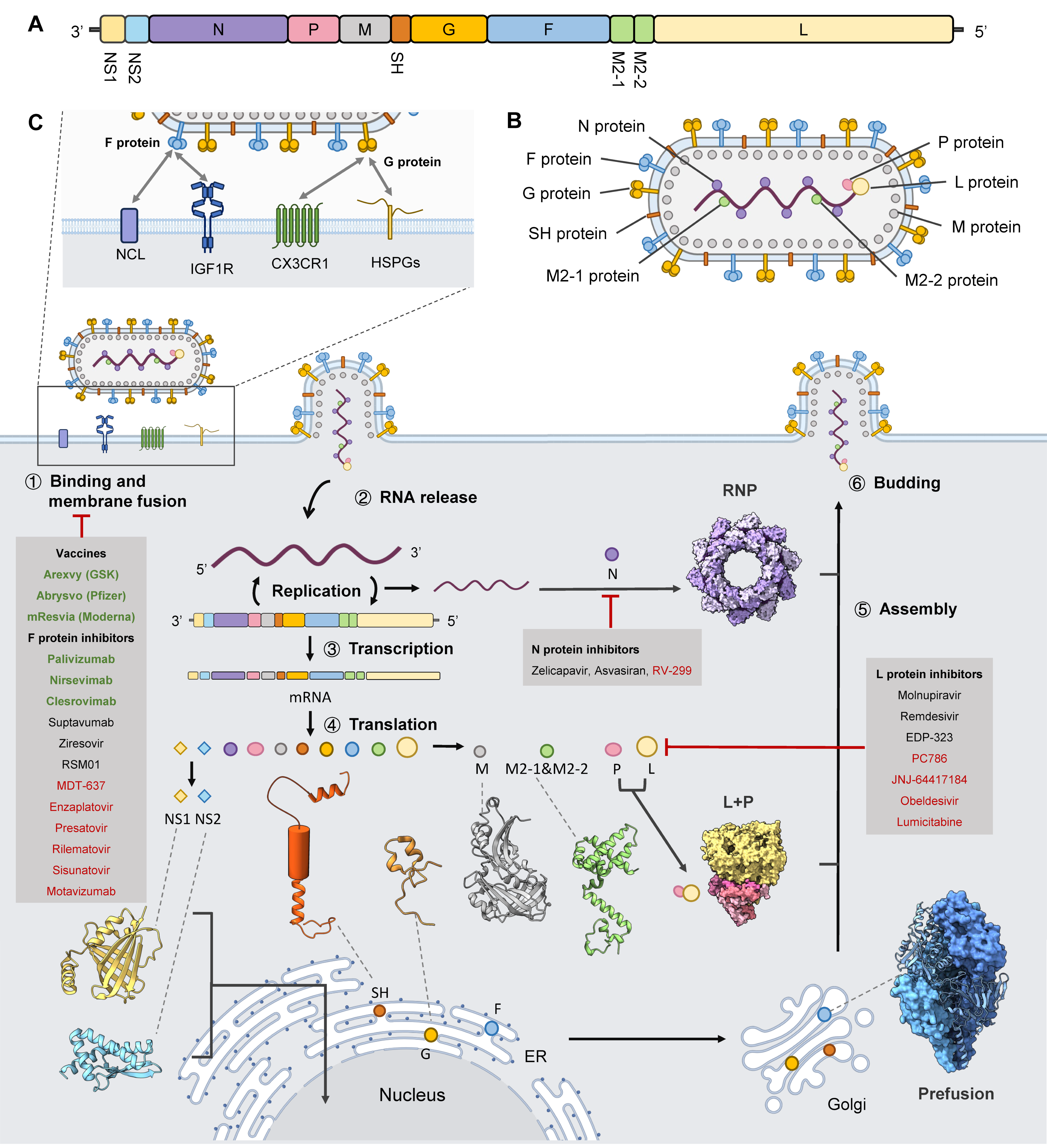
2. RSV Genome and Virion Architecture
2.1. Genome
2.2. Structures of RSV Components
3. RSV Life Cycle
3.1. Viral Attachment and Entry
3.2. Transcription and Genome Replication
3.3. Protein Translation
3.4. Assembly and Budding
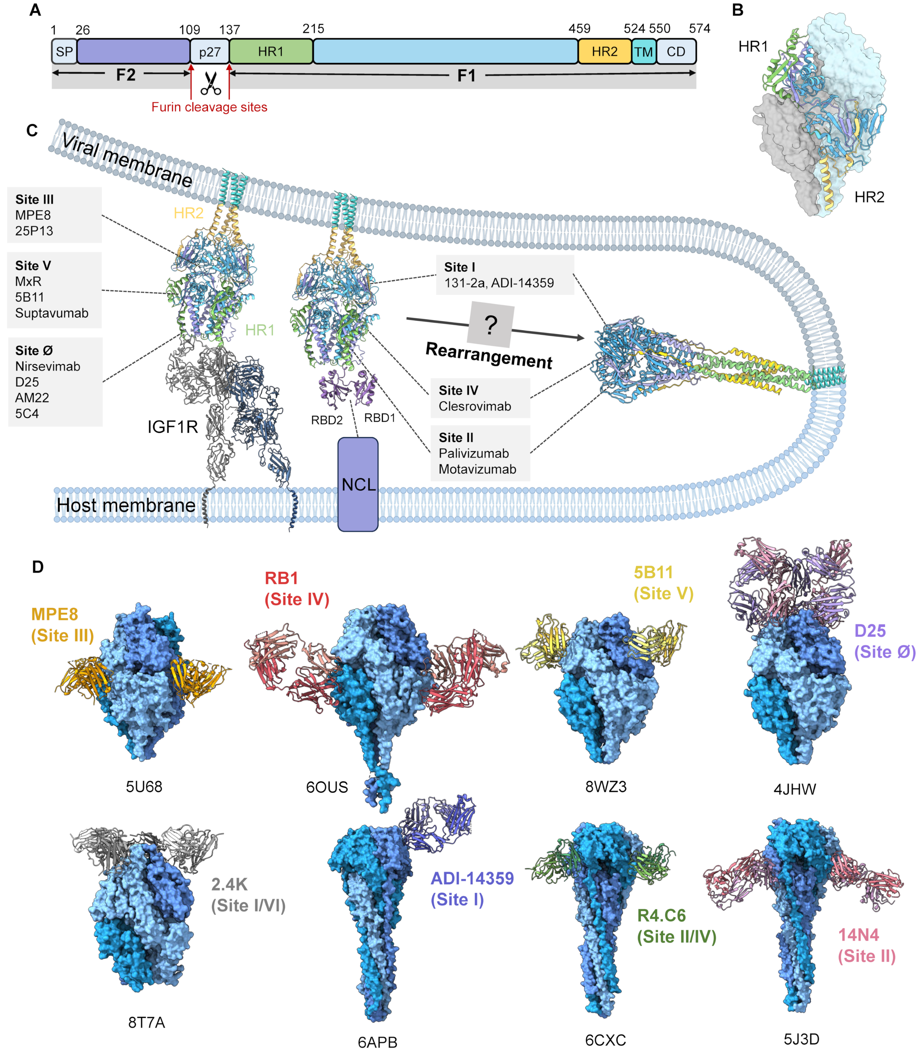
4. Fusion Machinery
5. Vaccine and Antiviral Design
5.1. F Protein Structure-Guided Design of Vaccine and Therapeutics
5.2. Structure-Guided Antiviral Development Beyond F
5.3. Advances in RSV Vaccines
5.4. Advances in RSV Therapeutic Agents
6. Future Perspectives
Funding
Institutional Review Board Statement
Informed Consent Statement
Data Availability Statement
Conflicts of Interest
References
- Zhu, D.; Cao, D.; Zhang, X. Virus structures revealed by advanced cryoelectron microscopy methods. Structure 2023, 31, 1348–1359. [Google Scholar] [CrossRef] [PubMed]
- Dutta, M.; Acharya, P. Cryo-electron microscopy in the study of virus entry and infection. Front. Mol. Biosci. 2024, 11, 1429180. [Google Scholar] [CrossRef] [PubMed]
- Calder, L.J.; Rosenthal, P.B. Cryomicroscopy provides structural snapshots of influenza virus membrane fusion. Nat. Struct. Mol. Biol. 2016, 23, 853–858. [Google Scholar] [CrossRef]
- Klein, S.; Cortese, M.; Winter, S.L.; Wachsmuth-Melm, M.; Neufeldt, C.J.; Cerikan, B.; Stanifer, M.L.; Boulant, S.; Bartenschlager, R.; Chlanda, P. SARS-CoV-2 structure and replication characterized by in situ cryo-electron tomography. Nat. Commun. 2020, 11, 5885. [Google Scholar] [CrossRef] [PubMed]
- Mendonca, L.; Howe, A.; Gilchrist, J.B.; Sheng, Y.; Sun, D.; Knight, M.L.; Zanetti-Domingues, L.C.; Bateman, B.; Krebs, A.S.; Chen, L.; et al. Correlative multi-scale cryo-imaging unveils SARS-CoV-2 assembly and egress. Nat. Commun. 2021, 12, 4629. [Google Scholar] [CrossRef]
- Chen, L.; Hikichi, Y.; Rey, J.S.; Akil, C.; Zhu, Y.; Veler, H.; Shen, Y.; Perilla, J.R.; Freed, E.O.; Zhang, P. Structural maturation of the matrix lattice is not required for HIV-1 particle infectivity. Sci. Adv. 2025, 11, eadv4356. [Google Scholar] [CrossRef]
- Benhaim, M.A.; Mangala Prasad, V.; Garcia, N.K.; Guttman, M.; Lee, K.K. Structural monitoring of a transient intermediate in the hemagglutinin fusion machinery on influenza virions. Sci. Adv. 2020, 6, eaaz8822. [Google Scholar] [CrossRef]
- Akil, C.; Xu, J.; Shen, J.; Zhang, P. Unveiling the structural spectrum of SARS-CoV-2 fusion by in situ cryo-ET. Nat. Commun. 2025, 16, 5150. [Google Scholar] [CrossRef]
- Bao, K.; Zhang, X.; Li, D.; Sun, W.; Sun, Z.; Wang, J.; Zhu, P. In situ structures of polymerase complex of mammalian reovirus illuminate RdRp activation and transcription regulation. Proc. Natl. Acad. Sci. USA 2022, 119, e2203054119. [Google Scholar] [CrossRef]
- Si, Z.; Zhou, K.; Tsao, J.; Luo, M.; Zhou, Z.H. Locations and in situ structure of the polymerase complex inside the virion of vesicular stomatitis virus. Proc. Natl. Acad. Sci. USA 2022, 119, e2111948119. [Google Scholar] [CrossRef]
- Xia, X.; Sung, P.Y.; Martynowycz, M.W.; Gonen, T.; Roy, P.; Zhou, Z.H. RNA genome packaging and capsid assembly of bluetongue virus visualized in host cells. Cell 2024, 187, 2236–2249 e2217. [Google Scholar] [CrossRef]
- Li, H.; Shao, L.; Liu, Z.; Liu, Q.; Xiang, Y. Molecular mechanisms of the viral encoded chaperone 100K in capsid folding and assembly of adenovirus. Nat. Commun. 2025, 16, 4013. [Google Scholar] [CrossRef] [PubMed]
- Watanabe, R.; Zyla, D.; Parekh, D.; Hong, C.; Jones, Y.; Schendel, S.L.; Wan, W.; Castillon, G.; Saphire, E.O. Intracellular Ebola virus nucleocapsid assembly revealed by in situ cryo-electron tomography. Cell 2024, 187, 5587–5603 e5519. [Google Scholar] [CrossRef] [PubMed]
- Wachsmuth-Melm, M.; Peterl, S.; O’Riain, A.; Makroczyova, J.; Fischer, K.; Krischuns, T.; Vale-Costa, S.; Amorim, M.J.; Chlanda, P. Visualizing influenza A virus assembly by in situ cryo-electron tomography. Nat. Commun. 2025, 16, 9394. [Google Scholar] [CrossRef]
- Dahmane, S.; Schexnaydre, E.; Zhang, J.; Singh, B.K.; Rosendal, E.; Chotiwan, N.; Sharma, K.B.; Nilsson, E.; Peters, M.B.A.; Yau, W.L.; et al. Cryo-electron tomography reveals coupled flavivirus replication, budding and maturation. Nat. Commun. 2026, 17, 828. [Google Scholar] [CrossRef]
- Liu, C.; Mendonca, L.; Yang, Y.; Gao, Y.; Shen, C.; Liu, J.; Ni, T.; Ju, B.; Liu, C.; Tang, X.; et al. The Architecture of Inactivated SARS-CoV-2 with Postfusion Spikes Revealed by Cryo-EM and Cryo-ET. Structure 2020, 28, 1218–1224 e1214. [Google Scholar] [CrossRef] [PubMed]
- Ni, T.; Mendonca, L.; Zhu, Y.; Howe, A.; Radecke, J.; Shah, P.M.; Sheng, Y.; Krebs, A.S.; Duyvesteyn, H.M.E.; Allen, E.; et al. ChAdOx1 COVID vaccines express RBD open prefusion SARS-CoV-2 spikes on the cell surface. iScience 2023, 26, 107882. [Google Scholar] [CrossRef]
- Ruckel, C.E.; Wolf, J.D.; Plemper, R.K. Status of advanced respiratory syncytial virus antiviral therapeutics 2025. Curr. Opin. Virol. 2025, 73, 101477. [Google Scholar] [CrossRef]
- Liang, X.; Yin, Y.; Lin, Y.; Chen, S.; Qian, Q.; Yuan, J.; Yang, L.; Yang, Y. Molecular and Cellular Mechanisms of Respiratory Syncytial Viral Infection: Its Implications for Prophylactic and Therapeutic Pharmaceuticals. MedComm (2020) 2025, 6, e70403. [Google Scholar] [CrossRef]
- Tang, K.; Wei, G.; Zhan, P. Breaking the impossible: The evolutionary journey of lenacapavir, a long-acting anti-HIV drug. Chin. Chem. Lett. 2025, 36, 110500. [Google Scholar] [CrossRef]
- Bester, S.M.; Wei, G.; Zhao, H.; Adu-Ampratwum, D.; Iqbal, N.; Courouble, V.V.; Francis, A.C.; Annamalai, A.S.; Singh, P.K.; Shkriabai, N.; et al. Structural and mechanistic bases for a potent HIV-1 capsid inhibitor. Science 2020, 370, 360–364. [Google Scholar] [CrossRef] [PubMed]
- Krempl, C.; Murphy, B.R.; Collins, P.L. Recombinant respiratory syncytial virus with the G and F genes shifted to the promoter-proximal positions. J. Virol. 2002, 76, 11931–11942. [Google Scholar] [CrossRef] [PubMed]
- Mufson, M.A.; Orvell, C.; Rafnar, B.; Norrby, E. Two distinct subtypes of human respiratory syncytial virus. J. Gen. Virol. 1985, 66, 2111–2124. [Google Scholar] [CrossRef] [PubMed]
- Blount, R.E., Jr.; Morris, J.A.; Savage, R.E. Recovery of cytopathogenic agent from chimpanzees with coryza. Proc. Soc. Exp. Biol. Med. 1956, 92, 544–549. [Google Scholar]
- Li, Y.; Wang, X.; Blau, D.M.; Caballero, M.T.; Feikin, D.R.; Gill, C.J.; Madhi, S.A.; Omer, S.B.; Simoes, E.A.F.; Campbell, H.; et al. Global, regional, and national disease burden estimates of acute lower respiratory infections due to respiratory syncytial virus in children younger than 5 years in 2019: A systematic analysis. Lancet 2022, 399, 2047–2064. [Google Scholar] [CrossRef]
- Ke, Z.; Dillard, R.S.; Chirkova, T.; Leon, F.; Stobart, C.C.; Hampton, C.M.; Strauss, J.D.; Rajan, D.; Rostad, C.A.; Taylor, J.V.; et al. The Morphology and Assembly of Respiratory Syncytial Virus Revealed by Cryo-Electron Tomography. Viruses 2018, 10, 446. [Google Scholar] [CrossRef]
- McLellan, J.S.; Chen, M.; Leung, S.; Graepel, K.W.; Du, X.; Yang, Y.; Zhou, T.; Baxa, U.; Yasuda, E.; Beaumont, T.; et al. Structure of RSV fusion glycoprotein trimer bound to a prefusion-specific neutralizing antibody. Science 2013, 340, 1113–1117. [Google Scholar] [CrossRef]
- McLellan, J.S.; Chen, M.; Joyce, M.G.; Sastry, M.; Stewart-Jones, G.B.; Yang, Y.; Zhang, B.; Chen, L.; Srivatsan, S.; Zheng, A.; et al. Structure-based design of a fusion glycoprotein vaccine for respiratory syncytial virus. Science 2013, 342, 592–598. [Google Scholar] [CrossRef]
- Tawar, R.G.; Duquerroy, S.; Vonrhein, C.; Varela, P.F.; Damier-Piolle, L.; Castagne, N.; MacLellan, K.; Bedouelle, H.; Bricogne, G.; Bhella, D.; et al. Crystal structure of a nucleocapsid-like nucleoprotein-RNA complex of respiratory syncytial virus. Science 2009, 326, 1279–1283. [Google Scholar] [CrossRef]
- Gilman, M.S.A.; Liu, C.; Fung, A.; Behera, I.; Jordan, P.; Rigaux, P.; Ysebaert, N.; Tcherniuk, S.; Sourimant, J.; Eleouet, J.F.; et al. Structure of the Respiratory Syncytial Virus Polymerase Complex. Cell 2019, 179, 193–204.e14. [Google Scholar] [CrossRef]
- Sibert, B.S.; Kim, J.Y.; Yang, J.E.; Ke, Z.; Stobart, C.C.; Moore, M.L.; Wright, E.R. Assembly of respiratory syncytial virus matrix protein lattice and its coordination with fusion glycoprotein trimers. Nat. Commun. 2024, 15, 5923. [Google Scholar] [CrossRef] [PubMed]
- Cao, D.; Gao, Y.; Liang, B. Structural Insights into the Respiratory Syncytial Virus RNA Synthesis Complexes. Viruses 2021, 13, 834. [Google Scholar] [CrossRef] [PubMed]
- Cao, D.; Gao, Y.; Chen, Z.; Gooneratne, I.; Roesler, C.; Mera, C.; D’Cunha, P.; Antonova, A.; Katta, D.; Romanelli, S.; et al. Structures of the promoter-bound respiratory syncytial virus polymerase. Nature 2024, 625, 611–617. [Google Scholar] [CrossRef] [PubMed]
- Girma, A. Biology of human respiratory syncytial virus: Current perspectives in immune response and mechanisms against the virus. Virus Res. 2024, 350, 199483. [Google Scholar] [CrossRef]
- Chatterjee, S.; Luthra, P.; Esaulova, E.; Agapov, E.; Yen, B.C.; Borek, D.M.; Edwards, M.R.; Mittal, A.; Jordan, D.S.; Ramanan, P.; et al. Structural basis for human respiratory syncytial virus NS1-mediated modulation of host responses. Nat. Microbiol. 2017, 2, 17101. [Google Scholar] [CrossRef]
- Pei, J.; Beri, N.R.; Zou, A.J.; Hubel, P.; Dorando, H.K.; Bergant, V.; Andrews, R.D.; Pan, J.; Andrews, J.M.; Sheehan, K.C.F.; et al. Nuclear-localized human respiratory syncytial virus NS1 protein modulates host gene transcription. Cell Rep. 2021, 37, 109803. [Google Scholar] [CrossRef]
- Han, B.; Wang, Y.; Zheng, M. Inhibition of autophagy promotes human RSV NS1-induced inflammation and apoptosis in vitro. Exp. Ther. Med. 2021, 22, 1054. [Google Scholar] [CrossRef]
- Mousa, J.J.; Kose, N.; Matta, P.; Gilchuk, P.; Crowe, J.E., Jr. A novel pre-fusion conformation-specific neutralizing epitope on the respiratory syncytial virus fusion protein. Nat. Microbiol. 2017, 2, 16271. [Google Scholar] [CrossRef]
- Langley, J.M.; MacDonald, L.D.; Weir, G.M.; MacKinnon-Cameron, D.; Ye, L.; McNeil, S.; Schepens, B.; Saelens, X.; Stanford, M.M.; Halperin, S.A. A Respiratory Syncytial Virus Vaccine Based on the Small Hydrophobic Protein Ectodomain Presented with a Novel Lipid-Based Formulation Is Highly Immunogenic and Safe in Adults: A First-in-Humans Study. J. Infect. Dis. 2018, 218, 378–387. [Google Scholar] [CrossRef]
- Mitra, R.; Baviskar, P.; Duncan-Decocq, R.R.; Patel, D.; Oomens, A.G. The human respiratory syncytial virus matrix protein is required for maturation of viral filaments. J. Virol. 2012, 86, 4432–4443. [Google Scholar] [CrossRef]
- Langedijk, A.C.; Bont, L.J. Respiratory syncytial virus infection and novel interventions. Nat. Rev. Microbiol. 2023, 21, 734–749, Erratum in Nat. Rev. Microbiol. 2024, 22, 587. [Google Scholar] [CrossRef] [PubMed]
- Lee, J.Y.; Chang, J. Recombinant baculovirus-based vaccine expressing M2 protein induces protective CD8(+) T-cell immunity against respiratory syncytial virus infection. J. Microbiol. 2017, 55, 900–908. [Google Scholar] [CrossRef] [PubMed]
- Haynes, L.M.; Caidi, H.; Radu, G.U.; Miao, C.; Harcourt, J.L.; Tripp, R.A.; Anderson, L.J. Therapeutic monoclonal antibody treatment targeting respiratory syncytial virus (RSV) G protein mediates viral clearance and reduces the pathogenesis of RSV infection in BALB/c mice. J. Infect. Dis. 2009, 200, 439–447. [Google Scholar] [CrossRef] [PubMed]
- Jones, H.G.; Ritschel, T.; Pascual, G.; Brakenhoff, J.P.J.; Keogh, E.; Furmanova-Hollenstein, P.; Lanckacker, E.; Wadia, J.S.; Gilman, M.S.A.; Williamson, R.A.; et al. Structural basis for recognition of the central conserved region of RSV G by neutralizing human antibodies. PLoS Pathog. 2018, 14, e1006935. [Google Scholar] [CrossRef]
- Battles, M.B.; McLellan, J.S. Respiratory syncytial virus entry and how to block it. Nat. Rev. Microbiol. 2019, 17, 233–245. [Google Scholar] [CrossRef]
- Gan, S.W.; Tan, E.; Lin, X.; Yu, D.; Wang, J.; Tan, G.M.; Vararattanavech, A.; Yeo, C.Y.; Soon, C.H.; Soong, T.W.; et al. The small hydrophobic protein of the human respiratory syncytial virus forms pentameric ion channels. J. Biol. Chem. 2012, 287, 24671–24689. [Google Scholar] [CrossRef]
- Cao, D.; Gao, Y.; Roesler, C.; Rice, S.; D’Cunha, P.; Zhuang, L.; Slack, J.; Domke, M.; Antonova, A.; Romanelli, S.; et al. Cryo-EM structure of the respiratory syncytial virus RNA polymerase. Nat. Commun. 2020, 11, 368. [Google Scholar] [CrossRef]
- Tanner, S.J.; Ariza, A.; Richard, C.A.; Kyle, H.F.; Dods, R.L.; Blondot, M.L.; Wu, W.; Trincao, J.; Trinh, C.H.; Hiscox, J.A.; et al. Crystal structure of the essential transcription antiterminator M2-1 protein of human respiratory syncytial virus and implications of its phosphorylation. Proc. Natl. Acad. Sci. USA 2014, 111, 1580–1585. [Google Scholar] [CrossRef]
- Pei, J.; Wagner, N.D.; Zou, A.J.; Chatterjee, S.; Borek, D.; Cole, A.R.; Kim, P.J.; Basler, C.F.; Otwinowski, Z.; Gross, M.L.; et al. Structural basis for IFN antagonism by human respiratory syncytial virus nonstructural protein 2. Proc. Natl. Acad. Sci. USA 2021, 118, e2020587118. [Google Scholar] [CrossRef]
- Merritt, T.N.; Pei, J.; Leung, D.W. Pathogenicity and virulence of human respiratory syncytial virus: Multifunctional nonstructural proteins NS1 and NS2. Virulence 2024, 15, 2283897. [Google Scholar] [CrossRef]
- Feldman, S.A.; Hendry, R.M.; Beeler, J.A. Identification of a linear heparin binding domain for human respiratory syncytial virus attachment glycoprotein G. J. Virol. 1999, 73, 6610–6617. [Google Scholar] [CrossRef] [PubMed]
- Griffiths, C.D.; Bilawchuk, L.M.; McDonough, J.E.; Jamieson, K.C.; Elawar, F.; Cen, Y.; Duan, W.; Lin, C.; Song, H.; Casanova, J.L.; et al. IGF1R is an entry receptor for respiratory syncytial virus. Nature 2020, 583, 615–619, Erratum in Nature 2020, 583, E22. [Google Scholar] [CrossRef] [PubMed]
- Mastrangelo, P.; Chin, A.A.; Tan, S.; Jeon, A.H.; Ackerley, C.A.; Siu, K.K.; Lee, J.E.; Hegele, R.G. Identification of RSV Fusion Protein Interaction Domains on the Virus Receptor, Nucleolin. Viruses 2021, 13, 261. [Google Scholar] [CrossRef] [PubMed]
- Piedra, F.A.; Qiu, X.; Teng, M.N.; Avadhanula, V.; Machado, A.A.; Kim, D.K.; Hixson, J.; Bahl, J.; Piedra, P.A. Non-gradient and genotype-dependent patterns of RSV gene expression. PLoS ONE 2020, 15, e0227558. [Google Scholar] [CrossRef]
- Cervantes-Ortiz, S.L.; Zamorano Cuervo, N.; Grandvaux, N. Respiratory Syncytial Virus and Cellular Stress Responses: Impact on Replication and Physiopathology. Viruses 2016, 8, 124. [Google Scholar] [CrossRef]
- Mazur, N.I.; Terstappen, J.; Baral, R.; Bardaji, A.; Beutels, P.; Buchholz, U.J.; Cohen, C.; Crowe, J.E., Jr.; Cutland, C.L.; Eckert, L.; et al. Respiratory syncytial virus prevention within reach: The vaccine and monoclonal antibody landscape. Lancet Infect. Dis. 2023, 23, e2–e21. [Google Scholar] [CrossRef]
- Shang, Z.; Tan, S.; Ma, D. Respiratory syncytial virus: From pathogenesis to potential therapeutic strategies. Int. J. Biol. Sci. 2021, 17, 4073–4091. [Google Scholar] [CrossRef]
- Henderson, G.; Murray, J.; Yeo, R.P. Sorting of the respiratory syncytial virus matrix protein into detergent-resistant structures is dependent on cell-surface expression of the glycoproteins. Virology 2002, 300, 244–254. [Google Scholar] [CrossRef]
- Schmitt, A.P.; Lamb, R.A. Escaping from the cell: Assembly and budding of negative-strand RNA viruses. Curr. Top. Microbiol. Immunol. 2004, 283, 145–196. [Google Scholar]
- Crank, M.C.; Ruckwardt, T.J.; Chen, M.; Morabito, K.M.; Phung, E.; Costner, P.J.; Holman, L.A.; Hickman, S.P.; Berkowitz, N.M.; Gordon, I.J.; et al. A proof of concept for structure-based vaccine design targeting RSV in humans. Science 2019, 365, 505–509. [Google Scholar] [CrossRef]
- Swanson, K.A.; Settembre, E.C.; Shaw, C.A.; Dey, A.K.; Rappuoli, R.; Mandl, C.W.; Dormitzer, P.R.; Carfi, A. Structural basis for immunization with postfusion respiratory syncytial virus fusion F glycoprotein (RSV F) to elicit high neutralizing antibody titers. Proc. Natl. Acad. Sci. USA 2011, 108, 9619–9624. [Google Scholar] [CrossRef] [PubMed]
- McLellan, J.S.; Yang, Y.; Graham, B.S.; Kwong, P.D. Structure of respiratory syncytial virus fusion glycoprotein in the postfusion conformation reveals preservation of neutralizing epitopes. J. Virol. 2011, 85, 7788–7796. [Google Scholar] [CrossRef]
- Mousa, J.J.; Sauer, M.F.; Sevy, A.M.; Finn, J.A.; Bates, J.T.; Alvarado, G.; King, H.G.; Loerinc, L.B.; Fong, R.H.; Doranz, B.J.; et al. Structural basis for nonneutralizing antibody competition at antigenic site II of the respiratory syncytial virus fusion protein. Proc. Natl. Acad. Sci. USA 2016, 113, E6849–E6858. [Google Scholar] [CrossRef] [PubMed]
- Xie, Q.; Wang, Z.; Ni, F.; Chen, X.; Ma, J.; Patel, N.; Lu, H.; Liu, Y.; Tian, J.H.; Flyer, D.; et al. Structure basis of neutralization by a novel site II/IV antibody against respiratory syncytial virus fusion protein. PLoS ONE 2019, 14, e0210749. [Google Scholar] [CrossRef]
- Peng, W.; Siborova, M.; Wu, X.; Du, W.; Schulte, D.; Pronker, M.F.; de Haan, C.A.M.; Snijder, J. Structural Basis for Postfusion-Specific Binding to the Respiratory Syncytial Virus F Protein by the Canonical Antigenic Site I Antibody 131-2a. ACS Infect. Dis. 2025, 11, 2357–2366. [Google Scholar] [CrossRef]
- Goodwin, E.; Gilman, M.S.A.; Wrapp, D.; Chen, M.; Ngwuta, J.O.; Moin, S.M.; Bai, P.; Sivasubramanian, A.; Connor, R.I.; Wright, P.F.; et al. Infants Infected with Respiratory Syncytial Virus Generate Potent Neutralizing Antibodies that Lack Somatic Hypermutation. Immunity 2018, 48, 339–349.e5. [Google Scholar] [CrossRef]
- Wen, X.; Mousa, J.J.; Bates, J.T.; Lamb, R.A.; Crowe, J.E., Jr.; Jardetzky, T.S. Structural basis for antibody cross-neutralization of respiratory syncytial virus and human metapneumovirus. Nat. Microbiol. 2017, 2, 16272. [Google Scholar] [CrossRef]
- Rossey, I.; Gilman, M.S.A.; Kabeche, S.C.; Sedeyn, K.; Wrapp, D.; Kanekiyo, M.; Chen, M.; Mas, V.; Spitaels, J.; Melero, J.A.; et al. Corrigendum: Potent single-domain antibodies that arrest respiratory syncytial virus fusion protein in its prefusion state. Nat. Commun. 2017, 8, 16165. [Google Scholar] [CrossRef]
- Tian, D.; Battles, M.B.; Moin, S.M.; Chen, M.; Modjarrad, K.; Kumar, A.; Kanekiyo, M.; Graepel, K.W.; Taher, N.M.; Hotard, A.L.; et al. Structural basis of respiratory syncytial virus subtype-dependent neutralization by an antibody targeting the fusion glycoprotein. Nat. Commun. 2017, 8, 1877. [Google Scholar] [CrossRef]
- Gilman, M.S.A.; Furmanova-Hollenstein, P.; Pascual, G.; Bvtw, A.; Langedijk, J.P.M.; McLellan, J.S. Transient opening of trimeric prefusion RSV F proteins. Nat. Commun. 2019, 10, 2105. [Google Scholar] [CrossRef] [PubMed]
- Jones, H.G.; Battles, M.B.; Lin, C.C.; Bianchi, S.; Corti, D.; McLellan, J.S. Alternative conformations of a major antigenic site on RSV F. PLoS Pathog. 2019, 15, e1007944. [Google Scholar] [CrossRef] [PubMed]
- Tang, A.; Chen, Z.; Cox, K.S.; Su, H.P.; Callahan, C.; Fridman, A.; Zhang, L.; Patel, S.B.; Cejas, P.J.; Swoyer, R.; et al. A potent broadly neutralizing human RSV antibody targets conserved site IV of the fusion glycoprotein. Nat. Commun. 2019, 10, 4153. [Google Scholar] [CrossRef] [PubMed]
- Harshbarger, W.; Tian, S.; Wahome, N.; Balsaraf, A.; Bhattacharya, D.; Jiang, D.; Pandey, R.; Tungare, K.; Friedrich, K.; Mehzabeen, N.; et al. Convergent structural features of respiratory syncytial virus neutralizing antibodies and plasticity of the site V epitope on prefusion F. PLoS Pathog. 2020, 16, e1008943. [Google Scholar] [CrossRef] [PubMed]
- Rossey, I.; Hsieh, C.L.; Sedeyn, K.; Ballegeer, M.; Schepens, B.; McLellan, J.S.; Saelens, X. A vulnerable, membrane-proximal site in human respiratory syncytial virus F revealed by a prefusion-specific single-domain antibody. J. Virol. 2021, 95, 10-1128. [Google Scholar] [CrossRef]
- Mukhamedova, M.; Wrapp, D.; Shen, C.H.; Gilman, M.S.A.; Ruckwardt, T.J.; Schramm, C.A.; Ault, L.; Chang, L.; Derrien-Colemyn, A.; Lucas, S.A.M.; et al. Vaccination with prefusion-stabilized respiratory syncytial virus fusion protein induces genetically and antigenically diverse antibody responses. Immunity 2021, 54, 769–780.e6. [Google Scholar] [CrossRef]
- Harshbarger, W.; Abeyrathne, P.D.; Tian, S.; Huang, Y.; Chandramouli, S.; Bottomley, M.J.; Malito, E. Improved epitope resolution of the prefusion trimer-specific antibody AM14 bound to the RSV F glycoprotein. MAbs 2021, 13, 1955812. [Google Scholar] [CrossRef]
- Caban, M.; Rodarte, J.V.; Bibby, M.; Gray, M.D.; Taylor, J.J.; Pancera, M.; Boonyaratanakornkit, J. Cross-protective antibodies against common endemic respiratory viruses. Nat. Commun. 2023, 14, 798. [Google Scholar] [CrossRef]
- Che, Y.; Gribenko, A.V.; Song, X.; Handke, L.D.; Efferen, K.S.; Tompkins, K.; Kodali, S.; Nunez, L.; Prasad, A.K.; Phelan, L.M.; et al. Rational design of a highly immunogenic prefusion-stabilized F glycoprotein antigen for a respiratory syncytial virus vaccine. Sci. Transl. Med. 2023, 15, eade6422. [Google Scholar] [CrossRef]
- Wen, X.; Suryadevara, N.; Kose, N.; Liu, J.; Zhan, X.; Handal, L.S.; Williamson, L.E.; Trivette, A.; Carnahan, R.H.; Jardetzky, T.S.; et al. Potent cross-neutralization of respiratory syncytial virus and human metapneumovirus through a structurally conserved antibody recognition mode. Cell Host Microbe 2023, 31, 1288–1300.e86. [Google Scholar] [CrossRef]
- McCool, R.S.; Musayev, M.; Bush, S.M.; Derrien-Colemyn, A.; Acreman, C.M.; Wrapp, D.; Ruckwardt, T.J.; Graham, B.S.; Mascola, J.R.; McLellan, J.S. Vaccination with prefusion-stabilized respiratory syncytial virus fusion protein elicits antibodies targeting a membrane-proximal epitope. J. Virol. 2023, 97, e0092923. [Google Scholar] [CrossRef]
- Sun, Y.; Liu, L.; Qiang, H.; Sun, H.; Jiang, Y.; Ren, L.; Jiang, Z.; Lei, S.; Chen, L.; Wang, Y.; et al. A potent broad-spectrum neutralizing antibody targeting a conserved region of the prefusion RSV F protein. Nat. Commun. 2024, 15, 10085. [Google Scholar] [CrossRef]
- Yang, Y.; Wang, R.; Guo, F.; Zhao, T.; Lei, Y.; Yang, Q.; Zeng, Y.; Yang, Z.; Ajavavarakula, T.; Tan, R.; et al. DS2 designer pre-fusion F vaccine induces strong and protective antibody response against RSV infection. npj Vaccines 2024, 9, 258. [Google Scholar] [CrossRef]
- Wasdin, P.T.; Johnson, N.V.; Janke, A.K.; Held, S.; Marinov, T.M.; Jordaan, G.; Gillespie, R.A.; Vandenabeele, L.; Pantouli, F.; Powers, O.C.; et al. Generation of antigen-specific paired-chain antibodies using large language models. Cell 2025, 188, 7206–7221.e16. [Google Scholar] [CrossRef] [PubMed]
- Zhang, Z.; Feng, R.; Zhang, L.; Yang, Q.; Chen, X.; Wang, X.; Nie, C.; Peng, W.; Wang, X.; Zhu, L.; et al. Discovery and molecular mechanism of potent neutralizing antibody from humanized mice with respiratory syncytial virus. PLoS Pathog. 2025, 21, e1013674. [Google Scholar] [CrossRef]
- Battles, M.B.; Langedijk, J.P.; Furmanova-Hollenstein, P.; Chaiwatpongsakorn, S.; Costello, H.M.; Kwanten, L.; Vranckx, L.; Vink, P.; Jaensch, S.; Jonckers, T.H.; et al. Molecular mechanism of respiratory syncytial virus fusion inhibitors. Nat. Chem. Biol. 2016, 12, 87–93. [Google Scholar] [CrossRef] [PubMed]
- Roymans, D.; Alnajjar, S.S.; Battles, M.B.; Sitthicharoenchai, P.; Furmanova-Hollenstein, P.; Rigaux, P.; Berg, J.V.D.; Kwanten, L.; Ginderen, M.V.; Verheyen, N.; et al. Therapeutic efficacy of a respiratory syncytial virus fusion inhibitor. Nat. Commun. 2017, 8, 167. [Google Scholar] [CrossRef]
- Sake, S.M.; Zhang, X.; Rajak, M.K.; Urbanek-Quaing, M.; Carpentier, A.; Gunesch, A.P.; Grethe, C.; Matthaei, A.; Ruckert, J.; Galloux, M.; et al. Drug repurposing screen identifies lonafarnib as respiratory syncytial virus fusion protein inhibitor. Nat. Commun. 2024, 15, 1173. [Google Scholar] [CrossRef] [PubMed]
- Yang, Q.; Xue, B.; Liu, F.; Lu, Y.; Tang, J.; Yan, M.; Wu, Q.; Chen, R.; Zhou, A.; Liu, L.; et al. Farnesyltransferase inhibitor lonafarnib suppresses respiratory syncytial virus infection by blocking conformational change of fusion glycoprotein. Signal Transduct. Target. Ther. 2024, 9, 144. [Google Scholar] [CrossRef]
- Zhu, Q.; McLellan, J.S.; Kallewaard, N.L.; Ulbrandt, N.D.; Palaszynski, S.; Zhang, J.; Moldt, B.; Khan, A.; Svabek, C.; McAuliffe, J.M.; et al. A highly potent extended half-life antibody as a potential RSV vaccine surrogate for all infants. Sci. Transl. Med. 2017, 9, eaaj1928. [Google Scholar] [CrossRef]
- Gilman, M.S.; Moin, S.M.; Mas, V.; Chen, M.; Patel, N.K.; Kramer, K.; Zhu, Q.; Kabeche, S.C.; Kumar, A.; Palomo, C.; et al. Characterization of a Prefusion-Specific Antibody That Recognizes a Quaternary, Cleavage-Dependent Epitope on the RSV Fusion Glycoprotein. PLoS Pathog. 2015, 11, e1005035. [Google Scholar] [CrossRef]
- Joyce, M.G.; Zhang, B.; Ou, L.; Chen, M.; Chuang, G.Y.; Druz, A.; Kong, W.P.; Lai, Y.T.; Rundlet, E.J.; Tsybovsky, Y.; et al. Iterative structure-based improvement of a fusion-glycoprotein vaccine against RSV. Nat. Struct. Mol. Biol. 2016, 23, 811–820. [Google Scholar] [CrossRef]
- Lee, Y.Z.; Han, J.; Zhang, Y.N.; Ward, G.; Braz Gomes, K.; Auclair, S.; Stanfield, R.L.; He, L.; Wilson, I.A.; Zhu, J. Rational design of uncleaved prefusion-closed trimer vaccines for human respiratory syncytial virus and metapneumovirus. Nat. Commun. 2024, 15, 9939. [Google Scholar] [CrossRef]
- Liang, Y.; Shao, S.; Li, X.Y.; Zhao, Z.X.; Liu, N.; Liu, Z.M.; Shen, F.J.; Zhang, H.; Hou, J.W.; Zhang, X.F.; et al. Mutating a flexible region of the RSV F protein can stabilize the prefusion conformation. Science 2024, 385, 1484–1491. [Google Scholar] [CrossRef]
- Bakkers, M.J.G.; Cox, F.; Koornneef, A.; Yu, X.; van Overveld, D.; Le, L.; van den Hoogen, W.; Vaneman, J.; Thoma, A.; Voorzaat, R.; et al. A foldon-free prefusion F trimer vaccine for respiratory syncytial virus to reduce off-target immune responses. Nat. Microbiol. 2024, 9, 3254–3267. [Google Scholar] [CrossRef]
- Miller, R.J.; Mousa, J.J. Structural basis for respiratory syncytial virus and human metapneumovirus neutralization. Curr. Opin. Virol. 2023, 61, 101337. [Google Scholar] [CrossRef]
- Peeples, M.E. Next-generation RSV vaccines avoid flipping out. Sci. Transl. Med. 2022, 14, eade9984. [Google Scholar] [CrossRef]
- Chang, L.A.; Phung, E.; Crank, M.C.; Morabito, K.M.; Villafana, T.; Dubovsky, F.; Falloon, J.; Esser, M.T.; Lin, B.C.; Chen, G.L.; et al. A prefusion-stabilized RSV F subunit vaccine elicits B cell responses with greater breadth and potency than a postfusion F vaccine. Sci. Transl. Med. 2022, 14, eade0424. [Google Scholar] [CrossRef] [PubMed]
- Corti, D.; Bianchi, S.; Vanzetta, F.; Minola, A.; Perez, L.; Agatic, G.; Guarino, B.; Silacci, C.; Marcandalli, J.; Marsland, B.J.; et al. Cross-neutralization of four paramyxoviruses by a human monoclonal antibody. Nature 2013, 501, 439–443. [Google Scholar] [CrossRef]
- Cheng, X.; Zhao, G.; Dong, A.; He, Z.; Wang, J.; Jiang, B.; Wang, B.; Wang, M.; Huai, X.; Zhang, S.; et al. A First-in-Human Trial to Evaluate the Safety and Immunogenicity of a G Protein-Based Recombinant Respiratory Syncytial Virus Vaccine in Healthy Adults 18-45 Years of Age. Vaccines 2023, 11, 999. [Google Scholar] [CrossRef] [PubMed]
- Terstappen, J.; Hak, S.F.; Bhan, A.; Bogaert, D.; Bont, L.J.; Buchholz, U.J.; Clark, A.D.; Cohen, C.; Dagan, R.; Feikin, D.R.; et al. The respiratory syncytial virus vaccine and monoclonal antibody landscape: The road to global access. Lancet Infect. Dis. 2024, 24, e747–e761. [Google Scholar] [CrossRef] [PubMed]
- Gatt, D.; Martin, I.; AlFouzan, R.; Moraes, T.J. Prevention and Treatment Strategies for Respiratory Syncytial Virus (RSV). Pathogens 2023, 12, 154. [Google Scholar] [CrossRef]
- Li, Q.; Li, H.; Li, Z.; Wang, Y. Vaccine and therapeutic agents against the respiratory syncytial virus: Resolved and unresolved issue. MedComm (2020) 2024, 5, e70016. [Google Scholar] [CrossRef]
- Blank, C. GSK’s Arexvy is First RSV Vaccine Approved for Older Adults. In Formulary Watch; Intellisphere LLC: Plainsboro, NJ, USA, 2023. [Google Scholar]
- Leroux-Roels, I.; Davis, M.G.; Steenackers, K.; Essink, B.; Vandermeulen, C.; Fogarty, C.; Andrews, C.P.; Kerwin, E.; David, M.P.; Fissette, L.; et al. Safety and Immunogenicity of a Respiratory Syncytial Virus Prefusion F (RSVPreF3) Candidate Vaccine in Older Adults: Phase 1/2 Randomized Clinical Trial. J. Infect. Dis. 2023, 227, 761–772. [Google Scholar] [CrossRef]
- Schwarz, T.F.; Hwang, S.J.; Ylisastigui, P.; Liu, C.S.; Takazawa, K.; Yono, M.; Ervin, J.E.; Andrews, C.P.; Fogarty, C.; Eckermann, T.; et al. Immunogenicity and Safety Following 1 Dose of AS01E-Adjuvanted Respiratory Syncytial Virus Prefusion F Protein Vaccine in Older Adults: A Phase 3 Trial. J. Infect. Dis. 2024, 230, e102–e110. [Google Scholar] [CrossRef]
- Patel, D.; Chawla, J.; Blavo, C. Use of the Abrysvo Vaccine in Pregnancy to Prevent Respiratory Syncytial Virus in Infants: A Review. Cureus 2024, 16, e68349. [Google Scholar] [CrossRef] [PubMed]
- Walsh, E.E.; Perez Marc, G.; Zareba, A.M.; Falsey, A.R.; Jiang, Q.; Patton, M.; Polack, F.P.; Llapur, C.; Doreski, P.A.; Ilangovan, K.; et al. Efficacy and Safety of a Bivalent RSV Prefusion F Vaccine in Older Adults. N. Engl. J. Med. 2023, 388, 1465–1477. [Google Scholar] [CrossRef] [PubMed]
- Qiu, X.; Xu, S.; Lu, Y.; Luo, Z.; Yan, Y.; Wang, C.; Ji, J. Development of mRNA vaccines against respiratory syncytial virus (RSV). Cytokine Growth Factor. Rev. 2022, 68, 37–53. [Google Scholar] [CrossRef] [PubMed]
- Shaw, C.A.; Mithani, R.; Kapoor, A.; Dhar, R.; Wilson, L.; El Asmar, L.; Schnyder-Ghamloush, S.; Schaefers, K.; August, A.; Stoszek, S.K.; et al. Safety, Tolerability, and Immunogenicity of an mRNA-Based Respiratory Syncytial Virus Vaccine in Healthy Young Adults in a Phase 1 Clinical Trial. J. Infect. Dis. 2024, 230, e637–e646. [Google Scholar] [CrossRef]
- Wilson, E.; Goswami, J.; Baqui, A.H.; Doreski, P.A.; Perez-Marc, G.; Zaman, K.; Monroy, J.; Duncan, C.J.A.; Ujiie, M.; Ramet, M.; et al. Efficacy and Safety of an mRNA-Based RSV PreF Vaccine in Older Adults. N. Engl. J. Med. 2023, 389, 2233–2244. [Google Scholar] [CrossRef]
- Jares Baglivo, S.; Polack, F.P. The long road to protect infants against severe RSV lower respiratory tract illness. F1000Res 2019, 8, F1000 Faculty Rev-610. [Google Scholar] [CrossRef] [PubMed]
- Yu, D.; Zhang, C.; Qi, Y.; Liu, Z.; Yang, D.; Zhao, N.; Ke, Z.; Lu, X.; Li, Y. RSV Vaccines: Targeting Prefusion F and G Proteins from Structural Design to Clinical Application. Vaccines 2025, 13, 1133. [Google Scholar] [CrossRef] [PubMed]
- Anastassopoulou, C.; Medic, S.; Ferous, S.; Boufidou, F.; Tsakris, A. Development, Current Status, and Remaining Challenges for Respiratory Syncytial Virus Vaccines. Vaccines 2025, 13, 97. [Google Scholar] [CrossRef] [PubMed]
- Jordan, E.; Kabir, G.; Schultz, S.; Silbernagl, G.; Schmidt, D.; Jenkins, V.A.; Weidenthaler, H.; Stroukova, D.; Martin, B.K.; De Moerlooze, L. Reduced Respiratory Syncytial Virus Load, Symptoms, and Infections: A Human Challenge Trial of MVA-BN-RSV Vaccine. J. Infect. Dis. 2023, 228, 999–1011. [Google Scholar] [CrossRef] [PubMed]
- Buchholz, U.J.; Cunningham, C.K.; Muresan, P.; Gnanashanmugam, D.; Sato, P.; Siberry, G.K.; Rexroad, V.; Valentine, M.; Perlowski, C.; Schappell, E.; et al. Live Respiratory Syncytial Virus (RSV) Vaccine Candidate Containing Stabilized Temperature-Sensitivity Mutations Is Highly Attenuated in RSV-Seronegative Infants and Children. J. Infect. Dis. 2018, 217, 1338–1346. [Google Scholar] [CrossRef]
- McFarland, E.J.; Karron, R.A.; Muresan, P.; Cunningham, C.K.; Libous, J.; Perlowski, C.; Thumar, B.; Gnanashanmugam, D.; Moye, J.; Schappell, E.; et al. Live Respiratory Syncytial Virus Attenuated by M2-2 Deletion and Stabilized Temperature Sensitivity Mutation 1030s Is a Promising Vaccine Candidate in Children. J. Infect. Dis. 2020, 221, 534–543. [Google Scholar] [CrossRef]
- Le Nouen, C.; McCarty, T.; Brown, M.; Smith, M.L.; Lleras, R.; Dolan, M.A.; Mehedi, M.; Yang, L.; Luongo, C.; Liang, B.; et al. Genetic stability of genome-scale deoptimized RNA virus vaccine candidates under selective pressure. Proc. Natl. Acad. Sci. USA 2017, 114, E386–E395. [Google Scholar] [CrossRef]
- Karron, R.A.; Luongo, C.; Mateo, J.S.; Wanionek, K.; Collins, P.L.; Buchholz, U.J. Safety and Immunogenicity of the Respiratory Syncytial Virus Vaccine RSV/DeltaNS2/Delta1313/I1314L in RSV-Seronegative Children. J. Infect. Dis. 2020, 222, 82–91. [Google Scholar] [CrossRef]
- Karron, R.A.; Luongo, C.; Woods, S.; Oliva, J.; Collins, P.L.; Buchholz, U.J.; Team, R.S. Evaluation of the Live-Attenuated Intranasal Respiratory Syncytial Virus (RSV) Vaccine RSV/6120/DeltaNS2/1030s in RSV-Seronegative Young Children. J. Infect. Dis. 2024, 229, 346–354. [Google Scholar] [CrossRef]
- Zar, H.J.; Simoes, E.A.F.; Madhi, S.A.; Ramilo, O.; Senders, S.D.; Shepard, J.S.; Laoprasopwattana, K.; Piedrahita, J.; Novoa, J.M.; Vargas, S.L.; et al. Clesrovimab for Prevention of RSV Disease in Healthy Infants. N. Engl. J. Med. 2025, 393, 1292–1303. [Google Scholar] [CrossRef]
- Rogovik, A.L.; Carleton, B.; Solimano, A.; Goldman, R.D. Palivizumab for the prevention of respiratory syncytial virus infection. Can. Fam. Physician 2010, 56, 769–772. [Google Scholar]
- Capella, C.; Chaiwatpongsakorn, S.; Gorrell, E.; Risch, Z.A.; Ye, F.; Mertz, S.E.; Johnson, S.M.; Moore-Clingenpeel, M.; Ramilo, O.; Mejias, A.; et al. Prefusion F, Postfusion F, G Antibodies, and Disease Severity in Infants and Young Children with Acute Respiratory Syncytial Virus Infection. J. Infect. Dis. 2017, 216, 1398–1406. [Google Scholar] [CrossRef]
- Griffin, M.P.; Yuan, Y.; Takas, T.; Domachowske, J.B.; Madhi, S.A.; Manzoni, P.; Simoes, E.A.F.; Esser, M.T.; Khan, A.A.; Dubovsky, F.; et al. Single-Dose Nirsevimab for Prevention of RSV in Preterm Infants. N. Engl. J. Med. 2020, 383, 415–425, Erratum in N. Engl. J. Med. 2020, 383, 698. [Google Scholar] [CrossRef]
- Syed, Y.Y. Clesrovimab: First Approval. Drugs 2025, 85, 1487–1492. [Google Scholar] [CrossRef] [PubMed]
- Rodriguez, W.J.; Kim, H.W.; Brandt, C.D.; Fink, R.J.; Getson, P.R.; Arrobio, J.; Murphy, T.M.; McCarthy, V.; Parrott, R.H. Aerosolized ribavirin in the treatment of patients with respiratory syncytial virus disease. Pediatr. Infect. Dis. J. 1987, 6, 159–163. [Google Scholar] [CrossRef] [PubMed]
- Witkowski, J.T.; Robins, R.K.; Sidwell, R.W.; Simon, L.N. Design, synthesis, and broad spectrum antiviral activity of 1-.beta.-D-ribofuranosyl-1,2,4-triazole-3-carboxamide and related nucleosides. J. Med. Chem. 1972, 15, 1150–1154. [Google Scholar] [CrossRef] [PubMed]
- Crotty, S.; Cameron, C.E.; Andino, R. RNA virus error catastrophe: Direct molecular test by using ribavirin. Proc. Natl. Acad. Sci. USA 2001, 98, 6895–6900. [Google Scholar] [CrossRef]
- Graci, J.D.; Cameron, C.E. Mechanisms of action of ribavirin against distinct viruses. Rev. Med. Virol. 2006, 16, 37–48. [Google Scholar] [CrossRef]
- Drysdale, S.B.; Green, C.A.; Sande, C.J. Best practice in the prevention and management of paediatric respiratory syncytial virus infection. Ther. Adv. Infect. Dis. 2016, 3, 63–71. [Google Scholar] [CrossRef]
- Tejada, S.; Martinez-Reviejo, R.; Karakoc, H.N.; Pena-Lopez, Y.; Manuel, O.; Rello, J. Ribavirin for Treatment of Subjects with Respiratory Syncytial Virus-Related Infection: A Systematic Review and Meta-Analysis. Adv. Ther. 2022, 39, 4037–4051. [Google Scholar] [CrossRef]
- Manothummetha, K.; Mongkolkaew, T.; Tovichayathamrong, P.; Boonyawairote, R.; Meejun, T.; Srisurapanont, K.; Phongkhun, K.; Sanguankeo, A.; Torvorapanit, P.; Moonla, C.; et al. Ribavirin treatment for respiratory syncytial virus infection in patients with haematologic malignancy and haematopoietic stem cell transplant recipients: A systematic review and meta-analysis. Clin. Microbiol. Infect. 2023, 29, 1272–1279. [Google Scholar] [CrossRef]
- Oey, A.; McClure, M.; Symons, J.A.; Chanda, S.; Fry, J.; Smith, P.F.; Luciani, K.; Fayon, M.; Chokephaibulkit, K.; Uppala, R.; et al. Lumicitabine, an orally administered nucleoside analog, in infants hospitalized with respiratory syncytial virus (RSV) infection: Safety, efficacy, and pharmacokinetic results. PLoS ONE 2023, 18, e0288271. [Google Scholar] [CrossRef]
- Coates, M.; Brookes, D.; Kim, Y.I.; Allen, H.; Fordyce, E.A.F.; Meals, E.A.; Colley, T.; Ciana, C.L.; Parra, G.F.; Sherbukhin, V.; et al. Preclinical Characterization of PC786, an Inhaled Small-Molecule Respiratory Syncytial Virus L Protein Polymerase Inhibitor. Antimicrob. Agents Chemother. 2017, 61, 10-1128. [Google Scholar] [CrossRef] [PubMed]
- Rhodin, M.H.J.; McAllister, N.V.; Castillo, J.; Noton, S.L.; Fearns, R.; Kim, I.J.; Yu, J.; Blaisdell, T.P.; Panarese, J.; Shook, B.C.; et al. EDP-938, a novel nucleoprotein inhibitor of respiratory syncytial virus, demonstrates potent antiviral activities in vitro and in a non-human primate model. PLoS Pathog. 2021, 17, e1009428. [Google Scholar] [CrossRef] [PubMed]
- Krarup, A.; Truan, D.; Furmanova-Hollenstein, P.; Bogaert, L.; Bouchier, P.; Bisschop, I.J.M.; Widjojoatmodjo, M.N.; Zahn, R.; Schuitemaker, H.; McLellan, J.S.; et al. A highly stable prefusion RSV F vaccine derived from structural analysis of the fusion mechanism. Nat. Commun. 2015, 6, 8143. [Google Scholar] [CrossRef] [PubMed]
- Papi, A.; Ison, M.G.; Langley, J.M.; Lee, D.G.; Leroux-Roels, I.; Martinon-Torres, F.; Schwarz, T.F.; van Zyl-Smit, R.N.; Campora, L.; Dezutter, N.; et al. Respiratory Syncytial Virus Prefusion F Protein Vaccine in Older Adults. N. Engl. J. Med. 2023, 388, 595–608. [Google Scholar] [CrossRef] [PubMed]
- Byrne, P.O.; McLellan, J.S. Principles and practical applications of structure-based vaccine design. Curr. Opin. Immunol. 2022, 77, 102209. [Google Scholar] [CrossRef]
- Gidwani, S.V.; Brahmbhatt, D.; Zomback, A.; Bassie, M.; Martinez, J.; Zhuang, J.; Schulze, J.; McLellan, J.S.; Mariani, R.; Alff, P.; et al. Engineered dityrosine-bonding of the RSV prefusion F protein imparts stability and potency advantages. Nat. Commun. 2024, 15, 2202. [Google Scholar] [CrossRef]
- Bonneux, B.; Shareef, A.; Tcherniuk, S.; Anson, B.; de Bruyn, S.; Verheyen, N.; Thys, K.; Conceicao-Neto, N.; Van Ginderen, M.; Kwanten, L.; et al. JNJ-7184, a respiratory syncytial virus inhibitor targeting the connector domain of the viral polymerase. Antivir. Res. 2024, 227, 105907. [Google Scholar] [CrossRef]
- Wolf, J.D.; Sirrine, M.R.; Cox, R.M.; Plemper, R.K. Structural basis of paramyxo- and pneumovirus polymerase inhibition by non-nucleoside small-molecule antivirals. Antimicrob. Agents Chemother. 2024, 68, e0080024. [Google Scholar] [CrossRef]



| Abbreviation | Full Name | Functional Category |
|---|---|---|
| NS1 | Non-structural protein 1 | Immune antagonist |
| NS2 | Non-structural protein 2 | Immune antagonist |
| N | Nucleoprotein | RNP component |
| P | Phosphoprotein | Polymerase cofactor |
| M | Matrix protein | Assembly and budding |
| SH | Small hydrophobic protein | Viroporin |
| G | Attachment glycoprotein | Viral attachment |
| F | Fusion glycoprotein | Membrane fusion |
| M2-1 | Transcription antitermination factor | Transcription regulation |
| M2-2 | Replication regulatory protein | Transcription–replication switch |
| L | Large polymerase protein | RdRp |
| Conformation | Binding Molecule | PDB ID | EMDB ID | Ref. | |
|---|---|---|---|---|---|
| Name | Type | ||||
| Postfusion | None | 3RKI | [61] | ||
| 3RRR | [62] | ||||
| Fab 14N4 | Neutralizing antibody | 5J3D | [63] | ||
| Fab R4.C6 | 6CXC | EMD-7774 | [64] | ||
| 131-2a Fab | Non-neutralizing antibody | 9HVW | EMD-52444 | [65] | |
| ADI-14359 | 6APB | [66] | |||
| Prefusion | Fab D25 | Neutralizing antibody | 4JHW | [27] | |
| MPE8 | 5U68 | [67] | |||
| F-VHH-4 | 5TOJ | [68] | |||
| F-VHH-L66 | 5TOK | [68] | |||
| Fab 5C4 | 5W23 | [69] | |||
| CR9501 | 6OE4 | [70] | |||
| CR9501 | 6OE5 | ||||
| motavizumab | |||||
| Fab RSD5 | 6DC3 | [71] | |||
| AM22 | 6DC5 | ||||
| RB1 | 6OUS | [72] | |||
| RSB1 | 6W52 | [73] | |||
| VHH Cl184 | 7LVW | [74] | |||
| Fab 32.4K and 01.4B | 7LUC | EMD-23520 | [75] | ||
| ADI-14442 | 7LUE | EMD-23521 | |||
| AM14 | 7MMN | [76] | |||
| AM14 | 7MPG | EMD-23933 | |||
| MxR Fabs | 8DG9 | EMD-27419 | [77] | ||
| AM14 and AM22 | 7UJA | EMD-26562 | [78] | ||
| RSV-199 Fab | 8DZW | EMD-27808 | [79] | ||
| 2.4K Fab | 8T7A | EMDB-41089 | [80] | ||
| 5B11 Fab | 8WZ3 | EMD-37945 | [81] | ||
| 5B11 Fab | 8WZ5 | EMD-37947 | |||
| 60 Fab | 8ZYM | EMD-60572 | [82] | ||
| RSV_2245 and RSV_3301 | 9MKN | EMD-48331 | [83] | ||
| PR306007 | 9U74 | EMD-63931 | [84] | ||
| Fab AM22 | Neutralizing antibody | 6APD | [66] | ||
| ADI-14359 | Non-neutralizing antibody | ||||
| JNJ-2408068 | Inhibitor | 5EA3 | [85] | ||
| JNJ-53718678 | 5KWW | [86] | |||
| lonafarnib | 8PHI | [87] | |||
| D25 fab | Neutralizing antibody | 8KG5 | EMD-37210 | [88] | |
| lonafarnib | inhibitor | ||||
| nirsevimab | Neutralizing antibody | 5UDC | [89] | ||
| Fab AM14 | 4ZYP | [90] | |||
| Motavizumab | |||||
| None | DS-Cav1, laid the structural foundation for prefusion F-based RSV vaccines | 4MMU | [28] | ||
| Improved prefusion trimer stability and structural homogeneity over DS-Cav1 | 5K6F | [91] | |||
| A more stable, conformationally closed prefusion F trimer with more precise presentation of neutralizing epitopes. | 8W3E | [92] | |||
| Improved prefusion F stability via dynamics-guided mutations | 8YE3 | EMD-39188 | [93] | ||
| Foldon-free preF; next-generation vaccine design | 9B2X | EMD-44117 | [94] | ||
| Type | Name | Company | Antigen | Development Phase | Population Target |
| Vaccine | Arexvy (RSVPreF3 OA) | GSK (London, UK) | RSV prefusion F protein (RSVPreF3) + AS01E adjuvant | Approved in 2023 | Adults ≥ 60 years |
| Abrysvo (RSVpreF) | Pfizer (New York, NY, USA) | Bivalent RSV prefusion F protein (RSV A & B) | Approved in 2023 | Adults ≥ 60 years Pregnant women (32–36 weeks gestation) for infant protection | |
| mRESVIA (mRNA-1345) | Moderna (Cambridge, MA, USA) | mRNA encoding RSV prefusion F protein | Approved in 2024 | Adults ≥ 60 years | |
| Type | Name | Company | Viral Target | Development Phase | Target Population |
| Monoclonal antibody | Palivizumab (Synagis) | AstraZeneca (MedImmune) (Gaithersburg, MD, USA) | F protein (site II) | Approved in 1998 | High-risk infants and young children |
| Nirsevimab (Beyfortus) | AstraZeneca/Sanofi (Cambridge, UK/Paris, France) | F protein (site Ø) | Approved in 2022 | All infants entering first RSV season; some up to 24 months | |
| Clesrovimab (ENFLONSIA™, MK-1654) | Merck (Kenilworth, NJ, USA) | F protein (site IV) | Approved in 2025 | Neonates and infants born during or entering first RSV season | |
| Motavizumab (MEDI-524) | AstraZeneca (MedImmune) (Gaithersburg, MD, USA) | F protein (site II) | Phase III, not approved by US FDA | High-risk infants | |
| Suptavumab (REGN2222) | Regeneron (Tarrytown, NY, USA) | F protein (site V) | Phase III, failed | Preterm infants | |
| RSM01 | Gates MRI (Cambridge, MA, USA) | F protein (site Ø) | Phase Ia completed | Healthy adults; intended for infant prophylaxis in later development | |
| Antiviral/ inhibitors | Ziresovir (AK0529) | Shanghai Ark Biopharmaceutical (Shanghai, China) | F protein | Phase II, Completed | Hospitalized infants |
| Antiviral/ inhibitors | Zelicapavir (EDP-938) | Enanta Pharmaceuticals (Watertown, MA, USA) | N protein | Phase IIa | High-risk patients |
| siRNA | Asvasiran | Alnylam (Cambridge, MA, USA) | N protein | Phase IIa/IIb | Lung transplant recipients/immunocompromised adults |
| Molnupiravir | Merck (MSD) (Rahway, NJ, USA) | L protein (RdRp) | Phase 2a | Healthy adults | |
| Remdesivir | Gilead Sciences (Foster City, CA, USA) | L protein (RdRp) | Phase II | Immunocompromised patients | |
| EDP-323 | Enanta Pharmaceuticals (Watertown, MA, USA) | L protein | Phase 2a | Healthy adults | |
| Antiviral drug | Ribavirin | Multiple (generic) | Viral RNA synthesis (nucleoside analogue; non-specific) | Approved (not recommended) | Severe RSV infection (historical use, mainly children) |
Disclaimer/Publisher’s Note: The statements, opinions and data contained in all publications are solely those of the individual author(s) and contributor(s) and not of MDPI and/or the editor(s). MDPI and/or the editor(s) disclaim responsibility for any injury to people or property resulting from any ideas, methods, instructions or products referred to in the content. |
© 2026 by the authors. Licensee MDPI, Basel, Switzerland. This article is an open access article distributed under the terms and conditions of the Creative Commons Attribution (CC BY) license.
Share and Cite
Huang, X.; Akıl, C.; Zhang, P. Structural Advances in Respiratory Syncytial Virus: Implications for Vaccine and Antiviral Development. Microorganisms 2026, 14, 1130. https://doi.org/10.3390/microorganisms14051130
Huang X, Akıl C, Zhang P. Structural Advances in Respiratory Syncytial Virus: Implications for Vaccine and Antiviral Development. Microorganisms. 2026; 14(5):1130. https://doi.org/10.3390/microorganisms14051130
Chicago/Turabian StyleHuang, Xuanwei, Caner Akıl, and Peijun Zhang. 2026. "Structural Advances in Respiratory Syncytial Virus: Implications for Vaccine and Antiviral Development" Microorganisms 14, no. 5: 1130. https://doi.org/10.3390/microorganisms14051130
APA StyleHuang, X., Akıl, C., & Zhang, P. (2026). Structural Advances in Respiratory Syncytial Virus: Implications for Vaccine and Antiviral Development. Microorganisms, 14(5), 1130. https://doi.org/10.3390/microorganisms14051130