Case Report

Cutaneous Mycobacterium chelonae Infection During Ibrutinib Treatment in Chronic Lymphocytic Leukemia: A Case Report

1
Dermatology Department, AULSS 2 Marca Trevigiana, Ospedale Ca’ Foncello, 31100 Treviso, Italy
2
Department of Medical Sciences, University of Trieste, 34151 Trieste, Italy
3
Microbiology Department, Ospedale Ca’ Foncello, 31100 Treviso, Italy
4
Department of Pathology, Azienda ULSS2 Marca Trevigiana, 31100 Treviso, Italy
*
Author to whom correspondence should be addressed.
Microorganisms 2026, 14(6), 1189; https://doi.org/10.3390/microorganisms14061189
Submission received: 24 April 2026 / Revised: 18 May 2026 / Accepted: 22 May 2026 / Published: 25 May 2026
(This article belongs to the Section Medical Microbiology)

Abstract

Mycobacterium chelonae is a rapidly growing nontuberculous mycobacterium (NTM) that can infect both immunocompetent and immunocompromised hosts. Cutaneous and soft tissue infections are the most common manifestations and occur more frequently in individuals with underlying immune dysfunction. Patients with chronic lymphocytic leukemia (CLL), particularly those receiving targeted therapies such as ibrutinib, may be at increased risk of opportunistic infections. The diagnostic workup, microbiological findings, antimicrobial susceptibility testing, and therapeutic approach adopted for a cutaneous M. chelonae infection arising in a CLL patient four months after the introduction of ibrutinib were described. Clinical course and surgical management are also reported. A 60-year-old beekeeper with B-cell CLL developed a progressive cutaneous lesion on the left lower limb within four months of starting ibrutinib. Culture of a skin biopsy identified M. chelonae. Antimicrobial therapy was initiated based on in vitro susceptibility testing, resulting in partial clinical improvement. Complete resolution required surgical excision of the infected tissue followed by skin grafting. The patient’s underlying hematologic disease, ongoing immunosuppression, and recent exposure to ibrutinib likely contributed to susceptibility and persistence of infection. This case highlights the increasing recognition of nontuberculous mycobacterial infections in immunocompromised individuals and underscores the importance of early diagnosis and susceptibility- guided therapy. Clinical response may be incomplete, and combined medical and surgical approaches may be required in selected cases. NTM infections should be considered in patients receiving Bruton’s tyrosine kinase inhibitors who present with persistent, atypical, or non-healing cutaneous lesions. However, the association between ibrutinib therapy and susceptibility to infection remains uncertain, as multiple predisposing factors may coexist. Increased awareness of this possible association, together with careful clinical evaluation, may facilitate earlier diagnosis and improved management.

1. Introduction

Mycobacterium chelonae (Mc) is a non-motile, non-spore-forming, Gram-positive, acid-fast, nonchromogenic, rapidly growing mycobacterium (RGM), belonging to the group of nontuberculous mycobacteria (NTM). It is classified in Group IV of the Runyon classification and was first isolated in 1903 by Freidmann from lung tissues of sea turtles (Chelona corticata). The genus name derives from the Greek words mykēs (fungus), baktērion (small rod), and chelōnē (turtle). RGMs are divided into six groups: the Mycobacterium fortuitum group, the M. chelonae/M. abscessus complex, the M. smegmatis group, the M. mucogenicum group, M. mageritense/M. wolinskyi, and the pigmented RGM group [1]. Mc is part of the M. chelonae/M. abscessus complex, and can be distinguished from M. abscessus by differences in the intergenic sequence (ITS) region; notably, Mc is characteristically susceptible to tobramycin, in contrast to M. abscessus [2]. Mc is ubiquitous in soil, water, aquatic animals, and drinking water distribution systems, as it is resistant to chlorine-based disinfection [3,4]. This environmental resilience contributes to its persistence in both community and healthcare settings, where it has been associated with nosocomial outbreaks linked to contaminated medical devices, solutions and surgical instruments [5,6]. Reported cases occur worldwide, with an overall increasing recognition of NTM infections in recent years [3]. No age, sex, or ethnic predilection has been identified, and person-to-person transmission has not been documented [7].
Clinically, Mc frequently causes skin and soft tissue infections (cellulitis, abscesses) [8], particularly on the extremities, where lower temperatures may favor bacterial proliferation. Lesions may evolve into pustules, hemorrhagic crusts, or abscesses and can follow trauma or invasive procedures. Ocular involvement represents the second most common manifestation [9]. Additional presentations include catheter-related infections and post-surgical infections following implants, kidney transplantation [10,11], endoscopy, laparoscopy [12], sclerotherapy, tattooing [13], dermal fillers injection [14], and acupuncture [15]. Sporotrichoid lymphocutaneous disease may occur [4], and disseminated disease is typically restricted to immunosuppressed hosts. Sweet’s syndrome has also been described as an associated manifestation [16].
Diagnosis relies on molecular techniques such as PCR [17], and PCR-restriction analysis (PRA) targeting heat shock protein genes [18]. Culture and antimicrobial susceptibility testing remain essential to guide therapy, as Mc, although usually sensitive to macrolides and aminoglycosides, may exhibit variable and sometimes unpredictable resistance patterns [19]. Here, we report the case of cutaneous M. chelonae infection involving the left calf and ankle in a patient with chronic lymphocytic leukemia (CLL), arising shortly after the initiation of ibrutinib therapy.

2. Case Report

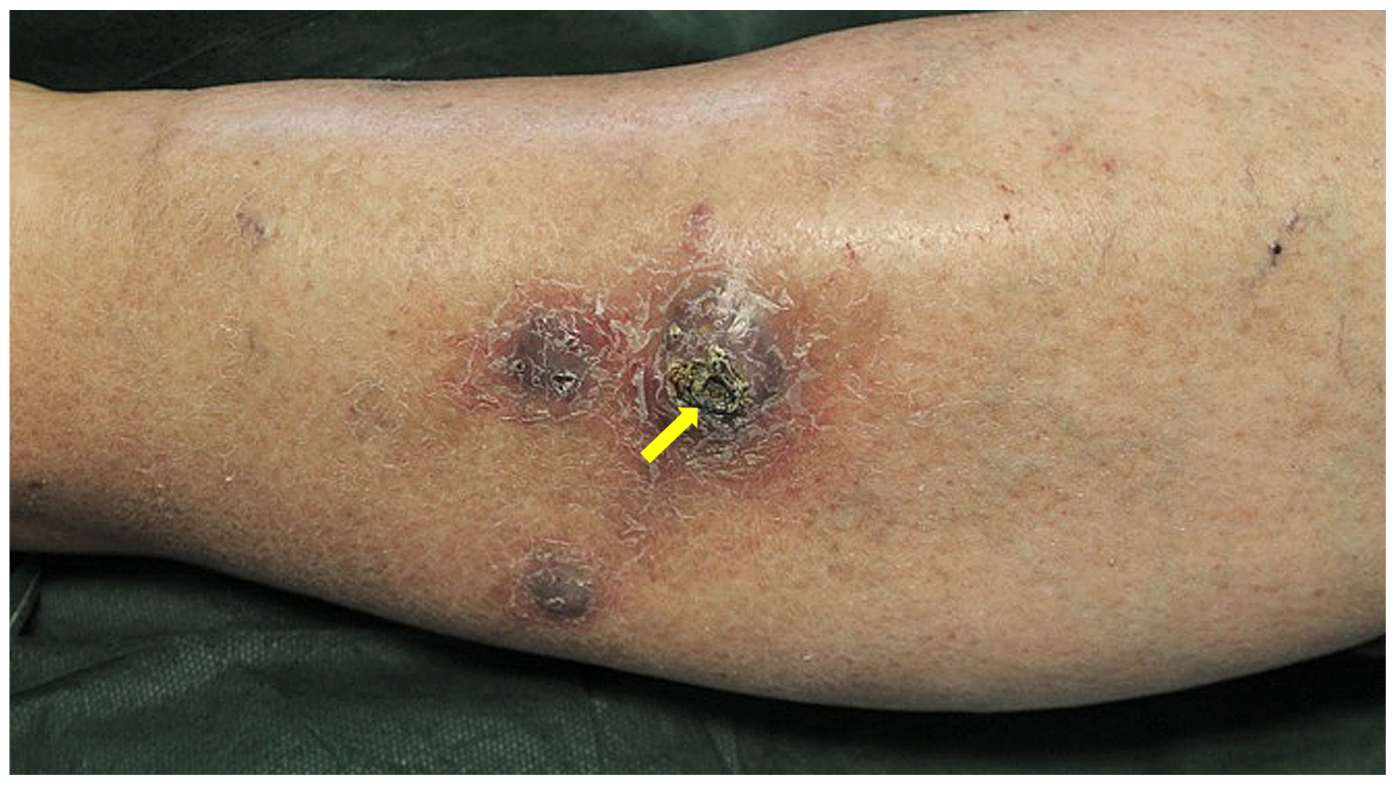
A 60-year-old beekeeper presented with violaceous, firm, painful, and swollen nodules on the left leg and lateral aspect of the ankle (Figure 1). His medical history included stage B B-cell chronic lymphocytic leukemia (CLL), according to Binet classification, with secondary hypogammaglobulinemia, type 2 diabetes mellitus, chronic obstructive pulmonary disease with bronchiectasis and recurrent pulmonary infections, obesity, obstructive sleep apnea syndrome, hypertension, and atrial fibrillation associated with ibrutinib therapy. For CLL, he had previously received standard chemotherapy; rituximab and idelalisib were discontinued due to disease progression. Before initiation of ibrutinib, complete blood count showed: white blood cells 3.7 × 10³/μL (neutrophils 47%, lymphocytes 37%, monocytes 10%, eosinophils 6%), red blood cells 5.28 × 10⁶/µL, and platelets 120 × 103/µL. The patient started ibrutinib therapy in August 2019, achieving good clinical control. In December 2019, ulcerated, painful erythematous–violaceous nodular lesions developed on the left calf and ankle (Figure 1). Biopsy specimens were obtained from the affected skin for histopathological and microbiological evaluation.
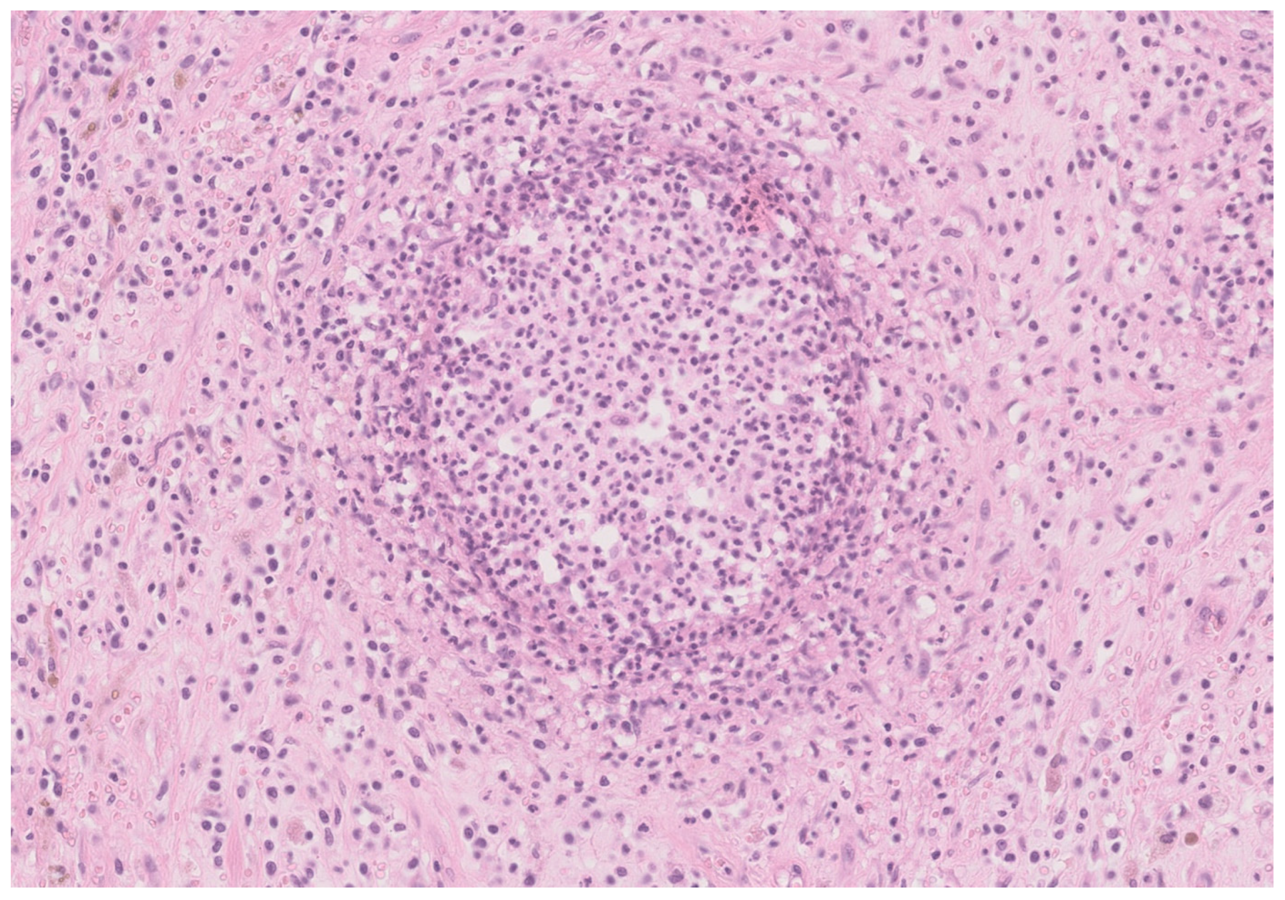
Histopathological analysis (Figure 2) revealed a mixed inflammatory infiltrate with peripheral epithelioid histiocytes, consistent with a suppurative granulomatous reaction, and numerous central neutrophils, a pattern commonly associated with atypical cutaneous mycobacterial infections in both immunocompromised and immunocompetent patients. Ziehl-Neelsen staining revealed the presence of acid-fast bacilli in the tissue.
Cultures for mycobacteria were performed using liquid medium (Mycobacterial Growth Indicator Tube, MGIT, Becton, Dickinson and Company, Franklin Lakes, NJ, USA), solid media (Löwenstein-Jensen, Becton, Dickinson and Company, Franklin Lakes, NJ, USA), and Middlebrook 7H11 agar (Becton, Dickinson and Company, Franklin Lakes, NJ, USA) incubated at 30 °C. Species identification was performed after 25 days of culture and achieved by MALDI-TOF mass spectrometry (Bruker Daltonik GmbH, Bremen, Germany), with subsequent confirmation by DNA probe-based assays (GenoType Mycobacterium CM and GenoType NTM-DR, Hain Lifescience GmbH, Nehren, Germany). Sequencing was not performed. No other cases of M. chelonae were identified in the laboratory during the same period, allowing us to reasonably exclude possible contamination. DNA strip testing for macrolide- and aminoglycoside-resistance genes was also performed. The initial biopsy culture was positive for Mycobacterium chelonae. The DNA-STRIP assay for genotypic resistance to macrolides showed no mutations in the erm and/or rrl genes, and the assay for aminoglycoside resistance showed no mutations in the rrs gene. An antibiogram of M. chelonae was also performed, demonstrating susceptibility to doxycycline (MIC = 0.25 µg/mL). The complete results are shown in Table 1.
Both standard bacterial and mycobacterial blood cultures, as well as sputum cultures and chest CT, showed no evidence of systemic dissemination. Based on antimicrobial susceptibility testing, a multidrug regimen was initiated under infectious disease supervision. The patient received intravenous imipenem for two weeks, followed by intravenous amikacin (1 g, three times per week- Monday, Wednesday, and Friday) for two months, linezolid (600 mg once daily) for two months, azithromycin (500 mg once daily), and doxycycline (100 mg twice daily) for four months. Doxycycline was included in the treatment regimen based on the antibiogram results and on available clinical evidence suggesting that this agent may contribute to the efficacy of combination therapies used to treat rapidly growing nontuberculous mycobacterial infections [20,21]. However, tetracyclines (including doxycycline) often appear ineffective against slow-growing nontuberculous mycobacteria [22], and further randomized controlled trials are needed to better define their role. Regular monitoring was performed, including twice-weekly assessment of serum creatinine, complete blood count, and alanine aminotransferase (ALT) levels, as well as weekly evaluation of vestibular and auditory function, ophthalmologic examination, and electrocardiography to monitor the QT interval. No significant adverse effects occurred. After four months of therapy, partial improvement of cutaneous lesions was observed, but complete resolution was not achieved. A second biopsy remained positive for M. chelonae, again without detectable resistance genes.
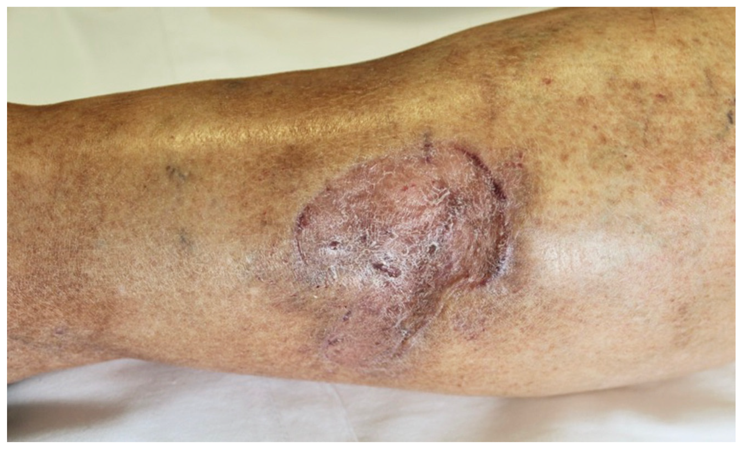
Given the persistence of lesions, a plastic surgery consultation was obtained. Surgical excision with skin grafting was performed, and postoperative cultures were negative. Antibiotic therapy was discontinued three months after surgery, with no evidence of recurrence (Figure 3).
At six-month follow-up after completion of antibiotic therapy (October 2020), microbiological culture performed on a biopsy specimen obtained from the lesion site was negative. Hematological evaluation showed a complete blood count with white blood cells of 22,140/μL (neutrophils 75.8%, lymphocytes 20.4%, monocytes 2.3%, eosinophils 1.2%, basophils 0.4%), red blood cells 4.80 × 10⁶/µL, Hemoglobin 14.9 g/dL and platelets 140 × 103/µL. Serum creatinine was 1.10 mg/dL. The patient was continuing ibrutinib therapy (two tablets daily), with a good clinical response. On physical examination, no abnormal lung sounds were detected, but palpable lymph nodes in the axillary and inguinal area. The liver and spleen were palpable 2 cm below the costal margin. Cardiac examination revealed normal heart sounds with a regular rhythm. Serum protein electrophoresis showed total proteins of 6.2 g/dL, with the following distribution: albumin 63.2%, α1-globulin 4.3%, α2-globulin 11.1%, β1-globulin 7.2%, β2-globulin 4.2%, and γ-globulin 10.0%. Immunoglobulin levels were: IgG 681 mg/dL, IgA 26 mg/dL, and IgM 17.6 mg/dL. No monoclonal components were detected. The patient remains under follow-up for the hematologic condition, and no recurrence has been observed as of February 2026.

3. Discussion

Mycobacterium chelonae is a rapidly growing non tuberculous mycobacterium commonly found in the environment, particularly in water, aquatic animals, and soil. It can survive under harsh conditions and cause a variety of infections, most frequently involving the skin and soft tissues. The patient’s occupational exposure as a beekeeper may also be relevant. Beekeeping involves frequent outdoor activities and direct contact with environmental reservoirs such as soil, water, and vegetation, all of which are recognized sources of nontuberculous mycobacteria, including M. chelonae. In addition, minor skin trauma, insect stings, or repeated micro-injuries to exposed areas may facilitate inoculation of the organism. Although no clear history of significant trauma or contaminated water exposure was documented in this case, the possibility of occupational inoculation to the affected limb cannot be excluded. These considerations highlight the importance of carefully exploring environmental and occupational exposures in patients presenting with cutaneous NTM infections.
Infections due to M. chelonae may be asymptomatic, but most commonly present with cutaneous and soft tissue manifestations, as observed in the present case. They occur more frequently in immunosuppressed individuals, although cases in immunocompetent hosts have also been reported. Pulmonary involvement is uncommon and is mainly described in patients with cystic fibrosis [23]. In immunosuppressed patients, including those receiving corticosteroids, monoclonal antibodies, or post-transplant immunosuppressive therapy, as well as in individuals with malignancy or chronic kidney disease, M. chelonae may cause invasive or disseminated disease. Disseminated cutaneous infection is typically characterized by multiple papules and pustules, often involving proximal extremities, the face and the upper trunk [24]. Other reported manifestations described in the literature include myositis [25], spondylitis [26], arthritis, sepsis [27], intra-abdominal abscess, and mediastinal infection [2]. In the present case, ibrutinib was considered a key predisposing factor. However, the patient also had several additional risk factors for M. chelonae infection, including chronic lymphocytic leukemia (CLL), secondary hypogammaglobulinemia, type 2 diabetes mellitus, chronic lung disease with bronchiectasis, recurrent infections, obesity, and prior chemoimmunotherapy. Each of these conditions may independently increase susceptibility to nontuberculous mycobacterial infections. Cases of NTM infection in patients with CLL receiving ibrutinib have been reported. In particular, one case described infection with the RGM M. chelonae; unlike the present case, that patient developed thrombocytopenia following antibiotic treatment [28]. Another report described a patient with CLL treated with ibrutinib who developed infection with Mycobacterium avium, a slow-growing NTM [29]. The clinical presentation is heterogeneous, ranging from localized cutaneous involvement to disseminated disease, and may be associated with variable treatment responses. Compared with previously published cases, our patient showed a localized cutaneous infection but required prolonged antimicrobial therapy and surgical management, highlighting the potential for a persistent clinical course even in the absence of systemic dissemination. However, the limited number of reported cases underscores the need for further studies to better define the true incidence and optimal management of NTM infections in this setting.
Ibrutinib inhibits Bruton’s tyrosine kinase (BTK), a key component of B-cell receptor signaling [30], and has been shown to impair bacterial clearance by reducing γδ T cell activation and CD107a degranulation, potentially affecting bacterial clearance [31]. Similarly, M. chelonae infection has been reported following treatment with alemtuzumab, a humanized anti-CD52 monoclonal antibody targeting lymphocytes and monocytes [32,33]. Interestingly, ibrutinib has also been associated with a potential beneficial effect in Mycobacterium tuberculosis (Mtb) infection, as it may inhibit intracellular bacterial growth in macrophages through modulation of autophagy pathways, including reduced p62 expression and increased LC3b levels [34].
Mycobacterium chelonae exhibits variable antimicrobial susceptibility, and treatment should therefore be individualized based on in vitro susceptibility testing in accordance with current guideline recommendations [35,36]. Although this species is often reported to be susceptible to agents such as amikacin, macrolides, linezolid, and, less consistently, imipenem, doxycycline, and clofazimine [37,38], these generalizations should be interpreted with caution. Treatment failures and recurrences are not uncommon [39], often reflecting multidrug resistance [39] or the emergence of resistance during therapy. Macrolides, particularly clarithromycin and azithromycin, are generally considered a cornerstone of therapy when susceptibility is confirmed; however, resistance may develop and compromise clinical outcomes. This may occur through point mutations in the 23S rRNA gene (e.g., at position 2058) [40]. Importantly, a distinction should be made between acquired resistance and inducible resistance mechanisms. Unlike Mycobacterium abscessus, in which inducible macrolide resistance mediated by erm genes is common, M. chelonae has traditionally been considered to lack functional erm genes, although recent evidence suggests that plasmid-mediated erm genes may confer inducible resistance in a minority of isolates [41,42]. These findings further emphasize the importance of susceptibility-guided therapy and careful microbiological monitoring during treatment. In refractory cases, surgical management, including debridement or excision with skin grafting, may be required, as in the present patient [43,44]. Additional therapeutic options for resistant infections include γ-interferon [45], omadacycline [8], and bacteriophage therapy [46]. Overall, treatment typically requires prolonged multidrug regimens administered over several weeks or months.

4. Conclusions

Mycobacterium chelonae is an emerging pathogen increasingly recognized as a cause of skin and soft tissue infections, particularly in immunocompromised hosts. This case highlights how underlying hematologic disease and treatment with ibrutinib may predispose patients to the development and progression of infection. Although antimicrobial therapy guided by susceptibility testing remains the cornerstone of management, clinical response may be incomplete, and combined medical and surgical approaches may be required in selected cases. Early recognition, prompt microbiological diagnosis, and careful therapeutic monitoring are essential to achieve clinical cure and prevent dissemination or recurrence. Growing evidence suggests that nontuberculous mycobacterial infections should be considered in patients receiving Bruton’s tyrosine kinase inhibitors who present persistent, atypical or non-healing cutaneous lesions. Increased awareness of this association, together with a multidisciplinary approach, may facilitate earlier diagnosis and more effective management of these challenging infections.

Author Contributions

Conceptualization, A.G., G.T. and S.B. (Serena Bergamo); methodology, S.B. (Serena Bergamo), G.M. and D.S.; formal analysis, S.B. (Serena Bergamo), G.M. and D.S.; investigation, S.B. (Serena Bergamo) and G.T.; data curation, S.B. (Serena Bonin); writing—original draft preparation, S.B. (Serena Bergamo), S.B. (Serena Bonin) and G.T.; writing—review and editing, S.B. (Serena Bergamo), S.B. (Serena Bonin), A.G., G.M., D.S. and G.T. All authors have read and agreed to the published version of the manuscript.

Funding

This research received no external funding.

Institutional Review Board Statement

The study was conducted in accordance with the Declaration of Helsinki. Ethical approval was not required for the publication of this case report, as it was conducted in accordance with institutional policies and supported by written informed consent obtained from the patient.

Informed Consent Statement

Informed consent was obtained from the patient involved in the study.

Data Availability Statement

The original contributions presented in this study are included in the article. Further inquiries can be directed to the corresponding author.

Conflicts of Interest

The authors declare no conflicts of interest.

Abbreviations

The following abbreviations are used in this manuscript:
BTKBruton’s tyrosine kinase
CLLChronic lymphocytic leukemia
McMycobacterium chelonae
MtbMycobacterium tuberculosis
NTMNontuberculous mycobacteria
RGMRapidly growing mycobacterium
PCRPolymerase Chain Reaction

References

  1. Brown-Elliott, B.A.; Philley, J.V. Rapidly Growing Mycobacteria. Microbiol. Spectr. 2017, 5, 703–723. [Google Scholar] [CrossRef]
  2. Nakamura, Y.; Yoshioka, D.; Miyagawa, S.; Yoshikawa, Y.; Hata, H.; Nakae, M.; Toda, K.; Sawa, Y. A case of Mycobacterium chelonae mediastinitis and acute humoral rejection after heart transplantation. J. Card. Surg. 2019, 34, 205–207. [Google Scholar] [CrossRef]
  3. Pinto-Gouveia, M.; Gameiro, A.; Ramos, L.; Cardoso, J.C.; Brites, M.M.; Tellechea, O.; Figueiredo, A. Mycobacterium chelonae Is an Ubiquitous Atypical Mycobacterium. Case Rep. Dermatol. 2015, 7, 207–211. [Google Scholar] [CrossRef]
  4. Tirado-Sanchez, A.; Bonifaz, A. Nodular Lymphangitis (Sporotrichoid Lymphocutaneous Infections). Clues to Differential Diagnosis. J. Fungi 2018, 4, 56. [Google Scholar] [CrossRef]
  5. Ferraro, V.; Caiazza, P.; Sorella, F.; Bisognin, F.; Crovara Pesce, C.M.; Dal Monte, P. Fast detection of non-tuberculous mycobacteria in medical device-water samples: Evaluation of the STANDARD M10 MTB/NTM cartridge. J. Hosp. Infect. 2026, 167, 43–47. [Google Scholar] [CrossRef]
  6. Inkster, T.; Peters, C.; Seagar, A.L.; Holden, M.T.G.; Laurenson, I.F. Investigation of two cases of Mycobacterium chelonae infection in haemato-oncology patients using whole-genome sequencing and a potential link to the hospital water supply. J. Hosp. Infect. 2021, 114, 111–116. [Google Scholar] [CrossRef] [PubMed]
  7. Akram, S.M.; Rathish, B.; Saleh, D. Mycobacterium chelonae Infection. Available online: https://www.ncbi.nlm.nih.gov/books/NBK430806/ (accessed on 28 November 2025).
  8. Frizzell, M.; Carr, E.; Brust, K. Omadacycline for treatment of Mycobacterium chelonae skin infection. Bayl. Univ. Med. Cent. Proc. 2020, 33, 610–611. [Google Scholar] [CrossRef]
  9. Kukla, R.; Nouzovska, K.; Ryskova, L.; Rozsivalova, P.; Pavlik, I.; Bostik, P. Successful treatment of Keratitis caused by Mycobacterium chelonae and an overview of previous cases in Europe. Ann. Clin. Microbiol. Antimicrob. 2024, 23, 92. [Google Scholar] [CrossRef] [PubMed]
  10. Yaseen, M.M.; Spencer, S.; Bhandari, S. Disseminated Mycobacterium chelonae infection in kidney transplant patients. BMJ Case Rep. 2024, 17, e260095. [Google Scholar] [CrossRef] [PubMed]
  11. Sukackiene, D.; Satkauskaite, D.; Sleivyte, U.; Vinikovas, A.; Ulianskaite, G.; Pamedys, J.; Raudonis, T.; Miglinas, M. Case Report: Cutaneous Mycobacterium chelonae infection in a kidney transplant recipient with long-term immunosuppression and eculizumab therapy. Front. Med. 2026, 12, 1728249. [Google Scholar] [CrossRef]
  12. Rajini, M.; Prasad, S.R.; Reddy, R.R.; Bhat, R.V.; Vimala, K.R. Postoperative infection of laparoscopic surgery wound due to Mycobacterium chelonae. Indian. J. Med. Microbiol. 2007, 25, 163–165. [Google Scholar] [CrossRef]
  13. Sergeant, A.; Conaglen, P.; Laurenson, I.F.; Claxton, P.; Mathers, M.E.; Kavanagh, G.M.; Tidman, M.J. Mycobacterium chelonae infection: A complication of tattooing. Clin. Exp. Dermatol. 2013, 38, 140–142. [Google Scholar] [CrossRef]
  14. Rodriguez, J.M.; Xie, Y.L.; Winthrop, K.L.; Schafer, S.; Sehdev, P.; Solomon, J.; Jensen, B.; Toney, N.C.; Lewis, P.F. Mycobacterium chelonae facial infections following injection of dermal filler. Aesthet. Surg. J. 2013, 33, 265–269. [Google Scholar] [CrossRef]
  15. Matic, S.; Teodosic, V.; Zagorac, S. Mycobacterium chelonae hand infection following acupuncture: A case report and literature review. Front. Med. 2024, 11, 1482236. [Google Scholar] [CrossRef] [PubMed]
  16. Theng, T.S.; Chan, Y.C.; Leow, Y.H.; Tan, S.H. Sweet’s syndrome associated with Mycobacterium chelonae and herpes simplex virus infections: A case report. Ann. Acad. Med. Singap. 2003, 32, 411–414. [Google Scholar] [CrossRef] [PubMed]
  17. Xu, T.; Zheng, Y.; Xue, R.; Li, R.; Zhang, W.; Sun, Z.; Song, Y. Retrospective analysis of non-tuberculous mycobacterial skin infections: Diagnostic accuracy, treatment efficacy, and PCR-based species detection in a Chinese primary care hospital. J. Clin. Microbiol. 2025, 63, e0106525. [Google Scholar] [CrossRef]
  18. Varma-Basil, M.; Garima, K.; Pathak, R.; Dwivedi, S.K.; Narang, A.; Bhatnagar, A.; Bose, M. Development of a novel PCR restriction analysis of the hsp65 gene as a rapid method to screen for the Mycobacterium tuberculosis complex and nontuberculous mycobacteria in high-burden countries. J. Clin. Microbiol. 2013, 51, 1165–1170. [Google Scholar] [CrossRef]
  19. Sardina, L.A.; Kaw, U.; Jour, G.; Knabel, D.; Dyck, R.M.; Procop, G.W.; Bergfeld, W.F.; Harrington, S.; Demkowicz, R.; Piliang, M.P. Diagnosis of Mycobacterium abscessus/chelonae complex cutaneous infection: Correlation of tissue culture and skin biopsy. J. Cutan. Pathol. 2020, 47, 321–327. [Google Scholar] [CrossRef]
  20. Daley, C.L.; Flume, P.A.; van Ingen, J.; Khare, R.; Mingora, C.M.; Nguyen, M.H.; Winthrop, K.L.; Zha, B.S. A systematic review of tetracyclines for nontuberculous mycobacteria: Focus on rapidly growing mycobacteria. Eur. Respir. Rev. 2025, 34, 250038. [Google Scholar] [CrossRef] [PubMed]
  21. Reil, I.; Spicic, S.; Barbic, L.; Duvnjak, S.; Kompes, G.; Benic, M.; Stojevic, D.; Cvetnic, Z.; Arapovic, J.; Zdelar-Tuk, M. Antimicrobial Resistance in Rapidly Growing Nontuberculous Mycobacteria among Domestic and Wild Animals Emphasizing the Zoonotic Potential. Microorganisms 2023, 11, 2520. [Google Scholar] [CrossRef] [PubMed]
  22. Wang, Y.; Zheng, H.; Wang, X.; Wang, X.; Liu, N.; Wang, L.; Liu, Z.; Meng, X.; Abullimiti, M.; Aihemuti, M.; et al. Molecular Characterization and Antibiograms of Nontuberculous Mycobacterium Infections in Kashgar, China. Curr. Microbiol. 2025, 82, 376. [Google Scholar] [CrossRef]
  23. Sermet-Gaudelus, I.; Le Bourgeois, M.; Pierre-Audigier, C.; Offredo, C.; Guillemot, D.; Halley, S.; Akoua-Koffi, C.; Vincent, V.; Sivadon-Tardy, V.; Ferroni, A.; et al. Mycobacterium abscessus and children with cystic fibrosis. Emerg. Infect. Dis. 2003, 9, 1587–1591. [Google Scholar] [CrossRef] [PubMed]
  24. Gilvaz, V.J.; Mody, E.; Abaalkhail, S. A Case of Disseminated Cutaneous Mycobacterium chelonae Infection During Treatment With Tofacitinib. J. Rheumatol. 2021, 48, 1623. [Google Scholar] [CrossRef] [PubMed]
  25. Hajjaji, N.; Cattier, B.; Lanotte, P.; Legras, A.; Perrotin, D. Mycobacterium chelonae myositis. Presse Med. 2004, 33, 1519–1520. [Google Scholar] [CrossRef] [PubMed]
  26. Dong, S.; Lin, Z.; Wu, S.; Cai, L. Spondylitis following bloodstream dissemination of Mycobacterium chelonae disseminated in an immunocompetent patient: A case report and literature review. J. Int. Med. Res. 2021, 49, 3000605211047766. [Google Scholar] [CrossRef]
  27. Lam, V.; Theyyunni, N. Septic arthritis due to non-tuberculous mycobacterium without effusion. Am. J. Emerg. Med. 2021, 43, 287.e5–287.e7. [Google Scholar] [CrossRef]
  28. Dousa, K.M.; Babiker, A.; Van Aartsen, D.; Shah, N.; Bonomo, R.A.; Johnson, J.L.; Skalweit, M.J. Ibrutinib Therapy and Mycobacterium chelonae Skin and Soft Tissue Infection. Open Forum Infect. Dis. 2018, 5, ofy168. [Google Scholar] [CrossRef]
  29. Diaco, N.D.; Strohdach, B.; Falkowski, A.L.; Hainc, N.; Brunner, P.; Rutishauser, J.; Jost, L.; Tarr, P.E. Psoas Abscess Due to Mycobacterium avium in A Patient with Chronic Lymphocytic Leukemia—Case Report and Review. J. Clin. Med. 2019, 8, 216. [Google Scholar] [CrossRef]
  30. Nam, H.Y.; Nam, J.H.; Yoon, G.; Lee, J.Y.; Nam, Y.; Kang, H.J.; Cho, H.J.; Kim, J.; Hoe, H.S. Ibrutinib suppresses LPS-induced neuroinflammatory responses in BV2 microglial cells and wild-type mice. J. Neuroinflamm. 2018, 15, 271. [Google Scholar] [CrossRef]
  31. Risnik, D.; Elias, E.E.; Keitelman, I.; Colado, A.; Podaza, E.; Cordini, G.; Vergara Rubio, M.; Fernandez Grecco, H.; Bezares, R.F.; Borge, M.; et al. The effect of ibrutinib on neutrophil and gammadelta T cell functions. Leuk. Lymphoma 2020, 61, 2409–2418. [Google Scholar] [CrossRef]
  32. Dungarwalla, M.; Field-Smith, A.; Jameson, C.; Riley, U.; Chapman, A.; Bunker, C.B.; Dearden, C.E.; Matutes, E. Cutaneous Mycobacterium chelonae infection in chronic lymphocytic leukaemia. Haematologica 2007, 92, e5–e6. [Google Scholar] [CrossRef]
  33. An, N.; Bian, K.; Li, C. Alemtuzumab for haematological malignancies. Ann. Hematol. 2025, 104, 2593–2603. [Google Scholar] [CrossRef]
  34. Hu, Y.; Wen, Z.; Liu, S.; Cai, Y.; Guo, J.; Xu, Y.; Lin, D.; Zhu, J.; Li, D.; Chen, X. Ibrutinib suppresses intracellular mycobacterium tuberculosis growth by inducing macrophage autophagy. J. Infect. 2020, 80, e19–e26. [Google Scholar] [CrossRef] [PubMed]
  35. Daley, C.L.; Iaccarino, J.M.; Lange, C.; Cambau, E.; Wallace, R.J., Jr.; Andrejak, C.; Böttger, E.C.; Brozek, J.; Griffith, D.E.; Guglielmetti, L.; et al. Treatment of Nontuberculous Mycobacterial Pulmonary Disease: An Official ATS/ERS/ESCMID/IDSA Clinical Practice Guideline. Clin. Infect. Dis. 2020, 71, e1–e36. [Google Scholar] [CrossRef] [PubMed]
  36. Lange, C.; Bottger, E.C.; Cambau, E.; Griffith, D.E.; Guglielmetti, L.; van Ingen, J.; Knight, S.L.; Marras, T.K.; Olivier, K.N.; Santin, M.; et al. Consensus management recommendations for less common non-tuberculous mycobacterial pulmonary diseases. Lancet Infect. Dis. 2022, 22, e178–e190, Erratum in Lancet Infect. Dis. 2022, 22, e73. https://doi.org/10.1016/S1473-3099(22)00068-8. [Google Scholar] [CrossRef]
  37. Shen, G.H.; Wu, B.D.; Hu, S.T.; Lin, C.F.; Wu, K.M.; Chen, J.H. High efficacy of clofazimine and its synergistic effect with amikacin against rapidly growing mycobacteria. Int. J. Antimicrob. Agents 2010, 35, 400–404. [Google Scholar] [CrossRef]
  38. Gaudencio, M.; Carvalho, A.; Bertao, M.I.; Barreiro, I.; Bessa, M.I.; Goncalves, A. Mycobacterium chelonae Cutaneous Infection: A Challenge for an Internist. Eur. J. Case Rep. Intern. Med. 2021, 8, 003013. [Google Scholar] [CrossRef]
  39. DuBow, A.; Morand, M.; Desy, D.; Krasny, M. Recurrence of cutaneous Mycobacterium chelonae infection: A case report. SAGE Open Med. Case Rep. 2019, 7, 2050313X19845231. [Google Scholar] [CrossRef] [PubMed]
  40. Vemulapalli, R.K.; Cantey, J.R.; Steed, L.L.; Knapp, T.L.; Thielman, N.M. Emergence of resistance to clarithromycin during treatment of disseminated cutaneous Mycobacterium chelonae infection: Case report and literature review. J. Infect. 2001, 43, 163–168. [Google Scholar] [CrossRef]
  41. Brown-Elliott, B.A.; Wallace, R.J., Jr.; Wengenack, N.L.; Workman, S.D.; Cameron, A.D.S.; Bush, G.; Hughes, M.D.; Melton, S.; Gonzalez-Ramirez, B.; Rodriguez, E.; et al. Emergence of Inducible Macrolide Resistance in Mycobacterium chelonae Due to Broad-Host-Range Plasmid and Chromosomal Variants of the Novel 23S rRNA Methylase Gene, erm(55). J. Clin. Microbiol. 2023, 61, e0042823. [Google Scholar] [CrossRef]
  42. Alexander, D.C.; Farquhar, T.; Adams, J.M.E.; Suchan, D.M.; Workman, S.D.; Wallace, R.J., Jr.; Brown-Elliott, B.A.; El-Halfawy, O.M.; Cameron, A.D.S. Macrolide resistance due to erm(55). Microbiol. Spectr. 2025, 13, e0239724. [Google Scholar] [CrossRef] [PubMed]
  43. Mannelli, V.K.; Rai, M.P.; Nemakayala, D.R.; Kadiri, N.P. Mycobacterium chelonae Developing Multidrug Resistance. BMJ Case Rep. 2018, 2018, bcr-2017-222569. [Google Scholar] [CrossRef]
  44. Wang, M.L.; Vosbikian, M.M.; Abboudi, J.; Beredjiklian, P.K. Treatment of a Chronic Multidrug-Resistant Cutaneous Mycobacterium chelonae Infection of the Hand with Wide Debridement and Skin-Grafting: A Case Report. JBJS Case Connect. 2014, 4, e102. [Google Scholar] [CrossRef] [PubMed]
  45. Jousse-Joulin, S.; Garre, M.; Guennoc, X.; Destombe, C.; Samjee, I.; Devauchelle-Pensec, V.; Saraux, A. Skin and joint infection by Mycobacterium chelonae: Rescue treatment with interferon gamma. Jt. Bone Spine 2007, 74, 385–388. [Google Scholar] [CrossRef]
  46. Little, J.S.; Dedrick, R.M.; Freeman, K.G.; Cristinziano, M.; Smith, B.E.; Benson, C.A.; Jhaveri, T.A.; Baden, L.R.; Solomon, D.A.; Hatfull, G.F. Bacteriophage treatment of disseminated cutaneous Mycobacterium chelonae infection. Nat. Commun. 2022, 13, 2313. [Google Scholar] [CrossRef] [PubMed]
Figure 1. Skin lesion on the left calf before initiation of antibiotic therapy. The yellow arrow indicates the site of sampling.
Microorganisms 14 01189 g001
Figure 2. Non-necrotizing epithelioid granuloma with central neutrophilic infiltration and a peripheral rim of lymphocytes (200× magnification).
Microorganisms 14 01189 g002
Figure 3. Left calf six months after surgical excision and skin grafting.
Microorganisms 14 01189 g003
Table 1. Antibiogram results for Mycobacterium chelonae.
AntibioticsInterpretationMIC (µg/mL)
AmikacinSusceptible8
CefoxitinResistant128
CiprofloxacinSusceptible1
ClarithromycinSusceptible0.5
DoxycyclineSusceptible0.25
ImipenemIntermediate16
LinezolidSusceptible8
MoxifloxacinSusceptible0.5
TobramycinSusceptible<1
Disclaimer/Publisher’s Note: The statements, opinions and data contained in all publications are solely those of the individual author(s) and contributor(s) and not of MDPI and/or the editor(s). MDPI and/or the editor(s) disclaim responsibility for any injury to people or property resulting from any ideas, methods, instructions or products referred to in the content.