Article

Isolation and Identification of an Endophytic Fungus Aspergillus sp. and Its Growth-Promoting Effects on Nymphaea candida Seedlings Through Modulation of the Rhizosphere Microbial Community

College of Marine Life Sciences, Ocean University of China, Qingdao 266003, China
*
Authors to whom correspondence should be addressed.
Microorganisms 2026, 14(5), 993; https://doi.org/10.3390/microorganisms14050993
Submission received: 19 March 2026 / Revised: 23 April 2026 / Accepted: 24 April 2026 / Published: 28 April 2026
(This article belongs to the Section Plant Microbe Interactions)

Abstract

Nymphaea candida Presl is a rare and endangered waterlily species, and cultivating robust seedlings suitable for artificial propagation has become a critical issue for the conservation of this species. In this study, Aspergillus sp., an endophytic fungus isolated from the roots of N. candida, showed the capability of solubilizing phosphate and potassium and producing siderophores. The application of Aspergillus sp. significantly increased leaf length, leaf width, leaf number, and root length of N. candida seedlings by 97.83%, 131.37%, 94.12%, and 171.25%, respectively. Meanwhile, Aspergillus sp. application significantly enhanced soil organic matter content, alkali-hydrolyzable nitrogen content, sucrase activity, and peroxidase activity by 6.57%, 31.62%, 23.26%, and 7.53%, respectively. Moreover, Aspergillus sp. enriched beneficial microorganisms including Cyanobium, Aquicella, and Cryptomycota to form a more stable rhizosphere soil microenvironment. Additionally, Aspergillus sp. upregulated genes involved in photosynthesis and photosynthesis–antenna protein pathways in N. candida leaves, with the expression levels of psbA, petG, and psbH significantly increasing by 2.17, 4.48, and 0.28-fold, respectively. Therefore, the endophytic fungus Aspergillus sp. might be a reliable tool for the propagation of N. candida seedlings, which would be helpful for the conservation of this rare and endangered aquatic plant species.

1. Introduction

The genus Nymphaea comprises approximately 50 species of perennial aquatic plants globally, with 5 native species in China [1]. Among them, Nymphaea candida Presl, a rare and endangered species, is found exclusively in the Xinjiang region, China. N. candida plays an essential role in water purification, habitat provision for wildlife, and maintenance of the structural and functional integrity of wetland ecosystems [2,3]. Over the past decade, the natural distribution area of N. candida population has been continuously declining due to the heavy seed predation by waterfowl, its inherently low natural germination rate, and the susceptibility to aquatic diseases during the germination process [4,5]. Currently, N. candida has been listed on both the IUCN Red List of Threatened Species and the National Key Protected Wild Plants List of China [6,7]. Given that natural reproduction is insufficient for the effective conservation of N. candida, it is urgent to conduct artificial seedling breeding of N. candida. Thus, how to promote the early growth of N. candida and obtain robust seedlings has become a critical issue for the conservation of this endangered species.
Plant growth-promoting bacteria could significantly promote the growth of aquatic plants due to their capabilities of nitrogen fixation, phosphate solubilization, and phytohormone secretion [8,9,10]. For instance, Bacillus subtilis and Pseudomonas fluorescens increased the fresh weight of hydroponically cultivated lettuce and celery by 144.4% and 300.9%, respectively [11]. Bacillus velezensis significantly increased the dry weight of rice (Oryza sativa) by 20.47% [12]. These bacteria could accelerate root development and enhance plant growth primarily by secreting growth hormones [13,14]. Compared with plant growth-promoting bacteria, the potential of plant growth-promoting fungi—particularly endophytic ones—to enhance hydrophyte growth has been substantially underestimated. Endophytic growth-promoting fungi, a group of fungi capable of colonizing internal plant tissues, could promote plant growth, enhance stress resistance, and inhibit pathogenic microorganisms [15]. The most extensively studied endophytic growth-promoting fungi primarily include species from the genera Trichoderma, Penicillium, and Talaromyces, with most studies focusing on their growth-promoting effect for terrestrial plants [16,17,18,19]. For example, Penicillium commune EP-5 significantly increased the root length of maize (Zea mays) by 39.87% through the production of indole-3-acetic acid (IAA) [20]. Aspergillus spp. enhanced the root growth in cassava (Manihot esculenta) by increasing soil nutrient availability via phosphate solubilization [21]. In addition, Trichoderma harzianum and Metarhizium anisopliae were reported to increase the fresh weight of water lettuce (Pistia stratiotes) by 23.33% and 10.3%, respectively [22]. We hypothesized that endophytic growth-promoting fungi isolated from N. candida might promote seedling growth and thereby provide a reliable tool for artificial propagation of N. candida seedlings.
In this study, endophytic fungi were firstly isolated and identified from the roots of N. candida. Their taxonomic status was determined using a combination of morphological and molecular biological methods, and their growth-promoting potential was also evaluated. Subsequently, the selected endophytic fungal strain was inoculated into a cultivation system of N. candida seedlings, and its effects on seedling growth were assessed by periodically measuring growth parameters, including leaf length, leaf width, leaf area, and root length. Furthermore, the changes in the soil physicochemical properties and enzyme activities of the rhizosphere soil of N. candida seedlings were measured to evaluate the impacts of the endophytic growth-promoting fungus on soil nutrient composition. Finally, high-throughput sequencing technologies were employed to analyze the effects of the endophytic growth-promoting fungus on the rhizosphere microbial community structure and the leaf transcriptome of N. candida seedlings to elucidate the potential growth-promoting mechanisms. The findings of this study would provide technical support for the seedling cultivation of N. candida.

2. Materials and Methods

2.1. Biological Materials and Reagents

N. candida plants were collected from Bosten Lake in the Xinjiang Uygur Autonomous Region, China, on 25 April 2024. Potato Dextrose Agar (PDA) medium used for the isolation of endophytic fungi, and Pikovskaya’s Agar Medium (PVK), Phosphate-solubilizing bacteria medium (OPM), Potassium bacteria Medium (PSM), and Chrome Azurol S Agar Medium (CAS) used for performance characterization, were purchased from Aladdin Biochemical Technology Co., Ltd. (Shanghai, China).

2.2. Isolation, Purification, and Performance Characterization of Endophytic Fungi

After transport to the laboratory, roots (5–6 cm in length) were excised from N. candida plants and rinsed with tap water for 5–10 min to remove surface debris. The roots were then cut into approximately 1 cm segments. Surface sterilization was performed by sequentially immersing the segments in 75% ethanol for 10 s, rinsing with sterile water, soaking in a 0.1% mercuric chloride solution for 12 min, and finally rinsing 5 times with sterile water [23]. The sterilized tissue segments were inoculated onto PDA medium in the dark at 28 °C for 3–5 days. Once the mycelia emerged around the tissue segments, a mycelial plug (approximately 5 mm in diameter) was excised using an inoculation needle and placed agar side down in the center of a fresh PDA plate for purification. This procedure was repeated until pure cultures were obtained, and the purified strains were streaked onto PDA slants. After the slants were fully covered with mycelia, they were temporarily stored at 4 °C for subsequent spore collection [24]. In addition, mycelial plugs (5 mm diameter) of each strain were inoculated onto the center of PVK, OPM, PSM, and CAS plates to assess the growth-promoting potential. These plates were incubated in the dark at 28 °C for 3–5 days, and the growth status of each strain on the respective functional media was observed.

2.3. Molecular Identification and Phylogenetic Tree Construction of Endophytic Fungi

Purified endophytic fungal strains were inoculated onto PDA plates and incubated at 28 °C for 48–96 h to observe and photograph colony morphology [25]. Subsequently, the purified endophytic fungal hyphae were ground with sterile glass beads, and the genomic DNA was extracted using Invitrogen Genomic DNA Extraction Kits (Invitrogen Corporation Shanghai Representative Office, Shanghai, China). NS1 (5′-GTAGTCATATGCTTGTCTC-3′)/NS6 (5′-GCATCACAGACCTGTTATTGCCTC-3′) primers were used for PCR amplification, and PCR products were analyzed by 1% agarose gel electrophoresis. The target band was recovered and purified using a purification kit, and the purified PCR products were sent for Sanger sequencing by Sangon Biotech Co., Ltd. (Shanghai, China). The obtained sequences were subjected to blast sequence homology comparison (https://blast.ncbi.nlm.nih.gov/Blast.cgi, accessed on 7 May 2025) with other fungal sequences in the National Center for Biotechnology Information (NCBI) database with a similarity greater than 99% [26]. Multiple sequence alignments of the tested strains and related reference strains were performed using MEGA software (version 7.0.14), and phylogenetic trees were constructed using the neighbor-joining method [27].

2.4. Preparation of Endophytic Fungal Inoculum

The purified endophytic fungal strain was inoculated onto PDA plates and incubated in the dark at 28 °C for 7 days until the plates were fully covered with mycelia. The mycelia were then scraped off using a sterile inoculation spoon, rinsed with sterile water, and filtered through two layers of gauze to remove mycelial fragments. Spores were collected from the filtrate, counted using a hemocytometer, adjusted to 1 × 10⁸ spores/mL with sterile water, and used as the inoculum in subsequent experiments [28].

2.5. Growth-Promoting Effect of Endophytic Fungi on N. candida Seedlings

A 60-day pot experiment was conducted starting on 30 May 2025 to investigate the effects of endophytic growth-promoting fungi on the growth of N. candida seedlings. Environmental conditions during the experiment: temperature was maintained at 28 ± 2 °C, relative humidity at 60 ± 5%, and light intensity at 200 μmol m−2 s−1 with a 14/10 h light/dark photoperiod. The fungal strain used was Aspergillus sp., a potential endophytic growth-promoting fungus isolated from the roots of N. candida. Two groups were established: a control group (CK) and an Aspergillus sp. group. Each group contained three replicates, and each replicate consisted of 6 pots. Planting soil (a 1:1 mixture of peat and silt) was sterilized by high-temperature autoclaving and evenly distributed into 36 plastic planting boxes (30 cm × 20 cm × 12 cm), with each box filled to a depth of 8 cm. The boxes were placed in standing water for 48 h and then randomly divided into two groups. For the Aspergillus sp. group, N. candida seedlings at the two-leaf stage with uniform growth were randomly selected, and their roots were immersed in Aspergillus sp. inoculum for 20 min. Then, the seedlings were fixed in the planting boxes by inserting approximately 5 cm of the rhizome apex into the planting soil. Six planting boxes were placed in each glass tank, and the positions of the planting boxes were randomly changed daily to minimize errors caused by variations in light intensity. On the 7th, 14th, and 21st days post-transplantation, 5 mL of the Aspergillus sp. spore suspension (1 × 10⁸ spores/mL) was injected into the water of the Aspergillus sp. group using a sterile syringe. The control group was immersed in sterile water on the 1st day and received an equal volume of sterile water on the 7th, 14th, and 21st days post-transplantation. Leaf length, leaf width, and leaf number of N. candida were measured on the 14th, 28th, 42nd, and 60th days post-transplantation.

2.6. Sample Collection

Sixty days post-transplantation, soil samples were collected from the control and Aspergillus sp. groups. Intact root–soil complexes were carefully excavated by vertically digging along the edge of each plastic planting box to the bottom using a sterile shovel. Loosely adhering soil was gently shaken off. Rhizosphere soil, defined as the soil tightly adhering to the roots, was collected using a sterile brush and stored at −80 °C for 18S rRNA and 16S rRNA gene sequencing. Meanwhile, bulk soil from each planting box was collected, air-dried, and stored to determine the soil physicochemical properties. Root length was measured using a ruler, and the N. candida plants after soil removal were placed in sterile sampling bags and stored at −80 °C for subsequent leaf RNA extraction and transcriptome sequencing [29].

2.7. Determination of Soil Physicochemical Properties

Air-dried soil samples from each planting box were ground and passed through 10- and 100-mesh sieves [30]. The contents of alkali-hydrolyzable nitrogen (AN), soil organic matter (SOM), soil sucrose (SC) and peroxidase activities (POD) of the soil were determined using the alkaline diffusion method [31], potassium dichromate (K2Cr2O7) volumetric method with dilution heat [32], and specific assay kits (Solarbio, Beijing, China), respectively.

2.8. Rhizosphere Soil DNA Extraction and High-Throughput Sequencing

Total DNA was extracted from rhizosphere soil samples using the FastDNA SPIN Kit for Soil (MP Biomedicals, Solon, OH, USA), and the extracted genomic DNA was examined by electrophoresis on a 1% agarose gel. PCR amplification was performed using universal primers for the 16S rRNA gene (27F-1492R: 5′-AGRGTTYGATYMTGGCTCAG-3′ and 5′-RGYTACCTTGTTACGACTT-3′) and the 18S rRNA gene (EukA-EukB: 5′-AACCTGGTTGCTGCCAGTGCAGT-3′ and 5′-TGATCCTTGCAGGTTCACCTAC-3′) [33]. The PCR program consisted of an initial denaturation at 98 °C for 30 s; 32 cycles of denaturation at 98 °C for 10 s, annealing at 54 °C for 30 s, and extension at 72 °C for 45 s; and a final extension at 72 °C for 10 min. PCR products were pooled and detected by 2% agarose gel electrophoresis, and target bands were excised and purified using the AxyPrepDNA Gel Extraction Kit (CA, USA). Purified products were eluted with Tris-HCl and checked by 2% agarose electrophoresis [34]. Paired-end sequencing was performed on a PacBio platform at Shanghai Lingen Biotechnology Co., Ltd. (Shanghai, China). High-fidelity (hifi) reads were generated using SMRT Link Analysis software (version 11.0). Barcode-CCS sequence identification was performed using Lima (version 2.6.99). Raw read data were trimmed and quality-controlled using QIIME2, and chimeric sequences were removed using DADA2. Representative amplicon sequence variants (ASVs) were assigned to the SILVA (version 13.8) or UNITE (version 8.0) database.

2.9. N. candida Leaf RNA Extraction and Transcriptome Sequencing

Healthy N. candida leaves (n = 6) with similar size were collected from the control and Aspergillus sp. groups, rinsed with sterile water, freeze-dried, and immediately ground. RNA was extracted from the disrupted cells using a TRIzol reagent kit (Thermo Fisher Scientific, Waltham, MA, USA). The quantity and quality of the isolated RNA samples were assessed using an Agilent 2100 Bioanalyzer (Agilent Technologies, Santa Clara, CA, USA) and agarose gel electrophoresis to check for RNA degradation. Upon quality approval, the samples were sent to Shanghai Majorbio Bio-pharm Technology Co., Ltd. (Shanghai, China) for sequencing on a NovaSeq X Plus platform. Following sequencing, de novo assembly of clean data from all samples was performed using Trinity as described by Sharma et al. [35]. The assembly results were optimized, evaluated, and subjected to expression level analysis. Based on the quantification results of expression levels, differential gene expression analysis was conducted between the two groups to identify differentially expressed genes (DEGs). DESeq2 software (v 1.46.0) was used for this analysis, with a screening threshold of |log2FC| ≥ 1 and p < 0.05.

2.10. Statistical Analysis

Data visualization was finalized using Origin 2019 software to ensure the standardization and readability of the figures. All data were first tested for normality using the Shapiro–Wilk test and for homogeneity of variances using Levene’s test. For normally distributed data with equal variances, a two-tailed Student’s t-test was used for comparisons between two groups. For data that did not meet normality or homogeneity assumptions, the Mann–Whitney U test was applied. Statistical significance was set at p < 0.05. Statistical analyses were performed using IBM SPSS Statistics 22 software, and permutational multivariate analysis of variance (PERMANOVA) was employed to analyze the differences in community structure. Differences in ASV relative abundance were compared using the DESeq2 package in R software (v 0.5.0). ASVs with p < 0.05 (FDR-adjusted) and |log2 (fold change)| > 2 were considered significantly different. Graphing was performed using GraphPad Prism (version 9.0.0.121). The R software tool (version 3.3.1) was used to count the number of common and unique ASVs across all experimental groups. Redundancy analysis (RDA) and associated plotting were performed using the vegan package (version 2.4.3) in R software. Expression levels for each transcript were calculated based on the transcripts per million reads (TPM) method to identify differentially expressed genes (DEGs) between the two sample groups. Cluster analysis was conducted using the Uparse algorithm (Uparse v7.0.1001) on all Effective Tags from all samples. Gene abundance was quantified using RSEM (v 1.3.3), and differential expression analysis was conducted with DESeq2 (v 1.46.0). KEGG pathway enrichment analysis was performed using the Python scipy package (v 1.17.0).

3. Results

3.1. Isolation and Identification of Endophytic Fungi from N. candida

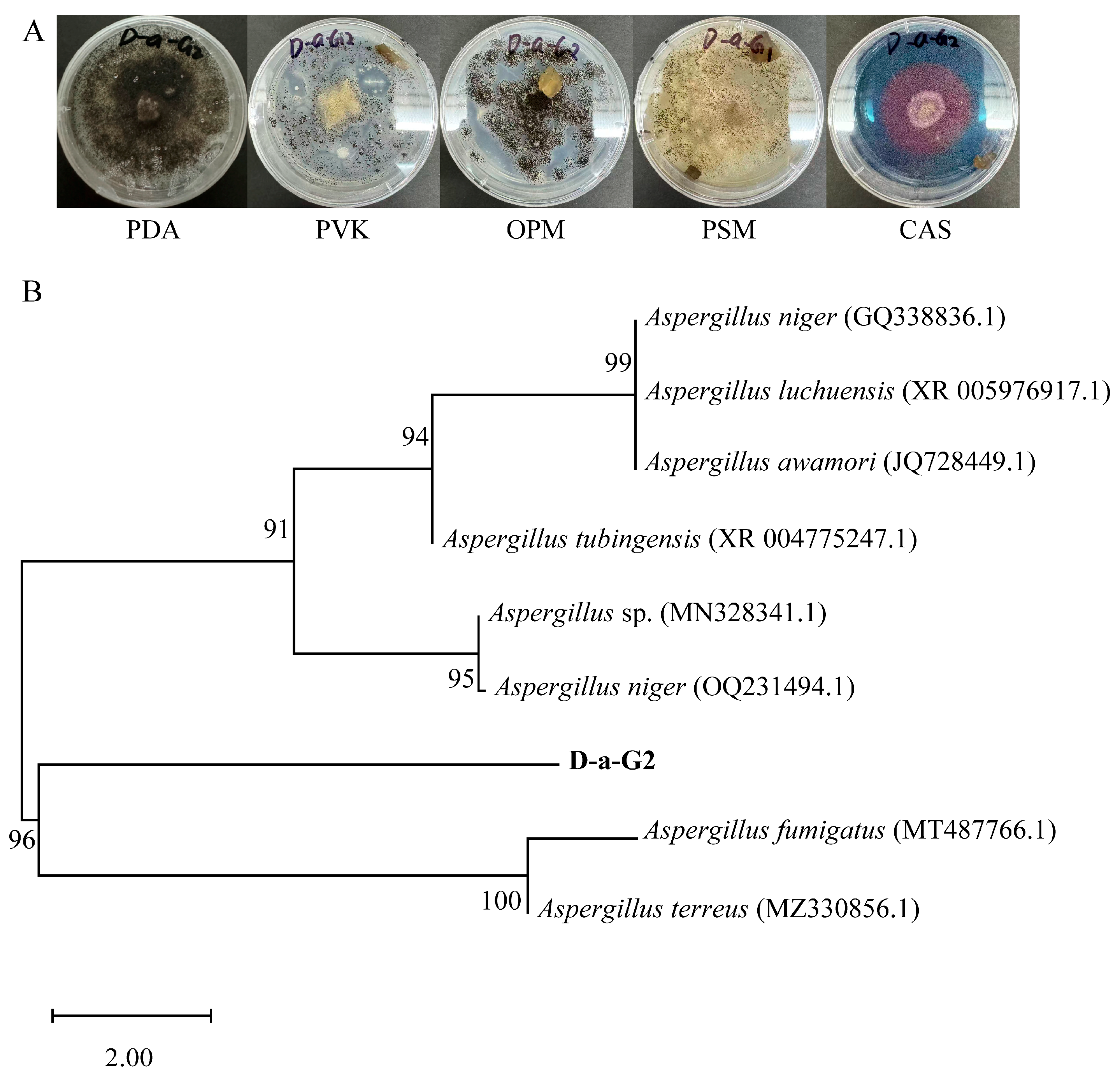
Nine endophytic fungal strains were isolated from the roots of N. candida (Table S1). Among them, strain D-a-G2 exhibited black coloration, an uneven surface, irregular colony margins, relatively light-colored mycelia, and loose association with the culture medium. This strain solubilizes organic and inorganic phosphate, solubilizes potassium, and produce siderophores (Figure 1A). The results showed that the sequence similarity between strain D-a-G2 and A. fumigatus exceeded 99%, but based on the current data, it was identified only at the genus level (Figure 1B). Thus, this fungus isolated from the roots of N. candida was identified as Aspergillus sp.

3.2. Effects of Aspergillus sp. on Leaf and Root Growth of N. candida Seedlings

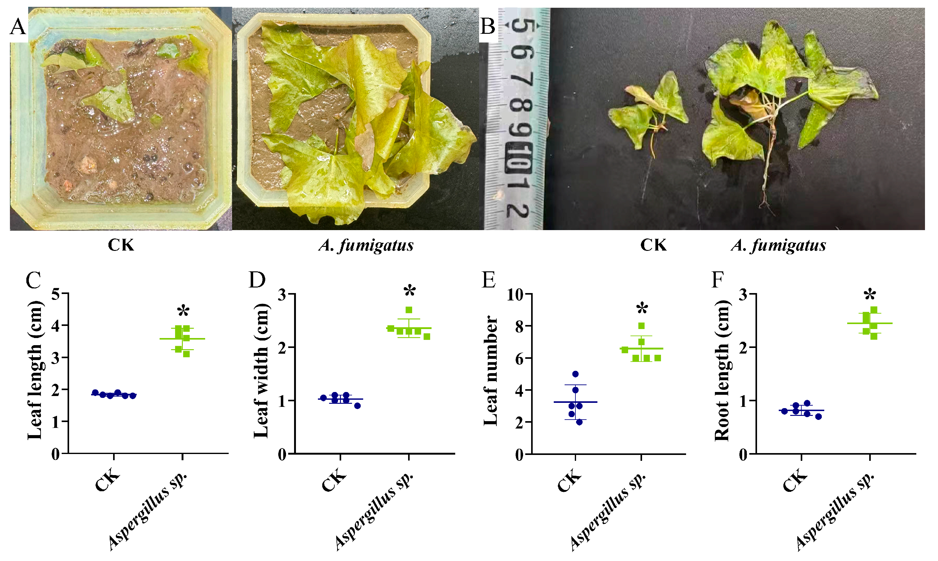
Compared with the control group, the inoculation of Aspergillus sp. for 60 days significantly increased leaf length, leaf width, and leaf number of N. candida seedlings by 97.83 ± 0.42%, 131.37 ± 0.36%, and 94.12 ± 2.45%, respectively (p < 0.05; Figure 2A,C–E). Notably, the root length of N. candida seedlings in the Aspergillus sp. group was 1.71-fold greater than that of the control group (p < 0.05; Figure 2B,F).

3.3. Effects of Aspergillus sp. on Soil Physicochemical Properties

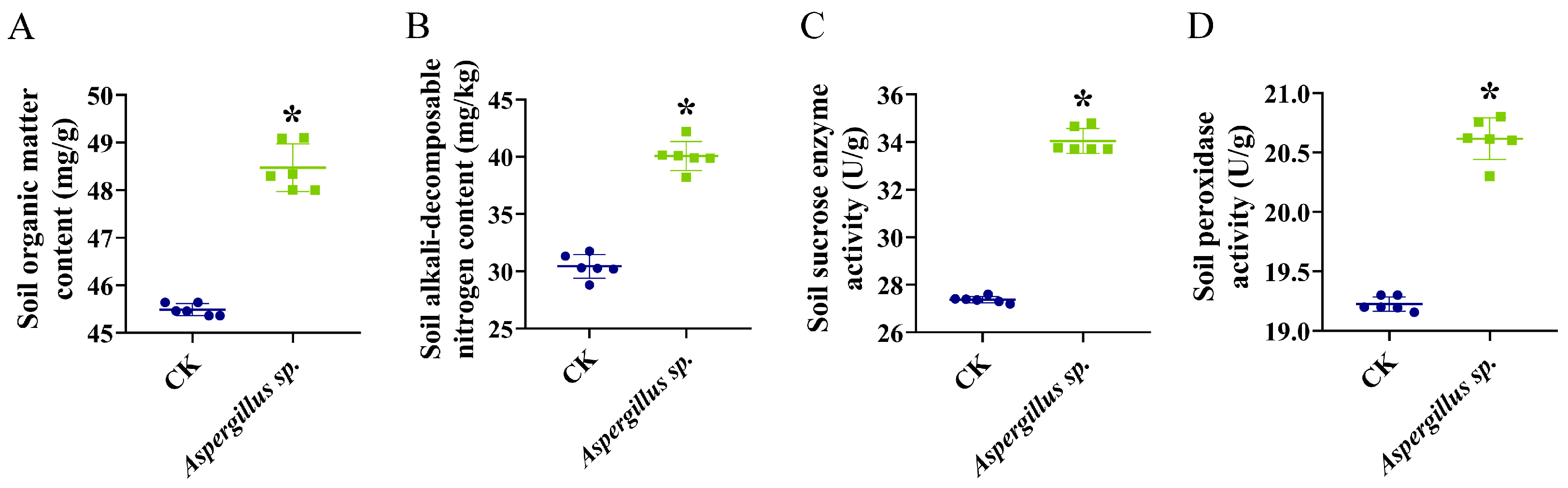
After the application of Aspergillus sp., soil organic matter content increased from 45.49 ± 0.13 g/kg to 48.47 ± 0.50 g/kg, alkali-hydrolyzable nitrogen content increased from 30.44 ± 1.03 mg/kg to 40.07 ± 1.27 mg/kg, sucrase activity increased from 27.38 ± 0.13 U/g to 34.06 ± 0.52 U/g, and peroxidase activity increased from 19.22 ± 0.06 U/g to 20.61 ± 0.18 U/g (p < 0.05; Figure 3A–D).

3.4. Effects of Aspergillus sp. on Rhizosphere Prokaryotic Microbial Communities

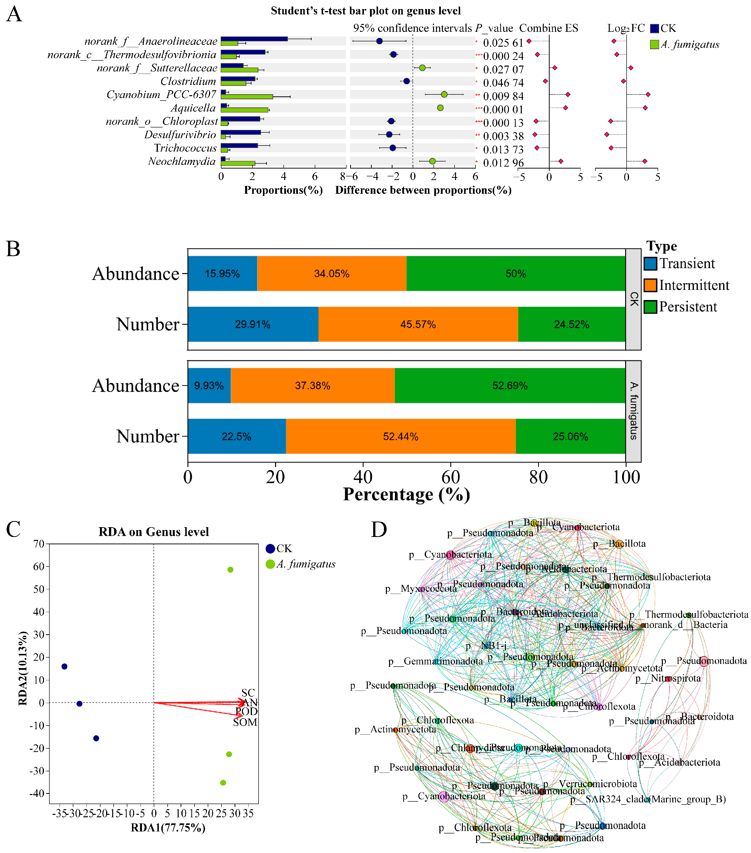
The application of Aspergillus sp. had no significant effect on the alpha and beta diversity of prokaryotic microorganisms in the rhizosphere soil of N. candida (Figure S1A–D). RDA results indicated that, compared with the control group, the rhizosphere prokaryotic microbial community in the Aspergillus sp. group was significantly positively correlated with soil sucrase activity (r2 = 0.99, p < 0.05; Figure 4C). Moreover, the relative abundances of Cyanobium-PCC-6307, Aquicella, and Neochlamydia in the Aspergillus sp. group significantly increased by 10.18, 7.08, and 6.78-fold, respectively (p < 0.05; Figure 4A). Meanwhile, the relative abundances of intermediate and persistent prokaryotic microbial communities significantly increased to 37.38% and 52.69%, respectively (Figure 4B). The application of Aspergillus sp. also increased the network density, modularity, and weighted average degree of the prokaryotic microbial co-occurrence network in the N. candida rhizosphere by 18.65%, 34.15%, and 42.73%, respectively (Figure 4D, Table S2).

3.5. Effects of Aspergillus sp. on Rhizosphere Eukaryotic Microbial Communities

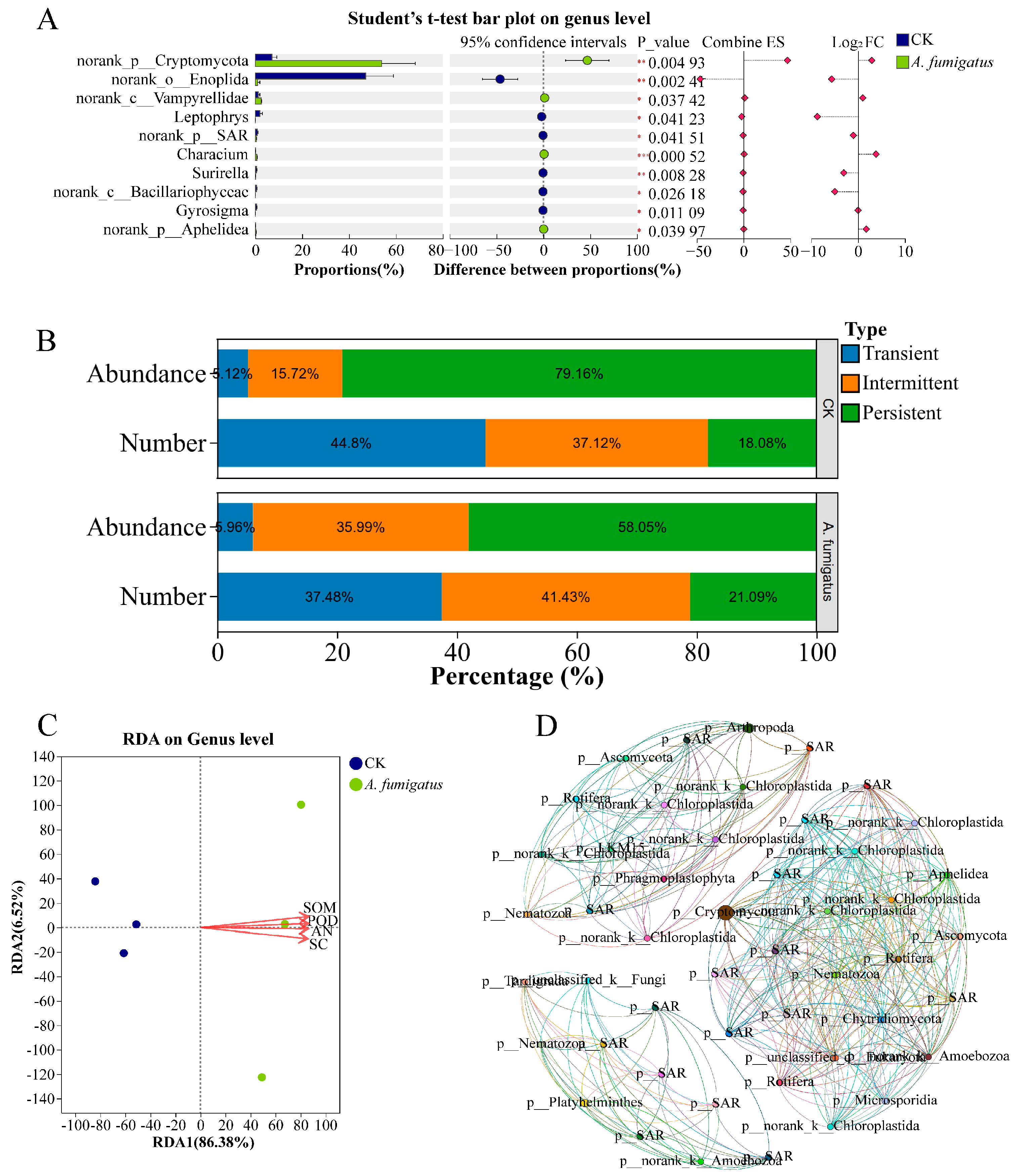
The application of Aspergillus sp. had no significant effect on alpha and beta diversity of eukaryotic microorganisms in the rhizosphere soil of N. candida (Figure S1C,D). RDA results indicated that, compared with the control group, the structure of the rhizosphere eukaryotic microbial community in the Aspergillus sp. group was positively correlated with soil organic matter content (r2 = 0.98, p < 0.05), alkali-hydrolyzable nitrogen content (r2 = 0.98, p < 0.05), sucrase activity (r2 = 0.98, p < 0.05), and peroxidase activity (r2 = 0.98, p < 0.05; Figure 5C). Aspergillus sp. significantly increased the relative abundances of norank-p-Cryptomycota, norank-c-Vampyrellidae, and Characium by 6.61, 1.01, and 13.19-fold, respectively (p < 0.05; Figure 5A). After the application of Aspergillus sp., the relative abundance of transient and intermediate eukaryotic microbial communities significantly increased to 5.96% and 35.99%, respectively (Figure 5B). Moreover, the application of Aspergillus sp. increased the network density, modularity, and weighted average degree of the eukaryotic microbial co-occurrence network in the N. candida rhizosphere by 4.19%, 2.02%, and 3.61%, respectively (Figure 5D; Table S2).

3.6. Effects of Aspergillus sp. on Gene Expression in N. candida Leaves

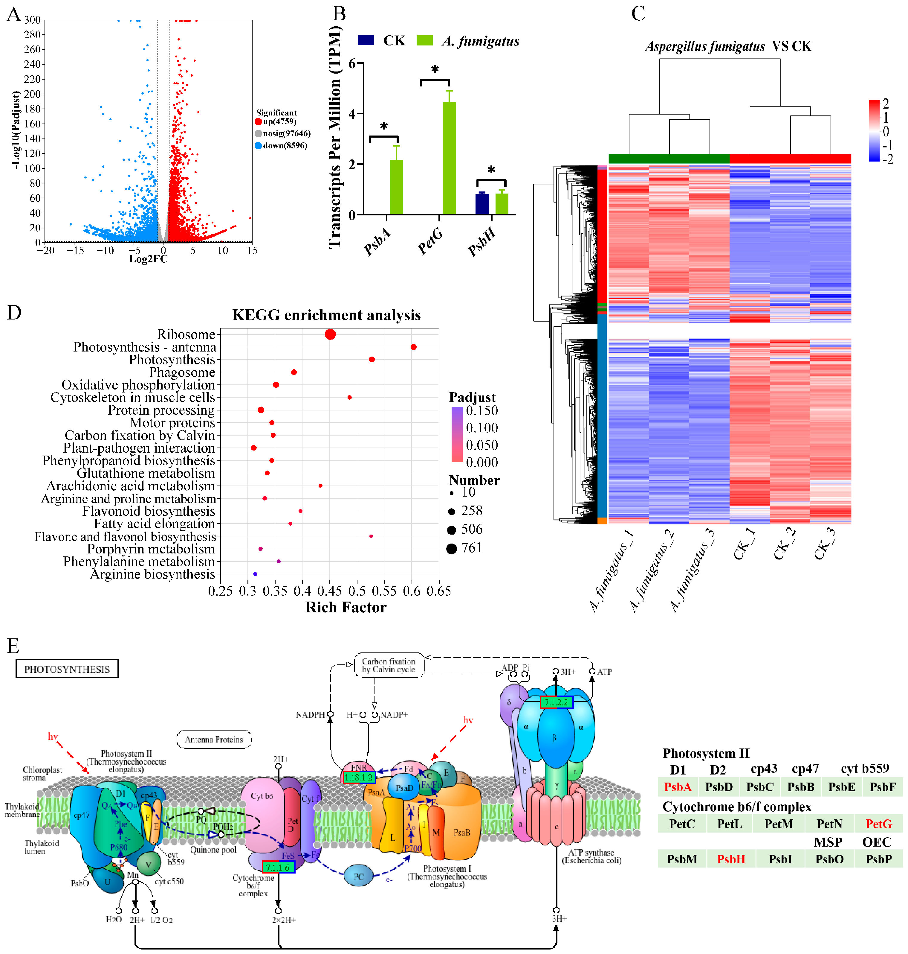
Compared with the control group, 4759 genes were upregulated and 8956 genes were downregulated in the leaves of N. candida from the Aspergillus sp. group (Figure 6A). Clustering analysis results showed that the unigenes from the CK and Aspergillus sp. groups clustered independently into two separate groups, Cluster I and Cluster II, indicating distinct gene expression profiles between the two treatments. High reproducibility was observed among parallel samples, indicating that the data were suitable for further analysis (Figure 6C). KEGG pathway enrichment analysis revealed that the most significantly enriched metabolic pathways in the Aspergillus sp. group mainly included “Ribosome”, “Photosynthesis-antenna proteins”, and “Photosynthesis” (Figure 6D). Analysis of DEGs showed that after Aspergillus sp. application, the transcripts per million (TPM) value of the gene encoding the photosystem II P680 reaction center D1 protein (psbA) in the photosynthesis pathway was significantly upregulated by 2.17-fold, and the TPM value of the gene encoding cytochrome b6-f complex subunit 5 (petG) significantly increased by 4.48-fold. The TPM value of the gene encoding the photosystem II PsbH protein (psbH) also significantly increased by 28.4% (p < 0.05; Figure 6B,E).

4. Discussion

In this study, an endophytic growth-promoting fungus, Aspergillus sp., was isolated from the roots of N. candida and demonstrated the ability to produce siderophores, solubilizing organic and inorganic phosphate, and solubilizing potassium. Although 18S rRNA sequence similarity (>99%) and morphological characteristics suggest that strain D-a-G2 is closely related to Aspergillus fumigatus, we refer to it as Aspergillus sp. due to the lack of multi-locus phylogenetic analysis (e.g., benA, caM, rpb2). Future studies will employ additional loci or whole-genome sequencing for definitive species identification [36]. Moreover, the application of Aspergillus sp. significantly increased leaf length, leaf width, leaf number, and root length of N. candida, as well as the photosynthetic efficiency in the leaves. Thus, Aspergillus sp. could significantly enhance the growth of the rare and endangered aquatic plant N. candida.
The growth of aboveground and belowground plant parts is relatively coordinated, and the improvement in leaf traits might be attributed to the increased root length of N. candida [37]. Previous studies showed that A. fumigatus exerted growth-promoting effects through the production of plant growth hormones and biostimulants, secretion of organic acids or phosphatases, and induction of plant resistance, which was similar to arbuscular mycorrhizal fungi [38,39,40,41]. In this study, Aspergillus sp. significantly increased the root length of N. candida by 1.7-fold. The Aspergillus nomius WLG2 strain could also promoted root growth of maize via producing growth-promoting substances auxin and flavonoids, resulting in a 60% increase in maize root length [42]. The promoting effects of A. fumigatus on both the leaves and roots of N. candida seedlings provide direct evidence for its potential as an efficient endophytic growth-promoting fungus.
The growth-promoting effect of Aspergillus sp. on N. candida might be attributed to improvements in soil physicochemical properties. After inoculation with Aspergillus sp., the contents of organic matter and alkali-hydrolyzable nitrogen in the cultivation soil of N. candida seedlings significantly increased by 6.57% and 31.62%, respectively. Moreover, the sucrase and peroxidase activities increased by 23.26% and 7.53%, respectively. Soil enzyme activities are important indicators of soil biological activity and nutrient cycling capacity [43]. Sucrase participates in the decomposition of soil organic carbon and carbon cycling, and its increase indicated an enhanced mineralization rate of organic matter in the soil, potentially providing plants with more available carbon sources and nutrients [44]. Peroxidase is closely associated with the polymerization of soil organic matter and humus formation, and its increase contributed to the improvement of the soil structure [45]. Studies have shown that A. fumigatus strains solubilize phosphate, converting insoluble phosphorus in the soil into forms that are absorbable by plants, thereby significantly increasing root length in Triticum aestivum and Vigna radiata by 25% [46,47]. Thus, Aspergillus sp. might improve soil health through organic matter degradation and nutrient cycling promotion, thereby enhancing the growth of N. candida seedlings.
Although the diversity of the rhizosphere microbial community was unaffected, Aspergillus sp. enriched different beneficial microorganisms and formed a specific functional microbial community. The application of Aspergillus sp. significantly enriched Cyanobium-PCC-6307, Aquicella, and Neochlamydia. Cyanobium-PCC-6307 is a dominant cyanobacterial population in paddy soil [48], and Aquicella is a potential plant growth-promoting bacterium and might increase chlorophyll content and bulb number in onion by 6.46% and 21.41%, respectively [49]. Neochlamydia, an endosymbiont of Vampyrellida, exhibited increased abundance typically associated with population dynamics of amoebae in the environment, directly demonstrating the changes in the rhizosphere microbial community structure of N. candida [50]. Eukaryotic microbial community analysis revealed that the Aspergillus sp. group significantly enriched norank-p-Cryptomycota, norank-c-Vampyrellidae, and Characium in the rhizosphere of N. candida. Among these, Cryptomycota is a fungal group that participated in organic matter decomposition [51], while Vampyrellidae could prey on pathogenic fungi and algae to promote nutrient cycling and play a positive role in controlling soilborne diseases and maintaining environmental health [52]. Characium, as a photosynthetic autotroph, could utilize light energy to produce oxygen and organic matter and might serve as a marker for enhanced primary productivity in the ecosystems [53]. In this study, the application of Aspergillus sp. reduced the relative abundance of transient microorganisms, but increased the relative abundances of intermediate and persistent microbial communities, suggesting an enhanced adaptability of the rhizosphere microbial community to environmental changes [54]. RDA analysis results showed that the rhizosphere microbial community of N. candida was significantly positively correlated with organic matter content, alkali-hydrolyzable nitrogen content, sucrase activity, and peroxidase activity in the rhizosphere soil, further confirming that Aspergillus sp. indirectly promoted host growth by improving the soil microenvironment [55]. Co-occurrence network analysis demonstrated that the application of Aspergillus sp. markedly increased the modularity of both prokaryotic and eukaryotic microbial communities in the N. candida rhizosphere, resulting in a more complex network structure. Thus, Aspergillus sp. could form a more stable rhizosphere soil microenvironment to promote the growth of N. candida seedlings.
For the leaves, the application of Aspergillus sp. significantly upregulated genes encoding the photosystem II P680 reaction center D1 protein (psbA), cytochrome b6-f complex subunit 5 (petG), and photosystem II PsbH protein (psbH) in the photosynthetic metabolic pathway by 2.17, 0.28 and 4.48-fold, respectively. The enhanced photosynthesis enabled plants to generate more photosynthetic products and provide energy for all plant life activities [56]. Moreover, the abundant photosynthetic product reserves provided plants with more energy and materials for defense responses when facing exogenous stresses, and thereby promoted plant growth [57]. For instance, A. oryzae and A. fumigatus enhanced the photosynthesis in maize leaves and significantly increased root length by 11.76% [42]. Additionally, the enrichment of “Photosynthesis–antenna proteins” pathway suggested that Aspergillus sp. might promote photosynthesis by enhancing light energy capture efficiency, which corroborated the observed significant increase in leaf area of N. candida [58]. The upregulation of photosynthetic genes suggests that Aspergillus sp. may enhance photosynthetic capacity. Future studies will directly measure photosynthetic parameters (e.g., net photosynthetic rate, chlorophyll fluorescence) to confirm this inference. We acknowledge that the lack of independent qPCR validation for the RNA-seq data is a limitation of this study. Future studies will include RT-qPCR validation of key photosynthetic genes (psbA, petG, psbH) to confirm the transcriptomic findings.

5. Conclusions

In conclusion, the endophytic growth-promoting fungus Aspergillus sp. isolated from the roots of N. candida could promote leaf and root development of N. candida seedlings by increasing the contents of organic matter and alkali-hydrolyzable nitrogen in rhizosphere soil and enhancing sucrase and peroxidase activities. Moreover, the application of Aspergillus sp. enriched beneficial microorganisms including Cyanobium, Aquicella, and Cryptomycota to form a more stable rhizosphere soil microenvironment to promote the growth of N. candida seedlings. Furthermore, Aspergillus sp. upregulated the genes involved in photosynthesis and photosynthesis-antenna protein pathways in N. candida leaves to improve light energy utilization efficiency. Thus, Aspergillus sp. could be utilized as a potential biofertilizer for the propagation of N. candida seedlings, which would be helpful for the conservation of this rare and endangered aquatic plant species.

Supplementary Materials

The following supporting information can be downloaded at: https://www.mdpi.com/article/10.3390/microorganisms14050993/s1. Table S1. Sequence alignment result of the endophytic fungi isolated from the roots of Nymphaea candida; Table S2. Rhizosphere microbial network indices of Nymphaea candida; Figure S1: Alpha diversity of prokaryotic and eukaryotic microbial communities in the rhizosphere soil of Nymphaea candida (A,B); Principal coordinate analysis (PCoA) of prokaryotic and eukaryotic microbial communities based on Bray-Curtis distance (C,D).

Author Contributions

Conceptualization, J.W.; Methodology, Y.X.; Formal analysis, Y.X.; Investigation, Y.X., J.Z., C.L. and Y.L.; Resources, J.W.; Writing—original draft, Y.X.; Writing—review & editing, J.W.; Supervision, J.W. All authors have read and agreed to the published version of the manuscript.

Funding

This work was supported by the Key Research and Development Program of Xinjiang Uygur Autonomous Region (2023B02037).

Institutional Review Board Statement

Not applicable.

Informed Consent Statement

Not applicable.

Data Availability Statement

The original contributions presented in this study are included in the article/Supplementary Material. Further inquiries can be directed to the corresponding authors.

Conflicts of Interest

The authors declare no conflict of interest.

References

  1. Qiu, P.; Liu, T.; Xu, Y.; Ye, C.; Zhang, R.; Wang, Y.; Jin, Q. Multi-omic dissection of the cold resistance traits of white water lily. Hortic. Res. 2024, 11, uhae093. [Google Scholar] [CrossRef] [PubMed]
  2. Borsch, T.; Hilu, K.W.; Wiersema, J.H.; Lohne, C.; Barthlott, W.; Wilde, V. Phylogeny of Nymphaea (Nymphaeaceae): Evidence from substitutions and microstructural changes in the chloroplast trnT-trnF region. Int. J. Plant Sci. 2007, 168, 639–671. [Google Scholar] [CrossRef]
  3. Kamdem, B.P.; Le Doux Kamto, E.; Aboubakar Pegnyemb, D.E.; Igne, F.E. Ethnomedicinal uses, phytochemistry, pharmacology, and Toxicity of the Genus Nymphaea L.: A Review. Curr. Bioact. Compd. 2022, 18, 2–40. [Google Scholar] [CrossRef]
  4. Dalziell, E.L.; Lewandrowski, W.; Merritt, D.J. Increased salinity reduces seed germination and impacts upon seedling development in Nymphaea L. (Nymphaeaceae) from northern Australia’s freshwater wetlands. Aquat. Bot. 2020, 165, 103235. [Google Scholar] [CrossRef]
  5. Arefin, M.A.; Rashid, F.; Islam, A. A review of biofuel production from floating aquatic plants: An emerging source of bio-renewable energy. Biofuels Bioprod. Biorefin. 2021, 15, 574–591. [Google Scholar] [CrossRef]
  6. Xing, Y.; Liu, C.; Cui, X.; Lv, H.; Wang, J. Isolation, identification, and control of Pathogenic Endophytic fungi in Nymphaea candida Presl tissue culture. Microorganisms 2025, 13, 1103. [Google Scholar] [CrossRef]
  7. Klok, P.F.; Van der Velde, G. Plant traits and environment: Floating leaf blade production and turnover of waterlilies. PeerJ 2017, 5, e3212. [Google Scholar] [CrossRef]
  8. Khan, M.B.; Sulaman, S.; Shabaan, M.; Fatima, A.; Hameed, A.; Zulfiqar, U.; Kabir, R.; Alarjani, K.M.; Reem, A.A. Exploring the phytostabilization potential of ryegrass (Lolium perenne L.) upon synergistic application of Cd-tolerant Pseudomonas fluorescens and organic amendments. Int. J. Phytoremediat. 2025, 27, 1314–1328. [Google Scholar] [CrossRef] [PubMed]
  9. Liu, T.; Li, D.; Tian, Y.; Liu, T.; Li, D.; Tian, Y.; Zhou, J.; Qiu, Y.; Li, D.; Liu, G.; et al. Enhancing nitrogen removal in constructed wetlands: The role of influent substrate concentrations in integrated vertical-flow systems. Environ. Sci. Ecotechnol. 2024, 21, 100411. [Google Scholar] [CrossRef]
  10. Leblebici, Z.; Aksoy, A. Growth and lead accumulation capacity of Lemna minor and Spirodela polyrhiza (Lemnaceae): Interactions with nutrient enrichment. Water Air Soil Pollut. 2011, 214, 175–184. [Google Scholar] [CrossRef]
  11. Wang, Q.Y.; Zhao, M.R.; Wang, J.Q.; Hu, B.; Chen, Q.; Qin, Y.; Zhang, G. Effects of microbial inoculants on agronomic characters, physicochemical properties and nutritional qualities of lettuce and celery in hydroponic cultivation. Sci. Hortic. 2023, 320, 112202. [Google Scholar] [CrossRef]
  12. Chen, Y.; Li, Y.; Fu, Y.; Jia, L.; Xu, Z.; Zhang, N.; Liu, Y.; Fan, X.; Xuan, W.; Xu, G.; et al. The beneficial rhizobacterium Bacillus velezensis SQR9 regulates plant nitrogen uptake via an endogenous signaling pathway. J. Exp. Bot. 2024, 75, 3388–3400. [Google Scholar] [CrossRef] [PubMed]
  13. Zhang, C.; Yu, Z.; Zhang, M.; Li, X.; Wang, M.; Li, L.; Li, X.; Ding, Z.; Tian, H. Serratia marcescens PLR enhances lateral root formation through supplying PLR-derived auxin and enhancing auxin biosynthesis in Arabidopsis. J. Exp. Bot. 2022, 73, 3711–3725. [Google Scholar] [CrossRef]
  14. López-Bucio, J.; Campos-Cuevas, J.C.; Hernández-Calderón, E.; Velásquez-Becerra, C.; Farías-Rodríguez, R.; Macías-Rodríguez, L.I.; Valencia-Cantero, E. Bacillus megaterium rhizobacteria promote growth and alter root-system architecture through an auxin-and ethylene-independent signaling mechanism in Arabidopsis thaliana. Mol. Plant-Microbe Interact. 2007, 20, 207–217. [Google Scholar] [CrossRef]
  15. Glick, B.R. Plant growth-promoting bacteria: Mechanisms and applications. Scientifica 2012, 2012, 963401. [Google Scholar] [CrossRef]
  16. Wei, Y.; Yang, H.; Hu, J.; Li, H.; Zhao, Z.; Wu, Y.; Li, J.; Zhou, Y.; Yang, K.; Yang, H. Trichoderma harzianum inoculation promotes sweet sorghum growth in the saline soil by modulating rhizosphere available nutrients and bacterial community. Front. Plant Sci. 2023, 14, 1258131. [Google Scholar] [CrossRef]
  17. Bi, Y.; Song, Y.; Bai, X.; Wang, S. DSE and its metabolites on Medicago sativa L. growth promotion and its potential for ecological restoration in mining areas. Coal Sci. Technol. 2023, 51, 90–99. [Google Scholar] [CrossRef]
  18. Liang, L.; Liu, B.; Huang, D.; Kuang, Q.; An, T.; Liu, S.; Liu, S.; Liu, R.; Xu, B.; Zhang, S.; et al. Arbuscular mycorrhizal fungi alleviate low phosphorus stress in maize genotypes with contrasting root systems. Plants 2022, 11, 3105. [Google Scholar] [CrossRef] [PubMed]
  19. Aljeddani, G.S.; Hamouda, R.A.; Abdelsattar, A.M.; Heikal, Y.M. Stress-responsive gene expression, metabolic, physiological, and agronomic responses by consortium nano-silica with Trichoderma against drought stress in bread wheat. Int. J. Mol. Sci. 2024, 25, 10954. [Google Scholar] [CrossRef] [PubMed]
  20. Khalil, A.M.A.; Hassan, S.E.D.; Alsharif, S.M.; Eid, A.M.; Ewais, E.E.; Azab, E.; Gobouri, A.A.; Elkelish, A.; Elkelish, A. Isolation and characterization of fungal endophytes isolated from medicinal plant Ephedra pachyclada as plant growth-promoting. Biomolecules 2021, 11, 140. [Google Scholar] [CrossRef]
  21. Khuna, S.; Kumla, J.; Srinuanpan, S.; Lumyong, S.; Suwannarach, N. Multifarious characterization and efficacy of three phosphate-solubilizing Aspergillus species as biostimulants in improving root induction of cassava and sugarcane stem cuttings. Plants 2023, 12, 3630. [Google Scholar] [CrossRef]
  22. Quesada Moraga, E. Entomopathogenic fungi as endophytes: Their broader contribution to IPM and crop production. Biocontrol Sci. Technol. 2020, 30, 864–877. [Google Scholar] [CrossRef]
  23. Wang, D.; Zhao, G. Study on surface sterilization method for tissue culture of Haloxylon ammodendron (CA Mey.) Bunge seeds. Agric. Biotechnol. 2012, 1, 26–28. [Google Scholar]
  24. Sheikholeslami, B.; Shukla, M.; Turi, C.; Harpur, C.; Saxena, P.K. Saving threatened plant species: Reintroduction of Hill’s thistle (Cirsium hillii. (Canby) Fernald) to its natural habitat. PLoS ONE 2020, 15, e0231741. [Google Scholar] [CrossRef]
  25. Watrud, L.S.; Martin, K.; Donegan, K.K.; Stone, J.K.; Coleman, C.G. Comparison of taxonomic, colony morphotype and PCR-RFLP methods to characterize microfungal diversity. Mycologia 2006, 98, 384–392. [Google Scholar] [CrossRef]
  26. Gama, D.D.S.; Santos, Í.A.F.M.; Abreu, L.M.D.; Flávio, H.V.D.M.; Whasley, F.D.; Patrícia, G.C. Endophytic fungi from Brachiaria grasses in Brazil and preliminary screening of Sclerotinia sclerotiorum antagonists. Sci. Agric. 2020, 77, e20180210. [Google Scholar] [CrossRef]
  27. Kumar, S.; Stecher, G.; Tamura, K. MEGA7: Molecular evolutionary genetics analysis version 7.0 for bigger datasets. Mol. Biol. Evol. 2016, 33, 1870–1874. [Google Scholar] [CrossRef] [PubMed]
  28. Geng, Y.; Chen, S.; Lv, P.; Li, Y.; Li, J.; Jiang, F.; Wu, Z.; Shen, Q.; Zhou, R. Positive role of Trichoderma harzianum in increasing plant tolerance to abiotic stresses: A review. Antioxidants 2025, 14, 807. [Google Scholar] [CrossRef] [PubMed]
  29. Ferreira, O.J.M.; Holanda, F.S.R.; Pedrotti, A.; Santos, L.D.V.; Silva-Mann, R. Root system of jatropha curcas provides resistance and strength to the soil. Commun. Soil Sci. Plant Anal. 2022, 53, 2955–2967. [Google Scholar] [CrossRef]
  30. Rossel, R.V.; McBratney, A.B. Soil chemical analytical accuracy and costs: Implications from precision agriculture. Aust. J. Exp. Agric. 1998, 38, 765–775. [Google Scholar] [CrossRef]
  31. De Vos, B.; Lettens, S.; Muys, B.; Deckers, J.A. Walkley–Black analysis of forest soil organic carbon: Recovery, limitations and uncertainty. Soil Use Manag. 2007, 23, 221–229. [Google Scholar] [CrossRef]
  32. Dixit, A.; Sahu, P.; Singh, V.R.; Dhruw, V. Physicochemical Study of Potassium Dichromate (K2Cr2O7) in 10% Sucrose-Water at Different Temperature. J. Pharm. Res. 2017, 5, 00140. [Google Scholar] [CrossRef][Green Version]
  33. Yang, H.; Min, G. New record of Kellicottia bostoniensis and redescription of two freshwater rotifers from Korea (Rotifera: Monogononta). Anim. Syst. Evol. Divers. 2020, 36, 222–227. [Google Scholar] [CrossRef]
  34. Yoon, K.Y.; Tan, W.S.; Tey, B.T.; Lee, K.W.; Ho, K.L. Native agarose gel electrophoresis and electroelution: A fast and cost-effective method to separate the small and large hepatitis B capsids. Electrophoresis 2013, 34, 244–253. [Google Scholar] [CrossRef]
  35. Li, H.; Li, C.; Song, X.; Liu, Y.; Gao, Q.; Zheng, R.; Li, J.; Zhang, P.; Liu, X. Impacts of continuous and rotational cropping practices on soil chemical properties and microbial communities during peanut cultivation. Sci. Rep. 2022, 12, 2758. [Google Scholar] [CrossRef]
  36. Silva, J.J.; Fungaro, M.H.P.; Wang, X.; Larsen, T.O.; Frisvad, J.C.; Taniwaki, M.H.; Iamanaka, B.T. Deep Genotypic Species Delimitation of Aspergillus Section Flavi Isolated from Brazilian Foodstuffs and the Description of Aspergillus annui sp. nov. and Aspergillus saccharicola sp. nov. J. Fungi 2022, 8, 1279. [Google Scholar] [CrossRef]
  37. Sharma, S.; Pandey, S.; Kulshreshtha, S.; Dubey, M. Biology and application of Chaetomium globosum as a biocontrol agent: Current status and future prospects. Microorganisms 2025, 13, 1646. [Google Scholar] [CrossRef]
  38. Zhao, H.; Wan, S.; Huang, Y.; Li, X.; Jiao, T.; Zhang, Z.; Ma, B.; Zhu, L.; Ma, F.; Li, M. The transcription factor MdBPC2 alters apple growth and promotes dwarfing by regulating auxin biosynthesis. Plant Cell 2024, 36, 585–604. [Google Scholar] [CrossRef]
  39. El-Shafey, N.M.; Marzouk, M.A.; Yasser, M.M.; Mahfouz, A.Y.; Daigham, G.E. Bio-stimulating effect of endophytic Aspergillus flavus AUMC 16068 and its respective ex-polysaccharides in lead stress tolerance of Triticum aestivum plant. Sci. Rep. 2024, 14, 11952. [Google Scholar] [CrossRef]
  40. El-Rahman, A.F.A.; Shaheen, H.M.; El-Aziz, A.R.M.; Gamal, A.; Attia, M.S.; Hashem, A.H. Innovative fungal bioagents: Producing siderophores, IAA, and HCN to support plants under salinity stress and combat microbial plant pathogens. Microb. Cell Factories 2025, 24, 246. [Google Scholar] [CrossRef]
  41. Abdelaziz, A.M.; El-Wakil, D.A.; Attia, M.S.; Omar, M.A.; Hamada, A.; Amr, H.H. Inhibition of Aspergillus flavus growth and aflatoxin production in Zea mays L. using endophytic Aspergillus fumigatus. J. Fungi 2022, 8, 482. [Google Scholar] [CrossRef]
  42. Niaz, K.; Rauf, M.; Arif, M.; Hamayun, M.; Gul, H.; Hashem, A.; Allah, E.A.; Wu, Q. Drought-tolerant fungal microbes, Aspergillus oryzae and Aspergillus fumigatus, elevate physiohormonal and antioxidant responses of maize under drought stress. Front. Microbiol. 2024, 15, 1488639. [Google Scholar] [CrossRef]
  43. Mehmood, A. Effect of Indole-3-Acetic Acid and Flavonoids on Association of Maize Roots and Phytostimulant Endophytic Fungi. Doctoral Dissertation, Abdul Wali Khan University, Mardan, Pakistan, 2019. [Google Scholar]
  44. Daunoras, J.; Kačergius, A.; Gudiukaitė, R. Role of soil microbiota enzymes in soil health and activity changes depending on climate change and the type of soil ecosystem. Biology 2024, 13, 85. [Google Scholar] [CrossRef] [PubMed]
  45. Taggart, M.G.; Baah, D.S.; Allen, S.; Khan, Z.; Arnscheidt, J.; Jordan, P.; O’Hagan, B.M.G.; Ibrahim, A.D.; Rao, J.R.; Ternan, N.G. Fitting soil extracellular enzyme activity into the complex network of abiotic and biotic soil properties often associated with soil health. Front. Microbiol. 2025, 16, 1638267. [Google Scholar] [CrossRef] [PubMed]
  46. Wang, L.; Hamel, C.; Lu, P.; Wang, J.; Sun, D.; Wang, Y.; Lee, S.; Gan, G. Using enzyme activities as an indicator of soil fertility in grassland-an academic dilemma. Front. Plant Sci. 2023, 14, 1175946. [Google Scholar] [CrossRef] [PubMed]
  47. Kumar, V.; Prasher, I.B. Phosphate solubilization and indole-3-acetic acid (IAA) produced by Colletotrichum gloeosporioides and Aspergillus fumigatus strains isolated from the rhizosphere of Dillenia indica L. Folia Microbiol. 2023, 68, 219–229. [Google Scholar] [CrossRef]
  48. Wichaphian, A.; Kamngoen, A.; Pathom-Aree, W.; Maneechote, W.; Khuendee, T.; Chromkaew, Y.; Cheirsilp, B.; Shyu, D.J.H.; Srinuanpan, S. Integrating microalgal Chlorella biomass and biorefinery residues into sustainable agriculture and food production: Insights from lettuce cultivation. Foods 2025, 14, 808. [Google Scholar] [CrossRef]
  49. Simarmata, R.; Widowati, T.; Christita, M.; Rahayu, F.W.P.; Christita, M.; Khairina, Y.; Erdayani, E.; Khumairah, F.H. Rhizosphere bacteriome of Allium cepa after the application of chemical and endophyte-based fertilizer. Acta Ecol. Sin. 2023, 43, 1138–1148. [Google Scholar] [CrossRef]
  50. Horn, M.; Wagner, M. Evidence for additional genus-level diversity of Chlamydiales in the environment. FEMS Microbiol. Lett. 2001, 204, 71–74. [Google Scholar] [CrossRef][Green Version]
  51. Sista Kameshwar, A.K.; Qin, W. Systematic review of publicly available non-Dikarya fungal proteomes for understanding their plant biomass-degrading and bioremediation potentials. Bioresour. Bioprocess. 2019, 6, 30. [Google Scholar] [CrossRef]
  52. Jiang, M.; Gong, Y.; Tikhonenkov, D. Feeding of Vampirellid Amoeba (Leptophryidae) on Cyanobacteria. Inland Water Biol. 2025, 18, 1504–1509. [Google Scholar] [CrossRef]
  53. Krings, M.; Harper, C.J.; Taylor, E.L.; Kerp, H. Early Devonian (~410 mya) microfossils resembling Characiopsis (Tribophyceae) and Characium (Chlorophyceae). J. Phycol. 2017, 53, 720–724. [Google Scholar] [CrossRef]
  54. Ma, S.; Zhao, D.; Han, X.; Peng, Y.; Ren, T.; Wang, M.; Wan, J.; Ding, J.; Du, X.; Zhao, F.; et al. New application of Aspergillus versicolor in promoting plant growth after suppressing sterigmatocystin production via genome mining and engineering. Microb. Biotechnol. 2023, 16, 139–147. [Google Scholar] [CrossRef]
  55. Chauhan, P.; Singh, M.; Sharma, A.; Chadha, P.; Kaur, A. Plant growth-promoting endophytic fungus Aspergillus niger VN2 enhances growth, regulates oxidative stress and protects DNA damage in Vigna radiata under salt stress. BMC Microbiol. 2025, 25, 585. [Google Scholar] [CrossRef] [PubMed]
  56. Bilal, L.; Asaf, S.; Hamayun, M.; Gul, H.; Iqbal, A.; Ullah, I.; Lee, I.; Hussain, A. Plant growth-promoting endophytic fungi Asprgillus fumigatus TS1 and Fusarium proliferatum BRL1 produce gibberellins and regulates plant endogenous hormones. Symbiosis 2018, 76, 117–127. [Google Scholar] [CrossRef]
  57. Tarafdar, J.C.; Marschner, H. Dual inoculation with Aspergillus fumigatus and Glomus mosseae enhances biomass production and nutrient uptake in wheat (Triticum aestivum L.) supplied with organic phosphorus as Na-phytate. Plant Soil 1995, 173, 97–102. [Google Scholar] [CrossRef]
  58. Ren, H.; Wang, L.; Ma, Q.; Wu, G. Progress in Biosynthetic Pathways of Brassinosteroids. Chin. Bull. Bot. 2015, 50, 768–778. [Google Scholar] [CrossRef]
Figure 1. Morphological and molecular identification of the endophytic growth-promoting fungus isolated from Nymphaea candida roots. (A) Morphology of Aspergillus sp. on Potato Dextrose Agar (PDA), Pikovskaya’s Agar Medium (PVK), Phosphate-solubilizing bacteria medium (OPM), Potassium bacteria Medium (PSM), and Chrome Azurol S Agar Medium (CAS). (B) Neighbor-joining phylogenetic tree constructed based on the 18S rRNA gene sequence of the endophytic growth-promoting fungus Aspergillus sp. obtained from Nymphaea candida in this study. The strain numbers shown in bold represent the isolates obtained in this study. The species name indicates the molecularly identified species and D-a-G2 is the strain number. GenBank accession numbers are shown in parentheses after the strain names. The branching pattern was generated using the neighbor-joining method, bootstrap analysis was performed with 1000 replicates, and the scale bar represents 0.5 nucleotide substitutions per site.
Microorganisms 14 00993 g001
Figure 2. Effects of Aspergillus sp. on leaf morphology (A), root morphology (B), leaf length (C), leaf width (D), leaf number (E), and root length (F) of Nymphaea candida seedlings. All experimental groups had six replicates, and data are presented as mean ± standard deviation (n = 6). Statistical significance was indicated as follows: * p < 0.05. Statistical significance was determined by two-tailed Student’s t-test.
Microorganisms 14 00993 g002
Figure 3. Effects of Aspergillus sp. on organic matter content (A), alkali-hydrolyzable nitrogen content (B), sucrase activity (C), and peroxidase activity (D) in the cultivation soil of Nymphaea candida seedlings. All experimental groups had six replicates, and data are presented as mean ± standard deviation (n = 6). Statistical significance was indicated as follows: * p < 0.05. Statistical significance was determined by two-tailed Student’s t-test.
Microorganisms 14 00993 g003
Figure 4. Effects of Aspergillus sp. on the composition and structure of prokaryotic microbial communities in the rhizosphere soil of Nymphaea candida seedlings. (A) Bar chart comparing the two groups showing significant differences in the mean relative abundance of the same taxonomic unit between different groups. Statistical significance is indicated as follows: * 0.01 < p < 0.05, ** 0.001 < p < 0.01, *** p < 0.001. (B) Environmental sensitivity of various types of prokaryotic microorganisms in the rhizosphere soil of Nymphaea candida from different experimental groups. (C) Response (RDA) analysis of the effects of key physicochemical indicators of the Nymphaea candida cultivation environment on rhizosphere prokaryotic microbial communities following Aspergillus sp. application. Soil organic matter (SOM), alkali-hydrolyzable nitrogen (AN), sucrase (SC), and peroxidase (POD). (D) Co-occurrence network analysis of prokaryotic microorganisms in the rhizosphere soil of the Aspergillus sp. group. Node colors were defined by different genera, and node sizes were defined by degree. Co-occurrence networks were constructed based on the relative abundances of prokaryotic microbial genera. Spearman’s correlation coefficients were calculated using the “psych” package (v 2.6.3) in R. Significant correlations (|r| > 0.6, p < 0.05) were used to build networks using the “igraph” package (v 2.2.3). Topological parameters (network density, modularity, and weighted average degree) were calculated to compare network complexity and stability between the control and Aspergillus sp. treatment groups.
Microorganisms 14 00993 g004
Figure 5. Effects of Aspergillus sp. on the composition and structure of eukaryotic microbial communities in the rhizosphere soil of Nymphaea candida seedlings. (A) Bar chart comparing the two groups showing significant differences in the mean relative abundance of the same taxonomic unit between different groups. Statistical significance is indicated as follows: * 0.01 < p < 0.05, ** 0.001 < p < 0.01, *** p < 0.001. (B) Environmental sensitivity of various types of eukaryotic microorganisms in the rhizosphere soil of Nymphaea candida from different experimental groups. (C) Response (RDA) analysis of the effects of key physicochemical indicators of the Nymphaea candida cultivation environment on rhizosphere eukaryotic microbial communities following Aspergillus sp. application. Soil organic matter (SOM), alkali-hydrolyzable nitrogen (AN), sucrase (SC), and peroxidase (POD). (D) Co-occurrence network analysis of eukaryotic microorganisms in the rhizosphere soil of the Aspergillus sp. group. Node colors were defined by different genera, and node sizes were defined by degree. Co-occurrence networks were constructed based on the relative abundances of eukaryotic microbial genera. Spearman’s correlation coefficients were calculated using the “psych” package in R. Significant correlations (|r| > 0.6, FDR-adjusted p < 0.05) were used to build networks using the “igraph” package. Topological parameters (network density, modularity, and weighted average degree) were calculated to compare network complexity and stability between the control and A. fumigatus treatment groups.
Microorganisms 14 00993 g005
Figure 6. Effects of Aspergillus sp. on gene expression in the transcriptome of Nymphaea candida leaves. (A) Volcano plot showing the distribution of differentially expressed genes in Nymphaea candida leaves from the Aspergillus sp. Group. (B) Relative quantification of upregulated genes in the photosynthesis pathway (psbA, petG, psbH) of the Aspergillus sp. group compared to the control group. Statistical significance is indicated as follows: * p < 0.05, (C) Clustering heatmap of differentially expressed genes in the leaf transcriptome of Nymphaea candida. (D) Top 20 KEGG enrichment pathways in the Aspergillus sp. group compared to the control group. (E) KEGG reference photosynthesis pathway map onto which the differentially expressed genes identified in N. candida leaves were mapped. Genes with significant upregulation (psbA, petG, psbH) are highlighted in red.
Microorganisms 14 00993 g006
Disclaimer/Publisher’s Note: The statements, opinions and data contained in all publications are solely those of the individual author(s) and contributor(s) and not of MDPI and/or the editor(s). MDPI and/or the editor(s) disclaim responsibility for any injury to people or property resulting from any ideas, methods, instructions or products referred to in the content.