Article

Probiotics Exert Colonization Resistance Against F. nucleatum subsp. polymorphum: Disruption by Antibiotics and Underlying Molecular Mechanisms

1
Hospital of Stomatology, Sun Yat-sen University, Guangzhou 510055, China
2
Guangdong Provincial Key Laboratory of Stomatology, Sun Yat-sen University, Guangzhou 510055, China
*
Authors to whom correspondence should be addressed.
These authors contributed equally to this work.
Microorganisms 2026, 14(5), 965; https://doi.org/10.3390/microorganisms14050965 (registering DOI)
Submission received: 22 March 2026 / Revised: 15 April 2026 / Accepted: 21 April 2026 / Published: 24 April 2026
(This article belongs to the Section Antimicrobial Agents and Resistance)

Abstract

Fusobacterium nucleatum (F. nucleatum), a key oral pathogen, promotes colorectal cancer (CRC) progression via gut translocation. Although gut probiotics provide colonization resistance against pathogens, antibiotic-induced dysbiosis may facilitate F. nucleatum integration and increase the risk of CRC. The mechanisms underlying probiotic—F. nucleatum antagonism and antibiotic modulation remain unclear. A 33-strain probiotic consortium and F. nucleatum subsp. Polymorphum (F. polymorphum) ATCC 10953 were co-cultured. The inhibitory effects of probiotics on F. nucleatum and the impacts of antibiotics (ABXs) on the microbial community structure in the co-culture system and on the probiotic-mediated inhibition of F. nucleatum were evaluated using spent medium assays, plate confrontation tests, growth curves, qRT-PCR, metagenomic sequencing, and transcriptomics. Hydrogen peroxide/pH/lysine assays and coaggregation models were performed to probe the associated mechanisms. Probiotics strongly inhibited the growth of F. nucleatum in a dose-dependent manner, primarily via organic acids, while F. nucleatum enriched amino acid/vitamin biosynthesis pathways without major growth suppression. Antibiotics weakened probiotic antagonism, shifted species abundance (↓ L. plantarum, ↑ L. paracasei), induced adaptive stress responses in F. nucleatum (↑ nucleotide metabolism, propanediol degradation, pdxS), and reduced lysine biosynthesis. Lysine supplementation restored probiotic abundance and disrupted F. nucleatum coaggregation. Multi-strain probiotics exert potent colonization resistance effects against F. nucleatum, mainly through organic acids and metabolic interference. Antibiotic-induced dysbiosis impairs this protective effect and may promote the persistence of F. nucleatum, which has been implicated in CRC risk. Targeted probiotic strategies may offer novel preventive approaches.

1. Introduction

Colorectal cancer (CRC) is now considered one of the most common malignant tumors. In 2022, the global incidence of CRC ranked third, while its mortality rate ranked second, posing a serious threat to human health [1].
Fusobacterium nucleatum (F. nucleatum), a Gram-negative obligate anaerobe, mainly colonizes the oral cavity in healthy populations. Due to its bridging bacteria role, it represents a key factor in the colonization and growth of periodontal pathogens [2], and is considered to be closely related to the occurrence and development of periodontitis.
In the past few years, studies have shown that F. nucleatum can localize to CRC tissues through the oral–intestinal pathway and periodontitis-related blood circulation, promoting the development of CRC [3,4]. Numerous studies have indicated that F. nucleatum is closely related to the development of CRC and treatment resistance [5,6]. Some studies have proposed a “double-hit” model for CRC occurrence, in which somatic mutations serve as the first hit, while F. nucleatum acts as the second hit, exacerbating tumor development through its enrichment in CRC tissues. Therefore, F. nucleatum is identified as a “promoter” of CRC [5,7].
Recent taxonomic studies have refined the subspecies classification of F. nucleatum and revealed distinct associations with CRC. Specifically, F. nucleatum subsp. animalis, subsp. vincentii, and subsp. watanabei are the predominant subspecies enriched in CRC tissues, whereas subsp. polymorphum is more commonly found in the oral cavity and is less frequently detected in CRC specimens [8]. Nevertheless, subsp. polymorphum remains a representative oral isolate [9] and has been widely employed to study the initial colonization, interspecies interactions, and metabolic adaptations of fusobacteria along the oral–gut axis [10,11].
It is noteworthy that the precise mechanisms by which F. nucleatum translocates from the oral cavity to the intestine, colonizes the intestinal microbiota, and subsequently causes disease remain unclear.
Gut microbiota is an important biological barrier in the human body, playing a key role in host immunity and nutrition. Dysbiosis of the gut microbiota is one of the main risk factors for CRC occurrence [12]. The gut microbiota can exhibit antagonistic properties against pathogens, known as colonization resistance. The ecological diversity of the gut microbiota and its interactions drive the community’s colonization resistance [13].
Studies have demonstrated that the intestinal probiotic community in healthy individuals shows significant resistance against F. nucleatum; meanwhile, in CRC patients, the intestinal microbial community shows significant imbalance, with reduced abundance of probiotic communities that maintain intestinal ecological stability and relative enrichment of F. nucleatum, thus promoting inflammatory responses and metabolic disorders [14].
Maintaining gut microbiota stability is crucial, and the widespread use of antibiotics is one of the most significant factors disrupting this balance. Large cohort studies have shown that antibiotic exposure is associated with increased CRC risk and exhibits dose dependence [15,16]. Studies have indicated that, after antibiotic use, the abundance of pathogens in the intestine, such as Fusobacteria, Porphyromonas, and Enterobacteriaceae, may increase; these bacteria are related to CRC and may form biofilms, promoting the initiation of carcinogenesis [17]. The estimated global antibiotic consumption in 2010 was 70 billion single doses, equivalent to 10 doses per person; this number continues to grow steadily, highlighting the severity of this public health problem [18].
Therefore, this study aimed to elucidate how the intestinal probiotic community interacts with F. nucleatum and whether microbiota dysbiosis caused by antibiotic abuse may facilitate the integration of F. nucleatum into the intestinal microbiota, potentially contributing to CRC risk. The above questions remain unanswered in the existing literature.
This study provides new ideas for precision microbiome intervention strategies based on probiotic communities (e.g., specific strain combinations and metabolite-targeted delivery), focusing on CRC prevention and treatment, as well as strategies for rebuilding the microbiota structure and function after antibiotic treatment.

2. Materials and Methods

2.1. Bacterial Strains and Culture Conditions

F. nucleatum subsp. polymorphum (F. polymorphum) ATCC 10953, Lactiplantibacillus plantarum ATCC 8014, and Lacticaseibacillus paracasei ATCC 334 were acquired from Guangdong Microbial Culture Collection Center (GDMCC, Guangzhou, China). Intestinal probiotics were acquired from Ranyi Technology Co., Ltd. (Shenzhen, China) (order number 1885748307538553773). The selected intestinal probiotics consisted of 33 well-characterized strains belonging to 9 genera (Lactiplantibacillus, Lactobacillus, Lacticaseibacillus, Ligilactobacillus, Limosilactobacillus, Bifidobacterium, Lactococcus, Streptococcus, Pediococcus). All genera and species included in this study possess Qualified Presumption of Safety (QPS) status according to the European Food Safety Authority (EFSA) [19], indicating that they are considered safe for deliberate use in food and feed. These taxa are also widely recognized as core probiotic genera by the International Scientific Association for Probiotics and Prebiotics (ISAPP) [20] and have been extensively documented in numerous human and animal studies regarding their ability to modulate the gut microbiota, inhibit pathogens, and confer health benefits [21,22].
The probiotics used in this study include Lactiplantibacillus plantarum LP4, Lactiplantibacillus plantarum CN2018, Lactobacillus acidophilus La28, Lactobacillus acidophilus L837, Lacticaseibacillus paracasei YMC1069, Lacticaseibacillus paracasei L578, Lacticaseibacillus rhamnosus RL519, Lacticaseibacillus rhamnosus LR863, Lacticaseibacillus rhamnosus L839, Lacticaseibacillus casei L560, Swiss Lactobacillus L551, Lactobacillus gasseri L838, Ligilactobacillus salivarius L663, Limosilactobacillus fermentum L665, Lactobacillus bulgaricus L574, Lactobacillus bulgaricus L8, Limosilactobacillus reuteri L840, Bifidobacterium bifidum TMC3115, Bifidobacterium longum subsp. infantis L998, Bifidobacterium animalis subsp. lactis BAL531, Bifidobacterium longum L693, Bifidobacterium adolescentis L996, Bifidobacterium breve L956, Lactococcus lactis S133, Lactococcus lactis S28, Lactococcus lactis S52, Lactococcus lactis S29, Streptococcus thermophilus S1, Streptococcus thermophilus S2, Streptococcus thermophilus S131, Streptococcus thermophilus S709, Streptococcus thermophilus S83, and Pediococcus pentosaceus S698.
The probiotics were anaerobically cultured in Brain Heart Infusion (BHI) medium (Difco, Detroit, MI, USA) at 37 °C (85% N2, 10% H2, 5% CO2). F. nucleatum was anaerobically cultured in BHI medium containing 5 g/L yeast extract (Thermo Fisher Scientific, Waltham, MA, USA), 0.4 g/L L-cysteine HCl (Amresco, Solon, OH, USA), 0.005 g/L hemin (Solarbio, Beijing, China), and 0.001 g/L vitamin K (Ronshyn, Shanghai, China) (BHIH medium) at 37 °C. F. nucleatum and the probiotics were cultured to the late logarithmic phase for later use.

2.2. Susceptibility of Probiotics and F. nucleatum to Antibiotics

The minimal inhibitory concentrations (MICs) of an antibiotic cocktail (ABXs, consisting of 1 g/L ampicillin, 1 g/L neomycin sulfate, 1 g/L metronidazole, and 0.5 g/L vancomycin) (Solarbio, China) [23], with respect to F. nucleatum or the probiotics, was determined via Clinical and Laboratory Standards Institute (CLSI)-recommended broth microdilution (M07) with minor modifications for anaerobes. The probiotic consortium was tested as a single standardized mixed inoculum (total 1.0 × 10⁶ CFU/mL), with the MIC defined as the lowest ABX concentration inhibiting visible growth of the entire pool. This pooled approach is consistent with recently validated P-AST methods for polymicrobial consortia that meet CLSI performance criteria [24,25].

2.3. Preparation of ABX-Pretreated Probiotics and Their Spent Medium

Probiotics were treated with ABXs at the MIC or half-MIC (hMIC) and cultured anaerobically at 37 °C for 24 h. Cells were harvested by centrifugation (4000× g, 15 min, 4 °C), washed three times with PBS, and resuspended in fresh BHI medium to a concentration of 108 CFU/mL to obtain ABX-pretreated probiotic suspensions.
To prepare the spent medium of ABX-pretreated probiotics, the above bacterial suspensions were diluted 1:30 in BHI medium and incubated anaerobically at 37 °C for an additional 24 h. The cultures were then centrifuged (4000× g, 15 min, 4 °C) and the supernatant was filtered through a 0.22 μm sterile filter to obtain the cell-free supernatant (CFS). The CFS was mixed with BHIH medium at a volume ratio of 1:9 to prepare the final spent media, designated as SM_MIC and SM_hMIC. The spent medium from untreated probiotics was prepared following the same protocol and designated as SM.

2.4. Spent Medium Assay

A spent medium assay was used to examine the inhibitory effect of the probiotic (untreated or ABX-pretreated) or F. nucleatum CFS. The CFS was collected from late-logarithmic phase cultures via centrifugation (4000× g, 15 min, 4 °C) and 0.22 μm filtration.
For the probiotic CFS (from untreated or ABX-pretreated cells), serial dilutions in BHIH medium (1:9 to 1:1) were prepared. Then, 1 mL of F. nucleatum (109 CFU/mL) was added to 10 mL of each dilution; F. nucleatum in BHIH medium alone served as the control.
For the F. nucleatum CFS, the same dilution protocol was applied using BHI medium, followed by the addition of probiotics, with probiotics in BHI medium serving as the control.
In a separate experiment, to test the effect of ABX pretreatment on spent medium activity, F. nucleatum (109 CFU/mL, 1 mL) was added to 10 mL of SM, SM_MIC, or SM_hMIC (prepared as in Section 3) and cultured anaerobically for 24 h, with untreated F. nucleatum serving as the control.
In all setups, cultures were incubated anaerobically at 37 °C, and optical density at 600 nm was measured every 4 h using a microplate reader (Biotek, Winooski, VT, USA). Due to the prolonged (24 h) anaerobic incubation required for F. nucleatum and the probiotic consortium, and to minimize the contamination risk associated with repeated sampling from the same flask, we adopted an alternative design: for each time point, independent cultures (biological replicates, n = 3) were prepared and measured only once. This approach generates independent measurements, allowing for statistical analysis at each time point without the need to correct for repeated measures.

2.5. Plate Confrontation Test

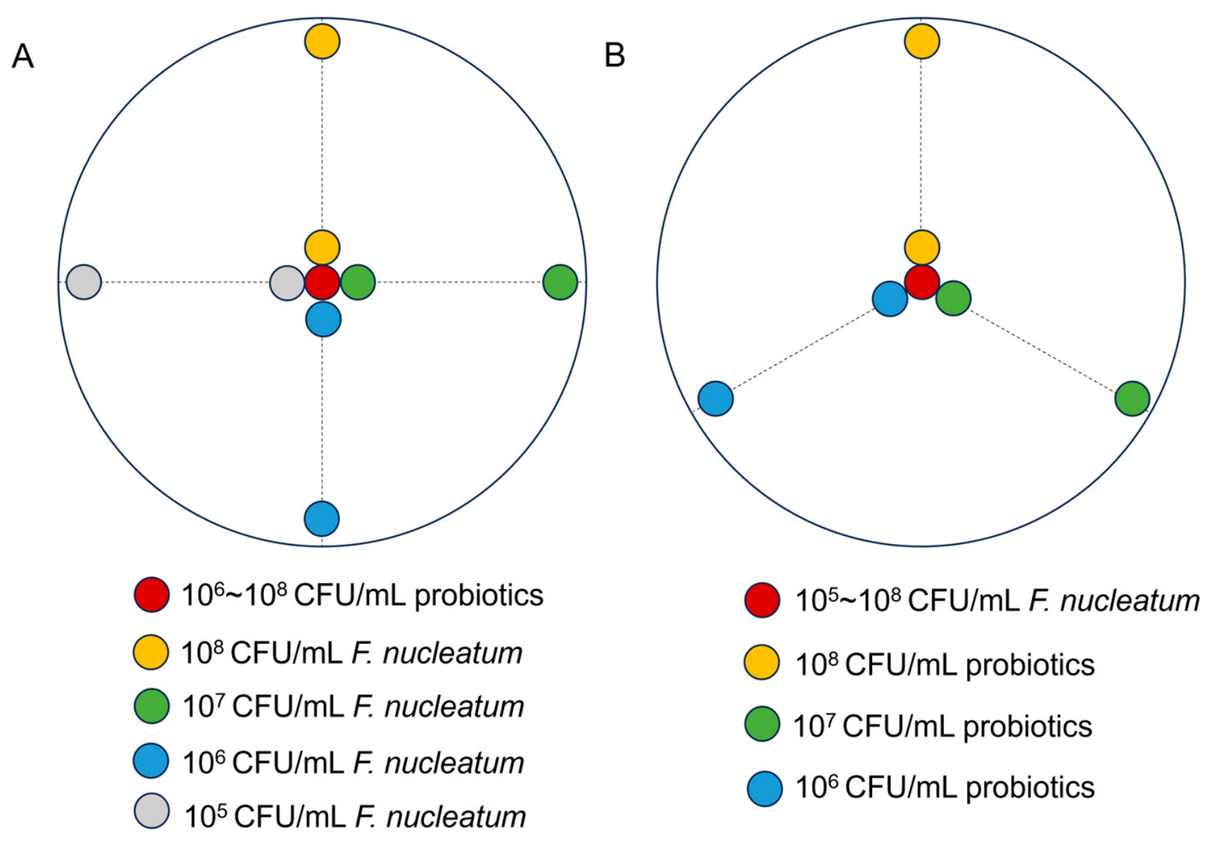
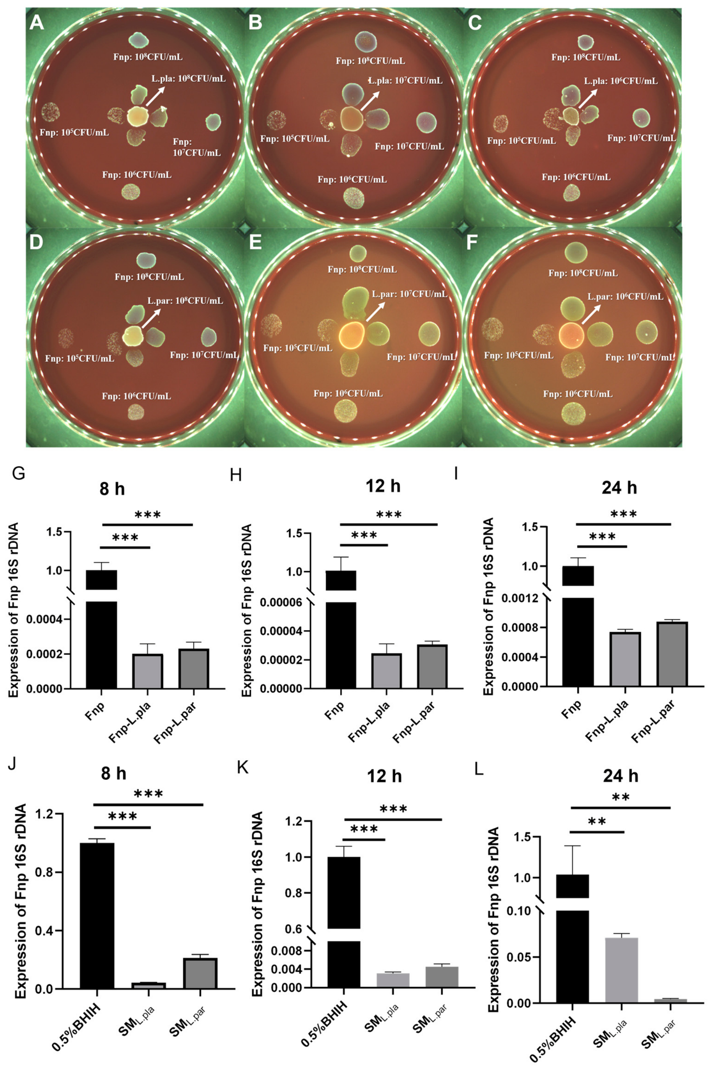
The mutual inhibitory effects between probiotics and F. nucleatum were determined using the plate confrontation test. Probiotics were first treated with or without ABX, as described in Section 2.3. After treatment, the bacterial suspensions were adjusted to different concentrations (106–108 CFU/mL), and 10 μL of each was dropped onto the center of BHI blood agar plates. Then, 10 μL of F. nucleatum at different concentrations (105–108 CFU/mL) was dropped onto the plate placed 40 mm away from the center. F. nucleatum at the same concentration was dropped onto the edge of the plate as the negative control (Figure 1A).
Conversely, in the reverse setup, F. nucleatum (105–108 CFU/mL) was spotted at the center, with probiotic droplets placed 40 mm away, and a corresponding negative control was included (Figure 1B).

2.6. RNA Extraction and qRT-PCR from Co-Culture

Co-culture concentrations were selected based on preceding assays to ensure clear inhibition while retaining sufficient bacterial yield. Accordingly, F. nucleatum (106 CFU/mL) was mixed with an equal volume of probiotics (108 CFU/mL) and incubated anaerobically at 37 °C for 24 h, with a F. nucleatum monoculture serving as the control. Total RNA was extracted using RNAzol® RT (MRC, Cincinnati, OH, USA) according to the manufacturer’s instructions, including lysozyme treatment, phase separation, and isopropanol precipitation. RNA was quantified using a NanoDrop 2000 spectrophotometer (NanoDrop Technologies, Wilmington, DE, USA). Reverse transcription was performed with PrimeScript™ RT Master Mix (TAKARA, Kusatsu, Shiga, Japan), followed by quantitative PCR using SYBR® Premix Ex Taq™ II (TAKARA) on a LightCycler 96 system (Roche, Basel, Switzerland) to obtain cDNA. The 2−ΔΔCt method was used to quantify fold changes relative to the F. nucleatum 16S rRNA gene. The 16S rRNA transcript level reflects the metabolic activity of F. nucleatum, as rRNA content is positively correlated with bacterial growth rate and ribosome synthesis activity [26,27,28]. Under standardized culture conditions, 16S rRNA-targeted RT-qPCR has been shown to correlate well with CFU counts [29,30]; therefore, in our controlled in vitro system, the RT-qPCR signal is interpreted as representing both physiological activity and, to a meaningful extent, relative bacterial abundance. The primer sequences were F-CTTAGGAATGAGACAGAGATG and R-TGATGGTAACATACGAAAGG.
The 16S rRNA transcript level reflects the metabolic activity of F. nucleatum, as rRNA content is positively correlated with bacterial growth rate and ribosome synthesis activity.

2.7. Metagenomic Sequencing and Analysis

Total DNA was extracted from co-culture and control samples using the TIANamp Bacteria DNA Kit (TIANGEN, Beijing, China), according to the manufacturer’s instructions. and then subjected to draft-genome sequencing on DNBSEQ-G400. Following the removal of low-quality sequences and adapters, the reads were de novo assembled using the MEGAHIT software v0.1-beta [31]. Assembled contigs with a length less than 300 bp were discarded in the following analysis. Genes were predicted over contigs using MetaGeneMark v3.38 [32]. Redundant genes were removed using CD-HIT v4.8.1 [33] based on identity and coverage cutoffs of 95% and 90%, respectively. To construct a gene abundance matrix, the Salmon v1.6.0 [34] software was used for quantification. Differentially enriched KEGG pathways were identified according to reporter scores. Alpha diversity was quantified by means of the Shannon index, chao1 index, and Simpson index using the relative abundance profiles at the gene, genus, and KO levels with the R package, respectively. Beta diversity was calculated using the Bray–Curtis distance based on the species-level relative abundance matrix. Principal coordinate analysis (PCoA) was performed for visualization. Raw data were deposited in the Sequence Read Archive (SRA) under Bioproject accession numbers PRJNA1415463 and PRJNA1415878.
Detailed metagenomic analysis parameters: Sequencing depth averaged 10 GB per sample. Three biological replicates were used per group (IP, Fnp-IP, Fnp-IP_hMIC, Fnp-IP_MIC). Assembly was performed using MEGAHIT v1.2.9. Gene prediction was conducted using MetaGeneMark v3.38. Redundant genes were removed using CD-HIT v4.8.1 with identity and coverage thresholds of 95% and 90%, respectively. Gene abundance was normalized using FPKM (Fragments per Kilobase per Million). For taxonomic classification, Kraken2 v2.1.2 was used with a custom database comprising NCBI NT (k2_pluspf_16gb_20240112) and UHGG v2.0; species abundance was estimated using Bracken2 v2.6.2. Alpha diversity and beta diversity were calculated using a species-level relative abundance matrix. For KEGG pathway enrichment, reporter scores were calculated according to the method of Patil and Nielsen [35]. Briefly, KO-level differential abundances were integrated into pathway-level Z-scores, which were then corrected by permutation (1000 times) to obtain a standard normal distribution; a reporter score with |Z| > 1.65 was considered significant (p < 0.05, one-tailed).

2.8. RNA Sequencing Transcriptome

RNA sequencing was conducted using the DNBSEQ platform (PE150 read length, average sequencing depth 4.36 Gb per sample). SOAPnuke (v1.6.5) was applied to filter reads and obtain clean reads; reads with adapters, more than 1% unknown bases, and low quality were removed. The clean reads were mapped to the Fusobacterium polymorphum ATCC 10953 reference genome GCF_037900625.1_ASM3790062v1 using HISAT2 V2.0.4. Gene expression levels were calculated using Bowtie2 V2.4.5 and RSEM V1.3.1. The library preparation strategy is illustrated in Figure S1. Briefly, total RNA was subjected to rRNA depletion using DNA probes that hybridize to rRNA, followed by RNase H digestion and DNase I treatment. After fragmentation, random N6 primers were used for reverse transcription, followed by second-strand cDNA synthesis. The double-stranded cDNA was end-repaired, A-tailed, and ligated to adapters. After PCR amplification, the product was denatured and circularized to obtain a single-stranded circular DNA library. The libraries were stranded. RNA was extracted from F. nucleatum monocultures after 24 h treatment with the probiotic supernatant or control medium. No probiotics were present during RNA extraction; thus, all sequenced reads originated from F. nucleatum. Three biological replicates were used per condition (Fn, YSJ, YSJ_MIC, YSJ_hMIC). All 12 samples passed read-level filtering (removal of adapters, low-quality reads, and reads with >1% N bases); no sample was excluded based on total reads, mapping rate, or correlation. Stringent criteria, including Log2FC > 1 and a false discovery rate (FDR < 0.001), were applied to filter differentially expressed genes (DEGs).
Data mining and figure presentation, including KEGG classification and GO classification analysis, were performed using the BGI in-house customized data mining system, Dr. Tom (https://biosys.bgi.com/). RNA sequencing data supporting the findings of this study were deposited in the NCBI SRA under accession number PRJNA1415531. qRT-PCR was used to validate changes in mRNA expression of DEGs. Gene-specific primer sequences are listed in Table S1.

2.9. Hydrogen Peroxide Content Determination and Depletion Experiment

The CFS of the probiotics was collected, and the content of hydrogen peroxide (H2O2) was determined using the hydrogen peroxide content detection kit, according to the manufacturer’s recommendations (Solabrio, Beijing, China).
The H2O2 in the probiotic CFS was depleted by 10 μg/mL catalase (Solarbio, Beijing, China). We added 300 μL of 109 CFU/mL F. nucleatum to 3 mL of the probiotic CFS with or without depletion of H2O2. We then prepared 0.5% BHIH_ H2O2 medium based on the H2O2 concentration in the CFS, and added catalase to ensure complete H2O2 depletion. 0.5% BHIH_ H2O2 medium with or without hydrogen peroxide depletion was used as the negative control group.
The bacterial suspensions were cultured at 37 °C anaerobically for 8 h, 12 h, and 24 h, and the optical densities were measured at a wavelength of 600 nm using a microplate reader (Biotek, Winooski, VT, USA).

2.10. Effect of pH of Probiotic CFS on F. nucleatum

The pH of the probiotic CFS was determined, and was then adjusted to 7.45 by adding K2HPO4. The CFS with or without pH adjustment was mixed with BHIH medium at a 1:9 ratio. We added 300 μL of 109 CFU/mL F. nucleatum to 3 mL of the probiotic CFS with or without pH adjustment. BHIH medium (adjusted to pH 3.45 or unadjusted) was used as the negative control.
The bacterial suspensions were cultured at 37 °C anaerobically for 8 h, 12 h, and 24 h, and the optical densities were measured at a wavelength of 600 nm using a microplate reader (Biotek, Winooski, VT, USA).

2.11. Lysine Content Determination and Supplementation Experiment

The CFS of the probiotics treated with or without ABXs and the CFS of F. nucleatum cultured with or without probiotics were collected, and the content of lysine was determined by using a lysine content detection kit, according to the manufacturer’s recommendations (Aidisheng, Yancheng, China). Based on the lysine concentration in the probiotic suspension, L-lysine was used to supplement the lysine concentration of the ABX-treated probiotic suspension to 11 mg/mL. Based on the lysine concentration in the co-culture suspension of probiotics and F. nucleatum, the lysine concentration in the F. nucleatum and ABX-treated probiotic suspension was supplemented to 15 mg/mL. The bacterial suspensions were cultured at 37 °C anaerobically for 24 h. Then, the number of probiotics with or without ABX treatment was confirmed by plate counting, and the metabolic activity of F. nucleatum was validated via qRT-PCR.

2.12. Establishment of Coaggregation Model

Coaggregation buffer (CAB) (150 mM NaCl, 1 mM Tris, pH 8.0, 0.1 mM CaCl2, 0.1 mM MgCl2) was prepared as previously described [36]. The intergeneric coaggregation model of probiotics and F. nucleatum was established as previously described [37,38,39]. Briefly, probiotics and F. nucleatum in the late logarithmic growth phase were washed twice and resuspended in CAB, adjusted to a final concentration of 109 CFU/mL. The probiotics and F. nucleatum were allowed to coaggregate by mixing equal suspensions of bacterial cells in a reaction tube, which was then vortexed for 20 s and left undisturbed anaerobically at room temperature for 10 min. After incubation, coaggregated probiotics and F. nucleatum were pelleted via low-speed (200× g) centrifugation for 1 min, while non-coaggregated bacteria were dispersed in the supernatant, which was collected carefully and measured for OD600nm (ODIP-Fn).
The degree of coaggregation was evaluated using the coaggregation index (CI), calculated according to the following formula: C I = O D I P + O D F n O D I P F n O D I P + O D F n × 100 % [40,41]. ODIP and ODFn represent the optical density of the probiotics and F. nucleatum at 600 nm, respectively.

2.13. Statistical Analysis

Statistical analysis and graphing were performed using Graphpad Prism 9.5.0. t-tests were used for inter-group comparisons, with p < 0.05 considered to indicate significant differences. Wilcoxon/Kruskal and KEGG pathway enrichment analyses (report score method) were performed to mine differences in species composition and functional composition between metagenomic samples. One-way analysis of variance (one-way ANOVA) was used for multi-group comparisons; if significant (p < 0.05), Dunnett’s correction and Tukey’s HSD were used for multiple comparisons. Dunnett’s correction and Tukey’s HSD were used to adjust p-values to Q-values, with Q-value < 0.05 considered to indicate significant differences.

3. Results

3.1. F. nucleatum Significantly Enriched Metabolic Pathways in the Probiotic Consortium

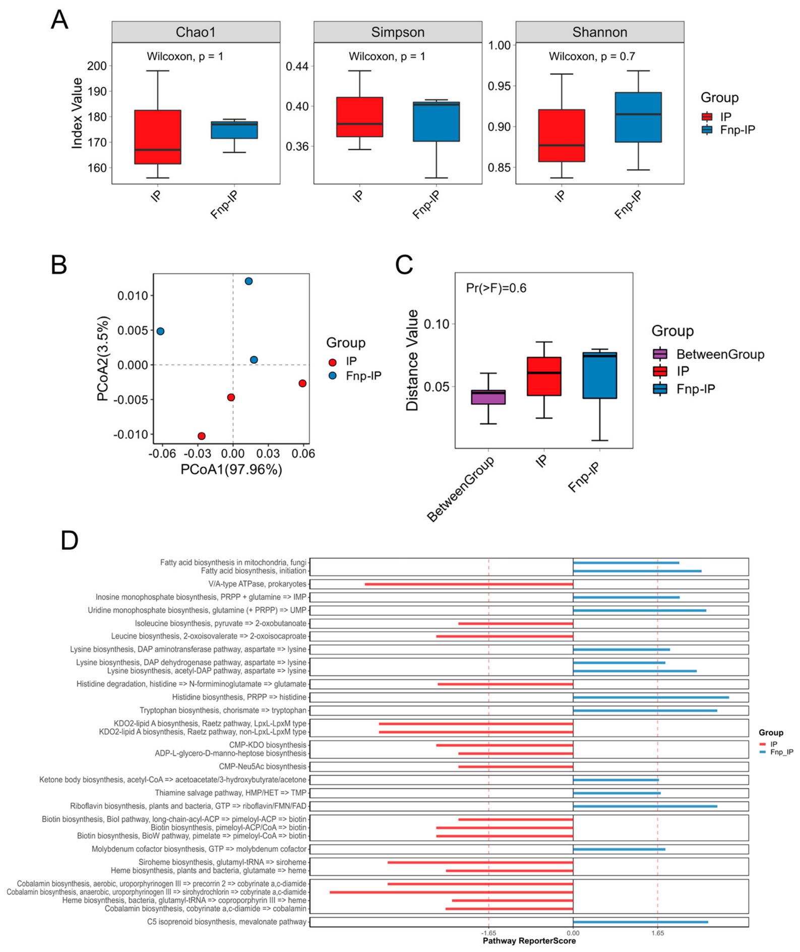
Metagenomic sequencing analysis was performed after co-culturing F. nucleatum with probiotics. Alpha diversity analysis showed (Figure 2A) no significant differences in species richness and evenness between the co-culture group (Fnp-IP) and the solo probiotic group (IP) (p > 0.05). Beta diversity was visualized via PCoA based on the Bray–Curtis distance. PERMANOVA (999 permutations) did not detect a statistically significant difference in overall community composition between the IP and Fnp-IP groups (p = 0.6, R2 = 0.10; Figure 2B,C).
KEGG pathway enrichment analysis showed (Figure 2D) that significantly enriched pathways in the Fnp-IP group included histidine biosynthesis, tryptophan biosynthesis, riboflavin biosynthesis, C5 isoprenoid biosynthesis, mevalonate pathway, fatty acid biosynthesis, and lysine biosynthesis, while enriched pathways in the IP group included isoleucine biosynthesis, biotin biosynthesis, cobalamin biosynthesis, siroheme biosynthesis, leucine biosynthesis, and histidine degradation.

3.2. Probiotics Significantly Inhibit the Growth of F. nucleatum, While F. nucleatum Does Not Obviously Affect Probiotic Growth

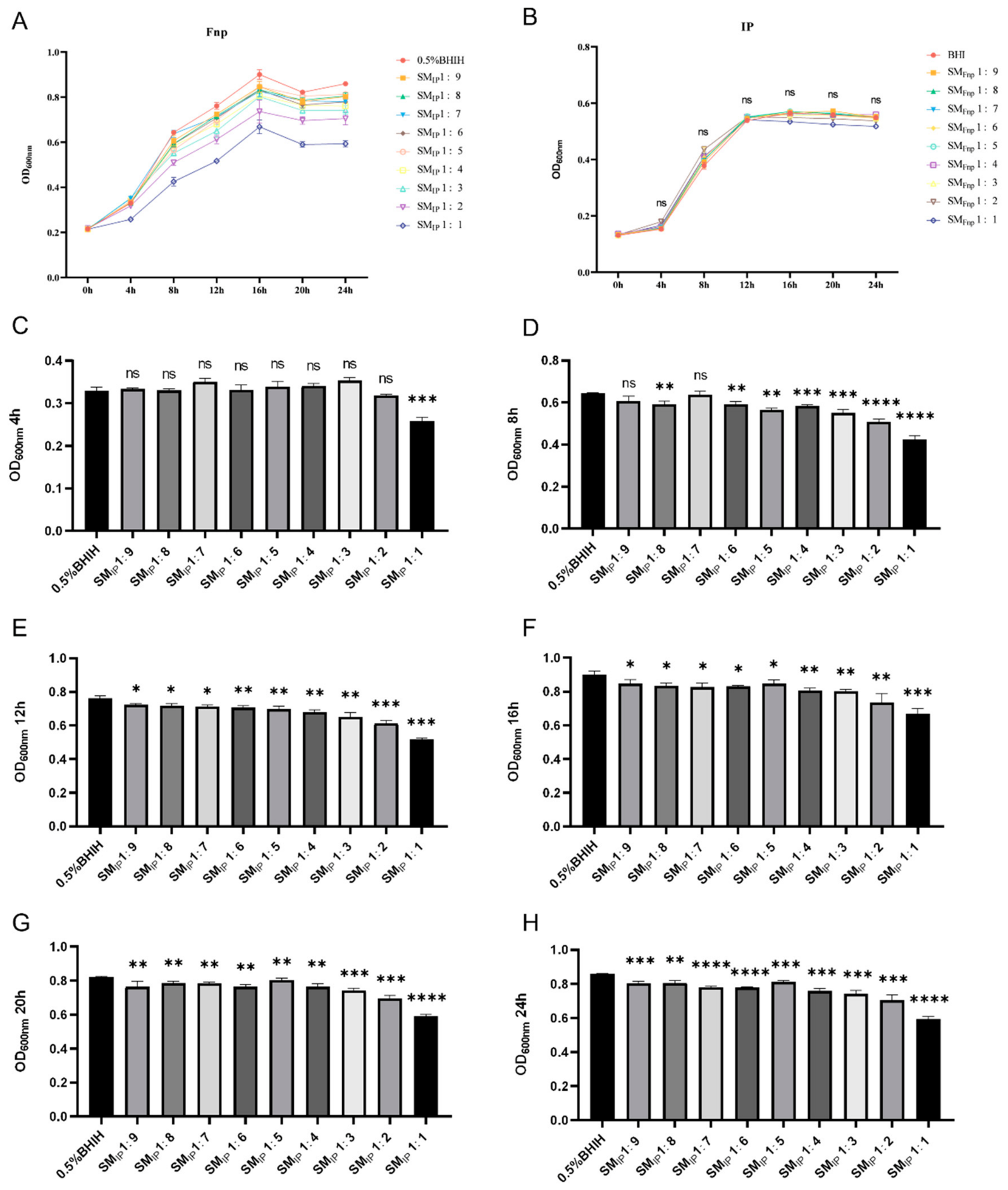
The growth curve results show that probiotics and F. nucleatum entered the stationary phase at the 12th and 16th hours of culture, respectively (Figure 3A,B). The spent medium from probiotics significantly reduced the bacterial concentration of F. nucleatum in the stationary phase, and the inhibitory effect was dose-dependent, but did not affect the time taken to enter the stationary phase (Figure 3A,C–H). In contrast, the spent medium of F. nucleatum had no significant effect on the growth of probiotics (Figure 3B).
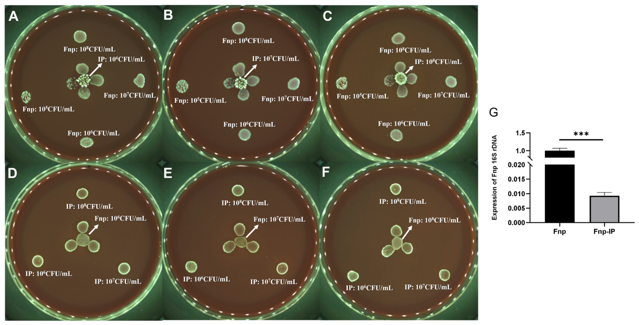
The plate confrontation experiment further confirmed that probiotics had a concentration-dependent inhibitory effect on the growth of F. nucleatum (Figure 4A–C), while F. nucleatum had no significant inhibition on probiotics (Figure 4D–F). qRT-PCR detection of F. nucleatum 16S rRNA gene expression in the co-culture system showed that its expression level in the Fnp-IP group was significantly lower than in the monoculture group (Figure 4G), suggesting that although F. nucleatum can integrate into the probiotic community and carry out metabolic activities, its bacterial count is significantly reduced, and its physiological activity is significantly inhibited.

3.3. Probiotic Metabolites Significantly Regulate the Gene Transcription Levels of F. nucleatum

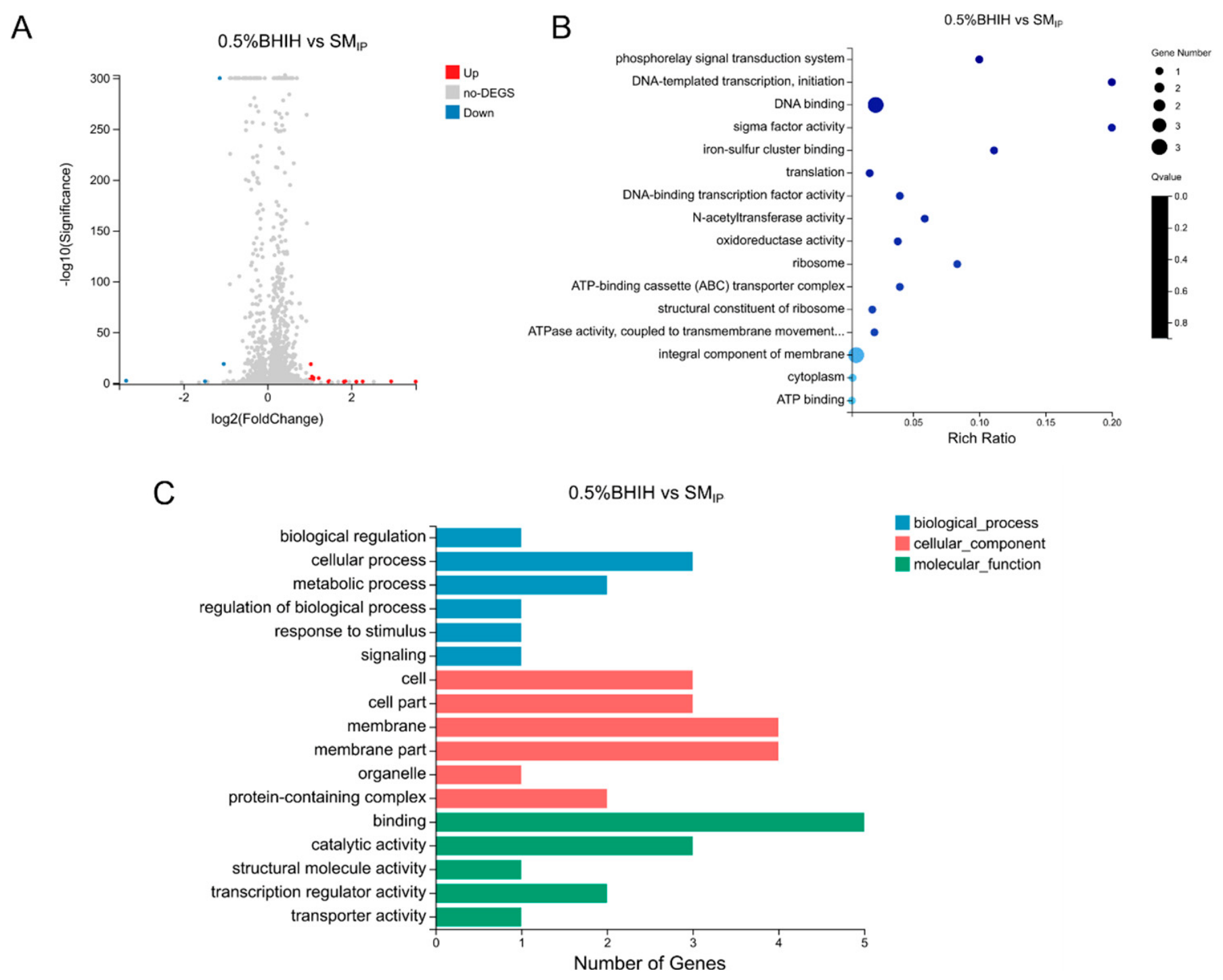
After treatment with probiotic supernatant, a total of eight genes in F. nucleatum showed significant expression changes, with six upregulated (including ABC transporter system ATP-binding protein, protoporphyrinogen III oxidase, GNAT family protein, LytTR family protein, and two genes of unknown function) and two downregulated (50S ribosomal protein L34 and 1 gene of unknown function) (Figure 5A). GO functional enrichment analysis showed that differentially expressed genes were mainly enriched in phosphorelay signal transduction system and DNA-templated transcription and initiation-related gene sets (Figure 5B,C). KEGG pathway classification indicated that these genes were mainly concentrated in the translation pathway.

3.4. Antibiotics Weaken the Inhibitory Effect of Probiotics on F. nucleatum and Affect Transcriptional Regulation

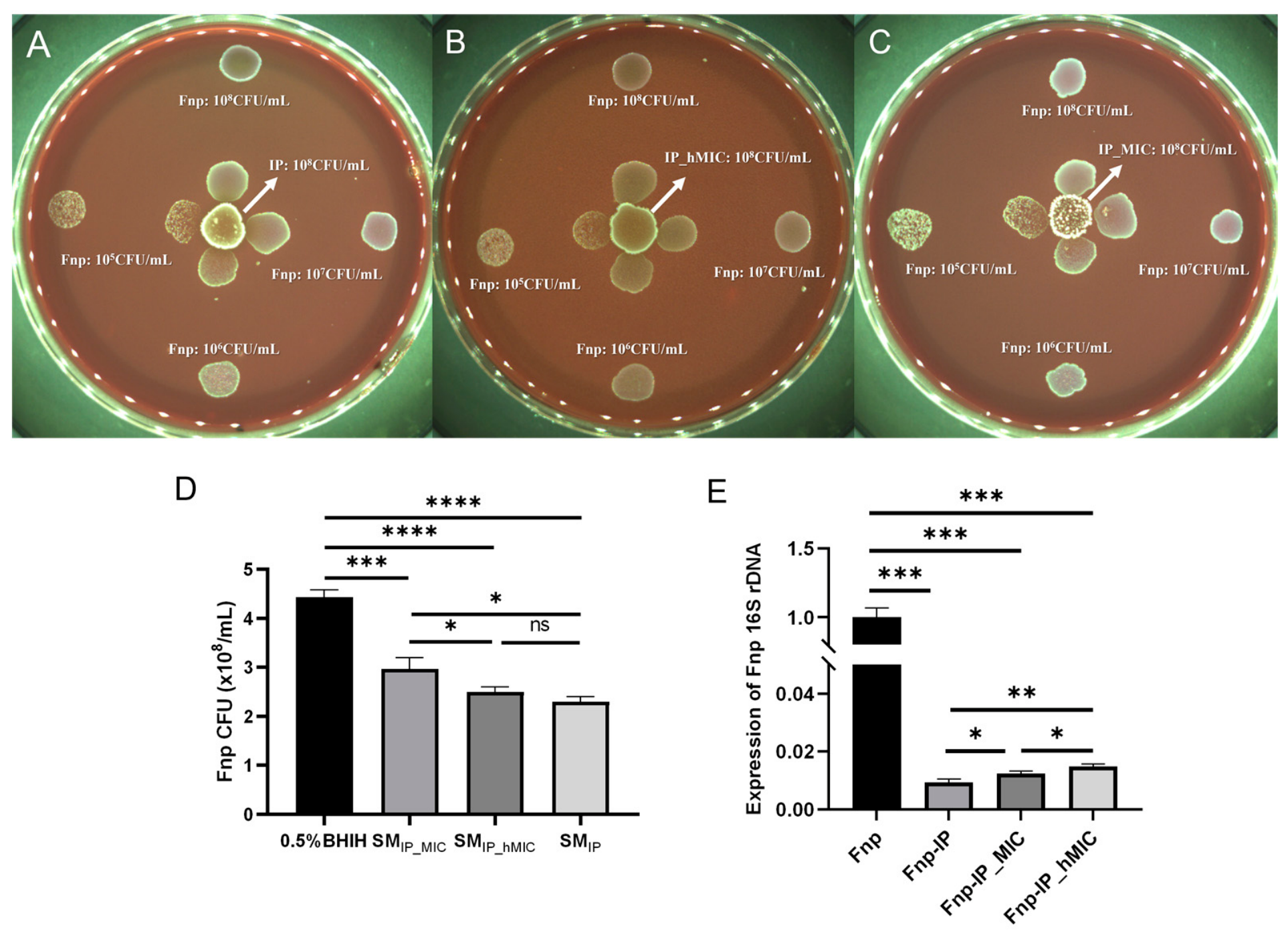
We found that the MIC of ABX against probiotics was 0.78 μg/mL and the hMIC was 0.39 μg/mL, while the MIC against F. nucleatum was below 0.195 μg/mL. Both plate confrontation experiments and spent medium experiments indicated that, after ABX treatment, the inhibitory effect of probiotics on F. nucleatum growth was weakened (Figure 6A,B). The qRT-PCR results further showed that, after ABX treatment, 16S rRNA gene expression of F. nucleatum was upregulated in the co-culture system (Figure 6C), indicating that antibiotics weakened the antagonistic effect of probiotics.
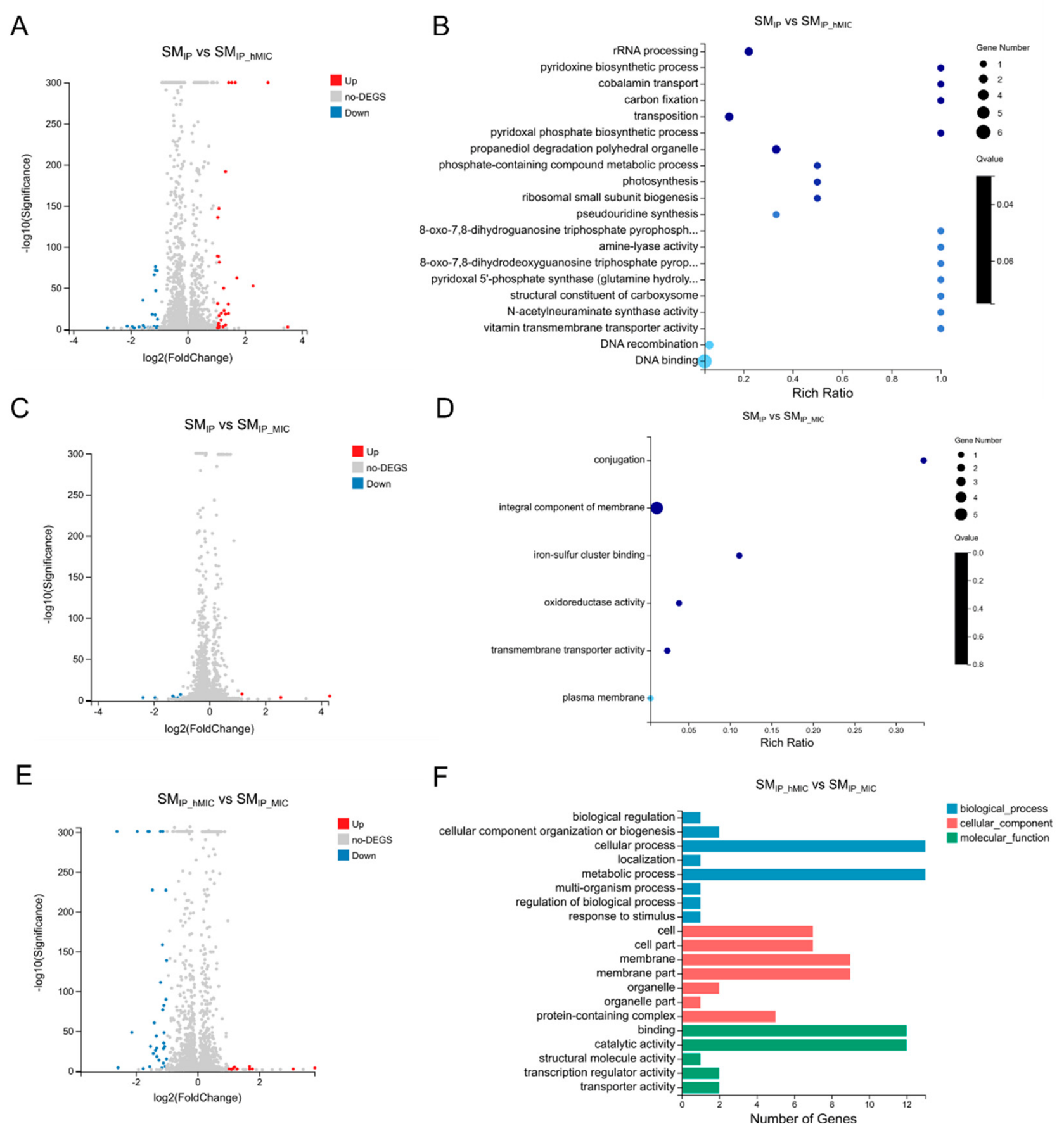
Transcriptomic analysis revealed that the probiotic supernatant treated with ABX at hMIC (SMIP-hMIC) induced more gene expression changes in F. nucleatum; in particular, compared to the SMIP group, nucleic acid metabolism, nucleotide metabolism, and propanediol degradation-related genes were upregulated 2.0–2.4-fold, acetylation-related genes were upregulated 2.6-fold, the pyridoxine synthesis gene pdxS was upregulated 6.8-fold, and transmembrane transport-related genes were downregulated 2.0–3.0-fold (Figure 7A). GO enrichment analysis suggested that DEGs were involved in multiple processes such as propanediol degradation polyhedral organelle, rRNA processing, and pyridoxine biosynthetic process (Figure 7B and Figure S2). KEGG analysis showed that these genes were significantly enriched in the sulfur metabolism pathway (Figure S3).
Compared to the SMIP group, the probiotic supernatant treated with ABXs at MIC (SMIP-MIC) induced the upregulation of five genes and the downregulation of three genes in F. nucleatum (Figure 7C). GO enrichment analysis suggested that DEGs were involved in the integral component of membrane and iron–sulfur cluster binding (Figure 7D and Figure S4).
Compared to the SMIP-hMIC group, SMIP-MIC upregulated 2 genes and downregulated 31 genes in F. nucleatum (Figure 7E). GO functional classification analysis suggested that DEGs were involved in multiple processes, such as biological process, cellular process, and metabolic process (Figure 7F). KEGG functional classification suggested that these genes were involved in pathways such as cellular community–prokaryotes, membrane transport, metabolism, energy metabolism, and the metabolism of cofactors and vitamins (Figure S5).

3.5. ABX Alters the Community Composition of Probiotics in the Co-Culture System, with Significant Changes in the Abundance of L. plantarum and L. paracasei

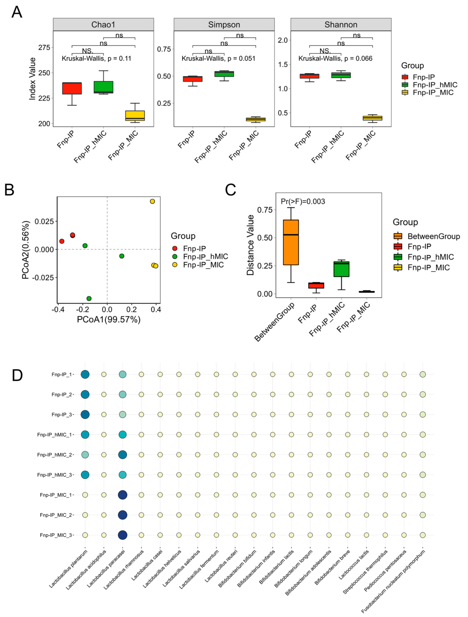
Alpha diversity analysis showed no significant changes in species richness and evenness in the Fnp-IP_hMIC and Fnp-IP_MIC groups compared to the Fnp-IP group (Figure 8A). Beta diversity was visualized by PCoA based on the Bray–Curtis distance. PERMANOVA (999 permutations) confirmed a significant difference in overall community composition among the three groups (Figure 8B,C; p = 0.003, R2 = 0.92), indicating that ABX treatment significantly altered the community structure of the probiotic consortium.
The species abundance bubble plot shows that the species with the most significant changes after ABX treatment were L. plantarum and L. paracasei: the abundance of L. plantarum decreased, while that of L. paracasei increased (Figure 8D). MIC determination further confirmed that L. plantarum was more sensitive to ABX (MIC = 0.49 μg/mL), while L. paracasei was relatively resistant (MIC = 1.95 μg/mL).

3.6. L. plantarum and L. paracasei Cells and Their Metabolites Both Inhibit F. nucleatum Growth

The growth curves showed that L. plantarum and L. paracasei both entered the stationary phase at the 21st hour of culture (Figure S6). Plate confrontation experiments indicated that both exerted concentration-dependent inhibitory effects on F. nucleatum (Figure 9A–F). The qRT-PCR results showed that, in separate co-cultures with these two lactobacilli, 16S rRNA gene expression of F. nucleatum was significantly lower than that in the monoculture group (Figure 9G–I). In addition, their CFSs also significantly inhibited the physiological activity of F. nucleatum (Figure 9J–L).
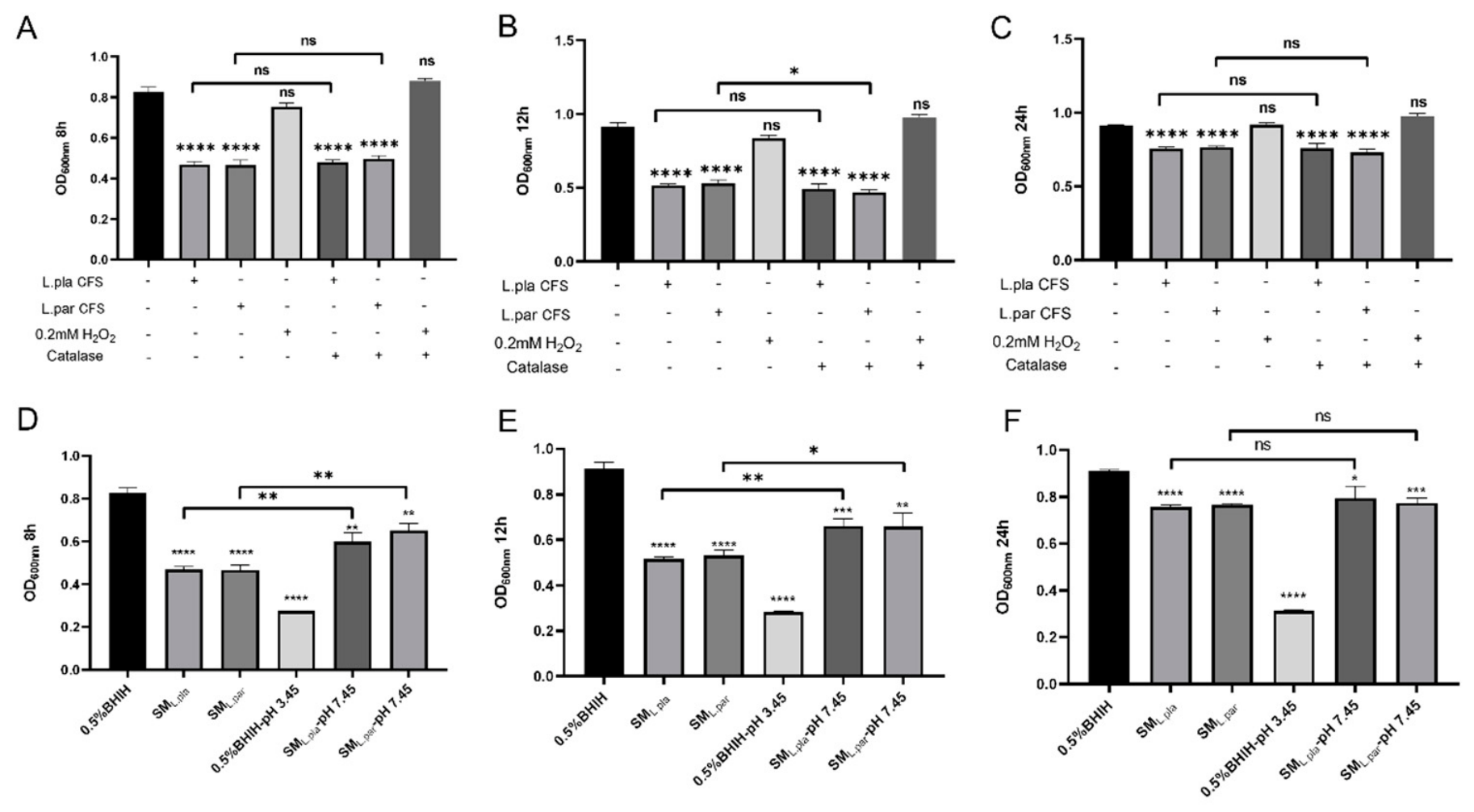
To explore their antibacterial substances, we tested the effects of hydrogen peroxide and organic acids in the supernatant. After hydrogen peroxide was depleted, the antibacterial activity of the supernatant did not change significantly (Figure 10A–C), indicating that hydrogen peroxide was not the main antibacterial component. However, after adjusting the supernatant pH from 3.45 to 7.45, its inhibitory effect was significantly weakened in the early culture stages (8 h, 12 h); at 24 h, even after pH neutralization, the final bacterial concentration of F. nucleatum was still significantly lower than that of the control group (Figure 10D–F). This indicates that organic acids in the supernatant may inhibit F. nucleatum proliferation in the early growth phase and delay its entry into the stationary phase or reduce the bacterial concentration in the stationary phase.

3.7. Lysine Restores the Abundance of ABX-Treated Probiotics and Interferes with Coaggregation Between Probiotics and F. nucleatum

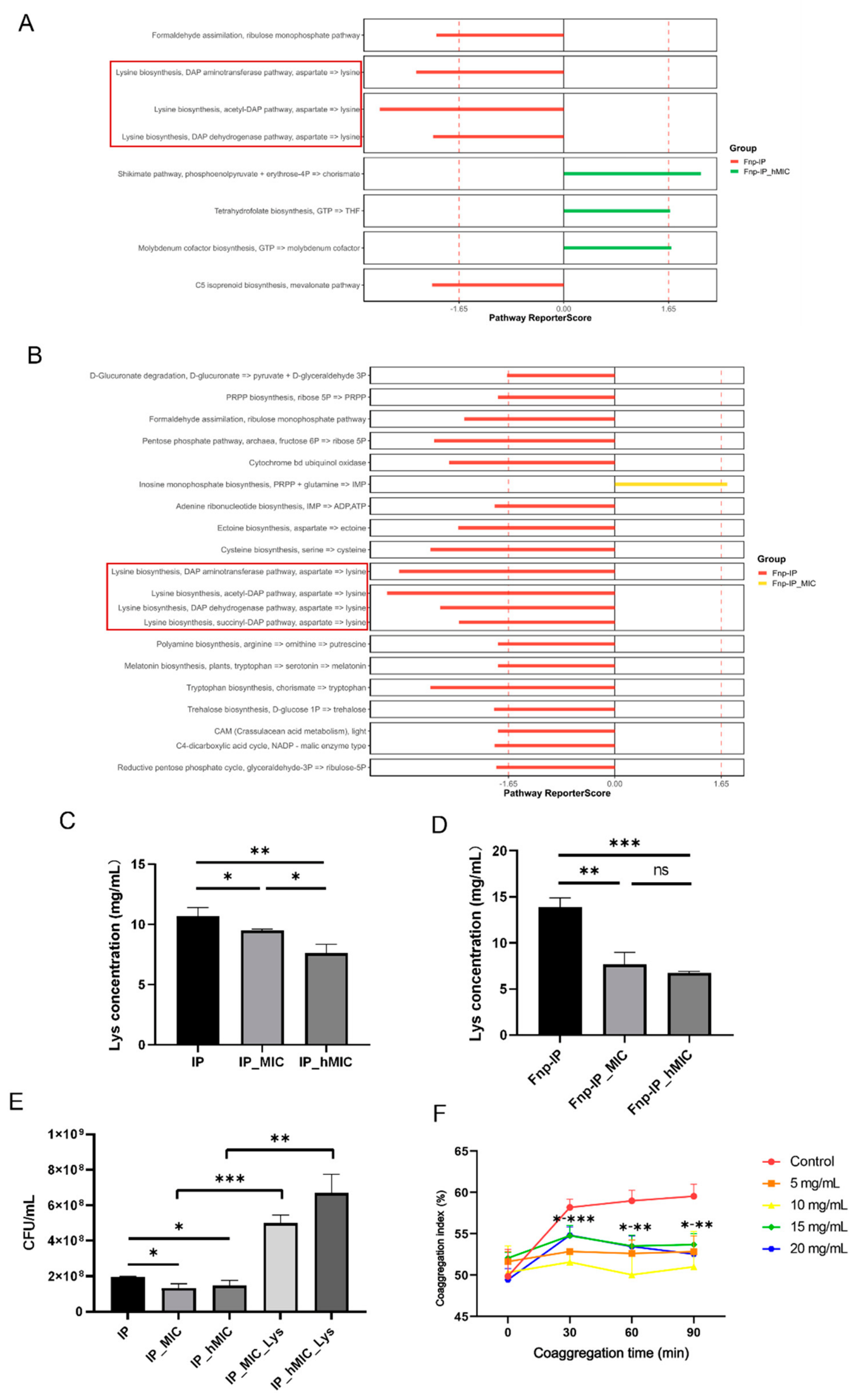
Metagenomic analysis showed that the lysine biosynthesis pathway was significantly enriched in the co-culture of probiotics and F. nucleatum, while the enrichment level of this pathway decreased after ABX treatment (Figure 11A,B). Experiments confirmed that the lysine concentration in the supernatant of ABX-treated probiotics was significantly reduced (Figure 11C,D), and the probiotic quantity also decreased; after lysine supplementation, the probiotic quantity was restored (Figure 11E).
Further measurements of the coaggregation index found that as lysine concentration increased, the coaggregation ability of F. nucleatum with probiotics significantly decreased (Figure 11F). Based on these findings, it is plausible that lysine alleviates the inhibitory effect of ABXs on probiotics and promotes colonization resistance, potentially through interference with interbacterial co-aggregation; however, further studies are required to confirm this mechanism.

4. Discussion

This study explores the interactions between F. nucleatum and intestinal probiotics and their potential significance in the occurrence and development of CRC. Through in vitro co-culture systems, we observed the impact of F. nucleatum on the composition and function of the probiotics and further investigated the inhibitory effects of these probiotics and their metabolites on the growth of F. nucleatum and the associated mechanisms. In addition, we examined the effects of antibiotic intervention on probiotic functions and the interactions between F. nucleatum and probiotics.
We found that, when F. nucleatum is co-cultured with probiotics, although it does not significantly inhibit the overall growth of the probiotics, it can alter their metabolic pathways. Mechanistic analysis through metagenomic sequencing revealed that, after co-culture of F. nucleatum with probiotics, pathways such as isoleucine, leucine biosynthesis, and histidine degradation in the microbial community were significantly downregulated. Beta diversity analysis (PCoA based on Bray–Curtis distance) did not detect a statistically significant shift in the overall community composition under our experimental conditions. This may be due to the limited sample size, which reduces statistical power to detect moderate changes, or to the fact that only a subset of species changed in abundance, and the changes in different species may have occurred in opposite directions, offsetting each other in the overall distance calculation. Nevertheless, previous studies have reported that, in mouse models colonized by F. nucleatum, the intestinal microbial composition was altered and related pathways such as amino acid metabolism and vitamin synthesis were downregulated without complete stagnation of microbial growth [42]. Supplementation with probiotics such as L. plantarum can restore homeostasis through negative regulation and promote the generation of beneficial metabolites [42]. These results suggest that F. nucleatum may primarily alter the probiotic community by inducing metabolic reprogramming rather than direct toxic killing.
In the metagenomic sequencing experiment, KEGG pathway enrichment analysis revealed that riboflavin synthesis and fatty acid synthesis were significantly enriched in the F. nucleatum–probiotic co-culture system. The literature indicates that probiotics can produce vitamin B2 through the riboflavin synthesis pathway, which can act as a signaling molecule involved in bacterial quorum sensing and host–bacteria interactions, improving the intestinal redox state and promoting anaerobic probiotic growth, thereby indirectly inhibiting the colonization of pathogens such as F. nucleatum [43,44]. In addition, short-chain fatty acids (such as butyrate) produced by probiotics through the fatty acid synthesis pathway can directly inhibit the growth of F. nucleatum, restore intestinal butyrate balance, enhance barrier function, and overcome chemotherapy resistance [45].
This study further found that the probiotic supernatant can significantly inhibit the growth of F. nucleatum and regulate the transcription levels of multiple genes. Among them, ABC transporter system ATP-binding protein, protoporphyrinogen III oxidase, and LytTR family protein gene expression were upregulated, while 50S ribosomal protein L34 gene expression was downregulated. Reports have indicated that, under acid–base stress, ABC transporter expression in F. nucleatum can be upregulated 2–3-fold to maintain ion and nutrient uptake and osmotic pressure balance [46]. Protoporphyrinogen III oxidase is a key enzyme in anaerobic heme synthesis, and its upregulation helps to cope with iron/oxidative stress, enhancing heme acquisition to support respiratory chain function [47]; the LytTR family protein also participates in regulating iron acquisition [48]. These changes suggest that the probiotic supernatant may force F. nucleatum to activate the anaerobic heme synthesis pathway in order to maintain redox homeostasis through mechanisms such as iron chelation. The downregulation of ribosomal protein L34 expression may reduce ribosome assembly, conserve energy, and regulate nucleocytoplasmic translocation [49], indicating that under the pressure of probiotic metabolites, F. nucleatum may slow protein synthesis and shift to a defensive state.
Antibiotic treatment may lead to gut microbiota dysbiosis and induce resistance gene production, thereby increasing the risk of CRC. Therefore, this study further explored the impacts of antibiotic treatment on probiotic inhibition against F. nucleatum. For this purpose, a broad-spectrum antibiotic “cocktail” combination, known to reduce intestinal microbial diversity and promote CRC development, was selected [50]. In clinical antibiotic therapy, blood concentrations often fluctuate, with prolonged periods at sub-MIC or near-MIC levels. At full MIC, probiotics may be partially killed, preventing effective supernatant production or observation of subsequent interactions. In contrast, hMIC allows for probiotic survival and the secretion of adaptive metabolites. Therefore, this study employed both MIC and hMIC concentrations to investigate the mechanisms of “residual resistance” and to simulate probiotic functional decline under gut microbiota dysbiosis after antibiotic treatment [51]. We found that, after 24 h of ABX treatment (MIC or hMIC), the direct and indirect inhibitory effects of probiotics on F. nucleatum were significantly weakened, indicating that antibiotics may disrupt the niche resistance of probiotics, creating an advantage for F. nucleatum growth.
Antibiotics also affected the transcriptional regulation of probiotic supernatant on F. nucleatum. In this study, we found that compared to the SMIP group, the SMIP-hMIC group led to significant upregulation of nucleotide metabolism, propanediol degradation, acetylation-related genes, and the pyridoxine synthesis gene pdxS in F. nucleatum. Studies have indicated that, under stress conditions such as hypoxia, nucleotide metabolism-related gene expression in F. nucleatum can be upregulated about 2-fold, helping to enhance its persistence in adverse environments [52]. In inflammatory intestinal environments, F. nucleatum can integrate symbiotic bacterial metabolism by upregulating propanediol/glycolysis degradation pathways (2–2.5-fold), competing for resources and generating energy [53]. In the σ^E stress response, acetyltransferase genes are upregulated 2–3-fold, modulating virulence and resistance through protein modification to cope with envelope or nutrient stress [54,55]. At the same time, in global stress responses, σ^E-mediated pdxS-like vitamin synthesis gene expression can be significantly upregulated about 6-fold. These transcriptional changes reflect the adaptive response of F. nucleatum to the probiotic supernatant after ABX treatment, involving σ^E-mediated global stress responses to enhance nutrient acquisition, detoxification, and protein modification capabilities, while downregulating non-essential transport processes to maintain survival. This adaptive mode may further strengthen the colonization and persistence of F. nucleatum in the intestinal or tumor microenvironment under antibiotic-induced dysbiosis.
Compared to the SMIP-hMIC group, the SMIP-MIC group showed significant downregulation of GNAT family protein-related and MarR family protein-related genes in F. nucleatum. These two types of genes are both related to environmental stress responses, which suggests that as the antibiotic concentration increases, the stress pressure caused by probiotic metabolites on F. nucleatum may further decrease, leading to downregulation of the expression of corresponding stress genes in F. nucleatum.
Within the probiotic community, the species with the most significant changes after ABX treatment were L. plantarum and L. paracasei, with the former’s abundance decreasing and the latter’s increasing. Although our metagenomic analysis does not resolve individual strains, this does not compromise the subsequent single-strain validation experiments, which confirmed the differential ABX sensitivity of L. plantarum and L. paracasei. This may stem from L. plantarum’s greater sensitivity to certain components in ABXs (such as β-lactams), while L. paracasei can gain a relative advantage in competition through resistance mechanisms such as membrane changes or metabolic adaptations [56,57]. These two lactobacilli, as important intestinal probiotics, can antagonize pathogens through multiple mechanisms, including niche competition, bacteriocin secretion, short-chain fatty acids, organic acids, and hydrogen peroxide [58].
We also found that the main active substances in the CFS of L. plantarum and L. paracasei inhibiting F. nucleatum are organic acids. The CFS of both probiotics can inhibit the proliferation of F. nucleatum in the early growth phase, prolong its lag phase, and reduce the bacterial concentration upon entering the stationary phase. The literature reports that organic acids in lactobacillus CFS can extend the lag phase of F. nucleatum by 2–6 h and inhibit its division rate, leading to a 20–60% decrease in stationary phase bacterial concentration [59,60]. The associated mechanism may involve organic acids diffusing into bacterial cells, depleting ATP, and inhibiting key enzyme activities such as glycolysis, rather than direct bactericidal action, thereby helping probiotics to gain an advantage in intestinal ecological competition [59,61].
F. nucleatum, an anaerobic bacterium capable of migrating from the oral cavity to the gastrointestinal tract, resists gastric acid (HCl, pH ≈ 1.5) primarily through membrane lipid remodeling, including the synthesis of erucic acid to enhance membrane stability and maintain cellular integrity [62]. Although resistant to this strong acid, it remains susceptible to probiotic-derived weak organic acids (e.g., lactic acid and SCFAs such as butyric, propionic, and acetic acids), which inhibit its growth via multiple pathways [63,64]. Unlike HCl, which fully dissociates into impermeable H+ and Cl ions, exerting mainly extracellular stress, weak organic acids (pKa ≈ 3.8–4.8) exist predominantly in their undissociated, lipophilic form (HA) at acidic extracellular pH. This neutral HA passively diffuses across the lipid bilayer without transporters or ion pumps [65]. Once inside the cell, HA dissociates, causing intracellular acidification, disruption of proton motive force, ATP depletion, and inhibition of glycolysis—explaining their superior inhibitory effect on F. nucleatum colonization compared to strong acids [65].
This study found that the lysine synthesis pathway was significantly enriched when probiotics were co-cultured with F. nucleatum, while the enrichment of this pathway decreased after ABX treatment. Studies have indicated that antibiotics often interfere with amino acid synthesis and uptake in probiotics, leading to lysine depletion (20–50% decrease), thereby inhibiting protein synthesis and growth; lysine supplementation can alleviate this effect by enhancing proton motive force or buffering pH, promoting probiotic abundance recovery [66,67,68]. In addition to the lysine pathway, other metabolic pathways (e.g., those related to nucleotide metabolism, stress responses, and vitamin synthesis) also showed altered enrichment after ABX treatment (Figure 11A,B), which warrant further investigation in future studies.
Lysine plays an important role in biological metabolism, growth, and development, and is an essential amino acid for maintaining overall health and normal physiological functions of the body [69]. Lysine acetylation is currently one of the most widespread forms of post-translational protein modification known in bacteria, which is involved in various regulatory processes such as bacterial transcription, translation, metabolism, and stress responses [70,71,72]. Cohort studies have shown that blood lysine levels are negatively associated with the risk of colorectal cancer [73]. Intestinal probiotics can convert lysine into butyrate while releasing ammonium ions, serving as an energy source for colon cells and helping to prevent inflammation and cancer development [74,75,76]. Meanwhile, lysine may interfere with its mediated interbacterial coaggregation by competitively binding to the adhesin RadD of F. nucleatum [77], thereby inhibiting F. nucleatum’s cross-niche colonization and integration into the gut microbiota. It has been reported that lysine can inhibit the expression and oligomerization of the hemolytic toxin Hla in Staphylococcus aureus (S. aureus), thereby reducing its virulence expression and colonization in food models (such as milk). In mouse and Caco-2 cell models, lysine alleviates the intestinal damage caused by S. aureus, demonstrating that it can prevent the colonization of pathogenic bacteria in food and the intestine by reducing bacterial attachment and virulence [78].
In periodontal disease patients with F. nucleatum overgrowth [79], the bacterium may translocate via the oral–gut axis to colonize the intestine, thereby promoting CRC initiation and progression [3,4]. Our findings demonstrate that lysine significantly inhibits F. nucleatum’s coaggregation with probiotics, likely by competitively binding to the adhesin RadD on the bacterial surface. This disruption impairs F. nucleatum’s ability to integrate into the gut microbial community, facilitating its clearance from the intestine rather than persistent colonization. Therefore, lysine supplementation or lysine-enriched probiotic strategies hold promising practical value as a non-antibiotic intervention to reduce F. nucleatum-mediated CRC risk in high-risk populations, such as those with chronic periodontitis.
Probiotics show potential for CRC prevention by inhibiting F. nucleatum colonization in the gut microbiota. Strains such as Bifidobacterium and Lactobacillus reduce the abundance of F. nucleatum through pH modulation, antimicrobial activity, and barrier formation, thereby attenuating CRC-associated inflammation and tumorigenesis [80,81]. This resistance to pathogenic colonization enhances microbial diversity and immune responses, offering a novel adjunct strategy for CRC prophylaxis [82]. Future clinical trials should validate these mechanisms in high-risk populations to optimize probiotic interventions.
We acknowledge that this study is in vitro and used a single F. nucleatum subspecies (subsp. polymorphum) and one antibiotic cocktail at fixed concentrations. The complexity of the 33-strain probiotic consortium means that our findings represent one plausible scenario among many possible ones. Different F. nucleatum subspecies (e.g., animalis, vincentii, watanabei), other antibiotic types, concentrations, or exposure times could yield different outcomes. Moreover, the metagenomic sample size was limited; thus, larger cohorts are needed to strengthen the conclusions. Additionally, the probiotic supernatant likely contains additional antimicrobial molecules (e.g., bacteriocins, reuterin) beyond those examined here; methods such as high-performance liquid chromatography (HPLC) would be required for full identification. Future in vivo studies using clinically relevant isolates and defined consortia are required to validate and extend our conclusions.

5. Conclusions

This study shows that probiotics and their metabolites inhibit the growth of F. nucleatum and modulate its stress- and metabolism-related genes. Antibiotic treatment weakens this inhibition and changes the stress response of F. nucleatum, which may enhance its adaptability in dysbiotic environments. Organic acids from L. plantarum and L. paracasei are key inhibitory effectors. Exogenous lysine restores antibiotic-reduced probiotic abundance and reduces coaggregation with F. nucleatum, suggesting a possible role in colonization resistance.
Together, these results suggest that probiotics might help to counteract F. nucleatum colonization via organic acids and metabolic interference, potentially contributing to gut homeostasis. However, antibiotic use could compromise this protective effect, which might favor the persistence of F. nucleatum and could thus be relevant to CRC risk. Further studies are needed to confirm these hypotheses in more complex systems.

Supplementary Materials

The following supporting information can be downloaded at: https://www.mdpi.com/article/10.3390/microorganisms14050965/s1. Figure S1: The library preparation strategy; Figure S2: Bubble plots of GO enrichment analysis (SMIP vs. SMIP_hMIC); Figure S3: Bubble plots of KEGG pathway enrichment analysis (SMIP vs. SMIP_hMIC); Figure S4: GO functional classification (SMIP vs. SMIP_MIC); Figure S5: KEGG functional classification analysis (SMIP_hMIC vs. SMIP_MIC); Figure S6: Growth curves of L. plantarum and L. paracasei in BHI. Table S1: Gene-specific primer sequences.

Author Contributions

W.H. and J.L. contributed equally to this work. Conceptualization, L.G.; data curation, W.H.; formal analysis, W.H. and L.G.; funding acquisition, L.G.; investigation, J.L., W.H. and P.C.; methodology, Z.L. and P.C.; project administration, L.G.; supervision, L.G. and Z.L.; visualization, J.L.; writing—original draft preparation, J.L.; writing—review and editing, L.G. and P.C. All authors have read and agreed to the published version of the manuscript.

Funding

This research was funded by the National Natural Science Foundation of China, grant number 81670982.

Institutional Review Board Statement

Not applicable.

Informed Consent Statement

Not applicable.

Data Availability Statement

The original contributions presented in this study are included in the article/Supplementary Material. Further inquiries can be directed to the corresponding authors. The raw data of the metagenomic sequencing were deposited in the Sequence Read Archive (SRA) under Bioproject accession numbers PRJNA1415463 and PRJNA1415878. The RNA sequencing data supporting the findings of this study were deposited in the NCBI SRA under accession number PRJNA1415531.

Conflicts of Interest

The authors declare no conflicts of interest.

References

  1. Bray, F.; Laversanne, M.; Sung, H.; Ferlay, J.; Siegel, R.L.; Soerjomataram, I.; Jemal, A. Global cancer statistics 2022: GLOBOCAN estimates of incidence and mortality worldwide for 36 cancers in 185 countries. CA Cancer J. Clin. 2024, 74, 229–263. [Google Scholar] [CrossRef]
  2. Brennan, C.A.; Garrett, W.S. Fusobacterium nucleatum—Symbiont, opportunist and oncobacterium. Nat. Rev. Microbiol. 2019, 17, 156–166. [Google Scholar] [CrossRef]
  3. Abed, J.; Maalouf, N.; Manson, A.L.; Earl, A.M.; Parhi, L.; Emgård, J.E.M.; Klutstein, M.; Tayeb, S.; Almogy, G.; Atlan, K.A.; et al. Colon Cancer-Associated Fusobacterium nucleatum May Originate from the Oral Cavity and Reach Colon Tumors via the Circulatory System. Front. Cell. Infect. Microbiol. 2020, 10, 400. [Google Scholar] [CrossRef]
  4. Rubinstein, M.R.; Baik, J.E.; Lagana, S.M.; Han, R.P.; Raab, W.J.; Sahoo, D.; Dalerba, P.; Wang, T.C.; Han, Y.W. Fusobacterium nucleatum promotes colorectal cancer by inducing Wnt/β-catenin modulator Annexin A1. EMBO Rep. 2019, 20, e47638. [Google Scholar] [CrossRef]
  5. Mima, K.; Nishihara, R.; Qian, Z.R.; Cao, Y.; Sukawa, Y.; Nowak, J.A.; Yang, J.; Dou, R.; Masugi, Y.; Song, M.; et al. Fusobacterium nucleatum in colorectal carcinoma tissue and patient prognosis. Gut 2016, 65, 1973–1980. [Google Scholar] [CrossRef]
  6. Hashemi Goradel, N.; Heidarzadeh, S.; Jahangiri, S.; Farhood, B.; Mortezaee, K.; Khanlarkhani, N.; Negahdari, B. Fusobacterium nucleatum and colorectal cancer: A mechanistic overview. J. Cell. Physiol. 2019, 234, 2337–2344. [Google Scholar] [CrossRef] [PubMed]
  7. Gethings-Behncke, C.; Coleman, H.G.; Jordao, H.W.T.; Longley, D.B.; Crawford, N.; Murray, L.J.; Kunzmann, A.T. Fusobacterium nucleatum in the Colorectum and Its Association with Cancer Risk and Survival: A Systematic Review and Meta-analysis. Cancer Epidemiol. Biomark. Prev. 2020, 29, 539–548. [Google Scholar] [CrossRef]
  8. Ou, S.; Wang, H.; Tao, Y.; Luo, K.; Ye, J.; Ran, S.; Guan, Z.; Wang, Y.; Hu, H.; Huang, R. Fusobacterium nucleatum and colorectal cancer: From phenomenon to mechanism. Front. Cell. Infect. Microbiol. 2022, 12, 1020583. [Google Scholar] [CrossRef] [PubMed]
  9. Karpathy, S.E.; Qin, X.; Gioia, J.; Jiang, H.; Liu, Y.; Petrosino, J.F.; Yerrapragada, S.; Fox, G.E.; Haake, S.K.; Weinstock, G.M.; et al. Genome sequence of Fusobacterium nucleatum subspecies polymorphum—A genetically tractable fusobacterium. PLoS ONE 2007, 2, e659. [Google Scholar] [CrossRef]
  10. Periasamy, S.; Chalmers, N.I.; Du-Thumm, L.; Kolenbrander, P.E. Fusobacterium nucleatum ATCC 10953 requires Actinomyces naeslundii ATCC 43146 for growth on saliva in a three-species community that includes Streptococcus oralis 34. Appl. Environ. Microbiol. 2009, 75, 3250–3257. [Google Scholar] [CrossRef] [PubMed]
  11. Hendrickson, E.L.; Wang, T.; Beck, D.A.C.; Dickinson, B.C.; Wright, C.J.; Lamont, R.J.; Hackett, M. Proteomics of Fusobacterium nucleatum within a model developing oral microbial community. MicrobiologyOpen 2014, 3, 729–751. [Google Scholar] [CrossRef]
  12. Nadeem, M.S.; Kumar, V.; Al-Abbasi, F.A.; Kamal, M.A.; Anwar, F. Risk of colorectal cancer in inflammatory bowel diseases. Semin. Cancer Biol. 2020, 64, 51–60. [Google Scholar] [CrossRef]
  13. Ducarmon, Q.R.; Zwittink, R.D.; Hornung, B.V.H.; van Schaik, W.; Young, V.B.; Kuijper, E.J. Gut Microbiota and Colonization Resistance against Bacterial Enteric Infection. Microbiol. Mol. Biol. Rev. 2019, 83, e00007-19. [Google Scholar] [CrossRef]
  14. Ranjbar, M.; Salehi, R.; Haghjooy Javanmard, S.; Rafiee, L.; Faraji, H.; Jafarpor, S.; Ferns, G.A.; Ghayour-Mobarhan, M.; Manian, M.; Nedaeinia, R. The dysbiosis signature of Fusobacterium nucleatum in colorectal cancer-cause or consequences? A systematic review. Cancer Cell Int. 2021, 21, 194, Erratum in Cancer Cell Int. 2022, 22, 134.. [Google Scholar] [CrossRef]
  15. Lu, S.S.M.; Mohammed, Z.; Häggström, C.; Myte, R.; Lindquist, E.; Gylfe, Å.; Van Guelpen, B.; Harlid, S. Antibiotics Use and Subsequent Risk of Colorectal Cancer: A Swedish Nationwide Population-Based Study. J. Natl. Cancer Inst. 2022, 114, 38–46. [Google Scholar] [CrossRef] [PubMed]
  16. Zhang, J.; Haines, C.; Watson, A.J.M.; Hart, A.R.; Platt, M.J.; Pardoll, D.M.; Cosgrove, S.E.; Gebo, K.A.; Sears, C.L. Oral antibiotic use and risk of colorectal cancer in the United Kingdom, 1989–2012: A matched case–control study. Gut 2019, 68, 1971–1978. [Google Scholar] [CrossRef]
  17. Pignatelli, P.; Nuccio, F.; Piattelli, A.; Curia, M.C. The Role of Fusobacterium nucleatum in Oral and Colorectal Carcinogenesis. Microorganisms 2023, 11, 2358. [Google Scholar] [CrossRef] [PubMed]
  18. Van Boeckel, T.P.; Gandra, S.; Ashok, A.; Caudron, Q.; Grenfell, B.T.; Levin, S.A.; Laxminarayan, R. Global antibiotic consumption 2000 to 2010: An analysis of national pharmaceutical sales data. Lancet Infect. Dis. 2014, 14, 742–750. [Google Scholar] [CrossRef]
  19. EFSA Panel on Biological Hazards (BIOHAZ); Allende, A.; Alvarez-Ordóñez, A.; Bortolaia, V.; Bover-Cid, S.; De Cesare, A.; Dohmen, W.; Guillier, L.; Jacxsens, L.; Nauta, M.; et al. Update of the list of QPS-recommended biological agents intentionally added to food or feeds as notified to EFSA. EFSA J. 2026, 24, e9823. [Google Scholar]
  20. Hill, C.; Guarner, F.; Reid, G.; Gibson, G.R.; Merenstein, D.J.; Pot, B.; Morelli, L.; Canani, R.B.; Flint, H.J.; Salminen, S.; et al. Expert consensus document. The International Scientific Association for Probiotics and Prebiotics consensus statement on the scope and appropriate use of the term probiotic. Nat. Rev. Gastroenterol. Hepatol. 2014, 11, 506–514. [Google Scholar] [CrossRef] [PubMed]
  21. Linares, D.M.; Gómez, C.; Renes, E.; Fresno, J.M.; Tornadijo, M.E.; Ross, R.P.; Stanton, C. Lactic Acid Bacteria and Bifidobacteria with Potential to Design Natural Biofunctional Health-Promoting Dairy Foods. Front. Microbiol. 2017, 8, 846. [Google Scholar] [CrossRef]
  22. Asnicar, F.; Manghi, P.; Fackelmann, G.; Baldanzi, G.; Bakker, E.; Ricci, L.; Piccinno, G.; Piperni, E.; Mladenovic, K.; Amati, F.; et al. Gut microorganisms associated with health, nutrition and dietary interventions. Nature 2026, 650, 450–458. [Google Scholar] [CrossRef] [PubMed]
  23. Wang, X.; Chen, M.; Su, Y.; Zhang, X.; Chen, J.; Huang, Z.; Xie, J.; Xie, Q.; He, L.; Su, L.; et al. A novel cholesterol-reducing mechanism of polygonati rhizoma: Dual action via Bacteroides-mediated cholesterol sulfonation and feedback inhibition of ACAT2 by sulfated metabolite. J. Ethnopharmacol. 2026, 355, 120619. [Google Scholar] [CrossRef]
  24. Festa, R.A.; Cockerill, F.R.; Pesano, R.L.; Haley, E.; Luke, N.; Mathur, M.; Chen, X.; Havrilla, J.; Percaccio, M.; Magallon, J.; et al. Pooled Antibiotic Susceptibility Testing for Polymicrobial UTI Performs Within CLSI Validation Standards. Antibiotics 2025, 14, 143. [Google Scholar] [CrossRef] [PubMed]
  25. Haley, E.; Cockerill, F.R.; Pesano, R.L.; Festa, R.A.; Luke, N.; Mathur, M.; Chen, X.; Havrilla, J.; Baunoch, D. Pooled Antibiotic Susceptibility Testing Performs Within CLSI Standards for Validation When Measured Against Broth Microdilution and Disk Diffusion Antibiotic Susceptibility Testing of Cultured Isolates. Antibiotics 2024, 13, 1214. [Google Scholar] [CrossRef]
  26. Pérez-Osorio, A.C.; Williamson, K.S.; Franklin, M.J. Heterogeneous rpoS and rhlR mRNA levels and 16S rRNA/rDNA (rRNA gene) ratios within Pseudomonas aeruginosa biofilms, sampled by laser capture microdissection. J. Bacteriol. 2010, 192, 2991–3000. [Google Scholar] [CrossRef]
  27. Lankiewicz, T.S.; Cottrell, M.T.; Kirchman, D.L. Growth rates and rRNA content of four marine bacteria in pure cultures and in the Delaware estuary. ISME J. 2016, 10, 823–832. [Google Scholar] [CrossRef]
  28. Falentin, H.; Henaff, N.; Le Bivic, P.; Deutsch, S.; Parayre, S.; Richoux, R.; Sohier, D.; Thierry, A.; Lortal, S.; Postollec, F. Reverse transcription quantitative PCR revealed persistency of thermophilic lactic acid bacteria metabolic activity until the end of the ripening of Emmental cheese. Food Microbiol. 2012, 29, 132–140. [Google Scholar] [CrossRef] [PubMed]
  29. Matsuda, K.; Tsuji, H.; Asahara, T.; Matsumoto, K.; Takada, T.; Nomoto, K. Establishment of an Analytical System for the Human Fecal Microbiota, Based on Reverse Transcription-Quantitative PCR Targeting of Multicopy rRNA Molecules. Appl. Environ. Microbiol. 2009, 75, 1961–1969. [Google Scholar] [CrossRef]
  30. Matsuda, K.; Tsuji, H.; Asahara, T.; Kado, Y.; Nomoto, K. Sensitive quantitative detection of commensal bacteria by rRNA-targeted reverse transcription-PCR. Appl. Environ. Microbiol. 2007, 73, 32–39. [Google Scholar] [CrossRef]
  31. Li, D.; Liu, C.; Luo, R.; Sadakane, K.; Lam, T. MEGAHIT: An ultra-fast single-node solution for large and complex metagenomics assembly via succinct de Bruijn graph. Bioinformatics 2015, 31, 1674–1676. [Google Scholar] [CrossRef] [PubMed]
  32. Zhu, W.; Lomsadze, A.; Borodovsky, M. Ab initio gene identification in metagenomic sequences. Nucleic Acids Res. 2010, 38, e132. [Google Scholar] [CrossRef]
  33. Fu, L.; Niu, B.; Zhu, Z.; Wu, S.; Li, W. CD-HIT: Accelerated for clustering the next-generation sequencing data. Bioinformatics 2012, 28, 3150–3152. [Google Scholar] [CrossRef]
  34. Patro, R.; Duggal, G.; Love, M.I.; Irizarry, R.A.; Kingsford, C. Salmon provides fast and bias-aware quantification of transcript expression. Nat. Methods 2017, 14, 417–419. [Google Scholar] [CrossRef] [PubMed]
  35. Patil, K.R.; Nielsen, J. Uncovering transcriptional regulation of metabolism by using metabolic network topology. Proc. Natl. Acad. Sci. USA 2005, 102, 2685–2689. [Google Scholar] [CrossRef]
  36. Cisar, J.O.; Kolenbrander, P.E.; McIntire, F.C. Specificity of coaggregation reactions between human oral streptococci and strains of Actinomyces viscosus or Actinomyces naeslundii. Infect. Immun. 1979, 24, 742–752. [Google Scholar] [CrossRef]
  37. Liang, J.; Huang, W.; Chan, P.; Guo, L. Interaction Between Enterococcus faecalis and Fusobacterium nucleatum Regulated Macrophage Transcriptional Profiling and Reprogrammed Cellular Immune and Metabolic Response. Microorganisms 2025, 13, 1351. [Google Scholar] [CrossRef]
  38. Liu, T.; Yang, R.; Zhou, J.; Lu, X.; Yuan, Z.; Wei, X.; Guo, L. Interactions Between Streptococcus gordonii and Fusobacterium nucleatum Altered Bacterial Transcriptional Profiling and Attenuated the Immune Responses of Macrophages. Front. Cell. Infect. Microbiol. 2022, 11, 783323. [Google Scholar] [CrossRef] [PubMed]
  39. Zhou, J.; Yuan, Z.; Yang, R.; Liu, T.; Lu, X.; Huang, W.; Guo, L. Coaggregated E. faecalis with F. nucleatum regulated environmental stress responses and inflammatory effects. Appl. Microbiol. Biotechnol. 2024, 108, 336. [Google Scholar] [CrossRef]
  40. Kaplan, A.; Kaplan, C.W.; He, X.; McHardy, I.; Shi, W.; Lux, R. Characterization of aid1, a novel gene involved in Fusobacterium nucleatum interspecies interactions. Microb. Ecol. 2014, 68, 379–387. [Google Scholar] [CrossRef]
  41. Lima, B.P.; Shi, W.; Lux, R. Identification and characterization of a novel Fusobacterium nucleatum adhesin involved in physical interaction and biofilm formation with Streptococcus gordonii. MicrobiologyOpen 2017, 6, e00444. [Google Scholar] [CrossRef]
  42. Wang, Y.; Li, J.; Ma, C.; Jiang, S.; Li, C.; Zhang, L.; Zhang, J. Lactiplantibacillus plantarum HNU082 inhibited the growth of Fusobacterium nucleatum and alleviated the inflammatory response introduced by F. nucleatum invasion. Food Funct. 2021, 12, 10728–10740. [Google Scholar] [CrossRef]
  43. Hossain, K.S.; Amarasena, S.; Mayengbam, S. B Vitamins and Their Roles in Gut Health. Microorganisms 2022, 10, 1168. [Google Scholar] [CrossRef] [PubMed]
  44. Bedani, R.; Cucick, A.C.C.; Albuquerque, M.A.C.D.; LeBlanc, J.G.; Saad, S.M.I. B-Group Vitamins as Potential Prebiotic Candidates: Their Effects on the Human Gut Microbiome. J. Nutr. 2024, 154, 341–353. [Google Scholar] [CrossRef]
  45. Zhang, H.; Tian, Y.; Xu, C.; Chen, M.; Xiang, Z.; Gu, L.; Xue, H.; Xu, Q. Crosstalk between gut microbiotas and fatty acid metabolism in colorectal cancer. Cell Death Discov. 2025, 11, 78. [Google Scholar] [CrossRef] [PubMed]
  46. Chew, J.; Zilm, P.S.; Fuss, J.M.; Gully, N.J. A proteomic investigation of Fusobacterium nucleatum alkaline-induced biofilms. BMC Microbiol. 2012, 12, 189. [Google Scholar] [CrossRef]
  47. Chen, P.; Skaar, E.P. Heme in Bacterial Pathogenesis and as an Antimicrobial Target. Chem. Rev. 2025, 125, 11120–11144. [Google Scholar] [CrossRef]
  48. Wu, Y.; Guo, S.; Chen, F.; Li, Y.; Huang, Y.; Liu, W.; Zhang, G. Fn-Dps, a novel virulence factor of Fusobacterium nucleatum, disrupts erythrocytes and promotes metastasis in colorectal cancer. PLoS Pathog. 2023, 19, e1011096. [Google Scholar] [CrossRef] [PubMed]
  49. Anand, S.; Korgaonkar, S.; Bose, C. Can Fusobacterium utilize nucleomodulins in the pathophysiology of CRC? Front. Cell. Infect. Microbiol. 2025, 15, 1644443. [Google Scholar] [CrossRef]
  50. Tan, J.; Gong, J.; Liu, F.; Li, B.; Li, Z.; You, J.; He, J.; Wu, S. Evaluation of an Antibiotic Cocktail for Fecal Microbiota Transplantation in Mouse. Front. Nutr. 2022, 9, 918098. [Google Scholar] [CrossRef]
  51. Dwyer, D.J.; Belenky, P.A.; Yang, J.H.; MacDonald, I.C.; Martell, J.D.; Takahashi, N.; Chan, C.T.Y.; Lobritz, M.A.; Braff, D.; Schwarz, E.G.; et al. Antibiotics induce redox-related physiological alterations as part of their lethality. Proc. Natl. Acad. Sci. USA 2014, 111, E2100–E2109. [Google Scholar] [CrossRef] [PubMed]
  52. Udayasuryan, B.; Zhou, Z.; Ahmad, R.N.; Sobol, P.; Deng, C.; Nguyen, T.T.D.; Kodikalla, S.; Morrison, R.; Goswami, I.; Slade, D.J.; et al. Fusobacterium nucleatum infection modulates the transcriptome and epigenome of HCT116 colorectal cancer cells in an oxygen-dependent manner. Commun. Biol. 2024, 7, 551. [Google Scholar] [CrossRef]
  53. Sakanaka, A.; Kuboniwa, M.; Shimma, S.; Alghamdi, S.A.; Mayumi, S.; Lamont, R.J.; Fukusaki, E.; Amano, A. Fusobacterium nucleatum Metabolically Integrates Commensals and Pathogens in Oral Biofilms. mSystems 2022, 7, e0017022. [Google Scholar] [CrossRef]
  54. Zhao, T.; Chen, J.; Liu, S.; Yang, J.; Wu, J.; Miao, L.; Sun, W. Transcriptome analysis of Fusobacterium nucleatum reveals differential gene expression patterns in the biofilm versus planktonic cells. Biochem. Biophys. Res. Commun. 2022, 593, 151–157. [Google Scholar] [CrossRef]
  55. Ponath, F.; Zhu, Y.; Cosi, V.; Vogel, J. Expanding the genetic toolkit helps dissect a global stress response in the early-branching species Fusobacterium nucleatum. Proc. Natl. Acad. Sci. USA 2022, 119, e2093507177. [Google Scholar] [CrossRef]
  56. Shi, Y.; Luo, J.; Narbad, A.; Chen, Q. Advances in Lactobacillus Restoration for β-Lactam Antibiotic-Induced Dysbiosis: A System Review in Intestinal Microbiota and Immune Homeostasis. Microorganisms 2023, 11, 179. [Google Scholar] [CrossRef]
  57. Shi, Y.; Zhao, X.; Zhao, J.; Zhang, H.; Zhai, Q.; Narbad, A.; Chen, W. A mixture of Lactobacillus species isolated from traditional fermented foods promote recovery from antibiotic-induced intestinal disruption in mice. J. Appl. Microbiol. 2018, 124, 842–854. [Google Scholar] [CrossRef] [PubMed]
  58. Rocchetti, M.T.; Russo, P.; Capozzi, V.; Drider, D.; Spano, G.; Fiocco, D. Bioprospecting Antimicrobials from Lactiplantibacillus plantarum: Key Factors Underlying Its Probiotic Action. Int. J. Mol. Sci. 2021, 22, 12076. [Google Scholar] [CrossRef]
  59. Thananimit, S.; Pahumunto, N.; Teanpaisan, R. Characterization of Short Chain Fatty Acids Produced by Selected Potential Probiotic Lactobacillus Strains. Biomolecules 2022, 12, 1829. [Google Scholar] [CrossRef]
  60. Zanetta, P.; Squarzanti, D.F.; di Coste, A.; Amoruso, A.; Pane, M.; Azzimonti, B. Growth Conditions Influence Lactobacillus Cell-Free Supernatant Impact on Viability, Biofilm Formation, and Co-Aggregation of the Oral Periodontopathogens Fusobacterium nucleatum and Porphyromonas gingivalis. Biomedicines 2023, 11, 859. [Google Scholar] [CrossRef] [PubMed]
  61. Kamianowski, C.; Kamianowski, A.; Nikitiuk, J.; Tomczyk, T.M.; Kindeusz, K.; Sierpińska, T.; Kamianowska, M. The Effect of Probiotic Lactobacillus Strains on Porphyromonas gingivalis, the Etiological Factor of Periodontal Diseases—A Systematic Review. Probiotics Antimicrob. Proteins 2026, 18, 3055–3093. [Google Scholar] [CrossRef]
  62. Gusmaulemova, A.; Kurentay, B.; Bayanbek, D.; Kulmambetova, G. Comparative insights into Fusobacterium nucleatum and Helicobacter pylori in human cancers. Front. Microbiol. 2025, 16, 1677795. [Google Scholar] [CrossRef]
  63. Kowsarkhizi, A.S.; Yousefi, B.; Rahimi, A.; Aliramezani, A. Targeting Fusobacterium nucleatum in colorectal cancer: Therapeutic strategies and future directions. Infect. Agents Cancer 2025, 20, 70. [Google Scholar] [CrossRef] [PubMed]
  64. Markowiak-Kopeć, P.; Śliżewska, K. The Effect of Probiotics on the Production of Short-Chain Fatty Acids by Human Intestinal Microbiome. Nutrients 2020, 12, 1107. [Google Scholar] [CrossRef]
  65. Yang, H.; Yu, Y.; Fu, C.; Chen, F. Bacterial Acid Resistance Toward Organic Weak Acid Revealed by RNA-Seq Transcriptomic Analysis in Acetobacter pasteurianus. Front. Microbiol. 2019, 10, 1616. [Google Scholar] [CrossRef] [PubMed]
  66. Deng, W.; Fu, T.; Zhang, Z.; Jiang, X.; Xie, J.; Sun, H.; Hu, P.; Ren, H.; Zhou, P.; Liu, Q.; et al. L-lysine potentiates aminoglycosides against Acinetobacter baumannii via regulation of proton motive force and antibiotics uptake. Emerg. Microbes Infect. 2020, 9, 639–650. [Google Scholar] [CrossRef] [PubMed]
  67. Yip, A.Y.G.; King, O.G.; Omelchenko, O.; Kurkimat, S.; Horrocks, V.; Mostyn, P.; Danckert, N.; Ghani, R.; Satta, G.; Jauneikaite, E.; et al. Antibiotics promote intestinal growth of carbapenem-resistant Enterobacteriaceae by enriching nutrients and depleting microbial metabolites. Nat. Commun. 2023, 14, 5094. [Google Scholar] [CrossRef]
  68. Hong, S.; Su, S.; Gao, Q.; Chen, M.; Xiao, L.; Cui, R.; Guo, Y.; Xue, Y.; Wang, D.; Niu, J.; et al. Enhancement of β-Lactam-Mediated Killing of Gram-Negative Bacteria by Lysine Hydrochloride. Microbiol. Spectr. 2023, 11, e0119823. [Google Scholar] [CrossRef]
  69. Holeček, M. Lysine: Sources, Metabolism, Physiological Importance, and Use as a Supplement. Int. J. Mol. Sci. 2025, 26, 8791. [Google Scholar] [CrossRef]
  70. Zhang, J.; Sprung, R.; Pei, J.; Tan, X.; Kim, S.; Zhu, H.; Liu, C.; Grishin, N.V.; Zhao, Y. Lysine acetylation is a highly abundant and evolutionarily conserved modification in Escherichia coli. Mol. Cell. Proteom. 2009, 8, 215–225. [Google Scholar] [CrossRef]
  71. Ouidir, T.; Kentache, T.; Hardouin, J. Protein lysine acetylation in bacteria: Current state of the art. Proteomics 2016, 16, 301–309. [Google Scholar] [CrossRef]
  72. Fang, Z.; Lai, F.; Cao, K.; Zhang, Z.; Cao, L.; Liu, S.; Duan, Y.; Yin, X.; Ge, R.; He, Q.; et al. Potential Role of Lysine Acetylation in Antibiotic Resistance of Escherichia coli. mSystems 2022, 7, e0064922. [Google Scholar] [CrossRef]
  73. Rothwell, J.A.; Bešević, J.; Dimou, N.; Breeur, M.; Murphy, N.; Jenab, M.; Wedekind, R.; Viallon, V.; Ferrari, P.; Achaintre, D.; et al. Circulating amino acid levels and colorectal cancer risk in the European Prospective Investigation into Cancer and Nutrition and UK Biobank cohorts. BMC Med. 2023, 21, 80. [Google Scholar] [CrossRef]
  74. Rampanelli, E.; Romp, N.; Troise, A.D.; Ananthasabesan, J.; Wu, H.; Gül, I.S.; De Pascale, S.; Scaloni, A.; Bäckhed, F.; Fogliano, V.; et al. Gut bacterium Intestinimonas butyriciproducens improves host metabolic health: Evidence from cohort and animal intervention studies. Microbiome 2025, 13, 15. [Google Scholar] [CrossRef]
  75. Leung, H.K.M.; Lo, E.K.K.; Chen, C.; Zhang, F.; Felicianna; Ismaiah, M.J.; El-Nezami, H. Probiotic Mixture Attenuates Colorectal Tumorigenesis in Murine AOM/DSS Model by Suppressing STAT3, Inducing Apoptotic p53 and Modulating Gut Microbiota. Probiotics Antimicrob. Proteins 2025, 17, 2227–2243. [Google Scholar] [CrossRef] [PubMed]
  76. Bui, T.P.N.; Ritari, J.; Boeren, S.; de Waard, P.; Plugge, C.M.; de Vos, W.M. Production of butyrate from lysine and the Amadori product fructoselysine by a human gut commensal. Nat. Commun. 2015, 6, 10062. [Google Scholar] [CrossRef]
  77. Wu, C.; Chen, Y.; Scheible, M.; Chang, C.; Wittchen, M.; Lee, J.H.; Luong, T.T.; Tiner, B.L.; Tauch, A.; Das, A.; et al. Genetic and molecular determinants of polymicrobial interactions in Fusobacterium nucleatum. Proc. Natl. Acad. Sci. USA 2021, 118, e2006482118. [Google Scholar] [CrossRef]
  78. Wan, Y.; Wang, X.; Bai, T.; Zheng, X.; Yang, L.; Li, Q.; Wang, X. Lysine Inhibits Hemolytic Activity of Staphylococcus aureus and Its Application in Food Model Contaminated with Staphylococcus aureus. Toxins 2022, 14, 867. [Google Scholar] [CrossRef]
  79. Han, Y.W. Fusobacterium nucleatum: A commensal-turned pathogen. Curr. Opin. Microbiol. 2015, 23, 141–147. [Google Scholar] [CrossRef] [PubMed]
  80. Liang, J.Q.; Zeng, Y.; Lau, E.Y.T.; Sun, Y.; Huang, Y.; Zhou, T.; Xu, Z.; Yu, J.; Ng, S.C.; Chan, F.K.L. A Probiotic Formula for Modulation of Colorectal Cancer Risk via Reducing CRC-Associated Bacteria. Cells 2023, 12, 1244. [Google Scholar] [CrossRef] [PubMed]
  81. Lawrence, G.W.; Begley, M.; Cotter, P.D.; Guinane, C.M. Potential Use of Biotherapeutic Bacteria to Target Colorectal Cancer-Associated Taxa. Int. J. Mol. Sci. 2020, 21, 924. [Google Scholar] [CrossRef] [PubMed]
  82. Zhang, S.; Cai, S.; Ma, Y. Association between Fusobacterium nucleatum and colorectal cancer: Progress and future directions. J. Cancer 2018, 9, 1652–1659. [Google Scholar] [CrossRef] [PubMed]
Figure 1. Diagram of the plate confrontation test. (A) Schematic illustration of the plate confrontation assay. The central spot represents the test strain (probiotics), the peripheral spot (40 mm away) represents the opposing test strain (F. nucleatum), and the negative control is the same as the peripheral strain but placed alone at the edge of the plate. The dashed lines indicate the approximate boundary of bacterial growth inhibition. (B) Reverse setup. The central spot represents F. nucleatum, the peripheral spot represents probiotics, and the negative control is placed at the edge of the plate as in (A).
Microorganisms 14 00965 g001
Figure 2. Metagenomic analysis of the probiotic consortium cultured with or without F. nucleatum (Fnp). (A) Alpha diversity (Shannon index) of probiotics alone (IP, n = 3) and probiotics co-cultured with Fnp (Fnp-IP, n = 3). Boxes show median ± IQR; whiskers indicate range. No significant difference was observed. (B) Principal coordinate analysis (PCoA) of beta diversity based on species abundance. Each dot represents one biological replicate (n = 3 per group). (C) PERMANOVA (Bray–Curtis, 999 permutations) detected no significant separation between IP and Fnp-IP (p = 0.6, R2 = 0.10). (D) KEGG pathway enrichment analysis. Significantly enriched pathways in Fnp-IP (blue) and IP (red) (p < 0.05). IP: probiotics alone; Fnp: F. nucleatum; Fnp-IP: co-culture of probiotics with F. nucleatum.
Microorganisms 14 00965 g002
Figure 3. Growth of F. nucleatum and probiotics, and dose-dependent inhibition of F. nucleatum by probiotic spent medium. (A) Growth curve of F. nucleatum cultured in 0.5% BHIH (control) or in spent medium of probiotics (SMIP, dilution factors 1:9 to 1:1). (B) Growth curve of probiotic consortium cultured in BHI or in spent medium of F. nucleatum (SMFnp). No significant inhibition was observed. (CH) Inhibition of F. nucleatum growth by increasing concentrations of probiotic spent medium (dilution factors 1:9 to 1:1) measured at different time points. IP: intestinal probiotics; Fnp: F. nucleatum; SM: spent medium. (n = 3 biological replicates, * p < 0.05, ** p < 0.01, *** p < 0.001, **** p < 0.001, one-way ANOVA with Dunnett’s correction.)
Microorganisms 14 00965 g003
Figure 4. Direct growth inhibition of F. nucleatum by probiotics assessed via plate confrontation and qRT-PCR. (AF) Representative images of BHI blood agar plates. For panels (AC), the central spot is probiotics (106–108 CFU/mL), and the peripheral spot (40 mm away) is F. nucleatum (105–108 CFU/mL). For panels (DF), the central spot is F. nucleatum, and the peripheral spot is probiotics. Negative controls (the same strain as the peripheral spot but placed alone at the edge of the plate) are shown in each panel. Increasing probiotic concentrations caused a clear zone of growth inhibition (AC), whereas varying F. nucleatum concentrations did not inhibit probiotic growth (DF). (G) The relative physiological activity and abundance of F. nucleatum were measured by means of 16S rRNA expression (qRT-PCR) after co-culture with probiotics. (n = 3 biological replicates, *** p < 0.001, t-test). Fnp-IP: F. nucleatum co-cultured with probiotics.
Microorganisms 14 00965 g004
Figure 5. Transcriptomic response of F. nucleatum to probiotic spent medium. (A) Volcano plot of differentially expressed genes (DEGs) in F. nucleatum after exposure to probiotic spent medium (SMIP) compared to control (BHI). Red: upregulated genes; blue: downregulated. Six genes were upregulated, and two were downregulated. (B) GO enrichment analysis (biological process) of DEGs. (C) KEGG pathway classification. SMIP: probiotic spent medium. (n = 3 biological replicates, Log2FC > 1, FDR < 0.001.).
Microorganisms 14 00965 g005
Figure 6. Antibiotic (ABX) treatment reduces the antagonistic activity of probiotics against F. nucleatum. (AC) Plate confrontation assay. Central spot: probiotics untreated (A), pretreated with hMIC (B) or MIC (C) ABXs. Peripheral spot (40 mm away) and edge negative control: F. nucleatum. ABX pretreatment reduced the inhibition zone. (D) Spent medium assay. F. nucleatum CFU/mL after 24 h exposure to supernatants from untreated probiotics (SMIP) or probiotics pretreated with hMIC (SMhMIC) or MIC (SMMIC) ABXs. MIC ABX pretreatment reduced inhibitory activity (higher CFU vs. SMIP). (E) qRT-PCR of F. nucleatum 16S rRNA after co-culture with probiotics, untreated or pretreated with hMIC or MIC ABXs. Both ABX-treated groups showed higher 16S rRNA levels vs. the untreated co-culture, indicating increased physiological activity and relative abundance. (n = 3 biological replicates, * p < 0.05, ** p < 0.01, *** p < 0.001, **** p < 0.0001, one-way ANOVA with Tukey’s HSD.)
Microorganisms 14 00965 g006
Figure 7. F. nucleatum transcriptome after exposure to probiotic supernatant from ABX-pretreated cultures. (A) Volcano plot of DEGs induced by SMIP vs. SMIP_hMIC; 25 genes were upregulated, 11 were downregulated. (B) GO enrichment analysis of DEGs from (A). (C) Volcano plot of DEGs induced by SMIP vs. SMIP_MIC; 5 were upregulated, 3 were downregulated. (D) GO enrichment of DEGs from (C). (E) Volcano plot of DEGs comparing SM IP_MIC vs. SMhMIC; 2 were upregulated, 31 were downregulated. (F) GO functional classification of DEGs from (E). SMIP: untreated probiotic supernatant; SM_hMIC and SMIP_MIC: supernatants from probiotics pretreated with half-MIC or MIC of ABXs (n = 3 biological replicates, Log2FC > 1, FDR < 0.001).
Microorganisms 14 00965 g007
Figure 8. The effect of ABX pretreatment on the community composition of the probiotic consortium during co-culture with F. nucleatum. (A) The alpha diversity of Fnp-IP (co-culture without ABX), Fnp-IP_hMIC (half-MIC ABX pretreatment), and Fnp-IP_MIC (MIC ABX pretreatment). No significant difference among groups. (B) PCoA of beta diversity based on Bray–Curtis distance. Each dot represents one biological replicate (n = 3 per group). (C) PERMANOVA (999 permutations) revealed significant separation among the three groups (p = 0.003, R2 = 0.92). (D) Species abundance bubble plot. The relative abundance of L. plantarum (decreased) and L. paracasei (increased) changed significantly after ABX treatment. Bubble size represents the mean relative abundance.
Microorganisms 14 00965 g008
Figure 9. Two representative probiotic strains, L. plantarum and L. paracasei, inhibit F. nucleatum growth and activity. (AF) Plate confrontation assay. Central spot: L. plantarum (AC) or L. paracasei (DF) (106–108 CFU/mL); peripheral spot (40 mm away) and edge negative control: F. nucleatum (107 CFU/mL). Dose-dependent inhibition zones were observed. (GI) qRT-PCR analysis of F. nucleatum 16S rRNA expression after co-culture with L. plantarum or L. paracasei, reflecting changes in both physiological activity and relative abundance. (*** p < 0.001, t-test). (JL) Effect of CFS from L. plantarum or L. paracasei on F. nucleatum 16S rRNA expression. CFS significantly reduced F. nucleatum activity and load. (** p < 0.01, *** p < 0.001, t-test). L. pla: L. plantarum; L. par: L. paracasei; CFS: cell-free supernatant. n = 3 biological replicates.
Microorganisms 14 00965 g009
Figure 10. The effect of hydrogen peroxide and organic acids on the inhibitory activity of L. plantarum and L. paracasei CFS. (AC) The growth of F. nucleatum in the CFS of L. plantarum or L. paracasei before and after hydrogen peroxide depletion (catalase treatment). No significant difference in growth. (DF) The growth of F. nucleatum in the CFS before and after pH adjustment to 7.45 (neutralized). The inhibitory effect was partially lost at early time points (8 h, 12 h), but at 24 h the final cell density remained significantly lower than that of the control. Control: 0.5% BHIH medium with or without pH adjustment to 3.45. (n = 3 biological replicates, * p < 0.05, ** p < 0.01, *** p < 0.001, **** p < 0.0001, one-way ANOVA with Dunnett’s correction.)
Microorganisms 14 00965 g010
Figure 11. Lysine restores ABX-treated probiotics and reduces coaggregation with F. nucleatum. (A,B) KEGG pathway enrichment comparing Fnp-IP (red) vs. Fnp-IP_hMIC (green) (A) and Fnp-IP (red) vs. Fnp-IP_MIC (yellow) (B). The lysine biosynthesis pathway (red bar) was significantly enriched in Fnp-IP but lost after ABX treatment. (C,D) The lysine concentration in probiotic CFS (C) and in co-culture supernatants (D). ABX treatment significantly reduced lysine levels (n = 3, * p < 0.05, ** p < 0.01, *** p < 0.001, one-way ANOVA with Tukey’s HSD). (E) Total CFU counts of ABX-treated probiotics before and after lysine supplementation (11 mg/mL). Lysine restored probiotic abundance. (n = 3, * p < 0.05, ** p < 0.01, *** p < 0.001, t-test). The horizontal lines with asterisks (e.g., *-** or *-***) indicate significant differences between the indicated concentration groups. (F) The coaggregation index (CI) between probiotics and F. nucleatum at increasing lysine concentrations (0–20 mg/mL). Lysine dose-dependently reduced coaggregation (n = 3, *** p < 0.001, one-way ANOVA with Dunnett’s correction). Fnp-IP: co-culture without ABX; Fnp-IP_hMIC: half-MIC ABX pretreatment; Fnp-IP_MIC: MIC ABX pretreatment.
Microorganisms 14 00965 g011
Disclaimer/Publisher’s Note: The statements, opinions and data contained in all publications are solely those of the individual author(s) and contributor(s) and not of MDPI and/or the editor(s). MDPI and/or the editor(s) disclaim responsibility for any injury to people or property resulting from any ideas, methods, instructions or products referred to in the content.