Article

Multilocus Sequence Typing Reveals New Insights into the Population Structure and Genetic Diversity of Lactococcus spp. from Brazilian Fish


by
Guilherme Campos Tavares
1,*,
Sarah Portes Carneiro
1,
Angelo Carlo Chaparro Barbanti
2,
Angélica Emanuely Costa do Rosário
2,
Helena Caldeira Matos
1,
Cynthia Rafaela Monteiro da Silva Maia
2,
Henrique Lopes Costa
1,
Renata Catão Egger
1,
Luiz Fagner Ferreira Nogueira
1,
Júlio César Câmara Rosa
1,
Felipe Luiz Pereira
3,4,
Fabiana Pilarski
5,
Silvia Umeda Gallani
2,
Esteban Soto
6,
Carlos Augusto Gomes Leal
1 and
Henrique César Pereira Figueiredo
1
1
Department of Preventive Veterinary Medicine, School of Veterinary Medicine, Federal University of Minas Gerais (UFMG), Belo Horizonte 31270-901, Minas Gerais, Brazil
2
Postgraduate Program in Aquaculture, Nilton Lins University, Manaus 69058-030, Amazonas, Brazil
3
Department of Quantitative Health Science, Mayo Clinic, Jacksonville, FL 32224, USA
4
Department of Neuroscience, Mayo Clinic, Jacksonville, FL 32224, USA
5
Laboratory of Microbiology and Parasitology of Aquatic Organisms, Aquaculture Center of São Paulo State University (UNESP), Jaboticabal 14884-900, São Paulo, Brazil
6
Department of Medicine & Epidemiology, School of Veterinary Medicine, University of California, Davis, CA 95616, USA
*
Author to whom correspondence should be addressed.
Microorganisms 2026, 14(5), 1131; https://doi.org/10.3390/microorganisms14051131
Submission received: 6 April 2026 / Revised: 8 May 2026 / Accepted: 14 May 2026 / Published: 16 May 2026
(This article belongs to the Special Issue Research on Bacterial Pathogens in Fish)

Abstract

Lactococcosis has emerged as an economically and ecologically significant disease in aquatic animals worldwide. This study employed multilocus sequence typing (MLST) to investigate the genetic diversity of Lactococcus spp. strains from Brazilian fish species and evaluate their phylogenetic relationships with global isolates to elucidate potential epidemiological connections involving multiple host species and distinct geographic regions. A total of 55 isolates from different laboratories had their DNA extracted, followed by the amplification and sequencing of the internal fragments of seven housekeeping genes (als, atpA, tuf, gapC, gyrB, rpoC and galP). Sequence types (STs) and clonal complexes (CCs) were defined. An unrooted neighbor-joining phylogenetic tree was generated using allele profiles from this study and those previously reported from other aquatic animal species. The isolates comprised 29 STs (11 previously reported, 18 novel ones), which were grouped into species-specific CCs: CC5 (L. formosensis); CC4, CC17, CC62 (L. garvieae); CC24, CC29, CC97 (L. petauri). Considerable genetic divergence was observed, with L. formosensis and L. garvieae forming heterogeneous populations, while L. petauri was more homogeneous. These findings describe the MLST structure of the sampled isolates and should be interpreted as hypothesis-generating rather than population-level estimates of genotype prevalence. Phylogenetics confirmed groupings within the CCs and revealed additional phylogenetic clustering patterns. In conclusion, the Brazilian Lactococcus spp. strains analyzed in this study constitute a genetically diverse population based on their STs. MLST and phylogenetic analysis demonstrated genetic relatedness between the L. garvieae and L. formosensis isolates from this study and those from other aquatic animal species. In contrast, all the STs identified for L. petauri in this study were unrelated to the MLST lineages responsible for outbreaks in Brazilian Nile tilapia (Oreochromis niloticus) and North American rainbow trout (Oncorhynchus mykiss). This suggests that piscine L. petauri populations in the Americas evolved from distinct ancestral origins.

1. Introduction

Lactococcosis has emerged as an economically and ecologically significant disease in aquatic animals worldwide [1]. Disease outbreaks and associated mortality have been linked to infections caused by Lactococcus formosensis, L. garvieae and L. petauri in various fishes and prawn species, particularly in aquaculture systems [2,3,4,5,6,7,8]. Among the lactococcosis-causing bacteria (LCBs), L. petauri has been responsible for the most significant economic losses in commercial rainbow trout (Oncorhynchus mykiss) [9] and Nile tilapia (Oreochromis niloticus) [3] production in the Americas. Nevertheless, LCBs have been detected in a wide range of fish species, some of which are susceptible to natural infection or experimental challenge [10,11,12,13,14,15,16,17,18,19,20,21]. In addition to aquatic animals, these three pathogens have also been identified in terrestrial animals including humans, products destined for human consumption, and in the environment [22,23,24,25,26].
Given the broad range of hosts and wide geographic distribution of these pathogens, genetic characterization studies have become a critical tool in epidemiological investigations. Such studies help elucidate the pathogen’s genetic structure and assess genetic relatedness or the divergence among isolates [23]. Different molecular typing methods have been employed for the genotyping of strains of LCBs. Among the sequencing-based methods, MLST is the most widely adopted for assessing genetic diversity in bacterial pathogens, including those that affect aquatic host species [27,28].
MLST is a molecular typing technique that relies on sequencing internal fragments of housekeeping genes and has been extensively used to determine phylogenetic relationships among bacterial isolates [23], infer ancestral genotypes and trace evolutionary lineages [22]. For LCBs, the seven housekeeping genes analyzed—along with their corresponding proteins—are als (α-acetolactate synthase), atpA (ATP synthase α subunit), tuf (elongation factor EF-Tu), gapC (glyceraldehyde-3-phosphate dehydrogenase), gyrB (DNA gyrase β subunit), rpoC (RNA polymerase β subunit) and galP (galactose permease) [22]. The combination of alleles from these genes defines an allelic profile, which corresponds to a sequence type (ST). Genetic relatedness among isolates can be inferred by comparing these allelic profiles. Allele and ST designations can be used to classify strains into clonal complexes (CCs) or lineages, thus providing insights into population structure and evolutionary dynamics [29]. Furthermore, curated MLST databases, particularly those hosted by PubMLST [30], offer standardized nomenclature and facilitate phylogenetic analysis to infer evolutionary relationships [29]. This approach enables the differentiation of LCB strains isolated from a variety of hosts and from different geographic regions [3,22,23,24,25,26,31,32]. Based on previously published MLST data, the predominant STs identified in fish include: ST10 (cobia, rainbow trout), ST13 (rainbow trout), ST14 (rainbow trout), ST15 (trout), ST16 (yellowtail), ST17 (yellowtail), ST24 (Nile tilapia), ST34 (red tilapia), ST39 (tilapia), ST41 (bighead carp), ST43 (yellowtail), ST46 (Nile tilapia), ST47 (Nile tilapia), ST95 (greater amberjack, European seabass, gilthead seabream), ST139 (European seabass), ST157 (European seabass), and ST158 (European seabass) [3,23,26,31,32,33].
In Brazil, fish farming has become the leading economic activity within the aquaculture sector, with Nile tilapia serving as the primary commodity. Additionally, the production of native (particularly from the orders Characiformes and Siluriformes) and exotic fish species (such as carps, rainbow trout and striped catfish) has expanded nationwide [34]. However, the intensification of farming systems has facilitated the emergence of infectious diseases, especially those of bacterial origin. Among these, L. petauri has emerged as a major pathogen causing production losses in tilapia farming [3]. Furthermore, this and other Lactococcus species have been increasingly detected in native fish across the country [21]. Despite the importance of lactococcosis in national fish production, information on the genetic diversity of Brazilian strains remains scarce. A recent study evaluated the genetic diversity of isolates from LCBs derived from native fish species using PCR-based DNA fingerprinting techniques (REP-, RAPD-, and BOX-PCR) [21]. The result is consistent with significant genetic heterogeneity among L. garvieae strains, whereas L. petauri isolates exhibited a more homogeneous population [21]. To date, MLST analysis in Brazil has been restricted to L. garvieae and L. petauri isolates obtained from Nile tilapia from different commercial farms, revealing that there are only three STs (ST24, ST46 and ST47) in circulation [3]. However, no MLST data are available for LCBs strains from other fish species in the country, raising the question of whether these strains could belong to the same STs. This represents a critical knowledge gap since it remains unclear whether the genetic structure of Lactococcus spp. infecting non-Nile tilapia hosts mirrors that reported in tilapia-associated strains, or whether the same genotypes are shared between fish farms and wild fish populations. Furthermore, MLST-based surveillance is ideal for assessing potential cross-species transmission, elucidating potential epidemiological connections among diverse host species and across geographic regions, and supporting evidence-based biosecurity measures in Brazilian aquaculture.
Therefore, this study aimed to evaluate the genetic diversity and population structure of Brazilian LCB strains isolated from multiple fish species using the MLST approach, and to compare these findings with data from other aquatic host strains available in the PubMLST database.

2. Materials and Methods

2.1. Bacterial Strains and Identification

This study used a total of 55 Lactococcus spp. strains, comprising L. formosensis (n = 7), L. garvieae (n = 20) and L. petauri (n = 28) isolates. The strains were obtained from 16 fish species (Arapaima gigas, Brycon amazonicus, Carassius auratus, Cichla sp., Colossoma macropomum, Hoplias macrophtalmus, Hoplias malabaricus, Lophiosilurus alexandri, Pangasianodon hypophthalmus, Phractocephalus hemioliopterus, Pseudoplatystoma corruscans, Pseudoplatystoma fasciatum and a hybrid of Pseudoplatystoma, Pterophyllum scalare, Trichogaster lalius, Xiphophorus maculatus) from wild populations and commercial farms in six Brazilian states (Amazonas, Bahia, Mato Grosso do Sul, Minas Gerais, Pará and São Paulo) between 2012 and 2024 (Table 1) [21,35,36,37,38,39]. The isolates were obtained during routine diagnostic investigations of bacterial infections in fish conducted by the Laboratory of Aquatic Animal Diseases (AQUAVET, Veterinary School, Federal University of Minas Gerais, Belo Horizonte, Brazil), Laboratory of Applied Microbiology of Aquatic Organisms (LAMAO, Nilton Lins University, Manaus, Brazil), Laboratory of Microbiology and Parasitology of Aquatic Organisms (LAPOA, Aquaculture Center of São Paulo State University, São Paulo, Brazil), and Fisheries Institute (IP, São Paulo, Brazil). These isolates comprise the entire set of Lactococcus spp. strains obtained from the aforementioned laboratories and maintained in a culture collection, excluding those originating from Nile tilapia, representing a total of 26 different sampled sites. Furthermore, all the selected isolates were previously identified to the species level using matrix-assisted laser desorption ionization time-of-flight (MALDI-TOF) mass spectrometry (Bruker Daltonics, Bremen, Germany) [37] with the Bruker MALDI Biotyper database (v13.0.0.2) followed by gyrB sequencing [22] at the Laboratory of Aquatic Animal Diseases. The isolates were stored at −80 °C in BHI broth with 15% glycerol until use.

2.2. DNA Extraction

The selected Lactococcus spp. strains were cultured from cryopreserved stocks on de Man, Rogosa and Sharp (MRS) agar (Merck, Darmstadt, Germany) and incubated at 28 °C for 72 h under aerobic conditions. Colonies were harvested and resuspended in 180 µL of lysis buffer (20 mg/mL lysozyme, 20 mM Tris-HCl [pH 8.0], 2mM EDTA, and 1.2% Triton X-100), followed by incubation at 37 °C overnight. Bacterial DNA was extracted using the Maxwell® 16 Tissue DNA Purification kit (Promega, Madison, WI, USA) in accordance with the manufacturer’s protocol. The DNA concentration of each isolate was measured in duplicate using a NanoDrop® 2000 spectrophotometer (Thermo Fisher Scientific, Wilmington, DE, USA). The elution buffer from the extraction kit was used as the blank. The 260/280 nm absorbance ratio was used to assess sample purity, with values ranging from 1.8 to 2.0 deemed acceptable for subsequent steps. DNA samples were stored at −20 °C until further analysis.

2.3. Multilocus Sequence Typing

For the MLST analysis, the isolates were characterized by sequencing internal fragments of seven housekeeping genes, following a modified version of the previously described protocol [22]. In summary, PCR amplification was performed using the Gotaq® PCR Core System kit (Promega, Madison, WI, USA) in 25 µL reaction volumes containing 100 ng of DNA template (2 µL) and 23 µL of PCR mix (1× PCR buffer, 0.2 µM of each primer [Table 2], 0.2 mM dNTPs, 1.5 mM MgCl2, 0.625 U of DNA polymerase, and nuclease-free water). The primers were synthesized and purified by Invitrogen (Thermo Fisher Scientific, Wilmington, DE, USA). The strain LG01-13 and nuclease-free water were used as positive and negative controls, respectively, in all of the assays.
Amplification of als, tuf, gapC, gyrB, rpoC and galP was conducted in a 96-well thermal cycler (Veriti®, Applied Biosystems, Foster City, CA, USA) under the following conditions: initial denaturation at 95 °C for 5 min; 35 cycles of 94 °C for 45 s, 56–58 °C (primer-specific, see Table 2) for 45 s, and 72 °C for 70 s; and a final extension at 72 °C for 5 min. The atpA gene was amplified using a touchdown protocol: initial denaturation at 95 °C for 5 min; 3 cycles of 95 °C for 60 s, 56 °C for 135 s, and 72 °C for 75 s; followed by 30 cycles of 95 °C for 35 s, 56 °C for 75 s, and 72 °C for 75 s; with a final extension at 72 °C for 7 min.
Amplicons were size verified by capillary electrophoresis (QIAxcel Advanced System, Qiagen, Hilden, Germany) using the QX DNA Screening kit (Qiagen) according to the manufacturer’s protocol. PCR products were then purified using the Agencourt AMPure® XP kit (Beckman Coulter, Brea, CA, USA). Sanger sequencing was performed using the BigDye® Terminator v3.1. Cycle Sequencing kit (Applied Biosystems) in a genetic analyzer (ABI 3500, Applied Biosystems). Sequences were checked by visual inspection of electropherograms and quality scores using Geneious Prime v.2022.2.2 (Dotmatics, Boston, MA, USA). Subsequently, contigs were assembled and manually curated using the same software.

2.4. Data Analysis

To determine the allelic profiles and STs for each isolate, the assembled contigs were analyzed against the L. garvieae typing scheme in the PubMLST database (https://pubmlst.org/organisms/lactococcus-garvieae accessed on 5 August 2025) [30]. The numbers of alleles (haplotypes), polymorphic sites, haplotypic diversity, and nucleotide diversity were calculated using the software DnaSP v.6.12 [40]. Additional Lactococcus spp. strains isolated from aquatic animals with publicly available genome sequences in GenBank databases [41,42], but without prior ST designations in the literature, were selected for analysis (Table S1). The corresponding FASTA sequences were retrieved and subsequently uploaded to the PubMLST database via the BIGSdb platform for automated in silico analysis. The combination of the seven allele numbers for each isolate was used to define ST. Novel allelic profiles and STs were assigned to both the newly sequenced strains in this study and the previously deposited genomes.
The genetic relationships among the LCB isolates were inferred using the goeBURST algorithm [43,44], performed in PHYLOViZ software v.2.0 [45]. Clonal complexes were defined as closely related STs based on single-locus variants (SLVs) using the PHYLOViZ software’s default parameters. The novel STs and CCs identified in this study were designated with the prefix ‘n’ preceding the ST, or the CC number.
To examine the phylogenetic relationships among the LCB isolates, we constructed an unrooted phylogenetic tree incorporating both novel allele profiles from this study and previously reported alleles from diverse animal aquatic species worldwide (Table S2). The seven housekeeping gene sequences were concatenated and the isolate sequences composed by all the loci were aligned using ClustalW implemented in MEGA12 v.12.1 [46]. Phylogenetics relationships were inferred using the Neighbor-Joining method [47] based on the Tamura–Nei model [48]. Branch support was assessed using 1000 bootstrap replicates to evaluate topological robustness [49]. Evolutionary analyses were conducted in MEGA12 [46]. The resulting phylogenetic trees were visualized using iTOL v.6 online tool [50].
ST and CC distributions were summarized as counts and proportions according to bacterial species, host species, Brazilian state/region, production origin, tissue and year. ST-level diversity was quantified using Simpson’s diversity index discriminatory index [51], calculated as D = 1 − [Σnj(nj − 1)]/[N(N − 1)], where nj is the number of isolates belonging to the ST and N is the total number of isolates. Approximate 95% confidence intervals were calculated for the diversity estimates. Exploratory associations between STs/CCs and metadata were assessed only for interpretable contingency tables. Fisher’s exact test was used for 2 × 2 comparisons, and permutation chi-square tests were used for multi-category tables. A nominal significance threshold of α = 0.05 was adopted; when multiple exploratory tests were considered jointly, Benjamini–Hochberg false-discovery-rate adjustment was applied. Missing metadata were not imputed. Isolates lacking complete MLST profiles were excluded from analyses requiring ST or CC assignment.

3. Results

3.1. MLST Analysis

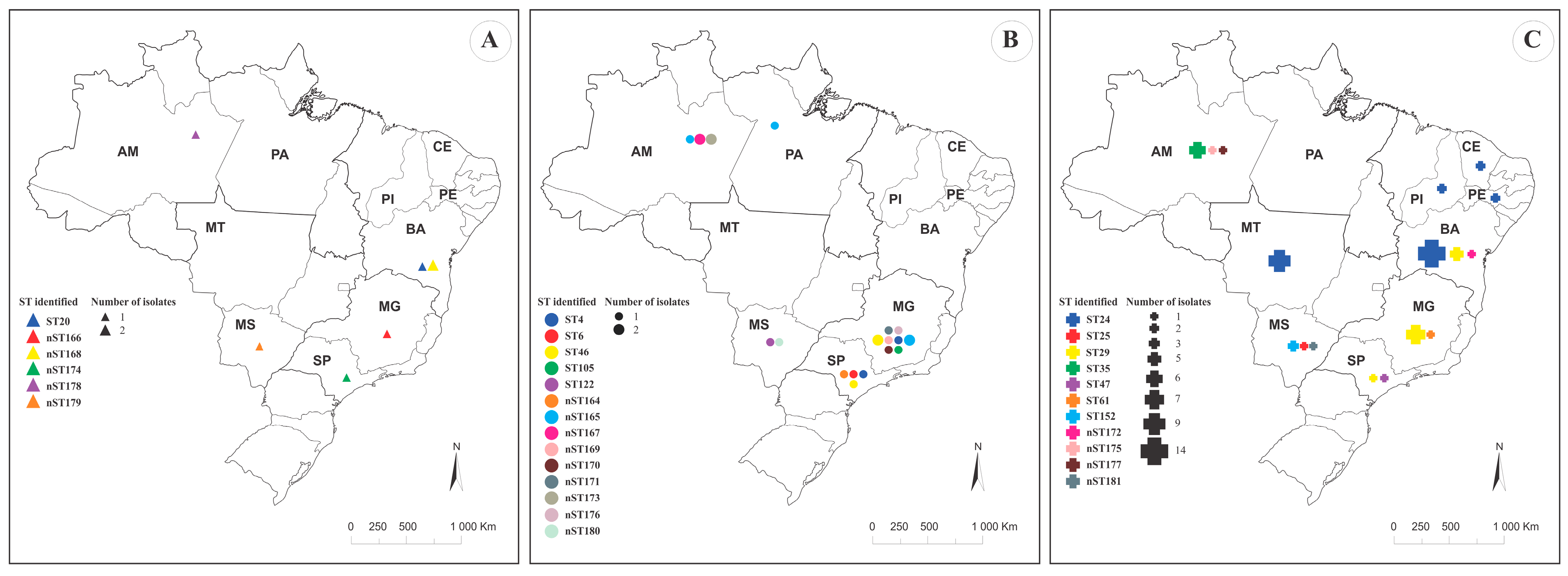
Evaluation of the DNA polymorphism for each allele within the MLST scheme revealed high haplotype diversity paired with low nucleotide diversity (Table 2). This pattern suggests a rapid population expansion from a small number of individuals alongside the generation of new mutations, supporting the hypothesis of varying evolutionary rates across the evaluated loci. Consequently, our MLST analysis revealed that the allelic profiles of the 55 LCB strains evaluated in this study grouped the isolates into 29 distinct STs (Table 3). The map of the distribution of L. formosensis, L. garvieae, and L. petauri STs is shown in Figure 1. Overall, the 55 isolates were assigned to 29 STs, of which 18/29 (62.1%) were novel. The overall ST-level Simpson diversity index was 0.929 (95% CI, 0.883–0.975). ST diversity (permutation test, p < 0.001) and CC diversity (permutation test, p < 0.001) differed among species.
Analysis of the 67 LCB genome sequences isolated from aquatic animals and subjected to MLST analysis in PubMLST identified 20 STs, including 10 novel STs. Only the ERR5094895 strain (from rainbow trout in Poland) lacked an assigned ST due to the absence of the als gene in its genome sequence (Table S1).
The L. formosensis strains used in this study were grouped into 6 different STs, including one previously reported (ST20) and five novel STs (nST166, nST168, nST174, nST178 and nST179). All of these STs were characterized as singletons (Table 3, Figure 2). The Simpson’s diversity index (SDI) value was 0.952 (95% CI, 0.857–1.000).
The L. garvieae strains grouped into 14 different STs, including five previously reported STs (ST4, n = 2; ST6, n = 1; ST46, n = 1; ST105, n = 1; ST122, n = 1) and nine novel STs (nST164, n = 1; nST165, n = 4; nST167, n = 2; nST169, n = 1; nST170, n = 1; nST171, n = 1; nST173, n = 2; nST176, n = 1; and nST180, n = 1). ST4 and ST122 were grouped into CC4 (3/55 isolates). ST46 and nST180 clustered into CC17 (2/55 isolates), and the nST176 belongs to CC62 (1/55 isolates). The nST165 and nST173 clustered together, but differed only in the galP gene allele (a 4-nucleotide divergence), without forming a distinct clonal complex. Finally, ST6, ST105, nST164, nST167, nST169, nST170 and nST171 were characterized as singletons (Table 3, Figure 2). The SDI value was 0.953 (95% CI, 0.903–1.000). For L. garvieae, exploratory permutation testing did not provide robust evidence of an association between ST and geographic region (p = 0.070), although a non-significant trend was observed. Likewise, there is no significant association between region and CC (p = 0.156).
The L. petauri strains grouped into nine different STs, including five previously reported STs (ST25, n = 1; ST29, n = 13; ST35, n = 6; ST61, n = 1; ST152, n = 3) and four novel STs (nST172, n = 1; nST175, n = 1; nST177, n = 1; nST181, n = 1). ST29, ST35 and ST152 were grouped into CC29 (22/55 isolates). In this isolate collection, ST35 was detected only among isolates from Amazonas, whereas ST152 was detected only among isolates from Mato Grosso do Sul. These patterns should be interpreted as descriptive spatial clustering within the sampled isolates, rather than evidence of state-specific STs. ST29, however, was detected in different hosts and across various geographical regions. ST61, nST177 and nST181 clustered with ST27, ST53 and ST47, respectively, but did not form distinct CCs. Finally, ST25, nST172 and nST175 were characterized as singletons (Table 3, Figure 2). The SDI value was 0.746 (95% CI 0.612–0.880). In L. petauri, exploratory permutation testing indicated that ST distribution differed by geographic region (p < 0.001) and state (p = 0.003). This pattern was mainly driven by ST35 among Amazonas isolates, ST152 among Mato Grosso do Sul isolates, and ST29 among isolates from Bahia, Minas Gerais and São Paulo. Because the contingency table was sparse, this result was interpreted as descriptive clustering within the analyzed collection rather than evidence of population-level geographic specificity. However, when considering CC level, there is no clear evidence of region (p = 0.879) or state (p = 0.918) distribution.
Overall, the collection showed high ST-level diversity, but diversity differed among species. L. formosensis and L. garvieae showed high ST richness relative to sample size, whereas L. petauri showed lower ST-level diversity because most isolates belonged to ST29 and nCC29. Table 4 summarizes the ST diversity and CC information for all studied isolates, as well as stratified by species. The observed clonal complexes (CCs) were significantly associated with specific species: CC5 with L. formosensis; CC4, CC17, and CC62 with L. garvieae; and CC24, CC29, and CC97 with L. petauri (p < 0.001).

3.2. Phylogenetic Relatedness Between Fish Isolates

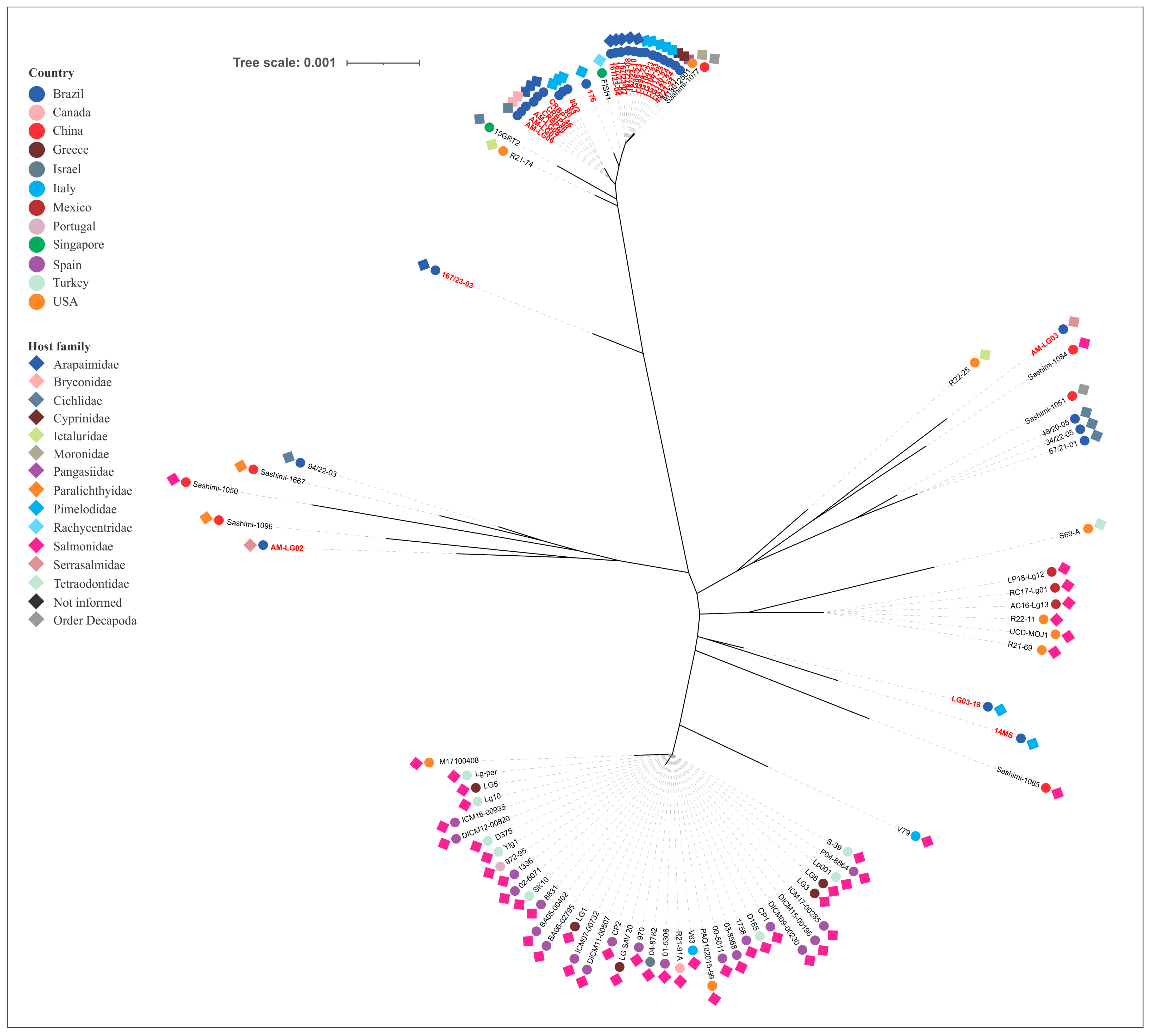
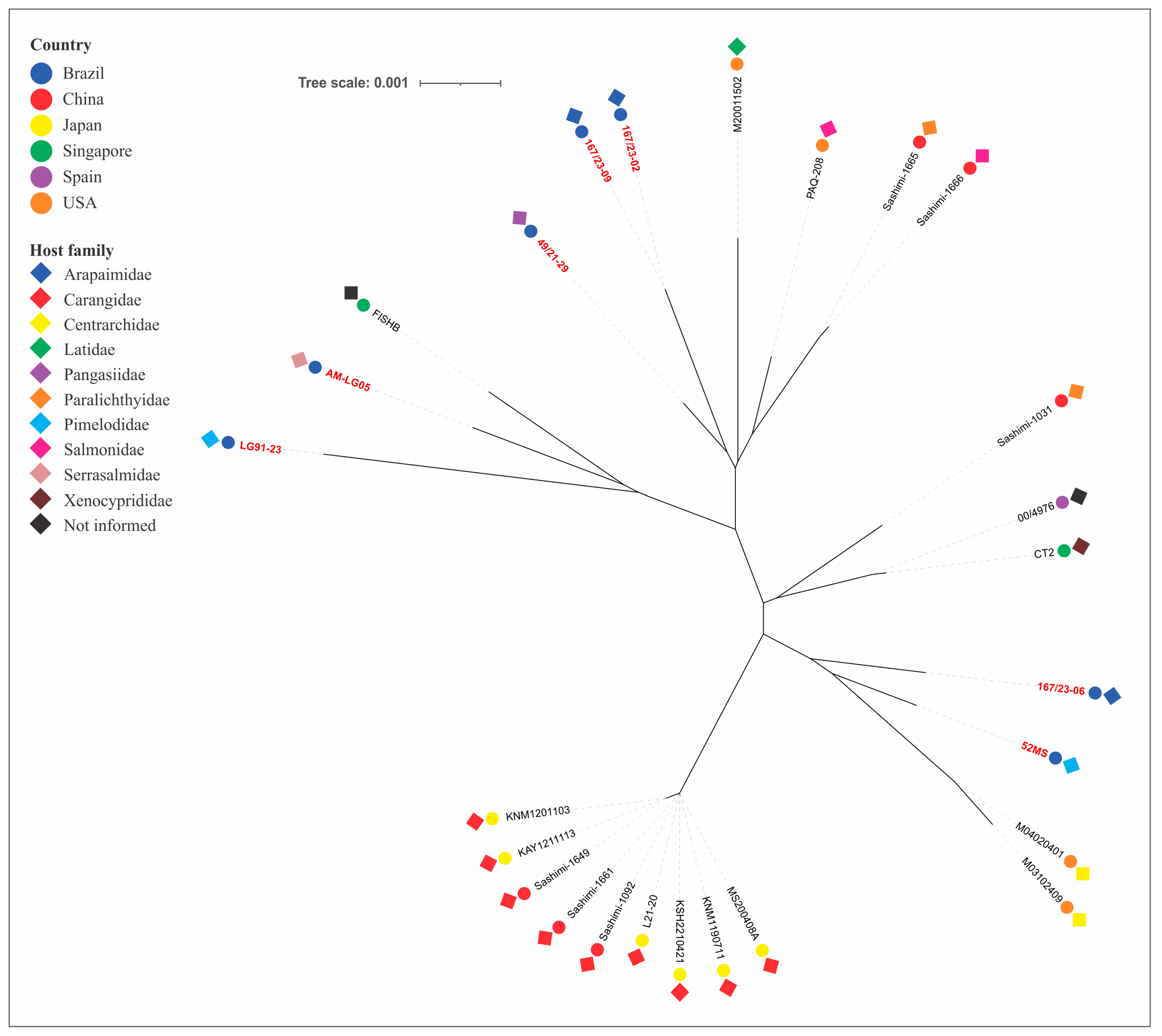
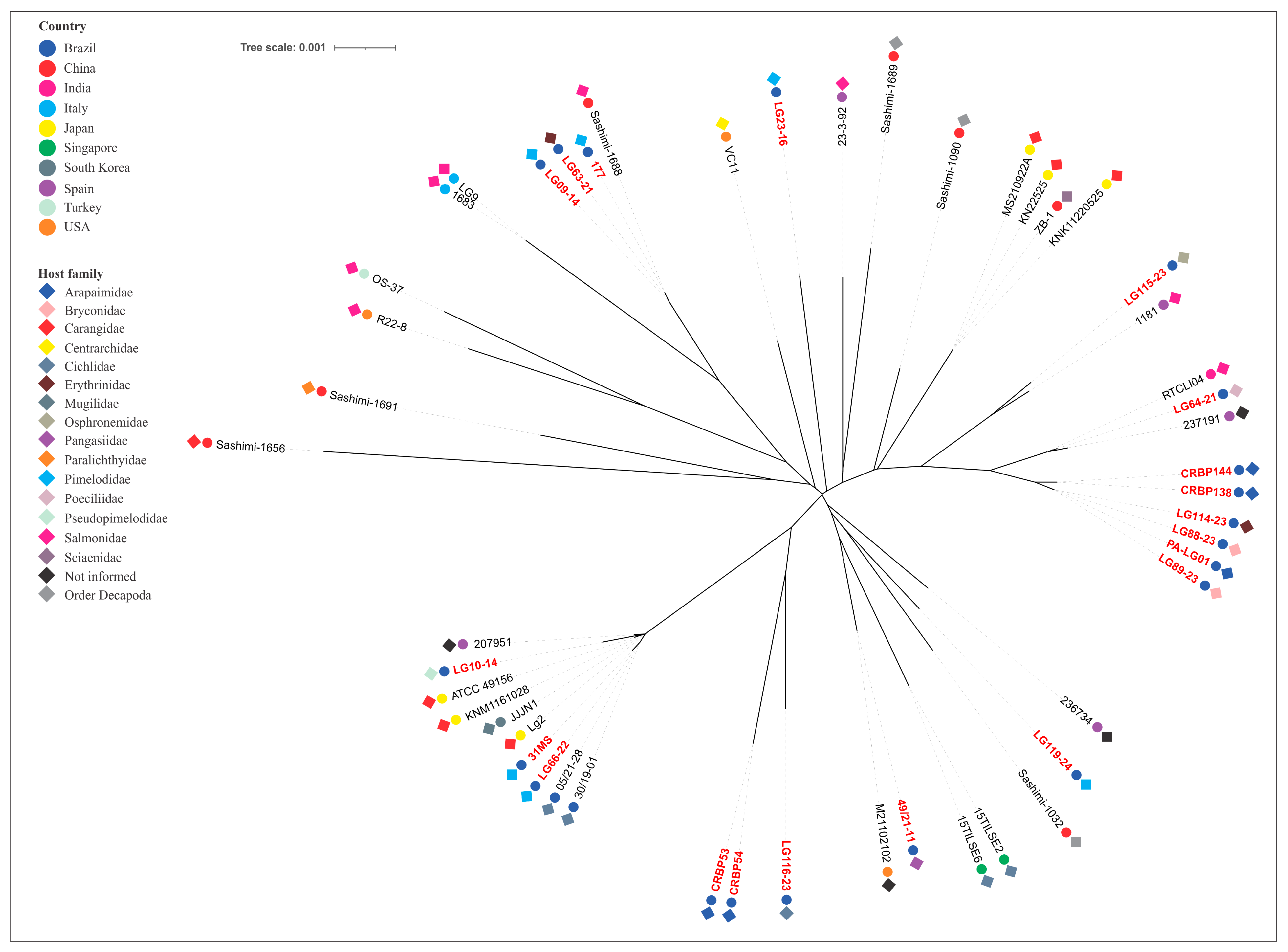
The phylogenetic tree, constructed from concatenated MLST allele sequences of piscine L. formosensis, L. garvieae, and L. petauri, are presented in Figure 3, Figure 4 and Figure 5, respectively.
The L. formosensis strains clustered into five major groups, with the strains reported in this study forming three distinct clusters. These exhibited phylogenetic divergence from isolates obtained from marine fish of the Carangidae family (ST56 and ST115) from Japan and China (Figure 3).
The L. garvieae strains clustered into fourteen groups. The Brazilian isolates that were not from tilapia grouped independently or alongside other aquatic animal isolates worldwide within eight of these groups. The LG10-14 (ST105), LG66-22 (ST46) and 31MS (nST180) strains clustered with isolates obtained from disease outbreaks in Nile tilapia in Brazil (Figure 4).
The L. petauri strains clustered into nine distinct phylogenetic groups. The Brazilian isolates that were not from tilapia were distributed among five of these clusters, with the majority (82.1%) forming a single predominant cluster. The analysis revealed genetic divergence between these isolates and those obtained from disease outbreaks in rainbow trout in Europe (ST14), the United States (nST145) and Mexico (nST145). Notably, AM-LG02 and AM-LG03 strains clustered with ST47 and ST24 isolates, respectively, which originated from disease outbreaks in Brazilian tilapia farms (Figure 5).

4. Discussion

The present study investigated the population structure and genetic profile of a set of LCB strains obtained from different fish species in Brazil, using MLST as the genotyping method. Based on sequences deposited in the PubMLST database for L. garvieae—including the isolates reported in this study—80 STs were assigned to isolates derived from aquatic animals (Figure 2), which include isolates from both clinical disease cases, stool samples and fish meat products. Among these, 18 STs belong to L. formosensis, 29 to L. petauri and 33 to L. garvieae (Table 5), highlighting the genetic heterogeneity among these bacterial species.
When the MLST scheme was first developed by Ferrario et al. [22], all the isolates were believed to belong to L. garvieae, revealing two distinct genetic populations within the analyzed collection of strains. Subsequent studies, using strains from diverse sources (human, animal, food and environmental), identified a wide range of STs, which indicates the genetic heterogeneity of L. garvieae [23,24,31,32]. However, a study conducted in 2017 redefined the L. garvieae subgroup A as a new species, named L. petauri, and suggested the reassignment of previously characterized isolates [52]. Consequently, various studies have been conducted to improve the speciation within the genus Lactococcus [21,42,53]. It was only after 2023 that the first studies using the MLST approach to differentiate genetic profiles among LCB species were published [3,6,25,26], demonstrating high and comparable genetic diversity within each species, based on isolates from both human and animal sources [26]. During this same period, our research team constructed the L. garvieae MLST scheme in the PubMLST database. Since then, we have curated all the newly deposited sequences—including alleles, isolates and genomes—to ensure standardized nomenclature for major STs and CCs, integrating and consolidating data from LCB strains, and providing a comprehensive analysis of their genetic and epidemiological characteristics. Thus, by sequencing the seven housekeeping genes of our isolates and utilizing the PubMLST database (accessed on 5 August 2025), it was possible to compare the population structure and phylogenetic relationships of Lactococcus spp. strains obtained from fish in Brazil, with those of other countries.
Our results for this dataset demonstrate that the LCB isolates from the fish belong to 11 previously established STs. ST4 and ST122 were previously identified in animal-derived products, including fish meat, from China, Italy and Spain [22,26]. Notably, ST4, ST20, ST29 and ST105 have been associated with human diseases in China, Singapore, Spain and the United States [22,23,26,32]. Additional epidemiological findings include: ST6 reported in vegetable isolates from Italy; ST61 detected in water samples from Spain; ST25, ST35 and ST152 identified in human and swine fecal samples from China and Spain [22,23]. Among the previously reported STs, only ST6 and ST46 have been found in diseased fish, in the United States and Brazil, respectively [3,42]. The high values of the SDI show a considerable genetic divergence among the isolates evaluated, with L. formosensis and L. garvieae having greater ST richness and higher ST-level diversity in this study while L. petauri collection suggested a less diverse population.
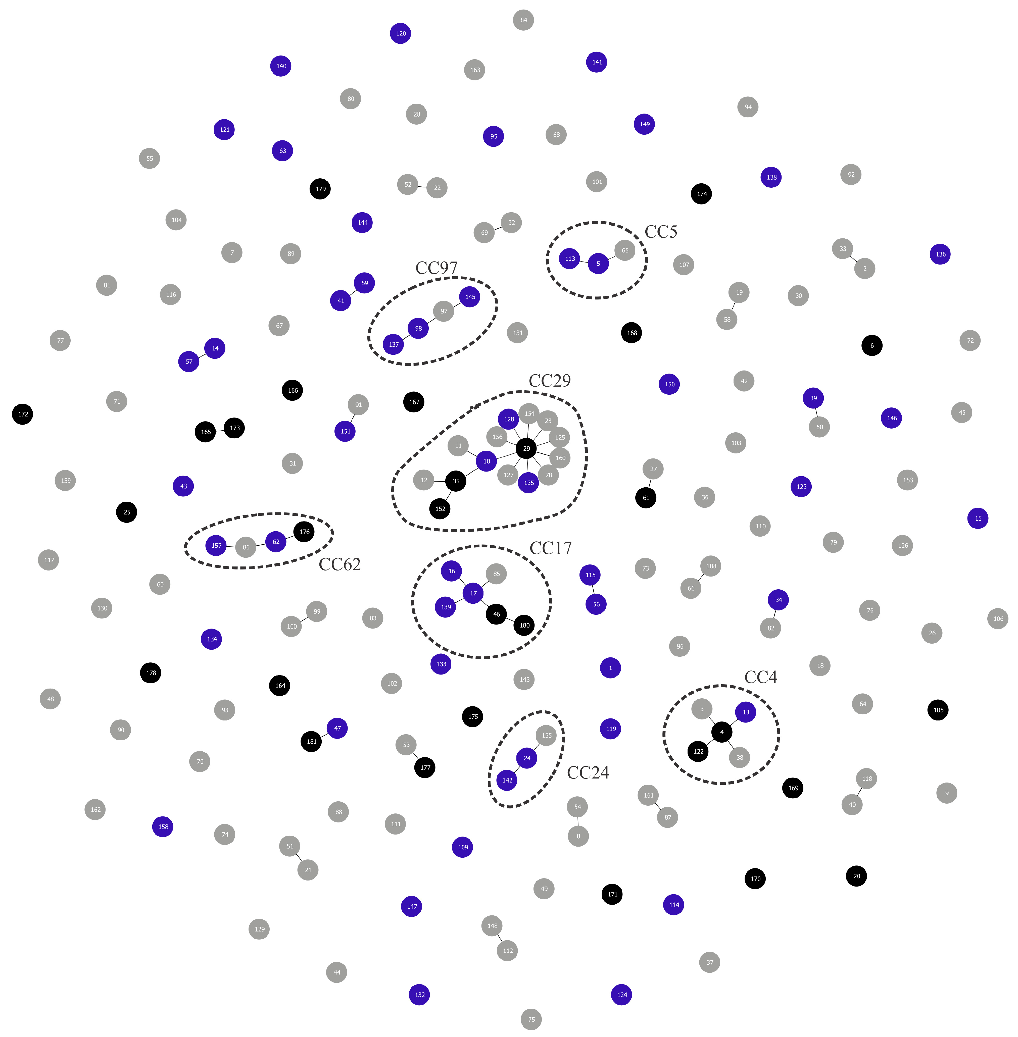
It is important to mention that when evaluating the ancestry of the isolates through CC analysis, no cluster comprising three or more STs formed exclusively by isolates from aquatic animals was observed (Figure 2).
L. garvieae CC4, which groups ST4 (LG09-14 and LG63-21 strains) and ST122 (177 strain) identified in this study, appears to be associated with isolates from animal-derived products, particularly samples originating from the European continent [26]. Nonetheless, ST13, which also belongs to this CC, includes isolates from rainbow trout in Italy [22,42]. On the other hand, L. garvieae CC17 appears to harbor more STs (ST16, ST17, ST46, and ST139) associated with clinical manifestations of disease in fish [3,22,23,25]. This corroborates results from our study, as 31MS (nST180) and LG66-22 (ST46) strains, isolated from diseased Pseudoplatystoma fasciatum and Phractocephalus hemioliopterus, respectively, grouped within this CC. A previous study assessed the pathogenicity of the 31MS strain through experimental infection (107 CFU/fish) in Pseudoplatystoma spp. [54]. During the 21-day monitoring period, 10.6% of the animals exhibited clinical signs of diseases; however, no mortality was observed. Conversely, the LG66-22 strain belongs to the same ST identified in disease outbreaks affecting Nile tilapia in Brazil in 2019 and 2021 [3]. Since the pathogenicity of this specific ST has not been evaluated, future laboratory controlled challenges comparing the susceptibility of Nile tilapia and Phractocephalus hemioliopterus is warranted to better understand the pathogenicity of this ST. Finally, L. garvieae CC62 includes isolates from fish in India and Spain (ST62 and ST157) [33,42], and is grouped with a strain from the current study, LG64-21 (nST176), which was obtained from an ornamental fish species. Although a few LCB isolates from ornamental fish were included in this study, the two L. garvieae strains possess different STs, showing no apparent consistent host- or region-associated pattern within the limits of this uneven isolate collection.
L. formosensis CC5 is also predominantly associated with isolates from animal-derived products, including fish meat from China (ST5 and ST113) [26]. In the current study, we did not identify any isolates belonging to this CC.
L. petauri CC24 comprises isolates associated with diseases in fish (Nile tilapia–ST24; catfish–nST142) and humans (ST24), as well from human (ST24) and swine (ST155) feces [3,23,32,42]. ST24 has been the predominant genetic profile among L. petauri isolates obtained from Nile tilapia in different types of commercial production and different geographic regions in Brazil between 2020 and 2022, and its pathogenicity and high virulence for this aquatic host were confirmed [3]. Interestingly, none of the isolates evaluated in this study shared this ST or belonged to CC24, suggesting that these isolates may have emerged from a distinct ancestor. On the other hand, L. petauri CC29 clustered isolates from diverse sources and was the largest CC identified in this study. CC29 clustered isolates from human feces [22,23], fish (cobia and European seabass) and prawn sashimi, such as ST10, ST128 and ST135 [26,32,42]. A total of 22 out of 28 L. petauri strains from our study belong to this CC, indicating that isolates of this bacterial species tend to have a more homogeneous genetic profile compared to the other two bacterial species investigated. Other studies utilizing different genotyping methods (DNA fingerprinting approaches) also revealed a more homogeneous population for L. petauri strains [21]. Finally, L. petauri CC97 contains isolates linked to diseases in fish (ST98 and nST145) and fish meat (ST137) [26,42]. Among these, nST145 has been associated with major mortality outbreaks in rainbow trout in the United States and Mexico between 2016 and 2020 [9,55]. No isolate from this study grouped within this CC.
Other genetic relationships were also identified via goeBURST analysis. For L. petauri: ST27 (human feces, Spain) and ST61 (LG03-18 strain); ST47 (Nile tilapia, Brazil) and nST181 (14MS strain); ST53 (bovine mastitis, Spain) and nST177 (AM-LG03 strain); ST34 (red tilapia, Singapore) and ST82 (human feces, China). For L. garvieae: nST165 (LG88-23, LG89-23 and PA-LG01 strains) and nST173 (CRBP138 and CRBP144) from Amazonian fish species; ST39 (tilapia, Singapore) and ST50 (bovine mastitis, Spain). And for L. formosensis: ST41 (carp, Singapore) and ST59 (fish, Spain); ST91 (bovine mastitis, China) and ST151 (barramundi, USA) [25,32,42]. Furthermore, our study observed that many isolates were singletons (lacking a common ancestor with other isolates), underscoring the significant genetic heterogeneity of these bacteria. Despite this, there are currently 181 STs deposited in the PubMLST database. In the future, with the addition of more isolates and allelic profiles, new population structure relationships among LCBs may be revealed.
Since the Lactococcus spp. isolates deposited in the PubMLST database originate from diverse sources, such as clinical samples from humans and animals, dairy and meat products, feces from healthy individuals, and even vegetables, uncertainties remain regarding the public health and food safety implications of these pathogens. Human infections have been reported following the handling or ingestion of raw fish and seafood [56,57,58], with the latter considered the most probable source of infection. For this reason, LCBs (especially L. garvieae) are considered potential zoonotic bacteria. However, the microbiological and molecular data available to support this hypothesis are scarce, as foodborne transmission from fish and seafood is usually inferred from clinical histories, with limited epidemiological evidence to confirm the transmission of the bacteria between food and humans [58]. Although our study and others in the scientific literature have demonstrated that isolates from different sources can share the same ST [22,23,26,31], it is currently not possible to determine whether a specific ST or CC is safe or potentially hazardous to humans.
Phylogenetic analysis of the concatenated housekeeping genes was used to reconstruct the evolutionary relationships among the strains of the tested bacterial species. As expected, the analysis enabled the grouping of strains within the same CC. However, it also revealed arrangements that represented double- or triple-locus variants. The analysis revealed that our L. formosensis strains are closely related to others obtained from largemouth bass (nST140 and nST141), barramundi (nST151), and rainbow trout (nST150) in the USA; from a fish with no designated species in Singapore (ST43); and from salmon (ST113) and flounder (ST5) sashimi in China [26,32,42]. The L. garvieae strains are related to those obtained from salmon (ST122) and prawn (ST119) sashimi (China), rainbow trout (ST62, India; ST63, Spain), European seabass (ST157 and ST158, Spain, [33]), unspecified fish species (ST6, USA), tilapia (ST39, Singapore; ST46, Brazil), yellowtail (ST16 and ST17, Japan), and so-iuy mullet (ST17, South Korea). This broad host range demonstrates a lack of host specificity and no clear phylogenetic distinction based on geographic origin. Conversely, the L. petauri strains demonstrated a more intriguing genetic relationship. Most of our isolates (those related to CC29) are genetically linked to other isolates obtained from tilapia (ST34, Singapore), cobia (ST10, Singapore), hybrid catfish (nST142, USA), European seabass (ST128, USA) and prawn sashimi (ST135, China) [26,32,42]. Isolates from clinical cases of piscine lactococcosis in trout were divided into two distinct phylogenetic clades: one associated with ST14, ST57 and nST146, identified primarily in European countries (with a single representative from the USA and Canada), and another clade associated with nST145, which, as previously mentioned, is linked to recent and impactful outbreaks in North America. This division presents a strong geographical signal of diversification among trout isolates. Our isolates are not phylogenetically related to these clades, indicating they evolved from different ancestors. In contrast, two of our L. petauri strains (AM-LG02 and AM-LG03), despite some phylogenetic distance, share a common ancestor with isolates associated with disease outbreaks in Nile tilapia in Brazil. Both isolates were obtained from the intestine of Colossoma macropomum. In an experimental infection study in this same aquatic host, the AM-LG02 strain did not cause any macroscopic or microscopic alterations in the challenged animals [59]. Therefore, future studies should use this and other LCB isolates in challenge experiments with tilapia to verify their pathogenic potential for this species.

5. Conclusions

This study provides new insights into the genetic diversity of Brazilian Lactococcus spp. strains isolated from different fish species, using an MLST approach. The analysis revealed that LCB isolates constitute a genetically diverse population based on their STs. Specifically, L. garvieae and L. formosensis exhibited greater heterogeneity compared to L. petauri, for which the majority of isolates belonged to a single clonal complex (CC29). MLST and phylogenetic analysis demonstrated genetic relatedness between the L. garvieae and L. formosensis isolates from this study and those from other aquatic animal species deposited in the PubMLST database. Regarding the L. petauri strains, all the STs identified in this study were unrelated to the MLST lineages responsible for outbreaks in Brazilian Nile tilapia and North American rainbow trout. This suggests that piscine L. petauri populations in the Americas evolved from distinct ancestral origins. However, phylogenetic analysis and MLST data showed that, although they belong to different CC, two isolates from Colossoma macropomum are genetically closely related to isolates from Nile tilapia in Brazil. Therefore, future studies, particularly those employing a whole-genome sequencing approach, are necessary to better elucidate the ancestral relationship between these strains. The results obtained herein provide a better understanding of the genetic diversity of Lactococcus spp. populations in fish from Brazil. Additionally, these findings will contribute to future molecular epidemiology studies, facilitating the selection of candidate strains for whole-genome sequencing, as well as the assessment of the pathogenesis and evolutionary aspects of LCBs using in vivo (experimental infection) or in vitro (transcriptomics and proteomics) models.

6. Limitations

Quantitatively, the Brazilian non-tilapia collection analyzed here represented a substantial fraction of the aquatic-animal ST diversity currently available in PubMLST. Among aquatic animal-derived STs, this study identified 6/18 (33.3%) L. formosensis STs, 14/33 (42.4%) L. garvieae STs and 9/29 (31.0%) L. petauri STs. Overall, the 29 STs detected in this study accounted to 29/80 (36.3%) of all aquatic animal-derived STs in the dataset analyzed, and 18/29 (62.1%) were novel. Compared with previous Brazilian MLST data for Nile tilapia, which reported only ST24 and ST47 for L. petauri and ST46 for L. garvieae, the present non-tilapia collection substantially expands the known Brazilian ST spectrum. However, this study was designed as a retrospective descriptive MLST survey of Brazilian Lactococcus spp. isolates from non-tilapia fish, rather than as a population-based epidemiological study. Therefore, ST and CC frequencies should be interpreted as patterns observed within this isolate collection, rather than as estimates of genotype prevalence in Brazilian aquaculture. Further, because host species, tissue, year and production origin were unevenly distributed and many categories were represented by one or two isolates, formal association tests for these variables were considered underpowered and were not used to support epidemiological conclusions.

Supplementary Materials

The following supporting information can be downloaded at: https://www.mdpi.com/article/10.3390/microorganisms14051131/s1, Table S1: Lactococcus ssp. strains isolated from aquatic animals with publicly available genome sequences in GenBank databases included in this study; Table S2: Lactococcus spp. strains isolated from diverse animal aquatic species with previously reported alleles and sequence type included in this study to phylogenetic analyses.

Author Contributions

G.C.T., F.P., S.U.G., C.A.G.L. and H.C.P.F. conceived and designed the experiments. S.P.C., A.C.C.B., A.E.C.d.R., H.C.M., C.R.M.d.S.M., H.L.C. and R.C.E. performed the microbiological analyses, DNA extraction, and PCR amplification. J.C.C.R. conducted the Sanger sequencing. F.L.P. developed the L. garvieae MLST scheme for inclusion in PubMLST database. G.C.T. and L.F.F.N. performed analyses and visualization of the data. G.C.T. wrote the manuscript and coordinated all analyses of the project. F.L.P., F.P., E.S., C.A.G.L. and H.C.P.F. contributed substantially to data interpretation and to revisions of the manuscript. All authors have read and agreed to the published version of the manuscript.

Funding

This study was financed in part by the Coordenação de Aperfeiçoamento de Pessoal de Nível Superior—Brasil (CAPES), through the PROCAD/Amazônia (grant number 88881.200614/2018-01) and PDPG-CAPES calls; Fundação de Amparo à Pesquisa do Estado de Minas Gerais (FAPEMIG, grant numbers APQ-01227-22, APQ-04309-22 and PPM-00779-18), and Fundação de Amparo à Pesquisa do Estado do Amazonas (FAPEAM, grant number 01.02.016301.03071/2022-11).

Institutional Review Board Statement

Not applicable.

Informed Consent Statement

Not applicable.

Data Availability Statement

The data that support the findings of this study are available on request from the corresponding author.

Acknowledgments

During the preparation of this manuscript/study, the author(s) used Gemini 3 Pro (Google) to improve the language, clarity, and readability of this manuscript. After using this tool, the authors reviewed and edited the content as needed and take full responsibility for the final version of the publication.

Conflicts of Interest

The authors declare no conflicts of interest.

References

  1. Shahin, K.; Abdel-Glil, M.; Saticıoğlu, I.B.; Duman, M.; Altun, S.; Colussi, S.; Esposito, G.; Acutis, P.L.; Marino, P.; Spondler, B.; et al. Diving Into the Depths: Unveiling the Main Etiologies of Piscine Lactococcosis With a Novel Multiplex QPCR Assay. J. Fish Dis. 2025, 48, e14147. [Google Scholar] [CrossRef]
  2. Rao, S.; Pham, T.H.; Poudyal, S.; Cheng, L.-W.; Nazareth, S.C.; Wang, P.-C.; Chen, S.-C. First Report on Genetic Characterization, Cell-Surface Properties and Pathogenicity of Lactococcus Garvieae, Emerging Pathogen Isolated from Cage-Cultured Cobia (Rachycentron canadum). Transbound. Emerg. Dis. 2022, 69, 1197–1211. [Google Scholar] [CrossRef]
  3. Egger, R.C.; Rosa, J.C.C.; Resende, L.F.L.; de Pádua, S.B.; de Oliveira Barbosa, F.; Zerbini, M.T.; Tavares, G.C.; Figueiredo, H.C.P. Emerging Fish Pathogens Lactococcus Petauri and L. Garvieae in Nile Tilapia (Oreochromis niloticus) Farmed in Brazil. Aquaculture 2023, 565, 739093. [Google Scholar] [CrossRef]
  4. Abraham, T.; Yazdi, Z.; Littman, E.; Shahin, K.; Heckman, T.I.; Quijano Cardé, E.M.; Nguyen, D.T.; Hu, R.; Adkison, M.; Veek, T.; et al. Detection and Virulence of Lactococcus Garvieae and L. Petauri from Four Lakes in Southern California. J. Aquat. Anim. Health 2023, 35, 187–198. [Google Scholar] [CrossRef] [PubMed]
  5. Salogni, C.; Bertasio, C.; Accini, A.; Gibelli, L.R.; Pigoli, C.; Susini, F.; Podavini, E.; Scali, F.; Varisco, G.; Alborali, G.L. The Characterisation of Lactococcus Garvieae Isolated in an Outbreak of Septicaemic Disease in Farmed Sea Bass (Dicentrarchus labrax, Linnaues 1758) in Italy. Pathogens 2024, 13, 49. [Google Scholar] [CrossRef]
  6. Salif, M.; Ogawa, R.; Mikami, A.; Daibata, M.; Imajoh, M. Complete Genome Sequence of Lactococcus Garvieae Isolated from a Greater Amberjack (Seriola dumerili) Farmed in Japan in 2022. Microbiol. Resour. Announc. 2024, 13, e00436-24. [Google Scholar] [CrossRef] [PubMed]
  7. Balan, R.; Pandey, S.; Wang, P.-C.; Byadgi, O.V.; Chen, S.-C. Insights on the Virulence and Genomic Features of Lactococcus Garvieae Isolated from Giant Freshwater Prawn Macrobrachium Rosenbergii (de Man 1879). J. Fish Dis. 2024, 47, e14011. [Google Scholar] [CrossRef] [PubMed]
  8. Wongkaew, J.; Chatchaiphan, S.; Taengphu, S.; Dong, H.T.; Senapin, S.; Piyapattanakorn, S. Identification and Pathogenicity of Lactococcus Species in Nile Tilapia (Oreochromis niloticus) and Asian Sea Bass (Lates calcarifer). J. Fish Dis. 2025, 48, e14113. [Google Scholar] [CrossRef]
  9. Shahin, K.; Veek, T.; Heckman, T.I.; Littman, E.; Mukkatira, K.; Adkison, M.; Welch, T.J.; Imai, D.M.; Pastenkos, G.; Camus, A.; et al. Isolation and Characterization of Lactococcus Garvieae from Rainbow Trout, Onchorhyncus Mykiss, from California, USA. Transbound. Emerg. Dis. 2022, 69, 2326–2343. [Google Scholar] [CrossRef]
  10. Chang, P.H.; Lin, C.W.; Lee, Y.C. Lactococcus Garvieae Infection of Cultured Rainbow Trout, Oncorhynchus Mykiss, in Taiwan and Associated Biophysical Characteristics and Histopathology. Bull. Eur. Assoc. Fish Pathol. 2002, 22, 319–327. [Google Scholar]
  11. Kang, S.H.; Shin, G.W.; Shin, Y.S.; Palaksha, K.J.; Kim, Y.R.; Yang, H.H.; Lee, E.Y.; Lee, E.G.; Huh, N.E.; Ju, O.M.; et al. Experimental Evaluation of Pathogenicity of Lactococcus Garvieae in Black Rockfish (Sebastes Schlegeli). J. Vet. Sci. 2004, 5, 387–390. [Google Scholar] [CrossRef]
  12. Bondavalli, F.; Colussi, S.; Pastorino, P.; Zanoli, A.; Bezzo Llufrio, T.; Fernández-Garayzábal, J.F.; Acutis, P.L.; Prearo, M. First Report of Lactococcus Petauri in the Pumpkinseed (Lepomis gibbosus) from Candia Lake (Northwestern Italy). Fishes 2024, 9, 117. [Google Scholar] [CrossRef]
  13. Esposito, G.; Bignami, G.; Colussi, S.; Pastorino, P.; Bondavalli, F.; Fioravanti, M.; Bozzetta, E.; Acutis, P.; Valentino, A.; Rizzi, R.; et al. Expanding Horizons: The First Reported Outbreak of Piscine Lactococcosis in Farmed Gilthead Seabream Sparus Aurata in the Northern Tyrrhenian Sea. J. Fish Dis. 2025, 48, e14121. [Google Scholar] [CrossRef]
  14. Fukuda, Y.; Tue, Y.; Oinaka, D.; Wada, Y.; Yamashita, A.; Urasaki, S.; Yoshioka, S.; Kimoto, K.; Yoshida, T. Pathogenicity and Immunogenicity of Non-Agglutinating Lactococcus Garvieae with Anti-KG- Phenotype Rabbit Serum in Seriola spp. Fish Pathol. 2015, 50, 200–206. [Google Scholar] [CrossRef][Green Version]
  15. Meyburgh, C.M.; Bragg, R.R.; Boucher, C.E. Lactococcus Garvieae: An Emerging Bacterial Pathogen of Fish. Dis. Aquat. Organ. 2017, 123, 67–79. [Google Scholar] [CrossRef]
  16. Fukushima, H.C.S.; Leal, C.A.G.; Cavalcante, R.B.; Figueiredo, H.C.P.; Arijo, S.; Moriñigo, M.A.; Ishikawa, M.; Borra, R.C.; Ranzani-Paiva, M.J.T. Lactococcus Garvieae Outbreaks in Brazilian Farms Lactococcosis in Pseudoplatystoma sp.—Development of an Autogenous Vaccine as a Control Strategy. J. Fish Dis. 2017, 40, 263–272. [Google Scholar] [CrossRef]
  17. Pastorino, P.; Vela Alonso, A.I.; Colussi, S.; Cavazza, G.; Menconi, V.; Mugetti, D.; Righetti, M.; Barbero, R.; Zuccaro, G.; Fernández-Garayzábal, J.F.; et al. A Summer Mortality Outbreak of Lactococcosis by Lactococcus Garvieae in a Raceway System Affecting Farmed Rainbow Trout (Oncorhynchus mykiss) and Brook Trout (Salvelinus fontinalis). Animals 2019, 9, 1043. [Google Scholar] [CrossRef]
  18. Choi, H.J.; Hur, J.W.; Cho, J.B.; Park, K.H.; Jung, H.J.; Kang, Y.J. Introduction of Bacterial and Viral Pathogens from Imported Ornamental Finfish in South Korea. Fish Aquat. Sci. 2019, 22, 5. [Google Scholar] [CrossRef]
  19. Cardoso, P.H.M.; Moreno, L.Z.; de Oliveira, C.H.; Gomes, V.T.M.; Silva, A.P.S.; Barbosa, M.R.F.; Sato, M.I.Z.; Balian, S.C.; Moreno, A.M. Main Bacterial Species Causing Clinical Disease in Ornamental Freshwater Fish in Brazil. Folia Microbiol. 2021, 66, 231–239. [Google Scholar] [CrossRef] [PubMed]
  20. Neupane, S.; Rao, S.; Yan, W.-X.; Wang, P.-C.; Chen, S.-C. First Identification, Molecular Characterization, and Pathogenicity Assessment of Lactococcus Garvieae Isolated from Cultured Pompano in Taiwan. J. Fish Dis. 2023, 46, 1295–1309. [Google Scholar] [CrossRef] [PubMed]
  21. Barbanti, A.C.C.; do Rosário, A.E.C.; da Silva Maia, C.R.M.; Rocha, V.P.; Costa, H.L.; Trindade, J.M.; Nogueira, L.F.F.; Rosa, J.C.C.; Ranzani-Paiva, M.J.T.; Pilarski, F.; et al. Genetic Characterization of Lactococcosis-Causing Bacteria Isolated from Brazilian Native Fish Species. Aquaculture 2024, 593, 741305. [Google Scholar] [CrossRef]
  22. Ferrario, C.; Ricci, G.; Milani, C.; Lugli, G.A.; Ventura, M.; Eraclio, G.; Borgo, F.; Fortina, M.G. Lactococcus Garvieae: Where Is It From? A First Approach to Explore the Evolutionary History of This Emerging Pathogen. PLoS ONE 2013, 8, e84796. [Google Scholar] [CrossRef]
  23. Reguera-Brito, M.; Galán-Sánchez, F.; Blanco, M.M.; Rodríguez-Iglesias, M.; Domínguez, L.; Fernández-Garayzábal, J.F.; Gibello, A. Genetic Analysis of Human Clinical Isolates of Lactococcus Garvieae: Relatedness with Isolates from Foods. Infect. Genet. Evol. 2016, 37, 185–191. [Google Scholar] [CrossRef] [PubMed]
  24. Thiry, D.; Billen, F.; Boyen, F.; Duprez, J.-N.; Quenault, H.; Touzain, F.; Blanchard, Y.; Clercx, C.; Mainil, J.G. Genomic Relatedness of a Canine Lactococcus Garvieae to Human, Animal and Environmental Isolates. Res. Vet. Sci. 2021, 137, 170–173. [Google Scholar] [CrossRef]
  25. Lin, Y.; Han, J.; Barkema, H.W.; Wang, Y.; Gao, J.; Kastelic, J.P.; Han, B.; Qin, S.; Deng, Z. Comparative Genomic Analyses of Lactococcus Garvieae Isolated from Bovine Mastitis in China. Microbiol. Spectr. 2023, 11, e02995-22. [Google Scholar] [CrossRef] [PubMed]
  26. Chan, Y.-X.; Cao, H.; Jiang, S.; Li, X.; Fung, K.-K.; Lee, C.-H.; Sridhar, S.; Chen, J.H.-K.; Ho, P.-L. Genomic Investigation of Lactococcus Formosensis, Lactococcus Garvieae, and Lactococcus Petauri Reveals Differences in Species Distribution by Human and Animal Sources. Microbiol. Spectr. 2024, 12, e00541-24. [Google Scholar] [CrossRef] [PubMed]
  27. Evans, J.J.; Bohnsack, J.F.; Klesius, P.H.; Whiting, A.A.; Garcia, J.C.; Shoemaker, C.A.; Takahashi, S. Phylogenetic Relationships among Streptococcus Agalactiae Isolated from Piscine, Dolphin, Bovine and Human Sources: A Dolphin and Piscine Lineage Associated with a Fish Epidemic in Kuwait Is Also Associated with Human Neonatal Infections in Japan. J. Med. Microbiol. 2008, 57, 1369–1376. [Google Scholar] [CrossRef]
  28. Barony, G.M.; Tavares, G.C.; Pereira, F.L.; Carvalho, A.F.; Dorella, F.A.; Leal, C.A.G.; Figueiredo, H.C.P. Large-Scale Genomic Analyses Reveal the Population Structure and Evolutionary Trends of Streptococcus Agalactiae Strains in Brazilian Fish Farms. Sci. Rep. 2017, 7, 13538. [Google Scholar] [CrossRef]
  29. Maiden, M.C.J.; van Rensburg, M.J.J.; Bray, J.E.; Earle, S.G.; Ford, S.A.; Jolley, K.A.; McCarthy, N.D. MLST Revisited: The Gene-by-Gene Approach to Bacterial Genomics. Nat. Rev. Microbiol. 2013, 11, 728–736. [Google Scholar] [CrossRef]
  30. Jolley, K.A.; Bray, J.E.; Maiden, M.C.J. Open-Access Bacterial Population Genomics: BIGSdb Software, the PubMLST.Org Website and Their Applications. Wellcome Open Res. 2018, 3, 124. [Google Scholar] [CrossRef]
  31. Kozakai, M.; Matsumoto, C.; Matsumoto, M.; Takakura, A.; Matsubayashi, K.; Satake, M. Different Growth Kinetics in Blood Components and Genetic Analysis of Lactococcus Garvieae Isolated from Platelet Concentrates. Transfusion 2020, 60, 1492–1499. [Google Scholar] [CrossRef]
  32. Lin, Y.S.; Kweh, K.H.; Koh, T.H.; Lau, Q.C.; Abdul Rahman, N.B. Genomic Analysis of Lactococcus garvieae Isolates. Pathology 2020, 52, 700–707, Correction in Pathology 2021, 53, 295. [Google Scholar] [CrossRef]
  33. González-Martín, D.; Ubieto, M.; del Caso, S.; Planas, E.; Ruiz-Zarzuela, I.; Sanz, C.; Arnal, J.L. Comparative Molecular and Antimicrobial Analysis of Lactococcus Garvieae and Lactococcus Petauri from Marine and Freshwater Fish Farms in the Mediterranean. Animals 2026, 16, 277. [Google Scholar] [CrossRef]
  34. Peixe, B.R. Anuário Brasileiro da Piscicultura; Peixe BR: Brasília, Brazil, 2026. [Google Scholar]
  35. Sebastião, F.A.; Furlan, L.R.; Hashimoto, D.T.; Pilarski, F. Identification of Bacterial Fish Pathogens in Brazil by Direct Colony PCR and 16S RRNA Gene Sequencing. Adv. Microbiol. 2015, 5, 409–424. [Google Scholar] [CrossRef]
  36. Do Rosário, A.E.C.; Henrique, M.C.; da Costa, É.J.C.; Barbanti, A.C.C.; Tavares, G.C. Surtos de Bacteriose Em Juvenis de Pirarucu (Arapaima gigas) Provevientes de Pisciculturas Amazônicas e Avaliação Da Sensibilidade de Aeromonas Jandaei a Antimicrobianos. In Enfermidades Parasitárias e Bacterianas na Piscicultura Brasileira: Insights e Perspectivas; da Cruz, M.G., da Castro, J.S., Jerônimo, G.T., Eds.; i-EDUCAM, 2023; pp. 33–46. [Google Scholar]
  37. Assis, G.B.N.; Pereira, F.L.; Zegarra, A.U.; Tavares, G.C.; Leal, C.A.; Figueiredo, H.C.P. Use of MALDI-TOF Mass Spectrometry for the Fast Identification of Gram-Positive Fish Pathogens. Front. Microbiol. 2017, 8, 1492. [Google Scholar] [CrossRef]
  38. Tavares, G.C.; de Queiroz, G.A.; Assis, G.B.N.; Leibowitz, M.P.; Teixeira, J.P.; Figueiredo, H.C.P.; Leal, C.A.G. Disease Outbreaks in Farmed Amazon Catfish (Leiarius Marmoratus x Pseudoplatystoma Corruscans) Caused by Streptococcus Agalactiae, S. Iniae, and S. Dysgalactiae. Aquaculture 2018, 495, 384–392. [Google Scholar] [CrossRef]
  39. Rosário, A.E.; Barbanti, A.C.; Matos, H.C.; Maia, C.R.; Trindade, J.M.; Nogueira, L.F.; Pilarski, F.; Gallani, S.U.; Leal, C.A.; Figueiredo, H.C.; et al. Antimicrobial Resistance in Lactococcus Spp. Isolated from Native Brazilian Fish Species: A Growing Challenge for Aquaculture. Microorganisms 2024, 12, 2327. [Google Scholar] [CrossRef]
  40. Rozas, J.; Ferrer-Mata, A.; Sánchez-DelBarrio, J.C.; Guirao-Rico, S.; Librado, P.; Ramos-Onsins, S.E.; Sánchez-Gracia, A. DnaSP 6: DNA Sequence Polymorphism Analysis of Large Data Sets. Mol. Biol. Evol. 2017, 34, 3299–3302. [Google Scholar] [CrossRef] [PubMed]
  41. Mahmoud, M.M.; Abdelsalam, M.; Kawato, S.; Harakawa, S.; Kawakami, H.; Hirono, I.; Kondo, H. Comparative Genome Analyses of Three Serotypes of Lactococcus Bacteria Isolated from Diseased Cultured Striped Jack (Pseudocaranx dentex). J. Fish Dis. 2023, 46, 829–839. [Google Scholar] [CrossRef] [PubMed]
  42. Heckman, T.I.; Yazdi, Z.; Older, C.E.; Griffin, M.J.; Waldbieser, G.C.; Chow, A.M.; Silva, I.M.; Anenson, K.M.; García, J.C.; LaFrentz, B.R.; et al. Redefining Piscine Lactococcosis. Appl. Environ. Microbiol. 2024, 90, e02349-23. [Google Scholar] [CrossRef]
  43. Feil, E.J.; Li, B.C.; Aanensen, D.M.; Hanage, W.P.; Brian, G. Spratt EBURST: Inferring Patterns of Evolutionary Descent among Clusters of Related Bacterial Genotypes from Multilocus Sequence Typing Data. J. Bacteriol. 2004, 186, 1518–1530. [Google Scholar] [CrossRef]
  44. Francisco, A.P.; Bugalho, M.; Ramirez, M.; Carriço, J.A. Global Optimal EBURST Analysis of Multilocus Typing Data Using a Graphic Matroid Approach. BMC Bioinform. 2009, 10, 152. [Google Scholar] [CrossRef]
  45. Nascimento, M.; Sousa, A.; Ramirez, M.; Francisco, A.P.; Carriço, J.A.; Vaz, C. PHYLOViZ 2.0: Providing Scalable Data Integration and Visualization for Multiple Phylogenetic Inference Methods. Bioinformatics 2017, 33, 128–129. [Google Scholar] [CrossRef]
  46. Kumar, S.; Stecher, G.; Suleski, M.; Sanderford, M.; Sharma, S.; Tamura, K. MEGA12: Molecular Evolutionary Genetic Analysis Version 12 for Adaptive and Green Computing. Mol. Biol. Evol. 2024, 41, msae263. [Google Scholar] [CrossRef]
  47. Saitou, N.; Nei, M. The Neighbor-Joining Method: A New Method for Reconstructing Phylogenetic Trees. Mol. Biol. Evol. 1987, 4, 406–425. [Google Scholar] [CrossRef] [PubMed]
  48. Tamura, K.; Nei, M. Estimation of the Number of Nucleotide Substitutions in the Control Region of Mitochondrial DNA in Humans and Chimpanzees. Mol. Biol. Evol. 1993, 10, 512–526. [Google Scholar] [CrossRef]
  49. Felsenstein, J. Confidence Limits on Phylogenies: An Approach Using the Bootstrap. Evolution 1985, 39, 783–791. [Google Scholar] [CrossRef] [PubMed]
  50. Letunic, I.; Bork, P. Interactive Tree of Life (ITOL) v6: Recent Updates to the Phylogenetic Tree Display and Annotation Tool. Nucleic Acids Res. 2024, 52, W78–W82. [Google Scholar] [CrossRef]
  51. Hunter, P.R.; Gaston, M.A. Numerical Index of the Discriminatory Ability of Typing Systems: An Application of Simpson’s Index of Diversity. J. Clin. Microbiol. 1988, 26, 2465–2466. [Google Scholar] [CrossRef] [PubMed]
  52. Goodman, L.B.; Lawton, M.R.; Franklin-Guild, R.J.; Anderson, R.R.; Schaan, L.; Thachil, A.J.; Wiedmann, M.; Miller, C.B.; Alcaine, S.D.; Kovac, J. Lactococcus Petauri Sp. Nov., Isolated from an Abscess of a Sugar Glider. Int. J. Syst. Evol. Microbiol. 2017, 67, 4397–4404. [Google Scholar] [CrossRef]
  53. Vela, A.I.; del Mar Blanco, M.; Colussi, S.; Kotzamanidis, C.; Prearo, M.; Altinok, I.; Acutis, P.L.; Volpatti, D.; Alba, P.; Feltrin, F.; et al. The Association of Lactococcus Petauri with Lactococcosis Is Older than Expected. Aquaculture 2024, 578, 740057. [Google Scholar] [CrossRef]
  54. Rodrigues, R.A.; do Nascimento Silva, A.L.; Siqueira, M.S.; Pilarski, F.; Leal, C.R.B.; Kuibida, K.V.; de Campos, C.M.; Fernandes, C.E. Hematological, Biochemical, and Histopathological Responses in Sorubim Pseudoplatystoma Spp. Experimentally Infected with Lactococcus Garvieae. Aquac. Int. 2020, 28, 1907–1923. [Google Scholar] [CrossRef]
  55. Ortega, C.; Irgang, R.; Valladares-Carranza, B.; Collarte, C.; Avendaño-Herrera, R. First Identification and Characterization of Lactococcus Garvieae Isolated from Rainbow Trout (Oncorhynchus Mykiss) Cultured in Mexico. Animals 2020, 10, 1609. [Google Scholar] [CrossRef]
  56. Kim, J.H.; Go, J.; Cho, C.R.; Kim, J.I.; Lee, M.S.; Park, S.C. First Report of Human Acute Acalculous Cholecystitis Caused by the Fish Pathogen Lactococcus Garvieae. J. Clin. Microbiol. 2013, 51, 712–714. [Google Scholar] [CrossRef]
  57. Wang, C.-Y.C.; Shie, H.-S.; Chen, S.-C.; Huang, J.-P.; Hsieh, I.-C.; Wen, M.-S.; Lin, F.-C.; Wu, D. Lactococcus Garvieae Infections in Humans: Possible Association with Aquaculture Outbreaks. Int. J. Clin. Pract. 2007, 61, 68–73. [Google Scholar] [CrossRef] [PubMed]
  58. Gibello, A.; Galán-Sánchez, F.; Blanco, M.M.; Rodríguez-Iglesias, M.; Domínguez, L.; Fernández-Garayzábal, J.F. The Zoonotic Potential of Lactococcus Garvieae: An Overview on Microbiology, Epidemiology, Virulence Factors and Relationship with Its Presence in Foods. Res. Vet. Sci. 2016, 109, 59–70. [Google Scholar] [CrossRef]
  59. Rosário, A.E.C.; Reis, F.Y.T.; Barbanti, A.C.C.; da Costa, É.J.C.; da Silva Maia, C.R.M.; Kotzent, S.; Marcelino, S.A.C.; Pierezan, F.; Valladão, G.M.R.; Luz, R.K.; et al. Susceptibility and Clinicopathological Findings of Three Amazonian Fishes Experimentally Infected with Lactococcus spp. Preprints 2025. [Google Scholar] [CrossRef]
Figure 1. Map of the distribution of L. formosensis (A), L. garvieae (B) and L. petauri (C) sequence types (ST) identified in this study according to Brazilian state. Different colors represent the different STs and symbol sizes are proportional to the number of isolates per ST. Lactococcus garvieae ST46, L. petauri ST24 and ST47 Nile tilapia-derived isolates were added to demonstrate Brazilian genetic diversity.
Microorganisms 14 01131 g001
Figure 2. Global optimal eBURST analysis of all sequence types (ST) available to date in the Lactococcus garvieae typing scheme in the PubMLST database. Each circle represents an ST. Each number inside a circle represents an ST. Blue circles represent ST isolated from fish or prawns, black circles represent ST observed in this study, and grey circles represent other STs deposited in the PubMLST database. Black lines represent single-locus variants. STs highlighted in dashed lines form a clonal complex.
Microorganisms 14 01131 g002
Figure 3. Phylogenetic tree of Lactococcus formosensis strains obtained from aquatic animals. The isolate’s name in red denotes strains from this study. The colors of the circles and diamonds indicate the isolate’s country of origin and host origin, respectively.
Microorganisms 14 01131 g003
Figure 4. Phylogenetic tree of Lactococcus garvieae strains obtained from aquatic animals. The isolate’s name in red denotes strains from this study. The colors of the circles and diamonds indicate the isolate’s country of origin and host origin, respectively.
Microorganisms 14 01131 g004
Figure 5. Phylogenetic tree of Lactococcus petauri strains obtained from aquatic animals. The isolate’s name in red denotes strains from this study. The colors of the circles and diamonds indicate the isolate’s country of origin and host origin, respectively.
Microorganisms 14 01131 g005
Table 1. Metadata of the 55 Lactococcus spp. analyzed in this study.
IsolateSpeciesHostOriginTissueYearStateCollection SiteCulture
Collection
Reference
167/23-02L. formosensisArapaima gigasFarmedBrain2023BA1AQUAVET[21]
167/23-06L. formosensisArapaima gigasFarmedBrain2023BA1AQUAVET[21]
167/23-09L. formosensisArapaima gigasFarmedKidney2023BA1AQUAVET[39]
AM-LG05L. formosensisColossoma macropomumFarmedIntestine2022AM2LAMAO[21]
49/21-29L. formosensisPangasianodon hypophthalmusFarmedBrain2021SP3AQUAVETThis study
52MSL. formosensisPseudoplatystoma fasciatumFarmedBrain2012MS4LAPOA[35]
LG91-23L. formosensisPseudoplatystoma sp.FarmedBrain2023MG5AQUAVET[21]
CRBP53L. garvieaeArapaima gigasFarmedIntestine2023AM6LAMAO[21]
CRBP54L. garvieaeArapaima gigasFarmedIntestine2023AM6LAMAO[21]
CRBP138L. garvieaeArapaima gigasFarmedIntestine2023AM7LAMAO[21]
CRBP144L. garvieaeArapaima gigasFarmedIntestine2023AM7LAMAO[21]
PA-LG01L. garvieaeArapaima gigasFarmedBrain2018PA8LAMAO[36]
LG88-23L. garvieaeBrycon amazonicusFarmedBrain2023MG5AQUAVET[21]
LG89-23L. garvieaeBrycon amazonicusFarmedKidney2023MG5AQUAVET[21]
LG116-23L. garvieaeCichla sp.WildBrain2023MG9AQUAVET[39]
LG63-21L. garvieaeHoplias macrophtalmusFarmedKidney2021MG10AQUAVET[21]
LG114-23L. garvieaeHoplias malabaricusWildBrain2023AM11AQUAVET[39]
LG10-14L. garvieaeLophiosilurus alexandriFarmedBrain2014MG12AQUAVET[37]
49/21-11L. garvieaePangasianodon hypophthalmusFarmedKidney2021SP3AQUAVETThis study
LG66-22L. garvieaePhractocephalus hemioliopterusFarmedKidney2022MG13AQUAVET[21]
LG09-14L. garvieaePseudoplatystoma corruscansFarmedKidney2014SP14AQUAVET[37]
LG23-16L. garvieaePseudoplatystoma corruscansFarmedBrain2016SP15AQUAVET[38]
177L. garvieaePseudoplatystoma fasciatumFarmedBrain2012MS16IP[16]
31MSL. garvieaePseudoplatystoma fasciatumFarmedKidney2012MS4LAPOA[35]
LG119-24L. garvieaePseudoplatystoma sp.FarmedBrain2024MG17AQUAVET[39]
LG115-23L. garvieaeTrichogaster laliusFarmedKidney2023MG18AQUAVETThis study
LG64-21L. garvieaeXiphophorus maculatusFarmedKidney2021MG19AQUAVETThis study
167/23-03L. petauriArapaima gigasFarmedKidney2023BA1AQUAVET[39]
167/23-04L. petauriArapaima gigasFarmedKidney2023BA1AQUAVET[39]
167/23-05L. petauriArapaima gigasFarmedKidney2023BA1AQUAVET[39]
167/23-07L. petauriArapaima gigasFarmedKidney2023BA1AQUAVET[39]
167/23-08L. petauriArapaima gigasFarmedKidney2023BA1AQUAVET[39]
167/23-10L. petauriArapaima gigasFarmedSpleen2023BA1AQUAVET[39]
CRBP89L. petauriArapaima gigasFarmedIntestine2023AM20LAMAO[21]
CRBP98L. petauriArapaima gigasFarmedIntestine2023AM20LAMAO[21]
CRBP146L. petauriArapaima gigasFarmedIntestine2023AM20LAMAO[21]
AM-LG07L. petauriBrycon amazonicusFarmedBrain2022AM21LAMAO[21]
AM-LG08L. petauriBrycon amazonicusFarmedBrain2022AM21LAMAO[21]
LG120-24L. petauriCarassius auratusFarmedKidney2024MG22AQUAVETThis study
LG121-24L. petauriCarassius auratusFarmedKidney2024MG22AQUAVETThis study
AM-LG02L. petauriColossoma macropomumFarmedIntestine2020AM23LAMAO[21]
AM-LG03L. petauriColossoma macropomumFarmedIntestine2022AM2LAMAO[21]
49/21-21L. petauriPangasianodon hypophthalmusFarmedKidney2021SP3AQUAVETThis study
LG03-18L. petauriPseudoplatystoma corruscansFarmedBrain2018MG24AQUAVET[21]
14MSL. petauriPseudoplatystoma fasciatumFarmedKidney2012MS4LAPOA[35]
176L. petauriPseudoplatystoma fasciatumFarmedBrain2012MS16IP[16]
86L. petauriPseudoplatystoma sp.FarmedBrain2012MS16IP[16]
89/2L. petauriPseudoplatystoma sp.FarmedBrain2012MS16IP[16]
93L. petauriPseudoplatystoma sp.FarmedBrain2012MS16IP[16]
LG86-23L. petauriPseudoplatystoma sp.FarmedKidney2023MG5AQUAVET[21]
LG94-23L. petauriPseudoplatystoma sp.FarmedBrain2023MG5AQUAVET[21]
LG104-23L. petauriPseudoplatystoma sp.FarmedBrain2023MG5AQUAVET[21]
LG106-23L. petauriPseudoplatystoma sp.FarmedKidney2023MG5AQUAVET[21]
LG117-23L. petauriPseudoplatystoma sp.FarmedKidney2023MG25AQUAVET[39]
AM-LG06L. petauriPterophyllum scalareFarmedLiver2022AM26LAMAOThis study
AM: Amazonas; BA: Bahia; MS: Mato Grosso do Sul; MG: Minas Gerais; PA: Pará; SP: São Paulo.
Table 2. Oligonucleotide primers used in the MLST assay for Lactococcus spp. strains and polymorphism observed for each gene.
GenePrimer Pairs (5′-3′)Annealing
Temperature (°C)
Size (bp)N° of AllelesN° of
Polymorphic Sites
Haplotypic
Diversity
Nucleotide Diversity
alsF: ATTCGGCTCAGACTTAGTTG
R: TTCAGCTGCTTCAACATCAA
588111001671.000 ± 0.00140.03195
atpAF: TAYRTYGGKGAYGGDATYGC
R: CCRCGRTTHARYTTHGCYTG
56803692361.000 ± 0.0020.03465
tufF: ATATGCGGCCGCCATYGGHCACGTBGACCA
R: AAAATATGCGGCCGCTCNCCNGGCATNACCAT
56809591701.000 ± 0.0030.01730
gapCF: AAGTTGGTATTAACGGTTTCG
R: AAGTGTACGAACGAGGTTAG
5682141511.000 ± 0.0050.00569
gyrBF: CATGCTGGTGGTAAATTTGG
R: GTCATCCATTTCTCCTAAACC
58827752041.000 ± 0.0020.05683
rpoCF: TTGGTCCACAAAAGGACTGG
R: TCACGTCCTTTTGCTTCCAT
58830661171.000 ± 0.0030.02684
galPF: TGGGGAAAATTTAAACCTTGG
R: ATCATCAGAACGGCTGGAAG
58812832131.000 ± 0.0020.05746
Table 3. Characteristics and allelic profiles of the Brazilian Lactococcus spp. isolates analyzed in this study.
IsolateSpeciesHostMLST
AlleleSTCC
alsatpAtufgapCgyrBrpoCgalP
167/23-02L. formosensisArapaima gigas226218320478n168Singleton
167/23-06L. formosensisArapaima gigas151014913151520Singleton
167/23-09L. formosensisArapaima gigas226218320478n168Singleton
49/21-29L. formosensisPangasianodon hypophthalmus100418320479n174Singleton
52MSL. formosensisPseudoplatystoma fasciatum9160149203381n179Singleton
AM-LG05L. formosensisColossoma macropomum9035149203881n178Singleton
LG91-23L. formosensisPseudoplatystoma sp.92450320473n166Singleton
177L. garvieaePseudoplatystoma fasciatum33425933122CC4
31MSL. garvieaePseudoplatystoma fasciatum128547271312n180CC17
49/21-11L. garvieaePangasianodon hypophthalmus55625556Singleton
CRBP53L. garvieaeArapaima gigas93615115726174n167Singleton
CRBP54L. garvieaeArapaima gigas93615115726174n167Singleton
CRBP138L. garvieaeArapaima gigas34592715281929n173-
CRBP144L. garvieaeArapaima gigas34592715281929n173-
LG09-14L. garvieaePseudoplatystoma corruscans33423334CC4
LG10-14L. garvieaeLophiosilurus alexandri60867104548105Singleton
LG23-16L. garvieaePseudoplatystoma corruscans88224625712071n164Singleton
LG63-21L. garvieaeHoplias macrophtalmus33423334CC4
LG64-21L. garvieaeXiphophorus maculatus87242715282729n176nCC62
LG66-22L. garvieaePhractocephalus hemioliopterus1286727131246CC17
LG88-23L. garvieaeBrycon amazonicus34592715281972n165-
LG89-23L. garvieaeBrycon amazonicus34592715281972n165-
LG114-23L. garvieaeHoplias malabaricus34592715281972n165-
LG115-23L. garvieaeTrichogaster lalius211312731675n169Singleton
LG116-23L. garvieaeCichla sp. 943522736276n170Singleton
LG119-24L. garvieaePseudoplatystoma sp.95696256377n171Singleton
PA-LG01L. garvieaeArapaima gigas34592715281972n165-
86L. petauriPseudoplatystoma sp.97323799152nCC29
93L. petauriPseudoplatystoma sp.97323799152nCC29
176L. petauriPseudoplatystoma fasciatum97341691725Singleton
14MSL. petauriPseudoplatystoma fasciatum3221727116n181-
167/23-03L. petauriArapaima gigas9467271182n172Singleton
167/23-04L. petauriArapaima gigas9734189929nCC29
167/23-05L. petauriArapaima gigas9734189929nCC29
167/23-07L. petauriArapaima gigas9734189929nCC29
167/23-08L. petauriArapaima gigas9734189929nCC29
167/23-10L. petauriArapaima gigas9734189929nCC29
49/21-21L. petauriPangasianodon hypophthalmus9734189929nCC29
89/2L. petauriPseudoplatystoma sp.97323799152nCC29
AM-LG02L. petauriColossoma macropomum6167357118n175Singleton
AM-LG03L. petauriColossoma macropomum892026224256n177-
AM-LG06L. petauriPterophyllum scalare973279935nCC29
AM-LG07L. petauriBrycon amazonicus973279935nCC29
AM-LG08L. petauriBrycon amazonicus973279935nCC29
CRBP89L. petauriArapaima gigas973279935nCC29
CRBP98L. petauriArapaima gigas973279935nCC29
CRBP146L. petauriArapaima gigas973279935nCC29
LG03-18L. petauriPseudoplatystoma corruscans336102711861-
LG86-23L. petauriPseudoplatystoma sp.9734189929nCC29
LG94-23L. petauriPseudoplatystoma sp.9734189929nCC29
LG104-23L. petauriPseudoplatystoma sp.9734189929nCC29
LG106-23L. petauriPseudoplatystoma sp.9734189929nCC29
LG117-23L. petauriPseudoplatystoma sp.9734189929nCC29
LG120-24L. petauriCarassius auratus9734189929nCC29
LG121-24L. petauriCarassius auratus9734189929nCC29
Table 4. Sequence type diversity and clonal-complex summary of isolates.
Group/SpeciesNumber of Isolates#STsNumber of New STsPredominant STST with Only 1 IsolateSimpson, IC 95%CC Summary
Total552918/29, 62.1%ST29, 13/55, 23.6%210.929, 0.883–0.975multiple CCs and many singletons
L. formosensis765/6, 83.3%nST168, 2/7, 28.6%50.952, 0.857–1.000all singletons
L. garvieae20149/14, 64.3%nST165, 4/20, 20.0%100.953, 0.903–1.000CC4: 3/20; CC17: 2/20; nCC62: 1/20; 14 with no defined CC
L. petauri2894/9, 44.4%ST29, 13/28, 46.4%60.746, 0.612–0.880nCC29: 22/28, 78.6%
Table 5. Number of aquatic animal-derived sequence types identified in this and previous studies, categorized by bacterial species.
Bacterial SpeciesST in Aquatic Animals/ST Total aSTs Identified in This StudySTs Identified in Other Studies
L. formosensis18/39ST20, nST166, nST168, nST174. nST178, nST179ST5, ST41, ST43, ST56, ST59, ST113, ST114, ST115, nST140, nST141, nST150, nST151
L. garvieae33/55ST4, ST6, ST46, ST105, ST122, nST164, nST165, nST167, nST169, nST170, nST171, nST173, nST176, nST180 ST1, ST13, ST16, ST17, ST39, ST62, ST63, ST95, ST109, ST119, ST120, ST121, ST123, ST124, ST139, nST144, nST147, ST157, ST158
L. petauri29/85ST25, ST29, ST35, ST61, ST152, nST172, nST175, nST177, nST181ST10, ST14, ST15, ST24, ST34, ST47, ST57, ST98, ST128, ST132, ST133, ST134, ST135, ST136, ST137, ST138, nST142, nST145, nST146, nST149
Lactococcus ssp. b0/2--
Total80/18129/18151/181
a Proportion of sequence types identified from aquatic animal isolates relative to the total number of STs deposited in PubMLST; b Strains currently classified as Lactococcus garvieae in PubMLST database but shown by genomic analysis to represent a distinct, yet taxonomically uncharacterized Lactococcus species [42].
Disclaimer/Publisher’s Note: The statements, opinions and data contained in all publications are solely those of the individual author(s) and contributor(s) and not of MDPI and/or the editor(s). MDPI and/or the editor(s) disclaim responsibility for any injury to people or property resulting from any ideas, methods, instructions or products referred to in the content.