Review

Magnesium Transporters as Crucial Regulators of Bacterial Survival and Pathogenicity

1
Department of Biotechnology, The Catholic University of Korea, Bucheon-si 14662, Republic of Korea
2
Life Sciences Institute, University of Michigan, Ann Arbor, MI 48109, USA
*
Author to whom correspondence should be addressed.
Microorganisms 2026, 14(5), 1033; https://doi.org/10.3390/microorganisms14051033
Submission received: 26 March 2026 / Revised: 27 April 2026 / Accepted: 28 April 2026 / Published: 1 May 2026

Abstract

Magnesium is an essential divalent cation required for adenosine triphosphate (ATP)-dependent reactions, nucleic acid metabolism, and ribosomal stability. Bacteria depend on specialized transport systems to maintain intracellular Mg2+ homeostasis as it cannot freely cross the phospholipid bilayer. During infection, host nutritional immunity restricts metal availability, and magnesium limitation within the phagosome compromises bacterial metabolism and stability. This review summarizes the major bacterial magnesium transport systems and their roles in survival and pathogenicity, with an emphasis on Salmonella and extension to clinically relevant ESKAPE pathogens. We focus on the PhoPQ-regulated MgtA, MgtB, and MgtC system, in which low magnesium, acidic pH, and other host-derived signals activate PhoPQ to induce mgt gene expression. MgtA and MgtB act as high-affinity P-type ATPases, whereas MgtC promotes bacterial survival within the intramacrophage environment by inhibiting bacterial F-type ATP synthase through specific interactions with subunit a. We also discuss CorA as a conserved channel for basal Mg2+ uptake and MgtE as a Mg2+-selective channel whose gating responds to intracellular Mg2+ and ATP. Finally, we consider the conservation and variation in these systems across pathogenic bacteria and their potential as therapeutic targets for antimicrobial development.

1. Introduction

Magnesium is an essential divalent cation and the most abundant free divalent cation in the intracellular environment [1]. It serves as a cofactor in more than 300 enzymatic reactions, including numerous ATP-dependent processes. Intracellular ATP primarily exists as an Mg2+–ATP complex, which represents the biologically active form recognized by most ATP-dependent enzymes [2]. DNA and RNA polymerases, as well as nucleases, require Mg2+ to catalyze phosphate transfer reactions, where magnesium contributes by stabilizing the active-site structure [3,4].
Magnesium must enter the cytosol through the cell membrane to perform its biological functions. However, the phospholipid bilayer carries a net negative charge, rendering it impermeable to cations [1]. Divalent ions such as magnesium face particular difficulties in crossing the membrane owing to their tightly bound hydration shells [5]. To overcome this barrier, organisms have evolved dedicated magnesium transporters that mediate selective Mg2+ uptake into cells.
Magnesium transporters have been identified in various organisms. MgtA/B, CorA, and MgtE are well-characterized Mg2+ transporters in bacteria. Each transporter has characteristic structural and regulatory features, but all share the common function of transporting magnesium into cells. The MgtC protein is not directly involved in Mg2+ transport, but it contributes to bacterial survival under magnesium-limited conditions. Recent evidence suggests that Mg2+ transporters perform additional roles beyond simple ion transport [6,7]. These proteins participate in sensing intracellular Mg2+ levels, regulating virulence gene expression, and modulating cellular energy metabolism. In pathogenic bacteria, Mg2+ transporters contribute to strategies that overcome host nutritional immunity during infection [8,9].
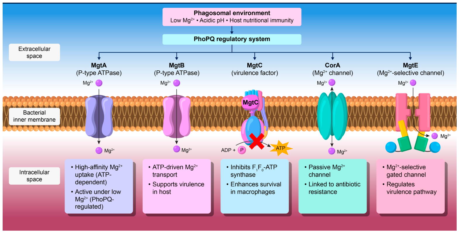
In this review, we discuss how Mg2+ transporters contribute to pathogen survival and virulence, particularly within macrophages. We also discuss recent findings regarding the functions and regulatory mechanisms of MgtA/B/C, CorA, and MgtE (Figure 1). In addition, we examine the conservation and variation in these systems among pathogens and highlight their potential as therapeutic targets for infectious diseases.

2. Pathogen Survival in the Phagosome: The MgtA/B/C System

Nutritional immunity is a host defense strategy that restricts the availability of essential metal ions required for pathogen survival, including Fe, Zn, Mn, and Mg2+, to suppress microbial function [8]. Magnesium has received less attention than either iron or zinc. However, recent studies have revealed that Mg2+ limitation also functions as an important component of nutritional immunity that is closely linked to the regulation of bacterial pathogenicity. Recent studies have provided direct evidence for this mechanism: the host endolysosomal cation channel MCOLN2 has been shown to actively deplete Mg2+ from pathogen-containing vacuoles, and ablation of MCOLN2 in human macrophages increased Salmonella enterica serovar Typhi intracellular replication by approximately 2.5-fold, confirming that Mg2+ deprivation constitutes an active host defense strategy [10]. In addition, cytoplasmic Mg2+ starvation has been found to trigger MgtC-dependent restriction of phosphate uptake, thereby limiting ATP precursor availability and coupling Mg2+ limitation to global metabolic suppression [11]. Mg2+ is essential for various biochemical processes in bacteria, and its limitation leads to ribosomal destabilization, ATP metabolic disruption, and impaired DNA/RNA integrity. Mg2+ ions neutralize the negative charges of phosphate groups in the rRNA backbone, and their depletion destabilizes ribosomal subunit assembly [3,4]. At the single-cell level, structural perturbation of the ribosome complex through deletion of ribosomal protein L34 has been shown to increase membrane hyperpolarization events and cell death, and supplementation of exogenous Mg2+ was sufficient to suppress hyperpolarization and restore wild-type growth rates, establishing a direct causal relationship between Mg2+ availability and ribosome-dependent survival [12]. Consequently, maintaining a low Mg2+ concentration within the phagosome alone can markedly reduce bacterial viability.
To counter this host-imposed restriction, pathogens have evolved high-affinity Mg2+ transporters, among which the Mgt family has been most extensively characterized, particularly in Salmonella and other enteric pathogens, and has been widely studied in the context of intramacrophage survival [5,13]. However, these proteins do not merely function as Mg2+ transporters. Instead, they directly influence bacterial survival during infection and regulate virulence gene expression.

2.1. Regulation of MgtA/B/C by the PhoPQ Two-Component System

The mgtA, mgtB, and mgtC genes have been most extensively studied in Salmonella spp. Therefore, unless otherwise stated, this review primarily refers to the findings derived from Salmonella-based studies when discussing the Mgt system. The expression of these genes is tightly regulated by the PhoPQ two-component system, which senses environmental Mg2+ levels and serves as a central regulator of bacterial survival strategies [14].
Thus, PhoPQ functions as more than a simple Mg2+-sensing system. It acts as a global regulator that coordinates multiple adaptive responses essential for bacterial survival within the macrophages. These responses include resistance to antimicrobial peptides (AMPs), lipopolysaccharide (LPS) modification, and membrane remodeling [15]. For example, PhoPQ regulates the expression of the PmrAB two-component regulatory system and pagP, which alter bacterial surface charge to enhance resistance against host cationic antimicrobial peptides (CAMPs) [16,17]. Notably, PhoPQ can sense multiple environmental signals, including low Mg2+ concentration and acidic pH [14,17]. Thus, Mg2+ limitation and low pH within the phagosomes activate PhoQ. This integrated sensing enables PhoP to precisely adjust gene expression under diverse environmental conditions.
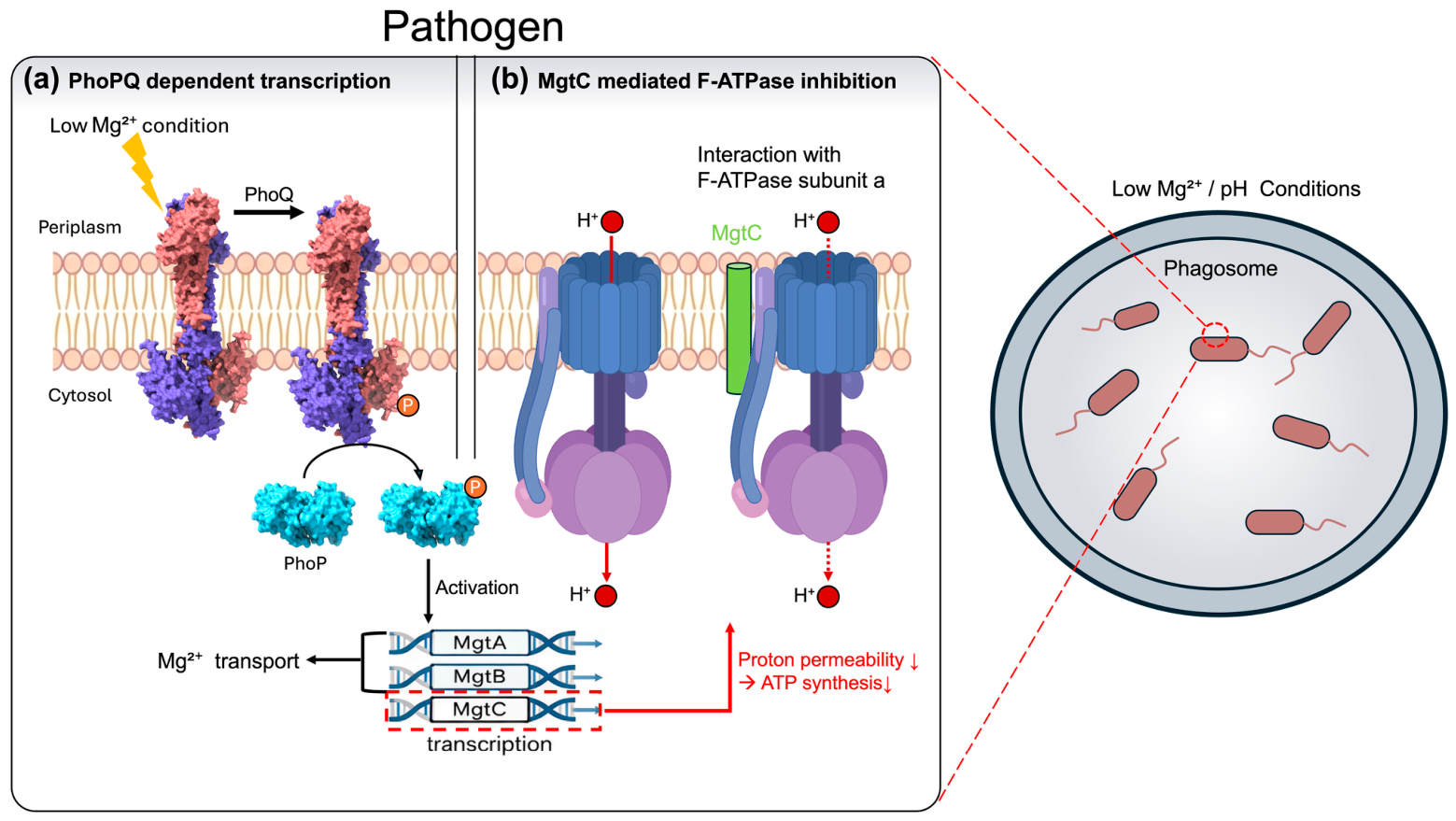
PhoQ is a histidine kinase located in the bacterial inner membrane. Under low external Mg2+ conditions, PhoQ undergoes autophosphorylation and subsequently transfers the phosphate group to the cytoplasmic response regulator PhoP, thereby activating it [18] (Figure 2). Activated PhoP promotes the transcription of numerous genes, including mgtA, mgtB, and mgtC. Thus, the PhoPQ system governs not only Mg2+ transporter expression but also multiple survival mechanisms, such as antimicrobial peptide resistance, membrane remodeling, and LPS modification, all of which are essential for successful infection [15] (Figure 2).

2.2. MgtA and MgtB: P-Type ATPases with Conserved Architecture

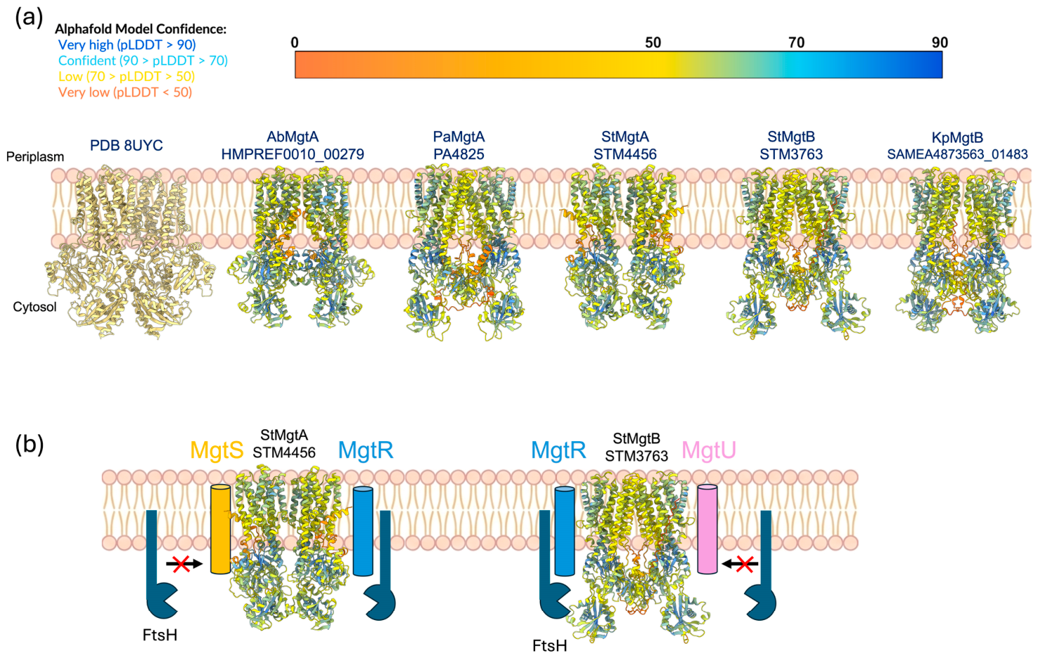
MgtA and MgtB are P-type ATPases that import Mg2+ into the cells with a high affinity through ATP hydrolysis. These proteins share approximately 50% sequence similarity [5]. The structural predictions generated by AlphaFold suggest that they possess similar architectures overall. Despite their structural resemblance, they exhibit distinct functional properties, particularly in their interactions with small membrane proteins. MgtA associates with MgtS, whereas MgtB interacts with MgtU. Specifically, MgtS is a small inner membrane peptide (31 amino acids) that stabilizes MgtA by directly interacting with its transmembrane domain, thereby preventing FtsH-mediated proteolytic degradation [19]. Similarly, MgtU plays an analogous protective role for MgtB. In the absence of MgtS or MgtU, the respective transporter is rapidly degraded, leading to reduced intracellular Mg2+ uptake and compromised bacterial survival within macrophages [19] (Figure 3). In contrast, both MgtA and MgtB interact with a regulatory peptide, MgtR, which enhances their susceptibility to FtsH-mediated degradation [20]. This regulatory mechanism enables bacteria to modulate transporter abundance in response to changes in magnesium levels. Recent studies suggest that the activities of these transporters can also vary depending on specific lipid compositions of the membrane, as MgtA has been shown to be activated by cardiolipin and is sensitive to free Mg2+ concentrations in vitro [21,22]. Together, these findings indicate that MgtA and MgtB are not merely simple ion pumps; instead, they are highly regulated transporters sensitive to various physiological factors, including membrane composition, protein complex formation, and cellular energy status.
In particular, MgtB is known to function as a direct regulator of virulence in several pathogens. In Yersinia species, MgtB enhances bacterial survival during the early stages of infection, and mgtB deletion markedly reduces virulence [23]. Moreover, studies in Salmonella have demonstrated that MgtB plays a critical role in maintaining virulence within macrophages expressing functional Slc11a1+/+ (also known as NRAMP1), a proton-coupled divalent cation transporter localized to the phagosomal membrane. Slc11a1 mediates pH-dependent efflux of Fe2+, Mn2+, and other divalent cations from the acidified phagosomal lumen into the cytoplasm, thereby imposing severe nutritional stress on intracellular pathogens [24,25]. Under these conditions, the absence of MgtB drastically decreases bacterial survival, underscoring its essential role in maintaining Mg2+ homeostasis during extreme nutritional stress.
Recent structural studies have revealed that MgtA, which shares high similarity with MgtB, was long thought to function as a monomer but instead forms a stable homodimer at 2.9 Å resolution, the first dimeric structure reported for any P-type ATPase [26]. This observation suggests that the regulatory mechanisms governing MgtA and MgtB function are more complex than previously appreciated, emphasizing the need for further structural investigation.

2.3. MgtC: A Virulence Factor That Does Not Transport Mg2+

MgtC does not directly transport Mg2+ but functions as an atypical virulence protein critical for bacterial survival under Mg2+-limiting conditions within macrophages [6,27]. Similar to MgtA and MgtB, MgtC is degraded by the FtsH protease upon its interaction with the regulatory peptide MgtR [28]. Recent findings have revealed that MgtC directly interacts with subunit a of the F1Fo ATP synthase to inhibit bacterial ATP synthesis [7]. This inhibition represents a survival strategy that enables bacteria to minimize ATP consumption and prioritize survival under energy-limited conditions such as those inside macrophages.
MgtC sustains PhoPQ activation through two distinct feedback mechanisms. First, reduced ATP levels further suppress the activity of ATP-dependent proteases (e.g., ClpS-ClpAP), thereby stabilizing regulatory proteins, such as PhoP against proteolysis [29]. Stabilization of PhoP subsequently leads to sustained mgtA/B/C expression, allowing MgtC to maintain PhoPQ activation through an indirect feedback loop [30].
Second, and independently, MgtC directly interacts with PhoP, promoting PhoP stability and creating a direct positive feedback loop to sustain transcription of mgtC itself, as well as mgtA and mgtB [13]. These regulatory mechanisms demonstrate that MgtC functions not as a single virulence factor but as a higher-order regulator coordinating bacterial survival under host-imposed stress [7].
MgtC has been predominantly identified in pathogenic bacteria and is rarely found or expressed in non-pathogenic strains [6,13,27]. This pattern suggests that MgtC has evolved to specialize in infection environments, highlighting its potential as a future therapeutic target against bacterial pathogenesis.

2.4. Mgt Homolog Distribution in Clinically Relevant Pathogens Beyond Salmonella

To date, MgtA/B/C has mainly been studied in Salmonella spp. However, genomic analyses (Table 1) have indicated the presence of Mgt homologs in various other pathogenic bacteria. Notably, the distribution of individual Mgt components varied across the species. Although Pseudomonas aeruginosa (P. aeruginosa) and Acinetobacter baumannii (A. baumannii) harbor MgtA and MgtC homologs, they lack a canonical MgtB ortholog, contrasting with the intact mgtCB operon in Salmonella. This raises intriguing evolutionary questions regarding the selective pressures that shaped the Mgt system in different pathogenic contexts. These pathogens belong to the clinically significant and antibiotic-resistant ESKAPE pathogen group. However, the structural features, regulatory mechanisms, interacting partners, and membrane lipid composition of the Mgt system in these non-Salmonella pathogens remain unknown. Further research into these areas will provide valuable insights into the conservation and pathogen-specific variations in the Mgt system, and potentially uncover novel targets for antimicrobial therapies. To complement the strain-level summary in Table 1, comparative phylogenetic analyses of MgtA/MgtB and MgtC homologs across these pathogens are provided as Supplementary Figure S1, with the underlying multiple sequence alignments included as Supplementary Data S1 and S2. A broader-scale phylogenetic analysis of MgtA homologs from 2450 bacterial strains, together with the corresponding sequence metadata, is provided as Supplementary Data S3-1 and S3-2.

3. CorA: A Conserved Mg2+ Channel with Emerging Roles in Pathogenesis

CorA is a highly conserved Mg2+ transporter, found in nearly all prokaryotic species [35]. Homologous proteins, such as Mrs2, are also present in the inner membrane of mitochondria in some eukaryotic cells. This high degree of conservation suggests that CorA is a fundamental transporter required for basic cellular functions. The physiological roles of CorA have been verified in a wide range of bacterial species, including Escherichia coli (E. coli), Salmonella, Mycobacterium, Bacillus subtilis, and Pseudomonas aeruginosa [36,37]. CorA functions as a transmembrane channel that facilitates the passive influx of Mg2+ into the cell, driven by an electrochemical gradient. This process does not require an energy input and occurs only when extracellular Mg2+ concentrations exceed intracellular levels. As such, CorA serves as the primary transporter for satisfying basal Mg2+ requirements under normal growth conditions [35].
CorA typically forms a pentameric complex in the membrane. Each subunit contains one transmembrane domain and one cytoplasmic regulatory domain [38]. Channel gating is controlled by Mg2+ binding to the cytoplasmic domain via a negative feedback mechanism. When the intracellular Mg2+ concentration increases, the channel closes, and when Mg2+ levels decrease, the channel opens [39]. The GxN (or GMN) motif plays a central role in both Mg2+ binding and gate regulation [40]. This motif allows the selective transport of divalent cations, including Mg2+. Binding of Mg2+ causes the channel to constrict, blocking ion flow, whereas Mg2+ release expands the channel, permitting influx. This structural regulation allows CorA to maintain intracellular Mg2+ homeostasis without energy expenditure, making it particularly advantageous under low-energy conditions [35,38].
CorA has traditionally been viewed as an essential transporter for core bacterial physiology. However, emerging evidence shows that CorA also participates in infection and pathogenesis. In Salmonella enterica serovar Typhimurium (S. Typhimurium), a single deletion of corA produces a relatively mild phenotype, likely because MgtA and the PhoPQ system provide compensatory Mg2+ uptake. However, the combined deletion of corA and phoP produces a synthetic genetic effect, resulting in marked growth reduction, diminished motility, and competitive disadvantage [41]. This indicates that CorA- and PhoPQ-regulated transporters function as complementary Mg2+ acquisition pathways. Additionally, CorA has been reported to mediate the nonselective transport of other divalent metal ions, such as Ca2+, Co2+, and Ni2+, in species including Pectobacterium versatile, Salmonella enterica, and E. coli [36,42]. These properties suggest that CorA contributes to maintaining ionic balance in complex infection environments. Such versatility implies that CorA functions not only in Mg2+ transport but also in adaptation to environmental stressors such as membrane stability, osmotic pressure, and pH fluctuations.
Notably, a recent study has revealed an association between CorA and antibiotic resistance. A recent study [43] revealed that corA deletion in Mycobacterium smegmatis increased its sensitivity to structurally diverse antibiotics. Resistance to fluoroquinolones and aminoglycoside antibiotics was also reduced. This phenotype was also observed in E. coli recombinantly expressing M. smegmatis CorA. These results suggest that CorA acts as a structural platform to maintain the function of antibiotic efflux systems. Although M. smegmatis is a non-pathogenic model organism, these findings have significant implications for pathogenic mycobacteria such as M. tuberculosis, where CorA homologs may similarly influence intrinsic antibiotic resistance. Future studies using pathogenic mycobacterial strains will be critical to validate this hypothesis.
CorA is highly conserved not only in pathogenic bacteria but also in non-pathogenic and commensal strains. This conservation indicates that CorA is a fundamental component of bacterial survival and is not restricted to virulent species [35]. However, in pathogenic contexts, CorA may interact with virulence regulatory systems to support adaptation to changing environmental conditions. Further studies are required to characterize CorA’s structural stability, ion selectivity, contribution to antibiotic sensitivity, and potential interplay with other transporters during infection. Defining CorA as a virulence accessory factor and exploring its role in antibiotic resistance mechanisms may open new opportunities for antimicrobial development.

4. MgtE: A Mg2+ Channel That Regulates Pathogenicity

MgtE is a Mg2+ channel widely conserved in both prokaryotes and eukaryotes [44]. The MgtE channel of bacteria is highly selective for Mg2+ and is involved in maintaining intracellular Mg2+ concentrations [45]. Structurally, MgtE is not an ATPase but rather an Mg2+-selective channel whose gating is regulated through structural changes induced by ATP binding [46]. When the intracellular Mg2+ and ATP concentrations are high, ATP binds to MgtE and stabilizes its closed conformation. This closed state prevents Mg2+ influx by obstructing the ion conduction pathway. Conversely, when intracellular Mg2+ and ATP concentrations are low, ATP dissociates from MgtE, resulting in the structural relaxation of the cytosolic domain and opening of the transport channel. These structural transitions occur without direct energy expenditure and allow MgtE to actively respond to fluctuations in cellular Mg2+ and ATP concentrations [46]. Crystallographic analysis of the MgtE cytoplasmic domain in complex with ATP has revealed that ATP coordinates between the N-terminal and CBS domains, inducing a conformational closure that directly occludes the ion conduction pathway [46]. Complementary cryo-EM analysis of full-length MgtE in the Mg2+-free state at near-atomic resolution further elucidated the open-to-closed gating transition, with specific rearrangements in TM2 and TM5 facilitating channel closure upon ATP binding [47].
MgtE of P. aeruginosa has been shown to function as more than a simple Mg2+ transporter and directly participates in infection persistence and pathogenicity [48,49]. The mutant strain lacking the mgtE gene showed altered phenotypes associated with cytotoxicity, including changes in exoenzyme expression and the regulation of biofilm-associated genes. It has been suggested that cytotoxicity regulation via Type III secretion system (T3SS) suppression could represent a critical step in chronic P. aeruginosa infections [48,49]. A previous study demonstrated gradual T3SS suppression following airway infection [50], and this T3SS regulation appears to be mediated by MgtE.
Co-culture experiments with P. aeruginosa and human-derived cystic fibrosis bronchial epithelial (CFBE) cells further demonstrated that MgtE deficiency significantly influenced T3SS expression, exoenzyme production, and biofilm-associated gene regulation, thereby altering bacterial cytotoxicity [51]. This finding suggests that MgtE is a regulatory factor closely integrated into the bacterial virulence network, extending its role beyond Mg2+ transport to include the direct modulation of pathogenic mechanisms essential for establishing chronic infections.

5. Mg2+ Transporters as Potential Therapeutic Targets

Mg2+ transport-related systems deserve attention as potential drug targets because their functions extend beyond Mg2+ uptake to directly support bacterial survival and virulence in host environments. Several features support their potential as therapeutic targets.
First, MgtC, for example, enhances bacterial survival within macrophages by altering cellular energy metabolism and helping pathogens withstand Mg2+-limited intracellular stress. As a result, it contributes to persistence in immune cells, which is a key feature of pathogenicity in several bacteria. MgtC is predominantly found in pathogenic bacteria and has no characterized mammalian counterpart, and the bacterial F1Fo ATP synthase subunit a targeted by MgtC differs substantially from the mitochondrial ATP synthase, reducing the likelihood of off-target effects. Cryo-EM studies have revealed that the bacterial Fo region consists of only three core subunits (ab2c9–15), in contrast to the more elaborate organization of the mammalian mitochondrial enzyme, which includes numerous supernumerary subunits absent from bacteria [52]. Moreover, a recent high-resolution (2.0–2.4 Å) structure of the P. aeruginosa ATP synthase identified species-specific features in the Fo region, including a unique ε-subunit inhibitory binding site and a coordinated metal ion (identified as zinc by mass spectrometry) capping the cytoplasmic proton channel, neither of which is found in mitochondrial counterparts [53]. These structural divergences support the feasibility of selective inhibitor design targeting the MgtC–ATP synthase interaction interface.
Second, the availability of high-resolution cryo-EM structures, including the recent MgtA dimer structure at 2.9 Å resolution [26] and the well-characterized CorA pentameric channel [38,39], provides a foundation for structure-based drug design (SBDD) approaches. Computational screening of compound libraries against MgtA’s ATP-binding site or the MgtC–F1Fo interaction interface could identify lead compounds for further development.
Third, the association between CorA and intrinsic antibiotic resistance in mycobacteria [43] suggests that targeting CorA function could serve as an adjuvant strategy to potentiate the activity of existing antibiotics. MgtE also appears to have consequences beyond Mg2+ homeostasis, as it has been linked to changes in cytotoxicity and T3SS regulation. Because these proteins influence infection-relevant outcomes, their inhibition could lead not only to impaired adaptation to host-imposed stress but also to the attenuation of bacterial virulence and persistence.
Although no specific inhibitors of bacterial Mg2+ transporters have yet advanced to clinical development, the convergence of structural data, infection-specific expression profiles, and validated virulence phenotypes positions this transporter family as a promising frontier for next-generation antimicrobial discovery.

6. Conclusions and Future Perspectives

Mg2+ is an essential metal ion required for the survival of all living organisms. Mg2+ is involved in almost all physiological processes, including cellular metabolism, gene expression, protein translation, and energy production. Because Mg2+ cannot pass through the cell membrane spontaneously, organisms have evolved specialized transporters to facilitate its selective uptake. Importantly, these transporters are now increasingly recognized not only for their role in ion transport but also for their direct involvement in bacterial survival and virulence under infection conditions.
This review focuses on the major Mg2+ transporters, namely MgtA, MgtB, MgtC, CorA, and MgtE, and examines their biochemical properties, structures, regulatory mechanisms, and functional diversity in pathogenic bacteria. Their roles are crucial for bacterial adaptation to the Mg2+-limited phagosomal environment within macrophages. MgtA and MgtB are high-affinity P-type ATPases expressed under the control of the PhoPQ system. They import Mg2+ into cells under low-Mg2+ conditions, supporting bacterial survival. Their protein stability is regulated through interactions with small regulatory peptides, and they are also responsive to specific membrane lipid compositions. MgtB has been shown to directly influence virulence in Yersinia and Salmonella. MgtB in Salmonella functions as an essential factor for survival in Slc11a1+/+ macrophages.
MgtC does not directly transport Mg2+ but acts as a strategic factor by suppressing ATP synthesis through interaction with subunit a of the F1Fo ATP synthase, stabilizing PhoP, and maintaining the PhoPQ system through dual positive feedback loops. This suggests that MgtC functions not merely as a virulence factor but as a higher-order regulator orchestrating bacterial survival strategies. CorA is a conserved pentameric channel for maintaining intracellular Mg2+ homeostasis. It is highly conserved in structure and operates through gating controlled by the GxN motif. Recent findings indicate that CorA in Mycobacterium smegmatis can influence antibiotic susceptibility, suggesting that it may also contribute to infection adaptation and resistance regulation. Validation in pathogenic mycobacterial species remains an important next step.
As discussed in Section 4, MgtE regulates its gating through ATP binding rather than hydrolysis. In P. aeruginosa, MgtE has been linked to T3SS regulation and infection-mode switching, positioning it as a multifunctional virulence-associated factor beyond its canonical role in Mg2+ transport. The structural, functional, and regulatory characteristics of the major bacterial Mg2+ transporters discussed in this review are summarized in Table 2.
These findings demonstrate that Mg2+ transporters play a wide range of roles that extend beyond basic survival. They contribute to virulence expression, infection persistence, and regulation of antibiotic resistance, highlighting their biological significance and potential as therapeutic targets. If transporters that are specifically expressed or activated in infection environments can be precisely inhibited, it may be possible to suppress pathogen survival while minimizing disruption to the host microbiota. This could provide a foundation for next-generation precision antimicrobial strategies.
However, the mechanisms by which these Mg2+ transporters are connected to virulence regulation remain incompletely understood. Further investigation is needed to clarify their regulatory elements, structure-based activation mechanisms, and pathogen-specific expression patterns. Multifunctional regulators like MgtC and MgtE, which extend beyond traditional transporter definitions, may become key research topics in future studies on infectious diseases. Ongoing research should include comparative analyses of transporter function across different pathogens, mapping of regulatory networks, and structure-based inhibitor design. These efforts will not only contribute to the control of infectious diseases but also provide important insights into the evolutionary dynamics of bacterial virulence.

Supplementary Materials

The following supporting information can be downloaded at: https://www.mdpi.com/article/10.3390/microorganisms14051033/s1, Supplementary Figure S1: Phylogenetic relationships of MgtA/MgtB and MgtC homologs in pathogenic bacteria; Supplementary Data S1: Multiple sequence alignment of MgtA and MgtB homologs from pathogenic bacteria; Supplementary Data S2: Multiple sequence alignment of MgtC homologs from pathogenic bacteria; Supplementary Data S3-1: Broad-scale phylogenetic tree of MgtA homologs from 2450 bacterial strains; Supplementary Data S3-2: Metadata for MgtA homolog sequences used in the broad-scale phylogenetic analysis.

Author Contributions

Conceptualization, S.H. and J.M.C.; investigation, S.H.; formal analysis, S.H. and J.M.C.; writing—original draft preparation, S.H.; writing—review and editing, S.H., Y.Y., and J.M.C.; visualization, S.H.; supervision, J.M.C.; project administration, J.M.C.; funding acquisition, J.M.C. All authors have read and agreed to the published version of the manuscript.

Funding

This work was supported by the Research Fund of The Catholic University of Korea in 2024 and by the National Research Foundation (NRF) of Korea through grants RS-2022-NR071689.

Institutional Review Board Statement

Not applicable.

Informed Consent Statement

Not applicable.

Data Availability Statement

All data supporting this review are contained within the article and the Supplementary Materials. No new primary datasets were generated.

Conflicts of Interest

The authors declare no conflicts of interest.

Abbreviations

AMPAntimicrobial peptide
ATPAdenosine triphosphate
CAMPsCationic antimicrobial peptides
CFBECystic fibrosis bronchial epithelial
ESKAPEEnterococcus faecium, Staphylococcus aureus, Klebsiella pneumoniae, Acinetobacter baumannii, Pseudomonas aeruginosa, and Enterobacter species
LPSLipopolysaccharide
PDBProtein Data Bank
pLDDTPredicted local distance difference test
SBDDStructure-based drug design
T3SSType III secretion system

References

  1. Romani, A.M. Cellular magnesium homeostasis. Arch. Biochem. Biophys. 2011, 512, 1–23. [Google Scholar] [CrossRef] [PubMed]
  2. Diamant, S.; Azem, A.; Weiss, C.; Goloubinoff, P. Effect of free and ATP-bound magnesium and manganese ions on the ATPase activity of chaperonin GroEL14. Biochemistry 1995, 34, 273–277. [Google Scholar] [CrossRef] [PubMed]
  3. Gesteland, R.F. Unfolding of Escherichia coli ribosomes by removal of magnesium. J. Mol. Biol. 1966, 18, 356–371. [Google Scholar] [CrossRef]
  4. Klein, D.J.; Moore, P.B.; Steitz, T.A. The contribution of metal ions to the structural stability of the large ribosomal subunit. RNA 2004, 10, 1366–1379. [Google Scholar] [CrossRef]
  5. Groisman, E.A.; Hollands, K.; Kriner, M.A.; Lee, E.-J.; Park, S.-Y.; Pontes, M.H. Bacterial Mg2+ homeostasis, transport, and virulence. Annu. Rev. Genet. 2013, 47, 625–646. [Google Scholar] [CrossRef]
  6. Miyashiro, T.; Goulian, M. Stimulus-dependent differential regulation in the Escherichia coli PhoQ–PhoP system. Proc. Natl. Acad. Sci. USA 2007, 104, 16305–16310. [Google Scholar] [CrossRef]
  7. Lee, E.-J.; Pontes, M.H.; Groisman, E.A. A bacterial virulence protein promotes pathogenicity by inhibiting the bacterium’s own F1Fo ATP synthase. Cell 2013, 154, 146–156. [Google Scholar] [CrossRef]
  8. Lopez, C.A.; Skaar, E.P. The Impact of Dietary Transition Metals on Host-Bacterial Interactions. Cell Host Microbe 2018, 23, 737–748. [Google Scholar] [CrossRef] [PubMed]
  9. Sheldon, J.R.; Laakso, H.A.; Heinrichs, D.E. Iron Acquisition Strategies of Bacterial Pathogens. Microbiol. Spectr. 2016, 4, 43–85. [Google Scholar] [CrossRef]
  10. Gibbs, K.D.; Wang, L.; Yang, Z.; Anderson, C.E.; Bourgeois, J.S.; Cao, Y.; Gaggioli, M.R.; Biel, M.; Puertollano, R.; Chen, C.C.; et al. Human variation impacting MCOLN2 restricts Salmonella Typhi replication by magnesium deprivation. Cell Genom. 2023, 3, 100290. [Google Scholar] [CrossRef]
  11. Bruna, R.E.; Kendra, C.G.; Groisman, E.A.; Pontes, M.H. Limitation of phosphate assimilation maintains cytoplasmic magnesium homeostasis. Proc. Natl. Acad. Sci. USA 2021, 118, e2021370118. [Google Scholar] [CrossRef] [PubMed]
  12. Lee, D.D.; Galera-Laporta, L.; Bialecka-Fornal, M.; Moon, E.C.; Shen, Z.; Briggs, S.P.; Garcia-Ojalvo, J.; Süel, G.M. Magnesium Flux Modulates Ribosomes to Increase Bacterial Survival. Cell 2019, 177, 352–360.e13. [Google Scholar] [CrossRef]
  13. Groisman, E.A.; Duprey, A.; Choi, J. How the PhoP/PhoQ System Controls Virulence and Mg2+ Homeostasis: Lessons in Signal Transduction, Pathogenesis, Physiology, and Evolution. Microbiol. Mol. Biol. Rev. 2021, 85, e0017620. [Google Scholar] [CrossRef]
  14. Groisman, E.A. The Pleiotropic Two-Component Regulatory System PhoP-PhoQ. J. Bacteriol. 2001, 183, 1835–1842. [Google Scholar] [CrossRef] [PubMed]
  15. Dalebroux, Z.D.; Matamouros, S.; Whittington, D.; Bishop, R.E.; Miller, S.I. PhoPQ regulates acidic glycerophospholipid content of the Salmonella Typhimurium outer membrane. Proc. Natl. Acad. Sci. USA 2014, 111, 1963–1968. [Google Scholar] [CrossRef] [PubMed]
  16. Dalebroux, Z.D.; Miller, S.I. Salmonellae PhoPQ regulation of the outer membrane to resist innate immunity. Curr. Opin. Microbiol. 2014, 17, 106–113. [Google Scholar] [CrossRef]
  17. Véscovi, E.G.; Soncini, F.C.; Groisman, E.A. Mg2+ as an extracellular signal: Environmental regulation of Salmonella virulence. Cell 1996, 84, 165–174. [Google Scholar] [CrossRef]
  18. Mao, M.; He, L.; Yan, Q. An updated overview on the bacterial PhoP/PhoQ two-component signal transduction system. Front. Cell. Infect. Microbiol. 2025, 15, 1509037. [Google Scholar] [CrossRef]
  19. Yeom, J.; Shao, Y.; Groisman, E.A. Small proteins regulate Salmonella survival inside macrophages by controlling degradation of a magnesium transporter. Proc. Natl. Acad. Sci. USA 2020, 117, 20235–20243. [Google Scholar] [CrossRef]
  20. Choi, E.; Lee, K.Y.; Shin, D. The MgtR regulatory peptide negatively controls expression of the MgtA Mg2+ transporter in Salmonella enterica serovar Typhimurium. Biochem. Biophys. Res. Commun. 2012, 417, 318–323. [Google Scholar] [CrossRef] [PubMed]
  21. Subramani, S.; Perdreau-Dahl, H.; Morth, J.P. The magnesium transporter A is activated by cardiolipin and is highly sensitive to free magnesium in vitro. Elife 2016, 5, e11407. [Google Scholar] [CrossRef]
  22. Weikum, J.; van Dyck, J.F.; Subramani, S.; Klebl, D.P.; Storflor, M.; Muench, S.P.; Abel, S.; Sobott, F.; Morth, J.P. The bacterial magnesium transporter MgtA reveals highly selective interaction with specific cardiolipin species. Biochim. Biophys. Acta Mol. Cell. Res. 2024, 1871, 119614. [Google Scholar] [CrossRef]
  23. Ford, D.C.; Joshua, G.W.P.; Wren, B.W.; Oyston, P.C.F. The importance of the magnesium transporter MgtB for virulence of Yersinia pseudotuberculosis and Yersinia pestis. Microbiology 2014, 160, 2710–2717. [Google Scholar] [CrossRef]
  24. Choi, E.; Choi, S.; Nam, D.; Park, S.; Han, Y.; Lee, J.-S.; Lee, E.-J. Elongation factor P restricts Salmonella’s growth by controlling translation of a Mg2+ transporter gene during infection. Sci. Rep. 2017, 7, 42098. [Google Scholar] [CrossRef] [PubMed]
  25. Cellier, M.F.; Courville, P.; Campion, C. Nramp1 phagocyte intracellular metal withdrawal defense. Microbes Infect. 2007, 9, 1662–1670. [Google Scholar] [CrossRef]
  26. Zeinert, R.; Zhou, F.; Franco, P.; Zöller, J.; Madni, Z.K.; Lessen, H.; Aravind, L.; Langer, J.D.; Sodt, A.J.; Storz, G.; et al. P-type ATPase magnesium transporter MgtA acts as a dimer. Nat. Struct. Mol. Biol. 2025, 32, 1633–1643. [Google Scholar] [CrossRef]
  27. Blanc-Potard, A.B.; Groisman, E.A. The Salmonella selC locus contains a pathogenicity island mediating intramacrophage survival. EMBO J. 1997, 16, 5376–5385. [Google Scholar] [CrossRef]
  28. Alix, E.; Blanc-Potard, A.B. Peptide-assisted degradation of the Salmonella MgtC virulence factor. EMBO J. 2008, 27, 546–557. [Google Scholar] [CrossRef] [PubMed]
  29. Yeom, J.; Groisman, E.A. Reduced ATP-dependent proteolysis of functional proteins during nutrient limitation speeds the return of microbes to a growth state. Sci. Signal. 2021, 14, eabc4235. [Google Scholar] [CrossRef] [PubMed]
  30. Yeom, J.; Wayne, K.J.; Groisman, E.A. Sequestration from Protease Adaptor Confers Differential Stability to Protease Substrate. Mol. Cell 2017, 66, 234–246.e235. [Google Scholar] [CrossRef]
  31. Guragain, M.; King, M.M.; Williamson, K.S.; Pérez-Osorio, A.C.; Akiyama, T.; Khanam, S.; Patrauchan, M.A.; Franklin, M.J. The Pseudomonas aeruginosa PAO1 Two-Component Regulator CarSR Regulates Calcium Homeostasis and Calcium-Induced Virulence Factor Production through Its Regulatory Targets CarO and CarP. J. Bacteriol. 2016, 198, 951–963. [Google Scholar] [CrossRef]
  32. Rang, C.; Alix, E.; Felix, C.; Heitz, A.; Tasse, L.; Blanc-Potard, A.B. Dual role of the MgtC virulence factor in host and non-host environments. Mol. Microbiol. 2007, 63, 605–622. [Google Scholar] [CrossRef] [PubMed]
  33. Moussouni, M.; Nogaret, P.; Garai, P.; Ize, B.; Vivès, E.; Blanc-Potard, A.B. Activity of a Synthetic Peptide Targeting MgtC on Pseudomonas aeruginosa Intramacrophage Survival and Biofilm Formation. Front. Cell. Infect. Microbiol. 2019, 9, 84. [Google Scholar] [CrossRef]
  34. Choi, E.; Nam, D.; Choi, J.; Park, S.; Lee, J.S.; Lee, E.J. Elongation factor P controls translation of the mgtA gene encoding a Mg2+ transporter during Salmonella infection. Microbiologyopen 2019, 8, e00680. [Google Scholar] [CrossRef]
  35. Franken, G.A.C.; Huynen, M.A.; Martínez-Cruz, L.A.; Bindels, R.J.M.; de Baaij, J.H.F. Structural and functional comparison of magnesium transporters throughout evolution. Cell. Mol. Life Sci. 2022, 79, 418. [Google Scholar] [CrossRef]
  36. Maguire, M.E. Magnesium transporters: Properties, regulation and structure. Front Biosci. 2006, 11, 3149–3163. [Google Scholar] [CrossRef]
  37. Papp, K.M.; Maguire, M.E. The CorA Mg2+ transporter does not transport Fe2+. J. Bacteriol. 2004, 186, 7653–7658. [Google Scholar] [CrossRef]
  38. Guskov, A.; Nordin, N.; Reynaud, A.; Engman, H.; Lundbäck, A.K.; Jong, A.J.; Cornvik, T.; Phua, T.; Eshaghi, S. Structural insights into the mechanisms of Mg2+ uptake, transport, and gating by CorA. Proc. Natl. Acad. Sci. USA 2012, 109, 18459–18464. [Google Scholar] [CrossRef]
  39. Matthies, D.; Dalmas, O.; Borgnia, M.J.; Dominik, P.K.; Merk, A.; Rao, P.; Reddy, B.G.; Islam, S.; Bartesaghi, A.; Perozo, E.; et al. Cryo-EM Structures of the Magnesium Channel CorA Reveal Symmetry Break upon Gating. Cell 2016, 164, 747–756. [Google Scholar] [CrossRef] [PubMed]
  40. Knoop, V.; Groth-Malonek, M.; Gebert, M.; Eifler, K.; Weyand, K. Transport of magnesium and other divalent cations: Evolution of the 2-TM-GxN proteins in the MIT superfamily. Mol. Genet. Genom. 2005, 274, 205–216. [Google Scholar] [CrossRef] [PubMed]
  41. Metaane, S.; Monteil, V.; Douché, T.; Giai Gianetto, Q.; Matondo, M.; Maufrais, C.; Norel, F. Loss of CorA, the primary magnesium transporter of Salmonella, is alleviated by MgtA and PhoP-dependent compensatory mechanisms. PLoS ONE 2023, 18, e0291736. [Google Scholar] [CrossRef]
  42. Kersey, C.M.; Dumenyo, C.K. Regulation of corA, the Magnesium, Nickel, Cobalt Transporter, and Its Role in the Virulence of the Soft Rot Pathogen, Pectobacterium versatile Strain Ecc71. Microorganisms 2023, 11, 1747. [Google Scholar] [CrossRef]
  43. Chatterjee, D.; Daya Manasi, A.R.; Rastogi, S.K.; Panda, A.P.; Biju, B.; Bhattacharyya, D.; Ghosh, A.S. Involvement of CorA of Mycobacterium smegmatis in exerting intrinsic resistance towards structurally unrelated antibiotics. J. Appl. Microbiol. 2024, 135, lxae298. [Google Scholar] [CrossRef]
  44. Moomaw, A.S.; Maguire, M.E. The unique nature of mg2+ channels. Physiology 2008, 23, 275–285. [Google Scholar] [CrossRef]
  45. Hattori, M.; Iwase, N.; Furuya, N.; Tanaka, Y.; Tsukazaki, T.; Ishitani, R.; Maguire, M.E.; Ito, K.; Maturana, A.; Nureki, O. Mg2+-dependent gating of bacterial MgtE channel underlies Mg2+ homeostasis. EMBO J. 2009, 28, 3602–3612. [Google Scholar] [CrossRef]
  46. Tomita, A.; Zhang, M.; Jin, F.; Zhuang, W.; Takeda, H.; Maruyama, T.; Osawa, M.; Hashimoto, K.; Kawasaki, H.; Ito, K.; et al. ATP-dependent modulation of MgtE in Mg2+ homeostasis. Nat. Commun. 2017, 8, 148. [Google Scholar] [CrossRef] [PubMed]
  47. Jin, F.; Sun, M.; Fujii, T.; Yamada, Y.; Wang, J.; Maturana, A.D.; Wada, M.; Su, S.; Ma, J.; Takeda, H.; et al. The structure of MgtE in the absence of magnesium provides new insights into channel gating. PLoS Biol. 2021, 19, e3001231. [Google Scholar] [CrossRef] [PubMed]
  48. Anderson, G.G.; Yahr, T.L.; Lovewell, R.R.; O’Toole, G.A. The Pseudomonas aeruginosa Magnesium Transporter MgtE Inhibits Transcription of the Type III Secretion System. Infect. Immun. 2010, 78, 1239–1249. [Google Scholar] [CrossRef] [PubMed]
  49. Chakravarty, S.; Melton, C.N.; Bailin, A.; Yahr, T.L.; Anderson, G.G. Pseudomonas aeruginosa Magnesium Transporter MgtE Inhibits Type III Secretion System Gene Expression by Stimulating rsmYZ Transcription. J. Bacteriol. 2017, 199, e00268-17. [Google Scholar] [CrossRef]
  50. Hauser, A.R. The type III secretion system of Pseudomonas aeruginosa: Infection by injection. Nat. Rev. Microbiol. 2009, 7, 654–665. [Google Scholar] [CrossRef]
  51. Coffey, B.M.; Akhand, S.S.; Anderson, G.G. MgtE is a dual-function protein in Pseudomonas aeruginosa. Microbiology 2014, 160, 1200–1213. [Google Scholar] [CrossRef] [PubMed]
  52. Guo, H.; Suzuki, T.; Rubinstein, J.L. Structure of a bacterial ATP synthase. Elife 2019, 8, e43128. [Google Scholar] [CrossRef]
  53. Sobti, M.; Gunn, A.P.; Brown, S.H.J.; Zavan, L.; Fraunfelter, V.M.; Wolfe, A.L.; McDevitt, C.A.; Steed, P.R.; Stewart, A.G. Distinct structural features of Pseudomonas aeruginosa ATP synthase revealed by cryo-electron microscopy. Nat. Commun. 2026, 17, 406. [Google Scholar] [CrossRef] [PubMed]
  54. Cleverley, R.M.; Kean, J.; Shintre, C.A.; Baldock, C.; Derrick, J.P.; Ford, R.C.; Prince, S.M. The Cryo-EM structure of the CorA channel from Methanocaldococcus jannaschii in low magnesium conditions. Biochim. Biophys. Acta 2015, 1848, 2206–2215. [Google Scholar] [CrossRef] [PubMed]
Figure 1. Overview of major bacterial Mg2+ transport systems and their roles in survival and pathogenicity. Low Mg2+, acidic pH, and host nutritional immunity activate the PhoPQ system, which induces MgtA, MgtB, and MgtC expression. MgtA and MgtB mediate high-affinity Mg2+ uptake, whereas MgtC supports bacterial survival in macrophages by modulating ATP synthase activity rather than by directly transporting Mg2+. In parallel, CorA functions as a conserved channel for basal Mg2+ uptake, and MgtE acts as an Mg2+-selective channel regulated by intracellular Mg2+ and ATP. This figure summarizes how Mg2+ transport and Mg2+-responsive systems contribute to bacterial adaptation and virulence.
Microorganisms 14 01033 g001
Figure 2. PhoPQ-dependent MgtA, MgtB, and MgtC regulation and MgtC-mediated control of cellular energy metabolism. (a) Under low Mg2+ conditions and acidic pH characteristic of the phagosome, PhoQ undergoes autophosphorylation and transfers the phosphate group to PhoP. Activated PhoP induces transcription of the mgtA, mgtB, and mgtC genes. MgtA and MgtB function as Mg2+ transporters and contribute to the maintenance of intracellular Mg2+ homeostasis. (b) MgtC interacts specifically with subunit a of the F1Fo ATP synthase and reduces ATP synthesis, thereby modulating cellular energy metabolism. This combined regulation supports bacterial survival under low-Mg2+ and low-pH conditions within the phagosome.
Microorganisms 14 01033 g002
Figure 3. Structural comparison of MgtA and MgtB and their regulation by small membrane peptides. (a) Experimentally determined structure of MgtA (PDB 8UYC) and AlphaFold-predicted models of MgtA and MgtB from different bacterial species in a membrane context. Models are colored according to AlphaFold confidence scores (pLDDT). Despite sharing approximately 50% sequence similarity, MgtA and MgtB display highly similar overall architectures, including conserved transmembrane and cytosolic domains, supporting their classification as closely related P-type ATPases. (b) Schematic representation of the differential regulation of MgtA and MgtB by small membrane peptides and the FtsH protease. MgtA is stabilized by interaction with MgtS, whereas MgtB is stabilized by interaction with MgtU, protecting each transporter from FtsH-mediated degradation. In contrast, the regulatory peptide MgtR promotes FtsH-dependent degradation of both MgtA and MgtB. This peptide-mediated control provides a mechanism to tune the cellular abundance of MgtA and MgtB in response to Mg2+ availability and other physiological conditions.
Microorganisms 14 01033 g003
Table 1. Distribution of MgtA, MgtB, and MgtC homologs in pathogenic bacteria. The table summarizes representative MgtA, MgtB, and MgtC homologs identified in pathogenic bacteria, including members of the ESKAPE pathogen group such as Pseudomonas aeruginosa, Acinetobacter baumannii, Enterococcus faecium, and Klebsiella pneumoniae. For each strain, the annotated protein name, UniProt accession number, and corresponding gene (ORF) identifier are listed [19,26,31,32,33,34].
StrainProteinUniProt IDGene (ORF)
P. aeruginosa (PAO1)MgtAQ9HUY5PA4825
MgtCQ9I0S6PA2558
MgtCQ9HVF6PA4635
MgtCQ9I1W7PA2148
A. baumannii (ATCC 19606)MgtAD0C699/A0ABX6CGY9HMPREF0010_00279
MgtCD0C6A0/A0ABX6CGZ0HMPREF0010_00280
E. faecium (BAA-472)MgtAQ3Y0B4HMPREF0351_10642
MgtCQ3Y0E8HMPREF0351_12381
S. typhimurium (LT2)MgtAP36640STM4456
MgtBP22036STM3763
MgtCP0CI70STM3764
K. pneumoniae (NCTC 13443)MgtAA0A086ICS8NCTC13443_01257
K. pneumoniae (5012STDY7626362)MgtBA0A486UQ81SAMEA4873563_01483
K. pneumoniae (IS43)MgtCW1DJ03PRK15385
Table 2. Comparison of major bacterial Mg2+ transporters.
FeatureMgtAMgtBMgtCCorAMgtE
TypeP-type ATPaseP-type ATPaseVirulence proteinPentameric channelMg2+-selective channel
EnergyATP hydrolysisATP hydrolysisN/A (inhibits F1Fo)PassivePassive (ATP-gated)
SignalLow Mg2+, pH (PhoPQ)Low Mg2+, pH (PhoPQ)Low Mg2+, pH (PhoPQ)Intracellular Mg2+Intracellular Mg2+/ATP
RegulationPhoPQ; MgtS/MgtR; FtsHPhoPQ; MgtU/MgtR; FtsHPhoPQ; MgtR/FtsH; PhoP loopsGxN motif gatingATP-binding gating
Virulence roleMg2+ uptake in phagosomeVirulence (Yersinia, Salmonella)F1Fo inhibition; PhoP stab.Antibiotic resistanceT3SS regulation
Key pathogensSalmonella, E. coli, P. aeruginosa, A. baumanniiSalmonella, Yersinia, K. pneumoniaeSalmonella, P. aeruginosa, A. baumanniiNearly all prokaryotesP. aeruginosa
StructureCryo-EM dimer/monomer (8UYC) [26]AlphaFold modelsNo high-res structure, AlphaFold modelsX-ray crystal [38]/Cryo-EM [54] pentamer X-ray crystal [45]/Cryo-EM structure binding with Fab [47]
Disclaimer/Publisher’s Note: The statements, opinions and data contained in all publications are solely those of the individual author(s) and contributor(s) and not of MDPI and/or the editor(s). MDPI and/or the editor(s) disclaim responsibility for any injury to people or property resulting from any ideas, methods, instructions or products referred to in the content.