


Abstract
Metabolic dysfunction-associated steatotic liver disease (MASLD) is an emerging global health challenge with limited effective therapeutic options. The gut microbiota, at the interface of host metabolism and immunity, acts as a critical disease modifier via the gut–liver axis. This review goes beyond cataloging its associations and synthesizes how intrinsic and extrinsic factors sculpt a permissive microbial ecosystem. These factors likely converge to establish a state of “metabolic dysbiosis”, fueling MASLD progression through three core mechanisms: compromised intestinal barrier integrity with immune activation, dysregulation of key microbial metabolite axes, and direct hepatic insult from gut-derived products. Next, we evaluate the translational landscape through a mechanism-informed precision framework, with an emphasis on how microbiome-based interventions could be aligned with non-invasive biomarkers increasingly used for MASLD risk stratification and treatment monitoring. By integrating evidence across scales, this review aims to frame a roadmap from microbiome correlations to causality-driven, personalized therapeutic strategies for MASLD.
1. Introduction
Metabolic dysfunction-associated steatotic liver disease (MASLD), previously referred to as “non-alcoholic fatty liver disease” (NAFLD), has become the most common chronic liver disease in the world [1,2]. Its clinical course gradually develops from simple liver steatosis to metabolic dysfunction-associated fatty hepatitis (MASH) and can further evolve into cirrhosis and hepatocellular carcinoma (HCC). In recent years, with an increasing prevalence of metabolic abnormalities such as obesity and type 2 diabetes, the incidence of MASLD and its potentially serious health hazards has surged [3]. This emerging health crisis thus imposes a great public health burden due to its association with high risks of cardiovascular events, cancer, type 2 diabetes, chronic kidney disease progression, and mortality [4].
Due to this disease’s insidious and asymptomatic nature, some MASLD patients present with irreversible advanced fibrosis or even HCC at the time of initial diagnosis [5]. Early detection is therefore of prime importance. However, in clinical practice, the current gold standard for MASLD diagnosis, liver biopsy, is an invasive method [6]. In addition, non-specific symptoms such as hepatitis often lead to misdiagnosis [7]. Therefore, there is an urgent need for effective, sensitive, and repeatable non-invasive biomarkers. Emerging evidence suggests that microbiota-associated signatures derived from stool, blood, or microbial metabolites may complement existing non-invasive tools (e.g., Controlled Attenuation Parameter, Magnetic Resonance Imaging - Proton Density Fat Fraction, fibrosis scores) for MASLD risk stratification and longitudinal monitoring [2,8,9]. Regarding treatment, poor patient adherence frequently leads to disappointing outcomes of lifestyle interventions, while approved drug treatment regimens are very limited [10]. This has formed a therapeutic impasse that refocuses on the gut microbiome. An increasing number of clinical trials on microbiota modulation now predict this as a likely future pillar of personalized MASLD management [11].
MASLD is a multi-system metabolic dysbiosis characterized by insulin resistance and metabolic syndrome [12]. Its pathogenesis is “multiple-hit” and includes liver lipid accumulation, inflammatory cascade reaction activation, intestinal microbiome dysbiosis, and genetic regulation [13]. The intestinal microbiome, containing trillions of bacteria-dominated microorganisms, as well as archaea, fungi, and viruses, is one of the key participants [13]. In human studies, MASLD-associated dysbiosis has been reported most consistently in adult cohorts enriched for obesity and/or type 2 diabetes, conditions that are closely intertwined with (and can confound) microbiome signatures attributed to MASLD itself [14]. Across such cohorts, reduced microbial diversity and enrichment of pathobiont-associated taxa (often including Proteobacteria/Enterobacteriaceae) together with depletion of fiber-associated genera (e.g., Bifidobacterium and Akkermansia) are recurrent observations, although the direction and magnitude of these shifts vary with metabolic background and study design [14]. In addition, a recent high-resolution multi-omics study reported trans-kingdom dysbiosis in a large, single-sex population (female nurses; 211 MASLD cases and 502 controls), identifying shifts in 66 bacterial species alongside viral taxa perturbations [15].
The gut and the liver display multidimensional interdependence: embryologically, the liver develops from the foregut; anatomically, it receives portal blood rich in microbial and metabolic products from the gut while secreting bile into the intestinal lumen. This bidirectional crosstalk has been termed the gut–liver axis [13]; through this axis, the gut microbiota has profound impacts on hepatic health. Despite progress, this field still faces numerous limitations that require critical interpretation of evidence. First, many findings only demonstrate correlations, necessitating further human studies to establish causality. Second, current trials primarily rely on animal models, the transferability of which to human models often remains uncertain. Third, existing research predominantly focuses on bacteria, with limited exploration of fungi, viruses, and archaea within the gut ecosystem [11]. This review will highlight these limitations and emphasize their critical importance in future research agendas.
This paper explores these lacunae by adopting a mechanistic and integrative perspective. First, we analyze how host factors program a MASLD-permissive microbiome. Then, we dissect the core causal pathways linking dysbiosis to hepatic pathology. Finally, we critically review therapeutic approaches by mapping them onto these mechanistic pillars, with the aim of accelerating the development of targeted, microbiome-informed precision medicine for MASLD.
2. Host and Behavioral Determinants of a MASLD-Permissive Microbiome
Gut microbiome composition and function are in continuous flux due to a complex interaction between host genetics and a multitude of modifiable lifestyle factors. The section deconstructs how these determinants interact at multiple levels to establish a dysbiotic state that either predisposes to or exacerbates MASLD.
2.1. Sex
Sex has a profound impact on gut microbiota composition and MASLD susceptibility. The disease is more prevalent in males [16,17,18]. The protective role of premenopausal estrogen is a central hypothesis. Indeed, premenopausal women have been observed to harbor a more abundant and metabolically favorable gut microbiota than their male counterparts [19,20,21,22].
The microbiota associated with MASLD exhibit sex-specific distributions. For instance, the genera Christensenella and Limosilactobacillus are more frequent in women, while Beduinibacterium and Anaerotruncus are more common in men [23]. Compared with healthy controls, male MASLD patients typically demonstrate reduced gut microbial α-diversity and distinct compositional shifts. In contrast, studies in female patients showed relatively higher α-diversity and opposite flora composition characteristics [24].
Animal models support these observations. When fed a high-fat diet, male mice exhibit more severe metabolic dysfunction, including increased fasting glucose, insulin, and alanine aminotransferase (ALT) levels [25]. The functional profiles of gut-derived metabolites also diverge between the sexes. A particularly important phenomenon observed in males is a change in the susceptibility of the liver to microbiota-derived inflammatory signaling, exemplified by increased hepatic expression of Toll-like receptor 4 (TLR4) and parallel elevations in levels of Gram-negative bacterial lipopolysaccharide (LPS) [18]. Thus, sex needs to be a required covariate for all gut microbiota analyses. Research into sex-specific differences in MASLD patients’ gut microbiota is essential. More elaborate knowledge of these dimorphic profiles and their disease-specific implications will prove essential to the development of sex-specific diagnostic and therapeutic approaches.
2.2. Genetics
MASLD pathogenesis arises from a complex interplay involving host genetics, epigenetics, and the gut microbiome. Family and twin studies confirm that the disease has a heritable component [26,27,28,29,30]. Twin cohort analyses in humans show that monozygotic twins have more similar microbiome profiles than dizygotic twins, supporting a heritability/host-genetic contribution to microbiome variation (observational human evidence) [31]. On the other hand, microbial metabolites have been shown (mainly in mechanistic preclinical models) to modulate host signaling/epigenetic regulation and may thereby contribute to disease progression, but direct causal evidence in humans remains limited [32,33].
Specific genetic variants modulate MASLD risk through interactions with the gut microbiome. For example, in obese youth, fecal abundances of Gemmiger and Oscillospira combined with the patatin-like phospholipase domain-containing protein 3 (PNPLA3) rs738409 risk variant predicted liver fat content [34]. Another study found that the PNPLA3 rs738409 G/G genotype is associated with reduced Desulfobacteraceae but increased fungal genera, including Candida and Aspergillus [35]. A novel mechanistic link was provided by preclinical genetic evidence (animal models) showing that intestinal TM6SF2 perturbation can worsen MASLD/MASH phenotypes alongside microbiome shifts, consistent with a gut–liver axis mechanism [36].
Collectively, these findings underscore the existence of dynamic gene–microbiome–metabolite axes in MASLD. Future research should employ integrated multi-omics approaches to delineate these complex interactions. Such mechanistic insights will be essential for developing genotype-directed, microbiome-informed therapeutic strategies.
2.3. Diet
Diet patterns are a major modifiable determinant of the intestinal microbiome and are strongly associated with MASLD risk/severity. However, much of the mechanistic evidence comes from preclinical studies, while human data are often observational [37,38,39,40,41]. Western-style dietary patterns have been shown to reshape gut microbiome composition and function across multiple study types, often shifting communities toward lower fiber-associated fermentation capacity and altered metabolite output [42]. In this framework, reduced short-chain fatty acid (SCFA) production and impaired barrier support represent plausible downstream consequences of low microbiota-accessible carbohydrate intake, providing a mechanistic link to metabolic dysfunction relevant to MASLD rather than a uniform, deterministic sequence in all individuals [42,43,44].
On the contrary, the Mediterranean diet can promote the benign transformation of the intestinal flora. These diet-induced bacterial flora changes can affect the host’s lipid/glucose homeostasis, bile acid metabolism, and immune function [45,46].
By analyzing specific micronutrients, the following mechanism is revealed: fiber-rich whole grains can reduce inflammation and improve barrier function [47]. A diet high in saturated fats can promote the growth of pro-inflammatory bacteria [48,49], while unsaturated fatty acids can enrich Bifidobacterium and butyrate-producing bacteria [50]. Emulsifiers and some food additives have been shown to alter the human gut microbiome in interventional settings (e.g., in a randomized controlled feeding study of carboxymethylcellulose in healthy adults), while other evidence comes from ex vivo human microbiota experiments and preclinical studies; therefore, their role in metabolic dysfunction should be described as supported but context-dependent rather than universally causal [51,52,53].
Therefore, dietary intervention has always been the basic and cost-effective core strategy of MASLD management, and targeted dietary adjustment provides a feasible path for restoring microbial and metabolic health.
2.4. Circadian Rhythm
A regular circadian rhythm is characterized by interconnected diurnal rhythms of the host circadian clock, liver metabolism, and gut microbiota [54]. This relationship is bidirectional: the host clock drives microbial rhythms by entrainment via feeding–fasting cycles [55,56,57,58], whereas bacterial metabolites, in turn, drive host transcriptional and metabolic oscillations [59]. Disruption of this harmony—as evidenced by high-fat diet (HFD) or aberrant light cycles—blunts microbial rhythms and is a proposed risk factor for MASLD.
In this context, time-restricted eating (TRE) shows metabolic benefits in preclinical models and has emerging interventional human evidence in MASLD; for example, one study reports a 4-week TRE regimen in 19 MASLD participants (human intervention) alongside mechanistic mouse experiments. Claims should thus be framed as supported by interventional evidence but still developing [60,61]. Interestingly, with regard to the beneficial effects of exercise, their magnitude may also depend on the timing of exercise training, as late-day exercise proves to be noticeably more effective in MASLD improvement, especially in reducing hepatic lipid accumulation, than early-day exercise [62].
2.5. Smoking
Smoking is common in patients with liver disease and is also independently associated with MASLD risk after adjusting for confounders [63]. Tobacco exposes the intestines to toxic compounds such as nicotine and aldehydes. Smoking is epidemiologically associated with microbiome alterations and MASLD risk, while mechanistic links involving barrier dysfunction and hepatic lipid accumulation are supported mainly by preclinical studies; thus, these effects should be described as potential pathways rather than established causation in humans [64,65].
Nicotine is an extensively studied alkaloid which, in combination with a high-fat diet, can have a harmful impact on the occurrence and progression of MASLD by upregulating the inflammatory response, increasing oxidative stress, promoting abdominal lipolysis, and improving liver fat synthesis [66]. Observational data show that smoking can lead to intestinal flora dysbiosis, which is usually characterized by changes in the proportion of Firmicutes/Bacteroidetes, an increase in Proteobacteria, and a decrease in beneficial genera such as Lactobacillus and Bifidobacterium. This may cause changes in the metabolite spectrum, including abnormal levels of ceramides, ethanol, LPS, and other substances [67,68]. Therefore, smoking may have a negative impact on MASLD progression through the lung–gut–liver axis, mediated by microbial metabolites. A key mechanism involves intestinal nicotine accumulation, which activates AMP-activated protein kinase α (AMPKα); this, in turn, stabilizes sphingomyelin phosphodiesterase 3 (SMPD3), increasing ceramide levels and driving MASH progression in nicotine-exposed mouse models [69].
Conversely, the gut microbiome can mitigate the damage. Bacteroides xylanisolvens degrades nicotine via the NicX enzyme, which decreases liver injury [69]. This exemplifies a complex lung–gut–liver axis in which microbial metabolism may modify environmental exposures.
2.6. Drugs
In addition to antibiotics, common drugs such as proton pump inhibitors (PPIs) and metformin have also been proven to have significant effects on the intestinal microbiome [70]. Among them, PPI use is observationally associated with reduced microbial diversity and compositional shifts [71]. Early PPI use may lead to longer flora dysbiosis and increase susceptibility to obesity [72]. Metformin treatment has been reported to alter gut microbiome composition in interventional human studies, and these changes may contribute to metabolic effects, though causal mediation in MASLD specifically remains to be fully established [73,74]. Studies suggest that this flora change is related to its metabolic improvement. Although the link between intestinal flora dysbiosis and MASLD pathogenesis has been confirmed, the question how commonly used prescription drugs affect the course of MASLD by regulating the microbiome is still a direction of insufficient research but with broad prospects.
There is a two-way interaction between drugs and the microbiome. Intestinal microorganisms can change the pharmacological properties of drugs, thus affecting their bioavailability, activity, and toxicity. These microbial-mediated drugs can modify individualized therapeutic responses, highlighting the important role of the microbiome in regulating drug safety and effectiveness (Figure 1).
Figure 1.
Host and behavioral factors affecting the MASLD gut–liver axis. This diagram explains the interaction between host endogenous and behavioral factors in shaping the composition and function of intestinal microorganisms. These interactions regulate the gut–liver axis and affect the occurrence and development of MASLD. MASLD: metabolic dysfunction-associated fatty liver disease.
3. Core Mechanisms: From Microbial Dysbiosis to Hepatic Pathology
Dysbiosis in MASLD is not merely a compositional shift but a functional alteration that disrupts key host pathways. We focus on three interconnected mechanistic axes that represent convergent functional pathways frequently implicated in MASLD while acknowledging that the relative contribution of each axis differs substantially across patient subgroups (Figure 2).
Figure 2.
The mechanism by which intestinal flora dysbiosis promotes MASLD progression through the gut–liver axis. Intestinal flora dysbiosis can participate in MASLD occurrence and development through multiple interrelated pathways. (A) Intestinal barrier damage and immune activation: Flora dysbiosis can destroy the close connection and increase intestinal permeability. This causes harmful substances such as LPS to be displaced into the portal circulation. In the liver, these molecules can activate TLRs on Kupffer cells and hepatic stellate cells, trigger NF-κB-mediated inflammatory reactions, and promote fibrosis. (B) Microbial metabolite dysregulation: Bile acid metabolism can affect the signaling pathways of FXR and TGR5, thereby regulating glucose homeostasis, lipid metabolism, and the inflammatory response. Short-chain fatty acids regulate lipid oxidation in the liver by regulating the secretion of GLP-1, PYY, and 5-HT, Treg cell differentiation, and macrophage polarization; choline deficiency will lead to an increase in TMA/TMAO, thus inhibiting the FXR signaling pathway and promoting mitochondrial dysfunction, macrophage death, and fibrosis; finally, intestinal ethanol and branched-chain amino acid metabolites can aggravate liver steatosis, inflammation, and mitochondrial dysfunction. LPS: lipopolysaccharide; TLRs: toll-like receptors; NF-κB: nuclear factor kappa-light-chain-enhancer of activated B cells; FXR: farnesoid X receptor; TGR5: Takeda G protein-coupled receptor 5; GLP-1: glucagon-like peptide-1; PYY: peptide YY; 5-HT: serotonin; TMA: trimethylamine; TMAO: trimethylamine N-oxide.
3.1. The Barrier–Immune Axis: From Leaky Gut to Hepatic Inflammation
The intestinal barrier is composed of a mechanical barrier, a chemical barrier, an immune barrier, and a biological barrier. It is an important line of defense that limits the entry of harmful pathogens and pathogenic factors into the blood circulation of the gastrointestinal tract [75]. Research based largely on animal models indicates that intestinal bacterial overgrowth and barrier dysfunction are key pathogenic factors, and their severity is positively linked to steatohepatitis progression in experimental settings [76,77].
In both animal models and selected human studies, an imbalanced intestinal microbiota has been associated with impaired tight junction integrity, facilitating translocation of LPS and other bacterial products into the portal circulation, a phenomenon most prominently observed in patients with advanced steatohepatitis rather than isolated steatosis [78,79]. As one of the most important pathogen-associated molecular patterns (PAMPs), LPS targets TLR4 on the surface of Kupffer cells and hepatic stellate cells, thus triggering NF-κB-mediated inflammation and fibrosis signaling pathways [80,81,82,83,84]. This inflammatory process will further destroy the integrity of the intestinal mucosa and form a vicious cycle that aggravates the entry of toxic substances.
At the molecular level, the PAMPs and damage-associated molecular patterns (DAMPs) produced by the intestine induce the formation of NOD-like receptor family pyrin domain containing 3 (NLRP3) inflammasome by activating TLR, leading to the activation of cysteine-aspartic acid protease-1 (caspase-1), cell death, and pro-inflammatory cytokine secretion, specifically of IL-1β and IL-18. These inflammatory mediators promote inflammatory cell infiltration, thus aggravating liver damage [85,86,87,88]. In addition, animal experimental data demonstrate that high fat concentration will enhance the palmitoylation of myeloid differentiation primary response 88 (MyD88), which, in turn, enhances TLR/MyD88 pathway activity and ultimately aggravates the inflammatory response of the liver and abnormal lipid metabolism [89,90]. Collectively, these findings show how an imbalance of the microbiota—namely, MASLD-associated gut dysbiosis—triggers the gut–liver axis, leading to the maintenance of liver inflammation, insulin resistance, and fibrosis.
While the detailed molecular cascade linking intestinal barrier dysfunction to hepatic inflammation has been well elucidated in animal models, emerging human studies provide key clinical validation for this axis. Patient data show that gut microbiota dysregulation—such as increased bile salt hydrolase activity—is associated with decreased tight junction protein expression and elevated plasma endotoxin levels, in addition to being positively correlated with the degree of liver fibrosis [91,92]. Intervention studies further demonstrate that modulating microbiota function through small-molecule inhibitors or metabolites (e.g., propionate) can improve the intestinal barrier and reduce systemic inflammation, partly involving the TLR/NF-κB pathway [91,93]. Portal circulation studies directly confirm the translocation of microbial products and their association with hepatic inflammatory activation [92]. Nevertheless, finer molecular mechanisms, such as NLRP3 inflammasome activation and MyD88 palmitoylation, remain primarily elucidated through animal models. Future human studies integrating direct intestinal permeability measurements with multi-omics analyses are needed to fully validate the clinical translational value of this axis as a therapeutic target.
In summary, intestinal flora imbalance can cause an inflammatory reaction in the liver, insulin resistance, and lipid metabolism disorders. This self-reinforcing cycle of intestinal barrier–microbiome–immune system interaction jointly promotes the occurrence and development of MASLD. Research on relevant biological mechanisms provides a theoretical basis for developing treatment strategies for the intestinal flora and inflammatory pathways (Figure 3).
Figure 3.
Molecular mechanisms of gut-derived PAMP-induced hepatic inflammation. Translocated gut-derived harmful substances, such as LPS, act as PAMPs. These are recognized by PRRs, including TLR4 and TLR9, leading to the activation of transcription factors such as NF-κB. This promotes pro-IL-1β and pro-IL-18 expressions. Subsequent NLRP3 inflammasome activation results in caspase-1-mediated cleavage of pro-IL-1β and pro-IL-18 into their active forms. IL-1β and IL-18 release exacerbates hepatic inflammation by recruiting neutrophils and monocytes, while inflammasome activation can also induce inflammatory cell death. PAMP: pathogen-associated molecular pattern; PRRs: pattern recognition receptors; TLR4: toll-like receptor 4; TLR9: toll-like receptor 9; pro-IL-1β: pro-interleukin-1beta; pro-IL-18: pro-interleukin-18.
However, there are still many challenges in this field. The gap between animal and human data, along with microbial specificity among individuals, underscores the necessity for human studies to validate causal relationships [94]. To advance this field, research must further develop human cohort studies to verify these mechanisms, accounting for microbiome heterogeneity when assessing barrier function and immune activation. This will be key to transforming the “barrier–immune” axis from a compelling mechanistic concept into a reliable therapeutic intervention target.
3.2. The Metabolic Axis: Microbial Metabolites as Hepatic Messengers
The gut microbiome directly affects host metabolism by a range of metabolites, particularly bile acids (BAs), short-chain fatty acids (SCFAs), and choline compounds, which cooperate to form the central metabolic axis implicated in MASLD development. Although the metabolites produced by the microbiome affect the process of liver lipid synthesis and oxidation, lipid clearance, and glucose metabolism through the gut–liver axis, these metabolites can also interact with inflammatory reactions and metabolic regulation through nuclear receptor pathways. This section systematically reviews the correlation mechanism between microbial metabolite-level disorder and the development of MASLD.
3.2.1. Bile Acids (BAs)
Intestinal bacteria in the colon are primarily responsible for converting primary bile acids to secondary bile acids. For example, the bile salt hydrolase (BSH) expressed by Lactobacillus and the 7α-/7β-dehydroxylase activity of Clostridium scindens jointly catalyze this transformation process [95,96]. In addition, Clostridium and other 7α-dehydroxylating bacteria can produce secondary bile acids—deoxycholic acid (DCA) and lithocholic acid (LCA). At the same time, 7β-hydroxysteroid dehydrogenase (7β-HSDH) expressed by certain bacteria can promote the conversion process of hydrophilic and hydrophobic bile acids, and mediate the conversion of ursodeoxycholic acid (UDCA) to chenodeoxycholic acid (CDCA) [97].
Bile acids can also modulate microbiota composition. For example, 3-sucCA can improve intestinal barrier function and diminish systemic inflammation by increasing Akkermansia muciniphila, thus exerting favorable effects on diet-induced MASLD in mouse models [98]. Similarly, 3-acetoDCA, which was discovered as a novel and distinct bile acid, can promote the growth of Lactobacillus, showing a mutual regulation of bile acids and the microbial ecosystem [99].
The disruption of bile acid homeostasis—characterized by enhanced production, impaired feedback regulation, and altered processing—is recognized as a central mechanism in the progression from MASLD to MASH. Clinical evidence from human studies indicates that MASLD patients exhibit elevated serum and fecal bile acid levels, which correlate positively with disease severity [100,101]. Consistently, human cohort analyses reveal that increased levels of the bile acid synthesis marker C4 and decreased expression of the enterokine fibroblast growth factor 19 (FGF19) reflect a breakdown in the physiological feedback loop regulating bile acid synthesis [100]. Complementary data from animal models and translational studies further demonstrate that elevated free fatty acids and reduced hepatic small heterodimer partner (SHP) expression upregulate cholesterol 7α-hydroxylase (CYP7A1) and Na+-taurocholate cotransporting polypeptide (NTCP), thereby accelerating bile acid biosynthesis and enterohepatic recirculation [102]. Moreover, advanced profiling in human cohorts indicates that not only the quantity but also the composition of the bile acid pool shifts in relation to the degree of insulin resistance [101]. This integrated evidence from clinical observations, animal models, and computational analyses [102,103,104] collectively underscores the pivotal role of bile acid dysregulation in MASLD pathogenesis.
Bile acid regulation requires the participation of two key receptors, farnesoid X receptor (FXR) and Takeda G protein-coupled receptor 5 (TGR5). FXR, which is mainly activated by primary bile acids, has an important impact on metabolism and inflammation through multiple pathways, including inhibiting bile acid synthesis through the FGF19/CYP7A1 pathway [101], enhancing insulin sensitivity through the phosphoinositide 3-kinase (PI3K)/protein kinase B (PKB/AKT) pathway [105,106], inhibiting de novo lipogenesis (DNL) gluconeogenesis by activating peroxisome proliferator-activated receptor (αPPARα) and AMP-activated protein kinase (AMPK) [105,106], and regulating inflammatory and fibrotic reactions through the mitogen-activated protein kinase (MAPK) and mammalian target of rapamycin (mTOR) pathways [105].
On the other hand, TGR5 is mainly stimulated by secondary bile acids [107]. TGR5 activation increases deiodinase 2 (DIO2) expression, which enhances the activation of thyroxine (T4) to triiodothyronine (T3), thus increasing energy expenditure in brown adipose tissue and skeletal muscle [108]. In addition, TGR5 receptor activation can promote glucagon-like peptide-1 (GLP-1) secretion [109,110]. Despite these specific effects, FXR activation in the gastrointestinal tract can regulate microorganism composition. This regulation enhances the TGR5/GLP-1 pathway through a feedback mechanism, thereby improving liver metabolism and insulin sensitivity [111]. The latest research also shows that the intestinal flora dysbiosis observed in MASLD not only leads to the upregulation of secondary bile acid levels in the ileum but also activates the TGR5 receptor on CD8+ T cells and finally activates the mTOR/oxidative phosphorylation (OXPHOS) signaling pathway. These processes cause ileum inflammation, bile acid imbalance, and liver FXR function decline, thus promoting the progression of MASLD [112].
In summary, FXR and TGR5, as key regulatory factors of bile acid metabolism, complement each other, although their mechanisms are different. In addition, the intestinal microbiota plays an important and fundamental regulatory role. Future research efforts should be based on an integral model of the intestinal and hepatic interfaces, advocating for individualization based on the profiling of bile acids and intestinal barrier integrity, followed by intervention based on FXR, TGR5, and their pathways to arrest the progress of MASLD.
3.2.2. Short-Chain Fatty Acids (SCFAs)
SCFAs, specifically acetate (C2), propionate (C3), and butyrate (C4), are by-products of the microbial fermentation of dietary fibers [113]. They are important not only for the energetic maintenance of the intestinal epithelium but for many other cellular processes as well [107]. The role of SCFAs in MASLD is multifaceted and cannot be categorized as uniformly protective or detrimental. Their net effect is determined by a complex interplay between dietary composition, host metabolic status, and the functional capacity of the gut microbiota.
The interplay between SCFAs, dietary factors, and the intestinal microbiota is highly dynamic. An enhanced Firmicutes/Bacteroidetes (F/B) ratio, which characterizes MASLD, is often evidenced in high-fat-induced MASLD murine models. Fermentable fibers like inulin and fructooligosaccharides were proved to increase SCFA production, decrease the F/B ratio, and limit the extraction of dietary energy, thereby mitigating MASLD [114]. Significantly, the disruption of the intestinal microbiota, which characterizes MASLD, could be related to age, as shown. Adult MASLD patients typically have low Bacteroidetes, whereas the opposite is true for children [115]. In vivo comparisons involving murine models have shown varying effects of different β-glucans on SCFA production: oat β-glucans were particularly potent in increasing the production of C2 and C3 and in enhancing SCFA-producing microbial populations. This study hinted that the prebiotic activity of fibers varies depending on their origin [116].
SCFAs play important roles in the pathophysiology of MASLD, and their mechanisms are wide-ranging, often synergistic, and sometimes counteracting. Regarding the metabolic aspect, C2 activates the AMPK pathway, which decreases the secretion of chylomicrons and enhanced lipid metabolism [117]. Activation of free fatty acid receptor 2 (FFAR2) by C2 or C3 induces the secretion of gastrointestinal hormones like GLP-1, Peptide YY (PYY), and serotonin (5-HT) [118]. Regarding the functional aspect, the metabolic impact of SCFAs as an energy source is context-dependent. Under conditions of a balanced, high-fiber diet, SCFAs support beneficial functions, including hepatic health and barrier maintenance. Conversely, in the context of a chronic high-fat, low-fiber diet—a hallmark of MASLD—the caloric contribution from SCFAs may add to the overall energy surplus and thus potentially favor metabolic disease progression [119,120].
C3 and C4 increase the intestinal process of gluconeogenesis, which leads to glucose that is detected by the glucose transporter 2 (GLUT2) receptor of the portal vein, thus initiating nerve pathways that decrease food intake and increase the sensitivity of the liver to insulin [121,122]. Immunologically, SCFAs exert an anti-inflammatory effect through reducing inflammation by modulating macrophage polarization, suppressing the infiltration of neutrophils and oxidative damage, and inducing the apoptosis of reactive immune cells. They help B cells mature, enhance mucosal immunity, and reduce the production of autoantibodies. On the other hand, their potential immunologic effect of enhancing Th1 and Th17 effector cell responses, particularly during certain conditions, underscores their complex regulatory role. They induce differentiation of regulatory T cells mediated through inhibition of histone deacetylase (HDAC) and engagement of the FFAR2 and free fatty acid receptor 3 (FFAR3) receptor pathways [123]. The various functions above render SCFAs potential therapeutic targets for the development of personalized treatments that could address the metabolic and immunologic disruptions of MASLD.
3.2.3. Choline, TMA, and TMAO
Choline is an important nutrient and cellular component of cell membranes. It can be obtained from the diet, e.g., from red meat and eggs, or it can be produced endogenously [13]. Recent findings suggest that an imbalance in choline metabolism, which is catalyzed by the presence of the intestinal microbiota, plays an important role in MASLD development and progression [124]. Bacterial species, specifically members of the Proteobacteria and Firmicutes, catalyze choline into trimethylamine (TMA) through the activity of choline TMA-lyase (CutC/D). TMA passes into the liver through the hepatic portal circulation, where it is oxidatively converted by flavin monooxygenase 3 (FMO3) to trimethylamine N-oxide (TMAO).
This mechanism can be understood from animal research as well, where it was observed that interleukin-33 (IL-33) disruption in mouse models led to an improvement in MASLD symptoms and a decrease in the levels of choline-metabolizing bacteria, including Lachnoclostridium, Desulfovibrio, and Prevotella. This reduction would translate into decreased levels of TMA and TMAO in the serum. Crucially, the levels of their precursor, choline, as well as those of L-carnitine, remained unaltered. This would prove that the observed metabolite levels are the result of microbial enzymatic activity instead of substrate limitation [76], suggesting that the reduction in specific choline-metabolizing bacteria mediated through the disruption of IL-33 protects against MASLD.
The pathway by which gut microbiota converts dietary choline into TMAO is considered one of the potential mechanism axes linking diet, microbiota, and MASLD. In animal models, choline deficiency or increased TMAO production driven by specific microbiota has been demonstrated to exacerbate hepatic steatosis and inflammation through mechanisms including suppression of FXR signaling, induction of mitochondrial dysfunction, and macrophage pyroptosis [125,126,127]. These studies provide crucial mechanistic insights into the causal chain linking choline, TMAO, and liver injury.
However, we must exercise caution when directly extrapolating this mechanism axis derived from animal studies to human MASLD. Although clinical research suggests that serum TMAO levels in MASLD patients correlate with disease severity, the causal relationship remains controversial [128]. Choline may not directly trigger MASLD but rather promote TMAO production against a backdrop of gut dysbiosis, thereby exacerbating existing hepatic inflammatory responses [129]. Choline exhibits bidirectional effects: in the general population, it may exert protective benefits by maintaining hepatic phospholipid metabolism; conversely, in MASLD patients with dysbiosis, choline is more readily converted into the pro-inflammatory mediator TMAO [130]. Furthermore, interindividual variations in microbiota composition significantly contribute to inconsistent research findings: for instance, choline intake correlates with increased stroke risk only in individuals harboring high TMA-producing bacteria [131]. Methodological limitations also warrant attention: existing human observational studies struggle to fully control for the effects of microbiota composition, host genetics, and other dietary variables [129,130,131]. This conflicting evidence suggests that TMAO may reflect host–microbiota co-metabolic states and overall metabolic dysfunction rather than act as an absolute independent pathogenic factor.
Thus, inconsistencies in the current research precisely reveal the highly context-dependent nature of choline metabolism. Future studies require the systematic elucidation of individualized “diet–microbiota–host” interaction patterns in finely stratified population cohorts, integrating metagenomic and metabolomic technologies. This approach will provide the foundation for precision nutritional interventions targeting MASLD.
3.2.4. Other Metabolites
Intestinal microbiota imbalance has been shown to be significantly associated with the generation of endogenous ethanol, an increasingly accepted contributor to MASLD pathology. Studies conducted on patients show a marked presence of ethanol-producing bacteria from Proteobacteria and particularly members of the family Enterobacteriaceae, as compared to a normal intestinal flora [132]. Their levels show a direct relationship with high concentrations of ethanol present in the portal venous system, which can reach 187 times the normal levels [133]. At a mechanism level, ethanol produced by microbial fermentation may destroy the integrity of the intestinal barrier, causing endotoxins to enter the downstream circulatory system; it can then trigger an inflammatory cascading reaction through the TLR pathway [134]. In addition, after ethanol circulates into the liver through the portal vein, it not only directly produces hepatotoxicity but also continues to stimulate the expression level of metabolic enzymes such as cytochrome P450 2E1 (CYP2E1). These enzymes catalyze the metabolic process of ethanol and produce oxidative stress reactions [135].
Aside from ethanol, the protein-fermenting activity of intestinal microbiota has both harmful and beneficial roles in MASLD. Branched-chain short-chain fatty acids (BCFAs) such as isovalerate and isobutyrate, which are metabolites of microbially broken-down protein, can impair the function of the hepatic tricarboxylic acid cycle, thus leading to steatosis [136]. In contrast, the indole compounds derived from the tryptophan metabolism of intestinal microbiota, such as indole-3-acetic acid (IAA) produced by Bifidobacterium species, were shown to be protective against the disease. These compounds decrease the circulating levels of endotoxins, block the activation of macrophages, interrupt the NF-κB pathway, and decrease hepatic inflammation and steatosis [137]. An imbalance between the two groups of metabolites appears to play an important role in the disease.
While microbial metabolites play important roles in the regulation of insulin sensitivity and liver function, most metabolomics analyses are performed after peripheral blood sampling, which would not necessarily provide an ideal representation of hepatic exposure. In a key publication in Cell Metabolism, Muñoz and coworkers compared metabolite levels in the portal and peripheral circulation of various mouse models that differed by diet and genetic background. They found that portal vein blood was specifically enriched with metabolites from the gastrointestinal tract. These metabolites activate PPARα, which can enhance fatty acid oxidation and improve mitochondrial function to regulate insulin sensitivity in the liver [138]. This study reveals an important mechanism for direct biochemical communication between the intestine and the liver through microbial metabolites enriched in the portal vein and provides new ideas for metabolic pathway research for the future development of MASLD therapy.
4. Therapeutic Strategies Targeting the Gut–Liver Axis
At present, MASLD management revolves around lifestyle measures, specifically diet and exercise. Pharmacologic therapy, such as the use of pioglitazone, vitamins E and D, and statins, exists mainly for the management of complications. However, the use of these measures is limited by the variability in their efficacy and safety [139]. In this context, a pragmatic precision framework may involve (i) microbiome or metabolite profiling, (ii) identification of dominant mechanistic axes (e.g., bile acid dysregulation or barrier dysfunction), (iii) targeted microbiome-modulating interventions, and (iv) evaluation using non-invasive metabolic or imaging-based endpoints.
As knowledge of the pivotal involvement of the intestinal microbiota in the development of MASLD continues to evolve, the use of the intestinal microbiota as an intervention target has come into focus as an innovative area of therapy, which can, to some degree, overcome the existing limitations. Importantly, the translational value of these strategies increasingly lies in their compatibility with non-invasive clinical endpoints, including imaging-based steatosis assessment and circulating metabolite profiles, which now dominate MASLD clinical decision-making [140,141]. This review particularly assesses the potential of the current most advanced microbiota-related treatments based on four major sets of therapy (Figure 4).
Figure 4.
Microbial-targeted intervention measures for MASLD. This figure clarifies the multidimensional treatment strategy of the targeted regulation of the intestinal microbiome and its interactions to treat MASLD. (A) Microbial-targeted therapy: This image shows the first generation of microbiome therapy, including probiotics, prebiotics, and synbiotics. This therapy can restore a healthy intestinal flora. Among them, FMT is used to rebuild microbial ecosystems. (B) Postbiotics: Postbiotics target microbial metabolic pathways, e.g., via bile acid receptor (FXR, TGR5) agonists or by increasing the level of beneficial metabolites (such as short-chain fatty acids) produced by symbiotic bacteria. (C) Bacterial flora–immune interaction: This image emphasizes the immunomodulatory effect of specific microorganisms and their metabolites. Metabolites such as HDA can activate the Nrf2 pathway to inhibit ferroptosis, while traditional Chinese medicine components can inhibit the TLR4/NF-κB pathway to reduce inflammation. (D) Dietary intervention: This image introduces dietary strategies such as the Mediterranean diet and TRE. These strategies can adjust the composition and function of the intestinal flora. FMT: fecal microbiota transplantation; HDA: hexadecanedioic acid; Nrf2: nuclear factor erythroid 2–related factor 2; TRE: time-restricted eating.
4.1. Microbiome-Targeted Therapies (MTT)
MTT provides an innovative way to treat MASLD by regulating the intestinal flora. The therapy mainly covers the following interventions: probiotics, prebiotics, synbiotics, and fecal microbiota transplantation (FMT) [142].
The initial microbial-targeted therapy, commonly known as the first-generation therapy, mainly relied on three types of intervention methods: probiotics; prebiotics; and a combination of the two, synbiotics. Clinical evidence indicates that specific probiotic strains (e.g., Lactobacillus acidophilus La5, Lactobacillus casei TMC, and Bifidobacterium lactis Bb12) may improve lipid metabolism, possibly via anti-inflammatory, barrier-enhancing, and antimicrobial mechanisms [143]. In a 10-week trial of mild hypercholesterolemic adults, a milk-based formula (2 × 10⁶ CFU/g per strain) significantly reduced total cholesterol and low-density lipoprotein cholesterol (LDL-c) while also improving intestinal transit. However, direct extrapolation to MASLD is limited by strain specificity, undefined dosing in liver disease, reliance on lipid-based (rather than hepatic histopathological) endpoints, and uncertain long-term safety and feasibility in MASLD populations. Therefore, probiotic applications in MASLD require validation through trials that employ standardized strains, optimized dosing, and liver-relevant clinical outcomes. The new generation of probiotic Akkermansia muciniphila, especially the membrane protein Amuc_1100, has shown potential benefits for metabolism despite being pasteurized, thus allowing for the creation of safe and effective products [144]. As far as prebiotics are concerned, there are potential therapeutic effects of inulin-type fructans that selectively increase Bifidobacterium and improve insulin sensitivity [145]. Though prebiotics are safe and available, gastrointestinal side effects are possible, as is the potential for liver toxicity [144]. The major potential benefit of synbiotics lies in their theoretical additive and/or synergistic effects, but there are no substantial clinical-data-supported advantages of synbiotics over the other treatments.
FMT is an emerging, novel, and increasingly understood treatment approach that works based on the restoration of a healthier and well-balanced community of intestinal microorganisms. Current evidence demonstrates that the efficacy of FMT in treating MASLD is both time-dependent and population-specific. FMT can improve insulin sensitivity and hepatic fat deposition in the short term (e.g., 6 weeks), but these effects are often transient and may diminish by 18 weeks [146]. Furthermore, FMT appears more effective in lean MASLD patients than in obese individuals [147]. These findings highlight the need for precise patient stratification (e.g., by BMI) and combination with lifestyle interventions to achieve sustained benefit. Regarding safety, FMT is generally well-tolerated, with only mild gastrointestinal symptoms reported and no serious adverse events documented [148]. Clinically, while FMT alone shows limited long-term effects, combining it with diet and exercise significantly reduces liver fat and inflammatory markers [149]. However, broader clinical adoption requires large-scale, long-term trials to optimize dosing and confirm efficacy [150]. To mitigate risks, strict donor screening (including multi-pathogen stool testing) and standardized delivery methods (e.g., colonoscopy or capsules) are essential to minimize infection and adverse event risks [150].
Although MTTs show promise, there are several challenges for their use as an intervention for MASLD. One major limitation is that their efficacy, especially regarding core histopathological outcomes like fibrosis, remains suboptimal. Most trials published to date are small and with short-term follow-up regarding safety and efficacy. Variability in the kind and dose of the probiotics, as well as the endpoint for each study, makes direct comparisons of the treatments difficult. In future, attention needs to focus on the rational selection and design of microbial strains, the optimization of protocols, and the search for mechanistically informative biomarkers that allow patient stratification. Most importantly, well-powered, carefully designed trials are necessary for proving the potential of MTTs as therapy and for incorporating them into future paradigms of MASLD therapy.
4.2. Postbiotics
Microbial-derived metabolites and their downstream signaling pathways, referred to collectively as postbiotics, provide potential for novel MASLD therapy, as they offer compensation for impaired host functions [151]. These metabolites represent an innovative category of disease-modifying agents that are theoretically effective in intervening in the host metabolism.
In this context, the modulation of bile acid signaling pathways has received special attention. Both FXR and TGR5 agonists have shown promise, but their use has remained relegated to preclinical investigations. Obeticholic acid (OCA) and similar acids, which are classical FXR agonists, have shown promise regarding improving liver histopathology and biochemical abnormalities, but their adverse effects, such as lipid abnormalities, itch, and constipation, limit their use [152]. Recent data suggest that FXRα FXRβ isoforms have differing influences on the lipid metabolism pathway of the liver, thus possibly allowing for the development of novel therapeutics that would improve efficacy and mitigate adverse effects [153].
In the case of TGR5, the efficacy of tissue-specific agonists varies. While intestinal and liver-specific agonists such as INT-777 show improvement and reduction in liver damage, there are no measurable hepatoprotective effects of systemically administered compounds, such as RO5527239 [154]. Alternatively, the strategy of bile acid sequestration offers an attractive approach that disrupts enterohepatic recirculation, and thus, impacts lipid and glucose metabolism [155,156]. Apical sodium-dependent bile acid transporters have already been confirmed to be effective for hyperlipidemia, but the same must be proven for the present model of MASLD [157].
SCFAs, especially propionate, have proved to be novel, potent microbiota-derived agents that are able to directly influence important pathophysiological aspects of MASLD. This includes decreased lipid deposition, remodeled visceral adipose tissue, and improved energy balance. Mechanistic research provides compelling preclinical evidence for the regulatory effects of SCFAs on the metabolic network at the gut–liver interface [107,139]. Moreover, it was observed that prebiotic supplements are efficient enough to increase the levels of SCFAs as they promote the growth of beneficial bacteria. For instance, study showed that the theabrownins compound derived from Qingzhuan tea (QTB) significantly upregulated the abundance of SCFA-producing microbe populations and enhanced colon SCFA content, which thus inhibited the development of MASLD [158]. However, the potency of SCFA-based treatments appears to be context-dependent. Although sodium butyrate, for example, exerts favorable metabolic effects on healthy people, it loses efficacy for those with pre-existing metabolic problems [159]. This means that tailored treatments are needed.
Nevertheless, despite their broad potential for MASLD, postbiotics and microbial metabolites face many translational hurdles. These include the presence of individual variability in treatments, the unavailability of standardized doses, and the incompletely investigated nature of the safety aspects. Future research efforts should mainly focus on the mechanistic elucidation of host and microbiome interactions and the design of novel, precision-medicine-based approaches.
4.3. Microbiota–Immune Interactions
An ever-growing attention has been focused on the symbiotic relationship between the intestinal microbiome and the immune system of the host as it pertains to the regulatory process involved in the development and progression observed in MASLD. Recent research efforts emphasize the potency of specific microbial species, specifically Akkermansia muciniphila, to modulate the immune system present within the liver. More specifically, the findings demonstrate the degradation of immunosuppressed myeloid-derived suppressor cells (MDSCs) and M2 macrophages, as well as the enhancement of the effector capabilities of CD4+ and CD8+ T lymphocytes [160]. These elements provide an underlying mechanism that would support the comparable administration of microbiome-specific treatments and immune system restimulation therapies, particularly applicable within the setting of MASLD-associated HCC.
In addition to microbial species, some gut-derived metabolites have shown immunomodulatory and hepatoprotective properties. Hexadecanedioic acid (HDA), an active compound derived from Bacteroides uniforms, exerts liver-protecting activity by triggering the Nrf2/SLC7A11/GPX4 pathway [161]. For instance, the use of the Chinese medicine compound Danggui Shaoyao San was shown to alleviate liver inflammation by modulating intestinal microbial populations and the TLR4/NF-κB pathway [162,163]. This provides some impetus for an integrative model of therapy that uses the best of both traditional and modern knowledge regarding the microbiome for the management of MASLD.
Looking ahead, there are several challenging areas that deserve exploration. These include the design of delivery platforms for targeted metabolites and combination therapies that leverage the strengths of microbial therapy and immunotherapeutic approaches, as well as the use of multi-omics-based paradigms that would support personalized therapy. Overcoming translational hurdles, such as the use of tissue-specific targeting and addressing variability, will surely be the key. Simultaneously, integrating new tools derived from the field of synthetic biology could provide an impetus for developing precision immunotherapeutic approaches for MASLD therapy.
4.4. Dietary Interventions
Intestinal barrier function damage and microbiota dysbiosis are the core links of the pathogenesis of MASLD. A growing body of evidence shows that individualized nutritional interventions can alleviate such abnormalities [164]. The application of nutritional intervention strategies such as the Mediterranean diet and time-restricted eating (TRE), as well as the use of functional food ingredients, has shown the potential to reduce liver steatosis and inflammation by regulating the intestinal flora and its metabolites.
At the core of the Mediterranean diet lies the intake of olive oil, fish, and whole grains, focusing on improving the level of dietary fiber and promoting the proliferation of beneficial bacteria such as Clostridium leptum and Eubacterium rectale. It is the most fully studied and effective MASLD management model at present. This model plays an important role in improving liver inflammation and insulin resistance symptoms by increasing the production of SCFAs, enhancing intestinal epithelial barrier function, and reducing LPS penetration [165]. Several trials have shown that after an observation period of twelve weeks, there was a significant reduction in liver fats, as measured by CAP values, in MASLD patients following the Mediterranean diet. However, observation periods in most trials only last 6–12 weeks, and there is very limited evidence available regarding the long-term efficacy and sustainability of the process [166].
In addition to nutritional content, the timing of eating the nutrient-laden foods becomes an important consideration. TRE methods that are based on eating according to the circadian clock help modulate the metabolism of both the host and the microbial populations [167]. Alongside such interventions, some functional foods show the capability to modulate the microbiome and provide the desired hepatoprotection. For example, 12 weeks of flaxseed supplement administration has been shown to increase the abundance of beneficial microbe levels and reduce hepatic lipid metabolism [168]. Similar benefits were observed for the use of fermented soybeans, which were shown to induce fatty acid oxidation, modulate the intestinal microbiota, and help decrease hepatic steatosis [169]. In addition, active molecules like walnut green husk polysaccharide (WGHP) were shown to have improved efficacy in alleviating high-fat diet-induced MASLD in animal models by increasing the secretion of SCFAs and modulating the intestinal microbiota [170].
Although the potential of such treatments for the prevention and management of various diseases appears well established, there are some limitations to the present state of research on dietary interventions. In fact, most of the existing studies involve short-term follow-up periods and are often conducted using surrogate endpoints as opposed to histopathologic endpoints, without normalizing for the evaluation of fibrosis degree [9]. To advance current research, there is a pressing need for the integration of multi-omics analyses and mathematical modeling to predict individual susceptibility to nutritional therapies. Furthermore, the potential synergies associated with the combination of nutritional therapy, pharmacologic therapy, and exercise could provide the necessary tools for effective MASLD management.
To provide a concise overview of key clinical studies investigating gut–liver axis-targeted interventions in MASLD populations, a list of representative trials is compiled in Table 1. This summary highlights the diversity of approaches—from traditional probiotics and prebiotics to fecal microbiota transplantation and dietary modifications—as well as the variability in study designs, patient populations, and outcomes. Notably, while some interventions show promising effects on metabolic parameters, hepatic fat reduction, and microbiota modulation, the heterogeneity in study duration, sample sizes, and primary endpoints underscores the need for standardized, well-powered trials to establish robust efficacy and optimize therapeutic protocols.
Table 1.
Summary of human clinical trials on microbiome-targeted therapies for MASLD.
5. Future Perspectives and Emerging Frontiers
The microbiome research environment, as it pertains to MASLD, appears to be evolving conceptually from one that focuses on bacteria to one that acknowledges the intricate, multi-kingdom community that inhabits the gastrointestinal tract. This integrative perspective offers innovative mechanistic and therapeutic hints for the gut–liver interface. Specifically, three research fronts, focused on using bacteriophages, antifungal therapies, and metabolites, represent attractive themes for precision microbial modulation.
5.1. Bacteriophage Therapy
Although bacterial communities are the main focus of research on the gut microbiome, the gut virome, especially the phage component, has emerged as an important but largely uncharted factor that influences the disease process. In the advanced stages of MASLD, there are some alterations, such as decreased viral diversity and an excessive proliferation of phages that primarily lyse Escherichia, Enterobacteria, and Enterococcus phages [171].
Despite growing attention to such virome disruptions, there are important outstanding questions. Are such disruptions causal for hepatic pathology, or are they sequelae, and how do phages exert their effects, especially as they translate into host immunometabolic responses?
The inherent specificity of bacteriophages for their bacterial targets makes the phage system an attractive tool for targeted microbiota editing. Concept validation experiments have already shown the potential of phage mixtures for reducing liver damage in high-alcohol-producing Klebsiella pneumoniae (HiAlc Kpn), the steatohepatitis-associated pathobiont, without disrupting the commensal microorganism community [172]. These data confirm the potential use of phage therapy for the precision manipulation of the intestinal microbiota.
Nevertheless, there are many translational hurdles that still exist. These include developing phage-resistant bacterial strains [173], induction of anti-phage immune responses in the host [174,175], and uncertainties regarding the optimum dosage, routes, and duration of phage therapy. The realization of the translational potential of phage therapy would therefore require the development of designed phage cocktails and the use of genetically engineered phages that possess superior pharmacological properties, as well as the development of phage–drug delivery modes.
5.2. Antifungal Therapies
Although fungi account for only a small percentage of the total intestinal microbiota, their functional impact is now increasingly understood. The intestinal mycobiome plays an important role in disease pathology as a consequence of microbial interaction, immune system modulation, and the release of biologically active metabolites. In the case of MASLD, the involvement of fungal dysbiosis was implied based on the finding of differences in the distribution of the fungal community and an increase in the levels of serum anti-Candida albicans IgG [176], an indicator of active disease, leading to hepatic steatosis and inflammation as a result of intestinal Candida albicans colonization [177].
Therapeutic approaches directed against the microbiota are now beginning to emerge. The use of the probiotic yeast species Saccharomyces boulardii has already been observed to reduce liver steatosis, inflammation, and fibrosis in various mouse model organisms [178,179]. Similarly, various compounds derived from fungi, such as β-glucans from Aureobasidium pullulans, have been discovered to possess liver hepatoprotective properties [180].
An important finding is that Fusarium foetens represents a constant intestinal symbiont of the mouse. It provides a protective influence against MASLD by means of the metabolite mechanism. The fungal metabolite FF-C1 was shown to directly interact with the host enzyme ceramide synthase 6 (CerS6) and, as such, to influence sphingolipid metabolism and alleviate the resultant steatohepatitis [181]. This result reveals a new fungal and host metabolic pathway. In combination, these findings highlight the challenges and opportunities for extending the range of MASLD treatments. Multi-kingdom microbiome therapies, encompassing phage and fungal metabolomics components, represent uncharted territory for innovative, mechanism-specific treatments for metabolic liver disease.
5.3. Engineered Bacterial Therapy
Probiotic agents engineered with genetic elements are an entirely new approach compared to the traditional method and provide unprecedented control over the temporal and spatial distribution of active compounds exerting their effects within the gastrointestinal system. By genetically engineering the targeted bacterial probiotics, it becomes feasible to repurpose the bacteria to produce and secrete their respective peptides and proteins.
This method has already been validated. For example, an engineered interleukin 22 (IL-22)-producing Lactobacillus reuteri significantly alleviates steatohepatitis [182]. Furthermore, engineered glucagon-like peptide 1 (GLP-1)-producing Escherichia coli Nissle 1917 and Lactobacillus gasseri significantly improve blood glucose metabolism and alleviate hepatic steatosis [183,184].
The use of CRISPR-Cas systems opens up the future possibility of genetic editing of the resident microbiota. This would make it feasible for the resident microbial populations to be directed to produce disease-modifying factors, such as the proteins that are deficient during pathological conditions [185,186].
Although there are very hopeful preclinical results, the translation phase is, as of now, only in the infancy stage. The hurdles that it needs to overcome are the lack of optimal dose regimen development, the scalability of the ongoing trials, and the dominance of homogenous study designs that ignore the obvious heterogeneity of the underlying MASH pathology. To overcome such shortcomings, researchers need to develop a precision design that focuses on designing and developing bacterial strains for remedying defined functional impairments through thorough phenotyping.
Together, these new technologies represent the advent of targeted, mechanism-based treatments, as opposed to the broad-spectrum manipulation of the microbiota. The future of microbiome-based therapy for MASLD, therefore, would seem to lie in the use of phage-guided reduction in pathogenic populations, followed by the use of protective metabolites from fungi, and the use of engineered bacteria for the delivery of curative proteins. This, of course, would necessitate the close study of the individualized microbiome and the use of multi-omics information for the purposes of effective stratification and personalized therapy. In other words, microbiome-based therapy serves as an effective means of implementing causal, disease-modifying responses.
5.4. Gut–Brain Axis: Mechanistic and Therapeutic Insights for MASLD
Beyond direct gut–liver communication, experimental evidence indicates that microbiota-mediated gut–brain signaling can modulate host metabolic and inflammatory states relevant to MASLD [187,188,189,190]. Specifically, one study has provided direct experimental evidence that gut microbial metabolites engage vagal afferent signaling to the brainstem, thereby defining a mechanistic gut–brain communication axis relevant to systemic metabolic regulation [191]. In addition, defined microbial functions have been shown to generate neuroactive metabolites that directly alter brain activity and behavior, establishing a causal gut-to-brain axis in liver disease models [192]. Microbiota-driven immune signaling has further been demonstrated to affect brain–metabolic crosstalk through cytokine- and barrier-dependent mechanisms [193]. Together, these experimentally defined pathways suggest that microbiome-based interventions may indirectly modify MASLD progression by targeting gut–brain circuits. Although not yet disease-specific, transcutaneous vagus nerve stimulation has been shown to modulate gastric motility in healthy volunteers [194], indicating the feasibility of targeting gut–brain axis functions in human physiology.
6. Conclusions
The gut–liver axis represents an integrative nodal point for the intricate relationship between host genetics, the environment, and the intestinal microbiome in MASLD development. It would be fruitful to reinterpret the relationship between the intestinal microbiome and the development of MASLD. This relationship would no longer be one of simple imbalance but would become one of functional disruption influencing hepatic lipid processing. This review synthesizes current evidence to propose an integrated perspective on MASLD pathogenesis, highlighting how host factors shape gut microbiome functionality along three interrelated axes.
Although many advances have been made, the community stands at a critical inflection point. There is now an urgent need to shift the focus from association-centric paradigms to the development of causality-informed, biological models founded on human biology. Future interventions should shift from generalized supplement approaches to developing new therapies that can manipulate individual functions of microorganisms, and trends need to shift beyond merely focusing on bacterial components.
In order for such an agenda to progress, multi-omics approaches need to be integrated for the purposes of patient phenotyping and stratification, and combination therapies targeting ecological, functional, and immunometabolic aspects of the disease need to be designed. At the same time, the utilization of novel technologies such as microbial therapy and the use of bacteriophages needs to be examined and assessed.
Overall, the therapeutic strategies explored in this paper span a broad spectrum, from gut microbiota modulation to dietary interventions and emerging technologies. Collectively, these approaches outline a potential pathway for improving MASLD management. We must shift our research focus from identifying correlations to uncovering causal relationships, as well as upgrade interventions from broad-spectrum modulation to mechanism-specific targeting, thereby advancing more personalized therapeutic strategies. However, realizing this vision requires overcoming several critical translational hurdles: precise patient stratification, validation of causal mechanisms in humans, and demonstration of long-term efficacy and safety. Future research should integrate multi-omics data and employ innovative trial designs, which are essential for exploring the feasibility of treating MASLD through modulation of the gut–liver axis.
Author Contributions
Conceptualization, J.Z., B.Z., Z.B. and Y.Z.; investigation, J.Z. and B.Z.; writing—original draft preparation, J.Z. (Section 1, Section 3 and Section 4) and B.Z. (Section 2, Section 5 and Section 6); writing—review and editing, J.Z.; visualization, J.Z. and B.Z.; project administration, J.Z., Z.B.; funding acquisition, T.W. and Y.Z.; supervision, T.W. and Y.Z. All authors have read and agreed to the published version of the manuscript.
Funding
This review was funded by grants from the National Natural Science Foundation of China (Nos. T2341015, 32271182 and 8257103456) and the Fundamental Research Funds for the Central Universities (No. 2024300382).
Institutional Review Board Statement
Not applicable.
Informed Consent Statement
Not applicable.
Data Availability Statement
No new data were created or analyzed in this study. Data sharing is not applicable to this article.
Acknowledgments
The authors would like to thank Xiaodong Shan for his valuable professional advice and guidance during the revision of this manuscript. His insights on strengthening the clinical perspective, particularly regarding the interpretation of results, the discussion of clinical relevance, and the refinement of terminology, were greatly appreciated.
Conflicts of Interest
The authors declare no conflicts of interest.
References
- Effenberger, M.; Grander, C.; Grabherr, F.; Tilg, H. Nonalcoholic Fatty Liver Disease and the Intestinal Microbiome: An Inseparable Link. J. Clin. Transl. Hepatol. 2023, 11, 1498–1507. [Google Scholar] [CrossRef] [Scilit]
- Rinella, M.E.; Lazarus, J.V.; Ratziu, V.; Francque, S.M.; Sanyal, A.J.; Kanwal, F.; Romero, D.; Abdelmalek, M.F.; Anstee, Q.M.; Arab, J.P.; et al. A multisociety Delphi consensus statement on new fatty liver disease nomenclature. J. Hepatol. 2023, 79, 1542–1556. [Google Scholar] [CrossRef]
- Koliaki, C.; Dalamaga, M.; Kakounis, K.; Liatis, S. Metabolically Healthy Obesity and Metabolic Dysfunction-Associated Steatotic Liver Disease (MASLD): Navigating the Controversies in Disease Development and Progression. Curr. Obes. Rep. 2025, 14, 46. [Google Scholar] [CrossRef] [Scilit] [PubMed]
- Riley, D.R.; Hydes, T.; Hernadez, G.; Zhao, S.S.; Alam, U.; Cuthbertson, D.J. The synergistic impact of type 2 diabetes and MASLD on cardiovascular, liver, diabetes-related and cancer outcomes. Liver Int. Off. J. Int. Assoc. Study Liver 2024, 44, 2538–2550. [Google Scholar] [CrossRef] [Scilit]
- Friedman, S.L. Hepatic Fibrosis and Cancer: The Silent Threats of Metabolic Syndrome. Diabetes Metab. J. 2024, 48, 161–169. [Google Scholar] [CrossRef] [Scilit] [PubMed]
- Castera, L. Diagnosis of non-alcoholic fatty liver disease/non-alcoholic steatohepatitis: Non-invasive tests are enough. Liver Int. Off. J. Int. Assoc. Study Liver 2018, 38, 67–70. [Google Scholar] [CrossRef] [Scilit]
- Cusi, K.; Isaacs, S.; Barb, D.; Basu, R.; Caprio, S.; Garvey, W.T.; Kashyap, S.; Mechanick, J.I.; Mouzaki, M.; Nadolsky, K.; et al. American Association of Clinical Endocrinology Clinical Practice Guideline for the Diagnosis and Management of Nonalcoholic Fatty Liver Disease in Primary Care and Endocrinology Clinical Settings: Co-Sponsored by the American Association for the Study of Liver Diseases (AASLD). Endocr. Pract. Off. J. Am. Coll. Endocrinol. Am. Assoc. Clin. Endocrinol. 2022, 28, 528–562. [Google Scholar] [CrossRef] [Scilit]
- Leung, H.; Long, X.; Ni, Y.; Qian, L.; Nychas, E.; Siliceo, S.L.; Pohl, D.; Hanhineva, K.; Liu, Y.; Xu, A.; et al. Risk assessment with gut microbiome and metabolite markers in NAFLD development. Sci. Transl. Med. 2022, 14, eabk0855. [Google Scholar] [CrossRef] [Scilit]
- Glass, O.; Filozof, C.; Noureddin, M.; Berner-Hansen, M.; Schabel, E.; Omokaro, S.O.; Schattenberg, J.M.; Barradas, K.; Miller, V.; Francque, S.; et al. Standardisation of diet and exercise in clinical trials of NAFLD-NASH: Recommendations from the Liver Forum. J. Hepatol. 2020, 73, 680–693. [Google Scholar] [CrossRef] [Scilit]
- Diehl, A.M.; Farpour-Lambert, N.J.; Zhao, L.; Tilg, H. Why we need to curb the emerging worldwide epidemic of nonalcoholic fatty liver disease. Nat. Metab. 2019, 1, 1027–1029. [Google Scholar] [CrossRef] [Scilit]
- Lau, H.C.-H.; Zhang, X.; Yu, J. Gut microbiome in metabolic dysfunction-associated steatotic liver disease and associated hepatocellular carcinoma. Nat. Reviews. Gastroenterol. Hepatol. 2025, 22, 619–638. [Google Scholar] [CrossRef] [Scilit]
- Steinberg, G.R.; Valvano, C.M.; De Nardo, W.; Watt, M.J. Integrative metabolism in MASLD and MASH: Pathophysiology and emerging mechanisms. J. Hepatol. 2025, 83, 584–595. [Google Scholar] [CrossRef] [Scilit] [PubMed]
- Leung, C.; Rivera, L.; Furness, J.B.; Angus, P.W. The role of the gut microbiota in NAFLD. Nat. Rev. Gastroenterol. Hepatol. 2016, 13, 412–425. [Google Scholar] [CrossRef] [Scilit]
- Aron-Wisnewsky, J.; Vigliotti, C.; Witjes, J.; Le, P.; Holleboom, A.G.; Verheij, J.; Nieuwdorp, M.; Clément, K. Gut microbiota and human NAFLD: Disentangling microbial signatures from metabolic disorders. Nat. Rev. Gastroenterol. Hepatol. 2020, 17, 279–297. [Google Scholar] [CrossRef] [Scilit]
- Kim, H.; Nelson, P.; Nzabarushimana, E.; Shen, J.; Jensen, J.; Bhosle, A.; Li, C.; Lee, N.; Everett, C.; Berdy, B.; et al. Multi-omic analysis reveals transkingdom gut dysbiosis in metabolic dysfunction-associated steatotic liver disease. Nat. Metab. 2025, 7, 1476–1492. [Google Scholar] [CrossRef] [Scilit] [PubMed]
- Lefebvre, P.; Staels, B. Hepatic sexual dimorphism—Implications for non-alcoholic fatty liver disease. Nat. Rev. Endocrinol. 2021, 17, 662–670. [Google Scholar] [CrossRef] [Scilit] [PubMed]
- Burra, P.; Bizzaro, D.; Gonta, A.; Shalaby, S.; Gambato, M.; Morelli, M.C.; Trapani, S.; Floreani, A.; Marra, F.; Brunetto, M.R.; et al. Clinical impact of sexual dimorphism in non-alcoholic fatty liver disease (NAFLD) and non-alcoholic steatohepatitis (NASH). Liver Int. 2021, 41, 1713–1733. [Google Scholar] [CrossRef] [Scilit]
- Pirola, C.J.; Landa, M.S.; Schuman, M.; García, S.I.; Salatino, A.; Sookoian, S. Metabolic dysfunction-associated steatotic liver disease exhibits sex-specific microbial heterogeneity within intestinal compartments. Clin. Mol. Hepatol. 2025, 31, 179–195. [Google Scholar] [CrossRef] [Scilit]
- Zhang, X.; Zhong, H.; Li, Y.; Shi, Z.; Ren, H.; Zhang, Z.; Zhou, X.; Tang, S.; Han, X.; Lin, Y.; et al. Sex- and age-related trajectories of the adult human gut microbiota shared across populations of different ethnicities. Nat. Aging 2021, 1, 87–100. [Google Scholar] [CrossRef] [Scilit]
- Santos-Marcos, J.A.; Rangel-Zuñiga, O.A.; Jimenez-Lucena, R.; Quintana-Navarro, G.M.; Garcia-Carpintero, S.; Malagon, M.M.; Landa, B.B.; Tena-Sempere, M.; Perez-Martinez, P.; Lopez-Miranda, J.; et al. Influence of gender and menopausal status on gut microbiota. Maturitas 2018, 116, 43–53, Erratum in Maturitas 2025 199, 108641. [Google Scholar] [CrossRef] [Scilit]
- Sinha, T.; Vich Vila, A.; Garmaeva, S.; Jankipersadsing, S.A.; Imhann, F.; Collij, V.; Bonder, M.J.; Jiang, X.; Gurry, T.; Alm, E.J.; et al. Analysis of 1135 gut metagenomes identifies sex-specific resistome profiles. Gut Microbes 2018, 10, 358–366. [Google Scholar] [CrossRef] [Scilit]
- Santos-Marcos, J.A.; Mora-Ortiz, M.; Tena-Sempere, M.; Lopez-Miranda, J.; Camargo, A. Interaction between gut microbiota and sex hormones and their relation to sexual dimorphism in metabolic diseases. Biol. Sex Differ. 2023, 14, 4. [Google Scholar] [CrossRef] [Scilit] [PubMed]
- Mogna-Peláez, P.; Riezu-Boj, J.I.; Milagro, F.I.; Clemente-Larramendi, I.; Esteban Echeverría, S.; Herrero, J.I.; Elorz, M.; Benito-Boillos, A.; Tobaruela-Resola, A.L.; González-Muniesa, P.; et al. Sex-Dependent Gut Microbiota Features and Functional Signatures in Metabolic Disfunction-Associated Steatotic Liver Disease. Nutrients 2024, 16, 4198. [Google Scholar] [CrossRef] [Scilit] [PubMed]
- Shi, J.; Yang, Y.; Xu, W.; Cai, H.; Wu, J.; Long, J.; Cai, Q.; Zheng, W.; Flynn, C.R.; Shu, X.-O.; et al. Sex-Specific Associations between Gut Microbiome and Non-Alcoholic Fatty Liver Disease among Urban Chinese Adults. Microorganisms 2021, 9, 2118. [Google Scholar] [CrossRef] [Scilit]
- Yadav, A.K.; MacNeill, J.J.; Krylov, A.; Ashrafi, N.; Mimi, R.A.; Saxena, R.; Liu, S.; Graham, S.F.; Wan, J.; Morral, N. Sex- and age-associated factors drive the pathophysiology of MASLD. Hepatol. Commun. 2024, 8, e0523. [Google Scholar] [CrossRef] [Scilit]
- Daly, A.K.; Ballestri, S.; Carulli, L.; Loria, P.; Day, C.P. Genetic determinants of susceptibility and severity in nonalcoholic fatty liver disease. Expert Rev. Gastroenterol. Hepatol. 2011, 5, 253–263. [Google Scholar] [CrossRef] [Scilit]
- Guerrero, R.; Vega, G.L.; Grundy, S.M.; Browning, J.D. Ethnic Differences in Hepatic Steatosis: An insulin resistance paradox? Hepatology 2009, 49, 791–801. [Google Scholar] [CrossRef] [Scilit] [PubMed]
- Loomba, R.; Schork, N.; Chen, C.-H.; Bettencourt, R.; Bhatt, A.; Ang, B.; Nguyen, P.; Hernandez, C.; Richards, L.; Salotti, J.; et al. Heritability of Hepatic Fibrosis and Steatosis Based on a Prospective Twin Study. Gastroenterology 2015, 149, 1784–1793. [Google Scholar] [CrossRef] [Scilit]
- Schwimmer, J.B.; Celedon, M.A.; Lavine, J.E.; Salem, R.; Campbell, N.; Schork, N.J.; Shiehmorteza, M.; Yokoo, T.; Chavez, A.; Middleton, M.S.; et al. Heritability of Nonalcoholic Fatty Liver Disease. Gastroenterology 2009, 136, 1585–1592. [Google Scholar] [CrossRef] [Scilit]
- Chen, Y.; Du, X.; Kuppa, A.; Feitosa, M.F.; Bielak, L.F.; O’Connell, J.R.; Musani, S.K.; Guo, X.; Kahali, B.; Chen, V.L.; et al. Genome-wide association meta-analysis identifies 17 loci associated with nonalcoholic fatty liver disease. Nat. Genet. 2023, 55, 1640–1650. [Google Scholar] [CrossRef] [Scilit]
- Goodrich, J.K.; Waters, J.L.; Poole, A.C.; Sutter, J.L.; Koren, O.; Blekhman, R.; Beaumont, M.; Van Treuren, W.; Knight, R.; Bell, J.T.; et al. Human genetics shape the gut microbiome. Cell 2014, 159, 789–799. [Google Scholar] [CrossRef] [Scilit]
- Cuevas-Sierra, A.; Ramos-Lopez, O.; Riezu-Boj, J.I.; Milagro, F.I.; Martinez, J.A. Diet, Gut Microbiota, and Obesity: Links with Host Genetics and Epigenetics and Potential Applications. Adv. Nutr. 2019, 10, S17–S30. [Google Scholar] [CrossRef] [Scilit]
- Song, Q.; Zhang, X.; Liu, W.; Wei, H.; Liang, W.; Zhou, Y.; Ding, Y.; Ji, F.; Ho-Kwan Cheung, A.; Wong, N.; et al. Bifidobacterium pseudolongum-generated acetate suppresses non-alcoholic fatty liver disease-associated hepatocellular carcinoma. J. Hepatol. 2023, 79, 1352–1365. [Google Scholar] [CrossRef] [Scilit]
- Monga Kravetz, A.; Testerman, T.; Galuppo, B.; Graf, J.; Pierpont, B.; Siebel, S.; Feinn, R.; Santoro, N. Effect of Gut Microbiota and PNPLA3 rs738409 Variant on Nonalcoholic Fatty Liver Disease (NAFLD) in Obese Youth. J. Clin. Endocrinol. Metab. 2020, 105, e3575–e3585. [Google Scholar] [CrossRef] [Scilit] [PubMed]
- Mascardi, M.F.; Mazzini, F.N.; Suárez, B.; Ruda, V.M.; Marciano, S.; Casciato, P.; Narvaez, A.; Haddad, L.; Anders, M.; Orozco, F.; et al. Integrated analysis of the transcriptome and its interaction with the metabolome in metabolic associated fatty liver disease: Gut microbiome signatures, correlation networks, and effect of PNPLA3 genotype. Proteomics 2023, 23, 2200414. [Google Scholar] [CrossRef] [Scilit]
- Zhang, X.; Lau, H.C.-H.; Ha, S.; Liu, C.; Liang, C.; Lee, H.W.; Ng, Q.W.-Y.; Zhao, Y.; Ji, F.; Zhou, Y.; et al. Intestinal TM6SF2 protects against metabolic dysfunction-associated steatohepatitis through the gut–liver axis. Nat. Metab. 2025, 7, 102–119, Erratum in Nat. Metab 2025, 7, 230. [Google Scholar] [CrossRef] [Scilit]
- Savard, C.; Tartaglione, E.V.; Kuver, R.; Haigh, W.G.; Farrell, G.C.; Subramanian, S.; Chait, A.; Yeh, M.M.; Quinn, L.S.; Ioannou, G.N. Synergistic interaction of dietary cholesterol and dietary fat in inducing experimental steatohepatitis. Hepatology 2013, 57, 81–92. [Google Scholar] [CrossRef] [Scilit] [PubMed]
- Szudzik, M.; Hutsch, T.; Chabowski, D.; Zajdel, M.; Ufnal, M. Normal caloric intake with high-fat diet induces metabolic dysfunction-associated steatotic liver disease and dyslipidemia without obesity in rats. Sci. Rep. 2024, 14, 22796. [Google Scholar] [CrossRef] [Scilit]
- Hamamah, S.; Iatcu, O.C.; Covasa, M. Dietary Influences on Gut Microbiota and Their Role in Metabolic Dysfunction-Associated Steatotic Liver Disease (MASLD). Nutrients 2024, 17, 143. [Google Scholar] [CrossRef] [Scilit]
- Jiménez-González, C.; Alonso-Peña, M.; Argos Vélez, P.; Crespo, J.; Iruzubieta, P. Unraveling MASLD: The Role of Gut Microbiota, Dietary Modulation, and AI-Driven Lifestyle Interventions. Nutrients 2025, 17, 1580. [Google Scholar] [CrossRef] [Scilit]
- Perazza, F.; Leoni, L.; Selvatici, B.; Girolami, F.; Bonalumi, A.; Beretta, A.; Ferri, S.; Petroni, M.L.; Piscaglia, F.; Ravaioli, F.; et al. Dietary Strategies to Modulate Gut Microbiota in Metabolic Dysfunction-Associated Steatotic Liver Disease (MASLD). Nutrients 2025, 17, 1906. [Google Scholar] [CrossRef] [Scilit]
- Ross, F.C.; Patangia, D.; Grimaud, G.; Lavelle, A.; Dempsey, E.M.; Ross, R.P.; Stanton, C. The interplay between diet and the gut microbiome: Implications for health and disease. Nat. Rev. Microbiol. 2024, 22, 671–686. [Google Scholar] [CrossRef] [Scilit]
- Sonnenburg, E.D.; Sonnenburg, J.L. Starving our Microbial Self: The Deleterious Consequences of a Diet Deficient in Microbiota-Accessible Carbohydrates. Cell Metab. 2014, 20, 779–786. [Google Scholar] [CrossRef] [Scilit]
- Tan, J.K.; Macia, L.; Mackay, C.R. Dietary fiber and SCFAs in the regulation of mucosal immunity. J. Allergy Clin. Immunol. 2023, 151, 361–370. [Google Scholar] [CrossRef] [Scilit]
- Fan, J.-G.; Cao, H.-X. Role of diet and nutritional management in non-alcoholic fatty liver disease. J. Gastroenterol. Hepatol. 2013, 28, 81–87. [Google Scholar] [CrossRef] [Scilit]
- Vancells Lujan, P.; Viñas Esmel, E.; Sacanella Meseguer, E. Overview of Non-Alcoholic Fatty Liver Disease (NAFLD) and the Role of Sugary Food Consumption and Other Dietary Components in Its Development. Nutrients 2021, 13, 1442. [Google Scholar] [CrossRef] [Scilit] [PubMed]
- Perler, B.K.; Friedman, E.S.; Wu, G.D. The Role of the Gut Microbiota in the Relationship Between Diet and Human Health. Annu. Rev. Physiol. 2023, 85, 449–468. [Google Scholar] [CrossRef] [Scilit] [PubMed]
- Zmora, N.; Suez, J.; Elinav, E. You are what you eat: Diet, health and the gut microbiota. Nat. Rev. Gastroenterol. Hepatol. 2019, 16, 35–56. [Google Scholar] [CrossRef] [Scilit] [PubMed]
- Lerma-Aguilera, A.M.; Pérez-Burillo, S.; Navajas-Porras, B.; León, E.D.; Ruíz-Pérez, S.; Pastoriza, S.; Jiménez-Hernández, N.; Cämmerer, B.-M.; Rufián-Henares, J.Á.; Gosalbes, M.J.; et al. Effects of different foods and cooking methods on the gut microbiota: An in vitro approach. Front. Microbiol. 2024, 14, 1334623. [Google Scholar] [CrossRef] [Scilit]
- Watson, H.; Mitra, S.; Croden, F.C.; Taylor, M.; Wood, H.M.; Perry, S.L.; Spencer, J.A.; Quirke, P.; Toogood, G.J.; Lawton, C.L.; et al. A randomised trial of the effect of omega-3 polyunsaturated fatty acid supplements on the human intestinal microbiota. Gut 2018, 67, 1974–1983. [Google Scholar] [CrossRef] [Scilit]
- Jarmakiewicz-Czaja, S.; Sokal-Dembowska, A.; Filip, R. Effects of Selected Food Additives on the Gut Microbiome and Metabolic Dysfunction-Associated Steatotic Liver Disease (MASLD). Medicina 2025, 61, 192. [Google Scholar] [CrossRef] [Scilit]
- Chassaing, B.; Compher, C.; Bonhomme, B.; Liu, Q.; Tian, Y.; Walters, W.; Nessel, L.; Delaroque, C.; Hao, F.; Gershuni, V.; et al. Randomized Controlled-Feeding Study of Dietary Emulsifier Carboxymethylcellulose Reveals Detrimental Impacts on the Gut Microbiota and Metabolome. Gastroenterology 2022, 162, 743–756. [Google Scholar] [CrossRef] [Scilit]
- Chassaing, B.; De Bodt, J.; Marzorati, M.; Van de Wiele, T.; Gewirtz, A.T. Dietary emulsifiers directly alter human microbiota composition and gene expression ex vivo potentiating intestinal inflammation. Gut 2017, 66, 1414–1427. [Google Scholar] [CrossRef] [Scilit]
- Bolshette, N.; Ibrahim, H.; Reinke, H.; Asher, G. Circadian regulation of liver function: From molecular mechanisms to disease pathophysiology. Nat. Rev. Gastroenterol. Hepatol. 2023, 20, 695–707. [Google Scholar] [CrossRef] [Scilit]
- Brooks, J.F.; Behrendt, C.L.; Ruhn, K.A.; Lee, S.; Raj, P.; Takahashi, J.S.; Hooper, L.V. The microbiota coordinates diurnal rhythms in innate immunity with the circadian clock. Cell 2021, 184, 4154–4167.E12. [Google Scholar] [CrossRef] [Scilit]
- Xie, X.; Xu, H.; Shu, R.; Sun, L.; Zhang, M.; Hu, Q.; Zhu, K.; Li, Z.; Wu, F. Clock gene Per3 deficiency disrupts circadian alterations of gut microbiota in mice. Acta Biochim. Biophys. Sin. 2023, 55, 2004–2007. [Google Scholar] [CrossRef] [Scilit] [PubMed]
- Heddes, M.; Altaha, B.; Niu, Y.; Reitmeier, S.; Kleigrewe, K.; Haller, D.; Kiessling, S. The intestinal clock drives the microbiome to maintain gastrointestinal homeostasis. Nat. Commun. 2022, 13, 6068. [Google Scholar] [CrossRef] [Scilit] [PubMed]
- Frazier, K.; Kambal, A.; Zale, E.A.; Pierre, J.F.; Hubert, N.; Miyoshi, S.; Miyoshi, J.; Ringus, D.L.; Harris, D.; Yang, K.; et al. High-fat diet disrupts REG3γ and gut microbial rhythms promoting metabolic dysfunction. Cell Host Microbe 2022, 30, 809–823.E6. [Google Scholar] [CrossRef] [Scilit] [PubMed]
- Thaiss, C.A.; Levy, M.; Korem, T.; Dohnalová, L.; Shapiro, H.; Jaitin, D.A.; David, E.; Winter, D.R.; Gury-BenAri, M.; Tatirovsky, E.; et al. Microbiota Diurnal Rhythmicity Programs Host Transcriptome Oscillations. Cell 2016, 167, 1495–1510.E12. [Google Scholar] [CrossRef] [Scilit]
- Xia, J.; Guo, W.; Hu, M.; Jin, X.; Zhang, S.; Liu, B.; Qiu, H.; Wang, K.; Zhuge, A.; Li, S.; et al. Resynchronized rhythmic oscillations of gut microbiota drive time-restricted feeding induced nonalcoholic steatohepatitis alleviation. Gut Microbes 2023, 15, 2221450. [Google Scholar] [CrossRef] [Scilit]
- Zhang, Y.; Wang, X.; Lin, J.; Liu, J.; Wang, K.; Nie, Q.; Ye, C.; Sun, L.; Ma, Y.; Qu, R.; et al. A microbial metabolite inhibits the HIF-2α-ceramide pathway to mediate the beneficial effects of time-restricted feeding on MASH. Cell Metab. 2024, 36, 1823–1838.E6. [Google Scholar] [CrossRef] [Scilit] [PubMed]
- Kovynev, A.; Ying, Z.; Zhang, S.; Olgiati, E.; Lambooij, J.M.; Visentin, C.; Guigas, B.; Ducarmon, Q.R.; Rensen, P.C.N.; Schönke, M. Timing Matters: Late, but Not Early, Exercise Training Ameliorates MASLD in Part by Modulating the Gut-Liver Axis in Mice. J. Pineal Res. 2024, 76, e70003. [Google Scholar] [CrossRef] [Scilit]
- Marti-Aguado, D.; Clemente-Sanchez, A.; Bataller, R. Cigarette smoking and liver diseases. J. Hepatol. 2022, 77, 191–205. [Google Scholar] [CrossRef] [Scilit]
- Gui, X.; Yang, Z.; Li, M.D. Effect of Cigarette Smoke on Gut Microbiota: State of Knowledge. Front. Physiol. 2021, 12, 673341. [Google Scholar] [CrossRef] [Scilit]
- Barouki, R.; Samson, M.; Blanc, E.B.; Colombo, M.; Zucman-Rossi, J.; Lazaridis, K.N.; Miller, G.W.; Coumoul, X. The exposome and liver disease—How environmental factors affect liver health. J. Hepatol. 2023, 79, 492–505. [Google Scholar] [CrossRef] [Scilit]
- Mignini, I.; Galasso, L.; Piccirilli, G.; Calvez, V.; Termite, F.; Esposto, G.; Borriello, R.; Miele, L.; Ainora, M.E.; Gasbarrini, A.; et al. Interplay of Oxidative Stress, Gut Microbiota, and Nicotine in Metabolic-Associated Steatotic Liver Disease (MASLD). Antioxidants 2024, 13, 1532. [Google Scholar] [CrossRef] [Scilit]
- Lee, S.H.; Yun, Y.; Kim, S.J.; Lee, E.-J.; Chang, Y.; Ryu, S.; Shin, H.; Kim, H.-L.; Kim, H.-N.; Lee, J.H. Association between Cigarette Smoking Status and Composition of Gut Microbiota: Population-Based Cross-Sectional Study. J. Clin. Med. 2018, 7, 282. [Google Scholar] [CrossRef] [Scilit]
- Tomoda, K.; Kubo, K.; Asahara, T.; Andoh, A.; Nomoto, K.; Nishii, Y.; Yamamoto, Y.; Yoshikawa, M.; Kimura, H. Cigarette smoke decreases organic acids levels and population of bifidobacterium in the caecum of rats. J. Toxicol. Sci. 2011, 36, 261–266. [Google Scholar] [CrossRef] [Scilit] [PubMed]
- Chen, B.; Sun, L.; Zeng, G.; Shen, Z.; Wang, K.; Yin, L.; Xu, F.; Wang, P.; Ding, Y.; Nie, Q.; et al. Gut bacteria alleviate smoking-related NASH by degrading gut nicotine. Nature 2022, 610, 562–568. [Google Scholar] [CrossRef] [Scilit]
- Weersma, R.K.; Zhernakova, A.; Fu, J. Interaction between drugs and the gut microbiome. Gut 2020, 69, 1510–1519. [Google Scholar] [CrossRef] [Scilit]
- Imhann, F.; Bonder, M.J.; Vich Vila, A.; Fu, J.; Mujagic, Z.; Vork, L.; Tigchelaar, E.F.; Jankipersadsing, S.A.; Cenit, M.C.; Harmsen, H.J.M.; et al. Proton pump inhibitors affect the gut microbiome. Gut 2016, 65, 740–748. [Google Scholar] [CrossRef] [Scilit]
- Stark, C.M.; Susi, A.; Emerick, J.; Nylund, C.M. Antibiotic and acid-suppression medications during early childhood are associated with obesity. Gut 2019, 68, 62–69. [Google Scholar] [CrossRef] [Scilit]
- Forslund, K.; Hildebrand, F.; Nielsen, T.; Falony, G.; Le Chatelier, E.; Sunagawa, S.; Prifti, E.; Vieira-Silva, S.; Gudmundsdottir, V.; Pedersen, H.K.; et al. Disentangling the effects of type 2 diabetes and metformin on the human gut microbiota. Nature 2015, 528, 262–266. [Google Scholar] [CrossRef] [Scilit] [PubMed]
- Wu, H.; Esteve, E.; Tremaroli, V.; Khan, M.T.; Caesar, R.; Mannerås-Holm, L.; Ståhlman, M.; Olsson, L.M.; Serino, M.; Planas-Fèlix, M.; et al. Metformin alters the gut microbiome of individuals with treatment-naive type 2 diabetes, contributing to the therapeutic effects of the drug. Nat. Med. 2017, 23, 850–858. [Google Scholar] [CrossRef] [Scilit]
- Benedé-Ubieto, R.; Cubero, F.J.; Nevzorova, Y.A. Breaking the barriers: The role of gut homeostasis in Metabolic-Associated Steatotic Liver Disease (MASLD). Gut Microbes 2024, 16, 2331460. [Google Scholar] [CrossRef] [Scilit]
- Hai, S.; Li, X.; Xie, E.; Wu, W.; Gao, Q.; Yu, B.; Hu, J.; Xu, F.; Zheng, X.; Zhang, B.-H.; et al. Intestinal IL-33 promotes microbiota-derived trimethylamine N -oxide synthesis and drives metabolic dysfunction-associated steatotic liver disease progression by exerting dual regulation on HIF-1α. Hepatology 2025, 82, 184–198. [Google Scholar] [CrossRef] [Scilit]
- He, S.; Cui, S.; Song, W.; Jiang, Y.; Chen, H.; Liao, D.; Lu, X.; Li, J.; Chen, X.; Peng, L. Interleukin-17 Weakens the NAFLD/NASH Process by Facilitating Intestinal Barrier Restoration Depending on the Gut Microbiota. mBio 2022, 13, e0368821. [Google Scholar] [CrossRef] [Scilit]
- Isayama, K.; Rini, D.M.; Yamamoto, Y.; Suzuki, T. Propionate regulates tight junction barrier by increasing endothelial-cell selective adhesion molecule in human intestinal Caco-2 cells. Exp. Cell Res. 2023, 425, 113528. [Google Scholar] [CrossRef] [Scilit] [PubMed]
- Violi, F.; Pignatelli, P.; Castellani, V.; Carnevale, R.; Cammisotto, V. Gut dysbiosis, endotoxemia and clotting activation: A dangerous trio for portal vein thrombosis in cirrhosis. Blood Rev. 2023, 57, 100998. [Google Scholar] [CrossRef] [Scilit]
- Patseas, D.; El-Masry, A.; Liu, Z.; Ramachandran, P.; Triantafyllou, E. Myeloid cells in chronic liver inflammation. Cell. Mol. Immunol. 2025, 22, 1237–1261. [Google Scholar] [CrossRef] [Scilit] [PubMed]
- Maia, C.M.d.A.; Vasconcelos, P.G.S.; Pasetto, S.; Godwin, W.C.; Silva, J.P.R.E.; Tavares, J.F.; Pardi, V.; Costa, E.M.M.d.B.; Murata, R.M. Anadenanthera colubrina regulated LPS-induced inflammation by suppressing NF-κB and p38-MAPK signaling pathways. Sci. Rep. 2024, 14, 16028. [Google Scholar] [CrossRef] [Scilit]
- An, L.; Wirth, U.; Koch, D.; Schirren, M.; Drefs, M.; Koliogiannis, D.; Nieß, H.; Andrassy, J.; Guba, M.; Bazhin, A.V.; et al. The Role of Gut-Derived Lipopolysaccharides and the Intestinal Barrier in Fatty Liver Diseases. J. Gastrointest. Surg. Off. J. Soc. Surg. Aliment. Tract 2022, 26, 671–683. [Google Scholar] [CrossRef] [Scilit] [PubMed]
- Mencin, A.; Kluwe, J.; Schwabe, R.F. Toll-like receptors as targets in chronic liver diseases. Gut 2009, 58, 704–720. [Google Scholar] [CrossRef] [Scilit]
- Zhang, Z.; Zhu, T.; Li, Y.; Yu, B.; Tao, H.; Zhao, H.; Cui, B. Butyrate Regulates Intestinal DNA Virome and Lipopolysaccharide Levels to Prevent High-Fat Diet-Related Liver Damage in Rats. J. Agric. Food Chem. 2025, 73, 8277–8289. [Google Scholar] [CrossRef] [Scilit]
- Yu, T.; Luo, L.; Xue, J.; Tang, W.; Wu, X.; Yang, F. Gut microbiota–NLRP3 inflammasome crosstalk in metabolic dysfunction-associated steatotic liver disease. Clin. Res. Hepatol. Gastroenterol. 2024, 48, 102458. [Google Scholar] [CrossRef] [Scilit] [PubMed]
- Chi, Q.; Xu, T.; He, Y.; Li, Z.; Tang, X.; Fan, X.; Li, S. Polystyrene nanoparticle exposure supports ROS-NLRP3 axis-dependent DNA-NET to promote liver inflammation. J. Hazard. Mater. 2022, 439, 129502. [Google Scholar] [CrossRef] [Scilit]
- Ramos-Tovar, E.; Muriel, P. NLRP3 inflammasome in hepatic diseases: A pharmacological target. Biochem. Pharmacol. 2023, 217, 115861. [Google Scholar] [CrossRef] [Scilit]
- Henao-Mejia, J.; Elinav, E.; Jin, C.; Hao, L.; Mehal, W.Z.; Strowig, T.; Thaiss, C.A.; Kau, A.L.; Eisenbarth, S.C.; Jurczak, M.J.; et al. Inflammasome-mediated dysbiosis regulates progression of NAFLD and obesity. Nature 2012, 482, 179–185. [Google Scholar] [CrossRef] [Scilit]
- You, M.; Zhang, Y.; Gao, M.; Zhao, W.; Wei, L.; Ruan, X.Z.; Chen, Y. Selenoprotein K-dependent MyD88 palmitoylation promotes hepatic metaflammation in high-fat diet fed mice. Free Radic. Biol. Med. 2025, 236, 144–159. [Google Scholar] [CrossRef] [Scilit]
- Yang, B.; Luo, W.; Wang, M.; Tang, Y.; Zhu, W.; Jin, L.; Wang, M.; Wang, Y.; Zhang, Y.; Zuo, W.; et al. Macrophage-specific MyD88 deletion and pharmacological inhibition prevents liver damage in non-alcoholic fatty liver disease via reducing inflammatory response. Biochim. Biophys. Acta. Mol. Basis Dis. 2022, 1868, 166480. [Google Scholar] [CrossRef] [Scilit]
- Jones, E.V.; Wang, Y.; Wei, W.; Reed, J.C.; Chaudhari, S.N.; Li, D.K.; Boursier, J.; Lang, S.; Demir, M.; Diehl, A.M.; et al. Bile salt hydrolase activity as a rational target for MASLD therapy. Gut Microbes 2026, 18, 2608437. [Google Scholar] [CrossRef] [Scilit]
- Baffy, G.; Portincasa, P. Gut Microbiota and Sinusoidal Vasoregulation in MASLD: A Portal Perspective. Metabolites 2024, 14, 324. [Google Scholar] [CrossRef] [Scilit] [PubMed]
- Liu, M.; Wang, Y.; Huang, H. Gut Microbiota-Derived Propionic Acid Mediates ApoA-I-Induced Amelioration of MASLD via Activation of GPR43-Ca(2+)-CAMKII-ATGL Hepatic Lipolysis. Int. J. Mol. Sci. 2026, 27, 468. [Google Scholar] [CrossRef] [Scilit]
- Chu, H.; Duan, Y.; Yang, L.; Schnabl, B. Small metabolites, possible big changes: A microbiota-centered view of non-alcoholic fatty liver disease. Gut 2019, 68, 359–370. [Google Scholar] [CrossRef] [Scilit]
- Song, Y.; Lau, H.C.; Zhang, X.; Yu, J. Bile acids, gut microbiota, and therapeutic insights in hepatocellular carcinoma. Cancer Biol. Med. 2023, 21, 144–162. [Google Scholar] [CrossRef] [Scilit]
- Song, Z.; Cai, Y.; Lao, X.; Wang, X.; Lin, X.; Cui, Y.; Kalavagunta, P.K.; Liao, J.; Jin, L.; Shang, J.; et al. Taxonomic profiling and populational patterns of bacterial bile salt hydrolase (BSH) genes based on worldwide human gut microbiome. Microbiome 2019, 7, 9. [Google Scholar] [CrossRef] [Scilit] [PubMed]
- Fuchs, C.D.; Simbrunner, B.; Baumgartner, M.; Campbell, C.; Reiberger, T.; Trauner, M. Bile acid metabolism and signalling in liver disease. J. Hepatol. 2025, 82, 134–153. [Google Scholar] [CrossRef] [Scilit]
- Nie, Q.; Luo, X.; Wang, K.; Ding, Y.; Jia, S.; Zhao, Q.; Li, M.; Zhang, J.; Zhuo, Y.; Lin, J.; et al. Gut symbionts alleviate MASH through a secondary bile acid biosynthetic pathway. Cell 2024, 187, 2717–2734.E33. [Google Scholar] [CrossRef] [Scilit] [PubMed]
- Ding, Y.; Luo, X.; Guo, J.; Xing, B.; Lin, H.; Ma, H.; Wang, Y.; Li, M.; Ye, C.; Yan, S.; et al. Identification of gut microbial bile acid metabolic enzymes via an AI-assisted pipeline. Cell 2025, 21, 6012–6027.E20. [Google Scholar] [CrossRef] [Scilit]
- Bechmann, L.P.; Kocabayoglu, P.; Sowa, J.-P.; Sydor, S.; Best, J.; Schlattjan, M.; Beilfuss, A.; Schmitt, J.; Hannivoort, R.A.; Kilicarslan, A.; et al. Free fatty acids repress small heterodimer partner (SHP) activation and adiponectin counteracts bile acid-induced liver injury in superobese patients with nonalcoholic steatohepatitis. Hepatology 2013, 57, 1394–1406. [Google Scholar] [CrossRef] [Scilit]
- Grzych, G.; Chávez-Talavera, O.; Descat, A.; Thuillier, D.; Verrijken, A.; Kouach, M.; Legry, V.; Verkindt, H.; Raverdy, V.; Legendre, B.; et al. NASH-related increases in plasma bile acid levels depend on insulin resistance. JHEP Rep. Innov. Hepatol. 2021, 3, 100222, Erratum in JHEP Rep. Innov. Hepatol. 2024, 6, 101097. [Google Scholar] [CrossRef] [Scilit] [PubMed]
- Jiao, N.; Baker, S.S.; Chapa-Rodriguez, A.; Liu, W.; Nugent, C.A.; Tsompana, M.; Mastrandrea, L.; Buck, M.J.; Baker, R.D.; Genco, R.J.; et al. Suppressed hepatic bile acid signalling despite elevated production of primary and secondary bile acids in NAFLD. Gut 2018, 67, 1881–1891. [Google Scholar] [CrossRef] [Scilit]
- Xie, H.; Wang, J.; Zhao, Q. Identification of potential metabolic biomarkers and immune cell infiltration for metabolic associated steatohepatitis by bioinformatics analysis and machine learning. Sci. Rep. 2025, 15, 16596. [Google Scholar] [CrossRef] [Scilit]
- Khalil, S.M.; de Souza, M.H.G.; de Oliveira, F.D.; Sato, E.D.B.d.S.; Meine, G.C. Efficacy and safety of aldafermin for the treatment of metabolic dysfunction-associated steatohepatitis: A systematic review and meta-analysis. Clin. Res. Hepatol. Gastroenterol. 2025, 49, 102579. [Google Scholar] [CrossRef] [Scilit]
- Saha, A.; Wood, E.; Omeragic, L.; Minkara, M.; Marma, K.; Gupta, S.D.; Ferdoush, J. Farnesoid X Receptor (FXR) Agonists and Protein Kinase Regulation in NAFLD and NASH: Mechanisms and Therapeutic Potential. Kinases Phosphatases 2025, 3, 16. [Google Scholar] [CrossRef] [Scilit]
- Jiao, Y.; Lu, Y.; Li, X.-Y. Farnesoid X receptor: A master regulator of hepatic triglyceride and glucose homeostasis. Acta Pharmacol. Sin. 2015, 36, 44–50. [Google Scholar] [CrossRef] [Scilit]
- Lang, S.; Schnabl, B. Microbiota and Fatty Liver Disease-the Known, the Unknown, and the Future. Cell Host Microbe 2020, 28, 233–244. [Google Scholar] [CrossRef] [Scilit]
- Watanabe, M.; Houten, S.M.; Mataki, C.; Christoffolete, M.A.; Kim, B.W.; Sato, H.; Messaddeq, N.; Harney, J.W.; Ezaki, O.; Kodama, T.; et al. Bile acids induce energy expenditure by promoting intracellular thyroid hormone activation. Nature 2006, 439, 484–489. [Google Scholar] [CrossRef] [Scilit]
- Maczewsky, J.; Kaiser, J.; Gresch, A.; Gerst, F.; Düfer, M.; Krippeit-Drews, P.; Drews, G. TGR5 Activation Promotes Stimulus-Secretion Coupling of Pancreatic β-Cells via a PKA-Dependent Pathway. Diabetes 2019, 68, 324–336. [Google Scholar] [CrossRef] [Scilit]
- Zhai, H.; Li, Z.; Peng, M.; Huang, Z.; Qin, T.; Chen, L.; Li, H.; Zhang, H.; Zhang, W.; Xu, G. Takeda G Protein-Coupled Receptor 5-Mechanistic Target of Rapamycin Complex 1 Signaling Contributes to the Increment of Glucagon-Like Peptide-1 Production after Roux-en-Y Gastric Bypass. eBiomedicine 2018, 32, 201–214. [Google Scholar] [CrossRef] [Scilit] [PubMed]
- Pathak, P.; Xie, C.; Nichols, R.G.; Ferrell, J.M.; Boehme, S.; Krausz, K.W.; Patterson, A.D.; Gonzalez, F.J.; Chiang, J.Y.L. Intestine farnesoid X receptor agonist and the gut microbiota activate G-protein bile acid receptor-1 signaling to improve metabolism. Hepatology 2018, 68, 1574–1588. [Google Scholar] [CrossRef] [Scilit]
- Zheng, C.; Wang, L.; Zou, T.; Lian, S.; Luo, J.; Lu, Y.; Hao, H.; Xu, Y.; Xiang, Y.; Zhang, X.; et al. Ileitis promotes MASLD progression via bile acid modulation and enhanced TGR5 signaling in ileal CD8+ T cells. J. Hepatol. 2024, 80, 764–777. [Google Scholar] [CrossRef] [Scilit]
- Brüssow, H.; Parkinson, S.J. You are what you eat. Nat. Biotechnol. 2014, 32, 243–245. [Google Scholar] [CrossRef] [Scilit] [PubMed]
- Yao, N.; Yang, Y.; Li, X.; Wang, Y.; Guo, R.; Wang, X.; Li, J.; Xie, Z.; Li, B.; Cui, W. Effects of Dietary Nutrients on Fatty Liver Disease Associated With Metabolic Dysfunction (MAFLD): Based on the Intestinal-Hepatic Axis. Front. Nutr. 2022, 9, 906511. [Google Scholar] [CrossRef] [Scilit] [PubMed]
- Boursier, J.; Diehl, A.M. Implication of gut microbiota in nonalcoholic fatty liver disease. PLoS Pathog. 2015, 11, e1004559. [Google Scholar] [CrossRef] [Scilit]
- Wu, R.; Zhang, X.; Qin, H.; Xia, X.; Yi, F.; Zhang, Y.; Zhang, R.; Lu, X.; Zhou, Y.; Xu, Y.; et al. Effects of three different dietary β-gulcans supplementation on the microbiota composition and short-chain fatty acid production in mice. BMC Nutr. 2025, 11, 172. [Google Scholar] [CrossRef] [Scilit] [PubMed]
- Zhu, M.; Dagah, O.M.A.; Silaa, B.B.; Lu, J. Thioredoxin/Glutaredoxin Systems and Gut Microbiota in NAFLD: Interplay, Mechanism, and Therapeutical Potential. Antioxidants 2023, 12, 1680. [Google Scholar] [CrossRef] [Scilit]
- Cornall, L.M.; Mathai, M.L.; Hryciw, D.H.; McAinch, A.J. The therapeutic potential of GPR43: A novel role in modulating metabolic health. Cell. Mol. Life Sci. CMLS 2013, 70, 4759–4770. [Google Scholar] [CrossRef] [Scilit]
- Sankarganesh, P.; Bhunia, A.; Ganesh Kumar, A.; Babu, A.S.; Gopukumar, S.T.; Lokesh, E. Short-chain fatty acids (SCFAs) in gut health: Implications for drug metabolism and therapeutics. Med. Microecol. 2025, 25, 100139. [Google Scholar] [CrossRef] [Scilit]
- Mann, E.R.; Lam, Y.K.; Uhlig, H.H. Short-chain fatty acids: Linking diet, the microbiome and immunity. Nat. Rev. Immunol. 2024, 24, 577–595. [Google Scholar] [CrossRef] [Scilit]
- Troy, S.; Soty, M.; Ribeiro, L.; Laval, L.; Migrenne, S.; Fioramonti, X.; Pillot, B.; Fauveau, V.; Aubert, R.; Viollet, B.; et al. Intestinal gluconeogenesis is a key factor for early metabolic changes after gastric bypass but not after gastric lap-band in mice. Cell Metab. 2008, 8, 201–211. [Google Scholar] [CrossRef] [Scilit]
- De Vadder, F.; Kovatcheva-Datchary, P.; Goncalves, D.; Vinera, J.; Zitoun, C.; Duchampt, A.; Bäckhed, F.; Mithieux, G. Microbiota-generated metabolites promote metabolic benefits via gut-brain neural circuits. Cell 2014, 156, 84–96. [Google Scholar] [CrossRef] [Scilit]
- Mukhopadhya, I.; Louis, P. Gut microbiota-derived short-chain fatty acids and their role in human health and disease. Nat. Rev. Microbiol. 2025, 23, 635–651. [Google Scholar] [CrossRef] [Scilit] [PubMed]
- Guerrerio, A.L.; Colvin, R.M.; Schwartz, A.K.; Molleston, J.P.; Murray, K.F.; Diehl, A.; Mohan, P.; Schwimmer, J.B.; Lavine, J.E.; Torbenson, M.S.; et al. Choline intake in a large cohort of patients with nonalcoholic fatty liver disease. Am. J. Clin. Nutr. 2012, 95, 892–900. [Google Scholar] [CrossRef] [Scilit]
- Tan, X.; Liu, Y.; Long, J.; Chen, S.; Liao, G.; Wu, S.; Li, C.; Wang, L.; Ling, W.; Zhu, H. Trimethylamine N-Oxide Aggravates Liver Steatosis through Modulation of Bile Acid Metabolism and Inhibition of Farnesoid X Receptor Signaling in Nonalcoholic Fatty Liver Disease. Mol. Nutr. Food Res. 2019, 63, e1900257. [Google Scholar] [CrossRef] [Scilit]
- Liu, J.; Li, C.; Yang, Y.; Li, J.; Sun, X.; Zhang, Y.; Liu, R.; Chen, F.; Li, X. Special correlation between diet and MASLD: Positive or negative? Cell Biosci. 2025, 15, 44. [Google Scholar] [CrossRef] [Scilit]
- Yamada, A.; Watanabe, A.; Nara, A.; Ishimaru, N.; Maeda, K.; Ido, Y.; Kotake, K.; Asano, M.; Shinohara, Y.; Yamamoto, T. Longitudinal Analysis of Mitochondrial Function in a Choline-Deficient L-Amino Acid-Defined High-Fat Diet-Induced Metabolic Dysfunction-Associated Steatohepatitis Mouse Model. Int. J. Mol. Sci. 2024, 25, 6193. [Google Scholar] [CrossRef] [Scilit]
- Tang, W.H.W.; Bäckhed, F.; Landmesser, U.; Hazen, S.L. Intestinal Microbiota in Cardiovascular Health and Disease: JACC State-of-the-Art Review. J. Am. Coll. Cardiol. 2019, 73, 2089–2105. [Google Scholar] [CrossRef] [Scilit]
- Taesuwan, S.; Kouvari, M.; McKune, A.J.; Panagiotakos, D.B.; Khemacheewakul, J.; Leksawasdi, N.; Rachtanapun, P.; Naumovski, N. Total choline intake, liver fibrosis and the progression of metabolic dysfunction-associated steatotic liver disease: Results from 2017 to 2020 NHANES. Maturitas 2025, 191, 108150. [Google Scholar] [CrossRef] [Scilit]
- Chai, C.; Chen, L.; Deng, M.G.; Liang, Y.; Liu, F.; Nie, J.Q. Dietary choline intake and non-alcoholic fatty liver disease (NAFLD) in U.S. adults: National Health and Nutrition Examination Survey (NHANES) 2017–2018. Eur. J. Clin. Nutr. 2023, 77, 1160–1166. [Google Scholar] [CrossRef] [Scilit]
- Goh, Y.Q.; Cheam, G.; Yeong, M.; Bhayana, N.; Thomson, A.; Zhang, J.; Xu, J.; Conway, P.; Shrestha, S.; Wang, Y. Ex vivo study on prebiotic & choline combination to modulate gut bacteria, enhance choline bioavailability, and reduce TMA production. Microbiome Res. Rep. 2025, 4, 21. [Google Scholar] [CrossRef] [Scilit] [PubMed]
- Zhu, L.; Baker, S.S.; Gill, C.; Liu, W.; Alkhouri, R.; Baker, R.D.; Gill, S.R. Characterization of gut microbiomes in nonalcoholic steatohepatitis (NASH) patients: A connection between endogenous alcohol and NASH. Hepatology 2013, 57, 601–609. [Google Scholar] [CrossRef] [Scilit]
- Meijnikman, A.S.; Davids, M.; Herrema, H.; Aydin, O.; Tremaroli, V.; Rios-Morales, M.; Levels, H.; Bruin, S.; de Brauw, M.; Verheij, J.; et al. Microbiome-derived ethanol in nonalcoholic fatty liver disease. Nat. Med. 2022, 28, 2100–2106. [Google Scholar] [CrossRef] [Scilit] [PubMed]
- Tilg, H.; Adolph, T.E.; Dudek, M.; Knolle, P. Non-alcoholic fatty liver disease: The interplay between metabolism, microbes and immunity. Nat. Metab. 2021, 3, 1596–1607. [Google Scholar] [CrossRef] [Scilit]
- Baker, S.S.; Baker, R.D.; Liu, W.; Nowak, N.J.; Zhu, L. Role of alcohol metabolism in non-alcoholic steatohepatitis. PLoS ONE 2010, 5, e9570. [Google Scholar] [CrossRef] [Scilit]
- Hoyles, L.; Fernández-Real, J.-M.; Federici, M.; Serino, M.; Abbott, J.; Charpentier, J.; Heymes, C.; Luque, J.L.; Anthony, E.; Barton, R.H.; et al. Molecular phenomics and metagenomics of hepatic steatosis in non-diabetic obese women. Nat. Med. 2018, 24, 1070–1080, Erratum in Nat. Med. 2018, 24, 1628. [Google Scholar] [CrossRef] [Scilit]
- Min, B.H.; Devi, S.; Kwon, G.H.; Gupta, H.; Jeong, J.-J.; Sharma, S.P.; Won, S.-M.; Oh, K.-K.; Yoon, S.J.; Park, H.J.; et al. Gut microbiota-derived indole compounds attenuate metabolic dysfunction-associated steatotic liver disease by improving fat metabolism and inflammation. Gut Microbes 2024, 16, 2307568. [Google Scholar] [CrossRef] [Scilit]
- Muñoz, V.R.; Moreau, F.; Soto, M.; Watanabe, Y.; Pham, L.D.; Zhong, J.; Zimmerman, S.; Brandao, B.B.; Girdhar, K.; Avila, J.; et al. Portal vein-enriched metabolites as intermediate regulators of the gut microbiome in insulin resistance. Cell Metab. 2025, 37, 2048–2065.E6. [Google Scholar] [CrossRef] [Scilit]
- Fang, J.; Yu, C.H.; Li, X.J.; Yao, J.M.; Fang, Z.Y.; Yoon, S.H.; Yu, W.Y. Gut dysbiosis in nonalcoholic fatty liver disease: Pathogenesis, diagnosis, and therapeutic implications. Front. Cell Infect. Microbiol. 2022, 12, 997018. [Google Scholar] [CrossRef] [Scilit]
- Qi, H.; Jiang, S.; Nan, J.; Guo, H.; Cheng, C.; He, X.; Jin, H.; Zhang, R.; Lei, J. Application and research progress of magnetic resonance proton density fat fraction in metabolic dysfunction-associated steatotic liver disease: A comprehensive review. Abdom. Radiol. 2025, 50, 185–197. [Google Scholar] [CrossRef] [Scilit]
- Tan, E.Y.; Muthiah, M.D.; Sanyal, A.J. Metabolomics at the cutting edge of risk prediction of MASLD. Cell Rep. Med. 2024, 5, 101853. [Google Scholar] [CrossRef] [Scilit]
- Sharpton, S.R.; Maraj, B.; Harding-Theobald, E.; Vittinghoff, E.; Terrault, N.A. Gut microbiome-targeted therapies in nonalcoholic fatty liver disease: A systematic review, meta-analysis, and meta-regression. Am. J. Clin. Nutr. 2019, 110, 139–149. [Google Scholar] [CrossRef] [Scilit]
- Chiu, H.-F.; Fang, C.-Y.; Shen, Y.-C.; Venkatakrishnan, K.; Wang, C.-K. Efficacy of Probiotic Milk Formula on Blood Lipid and Intestinal Function in Mild Hypercholesterolemic Volunteers: A Placebo-control, Randomized Clinical Trial. Probiotics Antimicrob. Proteins 2021, 13, 624–632. [Google Scholar] [CrossRef] [Scilit]
- Plovier, H.; Everard, A.; Druart, C.; Depommier, C.; Van Hul, M.; Geurts, L.; Chilloux, J.; Ottman, N.; Duparc, T.; Lichtenstein, L.; et al. A purified membrane protein from Akkermansia muciniphila or the pasteurized bacterium improves metabolism in obese and diabetic mice. Nat. Med. 2017, 23, 107–113. [Google Scholar] [CrossRef] [Scilit] [PubMed]
- Dewulf, E.M.; Cani, P.D.; Claus, S.P.; Fuentes, S.; Puylaert, P.G.B.; Neyrinck, A.M.; Bindels, L.B.; de Vos, W.M.; Gibson, G.R.; Thissen, J.-P.; et al. Insight into the prebiotic concept: Lessons from an exploratory, double blind intervention study with inulin-type fructans in obese women. Gut 2013, 62, 1112–1121. [Google Scholar] [CrossRef] [Scilit] [PubMed]
- Kootte, R.S.; Levin, E.; Salojärvi, J.; Smits, L.P.; Hartstra, A.V.; Udayappan, S.D.; Hermes, G.; Bouter, K.E.; Koopen, A.M.; Holst, J.J.; et al. Improvement of Insulin Sensitivity after Lean Donor Feces in Metabolic Syndrome Is Driven by Baseline Intestinal Microbiota Composition. Cell Metab. 2017, 26, 611–619.E6. [Google Scholar] [CrossRef] [Scilit] [PubMed]
- Xue, L.; Deng, Z.; Luo, W.; He, X.; Chen, Y. Effect of Fecal Microbiota Transplantation on Non-Alcoholic Fatty Liver Disease: A Randomized Clinical Trial. Front. Cell. Infect. Microbiol. 2022, 12, 759306. [Google Scholar] [CrossRef] [Scilit]
- He, C.; Zhou, F.; Fang, X. Meta-analysis of the effectiveness of fecal microbiota transplantation in the treatment of metabolic-associated fatty liver disease: A systematic review based on liver inflammation indicators and fat content. Medicine 2026, 105, e46886. [Google Scholar] [CrossRef] [Scilit]
- Wu, J.; Chen, X.; Qian, J.; Li, G. Clinical improvement effect of regulating gut microbiota on metabolic dysfunction-associated steatotic liver disease: Systematic review and meta-analysis of randomized controlled trials. Clin. Res. Hepatol. Gastroenterol. 2024, 48, 102397. [Google Scholar] [CrossRef] [Scilit]
- Jiménez-González, C.; Vallejo, I.; Sordo, D.; Medina-Méndez, J.M.; Alonso-Peña, M.; Crespo, J. Assessing the Therapeutic Potential of Fecal Microbiota Transplantation in Steatotic Liver Disease: Current Insights and Future Pathways. Curr. Hepatol. Rep. 2024, 23, 435–444. [Google Scholar] [CrossRef] [Scilit]
- Mimee, M.; Citorik, R.J.; Lu, T.K. Microbiome therapeutics—Advances and challenges. Adv. Drug Deliv. Rev. 2016, 105, 44–54. [Google Scholar] [CrossRef] [Scilit] [PubMed]
- Kamrul-Hasan, A.; Mondal, S.; Nagendra, L.; Sasikanth, T.; Ahammed, A.; Rahman, S.I.; Wickramarachchi, A.; Parajuli, N.; Khatiwada, S.; Dutta, D. Role of Obeticholic Acid, a Farnesoid X Receptor Agonist, in Nonalcoholic Fatty Liver Disease: A Systematic Review and Meta-analysis. TouchREVIEWS Endocrinol. 2024, 20, 54–61. [Google Scholar] [CrossRef] [Scilit] [PubMed]
- Ramos Pittol, J.M.; Milona, A.; Morris, I.; Willemsen, E.C.L.; van der Veen, S.W.; Kalkhoven, E.; van Mil, S.W.C. FXR Isoforms Control Different Metabolic Functions in Liver Cells via Binding to Specific DNA Motifs. Gastroenterology 2020, 159, 1853–1865.E10. [Google Scholar] [CrossRef] [Scilit]
- Gillard, J.; Picalausa, C.; Ullmer, C.; Adorini, L.; Staels, B.; Tailleux, A.; Leclercq, I.A. Enterohepatic Takeda G-Protein Coupled Receptor 5 Agonism in Metabolic Dysfunction-Associated Fatty Liver Disease and Related Glucose Dysmetabolism. Nutrients 2022, 14, 2707. [Google Scholar] [CrossRef] [Scilit]
- Fonseca, V.A.; Rosenstock, J.; Wang, A.C.; Truitt, K.E.; Jones, M.R. Colesevelam HCl improves glycemic control and reduces LDL cholesterol in patients with inadequately controlled type 2 diabetes on sulfonylurea-based therapy. Diabetes Care 2008, 31, 1479–1484. [Google Scholar] [CrossRef] [Scilit]
- Davidson, M.H. Interrupting bile-acid handling and lipid and glucose control: Effects of colesevelam on glucose levels. J. Clin. Lipidol. 2008, 2, S29–S33. [Google Scholar] [CrossRef] [Scilit]
- Karpen, S.J.; Kelly, D.; Mack, C.; Stein, P. Ileal bile acid transporter inhibition as an anticholestatic therapeutic target in biliary atresia and other cholestatic disorders. Hepatol. Int. 2020, 14, 677–689. [Google Scholar] [CrossRef] [Scilit]
- Chen, Y.; Xie, C.; Lei, Y.; Ye, D.; Wang, L.; Xiong, F.; Wu, H.; He, Q.; Zhou, H.; Li, L.; et al. Theabrownin from Qingzhuan tea prevents high-fat diet-induced MASLD via regulating intestinal microbiota. Biomed. Pharmacother. 2024, 174, 116582. [Google Scholar] [CrossRef] [Scilit]
- Bouter, K.; Bakker, G.J.; Levin, E.; Hartstra, A.V.; Kootte, R.S.; Udayappan, S.D.; Katiraei, S.; Bahler, L.; Gilijamse, P.W.; Tremaroli, V.; et al. Differential metabolic effects of oral butyrate treatment in lean versus metabolic syndrome subjects. Clin. Transl. Gastroenterol. 2018, 9, 155. [Google Scholar] [CrossRef] [Scilit]
- Wu, X.Q.; Ying, F.; Chung, K.P.S.; Leung, C.O.N.; Leung, R.W.H.; So, K.K.H.; Lei, M.M.L.; Chau, W.K.; Tong, M.; Yu, J.; et al. Intestinal Akkermansia muciniphila complements the efficacy of PD1 therapy in MAFLD-related hepatocellular carcinoma. Cell Rep. Med. 2025, 6, 101900. [Google Scholar] [CrossRef] [Scilit]
- Zhang, D.-Y.; Li, D.; Chen, S.-J.; Zhang, L.-J.; Zhu, X.-L.; Chen, F.-D.; Chen, C.; Wang, Q.; Du, Y.; Xiong, J.-X.; et al. Bacteroides uniformis-generated hexadecanedioic acid ameliorates metabolic-associated fatty liver disease. Gut Microbes 2025, 17, 2508433. [Google Scholar] [CrossRef] [Scilit]
- Gao, S.; An, Z.; Zhang, Q.; Sun, Q.; Huang, Q.; Shi, L.; Liu, W.; Gou, X.; Li, Y.; Xin, X.; et al. Danggui-Shaoyao-San protects against non-alcoholic steatohepatitis via modulation of hepatic APP protein, Lysosomal CTSB release, and NF-κB activation. Heliyon 2024, 10, e34213. [Google Scholar] [CrossRef] [Scilit]
- Yin, J.; Lu, J.; Lei, P.; He, M.; Huang, S.; Lv, J.; Zhu, Y.; Liu, Z.; Jiang, M. Danggui-Shaoyao-San Improves Gut Microbia Dysbiosis and Hepatic Lipid Homeostasis in Fructose-Fed Rats. Front. Pharmacol. 2021, 12, 671708. [Google Scholar] [CrossRef] [Scilit] [PubMed]
- Mouries, J.; Brescia, P.; Silvestri, A.; Spadoni, I.; Sorribas, M.; Wiest, R.; Mileti, E.; Galbiati, M.; Invernizzi, P.; Adorini, L.; et al. Microbiota-driven gut vascular barrier disruption is a prerequisite for non-alcoholic steatohepatitis development. J. Hepatol. 2019, 71, 1216–1228. [Google Scholar] [CrossRef] [Scilit]
- Barber, T.M.; Kabisch, S.; Pfeiffer, A.F.H.; Weickert, M.O. The Effects of the Mediterranean Diet on Health and Gut Microbiota. Nutrients 2023, 15, 2150. [Google Scholar] [CrossRef] [Scilit] [PubMed]
- George, E.S.; Reddy, A.; Nicoll, A.J.; Ryan, M.C.; Itsiopoulos, C.; Abbott, G.; Johnson, N.A.; Sood, S.; Roberts, S.K.; Tierney, A.C. Impact of a Mediterranean diet on hepatic and metabolic outcomes in non-alcoholic fatty liver disease: The MEDINA randomised controlled trial. Liver Int. Off. J. Int. Assoc. Study Liver 2022, 42, 1308–1322. [Google Scholar] [CrossRef] [Scilit]
- Zarrinpar, A.; Chaix, A.; Yooseph, S.; Panda, S. Diet and feeding pattern affect the diurnal dynamics of the gut microbiome. Cell Metab. 2014, 20, 1006–1017. [Google Scholar] [CrossRef] [Scilit] [PubMed]
- Tian, Y.; Zhou, Y.; Liao, W.; Xia, J.; Hu, Q.; Zhao, Q.; Zhang, R.; Sun, G.; Yang, L.; Li, L. Flaxseed powder supplementation in non-alcoholic fatty liver disease: A randomized controlled clinical trial. Food Funct. 2025, 16, 1389–1406. [Google Scholar] [CrossRef] [Scilit]
- Tung, Y.-C.; Liang, Z.-R.; Chou, S.-F.; Ho, C.-T.; Kuo, Y.-L.; Cheng, K.-C.; Lu, T.-J.; Chang, Y.-C.; Pan, M.-H. Fermented Soy Paste Alleviates Lipid Accumulation in the Liver by Regulating the AMPK Pathway and Modulating Gut Microbiota in High-Fat-Diet-Fed Rats. J. Agric. Food Chem. 2020, 68, 9345–9357. [Google Scholar] [CrossRef] [Scilit]
- Wang, G.; Zhang, Y.; Zhang, R.; Pan, J.; Qi, D.; Wang, J.; Yang, X. The protective effects of walnut green husk polysaccharide on liver injury, vascular endothelial dysfunction and disorder of gut microbiota in high fructose-induced mice. Int. J. Biol. Macromol. 2020, 162, 92–106. [Google Scholar] [CrossRef] [Scilit]
- Lang, S.; Demir, M.; Martin, A.; Jiang, L.; Zhang, X.; Duan, Y.; Gao, B.; Wisplinghoff, H.; Kasper, P.; Roderburg, C.; et al. Intestinal Virome Signature Associated With Severity of Nonalcoholic Fatty Liver Disease. Gastroenterology 2020, 159, 1839–1852. [Google Scholar] [CrossRef] [Scilit]
- Gan, L.; Feng, Y.; Du, B.; Fu, H.; Tian, Z.; Xue, G.; Yan, C.; Cui, X.; Zhang, R.; Cui, J.; et al. Bacteriophage targeting microbiota alleviates non-alcoholic fatty liver disease induced by high alcohol-producing Klebsiella pneumoniae. Nat. Commun. 2023, 14, 3215. [Google Scholar] [CrossRef] [Scilit] [PubMed]
- Blasco, L.; López-Hernández, I.; Rodríguez-Fernández, M.; Pérez-Florido, J.; Casimiro-Soriguer, C.S.; Djebara, S.; Merabishvili, M.; Pirnay, J.-P.; Rodríguez-Baño, J.; Tomás, M.; et al. Case report: Analysis of phage therapy failure in a patient with a Pseudomonas aeruginosa prosthetic vascular graft infection. Front. Med. 2023, 10, 1199657. [Google Scholar] [CrossRef] [Scilit]
- Dedrick, R.M.; Freeman, K.G.; Nguyen, J.A.; Bahadirli-Talbott, A.; Smith, B.E.; Wu, A.E.; Ong, A.S.; Lin, C.T.; Ruppel, L.C.; Parrish, N.M.; et al. Potent antibody-mediated neutralization limits bacteriophage treatment of a pulmonary Mycobacterium abscessus infection. Nat. Med. 2021, 27, 1357–1361. [Google Scholar] [CrossRef] [Scilit] [PubMed]
- Kaźmierczak, Z.; Majewska, J.; Miernikiewicz, P.; Międzybrodzki, R.; Nowak, S.; Harhala, M.; Lecion, D.; Kęska, W.; Owczarek, B.; Ciekot, J.; et al. Immune Response to Therapeutic Staphylococcal Bacteriophages in Mammals: Kinetics of Induction, Immunogenic Structural Proteins, Natural and Induced Antibodies. Front. Immunol. 2021, 12, 639570. [Google Scholar] [CrossRef] [Scilit]
- Demir, M.; Lang, S.; Hartmann, P.; Duan, Y.; Martin, A.; Miyamoto, Y.; Bondareva, M.; Zhang, X.; Wang, Y.; Kasper, P.; et al. The Fecal Mycobiome in Non-alcoholic Fatty Liver Disease. J. Hepatol. 2022, 76, 788–799. [Google Scholar] [CrossRef] [Scilit] [PubMed]
- Hartmann, P.; Schnabl, B. Fungal infections and the fungal microbiome in hepatobiliary disorders. J. Hepatol. 2023, 78, 836–851. [Google Scholar] [CrossRef] [Scilit]
- Everard, A.; Matamoros, S.; Geurts, L.; Delzenne, N.M.; Cani, P.D. Saccharomyces boulardii Administration Changes Gut Microbiota and Reduces Hepatic Steatosis, Low-Grade Inflammation, and Fat Mass in Obese and Type 2 Diabetic db/db Mice. mBio 2014, 5, e01011–e01014. [Google Scholar] [CrossRef] [Scilit]
- Yang, A.-M.; Lin, C.-Y.; Liu, S.-H.; Syu, G.-D.; Sun, H.-J.; Lee, K.-C.; Lin, H.-C.; Hou, M.-C. Saccharomyces Boulardii Ameliorates Non-alcoholic Steatohepatitis in Mice Induced by a Methionine-Choline-Deficient Diet Through Gut-Liver Axis. Front. Microbiol. 2022, 13, 887728. [Google Scholar] [CrossRef] [Scilit]
- Muramatsu, D.; Okabe, M.; Takaoka, A.; Kida, H.; Iwai, A. Aureobasidium pullulans produced β-glucan is effective to enhance Kurosengoku soybean extract induced Thrombospondin-1 expression. Sci. Rep. 2017, 7, 2831. [Google Scholar] [CrossRef] [Scilit]
- Zhou, S.; Li, M.; Wang, P.; Guo, C.; Zhang, J.; Luo, X.; Fan, Y.-C.; Chen, E.-Q.; Qi, X.; Chen, J.; et al. A symbiotic filamentous gut fungus ameliorates MASH via a secondary metabolite–CerS6–ceramide axis. Science 2025, 388, eadp5540. [Google Scholar] [CrossRef] [Scilit] [PubMed]
- Hendrikx, T.; Duan, Y.; Wang, Y.; Oh, J.-H.; Alexander, L.M.; Huang, W.; Stärkel, P.; Ho, S.B.; Gao, B.; Fiehn, O.; et al. Bacteria Engineered to Produce IL22 in Intestine Induce Expression of REG3G to Reduce Ethanol-induced Liver Disease in Mice. Gut 2018, 68, 1504–1515. [Google Scholar] [CrossRef] [Scilit]
- Duan, F.F.; Liu, J.H.; March, J.C. Engineered Commensal Bacteria Reprogram Intestinal Cells Into Glucose-Responsive Insulin-Secreting Cells for the Treatment of Diabetes. Diabetes 2015, 64, 1794–1803. [Google Scholar] [CrossRef] [Scilit]
- Ma, J.; Li, C.; Wang, J.; Gu, J. Genetically Engineered Escherichia coli Nissle 1917 Secreting GLP-1 Analog Exhibits Potential Antiobesity Effect in High-Fat Diet-Induced Obesity Mice. Obesity 2020, 28, 315–322. [Google Scholar] [CrossRef] [Scilit]
- Hamilton, J.R.; Chen, E.; Perez, B.S.; Sandoval Espinoza, C.R.; Kang, M.H.; Trinidad, M.; Ngo, W.; Doudna, J.A. In vivo human T cell engineering with enveloped delivery vehicles. Nat. Biotechnol. 2024, 42, 1684–1692. [Google Scholar] [CrossRef] [Scilit]
- Saeed, H.; Díaz, L.A.; Gil-Gómez, A.; Burton, J.; Bajaj, J.S.; Romero-Gomez, M.; Arrese, M.; Arab, J.P.; Khan, M.Q. Microbiome-centered therapies for the management of metabolic dysfunction-associated steatotic liver disease. Clin. Mol. Hepatol. 2025, 31, S94–S111. [Google Scholar] [CrossRef] [Scilit] [PubMed]
- Dong, T.S.; Mayer, E. Advances in Brain-Gut-Microbiome Interactions: A Comprehensive Update on Signaling Mechanisms, Disorders, and Therapeutic Implications. Cell Mol. Gastroenterol. Hepatol. 2024, 18, 1–13. [Google Scholar] [CrossRef] [Scilit] [PubMed]
- O’Riordan, K.J.; Moloney, G.M.; Keane, L.; Clarke, G.; Cryan, J.F. The gut microbiota-immune-brain axis: Therapeutic implications. Cell Rep. Med. 2025, 6, 101982. [Google Scholar] [CrossRef] [Scilit]
- Park, J.C.; Chang, L.; Kwon, H.K.; Im, S.H. Beyond the gut: Decoding the gut-immune-brain axis in health and disease. Cell Mol. Immunol. 2025, 22, 1287–1312. [Google Scholar] [CrossRef] [Scilit]
- Schneider, E.; O’Riordan, K.J.; Clarke, G.; Cryan, J.F. Feeding gut microbes to nourish the brain: Unravelling the diet-microbiota-gut-brain axis. Nat. Metab. 2024, 6, 1454–1478. [Google Scholar] [CrossRef] [Scilit]
- Jameson, K.G.; Kazmi, S.A.; Ohara, T.E.; Son, C.; Yu, K.B.; Mazdeyasnan, D.; Leshan, E.; Vuong, H.E.; Paramo, J.; Lopez-Romero, A.; et al. Select microbial metabolites in the small intestinal lumen regulate vagal activity via receptor-mediated signaling. iScience 2025, 28, 111699. [Google Scholar] [CrossRef] [Scilit] [PubMed]
- He, X.; Hu, M.; Xu, Y.; Xia, F.; Tan, Y.; Wang, Y.; Xiang, H.; Wu, H.; Ji, T.; Xu, Q.; et al. The gut-brain axis underlying hepatic encephalopathy in liver cirrhosis. Nat. Med. 2025, 31, 627–638. [Google Scholar] [CrossRef] [Scilit] [PubMed]
- Erny, D.; Hrabě de Angelis, A.L.; Jaitin, D.; Wieghofer, P.; Staszewski, O.; David, E.; Keren-Shaul, H.; Mahlakoiv, T.; Jakobshagen, K.; Buch, T.; et al. Host microbiota constantly control maturation and function of microglia in the CNS. Nat. Neurosci. 2015, 18, 965–977. [Google Scholar] [CrossRef] [Scilit] [PubMed]
- Steidel, K.; Krause, K.; Menzler, K.; Strzelczyk, A.; Immisch, I.; Fuest, S.; Gorny, I.; Mross, P.; Hakel, L.; Schmidt, L.; et al. Transcutaneous auricular vagus nerve stimulation influences gastric motility: A randomized, double-blind trial in healthy individuals. Brain Stimul. 2021, 14, 1126–1132. [Google Scholar] [CrossRef] [Scilit]
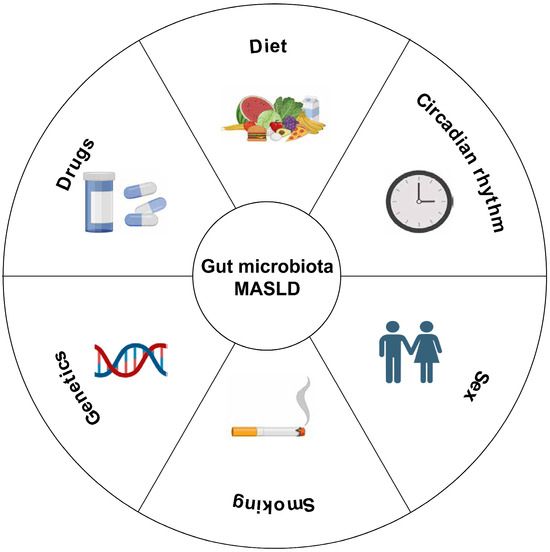
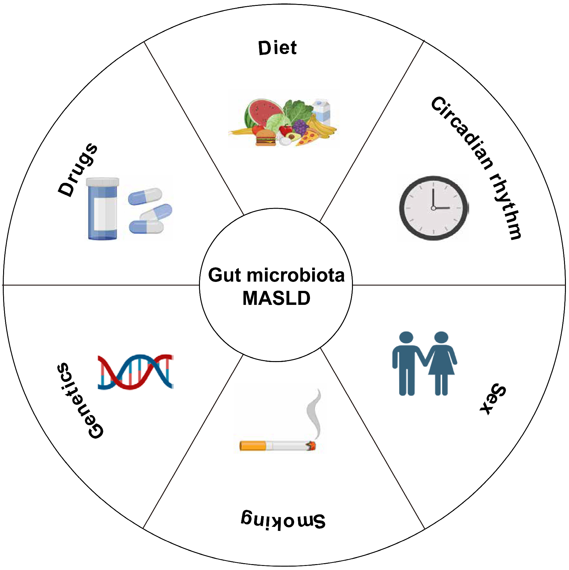
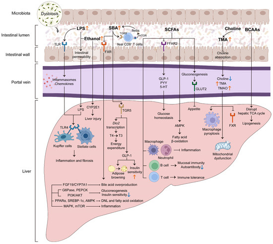
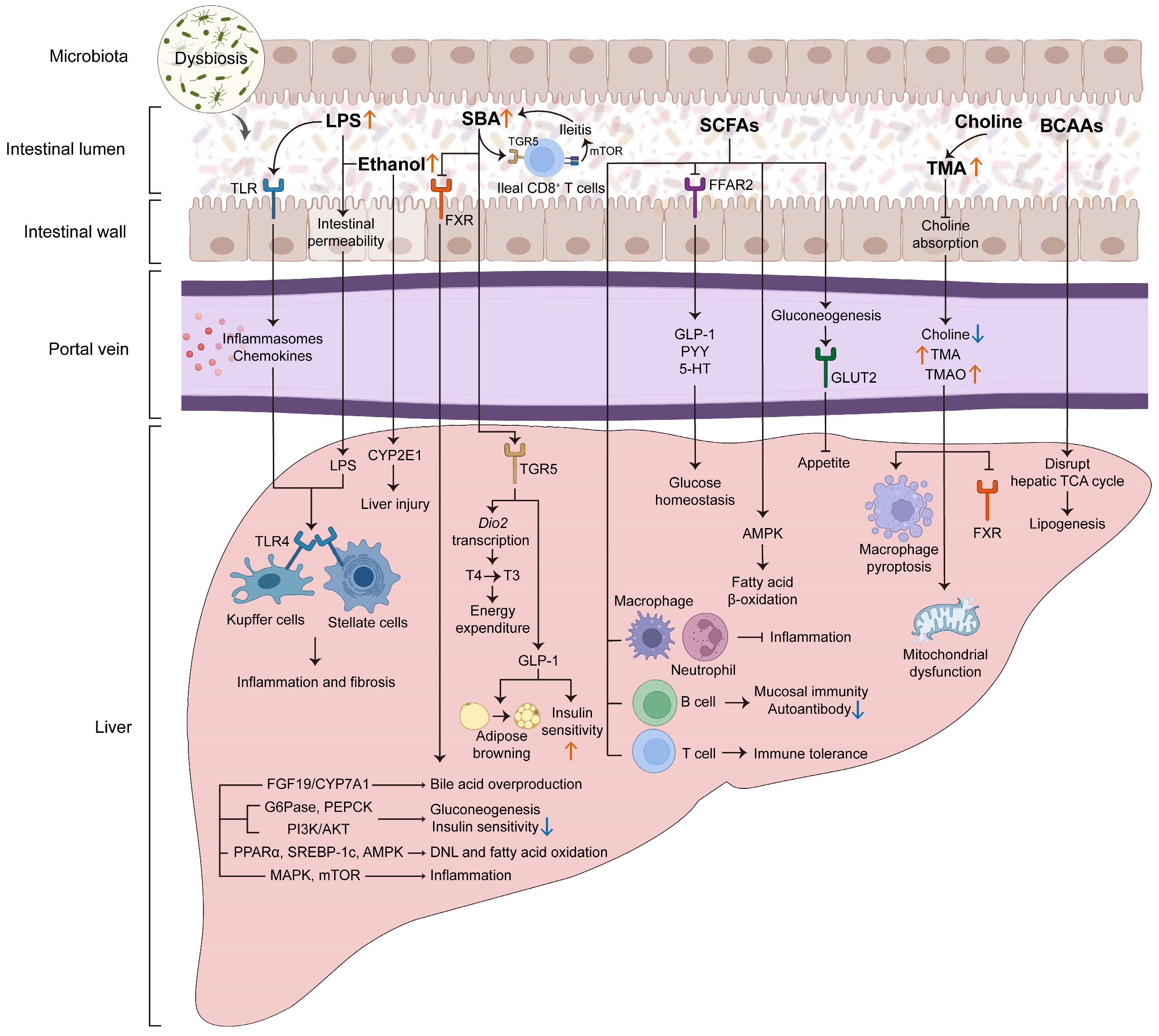
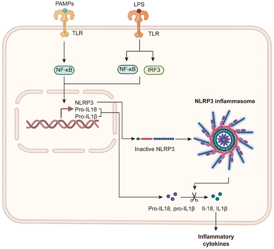
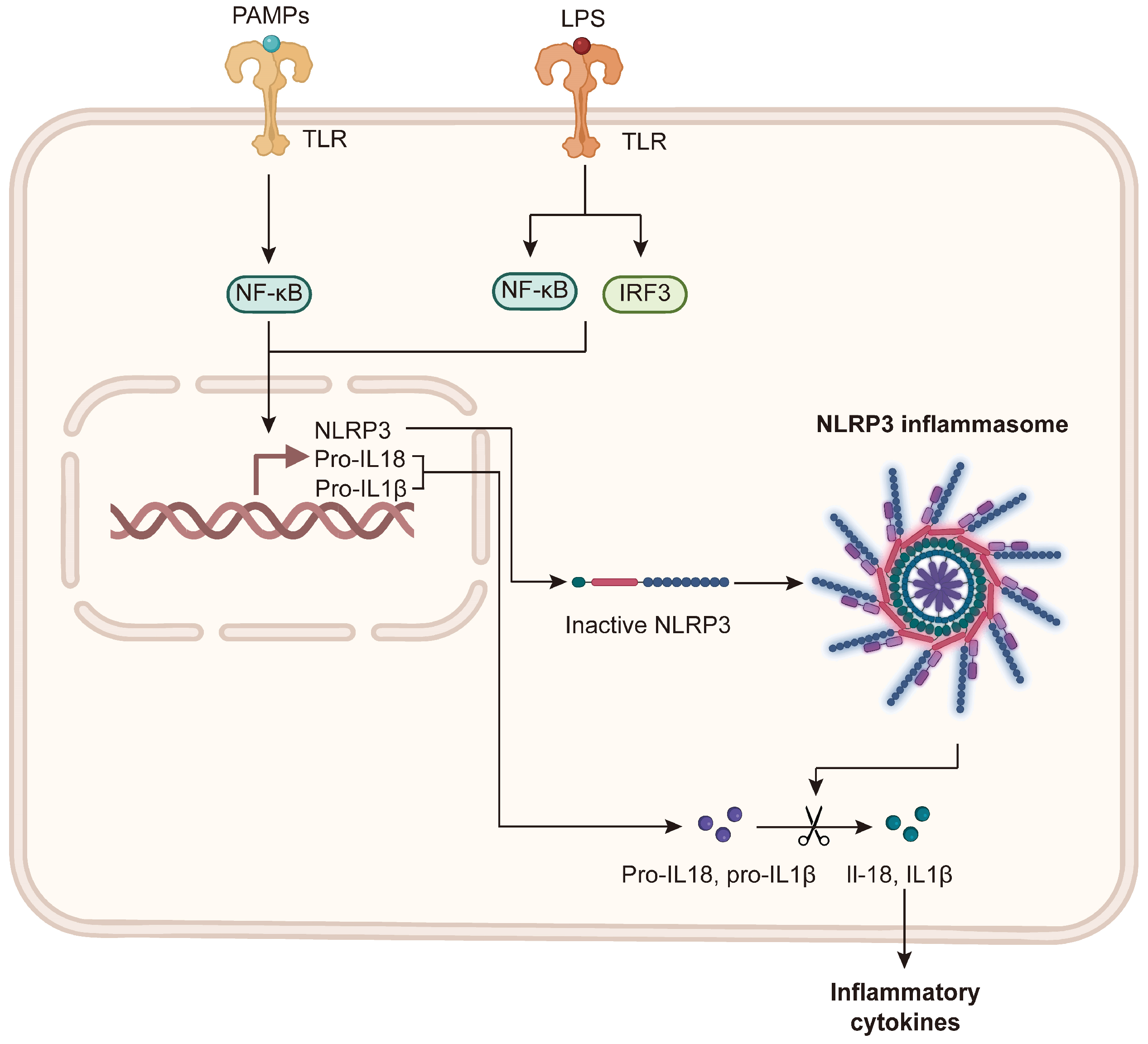
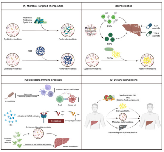
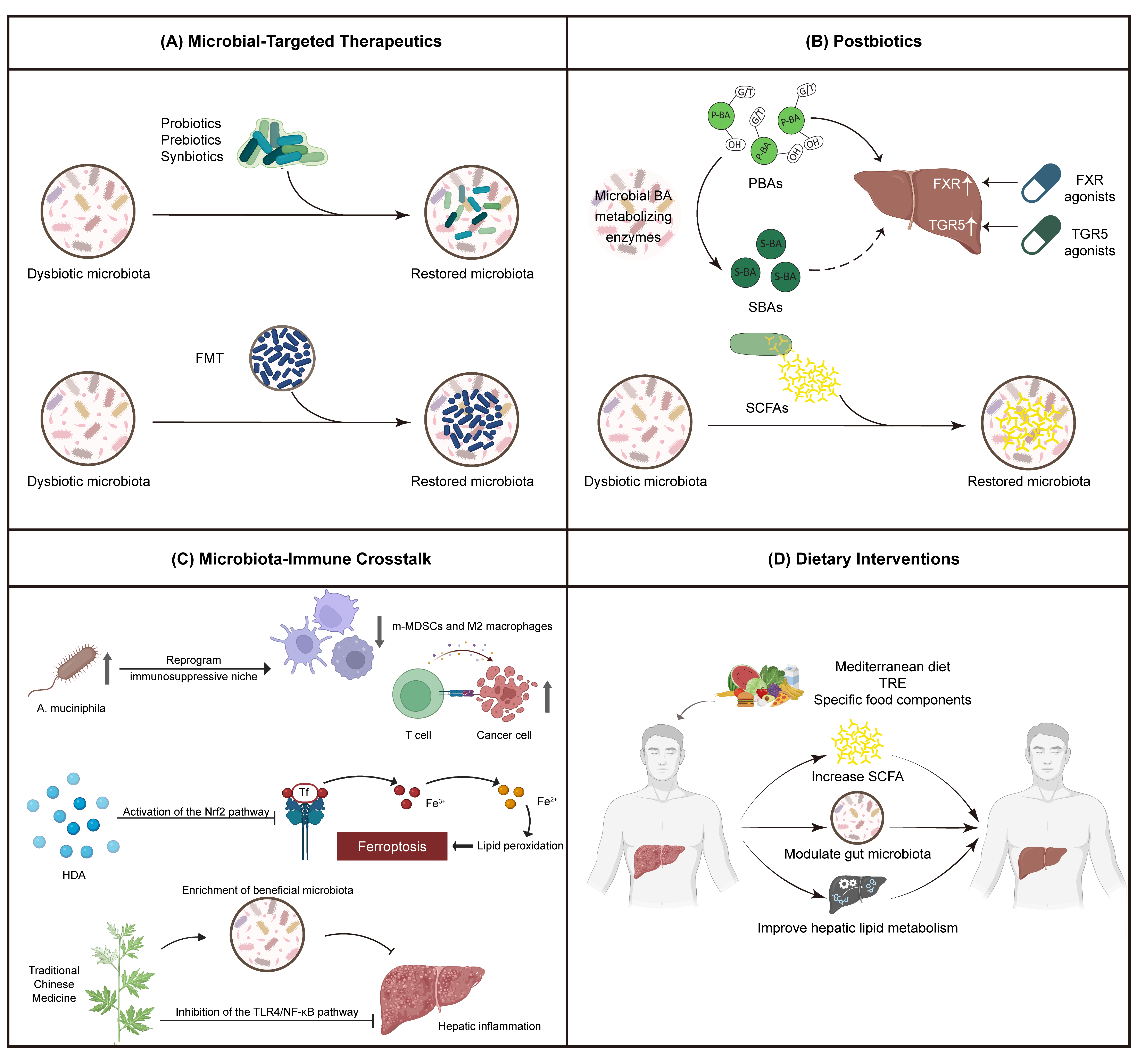

Disclaimer/Publisher’s Note: The statements, opinions and data contained in all publications are solely those of the individual author(s) and contributor(s) and not of MDPI and/or the editor(s). MDPI and/or the editor(s) disclaim responsibility for any injury to people or property resulting from any ideas, methods, instructions or products referred to in the content. |
© 2026 by the authors. Licensee MDPI, Basel, Switzerland. This article is an open access article distributed under the terms and conditions of the Creative Commons Attribution (CC BY) license.