Article

Sclerotinia Species Causing Lettuce Drop Disease in Serbia

by
Maja Živanović
1,
Milica Mihajlović
1,
Aleksandra Jovanović
2,
Jovana Hrustić
1,
Mira Vojvodić
3,
Brankica Pešić
1 and
Aleksandra Bulajić
3,*
1
Institute of Pesticides and Environmental Protection, Banatska 31b, 11080 Belgrade, Serbia
2
Institute for the Application of Nuclear Energy INEP, University of Belgrade, Banatska 31b, 11080 Belgrade, Serbia
3
Faculty of Agriculture, University of Belgrade, Nemanjina 6, 11080 Belgrade, Serbia
*
Author to whom correspondence should be addressed.
Microorganisms 2026, 14(1), 189; https://doi.org/10.3390/microorganisms14010189
Submission received: 4 December 2025 / Revised: 29 December 2025 / Accepted: 9 January 2026 / Published: 14 January 2026
(This article belongs to the Special Issue Diversity of Plant Pathogens)

Abstract

Sclerotinia spp. are globally distributed, devastating plant pathogens with a broad host range, including lettuce, on which they cause lettuce drop disease. To investigate the geographical distribution of lettuce drop incidence and the population structure of Sclerotinia sclerotiorum and S. minor in Serbia, 27 commercial lettuce fields across 12 administrative districts were surveyed. Sclerotinia spp. were confirmed at 10 localities, with S. sclerotiorum occurring more frequently. Co-occurrence of both species within the same field was recorded at only one location. Clear phenotypic and physiological differences were found between Sclerotinia species, as well as among isolates within each species. The two species differed in colony appearance, sclerotia production, virulence, growth rate, oxalic acid production, and tolerance to elevated osmotic pressure. Haplotype analysis of S. minor revealed the existence of 9 haplotypes arranged in a star-shaped network. These findings highlight the importance of considering both inter- and intraspecific variability of Sclerotinia species when evaluating their impact on crops, improving our understanding of Sclerotinia populations in lettuce, and supporting the development of effective management strategies.

1. Introduction

Lettuce (Lactuca sativa L.) is an important leafy vegetable cultivated extensively in both open-field and protected environments. Global lettuce production exceeded 28 million metric tons in 2023, with major producers including China, the United States, India, and Spain. Besides Spain, Italy, Turkey, and Belgium are the major lettuce-producing countries in Europe (FAO, https://www.fao.org/faostat/en/#data/QCL, accessed on 10 August 2025). Official data on lettuce production in Serbia are currently unavailable, despite being one of the most extensively cultivated vegetable crops in the country [1].
A wide spectrum of fungal and oomycete pathogens poses a persistent threat to lettuce production, often resulting in substantial reductions in yield and postharvest quality. Reported yield losses range from 5% to complete crop failure, depending on the pathogen and the disease incidence [2,3,4]. Lettuce drop, also known as Sclerotinia rot, is among the most economically significant diseases worldwide, affecting all types of lettuce, particularly leaf, romaine, and head lettuce [5,6]. While diseases caused by Sclerotinia species were typically sporadic in crop production, they have become increasingly persistent under intensive farming systems characterized by short crop rotations [7]. Yield losses in field-grown lettuce in the United Kingdom average around 10%, but can reach up to 50% under conditions of heavy or sustained precipitation [2]. Reported losses in Australia’s major lettuce-producing areas were between 10% and 45%, even when regular fungicide spray programs were implemented [8].
Two species of the genus Sclerotinia, S. sclerotiorum and S. minor, are known as the causal agents of Sclerotinia rot on lettuce and other composite vegetables [9,10]. The third lettuce drop causal agent, S. nivalis was recently isolated and identified in China [11]. Overall, S. sclerotiorum is one of the most devastating soilborne plant pathogens [12,13]. It is distributed in all continents, in more than 92 countries (CABI distribution map, UK, https://plantwiseplusknowledgebank.org, accessed on 10 August 2025), and has an extraordinarily broad host range, infecting more than 450 plant species from more than 75 families [9,13]. The hosts are mostly dicotyledonous herbaceous plants, although several monocotyledonous species were also recorded. S. minor is the most economically important in lettuce, sunflower, green bean, and peanut, even though it could cause substantial losses in other crops, especially if rotated with lettuce. They cause almost identical symptoms but have significantly different dispersal patterns and modes of infection [14].
Understanding the pathogenic potential and ecological success of Sclerotinia spp. requires an integrated assessment of their physiological traits and population structure. Oxalic acid production is a key virulence factor that facilitates host colonization through tissue acidification, suppression of plant defense responses, and cell wall degradation [15,16]; however, its quantitative contribution to pathogenicity may vary among species and isolates [12,17]. Tolerance to elevated osmotic pressure represents an important adaptive trait of soilborne pathogens, reflecting their ability to persist and grow under adverse environmental conditions such as high soil salinity or limited water availability, and may influence both soil survival and competitiveness during host infection [18]. In parallel, haplotype analysis provides insight into population genetic structure and diversity by revealing dominant lineages and patterns of genetic relatedness. A high degree of genetic diversity can also indicate a greater ability to adapt to changing environmental conditions and may reflect increased biological fitness of these genotypes [19]. Together, studies on haplotype composition, oxalic acid production, and osmotic stress tolerance provide a framework for exploring potential associations between population genetic structure and variation in key functional traits in Sclerotinia spp. associated with lettuce drop disease.
Data on the distribution and impact of Sclerotinia species in Serbia, particularly on leafy vegetables including lettuce, are scarce [20]. The only available record is a recent detection of S. minor as a lettuce pathogen [21], while there are no data on S. sclerotiorum as a lettuce pathogen. Serbian lettuce producers have expressed increasing concern and frequently requested management advice regarding lettuce drop disease. Therefore, a study was conducted to identify the distribution and species composition of Sclerotinia spp. causing lettuce drop disease in Serbia. The specific objectives of this study were to (1) identify Sclerotinia species associated with symptomatic lettuce plants in Serbia using conventional morphological and molecular methods; (2) assess the relative abundance and distribution of each Sclerotinia species across different lettuce-growing regions in Serbia; (3) characterize selected isolates of Sclerotinia spp. with respect to their virulence and morphological traits; (4) evaluate genetic diversity of the isolates through phylogenetic analyses and haplotype assessment; (5) examine physiological properties of the isolates such as growth rate and oxalic acid production and (6) determine the susceptibility of the isolates to high osmotic pressure as an indicator of their physiological adaptability to the environment.

2. Materials and Methods

2.1. Sclerotinia spp. Isolates

To assess the presence and spatial distribution of lettuce drop in Serbia, field surveys were conducted in the spring seasons of 2021 and 2022. Symptomatic lettuce plants, exhibiting poor or stunting growth and blanching of leaves, were collected from 27 commercial unheated plastic tunnel fields in 12 administrative districts of Serbia. The disease incidence was estimated by walking through the crop in a zigzag pattern and randomly rating 100 plants in three replicates. The number of samples collected per site was estimated based on field size (ranging from 0.2 to 2 ha) and the disease incidence and severity. Symptomatic tissues from sampled plants were cut into small pieces, surface sterilized with 0.5% sodium hypochlorite for 1 min, rinsed three times in sterile distilled water, placed on potato dextrose agar (PDA) supplemented with streptomycin (300 mg/mL), and incubated for 4–7 days at 25 °C. From the emerging fungal colonies, monohyphal-tip isolates were derived. The obtained isolates were stored at −80 °C in 20% glycerol for long-term preservation, while short-term storage was maintained on PDA slants at 4 °C.
All the obtained isolates were preliminarily identified based on colony morphology, growth rate, and the number and size of produced sclerotia after the incubation of 15 days on PDA medium at 25 °C in darkness [22].

2.2. Pathogenicity Test

To confirm the pathogenicity of all the obtained Sclerotinia spp. isolates, artificial inoculation of 4-leaf-stage lettuce plants cv. Majska kraljica, grown on commercial growth substrate (Floragard, Oldenburg, Germany) in 1 L pots, was conducted. Mycelial plugs, 10 mm in diameter, were excised from the margin of a 3-day-old colony grown on PDA and placed mycelium-side down on undamaged ground-level leaves of lettuce plants. Five plants were inoculated per isolate. Control plants were inoculated with sterile PDA plugs. Inoculated plants were enclosed in transparent plastic bags to maintain high humidity, and the bags were misted internally with water. Plants were kept in a growth chamber at 20 °C under a 13 h photoperiod. Plants were monitored daily for symptom development. Following symptom appearance, the pathogen was reisolated, and the morphological characteristics of the recovered isolates were compared with those of the challenging isolates.

2.3. Molecular Detection of Sclerotinia spp. Isolates

Preliminary identification of all obtained Sclerotinia-like isolates was confirmed with multiplex polymerase chain reaction (PCR) and respective specific primers. Total genomic DNA was isolated from mycelia of 7-day-old cultures grown on PDA using a method described by Harrington and Wingfield [23]. Primer pairs SMLcc2F/SMLcc2R, specific for S. minor, SSasprF/SSasprR for S. sclerotiorum, and STCadF/STCadR for S. trifoliorum, were used for amplifications of the Lcc2, Aspr, and Cad gene regions, respectively [24]. The multiplex PCR mix contained 12.5 μL of 2× Master mix (Fermentas, Vilnius, Lithuania), 1 μL of each primer, 1 μL of template DNA, and molecular-grade water up to a final volume of 25 μL. PCRs were performed in a Biometra Thermocycler (Analytik Jena GmbH+Co., Jena, Germany) with the following reaction conditions: an initial denaturation at 95 °C for 3 min, followed by 35 cycles of 95 °C for 30 s, 60 °C for 90 s, 72 °C for 90 s, and a final extension at 72 °C for 7 min. Negative controls were included by replacing template DNA with molecular-grade water. The PCR products were separated by electrophoresis in 2% agarose gels run in 1× Tris-borate EDTA buffer at 100 V constant voltage. The gels were stained with ethidium bromide, and the products were visualized and photographed under ultraviolet (UV) light.

2.4. Morphological Characterization

Based on geographic origin and colony morphology on PDA, 10 representative S. sclerotiorum and 10 S. minor isolates were selected for detailed phenotypic characterization. After 15 days of incubation on PDA at 25 °C in darkness, colony color, texture, and growth pattern were assessed. The presence, number, size, shape, and distribution of sclerotia within the Petri plate were also recorded [22]. To determine sclerotial size (length and width) 30 randomly selected sclerotia per isolate were measured using a ruler. Colony growth rate was determined after 2-day incubation on PDA in darkness and expressed in mm per day, following the methodology described by Morral et al. [25]. To study pigment production, isolates were incubated on PDA at 25 °C for 15 days, after which pigmentation was assessed by visually inspecting the reverse side of the plates [26]. All experiments were conducted twice, with four replicates per isolate.

2.5. Sequencing of Ribosomal DNA Internal Transcribed Spacer Region

The identity of selected representative Sclerotinia spp. isolates was further confirmed by amplification and sequencing of the internal transcribed spacer (ITS) region of ribosomal DNA (rDNA) using primers ITS1 and ITS4 and the same reaction mixture as described above. PCR amplifications were performed with an initial denaturation for 90 s at 94 °C, followed by 29 cycles consisting of a denaturation step for 30 s at 94 °C, primer annealing for 30 s at 55 °C, and extension for 30 s at 72 °C. The final extension step was performed at 72 °C for 9 min 30 s [27].
The amplified products were sequenced directly on automated equipment (Macrogen Inc., Seoul, Republic of Korea) in both directions using the same primers as for amplification. For each isolate, the consensus sequence covering the partial rDNA-ITS region was reconstructed using Pregap4 and Gap4 (v. 1.5) from the Staden Package and aligned using Clustal X under MEGA software version 6. All isolates were compared to all publicly available sequences using the Basic Local Alignment Search Tool (BLAST, version 2.13.0) algorithm in the National Center for Biotechnology Information (NCBI) database. The obtained sequences of Serbian Sclerotinia spp. isolates were deposited in the GenBank database, and their accession numbers were assigned.

2.6. Phylogenetic Analysis

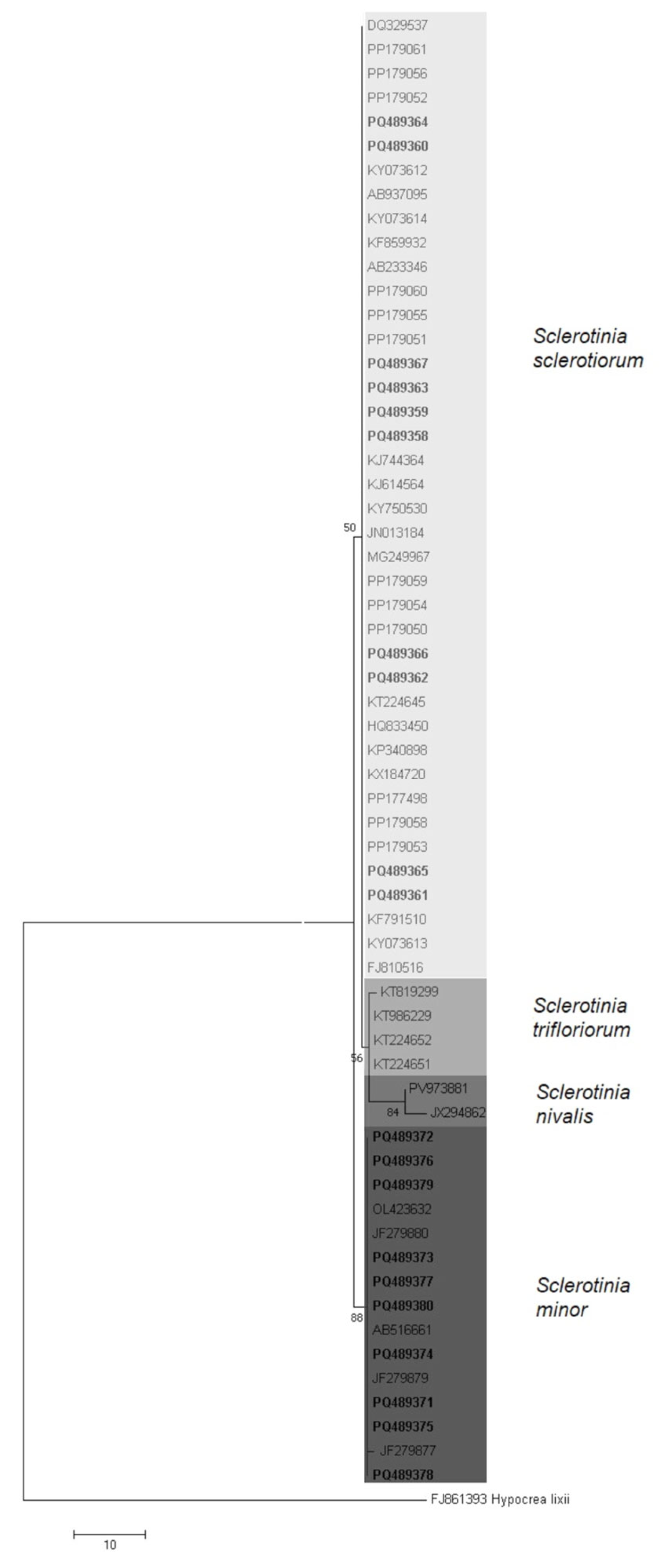
A phylogenetic tree was reconstructed based on 20 ITS sequences generated in this study and 42 previously published sequences representing S. sclerotiorum, S. minor, S. nivalis, and S. trifoliorum isolates from different hosts and geographic origins retrieved from GenBank (Table 1) using the Maximum Parsimony algorithm implemented in MEGA 6. The tree was evaluated with 1000 bootstrap replications to test clade stability, and bootstrap values < 50% were omitted. The sequence of Hypocrea lixii (GenBank Acc. No. FJ861393) was used as an outgroup reference species.

2.7. Haplotype Analysis of S. minor Sequences

As the haplotype analysis of S. sclerotiorum was recently performed [29], the present study was focused on S. minor population. The haplotype analyses of S. minor was based on all 99 available ITS region sequences in GenBank (accessed on 10 January 2025), along with 10 Serbian S. minor sequences. After a manual review and exclusion of short and/or sequences with degenerate codons, a total of 103 sequences were included in the analysis. All sequences were compared by calculating nucleotide identities (nt) using MEGA X software [32]. The number of haplotypes (h), the haplotype diversity (Hd), the identification of polymorphic sites (S) and the nucleotide diversity (P) of the ITS region were analyzed using DnaSP version 6.0 [33]. Haplotype composition and frequency were further investigated using PopART software version 1.7 (Population Analysis with Reticulate Trees) [34]. The mutual genealogical relationships among haplotypes were visualized using the Median Joining Network algorithm implemented in PopART software [35].

2.8. Virulence Test and Oxalic Acid Quantification

The virulence of 10 S. sclerotiorum and 10 S. minor isolates was assessed using a detached leaf inoculation assay [36]. Fully expanded, healthy lettuce leaves were used for inoculation. Leaves were arranged in plastic trays (12 × 20 cm) with two layers of moist blotter paper placed at the bottom to maintain humidity. Mycelial discs (10 mm in diameter) were excised from the actively growing margins of 72 h cultures of S. sclerotiorum and S. minor and placed on the upper side near the edge of each leaf. Leaves inoculated with sterile PDA plugs of the same size served as a control. Trays containing the inoculated leaves were incubated in a growth chamber set at 25 °C, 85–90% relative humidity, and a 10/14 h day/night photoperiod. Lesion length (in mm) along the main vein of the inoculated leaves was measured 72 h post-inoculation. A completely randomized design (CRD) was employed with three replicates per isolate. The experiment was conducted twice to ensure reproducibility.
Oxalic acid production by the selected 10 S. sclerotiorum and 10 S. minor isolates was quantified and compared [37]. The isolates were grown statically in flasks containing 50 mL of potato dextrose broth (PDB) (2% glucose and 0.4% fresh potato extract in distilled water) with three replicates per isolate. After a 3-day incubation at 22 ± 2 °C, the cultures were vacuum-filtered, and the dry weight of the mycelial fraction was determined after drying at 80 °C for 72 h. For oxalic acid quantification, supernatant (200 µL), bromophenol blue (BPB, 1 mM, 110 µL) (Centrohem, Stara Pazova, Serbia), sulfuric acid (1 M, 198 µL) (Sigma-Aldrich, Schnelldorf, Germany), potassium dichromate (100 mM, 176 µL) (Merck, Darmstadt, Germany) and distilled water (4.8 mL) were mixed and incubated at 60 °C for 10 min. Subsequently, the reaction was quenched by sodium hydroxide solution (0.75 M, 500 µL) (Fisher Scientific, Ottawa, ON, Canada). All absorbance readings were performed in triplicate at 600 nm using a Shimadzu UV-1800 UV/Visible scanning spectrophotometer (Shimadzu, Kyoto, Japan), with sterile PDB as the blank control. The calibration curve was prepared using known concentrations of oxalic acid (Oleohemija, Belgrade, Serbia), and the results were expressed as μg oxalic acid per mg of mycelial dry weight.

2.9. Susceptibility of Sclerotinia spp. Isolates to High Osmotic Pressure

The susceptibility of 10 representative S. sclerotiorum and 10 S. minor isolates to high osmotic pressure was studied according to a slightly modified method of Beever and Brien [38]. Sodium chloride (NaCl) was added to PDA to achieve a final concentration of 0.51 mol/dm3, while PDA amended with sterile distilled water served as a control. Mycelial plugs (10 mm in diameter) were excised from the actively growing margins of 3-day-old colonies and placed, mycelium side down, at the center of Petri plates. The plates were incubated at 25 °C, and the mycelial growth diameter was measured in two perpendicular directions after 2 days, excluding the diameter of the inoculation plug. The percentage of growth inhibition under osmotic stress was calculated relative to the control. The experiment was conducted twice, with four replicates per treatment.

2.10. Statistical Analyses

To evaluate differences both between S. sclerotiorum and S. minor species and among isolates within each species, the data on mycelial growth, lesion diameters on lettuce leaves (virulence assay), and oxalic acid production in PDB were subjected to statistical analysis using GraphPad Prism version 5.0 (GraphPad Software, Boston, MA, USA). Statistical significance was set at p < 0.05. To compare the two species, an unpaired two-tailed t-test was used. Prior to the t-test, data normality was assessed using the Shapiro–Wilk test, and homogeneity of variances was evaluated using the F-test. Where significant differences in variances were detected, Welch’s correction was applied. Differences among isolates within each species were analyzed separately using the non-parametric Kruskal–Wallis test, followed by Dunn’s multiple-comparison test. Relationships between virulence and the other studied variables (growth rate, oxalic acid production, and susceptibility to high osmotic pressure) were examined using Spearman’s rank correlation coefficient.

3. Results

3.1. Presence and Distribution of Sclerotinia spp. in Serbia

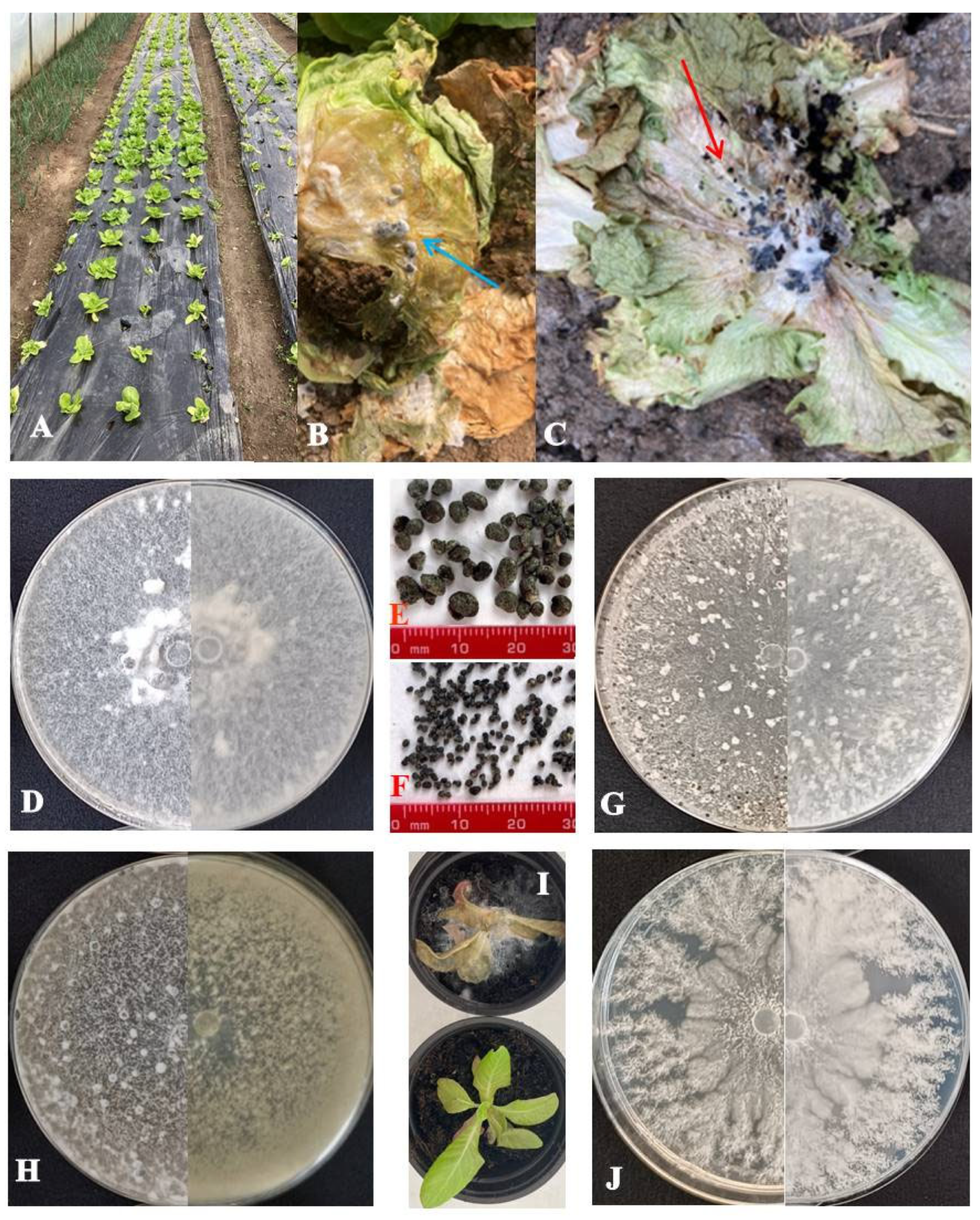
At 27 sites distributed across 12 administrative districts in the major lettuce-producing regions of Serbia (Figure 1, Table 2), lettuce plants exhibiting symptoms of wilting, poor growth, stunting, blanching, and leaf decay (Figure 2A–C) were collected. From the collected samples, 78 Sclerotinia-like colonies were recovered, initially with uniform colony appearance, but with visible differences between two groups of isolates after 5–6 days of incubation. The obtained isolates forming large sclerotia were preliminarily identified as S. sclerotiorum (43 out of 78), whereas the isolates with numerous small sclerotia were assigned as S. minor (35 out of 78). Using the species-specific primers, products of predicted size of 171 bp for S. sclerotiorum and 264 bp for S. minor isolates were amplified, confirming the identification of all isolates. Based on morphology and pathogenicity tests, besides Sclerotinia spp., as the most frequently isolated pathogens, various fungal/pseudofungal species, from genera Phoma, Verticillium, Alternaria, Plectosphaerella, Fusarium, Pythium, and Botrytis, were detected.
In the fields where Sclerotinia species were detected, the estimated incidence of lettuce drop ranged from 2% to 55%, with a mean value of 25.7% (Table 2). The highest incidence (55%) was recorded at the Potočanje locality, where symptoms included extensive leaf necrosis and the development of abundant white mycelium and numerous sclerotia on infected plants. In the other surveyed localities, the disease incidence was lower, ranging from 2% in Kraljevci (Srem District) to 40% in Irig (Srem District). S. sclerotiorum occurred at 7 locations in 6 districts, while S. minor was detected at 4 locations in 3 districts. Co-occurrence of both species was observed only at the Blace locality in the Toplica District (Figure 1, Table 2).

3.2. Pathogenicity

The pathogenicity of all 78 obtained Sclerotinia spp. isolates was confirmed by artificial inoculation of lettuce plants. After 3–7 days, all inoculated plants exhibited characteristic lettuce drop symptoms, while control plants remained asymptomatic (Figure 2I). No differences were observed in the timing of symptom appearance, nor in their severity. The pathogen was reisolated from symptomatic tissue following the previously described method, whereas no isolates were obtained from control plants. Koch’s postulates were fulfilled by verifying the identity of the reisolates based on their morphological characteristics.

3.3. Morphological Characterization of Sclerotinia spp. Isolates

Ten representative S. sclerotiorum isolates produced between 2.3 and 65 large sclerotia per plate, with diameters ranging from 0.67 to 5.83 mm (Figure 2E). Colony color varied from snow white (20% of isolates) to white (30%), whitish (40%), and 10% gray, as observed in isolate Ss 18-10 (Figure 2D,H). Mycelial density ranged from sparse to very dense, exhibiting textures described as floccose, woolly, or woolly-floccose. Most isolates lacked dark pigmentation, with the exception of Ss 18-10 and Ss 20-3, which displayed noticeable pigmentation. S. minor isolates produced 11.7 to 373.3 small sclerotia per plate (Figure 2F), within the diameter range of 0.20–2.50 mm. Colonies were snow white (10%), white (10%), or whitish (80%), with floccose, cottony, or woolly mycelium of variable density. All isolates exhibited regular growth with even margins, except S. minor isolate Sm 18-5, which displayed irregular growth characterized by a lobed margin (Figure 2G,J). Patterns of sclerotial distribution also differed between species. S. sclerotiorum isolates most frequently had edge and middle ring sclerotia arrangements (50%) or scattered patterns (40%), with the middle ring pattern occurring rarely. In contrast, S. minor isolates displayed predominantly uniform sclerotial distribution, with the scattered pattern prevailing (80%) and the edge ring pattern restricted to the isolates Sm 18-4 and Sm 18-5. In terms of the time required for the formation of sclerotia, the difference was not observed between species. For both species, sclerotia mostly appeared on the eighth day of incubation on PDA medium at 25 °C. None of the S. minor isolates produced dark pigment, while pigment production in S. sclerotiorum isolates was limited to isolates Ss 18-10 and Ss 20-3 (Table 3).
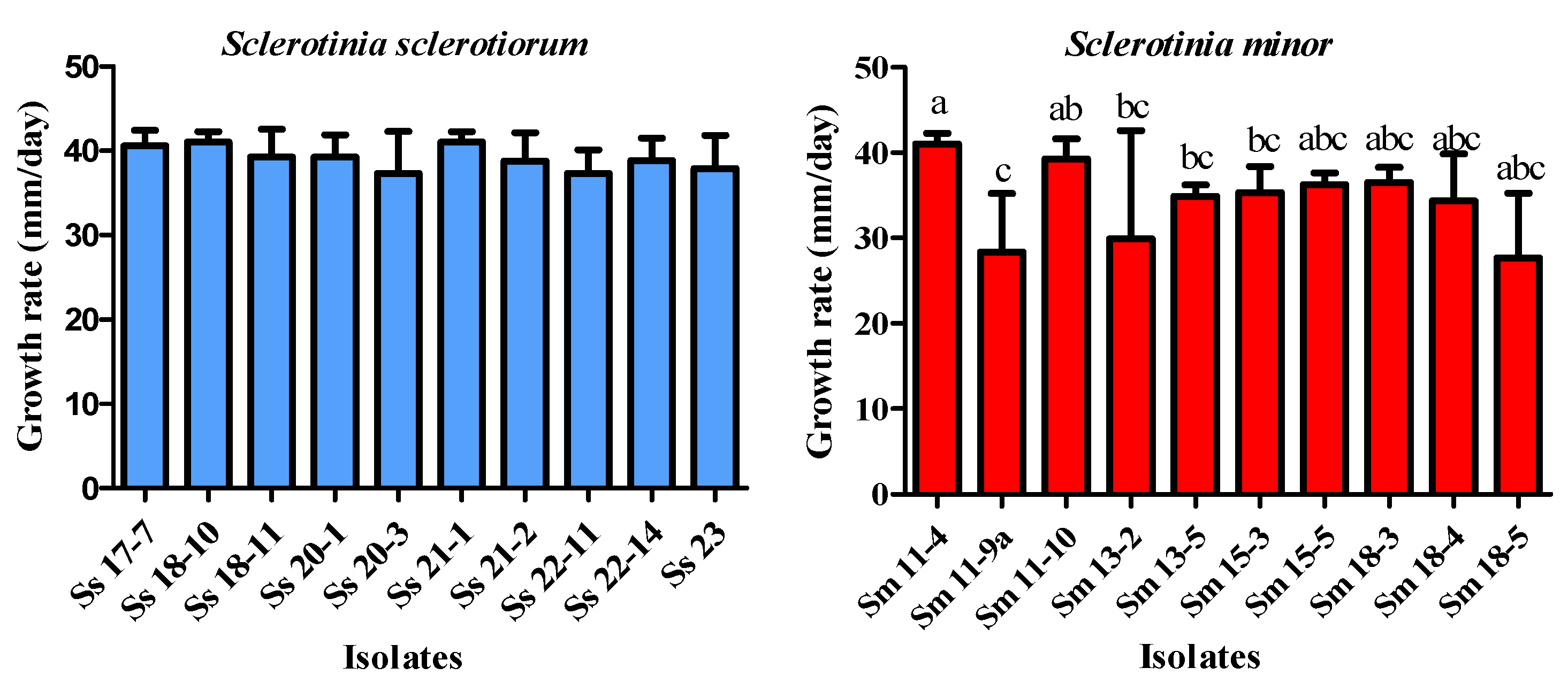
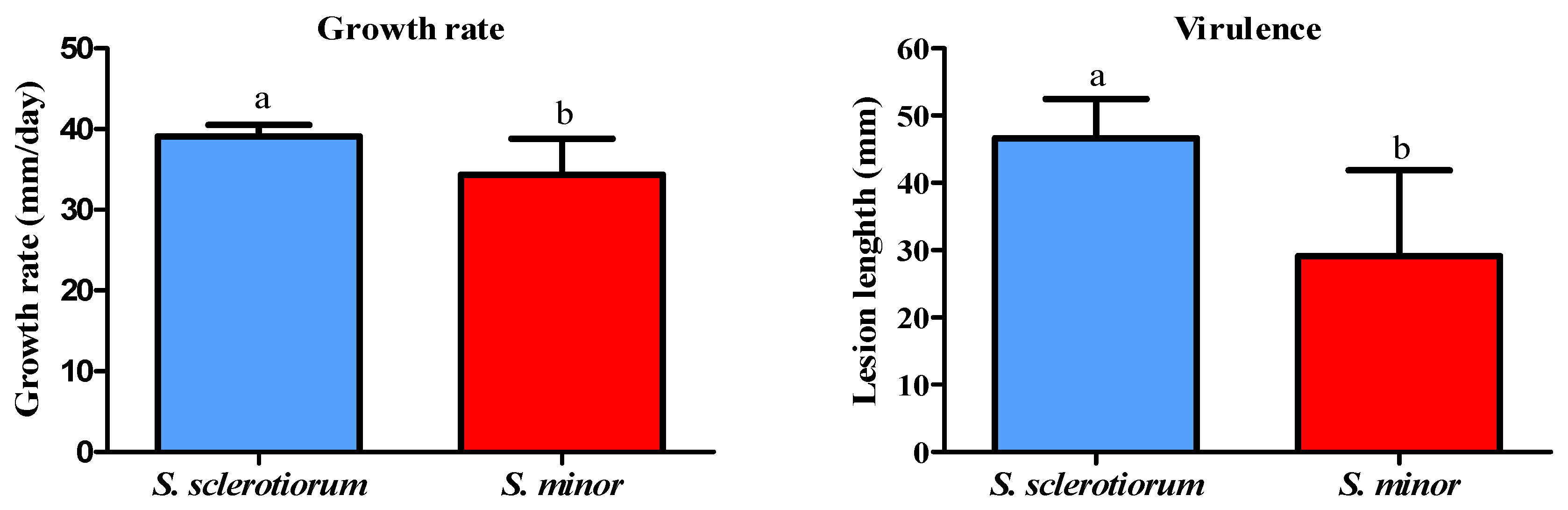
Colony growth rate varied significantly between species (t = 3.238, df = 10, p < 0.01). S. sclerotiorum exhibited a higher average growth (39.12 mm/day) compared to S. minor (34.19 mm/day), as it is presented in the Section 3.8. The difference in mycelial growth among S. sclerotiorum isolates was not significant (p = 0.0927), while the growth rate of S. minor isolates differed significantly (p < 0.01). For instance, a significant difference was observed between S. minor isolate Sm 11-4 and the isolates Sm 11-9a, Sm 13-2, Sm 13-5, and Sm 18-5. Differences were also observed between Sm 11-10 and Sm 18-5 (Figure 3).

3.4. Molecular Characterization and Phylogenetic Analysis

BLAST analysis showed that the ITS sequence of 10 Serbian S. sclerotiorum isolates had 100% nucleotide identity with GenBank S. sclerotiorum sequences, while sequences of 10 S. minor isolates had 100% nucleotide identity to S. minor from other parts of the world. Moreover, comparison of all ITS sequences obtained in this study revealed 100% nucleotide identity among isolates of the same species, and a difference of 3 nucleotides between Sclerotinia species.
The phylogenetic analysis based on ITS rDNA sequences resulted in a well-supported tree consistent with the previously established relationships among Sclerotinia species (Figure 4). Two major clusters corresponding to S. sclerotiorum and S. minor were observed. Within the first cluster, S. nivalis and S. trifoliorum formed distinct subgroups closely related to S. sclerotiorum. Isolates of S. sclerotiorum from Serbia grouped with reference isolates from Asia, Europe, and North America, while S. minor isolates clustered with sequences from Asia, Europe, and Australia. These results confirm the molecular identification and phylogenetic placement of Serbian Sclerotinia isolates within the established global lineages.

3.5. Haplotype Structure of S. minor Sequences

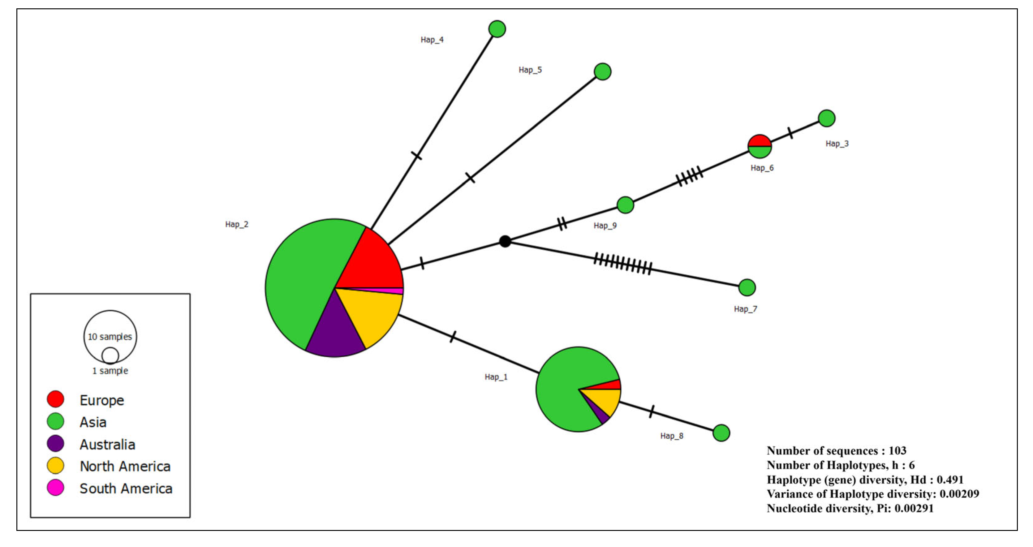
The total of 103 S. minor sequences from Europe, Asia, Australia, North and South America showed the presence of 9 haplotypes (Hap1–9) with 22 variable positions. Hap2 was the most widespread, with 69 sequences from different continents and hosts, including all Serbian sequences. Hap1 was the second most common, with 26 isolates. The remaining haplotypes, Hap3–9, were less common, with 1 to 2 sequences. Haplotype diversity (Hd) was 0.491, indicating a moderate level of genetic variation within the population. Haplotype diversity variance was 0.00209. Nucleotide diversity (Pi) was 0.00291 (0 to 15 nt difference), indicating an overall low level of nucleotide variability among the sequences analyzed. The haplotype network of S. minor showed a structure with Hap2 in the center, encompassing all Serbian sequences (Figure 5). All other haplotypes were directly linked to Hap2 by single or multiple mutations.

3.6. Virulence of Sclerotinia spp. Isolates

After a 3-day incubation, all 20 selected isolates of both Sclerotinia species caused necrotic lesions on inoculated lettuce leaves, whereas none of the leaves inoculated with sterile agar plugs developed symptoms.
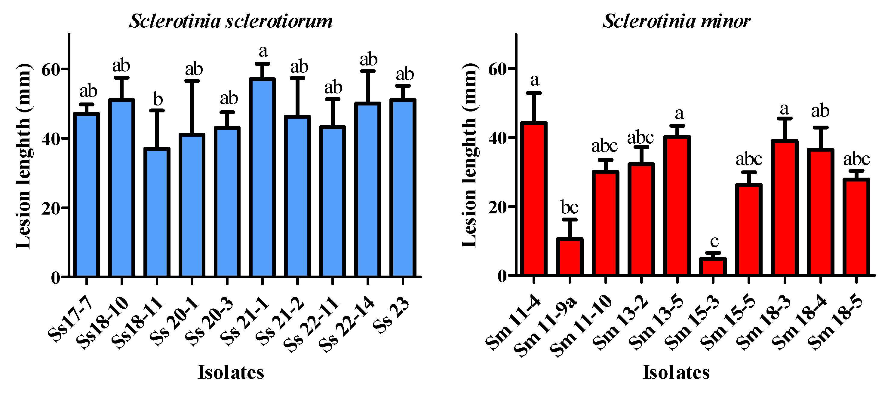
A statistically significant difference in virulence between the two species (t = 3.954, df = 12, p = 0.0019) was found, revealing that S. sclerotiorum is significantly more virulent under the tested conditions; an average lesion length caused by S. sclerotiorum was 46.64 mm, compared to 29.14 mm that was recorded for S. minor, as it is shown in the Section 3.8.
The difference in virulence among isolates within both species was also observed (Figure 6). S. sclerotiorum isolate Ss 21-1 was highly virulent and caused lesions 57 mm long, while the isolate Ss 18-11 was the least virulent (lesion length 37 mm). The S. minor isolates formed significantly smaller lesions (4.8–44.2 mm). A positive correlation was recorded between virulence and the growth rate of Sclerotinia spp. (r = 0.709, p = 0.0005).
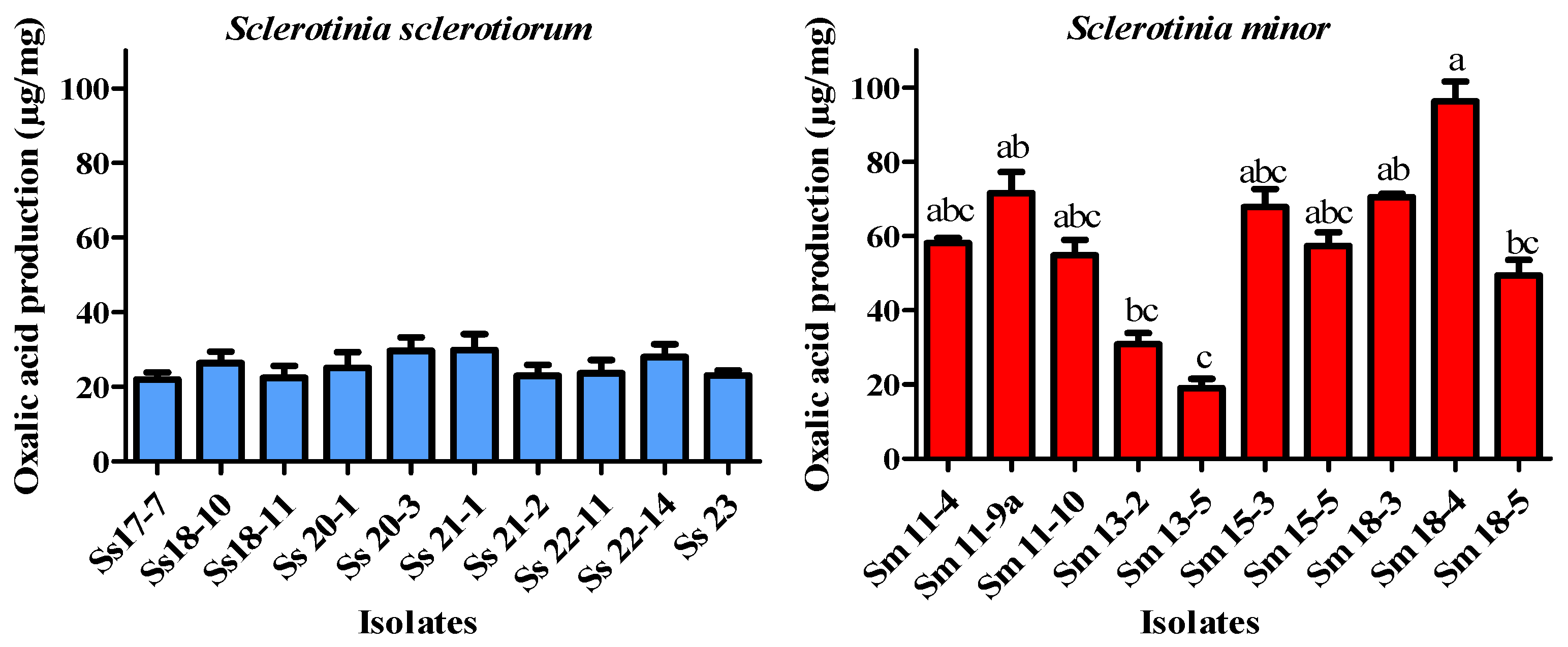
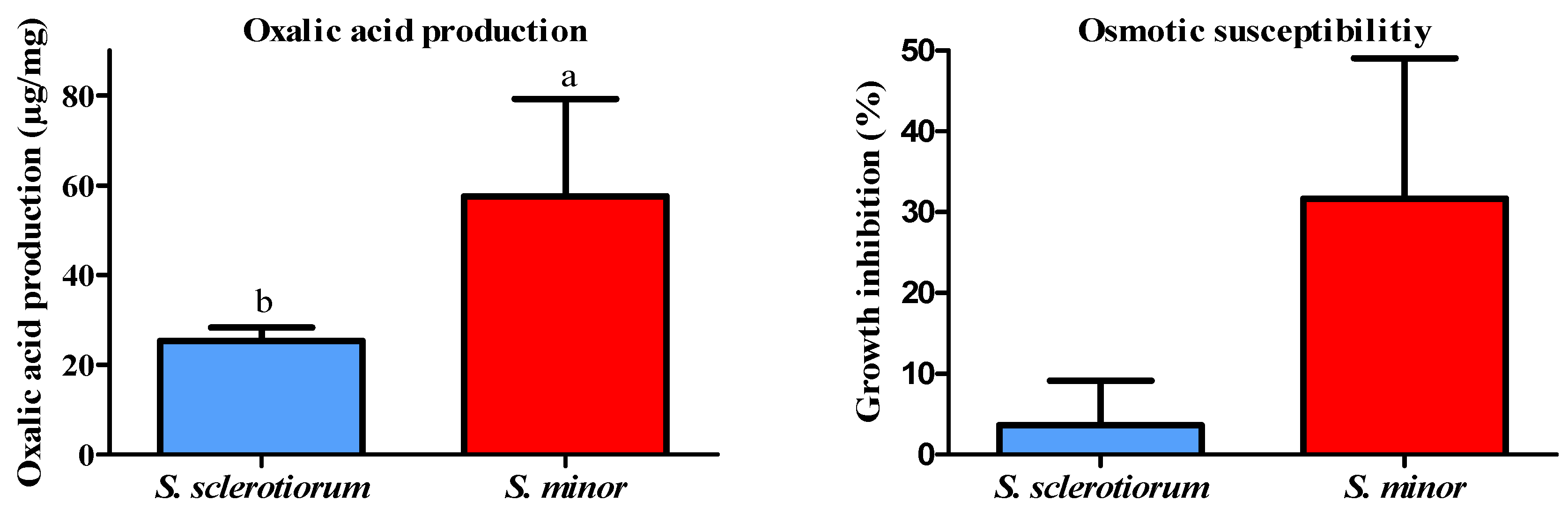
Given that oxalic acid is a major virulence factor in Sclerotinia spp. and is strongly linked to fungal pathogenicity, oxalic acid production was assessed in parallel with pathogenicity assays to provide a comprehensive evaluation of the isolates’ virulence. Statistical analysis revealed that S. sclerotiorum produced significantly less oxalic acid than S. minor (t = 4.654, df = 9, p = 0.0012). The highest production was recorded in S. minor isolate Sm 18-4 (96.3 µg/mg), whereas the lowest was found in the isolate Sm 13-5 (19.0 µg/mg). The isolates of S. sclerotiorum exhibited lower levels of oxalic acid production. The highest amount was detected in isolate Ss 21-1 (29.9 µg/mg) (Figure 7). A statistically significant correlation between virulence and oxalic acid production in both Sclerotinia species was not found (S. sclerotiorum: r = 0.353, p = 0.313; S. minor: r = −0.188, p = 0.607).

3.7. High Osmotic Pressure Susceptibility

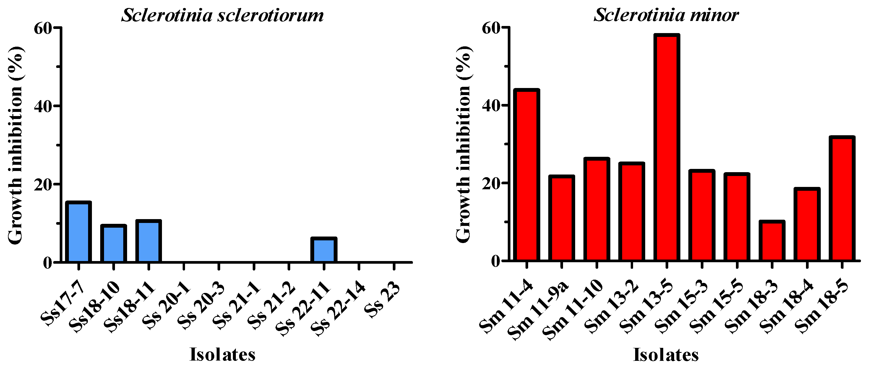
S. sclerotiorum was significantly less susceptible to high osmotic pressure compared to S. minor (t = 4.859, df = 10, p = 0.0007). The growth of 60% of S. sclerotiorum isolates was not inhibited on a PDA medium supplemented with 0.51 mol/dm3 NaCl. The remaining 40% of the isolates showed growth inhibition ranging from 6.2% to 15.3%. In contrast, all S. minor isolates exhibited growth inhibition under the same conditions, ranging from 10.1% to 58.1% compared to the control (Figure 8). No statistically significant correlation was found between isolate susceptibility to increased osmotic pressure and virulence in either of the two studied species.

3.8. Comparative Characterization of S. sclerotiorum and S. minor

Based on the overall comparison of all examined traits, significant differences between S. sclerotiorum and S. minor were observed. Differences were detected in growth, virulence, oxalic acid production, and tolerance to high osmotic pressure. S. sclerotiorum was characterized by greater mycelial growth, higher virulence, and increased tolerance to elevated osmotic pressure. In contrast, S. minor was found to produce higher levels of oxalic acid (Figure 9).

4. Discussion

This study provides the first comprehensive characterization of Sclerotinia species as the causal agents of lettuce drop disease in Serbia. Two closely related broad-host-range pathogens, S. sclerotiorum and S. minor are detected, both described to infect lettuce and other members of the Asteraceae family [9,10]. S. sclerotiorum is a widespread pathogen of sunflower in Serbia, causing significant yield losses [39,40]. It was also detected in green beans [41], faba beans [42], and cabbage [29]. The presence of S. minor in Serbia was described based on symptom presence [43], with no additional data including pathogenicity confirmation. Recently, S. minor was isolated for the first time in Serbia from symptomatic lettuce plants [21]. S. minor could become prevalent in crops such as basil, cauliflower, endive, escarole, and radicchio, particularly when these are rotated with infected lettuce [10].
Disease incidence was estimated using visual zigzag sampling approach, which may introduce sampling bias due to non-random field coverage. In fields with low disease incidence, spatial heterogeneity and aggregated symptom distribution may lead to under- or overestimation of the disease incidence. However, the zigzag approach was chosen to balance field coverage and practical feasibility and is commonly used in large-scale field assessments.
All studied isolates formed typical Sclerotinia colonies, producing black sclerotia, either large or small, consistent with the morphological descriptions of S. sclerotiorum and S. minor, respectively [44,45]. Based on sclerotial size, a reliable morphological criterion for distinguishing the two species [46], 55.1% of isolates derived in the present study were identified as S. sclerotiorum and 44.9% as S. minor. S. nivalis, a third species reported as a lettuce pathogen [11], was not detected. Sclerotinia spp. were detected in 8 districts across Serbia, with S. sclerotiorum being more frequently isolated. These findings align with those of Kim and Cho [45], who reported a lower isolation frequency of S. minor compared to S. sclerotiorum in surveyed locations in South Korea. S. sclerotiorum has also been published as a dominant species causing lettuce drop in the San Joaquin Valley in California, the USA [47], and in Norway [48]. Our results represent one of the first comprehensive studies on the distribution and prevalence of Sclerotinia species on lettuce in Europe. They are also among the few studies available worldwide. This significance is further accentuated by the considerable lack of recent data from most European countries, highlighting the need for sustained and expanded research in this field.
Almost all S. sclerotiorum isolates exhibited white or whitish, fluffy aerial mycelium. Only one grey-colored S. sclerotiorum isolate was identified. Similarly, S. minor isolates mostly formed whitish cottony mycelium, with no darkly pigmented isolates observed. Morphological diversity among S. sclerotiorum isolates has been documented across various crops [49,50,51,52,53,54]. Variability in colony color and growth patterns has been reported in isolates from Bangladesh [55], northeastern India [52], and Brazil [56]. Garg et al. [57] were the first to find three darkly pigmented isolates from Australia. Abreu and Souza [58] described Brazilian isolates ranging in color from white and beige to brown and black. In contrast to mycelial appearance, sclerotial size is a widely accepted morphological criterion for distinguishing between S. sclerotiorum and S. minor. In our study, we observed that the ranges of the average sclerotial size of the two species overlapped. Despite this overlap, the distinction between species remains reliable when considering the overall distribution of sclerotial sizes and their number per Petri plate. Therefore, species identification should take into account not only individual sclerotial size but also the typical size range and the abundance pattern, both of which support clear differentiation between the two species.
Morphological characteristics may provide reliable and accurate Sclerotina species discrimination if sclerotia are present. However, molecular methods ensure the most precise and fastest detection and characterization. Different molecular methods have been developed to discriminate Sclerotinia species [24,59,60,61,62]. The primers and multiplex PCR protocol described by Abd-Elmagid et al. [24] were tested in this study and confirmed as a fast, sensitive, reliable, and convenient diagnostic technique that successfully complemented the morphological identification of all Sclerotinia spp. isolates derived in this study. Molecular analysis proved that the ITS region is highly conserved for Sclerotinia species, with only a few nucleotide differences between the studied species. Phylogenetic analysis of 20 Serbian isolates confirmed a closer relationship of S. sclerotiorum with S. nivalis and S. trifoliorum than with S. minor, which is consistent with the previous findings [28,61].
The population structure of S. sclerotiorum, based on a comprehensive haplotype analysis, showed substantial uniformity and the presence of one dominant haplotype [29]. Before this study, there were scarce data on the population structure of S. minor at the global level. Several studies on the mycelial compatibility of S. minor have determined genetic diversity in the population, including the identification of 8 mycelial compatible groups (MCGs) among 95 isolates from lettuce and weeds in China [63] and 23 MCGs among isolates from lettuce in California, the USA [47]. Regardless of certain constraints of the conservative nature of the ITS region in Sclerotinia spp. in general, and in S. minor, our analysis of all available sequences of the worldwide population of S. minor implied a clear pattern and the presence of 9 haplotypes arranged in a star-shaped network. All S. minor sequences from Serbia were uniform, and all belonged to the dominant Hap2 haplotype. The central position of Hap2 suggests that it could be an ancestral haplotype or a haplotype with higher reproductive success. The fact that all other haplotypes were linked to Hap2 may indicate a clear diversification pattern and suggest recent population expansion or positive selection. Genotype characterization of the field population of S. minor [64] showed similar results and the presence of one dominant genotype comprising the largest number of isolates. The haplotype diversity and network analyzed in this study, in conjunction with the geography of origin and host plants, revealed that the most frequently detected haplotypes were Hap2 and Hap1 (comprising 69 and 26 isolates, respectively), detected on five and four continents, respectively, and pathogenic to 15 and 3 host plant species, respectively. This is consistent with previous descriptions that S. minor has a limited host range [10,63,65]. Although isolates from Hap2 are pathogenic to 15 different host plants, the majority are associated with a single family, Asteraceae, suggesting later diversification and adaptation to new hosts. The expansion of the host range likely reflects multiple factors involved in the co-evolution of pathogens and host plants [66], as has also been demonstrated for fungi in the Sclerotiniaceae [67].
Sclerotinia species are well-known necrotrophic plant pathogens that can effectively induce cell death in host plant tissues. They produce numerous lytic enzymes, including pectinases, cellulases, hemicellulases, and proteases, which aid in colonization and cause the breakdown of host cell walls. Alongside cell wall-degrading enzymes, oxalic acid, considered an essential virulence factor for Sclerotinia species, plays a crucial role in the infection process by suppressing host defense responses and inducing programmed cell death in plant cells [15,16,68]. This phytotoxin produced by the fungus serves multiple functions, including the disruption of plant cell membranes, inactivation of enzymes involved in plant defense mechanisms, and the manipulation of host signaling pathways to create a favorable environment for fungal colonization and infection [17,69,70]. Various studies demonstrated that higher oxalic acid-producing isolates were more virulent than those of low-producing isolates [17,71,72,73]; however, in the study of Li et al. [12], a correlation between oxalic acid production and pathogenicity was not found. In the present study, we did not observe a statistically significant correlation between oxalic acid production and virulence in either of the two investigated species. For S. sclerotiorum, the correlation was weakly positive, while for S. minor, it was weakly negative; however, in both cases, the correlations were not statistically significant. It is important to emphasize that the virulence in our study was assessed using detached leaf assays, which provide a standardized and controlled environment for evaluating isolate aggressiveness. While these assays allow a reliable comparison of relative pathogenic potential among isolates, they may not capture the full complexity of field conditions, including environmental variability and host responses. Therefore, the observed differences in virulence could be interpreted as relative aggressiveness under controlled conditions, rather than as absolute measures of field virulence. These findings suggest that although oxalic acid is an established virulence-associated metabolite in S. sclerotiorum, it does not fully account for virulence variation among isolates, and its contribution appears even less pronounced in S. minor. The contrasting tendencies between species likely reflect differences in pathogenic strategies and the multifactorial nature of virulence, including factors such as cell wall-degrading enzymes, pH modulation, effector secretion [15,70,73,74,75], and potentially, the timing and localization of oxalic acid production. Therefore, oxalic acid should be considered one component of a broader virulence repertoire. Future work integrating multiple biochemical and molecular determinants is warranted to fully resolve pathogenic variability. Additional studies incorporating larger sample sizes and assays under conditions that better reflect the natural environment are also required to elucidate the relationship between oxalic acid production and virulence in these species.
Microorganisms are continuously exposed to a variety of abiotic stressors, including ultraviolet radiation, temperature fluctuations, disruptions in osmotic homeostasis, and oxidative stress [18]. Understanding how they respond to such challenging conditions is essential not only for elucidating the ecophysiology of microorganisms in diverse natural habitats but also for advancing food safety and predicting pathogen virulence [76]. For instance, high concentrations of NaCl induce osmotic stress, which can significantly inhibit the mycelial growth of many fungi [18]. Araújo et al. [77] assessed osmotic susceptibility across 70 fungal strains representing 40 ecologically diverse species and found that fungi exhibiting a trophic dependency on hosts were more susceptible to high osmotic pressure. The results of the present study showed that S. minor was significantly more susceptible to increased osmotic pressure compared to S. sclerotiorum. This finding may indicate a higher level of physiological adaptability or osmotic and saline stress tolerance in S. sclerotiorum. This could partly explain its wider distribution and prevalence as a plant pathogen globally. In contrast, S. minor exhibited higher susceptibility to osmotic stress, suggesting potential limitations in its ecological adaptability under conditions of environmental stress. Given that osmotic stress can be a factor in soil environments, especially under drought or saline conditions, these physiological differences may affect species survival and virulence in the field. Therefore, further investigation of their environmental resilience, particularly that of S. minor, is warranted, given its recent emergence in this region [21].

5. Conclusions

This study provides new insights into lettuce drop disease in Serbia. Through pathogenicity and virulence assays, morphological characterization, and molecular analysis, S. sclerotiorum and S. minor were confirmed as the causal agents of the disease. A statistically significant difference between the two species was detected in growth rate, virulence, oxalic acid production, and osmotic susceptibility. No significant relationship between oxalic acid production and virulence was identified, further highlighting the multifactorial nature of pathogenicity in Sclerotinia species. Given the scarcity of up-to-date data on Sclerotinia populations in Europe, these findings fill a critical knowledge gap and provide a foundation for the development of more effective and targeted management strategies. They also serve as an essential reference for future research on lettuce and other horticultural crops.

Author Contributions

Conceptualization, A.B., B.P. and M.M.; methodology, M.Ž., B.P., J.H., M.M., A.J., A.B. and M.V.; software, B.P., M.V. and J.H.; validation, B.P., M.M. and A.B.; investigation, M.Ž., M.M., A.J., B.P., J.H., A.B. and M.V.; resources, B.P., M.M., A.J. and A.B.; writing—original draft preparation, M.Ž., M.M. and A.J.; writing—review and editing, M.Ž., B.P., J.H., M.M., A.J., A.B. and M.V.; visualization, M.Ž., M.M., M.V., J.H. and B.P.; funding acquisition, B.P., A.B. and A.J. All authors have read and agreed to the published version of the manuscript.

Funding

This project was supported by the Ministry of Education, Science and Technological Development of the Republic of Serbia grants 451-03-136/2025-03/200214; 451-03-137/2025-03/200116; and 451-03-136/2025-03/200019.

Institutional Review Board Statement

Not applicable.

Informed Consent Statement

Not applicable.

Data Availability Statement

The original contributions presented in this study are included in the article. Further inquiries can be directed to the corresponding author.

Conflicts of Interest

The authors declare no conflicts of interest.

Abbreviations

The following abbreviations are used in this manuscript:
bpBase pair
DNADeoxyribonucleic Acid
HapHaplotype
ITSInternal Transcribed Spacer
PCRPolymerase Chain Reaction
PDAPotato Dextrose Agar
ntNucleotide
SDStandard Deviation

References

  1. Ilić, S.Z.; Milenković, L.; Dimitrijević, A.; Stanojević, L.; Cvetković, D.; Kevrešan, Ž.; Mastilović, J. Light modification by color nets improve quality of lettuce from summer production. Sci. Hortic. 2017, 226, 389–397. [Google Scholar] [CrossRef]
  2. Young, C.S.; Clarkson, J.P.; Smith, J.A.; Watling, M.; Phelps, K.; Whipps, J.M. Environmental conditions influencing Sclerotinia sclerotiorum infection and disease development in lettuce. Plant Pathol. 2004, 53, 387–397. [Google Scholar] [CrossRef]
  3. Farmer, A.A. Population diversity and epidemiology of Bremia lactucae the cause of lettuce downy mildew. Doctoral Dissertation, The University of St Andrews, St Andrews, UK, 2024. [Google Scholar] [CrossRef]
  4. Hossain, M.M.; Sultana, F.; Rubayet, M.T.; Khan, S.; Mostafa, M.; Mishu, N.J.; Sabbir, M.A.A.; Akter, N.; Kabir, A.; Mostofa, M.G. White Mold: A Global Threat to Crops and Key Strategies for Its Sustainable Management. Microorganisms 2025, 13, 4. [Google Scholar] [CrossRef] [PubMed]
  5. Patterson, C.L.; Grogan, R.G. Differences in epidemiology and control of lettuce drop caused by Sclerotinia minor and S. sclerotiorum. Plant Dis. 1985, 69, 766–770. [Google Scholar] [CrossRef]
  6. Mamo, B.E.; Eriksen, R.L.; Adhikari, N.D.; Hayes, R.J.; Mou, B.; Simko, I. Epidemiological characterization of lettuce drop (Sclerotinia spp.) and biophysical features of the host identify soft stem as a susceptibility factor. PhytoFrontiers™ 2021, 1, 182–204. [Google Scholar] [CrossRef]
  7. O’Sullivan, C.A.; Belt, K.; Thatcher, L.F. Tackling Control of a Cosmopolitan Phytopathogen: Sclerotinia. Front. Plant Sci. 2021, 12, 707509. [Google Scholar] [CrossRef]
  8. Vallalta, O.; Porter, I.J. Development of Biological Controls for Sclerotinia Diseases of Horticultural Crops in Australasia; Horticultural Australia Ltd.: North Sydney, Australia, 2004. [Google Scholar]
  9. Boland, G.J.; Hall, R. Index of plant hosts of Sclerotinia sclerotiorum. Can. J. Plant Pathol. 1994, 16, 93–108. [Google Scholar] [CrossRef]
  10. Melzer, M.S.; Smith, E.A.; Boland, G.J. Index of plant hosts of Sclerotinia minor. Can. J. Plant Pathol. 1997, 19, 272–280. [Google Scholar] [CrossRef]
  11. Li, G.; Wang, D.; Huang, H.C.; Laroche, A. First report of Sclerotinia nivalis on lettuce in central China. Mycol. Res. 2000, 104, 232–237. [Google Scholar] [CrossRef]
  12. Li, Z.; Zhang, M.; Wang, Y.; Li, R.; Fernando, W.G. Mycelial Compatibility Group and Pathogenicity Variation of Sclerotinia sclerotiorum Populations in Sunflower from China, Canada and England. Plant Pathol. J. 2008, 7, 131–139. [Google Scholar] [CrossRef]
  13. Sharma, P.; Meena, P.D.; Verma, P.R.; Saharan, G.S.; Mehta, N.; Singh, D.; Kumar, A. Sclerotinia sclerotiorum (Lib.) de Bary causing Sclerotinia rot in oilseed Brassicas: A review. J. Oilseed Brass. 2015, 6, 1–44. [Google Scholar]
  14. Abawi, G.S.; Grogan, R.G. Epidemiology of diseases caused by Sclerotinia species. Phytopathology 1979, 69, 899–904. [Google Scholar] [CrossRef]
  15. Rollins, J.A.; Dickman, M.B. pH signaling in Sclerotinia sclerotiorum: Identification of a pacC/RIMI homolog. Appl. Environ. Microbiol. 2001, 67, 75–81. [Google Scholar] [CrossRef] [PubMed]
  16. Albert, D.; Dumonceaux, T.; Carisse, O.; Beaulieu, C.; Filion, M. Combining desirable traits for a good biocontrol strategy against Sclerotinia sclerotiorum. Microorganisms 2022, 10, 1189. [Google Scholar] [CrossRef]
  17. Cessna, S.G.; Sears, V.E.; Dickman, M.B.; Low, P.S. Oxalic acid, a pathogenicity factor for Sclerotinia sclerotiorum, suppresses the oxidative burst of the host plant. Plant Cell 2000, 12, 2191–2199. [Google Scholar] [CrossRef]
  18. Duran, R.; Cary, J.W.; Calvo, A.M. Role of the osmotic stress regulatory pathway in morphogenesis and secondary metabolism in filamentous fungi. Toxins 2010, 2, 367–381. [Google Scholar] [CrossRef]
  19. Ramdial, H.; Latchoo, R.K.; Hosein, F.N.; Rampersad, S.N. Phylogeny and haplotype analysis of fungi within the Fusarium incarnatum-equiseti species complex. Phytopathology 2017, 107, 109–120. [Google Scholar] [CrossRef]
  20. Živković, S.; Gavrilović, V.; Gašić, K.; Ristić, D.; Dimkić, I.; Stanković, S.; Fira, Ð. Antagonistic effect of Trichoderma harzianum and Bacillus sp. against the lettuce pathogen Sclerotinia sclerotiorum. In Proceedings of the VII International Scientific Agriculture Symposium,”Agrosym 2016”, Jahorina, Bosnia and Herzegovina, 6–9 October 2016; University of East Sarajevo, Faculty of Agriculture: Sarajevo, Bosnia and Herzegovina, 2016; pp. 1363–1368. [Google Scholar]
  21. Mihajlović, M.; Hrustić, J.; Grahovac, M.; Tanović, B. First Report of Sclerotinia minor on Lettuce in Serbia. Plant Dis. 2022, 106, 2754. [Google Scholar] [CrossRef]
  22. Mert-Türk, F.; Ipek, M.; Mermer, D.; Nicholson, P. Microsatellite and morphological markers reveal genetic variation within a population of Sclerotinia sclerotiorum from oilseed rape in the Çanakkale Province of Turkey. J. Phytopathol. 2007, 155, 182–187. [Google Scholar] [CrossRef]
  23. Harrington, T.C.; Wingfield, B.D. A PCR-based identification method for species of Armillaria. Mycologia 1995, 87, 280–288. [Google Scholar] [CrossRef]
  24. Abd-Elmagid, A.; Garrido, P.A.; Hunger, R.; Lyles, J.L.; Mansfield, M.A.; Gugino, B.K.; Smith, D.L.; Melouk, H.A.; Garzon, C.D. Discriminatory simplex and multiplex PCR for four species of the genus Sclerotinia. J. Microbiol. Methods 2013, 92, 293–300. [Google Scholar] [CrossRef]
  25. Morrall, R.A.A.; Duczek, L.J.; Sheard, J.W. Variations and correlations within and between morphology, pathogenicity, and pectolytic enzyme activity in Sclerotinia from Saskatchewan. Can. J. Bot. 1972, 50, 767–786. [Google Scholar] [CrossRef]
  26. Henson, J.M.; Butler, M.J.; Day, A.W. The dark side of the mycelium: Melanins of phytopathogenic fungi. Annu. Rev. Phytopathol. 1999, 37, 447–471. [Google Scholar] [CrossRef] [PubMed]
  27. White, T.J.; Bruns, T.D.; Lee, S.B.; Taylor, J.W. Amplification and direct sequencing of fungal ribosomal RNA genes for phylogenetics. In PCR Protocols: A Guide to Methods and Applications; Academic Press: San Diego, CA, USA, 1990; pp. 315–322. [Google Scholar] [CrossRef]
  28. Prova, A.; Akanda, A.M.; Islam, S.; Hossain, M.M. Characterization of Sclerotinia sclerotiorum, an emerging fungal pathogen causing blight in hyacinth bean (Lablab purpureus). Plant Pathol. J. 2018, 34, 367–380. [Google Scholar] [CrossRef] [PubMed]
  29. Pešić, B.; Mitrović, P.; Marjanović Jeromela, A.; Zanetti, F.; Mihajlović, M.; Hrustić, J.; Vojvodić, M.; Grkinić, M.; Bulajić, A. Morphological, Pathogenic and Molecular Characterization of Sclerotinia sclerotiorum, the Causal Agent of White Rot of Cabbage (Brassica oleracea var. capitata), in Serbia. Plants 2025, 14, 2478. [Google Scholar] [CrossRef]
  30. Zhou, R.J.; Xu, H.J.; Fu, J.F.; Yang, F.Y.; Liu, Z. First Report of Sclerotinia Rot of Chinese Atractylodes Caused by Sclerotinia nivalis in China. Plant Dis. 2012, 96, 1823. [Google Scholar] [CrossRef]
  31. Medvedeva, V.V.; Tarakanov, R.I.; Evseev, P.V.; Mazurin, E.S.; Chebanenko, S.I.; Beloshapkina, O.O.; Dzhalilov, F.S.-U.; Monakhos, S.G. First Detection of Sclerotinia nivalis on Carrot (Daucus carota subsp. sativus) in Russia and Comparative Pathogenicity of Sclerotinia Isolates on Carrot. Plants 2025, 14, 3487. [Google Scholar] [CrossRef]
  32. Kumar, S.; Stecher, G.; Li, M.; Knyaz, C.; Tamura, K. MEGA X: Molecular Evolutionary Genetics Analysis across Computing Platforms. Mol. Biol. Evol. 2018, 35, 1547–1549. [Google Scholar] [CrossRef]
  33. Rozas, J.; Ferrer-Mata, A.; Sánchez-DelBarrio, J.C.; Guirao-Rico, S.; Librado, P.; Ramos-Onsins, S.E.; Sánchez-Gracia, A. DnaSP 6: DNA sequence polymorphism analysis of large data sets. Mol. Biol. Evol. 2017, 34, 3299–3302. [Google Scholar] [CrossRef]
  34. Leigh, J.W.; Bryant, D.; Nakagawa, S. POPART: Full-feature software for haplotype network construction. Methods Ecol. Evol. 2015, 6, 1110–1116. [Google Scholar] [CrossRef]
  35. Bandelt, H.J.; Forster, P.; Röhl, A. Median-joining networks for inferring intraspecific phylogenies. Mol. Biol. Evol. 1999, 16, 37–48. [Google Scholar] [CrossRef]
  36. Kull, L.S.; Vuong, T.D.; Powers, K.S.; Eskridge, K.M.; Steadman, J.R.; Hartman, G.L. Evaluation of resistance screening methods for Sclerotinia stem rot of soybean and dry bean. Plant Dis. 2003, 87, 1471–1476. [Google Scholar] [CrossRef] [PubMed]
  37. Durman, S.B.; Menendez, A.B.; Godeas, A.M. Variation in oxalic acid production and mycelial compatibility within field populations of Sclerotinia sclerotiorum. Soil Biol. Biochem. 2005, 37, 2180–2184. [Google Scholar] [CrossRef]
  38. Beever, R.E.; Brien, H.M.R. A survey of resistance to the dicarboximide fungicides in Botrytis cinerea. N. Z. J. Agric. Res. 1983, 26, 391–400. [Google Scholar] [CrossRef]
  39. Maširević, S.; Dedić, B. Mass occurrence of sunflower head rot (Sclerotinia sclerotiorum) and its effect on yield in 2005. Zbornik Rad. Inst. Ratar. Povrt. 2006, 42, 87–98. [Google Scholar]
  40. Tančić, S.; Dedić, B.; Jocić, S.; Balalić, I.; Lačok, N.; Miladinović, D.; Miklič, V. Sclerotinia wilt occurrence on sunflower in Vojvodina, Serbia. Ratar. Povrt. 2011, 48, 353–358. [Google Scholar] [CrossRef]
  41. Radujkov, D.; Maširević, S.; Vujičić, J.; Tarlanović, J.; Vlajić, S. The appearance of Sclerotinia sclerotiorum on green beans and the examination of antifungal effect of Extrasol®. Res. J. Agric. Sci. 2015, 47, 183–187. [Google Scholar]
  42. Vasić, T.; Živković, S.; Marković, J.; Stanojević, I.; Filipović, S.; Terzić, D. Phytopathogenic fungi causers fungal diseases of the faba bean (Vicia faba L.) in Serbia. Biol. Nyssana 2019, 10, 17–21. [Google Scholar] [CrossRef]
  43. Marić, A.; Balaz, F.; Jasnic, S.; Sekulic, R. Important diseases and pests of vegetables in glasshouses of Voyvodina [Yugoslavia] with a particular reference to chemical control of Sclerotinia minor Jagg, Leveillula taurica (Lev.) Arn. and Trialeurodes vaporarium Westw. Acta Hortic. 1977, 58, 393–400. [Google Scholar] [CrossRef]
  44. Mordue, J.E.M.; Holliday, P.  Sclerotinia sclerotiorum. Descr. Fungi Bact. 1976, 52, Sheet-513. [Google Scholar] [CrossRef]
  45. Kim, W.G.; Cho, W.D. Occurrence of Sclerotinia Rot on Composite Vegetable Crops and the Causal Sclerotinia spp. Mycobiology 2002, 30, 41–46. [Google Scholar] [CrossRef]
  46. Ekins, M.G.; Aitken, E.A.B.; Goulter, K.C. Identification of Sclerotinia species. Australas. Plant Pathol. 2005, 34, 549–555. [Google Scholar] [CrossRef]
  47. Wu, B.M.; Subbarao, K.V. Analyses of lettuce drop incidence and population structure of Sclerotinia sclerotiorum and S. minor. Phytopathology 2006, 96, 1322–1329. [Google Scholar] [CrossRef] [PubMed]
  48. Nordskog, B.; Nærstad, R.; Herrero, M.L.; Sundheim, L.; Hermansen, A. Fungal pathogens in Norwegian lettuce fields. Acta Agric. Scand. B Soil Plant Sci. 2008, 58, 230–235. [Google Scholar] [CrossRef]
  49. Rahman, M.M.E.; Suzuki, K.; Islam, M.M.; Dey, T.K.; Harada, N.; Hossain, D.M. Molecular characterization, mycelial compatibility grouping, and aggressiveness of a newly emerging phytopathogen, Sclerotinia sclerotiorum, causing white mold disease in new host crops in Bangladesh. J. Plant Pathol. 2020, 102, 775–785. [Google Scholar] [CrossRef]
  50. Chaudhary, S.; Lal, M.; Sagar, S.; Tyagi, H.; Kumar, M.; Sharma, S.; Chakrabarti, S.K. Genetic diversity studies based on morpho-pathological and molecular variability of the Sclerotinia sclerotiorum population infecting potato (Solanum tuberosum L.). World J. Microbiol. Biotechnol. 2020, 36, 177. [Google Scholar] [CrossRef]
  51. Islam, M.R.; Akanda, A.M.; Hossain, M.M.; Hossain, M.M. First characterization of a newly emerging phytopathogen, Sclerotinia sclerotiorum causing white mold in pea. J. Basic Microbiol. 2021, 61, 923–939. [Google Scholar] [CrossRef]
  52. Borah, T.R.; Dutta, S.; Barman, A.R.; Helim, R.; Sen, K. Variability and host range of Sclerotinia sclerotiorum in Eastern and North Eastern India. J. Plant Pathol. 2021, 103, 809–822. [Google Scholar] [CrossRef]
  53. Chaudhary, S.; Lal, M.; Sagar, S.; Sharma, S.; Meena, A.L.; Kumar, M. Variation in oxalic acid production, mycelial compatibility and pathogenicity amongst isolates of Sclerotinia sclerotiorum causing white mold disease. Vegetos 2024, 37, 1294–1306. [Google Scholar] [CrossRef]
  54. Zamani-Noor, N.; Brand, S.; Wüsthoff, N.; Klocke, B.; Papenbrock, J. Diversity in Morphological Traits, Cultural Characteristics, and Virulence of Sclerotinia sclerotiorum Isolates in Oilseed rape in Germany. J. Crop Health. 2025, 77, 49. [Google Scholar] [CrossRef]
  55. Faruk, M.I.; Rahman, M. Collection, isolation and characterization of Sclerotinia sclerotiorum, an emerging fungal pathogen causing white mold disease. J. Plant Sci. Phytopathol. 2022, 6, 043–051. [Google Scholar] [CrossRef]
  56. Zanatta, T.P.; Kulczynski, S.M.; Guterres, C.W.; Fontana, D.C.; Meira, D.; Ceolin, E.L.; Balem, E.; Trevisan, M.; Paraginski, J.A.; Buffon, P.A. Morphological and pathogenic characterization of Sclerotinia sclerotiorum. J. Agric. Sci. 2019, 11, 302–313. [Google Scholar] [CrossRef]
  57. Garg, H.; Kohn, L.M.; Andrew, M.; Li, H.; Sivasithamparam, K.; Barbetti, M.J. Pathogenicity of morphologically different isolates of Sclerotinia sclerotiorum with Brassica napus and B. juncea genotypes. Eur. J. Plant Pathol. 2010, 126, 305–315. [Google Scholar] [CrossRef]
  58. Abreu, M.J.; Souza, E.A. Investigation of Sclerotinia sclerotiorum strains variability in Brazil. Genet. Mol. Res. 2015, 14, 6879–6896. [Google Scholar] [CrossRef] [PubMed]
  59. Freeman, J.; Ward, E.; Calderon, C.; McCartney, A. A polymerase chain reaction (PCR) assay for the detection of inoculum of Sclerotinia sclerotiorum. Eur. J. Plant Pathol. 2002, 108, 877–886. [Google Scholar] [CrossRef]
  60. Andrew, M.; Kohn, L.M. Single nucleotide polymorphism-based diagnostic system for crop-associated Sclerotinia species. Appl. Environ. Microbiol. 2009, 75, 5600–5606. [Google Scholar] [CrossRef]
  61. Baturo-Ciesniewska, A.; Groves, C.L.; Albrecht, K.A.; Grau, C.R.; Willis, D.K.; Smith, D.L. Molecular identification of Sclerotinia trifoliorum and Sclerotinia sclerotiorum isolates from the United States and Poland. Plant Dis. 2017, 101, 192–199. [Google Scholar] [CrossRef]
  62. Lee, D.J.; Lee, J.A.; Chae, D.H.; Jang, H.S.; Choi, Y.J.; Kim, D. Multiplex TaqMan qPCR Assay for Detection, Identification, and Quantification of Three Sclerotinia Species. Mycobiology 2022, 50, 382–388. [Google Scholar] [CrossRef]
  63. Yang, D.; Zhang, J.; Wu, M.; Chen, W.; Li, G.; Yang, L. Characterization of the mycelial compatibility groups and mating type alleles in populations of Sclerotinia minor in central China. Plant Dis. 2016, 100, 2313–2318. [Google Scholar] [CrossRef]
  64. Crutcher, F.K.; Henry-Gregory, M.A.; Wilkinson, H.H.; Duke, S.E.; Wheeler, T.; Kenerley, C.M. Characterization of Sclerotinia minor populations in Texas peanut fields. Plant Pathol. 2018, 67, 839–847. [Google Scholar] [CrossRef]
  65. Calderon, J.; Acker, S.; Elfar, K.; Greer, C.A.; Del Castillo Múnera, J. First report of Sclerotinia minor causing soft rot of Scabiosa atropurpurea in California. Plant Dis. 2024, 108, 3657. [Google Scholar] [CrossRef]
  66. Borah, T.R.; Dutta, S.; Barman, A.R. First report of Sclerotinia sclerotiorum on Mimosa pudica in India. New Dis. Rep. 2018, 38, 14. [Google Scholar] [CrossRef]
  67. Navaud, O.; Barbacci, A.; Taylor, A.; Clarkson, J.P.; Raffaele, S. Shifts in diversification rates and host jump frequencies shaped the diversity of host range among Sclerotiniaceae fungal plant pathogens. Mol. Ecol. 2018, 27, 1309–1323. [Google Scholar] [CrossRef] [PubMed]
  68. Zhu, Y.; Wu, C.; Deng, Y.; Yuan, W.; Zhang, T.; Lu, J. Recent advances in virulence of a broad host range plant pathogen Sclerotinia sclerotiorum: A mini-review. Front. Microbiol. 2024, 15, 1424130. [Google Scholar] [CrossRef]
  69. Kim, K.S.; Min, J.Y.; Dickman, M.B. Oxalic acid is an elicitor of plant programmed cell death during Sclerotinia sclerotiorum disease development. Mol. Plant-Microbe Interact. 2008, 21, 605–612. [Google Scholar] [CrossRef]
  70. Xu, L.; Li, G.; Jiang, D.; Chen, W. Sclerotinia sclerotiorum: An evaluation of virulence theories. Annu. Rev. Phytopathol. 2018, 56, 311–338. [Google Scholar] [CrossRef]
  71. EL-Argawy, E. Oxalic acid production by Sclerotinia sclerotiorum and its relation to pathogenicity. J. Plant Prot. Pathol. 2012, 3, 211–225. [Google Scholar] [CrossRef]
  72. Liu, L.; Free, S.J. Characterization of the Sclerotinia sclerotiorum cell wall proteome. Mol. Plant Pathol. 2016, 17, 985–995. [Google Scholar] [CrossRef]
  73. Marciano, P.; Di Lenna, P.; Magro, P. Oxalic acid, cell wall-degrading enzymes and pH in pathogenesis and their significance in the virulence of two Sclerotinia sclerotiorum isolates on sunflower. Physiol. Plant Pathol. 1983, 22, 339–345. [Google Scholar] [CrossRef]
  74. Guyon, K.; Balagué, C.; Roby, D.; Raffaele, S. Secretome analysis reveals effector candidates associated with broad host range necrotrophy in the fungal plant pathogen Sclerotinia sclerotiorum. BMC Genom. 2014, 15, 336. [Google Scholar] [CrossRef]
  75. Hossain, M.M.; Sultana, F.; Li, W.; Tran, L.-S.P.; Mostofa, M.G. Sclerotinia sclerotiorum (Lib.) de Bary: Insights into the Pathogenomic Features of a Global Pathogen. Cells 2023, 12, 1063. [Google Scholar] [CrossRef]
  76. Bremer, E.; Krämer, R. Responses of Microorganisms to Osmotic Stress. Annu. Rev. Microbiol. 2019, 73, 313–334. [Google Scholar] [CrossRef]
  77. Araujo, C.A.S.; Ferreira, P.C.; Pupin, B.; Dias, L.P.; Avalos, J.; Edwards, J.; Hallsworth, J.E.; Rangel, D.E.N. Osmotolerance as a determinant of microbial ecology: A study of phylogenetically diverse fungi. Fungal Biol. 2020, 124, 273–288. [Google Scholar] [CrossRef] [PubMed]
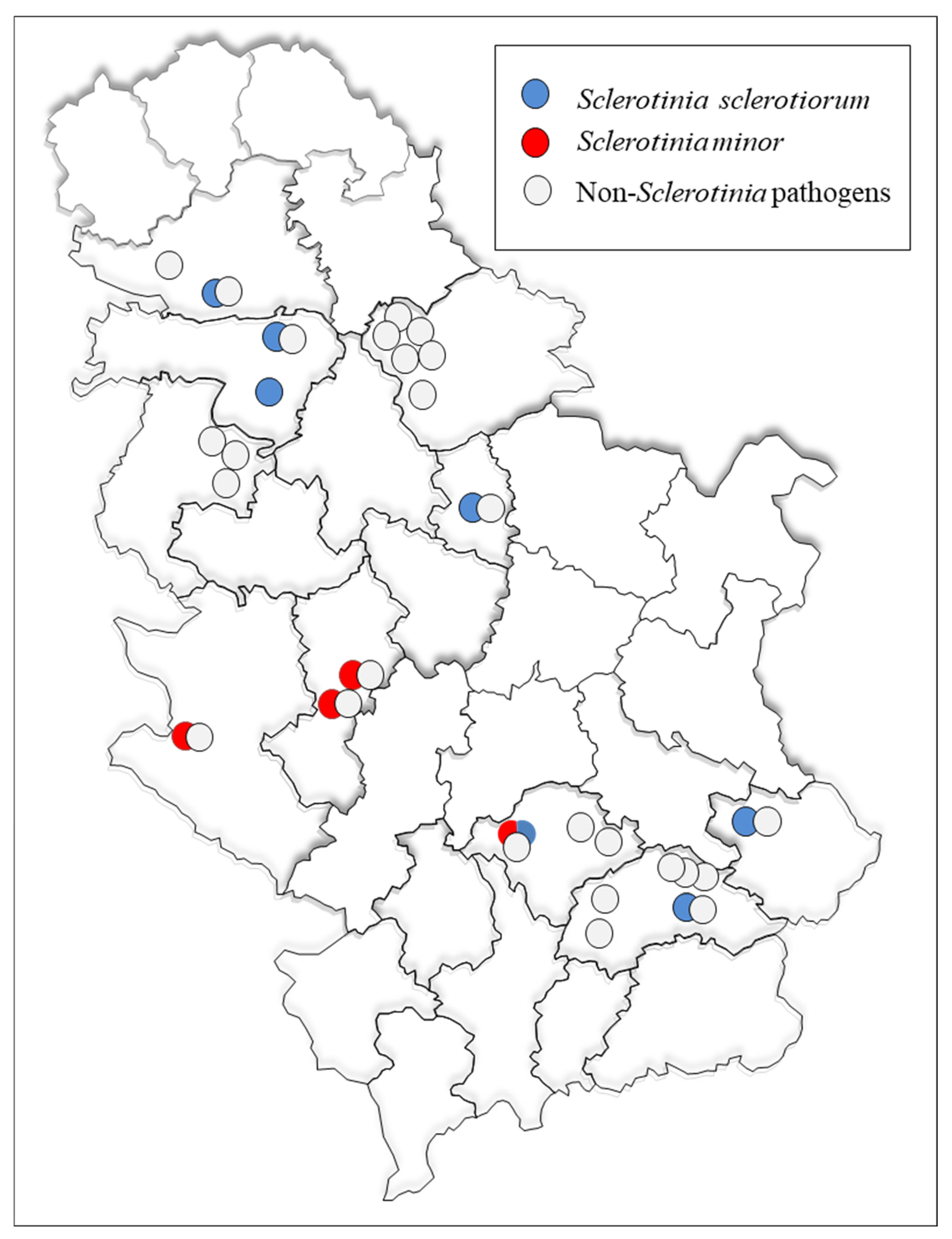
Figure 1. The geographic distribution of localities in Serbia included in the survey and detected Sclerotinia spp. species.
Microorganisms 14 00189 g001
Figure 2. Sclerotinia spp.—naturally infected lettuce plants with S. minor (A,C) and S. sclerotiorum (B) in the field with developed sclerotia (arrows); obverse (left) and reverse (right) appearance of snow-white (D) and gray (H) mycelium of S. sclerotiorum isolates on potato dextrose agar (PDA) after 15 days at 25 °C; obverse (left) and reverse (right) appearance of regular (G) and irregular (J) growth pattern of S. minor isolates on PDA after 15 days at 25 °C; Sclerotia of S. sclerotiorum (E) and S. minor (F) isolates; (I) pathogenicity test: inoculated plant (top) and control (bottom).
Microorganisms 14 00189 g002
Figure 3. Growth rate of Sclerotinia spp. isolates grown on potato dextrose agar at 25 °C in the dark. The error bars represent the standard deviation. Values marked with the same letters or absence of letters indicate no statistically significant difference.
Microorganisms 14 00189 g003
Figure 4. Maximum parsimony tree, constructed based on DNA sequences from fragments of the ITS rDNA from a subset of 20 Serbian isolates (10 Sclerotinia sclerotiorum, 10 Sclerotinia minor), 42 selected sequences of Sclerotinia species available in the GenBank database, and outgroup Hypocrea lixii (FJ861393). The tree was generated in MEGA 6. Bootstrap analyses were performed with 1000 replicates, and bootstrap values (>50%) are shown on the tree. All Serbian isolates are indicated in bold.
Microorganisms 14 00189 g004
Figure 5. Haplotype network of Sclerotinia minor based on sequences of the ITS region (n = 103; 93 sequences from GenBank and 10 Serbian S. minor sequences). Each circle depicts a unique haplotype, and the size of each circle is proportional to the number of sequences it represents. Nucleotide differences are denoted by the hatch marks across black lines connecting haplotypes, with each hatch mark representing a single nucleotide variation.
Microorganisms 14 00189 g005
Figure 6. Lesion length on inoculated lettuce leaves caused by selected Sclerotinia spp. isolates after 3-day incubation at 25 °C. The error bars represent the standard deviation. Values marked with the same letters or absence of letters indicate no statistically significant difference.
Microorganisms 14 00189 g006
Figure 7. Oxalic acid production of different isolates of Sclerotinia sclerotiorum and Sclerotinia minor after 3-day static incubation in 50 mL of potato-dextrose broth at 25 °C. The error bars represent the standard deviation. Values marked with the same letters or absence of letters indicate no statistically significant difference.
Microorganisms 14 00189 g007
Figure 8. Susceptibility of Sclerotinia sclerotiorum and Sclerotinia minor isolates to high osmotic pressure: inhibition of the mycelial growth on potato dextrose agar medium supplemented with 0.51 mol/dm3 NaCl after incubation for 2 days at 25 °C.
Microorganisms 14 00189 g008
Figure 9. Comparison of Sclerotinia sclerotiorum and Sclerotinia minor in terms of the mycelial growth, virulence, oxalic acid production, and susceptibility to high osmotic pressure. An average of 10 isolates of each species is presented. The error bars represent the standard deviation. Values marked with different letters indicate statistically significant difference.
Microorganisms 14 00189 g009aMicroorganisms 14 00189 g009b
Table 1. Isolates of Sclerotinia spp. included in phylogenetic analyses.
SpeciesAcc NoIsolateHostOriginSubmittedReference
Sclerotinia minorJF27988062907Tanacetum sp.Australia2011[28]
AB516661MAFF 238173-Japan2009[28]
JF27987945903A-Australia2011[28]
JF27987745802A-Australia2011[28]
OL42363215-2Lactuca sativaSerbia2021[21]
PQ489371Sm 11-4Lactuca sativaSerbia2021In this study
PQ489372Sm 11-9aLactuca sativaSerbia2021In this study
PQ489373Sm 11-10Lactuca sativaSerbia2021In this study
PQ489374Sm 13-2Lactuca sativaSerbia2021In this study
PQ489375Sm 13-5Lactuca sativaSerbia2021In this study
PQ489376Sm 15-3Lactuca sativaSerbia2022In this study
PQ489377Sm 15-5Lactuca sativaSerbia2022In this study
PQ489378Sm 18-3Lactuca sativaSerbia2022In this study
PQ489379Sm 18-4Lactuca sativaSerbia2022In this study
PQ489380Sm 18-5Lactuca sativaSerbia2022In this study
Sclerotinia sclerotiorumKX1847202Brassica oleracea var. capitataSri Lanka2017[28]
MG249967SSC2JHUGossypium hirsutumUSA2017[28]
AB233346MAFF 306676Vaccinium corymbosumJapan 2007[28]
KY07361316-042Capsella bursa-pastorisKorea2016[28]
KP340898SS-BO-SCBrassica oleracea var. capitataNew Mexico2015[28]
JN013184-Aquilegia flabellataItaly2011[28]
KF859932DAOM:241671-Canada2013[28]
DQ329537B23-Alaska2006[28]
HQ833450Ms85Morus albaChina2010[28]
KY750530SQC-000Oenanthe javanicaChina2017[28]
KY07361416-119Cucumis sativusKorea2016[28]
FJ810516ATCC:MYA-4521-USA2009[28]
KT224645Ss1212HATrifolium ambiguumPoland2015[28]
KJ614564JBARES2014AAllium tuberosumKorea2014[28]
AB937095MuRa-103Brassica napaJapan2014[28]
KJ744364PM27Beta vulgarisGermany2014[28]
KY07361215-030Lactuca sativaKorea2016[28]
KF791510PSHB1Lablab purpureusBangladesh2013[28]
PP179050BBrassica oleracea var. capitataSerbia2025[29]
PP179051B1Brassica oleracea var. capitataSerbia2025[29]
PP179052B2Brassica oleracea var. capitataSerbia2025[29]
PP179053B3Brassica oleracea var. capitataSerbia2025[29]
PP179054MBrassica oleracea var. capitataSerbia2025[29]
PP179057M1Brassica oleracea var. capitataSerbia2025[29]
PP179055M2Brassica oleracea var. capitataSerbia2025[29]
PP179056M3Brassica oleracea var. capitataSerbia2025[29]
PP179058M4Brassica oleracea var. capitataSerbia2025[29]
PP179059M5Brassica oleracea var. capitataSerbia2025[29]
PP179060M6Brassica oleracea var. capitataSerbia2025[29]
PP179061M7Brassica oleracea var. capitataSerbia2025[29]
PP177498SCHelianthus annuusSerbia2025[29]
PQ489358Ss 17-7Lactuca sativaSerbia2022In this study
PQ489359Ss 18-10Lactuca sativaSerbia2022In this study
PQ489360Ss 18-11Lactuca sativaSerbia2022In this study
PQ489361Ss 20-1Lactuca sativaSerbia2022In this study
PQ489362Ss 20-3Lactuca sativaSerbia2022In this study
PQ489363Ss 21-1Lactuca sativaSerbia2022In this study
PQ489364Ss 21-2Lactuca sativaSerbia2022In this study
PQ489365Ss 22-11Lactuca sativaSerbia2022In this study
PQ489366Ss 22-14Lactuca sativaSerbia2022In this study
PQ489367Ss 23Lactuca sativaSerbia2022In this study
Sclerotinia trifoliorumKT819299TN Sc10101Trigonella foenum-graecumTunisia2015[28]
KT224652St03TPTrifolium pretensePoland2015[28]
KT224651St02TPTrifolium pretensePoland2015[28]
KT986229St1813TPTrifolium pretensePoland2015[28]
Sclerotinia nivalisJX294862SN110812Atractylodes japonicaChina2012[30]
PV973881Sm8Daucus carota subsp. sativusRussia2023[31]
Hypocrea lixiiFJ8613932S12A-USA2009[28]
Table 2. Lettuce drop incidence in different districts in Serbia during 2021 and 2022.
YearDistrictLocationDisease Incidence 1No. of SamplesFungal Species Detected 2
2021South BanatGlogonj 110%7Phoma sp. (1) 3
Plectosphaerella spp. (3)
Glogonj 25%9Fusarium spp. (2)
Plectosphaerella sp. (1)
Glogonj 42–5%4Pythium sp. (1)
Glogonj 52%3Alternaria sp. (1)
West BačkaBački Brestovac2%6Fusarium sp. (1)
JablanicaNavalin 15%5Alternaria sp. (1)
Navalin 210%3Verticillium spp. (2)
Alternaria sp. (1)
Navalin 42%4Fusarium sp. (1)
Donja Lokošnica2%3Plectosphaerella spp. (3)
Priboj5–10%4Verticillium spp. (2)
Alternaria sp. (1)
ZlatiborPotočanje55%8Sclerotinia minor (5)
Fusarium sp. (1)
2022South BanatOpovo 11%7Plectosphaerella sp. (1)
Opovo 21%4Plectosphaerella sp. (1)
MačvaMrđenovac 12%1Fusarium sp. (1)
Mrđenovac 25%4Fusarium spp. (2)
Žabar5%1Plectosphaerella sp. (1)
MoravicaTrbušani 130%20Sclerotinia minor (19)
Botrytis spp. (3)
Trbušani 315%7Sclerotinia minor (3)
Botrytis sp. (1)
NišavaMerošina 125%3Fusarium spp. (3)
Merošina 21%1Fusarium sp. (1)
PirotBela Palanka35%25Sclerotinia sclerotiorum (10)
Botrytis sp. (1)
ToplicaBlace20%23Sclerotinia sclerotiorum (5)
Sclerotinia minor (8)
Botrytis sp. (1)
JablanicaLebane 230%12Sclerotinia sclerotiorum (10)
Botrytis spp. (3)
PodunavljeSmederevska Palanka15%7Sclerotinia sclerotiorum (5)
Botrytis sp. (2)
SremIrig40%12Sclerotinia sclerotiorum (11)
Botrytis sp. (1)
Kraljevci2%1Sclerotinia sclerotiorum (1)
South BačkaVeternik15%1Sclerotinia sclerotiorum (1)
Botrytis spp. (3)
1 Average disease incidence estimated by walking through the crop in a zigzag pattern and randomly rating 100 plants in three replicates. 2 Isolated pathogenic species identified to the genus level based on colony morphology and ITS sequencing. 3 The numbers in parentheses represent the number of isolates obtained.
Table 3. Cultural features of Sclerotinia spp. isolates on potato dextrose agar at 25 °C.
SpeciesIsolateColony ColorRelative Density and Appearance of Aerial MyceliumDark Pigment SynthesisTiming of Formation of Sclerotia (Day)Arrangement of SclerotiaAverage No of Sclerotia
± SD/Plate
Average Diameter of Sclerotia (mm)
Sclerotinia sclerotiorumSs 17-7Snow whiteWoolly- floccose, dense-8Edge and middle ring12.7 ± 8.11.67–5.17
Ss 18-10GreyFloccose, dense+9Scattered all around65.0 ± 8.70.67–2.17
Ss 18-11WhitishFloccose, low dense-7Edge and middle ring20.0 ± 13.00.83–3.33
Ss 20-1WhitishFloccose, very dense-7Scattered all around36.3 ± 5.11.17–3.33
Ss 20-3WhitishFloccose, dense+9Edge and middle ring24.0 ± 7.91.00–2.83
Ss 21-1Snow whiteWoolly, dense-8Edge and middle ring8.7 ± 1.52.33–5.83
Ss 21-2White Floccose, low dense-8Scattered all around21.7 ± 6.51.50–4.33
Ss 22-11WhiteFloccose, low dense-8Middle ring2.3 ± 0.63.67–5.00
Ss 22-14WhiteWoolly- floccose, low dense-7Edge and middle ring20.7 ± 3.50.90–4.00
Ss 23WhitishFloccose, dense-8Scattered all around14.3 ± 3.11.67–3.33
Sclerotinia minorSm 11-4WhitishFloccose, dense-6Scattered all around373.3 ± 23.10.50–1.50
Sm 11-9aSnow whiteWoolly, dense to very dense-8Scattered all around153.3 ± 30.60.40–2.33
Sm 11-10WhitishCottony- floccose, low dense-8Scattered all around120.0 ± 20.00.50–1.75
Sm 13-2WhitishFloccose, low dense to dense-6Scattered all around200.0 ± 40.00.50–2.00
Sm 13-5WhitishFloccose, low dense-8Scattered all around186.7 ± 61.10.50–1.50
Sm 15-3WhitishFloccose, dense-6Scattered all around140.0 ± 10.01.00–2.00
Sm 15-5WhiteFloccose, low dense to dense-9Scattered all around46.7 ± 11.51.00–2.50
Sm 18-3WhitishCottony- floccose, very dense-8Scattered all around95.0 ± 39.70.67–1.83
Sm 18-4WhitishCottony, dense-8Edge ring108.3 ± 20.20.50–1.83
Sm 18-5WhitishCottony, dense-9Edge ring11.7 ± 5.90.20–0.50
Disclaimer/Publisher’s Note: The statements, opinions and data contained in all publications are solely those of the individual author(s) and contributor(s) and not of MDPI and/or the editor(s). MDPI and/or the editor(s) disclaim responsibility for any injury to people or property resulting from any ideas, methods, instructions or products referred to in the content.