

Article

Clinical Significance of cfiA Positivity Detected by Matrix-Assisted Laser Desorption/Ionization Time-of-Flight Mass Spectrometry in Bacteroides fragilis Infections


1
Department of Pathology, United Christian Hospital, Hong Kong SAR, China
2
Department of Health Technology and Informatics, Faculty of Health and Social Science, The Hong Kong Polytechnic University, Hong Kong SAR, China
3
Department of Pathology, Tseung Kwan O Hospital, Hong Kong SAR, China
*
Author to whom correspondence should be addressed.
Microorganisms 2026, 14(1), 168; https://doi.org/10.3390/microorganisms14010168
Submission received: 11 December 2025 / Revised: 9 January 2026 / Accepted: 10 January 2026 / Published: 12 January 2026
(This article belongs to the Special Issue Advances in Clinical Infections and Antimicrobial Resistance)

Abstract

The MALDI-TOF MS Bruker Biotyper MBT subtyping IVD module enables the early detection of cfiA-positive Bacteroides fragilis (cfiA+ BF) during bacterial identification. However, the relationship between genetic positivity, phenotypic resistance, and clinical outcomes has not been fully elucidated. This retrospective study analyzed B. fragilis isolates from three Hong Kong hospitals between 2021 and 2025 to examine their prevalence and the clinical utility of MALDI-TOF MS in rapid cfiA detection. Antibiotic susceptibility testing, cfiA gene detection using MALDI-TOF MS, and Oxford Nanopore sequencing were performed. Medical records were reviewed, and univariate analyses and multivariate logistic regression were used to identify factors associated with cfiA positivity and 30-day all-cause mortality. Overall, B. fragilis exhibited a high rate of antibiotic resistance. Concomitant resistance to carbapenems and metronidazole was identified in three isolates. Among the 166 isolates, 40 (24.1%) were cfiA-positive. cfiA detection by MALDI-TOF MS showed 100% concordance with the gene sequencing results and correlated strongly with phenotypic carbapenem resistance (Φ = 0.82, p < 0.001 for meropenem; Φ = 0.70, p < 0.001 for ertapenem; Φ = 0.63, p < 0.001 for imipenem). Phylogenetic analysis revealed two distinct clusters corresponding to cfiA status, each exhibiting genetic diversity based on multi-locus sequence typing (MLST). The cfiA+ BF isolates demonstrated high-level phenotypic carbapenem resistance in the presence of upstream insertion sequences. The predominant sequence type (ST) among cfiA+ BF isolates was ST157, and 70% of ST157 isolates harbored IS1187 in the upstream region of cfiA. Gene sequencing also identified other emerging beta-lactamase genes blaOXA-347 and blaMUN. The 30-day all-cause mortality following B. fragilis infection was 13.3%, with independent predictors including a high Charlson Comorbidity Index (OR = 1.30; p = 0.02) and the absence of early source control (OR = 4.84; p = 0.03). This study highlights the widespread occurrence of cfiA+ BF in Hong Kong and the clinical significance of rapid cfiA detection. Continuous surveillance is essential to monitor the ongoing threat of antibiotic resistance in B. fragilis.

1. Introduction

Bacteroides fragilis is a gram-negative anaerobic bacterium and a key constituent of the colonic microbiota. It is an opportunistic pathogen capable of causing a variety of human infections, including bacteremia, and intra-abdominal, skin, and soft tissue infections. The global emergence of antibiotic resistance among B. fragilis strains has posed a significant threat to public health [1,2,3,4]. Infections caused by resistant B. fragilis have been associated with an increased risk of treatment failure and adverse clinical outcomes [5,6].
Antibiotic resistance in B. fragilis is mediated by multiple genes, among which cfiA is regarded as the predominant carbapenemase gene. Molecular studies have shown that cfiA-negative (cfiA− BF) and cfiA-positive (cfiA+ BF) B. fragilis strains belong to two genotypically distinct divisions, I and II, respectively [7,8], which differ in their geographic distribution and pathogenic potential [9]. Most division I isolates carry the cepA gene, which encodes a class A serine beta-lactamase conferring resistance to penicillins and cephalosporins. On the other hand, division II isolates possess the cfiA gene, which encodes a class B metallo-beta-lactamase that mediates resistance to carbapenems and other beta-lactam antibiotics. Notably, not all cfiA+ BF isolates exhibit phenotypic resistance because the gene may only be weakly expressed in the absence of an upstream mobile element [10].
Routine antibiotic susceptibility testing of anaerobes is uncommon in many clinical laboratories because it is labor-intensive and time-consuming. Phenotypic susceptibility testing usually takes several days to complete, and many genotypic methods require specialized technical expertise [11]. Consequently, antibiotic treatment is typically empirical, guided by expected susceptibility patterns and local resistance epidemiology. In the era of rising antibiotic resistance, there is a growing need for a rapid, accurate and cost-effective susceptibility testing method.
In recent years, the new MBT subtyping IVD module has been introduced for the matrix-assisted laser desorption/ionization time-of-flight mass spectrometry (MALDI-TOF MS) Bruker Biotyper (Bruker Daltonics, Bremen, Germany), enabling rapid differentiation between cfiA+ BF and cfiA− BF at the time of bacterial identification. Previous studies have confirmed its diagnostic accuracy, but the relationship between genetic positivity, phenotypic resistance and clinical outcomes has not been fully elucidated [12,13,14,15]. In Hong Kong, where the prevalence of cfiA+ BF is high [16], it is particularly important to investigate how this module may be useful in clinical practice.
The aim of this study was to characterize the phenotypic and genotypic resistance patterns of B. fragilis from three hospitals in Hong Kong. Additionally, this study evaluated the risk factors and clinical outcomes associated with cfiA+ BF infections. These findings may offer insights into laboratory diagnosis, antimicrobial stewardship and clinical decision-making.

2. Materials and Methods

2.1. Patient Identification and Isolate Collection

This retrospective observational study was conducted across three non-teaching hospitals (United Christian Hospital, Haven of Hope Hospital and Tseung Kwan O Hospital) within the Kowloon East Cluster in Hong Kong from 2021 to 2025. Both United Christian Hospital and Tseung Kwan O Hospital are acute-care hospitals, while Haven of Hope Hospital is an extended-care hospital. As of 31 March 2024, the three hospitals serve a population of approximately 1,171,600, with a total of 3010 beds. Cases with B. fragilis infections were identified through the Laboratory Information System. Due to variations in laboratory practice pertaining to strain preservation, isolates from all specimen types were retrieved from United Christian Hospital over a 14-month period (November 2023 to January 2025), whereas only blood culture isolates were retrieved from Tseung Kwan O Hospital and Haven of Hope Hospital over a 4-year period (January 2021 to January 2025). All isolates were stored at −70 °C before testing. If multiple isolates were obtained from the same patient during the study period, only the first isolate was included, regardless of the specimen type.

2.2. Isolate Identification

B. fragilis isolates were re-cultured on blood agar and incubated at 37 °C in the Concept Anaerobic Workstation (Baker Ruskinn, Bridgend, UK). These were identified down to the species level with identification scores ≥ 2.0 using a MALDI-TOF MS Bruker Biotyper equipped with the MBT IVD Library revision J (Bruker Daltonics, Bremen, Germany) according to the manufacturer’s instructions. The MBT subtyping IVD module simultaneously reported the cfiA status as a categorical output (“cfiA-positive” or “cfiA-negative”) at the time of bacterial identification. The cfiA subtyping result was not displayed if the internal threshold values of the algorithm were not met.

2.3. Antibiotic Susceptibility Testing

Antibiotic susceptibility testing was performed using the disk diffusion method based on the European Committee for Antimicrobial Susceptibility Testing (EUCAST) guidelines [17]. B. fragilis isolates were tested against the following antibiotic disks (Oxoid Ltd., Basingstoke, UK): ampicillin–sulbactam (10 µg/10 µg), amoxicillin–clavulanic acid (2 µg/1 µg), piperacillin–tazobactam (30 µg/6 µg), ertapenem (10 µg), imipenem (10 µg), meropenem (10 µg), metronidazole (5 µg), and clindamycin (2 µg). Bacterial suspensions adjusted to 1.0 McFarland standard were incubated with antibiotic disks on fastidious anaerobe agar supplemented with 5% defibrinated horse blood in the same anaerobic workstation for 16 to 20 h. B. fragilis ATCC 25285 and Clostridium perfringens ATCC 13124 were used for quality control. The antibiotic susceptibility was interpreted according to the EUCAST breakpoint guidelines version 15.0, which was valid from 1 January 2025 [18].

2.4. Genomic Sequencing

All B. fragilis isolates were subjected to Oxford Nanopore sequencing. Bacterial DNA was extracted using the QIAamp BiOstic Bacteremia DNA Kit (Qiagen, Hilden, Germany) following the manufacturer’s protocol. Sequencing libraries were prepared using the Nanopore-only Microbial Isolate Sequencing Solution (NO-MISS) protocol for cell cultures with the Rapid Barcoding Kit 96 V14 (SQK-RBK 114.96; Oxford Nanopore Technologies, Oxford, UK). The libraries were sequenced using the R10.4.1 flow cells on the GridION MK1 platform for 72 h. Basecalling was performed using Dorado Basecall Server v7.8.3 in super-accurate (SUP) mode and reads with a Phred quality score (Q score) less than 10 were filtered out. Sequencing yield and quality were assessed with NanoPlot v1.46.2 [19]. Quality-filtered reads were mapped against the B. fragilis NCTC 9343 reference genome (RefSeq accession number GCF_000025985.1) using minimap2 v2.30 [20]. Per-base coverage was calculated with modsdepth v0.3.12 [21], yielding a mean coverage depth of 139.1× ± 48.75×. Raw sequencing reads were assembled using Hybracter v0.11.2 [22], and the sequencing assemblies were analyzed using Bactopia v3.2.0 [23]. Species identification was performed using the Genome Taxonomy Database Toolkit (GTDB-Tk) v2.4.0 [24], and the assembly quality was assessed using CheckM v1.2.3 [25]. Multi-locus sequence typing (MLST), which was based on seven core gene fragments (groL, rpoB, recA, dnaJ, rprX, prfA and fusA), was conducted using mlst v2.23.0 [26]. Antibiotic resistance genes were identified using AMRFinderPlus v4.0.19 [27]. For cfiA+ BF, the genomic location of cfiA was also determined by AMRFinderPlus, and the upstream region was searched for potential insertion sequences with reference to the ISfinder database (https://isfinder.biotoul.fr, last update: 21 November 2025, access date: 2 December 2025) [28]. The sequencing data of all isolates have been deposited in the National Center for Biological Information (NCBI) BioProject database under the accession number PRJNA1372458.

2.5. Phylogenetic Analysis

Phylogenetic analysis was performed to assess the genetic relationships among isolates. Core genomes were identified using a pangenome-based approach, and recombination events were masked prior to tree construction. A maximum-likelihood phylogenetic tree was then generated using IQ-Tree v2.2.2.7 [29] with 1000 ultrafast bootstrap replicates, and branches with bootstrap support ≥ 70% were considered well-supported. The resulting tree was visualized and annotated using Interactive Tree of Life (iTOL) v7.2.2.

2.6. Clinical Data Collection

Clinical information was retrospectively extracted from electronic medical records for all patients. The following information was included: demographics (age, gender, residential care home), medical history (recent surgery, use of immunosuppressive therapy, prior antibiotic exposure), comorbidities, clinical characteristics (presumed source of B. fragilis infections, temperature, co-infecting microorganisms), interventions (antibiotics, non-surgical drainage, surgery), ICU admission, duration of hospitalization, and mortality. The Charlson Comorbidity Index was calculated to stratify the comorbidity burden.

2.7. Definitions

Comorbidities were identified based on the information documented by the physicians in the medical records at the time of specimen collection. Chronic kidney disease was defined as having an estimated glomerular filtration rate (eGFR) less than 60 mL/min/1.73 m2 persisting for at least 3 months, or a history of renal transplant. Heart disease encompassed all cardiac conditions, including ischemic heart disease, congestive heart failure, arrhythmia, cardiomyopathy, and structural heart disease. Recent surgery, use of immunosuppressive therapy, and antibiotic exposure referred to any such events occurring within 4 weeks preceding specimen collection. Presumed source of infection was assigned for bacteremia cases according to the most plausible clinical site of origin based upon a review of medical records. Fever was defined as a body temperature exceeding 37.8 °C measured within 24 h of specimen collection. Co-infecting microorganisms referred to laboratory-confirmed microorganisms identified from the same specimen from which B. fragilis was isolated. Empirical antibiotic treatment was defined as antibiotic treatment administered during the time of specimen collection prior to the availability of culture results. It was considered appropriate if the corresponding isolate was later confirmed to be susceptible to at least one of the empirical antibiotics administered within 24 h of specimen collection by phenotypic testing. Early source control was defined as any surgical procedure or drainage performed within 4 days of specimen collection, or if an effective drainage system was already in place at the time of specimen collection. All-cause mortality was defined as death from any cause occurring within 7, 30, or 90 days after specimen collection.

2.8. Statistical Analysis

Data were analyzed using the IBM SPSS Statistics for Windows software, version 31.0 (IBM Corp., Armonk, NY, USA). Comparisons were made between (1) infections caused by cfiA+ BF and cfiA− BF, and (2) survivors and non-survivors based on 30-day outcomes. Categorical variables are expressed as frequency counts and percentages. Continuous variables are expressed as means +/− standard deviations (SDs) if normally distributed or as medians with interquartile ranges (IQRs) if non-normally distributed. Univariate analyses were conducted using the Chi-square test or Fisher’s exact test for categorical variables, and the Student’s t-test or the Mann–Whitney U-test for continuous variables, as appropriate. All variables with a p-value < 0.25 in univariate analyses were included in multivariate logistic regression to identify independent risk factors for cfiA+ BF infection and 30-day all-cause mortality. To avoid multicollinearity, variables of similar nature were not included simultaneously. The results are presented as odds ratios (ORs) with 95% confidence intervals (CIs). Two-tailed p-values < 0.05 were considered statistically significant. The p-values were reported as exploratory and were not adjusted for multiplicity.

3. Results

3.1. Isolate Information

A total of 166 non-duplicate B. fragilis isolates [40 (24.1%) cfiA-positive and 126 (75.9%) cfiA-negative] were retrieved. In total, 100 isolates (60.2%) were from United Christian Hospital (November 2023 to January 2025), 51 isolates (30.7%) were from Tseung Kwan O Hospital (January 2021 to January 2025), and 15 isolates (9.0%) were from Haven of Hope Hospital (January 2021 to January 2025). The isolates were most commonly obtained from blood (42.2%), followed by skin and soft tissue (32.5%) and intra-abdominal sources (16.3%) (Supplementary Table S1). The majority of B. fragilis infections were polymicrobial, with at least one co-infecting microorganism identified in 71.1% of cases. The most frequently isolated co-pathogens were Escherichia coli (33.1%), Streptococcus anginosus group (15.7%), Staphylococcus aureus (10.2%), and Enterococcus species (9.0%). E. coli co-infections were more common in cfiA+ BF infections (50.0% vs. 27.8%, p = 0.009), and S. aureus co-infections occurred more often in cfiA− BF infections (13.5% vs. 0%, p = 0.01) (Supplementary Table S2).

3.2. Genotypic Characteristics

The 166 B. fragilis isolates belonged to 81 different sequence types (STs). Phylogenetic analysis showed that the strains were clustered into two major divisions, I and II, corresponding to cfiA status. The cepA gene was found in all except one isolate (99.2%) belonging to division I. Based on MLST, isolates sharing the same STs consistently belonged to the same division. However, within each division, isolates exhibited considerable genetic diversity. In division I (cfiA− BF), the most prevalent STs were ST178 (n = 11), ST1 (n = 8), ST4 (n = 7), ST74 (n = 7), ST17 (n = 5), and ST67 (n = 5). In division II (cfiA+ BF), the predominant STs were ST157 (n = 10), ST151 (n = 4), and ST52 (n = 3) (Figure 1). A total of 18 isolates (10.8%) carried the bft gene, all of which belonged to division I (cfiA− BF), with bft-1 being the predominant subtype. No specific association was observed between the ST and infection sites.
For the 40 division II (cfiA+ BF) isolates, upstream insertion sequences were identified in 19 of them (47.5%), all located within 100 bp of the cfiA start codon. The most prevalent insertion sequence type was IS1187 (n = 11), which was present in 70.0% of ST157 isolates and also detected in four other sequence types. The second and third most common insertion sequence types were IS613 (n = 4) and IS616 (n = 2), respectively (Supplementary Table S3).
The distribution of 29 antibiotic resistance genes conferring resistance to 7 classes of antibiotics among the B. fragilis isolates is summarized in Supplementary Table S4. The most prevalent resistance gene was tetQ (90.4%), followed by cepA (75.3%) and ermF (55.4%). On the other hand, the nimJ (1.2%), nimA (0.6%), and nimE (0.6%) genes for nitroimidazole resistance were rare. A small proportion of isolates, in both divisions I and II, harbored emerging beta-lactamase genes blaOXA-347 (6.0%), blaMUN-1 (3.0%), and blaMUN-5 (0.6%). When comparing between cfiA+ BF and cfiA− BF isolates, lnu(AN2) (18.3% vs. 5.0%; p = 0.04) and mef(En2) (18.3% vs. 5.0%; p = 0.04) were detected more often in cfiA− BF isolates. In contrast, blaOXA-347 (12.5% vs. 4.0%; p = 0.06) and erm(F) (67.5% vs. 51.6%; p = 0.08) tended to occur more frequently in cfiA+ BF isolates.

3.3. Phenotypic Antibiotic Resistance and Association with cfiA Status

According to the EUCAST clinical breakpoint guideline, more than half of the B. fragilis isolates exhibited resistance to amoxicillin–clavulanic acid (52.4%), ampicillin–sulbactam (51.8%), and clindamycin (51.2%). The rates of resistance to ertapenem (28.9%) and meropenem (27.7%) were higher than those to imipenem (16.3%). The lowest resistance rate was observed for metronidazole (13.9%).
The detection of cfiA using the MALDI-TOF MS Bruker Biotyper MBT subtyping IVD module demonstrated 100% concordance with the Oxford Nanopore sequencing results. A positive cfiA result was strongly associated with phenotypic carbapenem resistance, showing the highest correlation with meropenem (Φ = 0.82, p < 0.001) and the lowest with imipenem (Φ = 0.63, p < 0.001). A moderate association was also detected between cfiA positivity and phenotypic resistance to beta-lactam and beta-lactamase inhibitor combinations, with the strongest association observed for piperacillin–tazobactam (Φ = 0.43, p < 0.001). Phenotypically, cfiA+ BF isolates, especially those belonging to ST157, exhibited marked resistance to most antibiotics, except metronidazole. The level of carbapenem resistance differed with respect to the presence of upstream insertion sequences. All cfiA+ BF isolates with upstream insertion sequences exhibited phenotypic resistance to all three carbapenems, whereas 90.5% of isolates without such sequences remained phenotypically susceptible to at least one carbapenem (Supplementary Table S3). A similarly high level of resistance was also observed for isolates harboring blaOXA-347 and blaMUN-1. Conversely, the majority of cfiA− BF isolates were phenotypically susceptible to piperacillin–tazobactam (87.3%), carbapenems (88.9% to ertapenem; 96.8% to imipenem; 92.9% to meropenem), and metronidazole (84.9%) (Table 1). Three isolates (1.8%) displayed concomitant resistance to all three carbapenems and metronidazole. Furthermore, two of these isolates (1.2%) were resistant to all eight antibiotics tested in the study. Both were division II isolates, one belonging to ST157 and the other to ST202.

3.4. Clinical Characteristics of Patients with cfiA+ BF Infection

The median age of the patients was 73.0 (IQR 60.3–85.0), with the majority (65.7%) aged over 65. Slightly more female (53.0%) than male patients (47.0%) were included. There were no significant differences in terms of age and sex distribution with respect to cfiA status. Approximately half (48.8%) of the patients had a Charlson Comorbidity Index more than or equal to five. Common comorbidities included diabetes mellitus (35.5%), chronic kidney disease (24.1%) and active malignancy (21.1%). A greater proportion of patients with cfiA+ BF infections had a history of recent surgery (10.0% vs. 3.2%; p = 0.10) compared to those with cfiA− BF infections, although the difference was not statistically significant. No significant differences were observed between the cfiA+ BF and cfiA− BF groups regarding the presumed sources of infection or the presence of fever (Supplementary Table S5). Univariate analysis identified diabetes mellitus as a potential risk factor (p = 0.03) for cfiA+ BF infection. However, multivariate logistic regression did not identify any independent risk factors (Supplementary Table S6).
Most of the patients (84.9%) had received empirical antibiotics with expected activity against B. fragilis, primarily amoxicillin–clavulanic acid (56.0%) and piperacillin–tazobactam (17.5%). Appropriate empirical antibiotic use was less common in those with cfiA+ BF infections (43.6% vs. 55.6%; p = 0.19). Regarding targeted therapy, the most widely prescribed antibiotics were amoxicillin–clavulanic acid (36.1%), metronidazole (31.9%), and piperacillin–tazobactam (22.3%). Meropenem was used more often as targeted therapy (15.1%), particularly in those with cfiA+ BF infections (25.0%). Similarly, appropriate use of targeted antibiotics was less frequent in cfiA+ BF infections (62.5% vs. 75.4%; p = 0.11). The 30-day all-cause mortality following B. fragilis infection was 13.3%. Patients with cfiA+ BF infections appeared to have a higher likelihood of ICU admission (12.5% vs. 6.3%; p = 0.31) and prolonged hospital stay (40.5% vs. 27.5%; p = 0.13), and increased 7-day (7.5% vs. 3.2%; p = 0.36), 30-day (17.5% vs. 11.9%; p = 0.36), and 90-day (25.0% vs. 23.8%; p = 0.88) all-cause mortality, although none of these differences reached statistical significance (Table 2).

3.5. Predictors for 30-Day All-Cause Mortality Following B. fragilis Infection

Patients with B. fragilis infections were classified into survivor and non-survivor groups by their 30-day outcomes. Univariate analysis showed that the risk factors for 30-day all-cause mortality included a high Charlson Comorbidity Index (p < 0.001), active malignancy (p < 0.001), and the absence of early source control (p < 0.001). The mortality rate appeared to be similar regardless of the appropriateness of empirical or targeted antibiotics (Table 3). Multivariate logistic regression revealed that a high Charlson Comorbidity Index [OR = 1.30; 95% confidence interval (CI): 1.04–1.63, p = 0.02] and the absence of early source control [OR = 4.84; 95% confidence interval (CI): 1.18–19.75, p = 0.03] were independent risk factors for 30-day all-cause mortality (Table 4).

4. Discussion

B. fragilis is the leading cause of severe anaerobic infections, but the emergence of antibiotic resistance has complicated the selection of effective antibiotic therapy. The MALDI-TOF MS Bruker Biotyper MBT subtyping IVD module has demonstrated potential as a rapid and reliable tool for the detection of cfiA, the predominant carbapenemase gene in B. fragilis. In this study, we investigated the clinical significance of cfiA+ BF detection and evaluated the patient characteristics and outcomes associated with these infections.

4.1. MALDI-TOF MS as a Rapid and Useful Diagnostic Tool for cfiA Detection

Consistent with previous studies [12,13,14,15], the detection of cfiA using the MALDI-TOF MS Bruker Biotyper MBT subtyping IVD module demonstrated excellent concordance (100%) with gene sequencing results, confirming its reliability as a rapid diagnostic tool. The cfiA positivity correlated strongly with phenotypic carbapenem resistance and moderately with beta-lactam and beta-lactamase inhibitor combination resistance. Among individual carbapenems, the association was strongest for meropenem and weakest for imipenem. For example, while only 7.5% of cfiA+ BF isolates were phenotypically susceptible to meropenem, up to 42.5% of them were susceptible to imipenem. Similar patterns of differential carbapenem resistance have been reported in other studies [30], in which the minimal inhibitory concentrations (MICs) of cfiA+ BF were generally lower for imipenem than for meropenem. This difference may be related to the more efficient hydrolysis of meropenem by beta-lactamases [15,31]. Accordingly, meropenem may serve as a more sensitive phenotypic marker of cfiA-mediated resistance than imipenem [32].
Approximately half (47.5%) of the cfiA+ BF isolates in our cohort possessed upstream insertion sequences, and all of them demonstrated phenotypic resistance to all three carbapenems tested. These insertion sequence types are similar to those previously described in Hong Kong [33]. They are thought to act as strong promoters for cfiA expression and thereby confer high-level carbapenem resistance. In contrast, 90.5% of cfiA+ BF isolates without upstream insertion sequences displayed only partial carbapenem resistance, probably related to the low level of constitutive cfiA expression. Their resistance may also be mediated by other genetic determinants such as porin loss and the upregulation of efflux pumps [10,30,34,35,36,37]. Although such isolates may appear phenotypically susceptible, carbapenem should be used with caution in these cases, because insertion sequences can be acquired over time, and exposure to carbapenem may select for hetero-resistant mutants [38]. In addition, two isolates without detectable upstream insertion sequences also demonstrated high-level carbapenem resistance, which may be due to novel insertion sequences, other resistance genes, or the presence of efflux pumps [39]. At present, clinical data regarding the implications of the above microbiological findings are limited, and further studies incorporating genomic and functional analysis are warranted to elucidate the resistance mechanism and their clinical relevance. Nonetheless, it seems reasonable to consider alternative classes of antibiotics in all cases with cfiA+ BF infection. Metronidazole, for instance, may be considered due to its relatively low resistance. On the other hand, carbapenems remain as a suitable empirical therapy for cfiA− BF isolates, which are typically susceptible to carbapenems, as well as to piperacillin–tazobactam and metronidazole. When selecting empirical treatment for B. fragilis infections, clinicians should also consider the source of infection, disease severity, and likely co-pathogens. For example, piperacillin–tazobactam and meropenem may better penetrate into intra-abdominal abscesses than imipenem and ertapenem [40].

4.2. High Antibiotic Resistance and Risk of Multidrug Resistance in B. fragilis

Our study revealed an overall high prevalence of antibiotic resistance in B. fragilis. Among the 166 isolates, 40 (24.1%) were cfiA-positive. This prevalence is comparable to findings from Hong Kong [16] and mainland China [14], but higher than that reported in Europe [41]. B. fragilis ST157, which is usually found in Asia and characterized by marked carbapenem resistance [42], accounted for 25.0% of cfiA+ BF in our cohort. Nearly two-thirds (63.6%) of the isolates expressed high-level carbapenem resistance in the presence of upstream IS1187. Although other studies have also identified IS1187 in cfiA+ BF, direct comparison of the prevalence is difficult because of heterogeneity in sample types and study designs [33,43,44]. Phenotypically, more than half of the B. fragilis isolates exhibited resistance to amoxicillin–clavulanic acid, ampicillin–sulbactam, and clindamycin, with rates notably higher than those reported in previous studies [4,45,46,47].
The emergence of B. fragilis with multi-drug resistance in our cohort may pose additional challenges in clinical management. Three isolates showed concomitant resistance to metronidazole and all three carbapenems, and two of them even exhibited resistance to all eight antibiotics tested. This extensive pattern of resistance has been rare and mainly described in case reports. Treatment options for these isolates are limited, and the optimal antibiotic therapy remains uncertain [48,49].
The increased resistance observed in our cohort may be partly attributable to differences in testing methodologies and interpretative breakpoints. While previous studies typically assessed B. fragilis phenotypic susceptibility using the agar dilution or gradient diffusion method based on the Clinical and Laboratory Standard Institute (CLSI) or earlier EUCAST guidelines for anaerobes, we employed the disk diffusion method in accordance with the EUCAST 2025 guidelines because it provides species-specific breakpoints for B. fragilis and is less labor-intensive to perform. Traditionally, disk diffusion was not recommended for anaerobic sensitivity testing because it showed substantial variability in zone diameters and poor agreement with the reference methodology [50,51]. However, the new EUCAST disk diffusion method for anaerobic sensitivity has demonstrated an excellent overall categorical agreement with the CLSI agar dilution method, although its breakpoints appeared to be more stringent [52,53]. While the change in methodology may have contributed to the higher resistance rates, it is noteworthy that the CLSI breakpoints for anaerobes have remained largely unchanged for more than a decade. In contrast, the EUCAST disk diffusion method was recently evaluated, with breakpoints specific to B. fragilis introduced [54]. The higher resistance rates observed in our cohort may therefore reflect a genuine rise and should not be easily dismissed. Although our sample size was modest and derived from a single region, clinicians should be cautious of this apparent increasing trend of resistance and revise their empirical antibiotic regimes based on the latest local epidemiology.

4.3. Emerging Beta-Lactamase Genes Other than cfiA

While cfiA remains a key antibiotic resistance gene in B. fragilis, our study also identified other emerging beta-lactamase genes, including blaOXA-347 (6.0%), blaMUN-1 (3.0%) and blaMUN-5 (0.6%), which were detected in both phylogenetic divisions. The blaOXA-347 gene, encoding a type D beta-lactamase, was first reported in B. fragilis in 2015 and has also been found in Enterobacteriaceae and Flavobacteriaceae. It has been widely detected in fecal samples from both humans and animals, as well as in wastewater. Its association with mobile transposons has raised concern regarding inter-species horizontal gene transfer [55,56,57,58]. Meanwhile, the blaMUN gene encodes a type A beta-lactamase with extended spectrum beta-lactamase (ESBL)-like activity. Although blaMUN-1 was initially described in Bacteroides species, it has also been identified in Sutterella wadsworthensis, a member of the phylum Pseudomonadota, raising suspicion of possible interphylum transfer [59]. In our cohort, isolates harboring either blaOXA-347 or blaMUN-1 exhibited a resistance pattern similar to cfiA+ BF, showing increased resistance to most antibiotics except metronidazole. The emergence of these genes indicates ongoing diversification of resistance mechanisms in B. fragilis and reinforces the need for phenotypic susceptibility confirmation. Moreover, their potential for horizontal transfer is concerning due to the risk of resistance dissemination to other species and the environment. Continuous surveillance is therefore crucial to identify ongoing resistance threats.

4.4. cfiA+ BF Infections Associated with Higher Risk of Suboptimal Treatment and Possible Adverse Outcomes

In addition to microbiological and genotypic analysis of cfiA+ BF, our study also analyzed the clinical characteristics and outcomes of patients with these infections. In our cohort, the majority of patients with B. fragilis infections were aged 65 years or older and had a high Charlson Comorbidity Index, reflecting a population with substantial baseline frailty and comorbidities. Diabetes mellitus appeared to be associated with cfiA+ BF infection in univariate analysis, but this relationship was not maintained in multivariate logistic regression. The absence of independent clinical predictors suggested that risk factors for cfiA+ BF infection may be multifactorial or environmentally influenced, and MALDI-TOF MS is an invaluable tool for the rapid and accurate detection of these strains, especially in high-prevalence regions.
The emergence of cfiA+ BF strains poses challenges in antibiotic selection. Even though most patients received empirical and targeted antibiotics with expected activity against B. fragilis, a substantial proportion of these antibiotics were considered inappropriate based on phenotypic testing, especially those with cfiA+ BF infections. This finding suggests that the current practice of choosing an empirical antibiotic regime based on expected susceptibility may not be able to address the evolving resistance patterns. Additional genotypic and phenotypic susceptibility tests should be performed, especially for patients with severe infections.
We also evaluated the outcomes of patients with cfiA+ BF infections. Despite not reaching statistical significance, these infections were associated with a trend toward higher ICU admission rates, prolonged hospitalization, and increased all-cause mortality at 7, 30, and 90 days. Although our sample size was modest, these findings suggest that cfiA+ BF infections may be associated with adverse clinical outcomes, and that early detection may assist in guiding appropriate patient management.

4.5. Early Source Control May Improve the Management of B. fragilis Infections

Another notable observation is that the 30-day all-cause mortality following B. fragilis infection was higher among patients with substantial baseline comorbidities and in those without early source control. The importance of source control has been well-established in the management of infections that frequently involve anaerobes, such as intra-abdominal infections [60,61,62]. This is critical because anaerobic infections often result in necrotic tissue or abscess formation, where antibiotic penetration is limited. The low pH and the inactivating enzymes inside an abscess may further impair antibiotic activity [63]. Early source control reduces the bacterial load and improves the effectiveness of antibiotic therapy.
The appropriateness of antibiotic therapy was not identified as an independent predictor of 30-day all-cause mortality in our study, consistent with several other studies. Blairon et al. reported that the severity of underlying disease and the immunosuppression status were more strongly associated with mortality than the effectiveness of antimicrobial therapy [64], whereas Kovács et al. identified advanced age as the major predictor [65]. However, other studies [66,67,68], including a prospective multicenter observational study [5], have demonstrated a significant association between appropriate antimicrobial therapy and improved outcomes. Despite these conflicting results, the clinical importance of appropriate antibiotic therapy in optimizing outcomes of B. fragilis infection should not be overlooked. The discrepancies among studies may reflect variations in patient populations, sample sizes, study designs, and the adequacy of source control. In our cohort, for instance, the impact of antibiotic appropriateness may have been attenuated by the substantial comorbidity burden observed among patients.

4.6. Strengths and Limitations

The strength of this study lies in its integrated clinical, microbiological, and genotypic analysis for cfiA+ BF infections over a four-year period within an urban region. The number of infections was considerable, and data were identified for the vast majority of clinical and microbiological variables. However, several important limitations should be acknowledged. First, as a retrospective observational study, the analysis relied on existing medical records that may be incomplete or subject to information bias, and causal inferences may not be firmly established. In particular, some variables, such as the timing and adequacy of source control, may not have been consistently recorded. This could have led to misclassification and introduced bias into the observed associations. Nevertheless, the retrospective design enabled standardized data extraction, and our resulting dataset was sufficiently comprehensive to support statistical analysis. Second, the sample size was modest and limited to three hospitals, which may have reduced the statistical power and limited the generalizability of the findings to other populations. There was also a potential risk of selection bias because strain-saving practices differed across individual laboratories, and only culture-confirmed cases were included. Still, this study provides a useful summary of regional data by analyzing isolates from multiple hospitals and may offer insights relevant to regions with a similarly high prevalence of cfiA+ BF. Third, the appropriateness of antibiotic therapy was based on phenotypic in vitro susceptibility without consideration of other co-pathogens and pharmacokinetic factors such as tissue penetration. However, even under these favorable assumptions, a substantial proportion of patients still received inappropriate therapy based on phenotypic testing, suggesting that the problem may be even more pronounced in real-world practice. Fourth, a less stringent cutoff for variable selection has been used to allow for a more exploratory analysis because the number of previous studies was limited. Although this may increase the risk of overfitting, we identified potential predictors for 30-day all-cause mortality with statistical significance, which may warrant further analysis. Finally, a functional assay assessing resistance gene expression was not performed; hence, the exact relationship between the cfiA gene, upstream insertion sequences, and carbapenemase activity could not be clearly defined. Nonetheless, most of the insertion sequences detected in our cohort have been shown to possess promoter activity in cfA+ BF. Moreover, the strong correlation between cfiA and phenotypic carbapenem resistance, as well as the differential carbapenem resistance pattern observed among cfiA+ BF, is consistent with previous studies. In the future, large-cohort prospective studies assessing clinical outcomes when therapy is adjusted based on MALDI-TOF MS cfiA results, together with functional studies of cfiA expression and carbapenemase activity, would be valuable.

5. Conclusions

This study highlights the high prevalence of cfiA+ BF in Hong Kong and underscores the clinical significance of rapid cfiA detection using MALDI-TOF MS. A positive cfiA result was strongly correlated with phenotypic carbapenem resistance and moderately correlated with resistance to beta-lactam and beta-lactamase inhibitor combinations. A high rate of antibiotic resistance in B. fragilis was observed, accompanied by the emergence of new beta-lactamase genes. The detection of multidrug-resistant strains is particularly concerning, as it may further limit available treatment options. Continuous genotypic and phenotypic surveillance is recommended to monitor resistance trends. Incorporation of cfiA detection using MALDI-TOF MS into the diagnostic workflow for B. fragilis isolates may be beneficial in high-prevalence regions. When cfiA+ BF infection is confirmed, carbapenems should generally be avoided and alternative agents such as metronidazole may be considered, in conjunction with effective source control. Given the retrospective, single-region design of our study, these conclusions should be interpreted with caution, and validation in further large-scale prospective studies and meta-analyses is warranted.

Supplementary Materials

The following supporting information can be downloaded at: https://www.mdpi.com/article/10.3390/microorganisms14010168/s1. Table S1: Types of specimens from which B. fragilis was isolated; Table S2: Ten most common co-infecting microorganisms in B. fragilis infections; Table S3: Summary of upstream insertion sequences of cfiA-positive B. fragilis; Table S4: Distribution of antibiotic resistance genes; Table S5: Baseline demographics and clinical characteristics of patients with B. fragilis infections; Table S6: Multivariate logistic regression for risk factors of cfiA-positive B. fragilis infections; Table S7: Treatment and clinical outcomes of patients with B. fragilis infections.

Author Contributions

Conceptualization, W.-M.C., J.C.-K.C. and S.K.-Y.C.; methodology, W.-M.C., L.-K.L., J.C.-K.C., G.K.-H.S. and S.K.-Y.C.; formal analysis, W.-M.C., L.-K.L., J.C.-K.C. and S.K.-Y.C.; investigation, W.-M.C., L.-K.L. and J.C.-K.C.; resources, J.C.-K.C., S.-H.Y., R.S., G.K.-H.S. and S.K.-Y.C.; data curation, W.-M.C., L.-K.L. and J.C.-K.C.; writing—original draft preparation, W.-M.C., L.-K.L., J.C.-K.C. and S.K.-Y.C.; writing—review and editing, all authors; visualization, W.-M.C., J.C.-K.C. and S.K.-Y.C.; supervision, G.K.-H.S. and S.K.-Y.C.; project administration, W.-M.C., J.C.-K.C. and S.K.-Y.C. All authors have read and agreed to the published version of the manuscript.

Funding

This research received no external funding.

Institutional Review Board Statement

The study was conducted in accordance with the Declaration of Helsinki, and approved by the Institutional Review Board of Hospital Authority, Hong Kong, under the reference number CIRB-2024-019-4 on 6 May 2024.

Informed Consent Statement

Informed consent was waived in light of the retrospective nature of the study.

Data Availability Statement

The original contributions presented in this study are included in the article/Supplementary Materials. Further inquiries can be directed to the corresponding author.

Acknowledgments

We would like to thank Alison Yee-Ting Lam for her valuable advice on genome sequencing and phylogenetic analysis.

Conflicts of Interest

The authors declare no conflicts of interest.

Abbreviations

The following abbreviations are used in this manuscript:
cfiA+ BFcfiA-positive Bacteroides fragilis
cfiA− BFcfiA-negative Bacteroides fragilis
MLSTMulti-locus sequence typing
STSequence type
IQRInterquartile range
OROdds ratio
CIConfidence interval

References

  1. Reissier, S.; Penven, M.; Guérin, F.; Cattoir, V. Recent Trends in Antimicrobial Resistance among Anaerobic Clinical Isolates. Microorganisms 2023, 11, 1474. [Google Scholar] [CrossRef] [Scilit]
  2. Florisson, M.; Acar, Z.; Holzknecht, B.J.; Østergaard, C.; Holmgaard, D.B.; Dzajic, E.; Samulioniené, J.; Schønning, K.; Søes, L.M.; Wang, M.; et al. A seemingly considerable increase in antimicrobial resistance in the Bacteroides fragilis group from blood cultures—The second national study in Denmark. Infect. Dis. 2025, 57, 316–321. [Google Scholar] [CrossRef] [Scilit] [PubMed]
  3. Betriu, C.; Culebras, E.; Gómez, M.; López, F.; Rodríguez-Avial, I.; Picazo Juan, J. Resistance Trends of the Bacteroides fragilis Group over a 10-Year Period, 1997 to 2006, in Madrid, Spain. Antimicrob. Agents Chemother. 2008, 52, 2686–2690. [Google Scholar] [CrossRef] [Scilit] [PubMed]
  4. Fernández-Canigia, L.; Litterio, M.; Legaria, M.C.; Castello, L.; Predari, S.C.; Martino, A.D.; Rossetti, A.; Rollet, R.; Carloni, G.; Bianchini, H.; et al. First National Survey of Antibiotic Susceptibility of the Bacteroides fragilis Group: Emerging Resistance to Carbapenems in Argentina. Antimicrob. Agents Chemother. 2012, 56, 1309–1314. [Google Scholar] [CrossRef] [Scilit] [PubMed]
  5. Nguyen, M.H.; Yu, V.L.; Morris, A.J.; McDermott, L.; Wagener, M.W.; Harrell, L.; Snydman, D.R. Antimicrobial Resistance and Clinical Outcome of Bacteroides Bacteremia: Findings of a Multicenter Prospective Observational Trial. Clin. Infect. Dis. 2000, 30, 870–876. [Google Scholar] [CrossRef] [Scilit]
  6. Dubreuil, L.; Veloo, A.C.; Sóki, J. Correlation between antibiotic resistance and clinical outcome of anaerobic infections; mini-review. Anaerobe 2021, 72, 102463. [Google Scholar] [CrossRef] [Scilit]
  7. Wang, Y.; Wen, J.; Guo, B.; Zheng, W.; Wang, J. Genotypic and phenotypic diversity of carbapenem-resistant Bacteroides fragilis strains collected from different clinical origins. Anaerobe 2025, 91, 102924. [Google Scholar] [CrossRef] [Scilit]
  8. Gutacker, M.; Valsangiacomo, C.; Piffaretti, J.C. Identification of two genetic groups in Bacteroides fragilis by multilocus enzyme electrophoresis: Distribution of antibiotic resistance (cfiA, cepA) and enterotoxin (bft) encoding genes. Microbiology 2000, 146, 1241–1254. [Google Scholar] [CrossRef] [Scilit]
  9. Oles, R.E.; Terrazas, M.C.; Loomis, L.R.; Hsu, C.-Y.; Tribelhorn, C.; Belda-Ferre, P.; Ea, A.C.; Bryant, M.; Young, J.A.; Carrow, H.C.; et al. Pangenome comparison of Bacteroides fragilis genomospecies unveils genetic diversity and ecological insights. mSystems 2024, 9, e00516-24. [Google Scholar] [CrossRef] [Scilit]
  10. Edwards, R.; Read, P.N. Expression of the carbapenemase gene (cfiA) in Bacteroides fragilis. J. Antimicrob. Chemother. 2000, 46, 1009–1012. [Google Scholar] [CrossRef] [Scilit]
  11. Gajdács, M.; Spengler, G.; Urbán, E. Identification and Antimicrobial Susceptibility Testing of Anaerobic Bacteria: Rubik’s Cube of Clinical Microbiology? Antibiotics 2017, 6, 25. [Google Scholar] [CrossRef] [Scilit] [PubMed]
  12. Wybo, I.; De Bel, A.; Soetens, O.; Echahidi, F.; Vandoorslaer, K.; Van Cauwenbergh, M.; Piérard, D. Differentiation of cfiA-negative and cfiA-positive Bacteroides fragilis isolates by matrix-assisted laser desorption ionization-time of flight mass spectrometry. J. Clin. Microbiol. 2011, 49, 1961–1964. [Google Scholar] [CrossRef] [Scilit] [PubMed]
  13. Cordovana, M.; Kostrzewa, M.; Sóki, J.; Witt, E.; Ambretti, S.; Pranada, A.B. Bacteroides fragilis: A whole MALDI-based workflow from identification to confirmation of carbapenemase production for routine laboratories. Anaerobe 2018, 54, 246–253. [Google Scholar] [CrossRef] [Scilit] [PubMed]
  14. Wang, Y.; Guo, B.; Gao, X.; Wen, J.; Wang, Z.; Wang, J. High prevalence of cfiA positive Bacteroides fragilis isolates collected at a teaching hospital in Hohhot, China. Anaerobe 2023, 79, 102691. [Google Scholar] [CrossRef] [Scilit]
  15. Kawamoto, Y.; Kosai, K.; Ota, K.; Uno, N.; Sakamoto, K.; Hasegawa, H.; Izumikawa, K.; Mukae, H.; Yanagihara, K. Rapid detection and surveillance of cfiA-positive Bacteroides fragilis using matrix-assisted laser desorption ionization time-of-flight mass spectrometry. Anaerobe 2021, 72, 102448. [Google Scholar] [CrossRef] [Scilit]
  16. Ho, P.-L.; Chong, Y.Y.; Lok, Y.H.; Lai, E.L.-Y.; Liu, M.C.-J.; Tse, C.W.-S.; Chow, K.-H. Antimicrobial susceptibility of Bacteroides fragilis group organisms in Hong Kong by the tentative EUCAST disk diffusion method. Anaerobe 2017, 47, 51–56. [Google Scholar] [CrossRef] [Scilit]
  17. The European Committee on Antimicrobial Susceptibility Testing. EUCAST Disk Diffusion Methodology for Selected Rapidly Growing Anaerobic Bacteria on Fastidious Anaerobe Agar with Defibrinated Horse Blood (FAA-HB). [Version 2.0]. Available online: https://www.eucast.org/fileadmin/eucast/pdf/disk_test_documents/2023/EUCAST_disk_diffusion_methodology_for_rapidly_growing_anaerobic_bacteria_v_2.0_2023.pdf (accessed on 8 January 2026).
  18. The European Committee on Antimicrobial Susceptibility Testing. Breakpoint Tables for Interpretation of MICs and Zone Diameters. [Version 15.1]. Available online: https://www.eucast.org/fileadmin/src/media/PDFs/EUCAST_files/Breakpoint_tables/v_15.0_Breakpoint_Tables.pdf (accessed on 8 January 2026).
  19. De Coster, W.; Rademakers, R. NanoPack2: Population-scale evaluation of long-read sequencing data. Bioinformatics 2023, 39, btad311. [Google Scholar] [CrossRef] [Scilit]
  20. Li, H. Minimap2: Pairwise alignment for nucleotide sequences. Bioinformatics 2018, 34, 3094–3100. [Google Scholar] [CrossRef] [Scilit]
  21. Pedersen, B.S.; Quinlan, A.R. Mosdepth: Quick coverage calculation for genomes and exomes. Bioinformatics 2018, 34, 867–868. [Google Scholar] [CrossRef] [Scilit]
  22. Bouras, G.; Houtak, G.; Wick, R.R.; Mallawaarachchi, V.; Roach, M.J.; Papudeshi, B.; Judd, L.M.; Sheppard, A.E.; Edwards, R.A.; Vreugde, S. Hybracter: Enabling scalable, automated, complete and accurate bacterial genome assemblies. Microb. Genom. 2024, 10, 001244. [Google Scholar] [CrossRef] [Scilit]
  23. Petit, R.A., 3rd; Read, T.D. Bactopia: A Flexible Pipeline for Complete Analysis of Bacterial Genomes. mSystems 2020, 5, e00190-20. [Google Scholar] [CrossRef] [Scilit] [PubMed]
  24. Chaumeil, P.-A.; Mussig, A.J.; Hugenholtz, P.; Parks, D.H. GTDB-Tk: A toolkit to classify genomes with the Genome Taxonomy Database. Bioinformatics 2019, 36, 1925–1927. [Google Scholar] [CrossRef] [Scilit] [PubMed]
  25. Parks, D.H.; Imelfort, M.; Skennerton, C.T.; Hugenholtz, P.; Tyson, G.W. CheckM: Assessing the quality of microbial genomes recovered from isolates, single cells, and metagenomes. Genome Res. 2015, 25, 1043–1055. [Google Scholar] [CrossRef] [Scilit] [PubMed]
  26. Nielsen, F.D.; Skov, M.N.; Sydenham, T.V.; Justesen, U.S. Development and Clinical Application of a Multilocus Sequence Typing Scheme for Bacteroides fragilis Based on Whole-Genome Sequencing Data. Microbiol. Spectr. 2023, 11, e05111–e05122. [Google Scholar] [CrossRef] [Scilit]
  27. Feldgarden, M.; Brover, V.; Gonzalez-Escalona, N.; Frye, J.G.; Haendiges, J.; Haft, D.H.; Hoffmann, M.; Pettengill, J.B.; Prasad, A.B.; Tillman, G.E.; et al. AMRFinderPlus and the Reference Gene Catalog facilitate examination of the genomic links among antimicrobial resistance, stress response, and virulence. Sci. Rep. 2021, 11, 12728. [Google Scholar] [CrossRef] [Scilit]
  28. Siguier, P.; Perochon, J.; Lestrade, L.; Mahillon, J.; Chandler, M. ISfinder: The reference centre for bacterial insertion sequences. Nucleic Acids Res. 2006, 34, D32–D36. [Google Scholar] [CrossRef] [Scilit]
  29. Minh, B.Q.; Schmidt, H.A.; Chernomor, O.; Schrempf, D.; Woodhams, M.D.; von Haeseler, A.; Lanfear, R. IQ-TREE 2: New Models and Efficient Methods for Phylogenetic Inference in the Genomic Era. Mol. Biol. Evol. 2020, 37, 1530–1534. [Google Scholar] [CrossRef] [Scilit]
  30. Kierzkowska, M.; Majewska, A.; Karłowicz, K.; Pituch, H. Phenotypic and genotypic identification of carbapenem resistance in Bacteroides fragilis clinical strains. Med. Microbiol. Immunol. 2023, 212, 231–240. [Google Scholar] [CrossRef] [Scilit]
  31. Yang, Y.; Testa, R.T.; Bhachech, N.; Rasmussen, B.A.; Bush, K. Biochemical characterization of novel tetrahydrofuranyl 1β-methylcarbapenems: Stability to hydrolysis by renal dehydropeptidases and bacterial β-lactamases, binding to penicillin binding proteins, and permeability properties. Antimicrob. Agents Chemother. 1999, 43, 2904–2909. [Google Scholar] [CrossRef] [Scilit]
  32. Schwensen, S.A.; Acar, Z.; Sydenham, T.V.; Johansson, Å.C.; Justesen, U.S. Phenotypic detection of the cfiA metallo-β-lactamase in Bacteroides fragilis with the meropenem–EDTA double-ended Etest and the ROSCO KPC/MBL Confirm Kit. J. Antimicrob. Chemother. 2016, 72, 437–440. [Google Scholar] [CrossRef] [Scilit]
  33. Cao, H.; Liu, M.C.-J.; Tong, M.-K.; Jiang, S.; Lau, A.; Chow, K.-H.; Tse, C.W.-S.; Ho, P.-L. Diversity of genomic clusters and CfiA/cfiA alleles in Bacteroides fragilis isolates from human and animals. Anaerobe 2022, 75, 102567. [Google Scholar] [CrossRef] [Scilit]
  34. Sóki, J.; Fodor, E.; Hecht, D.W.; Edwards, R.; Rotimi, V.O.; Kerekes, I.; Urbán, E.; Nagy, E. Molecular characterization of imipenem-resistant, cfiA-positive Bacteroides fragilis isolates from the USA, Hungary and Kuwait. J. Med. Microbiol. 2004, 53, 413–419. [Google Scholar] [CrossRef] [Scilit] [PubMed]
  35. Kato, N.; Yamazoe, K.; Han, C.-G.; Ohtsubo, E. New Insertion Sequence Elements in the Upstream Region of cfiA in Imipenem-Resistant Bacteroides fragilis Strains. Antimicrob. Agents Chemother. 2003, 47, 979–985. [Google Scholar] [CrossRef] [Scilit] [PubMed]
  36. Ho, P.-L.; Chong, Y.-Y.; Ho, L.-Y.; Chen, J.H.-K.; Lai, E.L.-Y.; Lo, S.W.-U.; Tse, C.W.-S.; Chow, K.-H. Rapid detection of cfiA metallo-β-lactamase-producing Bacteroides fragilis by the combination of MALDI-TOF MS and CarbaNP. J. Clin. Pathol. 2017, 70, 868–873. [Google Scholar] [CrossRef] [Scilit] [PubMed]
  37. Wexler, H.M. Bacteroides: The good, the bad, and the nitty-gritty. Clin. Microbiol. Rev. 2007, 20, 593–621. [Google Scholar] [CrossRef] [Scilit]
  38. Baaity, Z.; von Loewenich, F.D.; Nagy, E.; Orosz, L.; Burián, K.; Somogyvári, F.; Sóki, J. Phenotypic and Molecular Characterization of Carbapenem-Heteroresistant Bacteroides fragilis Strains. Antibiotics 2022, 11, 590. [Google Scholar] [CrossRef] [Scilit]
  39. Yekani, M.; Rezaee, M.A.; Beheshtirouy, S.; Baghi, H.B.; Bazmani, A.; Farzinazar, A.; Memar, M.Y.; Sóki, J. Carbapenem resistance in Bacteroides fragilis: A review of molecular mechanisms. Anaerobe 2022, 76, 102606. [Google Scholar] [CrossRef] [Scilit]
  40. Cancela Costa, A.; Grass, F.; Andres Cano, I.; Desgranges, F.; Delabays, C.; Kritikos, A.; Glampedakis, E.; Buclin, T.; Duran, R.; Guery, B.; et al. Antibacterial and antifungal drug concentrations in intra-abdominal abscesses: A prospective clinical study. Antimicrob. Agents Chemother. 2024, 69, e0117824. [Google Scholar] [CrossRef] [Scilit]
  41. Jeverica, S.; Sóki, J.; Premru, M.M.; Nagy, E.; Papst, L. High prevalence of division II (cfiA positive) isolates among blood stream Bacteroides fragilis in Slovenia as determined by MALDI-TOF MS. Anaerobe 2019, 58, 30–34. [Google Scholar] [CrossRef] [Scilit]
  42. Kim, B.; Kim, M.; Lee, K.; Lee, Y. Clinical Outcomes and Molecular Characteristics of Bacteroides fragilis Infections. Ann. Lab. Med. 2025, 45, 223–227. [Google Scholar] [CrossRef] [Scilit]
  43. Demir, M.; Soki, J.; Tanrıverdi, E.S.; Özkul, C.; Mahmood, B.; Otlu, B.; Hazırolan, G. Molecular characterization and antibiotic resistance of clinical Bacteroides and related genera from a tertiary care center in Türkiye (Turkey). Anaerobe 2024, 90, 102912. [Google Scholar] [CrossRef] [Scilit] [PubMed]
  44. Nasoohian, N.; Fallahzadeh, E.; Sabour, S.; Soleiman-Meigooni, S.; Azimi, T. Carbapenem resistance in enterotoxigenic Bacteroides fragilis isolated from colonoscopy candidates. AMB Express 2025, 15, 186. [Google Scholar] [CrossRef] [Scilit]
  45. König, E.; Ziegler, H.P.; Tribus, J.; Grisold, A.J.; Feierl, G.; Leitner, E. Surveillance of Antimicrobial Susceptibility of Anaerobe Clinical Isolates in Southeast Austria: Bacteroides fragilis Group Is on the Fast Track to Resistance. Antibiotics 2021, 10, 479. [Google Scholar] [CrossRef] [Scilit] [PubMed]
  46. Jasemi, S.; Emaneini, M.; Ahmadinejad, Z.; Fazeli, M.S.; Sechi, L.A.; Sadeghpour Heravi, F.; Feizabadi, M.M. Antibiotic resistance pattern of Bacteroides fragilis isolated from clinical and colorectal specimens. Ann. Clin. Microbiol. Antimicrob. 2021, 20, 27. [Google Scholar] [CrossRef] [Scilit] [PubMed]
  47. Liu, C.-Y.; Huang, Y.-T.; Liao, C.-H.; Yen, L.-C.; Lin, H.-Y.; Hsueh, P.-R. Increasing Trends in Antimicrobial Resistance among Clinically Important Anaerobes and Bacteroides fragilis Isolates Causing Nosocomial Infections: Emerging Resistance to Carbapenems. Antimicrob. Agents Chemother. 2008, 52, 3161–3168. [Google Scholar] [CrossRef] [Scilit]
  48. Centers for Disease Control and Prevention (CDC). Multidrug-Resistant Bacteroides fragilis—Seattle, Washington, 2013. MMWR Morb. Mortal. Wkly. Rep. 2013, 62, 694–696. [Google Scholar]
  49. Wareham, D.W.; Wilks, M.; Ahmed, D.; Brazier, J.S.; Millar, M. Anaerobic Sepsis Due to Multidrug-Resistant Bacteroides fragilis: Microbiological Cure and Clinical Response with Linezolid Therapy. Clin. Infect. Dis. 2005, 40, e67–e68. [Google Scholar] [CrossRef] [Scilit]
  50. Nagy, E.; Boyanova, L.; Justesen, U.S. How to isolate, identify and determine antimicrobial susceptibility of anaerobic bacteria in routine laboratories. Clin. Microbiol. Infect. 2018, 24, 1139–1148. [Google Scholar] [CrossRef] [Scilit]
  51. Dubreuil, L.; Jehl, F.; Cattoen, C.; Bonnet, R.; Bru, J.P.; Caron, F.; Cattoir, V.; Courvalin, P.; Jarlier, V.; Lina, G.; et al. Improvement of a disk diffusion method for antibiotic susceptibility testing of anaerobic bacteria. French recommendations revisited for 2020. Anaerobe 2020, 64, 102213. [Google Scholar] [CrossRef] [Scilit]
  52. Stubhaug, T.T.; Giske, C.G.; Justesen, U.S.; Kahlmeter, G.; Matuschek, E.; Sundsfjord, A.; Skaare, D. Antimicrobial susceptibility testing of Bacteroides species by disk diffusion: The NordicAST Bacteroides study. Anaerobe 2023, 81, 102743. [Google Scholar] [CrossRef] [Scilit]
  53. Lee, Y.; Bae, M.-H.; Lee, H.; Kim, M.; Lee, K. Evaluation of the Disk Diffusion Test for Bacteroides fragilis Group Clinical Isolates. Ann. Lab. Med. 2025, 45, 70–76. [Google Scholar] [CrossRef] [Scilit]
  54. Bavelaar, H.; Justesen, U.S.; Morris, T.E.; Anderson, B.; Copsey-Mawer, S.; Stubhaug, T.T.; Kahlmeter, G.; Matuschek, E. Development of a EUCAST disk diffusion method for the susceptibility testing of rapidly growing anaerobic bacteria using Fastidious Anaerobe Agar (FAA): A development study using Bacteroides species. Clin. Microbiol. Infect. 2021, 27, 1695.e1–1695.e6. [Google Scholar] [CrossRef] [Scilit] [PubMed]
  55. Zangenah, S.; Andersson, A.F.; Özenci, V.; Bergman, P. Genomic analysis reveals the presence of a class D beta-lactamase with broad substrate specificity in animal bite associated Capnocytophaga species. Eur. J. Clin. Microbiol. Infect. Dis. 2017, 36, 657–662. [Google Scholar] [CrossRef] [Scilit] [PubMed]
  56. Zhu, D.; Wei, X.; Zhu, H.; Yang, Z.; Wang, M.; Jia, R.; Chen, S.; Liu, M.; Zhao, X.; Yang, Q.; et al. Emergence of plasmid-mediated tigecycline, β-lactam and florfenicol resistance genes tet(X), blaOXA-347 and floR in Riemerella anatipestifer isolated in China. Poult. Sci. 2022, 101, 102057. [Google Scholar] [CrossRef] [Scilit] [PubMed]
  57. Sydenham, T.V.; Sóki, J.; Hasman, H.; Wang, M.; Justesen, U.S.; ESGI. Identification of antimicrobial resistance genes in multidrug-resistant clinical Bacteroides fragilis isolates by whole genome shotgun sequencing. Anaerobe 2015, 31, 59–64. [Google Scholar] [CrossRef] [Scilit]
  58. Feng, Y.; Lu, X.; Zhao, J.; Li, H.; Xu, J.; Li, Z.; Wang, M.; Peng, Y.; Tian, T.; Yuan, G.; et al. Regional antimicrobial resistance gene flow among the One Health sectors in China. Microbiome 2025, 13, 3. [Google Scholar] [CrossRef] [Scilit]
  59. Gschwind, R.; Petitjean, M.; Fournier, C.; Lao, J.; Clermont, O.; Nordmann, P.; Mellmann, A.; Denamur, E.; Poirel, L.; Ruppé, E. Inter-phylum circulation of a beta-lactamase-encoding gene: A rare but observable event. Antimicrob. Agents Chemother. 2024, 68, e0145923. [Google Scholar] [CrossRef] [Scilit]
  60. Tellor, B.; Skrupky, L.P.; Symons, W.; High, E.; Micek, S.T.; Mazuski, J.E. Inadequate Source Control and Inappropriate Antibiotics are Key Determinants of Mortality in Patients with Intra-Abdominal Sepsis and Associated Bacteremia. Surg. Infect. 2015, 16, 785–793. [Google Scholar] [CrossRef] [Scilit]
  61. Sartelli, M.; Chichom-Mefire, A.; Labricciosa, F.M.; Hardcastle, T.; Abu-Zidan, F.M.; Adesunkanmi, A.K.; Ansaloni, L.; Bala, M.; Balogh, Z.J.; Beltrán, M.A.; et al. The management of intra-abdominal infections from a global perspective: 2017 WSES guidelines for management of intra-abdominal infections. World J. Emerg. Surg. 2017, 12, 29. [Google Scholar] [CrossRef] [Scilit]
  62. Bova, R.; Griggio, G.; Vallicelli, C.; Santandrea, G.; Coccolini, F.; Ansaloni, L.; Sartelli, M.; Agnoletti, V.; Bravi, F.; Catena, F. Source Control and Antibiotics in Intra-Abdominal Infections. Antibiotics 2024, 13, 776. [Google Scholar] [CrossRef] [Scilit]
  63. Wagner, C.; Sauermann, R.; Joukhadar, C. Principles of antibiotic penetration into abscess fluid. Pharmacology 2006, 78, 1–10. [Google Scholar] [CrossRef] [Scilit] [PubMed]
  64. Blairon, L.; De Gheldre, Y.; Delaere, B.; Sonet, A.; Bosly, A.; Glupczynski, Y. A 62-month retrospective epidemiological survey of anaerobic bacteraemia in a university hospital. Clin. Microbiol. Infect. 2006, 12, 527–532. [Google Scholar] [CrossRef] [Scilit]
  65. Kovács, K.; Nyul, A.; Lutz, Z.; Mestyán, G.; Gajdács, M.; Urbán, E.; Sonnevend, Á. Incidence and Clinical Characteristics of Anaerobic Bacteremia at a University Hospital in Hungary: A 5-Year Retrospective Observational Study. Antibiotics 2022, 11, 1326. [Google Scholar] [CrossRef] [Scilit]
  66. Cheng, C.W.; Lin, H.S.; Ye, J.J.; Yang, C.C.; Chiang, P.C.; Wu, T.S.; Lee, M.H. Clinical significance of and outcomes for Bacteroides fragilis bacteremia. J. Microbiol. Immunol. Infect. 2009, 42, 243–250. [Google Scholar]
  67. Kim, J.; Lee, Y.; Park, Y.; Kim, M.; Choi, J.Y.; Yong, D.; Jeong, S.H.; Lee, K. Anaerobic Bacteremia: Impact of Inappropriate Therapy on Mortality. Infect. Chemother. 2016, 48, 91–98. [Google Scholar] [CrossRef] [Scilit]
  68. Salonen, J.H.; Eerola, E.; Meurman, O. Clinical Significance and Outcome of Anaerobic Bacteremia. Clin. Infect. Dis. 1998, 26, 1413–1417. [Google Scholar] [CrossRef] [Scilit]
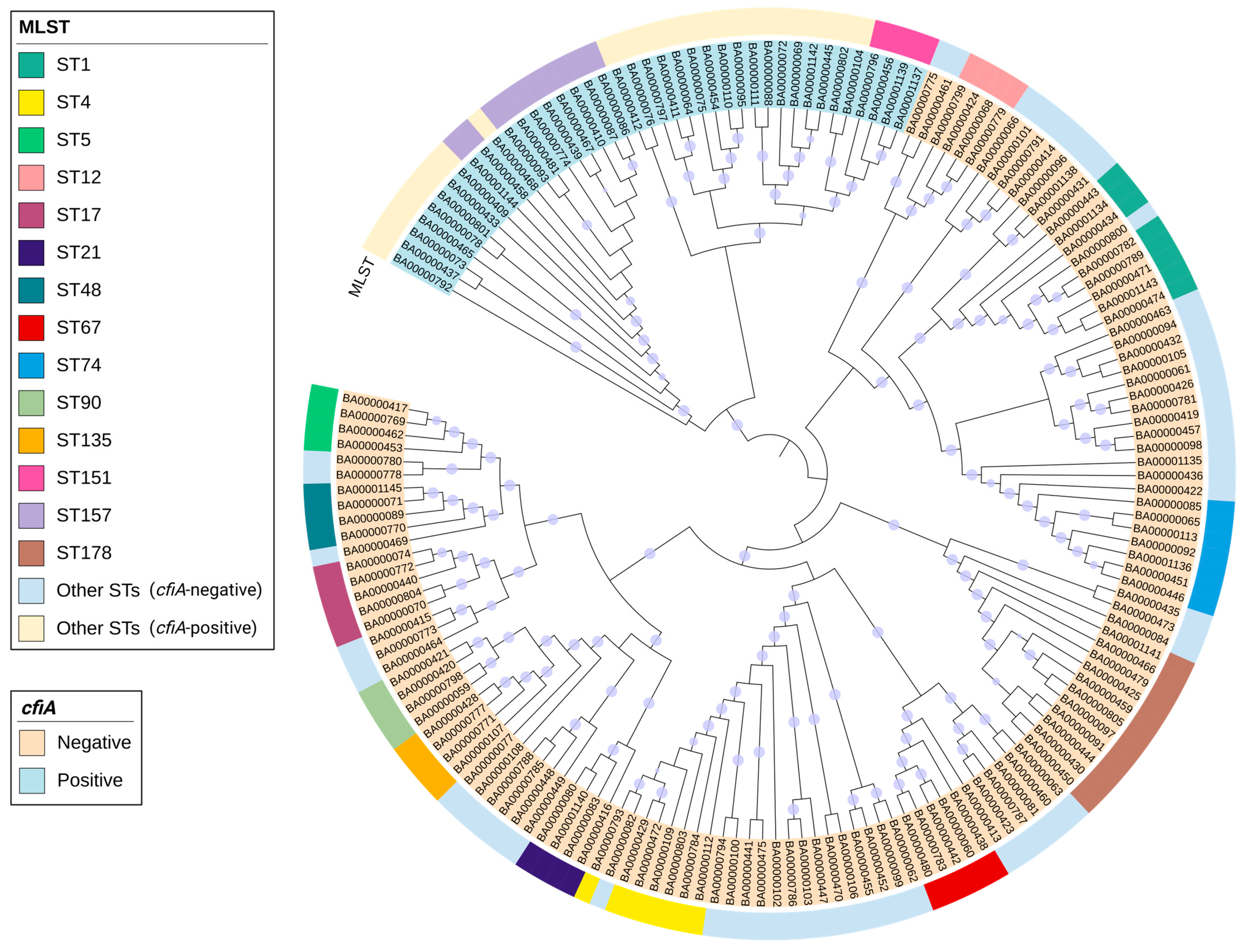
Figure 1. Maximum-likelihood phylogenetic tree of 166 B. fragilis isolates in Hong Kong based on core-genome alignment. The tree is midpoint-rooted, and branch lengths are not to scale. Branches with bootstrap support ≥70% are marked with purple dots. The strain numbers are prefixed with “BA”. Clades containing cfiA+ BF and cfiA− BF are shaded in blue and orange, respectively. The outer circle represents multi-locus sequencing (MLST) sequence types. Sequence types with ≥4 isolates are assigned specific color annotations, while all remaining sequence types are grouped as “Other STs (cfiA-negative)” and “Other STs (cfiA-positive)”.
Microorganisms 14 00168 g001
Table 1. Association between phenotypic antibiotic resistance and cfiA status of B. fragilis isolates.
AntibioticAll Isolates (n = 166)cfiA+ BF (n = 40)cfiA− BF (n = 126)Phi Coefficient (Φ)p-Value 1
Number (Percent)
Amoxicillin–clavulanic acid87 (52.4)32 (80.0)55 (43.7)0.31<0.001
Ampicillin–sulbactam86 (51.8)35 (87.5)51 (40.5)0.40<0.001
Piperacillin–tazobactam38 (22.9)22 (55.0)16 (12.7)0.43<0.001
Ertapenem48 (28.9)34 (85.0)14 (11.1)0.70<0.001
Imipenem27 (16.3)23 (57.5)4 (3.2)0.63<0.001
Meropenem46 (27.7)37 (92.5)9 (7.1)0.82<0.001
Metronidazole23 (13.9)4 (10.0)19 (15.1)0.060.42
Clindamycin85 (51.2)25 (62.5)60 (47.6)0.130.10
Abbreviations: cfiA+ BF, cfiA-positive Bacteroides fragilis; cfiA− BF, cfiA-negative Bacteroides fragilis. 1 p-values were calculated by comparing the cfiA+ BF with the cfiA− BF groups.
Table 2. Treatment and clinical outcomes of patients with B. fragilis infections.
VariableAll Patients (n = 166)cfiA+ BF (n = 40)cfiA− BF (n = 126)p-Value 1
Number (Percent)
Empirical
antibiotics
Amoxicillin–clavulanic acid93 (56.0)19 (47.5)74 (58.7)0.21
Piperacillin–tazobactam29 (17.5)6 (15.0)23 (18.3)0.64
Cefoperazone–sulbactam1 (0.6)1 (2.5)00.24
Meropenem8 (4.8)3 (7.5)5 (4.0)0.40
Ertapenem4 (2.4)1 (2.5)3 (2.4)1.00
Metronidazole10 (6.0)5 (12.5)5 (4.0)0.06
Clindamycin1 (0.6)1 (2.5)00.24
Absence of empirical antibiotics with expected activity against B. fragilis25 (15.1)6 (15.0)19 (15.1)0.99
Appropriate empirical antibiotics 287/165 (52.7)17/39 (43.6)70 (55.6)0.19
Targeted
antibiotics
Amoxicillin–clavulanic acid60 (36.1)12 (30.0)48 (38.1)0.35
Piperacillin–tazobactam37 (22.3)9 (22.5)28 (22.2)0.97
Cefoperazone–sulbactam1 (0.6)01 (0.79)1.00
Meropenem25 (15.1)10 (25.0)15 (11.9)0.04
Ertapenem9 (5.4)1 (2.5)8 (6.3)0.69
Metronidazole53 (31.9)11 (27.5)42 (33.3)0.49
Clindamycin4 (2.4)1 (2.5)3 (2.4)1.00
Absence of targeted antibiotics with expected activity against B. fragilis14 (8.4)5 (12.5)9 (7.1)0.33
Appropriate targeted antibiotics120 (72.3)25 (62.5)95 (75.4)0.11
InterventionsEarly source control84 (50.6)21 (52.5)63 (50.0)0.78
ICU admission13 (7.8)5 (12.5)8 (6.3)0.31
OutcomeDuration of hospital stay ≥ 30 days 348/157 (30.6)15/37 (40.5)33/120 (27.5)0.13
All-cause mortality7 days7 (4.2)3 (7.5)4 (3.2)0.36
30 days22 (13.3)7 (17.5)15 (11.9)0.36
90 days40 (24.1)10 (25.0)30 (23.8)0.88
Abbreviations: cfiA+ BF, cfiA-positive Bacteroides fragilis; cfiA− BF, cfiA-negative Bacteroides fragilis. 1 p-values were calculated by comparing the cfiA+ BF with the cfiA− BF groups. 2 One patient (cfiA+ BF) treated with cefoperazone–sulbactam only was excluded because the appropriateness of therapy could not be determined due to the absence of an EUCAST clinical breakpoint. 3 Nine outpatient patients (three cfiA+ BF and six cfiA− BF) were excluded.
Table 3. Univariate analysis of predictors for 30-day all-cause mortality following B. fragilis infections.
VariableAll Patients (n = 166)Deceased (n = 22)Survivors (n = 144)p-Value 1
Number (Percent)
DemographicsMedian age (IQR)73.0 (60.3–85.0)77.0 (63.5–85.0)72.5 (58.8–85.3)0.23
Age ≥ 65109 (65.7)16 (72.7)93 (64.6)0.45
Male gender78 (47.0)11 (50.0)67 (46.5)0.76
Residential care home24 (14.5)4 (18.2)20 (13.9)0.53
Charlson Comorbidity IndexMedian score (IQR)4.0 (2.0–6.8)7.5 (6.0–8.0)4.0 (2.0–6.0)<0.001
025 (15.1)025 (17.4)
1–218 (10.8)1 (4.5)17 (11.8)
3–442 (25.3)1 (4.5)41 (28.5)
≥ 581 (48.8)20 (90.9)61 (42.4)
ComorbiditiesDiabetes mellitus59 (35.5)11 (50.0)48 (33.3)0.13
Active malignancy35 (21.1)13 (59.1)22 (15.3)<0.001
Chronic kidney disease40 (24.1)7 (31.8)33 (22.9)0.36
Liver cirrhosis1 (0.6)01 (0.7)1.00
Heart disease32 (19.3)5 (22.7)27 (18.8)0.77
Medical historyRecent surgery8 (4.8)3 (13.6)5 (3.5)0.07
Recent immunosuppressant use10 (6.0)3 (13.6)7 (4.9)0.13
Antibiotic exposureRecent antibiotic use51 (30.7)10 (45.5)41 (28.5)0.11
Carbapenem7 (4.2)1 (4.5)6 (4.2)1.00
Beta-lactam/beta-lactamase inhibitor combination33 (19.9)5 (22.7)28 (19.4)0.77
Antibiotic treatmentAppropriate empirical antibiotics 287/165 (52.7)12 (54.5)75/143 (52.4)0.85
Appropriate targeted antibiotics120 (72.3)16 (72.7)104 (72.2)0.96
Presumed source of infectionAbdomen64 (38.6)12 (54.5)52 (36.1)0.10
Skin and soft tissue56 (33.7)2 (9.1)54 (37.5)0.009
Genital tract16 (9.6)1 (4.5)15 (10.4)0.70
Septicemia with uncertain source23 (13.9)6 (27.3)17 (11.8)0.09
Others7 (4.2)1 (4.5)6 (4.2)1.00
Clinical characteristicsFever (> 37.8 °C) 368/158 (43.0)12 (54.5)56/136 (41.2)0.24
cfiA+ BF40 (24.1)7 (31.8)33 (22.9)0.36
InterventionsAbsence of early source control82 (49.4)19 (86.4)63 (43.8)<0.001
ICU admission13 (7.8)2 (9.1)11 (7.6)0.68
1 p-values were calculated by comparing the cfiA+ BF with the cfiA− BF groups. 2 One patient (survivor) treated with cefoperazone–sulbactam only was excluded because the appropriateness of therapy could not be determined due to the absence of an EUCAST clinical breakpoint. 3 Eight patients (all survivors) without temperature records were excluded.
Table 4. Multivariate logistic regression for predictors of 30-day all-cause mortality following B. fragilis infections.
VariableOdds Ratio (OR)95% Confidence Interval (95% CI)p-Value
Age0.990.96–1.030.70
Charlson Comorbidity Index1.301.04–1.630.02
Recent surgery0.400.06–2.870.36
Recent immunosuppressant use0.710.11–4.760.72
Recent antibiotic use1.280.39–4.220.68
Presumed intra-abdominal infections2.040.38–12.680.45
Presumed skin and soft tissue infections0.570.06–5.200.62
Septicemia with uncertain source1.710.23–12.550.60
Fever0.910.30–2.740.86
Absence of early source control4.841.18–19.750.03
Disclaimer/Publisher’s Note: The statements, opinions and data contained in all publications are solely those of the individual author(s) and contributor(s) and not of MDPI and/or the editor(s). MDPI and/or the editor(s) disclaim responsibility for any injury to people or property resulting from any ideas, methods, instructions or products referred to in the content.


