
Group A Streptococcal Invasive Infections Among Children in Cyprus

 , and
, and
Abstract
1. Introduction
2. Materials and Methods
2.1. Setting
2.2. Study Design
2.3. Collection of Patient Data
2.4. Isolation of GAS
2.5. Molecular Identification of GAS by Real-Time PCR
2.6. Molecular Analysis of Isolated GAS Strains
2.7. Statistics
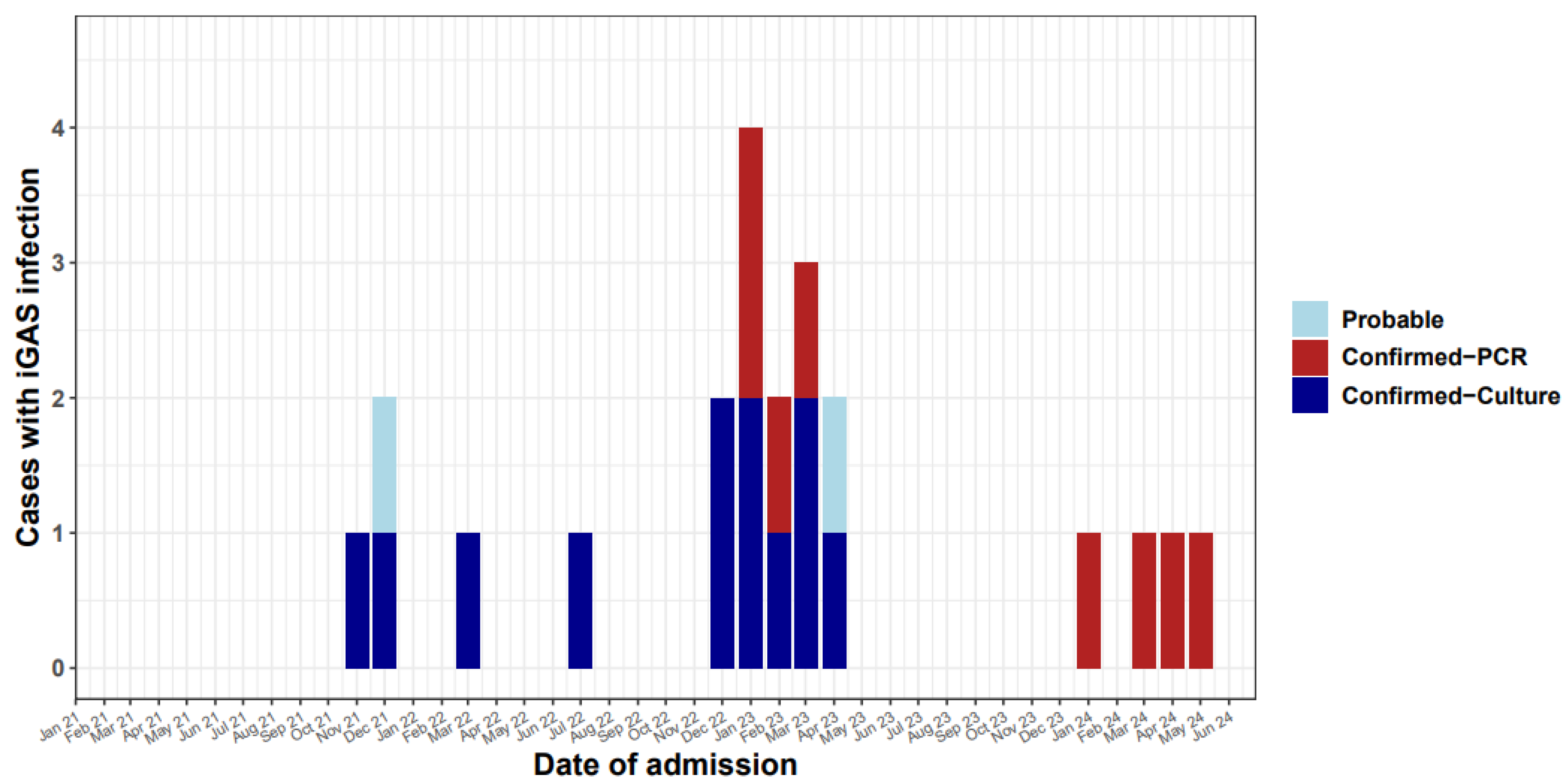
3. Results
3.1. Antimicrobial Resistance
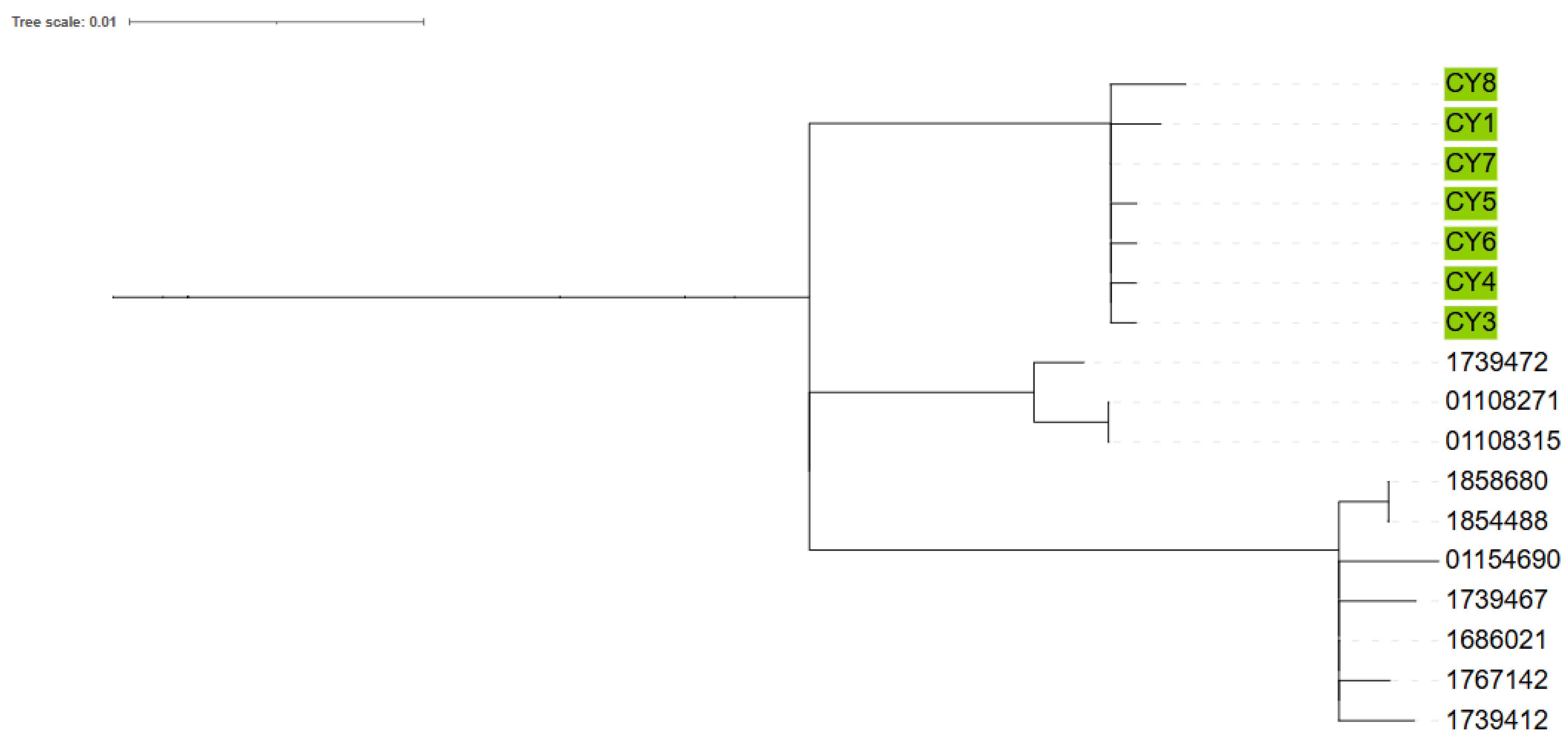
3.2. Emm Types
4. Discussion
5. Conclusions
Author Contributions
Funding
Institutional Review Board Statement
Informed Consent Statement
Data Availability Statement
Acknowledgments
Conflicts of Interest
References
- Stevens, D.L. Invasive Group A Streptococcus Infections. Clin. Infect. Dis. 1992, 14, 2–13. [Google Scholar] [CrossRef]
- Miller, K.M.; Lamagni, T.; Cherian, T.; Cannon, J.W.; Parks, T.; Adegbola, R.A.; Pickering, J.; Barnett, T.; Engel, M.E.; Manning, L.; et al. Standardization of Epidemiological Surveillance of Invasive Group A Streptococcal Infections. Open Forum Infect. Dis. 2022, 9, S31–S40. [Google Scholar] [CrossRef] [PubMed]
- Stevens, D.L.; Tanner, M.H.; Winship, J.; Swarts, R.; Ries, K.M.; Schlievert, P.M.; Kaplan, E. Severe Group A Streptococcal Infections Associated with a Toxic Shock-like Syndrome and Scarlet Fever Toxin A. N. Engl. J. Med. 1989, 321, 1–7. [Google Scholar] [CrossRef] [PubMed]
- Efstratiou, A.; Lamagni, T. Epidemiology of Streptococcus Pyogenes. In Streptococcus Pyogenes: Basic Biology to Clinical Manifestations; NIH: Bethesda, MD, USA, 2022. [Google Scholar]
- de Gier, B.; Marchal, N.; de Beer-Schuurman, I.; te Wierik, M.; Hooiveld, M.; de Melker, H.E.; van Sorge, N.M. Increase in Invasive Group A Streptococcal (Streptococcus Pyogenes) Infections (IGAS) in Young Children in the Netherlands, 2022. Eurosurveillance 2023, 28, 2200941. [Google Scholar] [CrossRef] [PubMed]
- Johannesen, T.B.; Munkstrup, C.; Edslev, S.M.; Baig, S.; Nielsen, S.; Funk, T.; Kristensen, D.K.; Jacobsen, L.H.; Ravn, S.F.; Bindslev, N.; et al. Increase in Invasive Group A Streptococcal Infections and Emergence of Novel, Rapidly Expanding Sub-Lineage of the Virulent Streptococcus Pyogenes M1 Clone, Denmark, 2023. Eurosurveillance 2023, 28, 2300291. [Google Scholar] [CrossRef]
- Barnes, M.; Youngkin, E.; Zipprich, J.; Bilski, K.; Gregory, C.J.; Dominguez, S.R.; Mumm, E.; McMahon, M.; Como-Sabetti, K.; Lynfield, R.; et al. Notes from the Field: Increase in Pediatric Invasive Group A Streptococcus Infections—Colorado and Minnesota, October–December 2022. MMWR Morb. Mortal. Wkly. Rep. 2023, 72, 265–267. [Google Scholar] [CrossRef]
- van Kempen, E.B.; Bruijning-Verhagen, P.C.J.; Borensztajn, D.; Vermont, C.L.; Quaak, M.S.W.; Janson, J.-A.; Maat, I.; Stol, K.; Vlaminckx, B.J.M.; Wieringa, J.W.; et al. Increase in Invasive Group a Streptococcal Infections in Children in the Netherlands, A Survey Among 7 Hospitals in 2022. Pediatr. Infect. Dis. J. 2023, 42, e122–e124. [Google Scholar] [CrossRef]
- Aboulhosn, A.; Sanson, M.A.; Vega, L.A.; Segura, M.G.; Summer, L.M.; Joseph, M.; McNeil, J.C.; Flores, A.R. Increases in Group A Streptococcal Infections in the Pediatric Population in Houston, TX, 2022. Clin. Infect. Dis. 2023, 77, 351–354. [Google Scholar] [CrossRef]
- Kizil, M.C.; Kara, Y.; Bozan, G.; Arda, S.; Durmaz, G.; Kilic, O.; Dinleyici, E.C. Consecutive Seven Serious Cases with Invasive Group A Streptococcal Infections at December 2022–January 2023. Pediatr. Infect. Dis. J. 2023, 42, e254–e255. [Google Scholar] [CrossRef]
- Statistical Services Population—Publications. Available online: https://www.cystat.gov.cy/en/PublicationList?s=46&utm_source=chatgpt.com (accessed on 2 January 2023).
- Statistical Service & Press and Information Office Census of Population and Housing 2021: Final Results. Available online: https://www.gov.cy/en/economy-and-finance/census-of-population-and-housing-2021-final-results/?utm_source=chatgpt.com (accessed on 2 January 2023).
- Breiman, R.F.; Davis, J.P.; Facklam, R.R. Defining the Group A Streptococcal Toxic Shock Syndrome. Rationale and Consensus Definition. JAMA 1993, 269, 390–391. [Google Scholar] [CrossRef]
- EUCAST Breakpoints 2022: Clinical Breakpoints and Dosing of Antibiotics. Available online: https://www.eucast.org/clinical_breakpoints (accessed on 2 January 2023).
- Darenberg, J.; Luca-Harari, B.; Jasir, A.; Sandgren, A.; Pettersson, H.; Schalen, C.; Norgren, M.; Romanus, V.; Norrby-Teglund, A.; Normark, B.H. Molecular and Clinical Characteristics of Invasive Group A Streptococcal Infection in Sweden. Clin. Infect. Dis. 2007, 45, 450–458. [Google Scholar] [CrossRef]
- Vieira, A.; Wan, Y.; Ryan, Y.; Li, H.K.; Guy, R.L.; Papangeli, M.; Huse, K.K.; Reeves, L.C.; Soo, V.W.C.; Daniel, R.; et al. Rapid Expansion and International Spread of M1UK in the Post-Pandemic UK Upsurge of Streptococcus Pyogenes. Nat. Commun. 2024, 15, 3916. [Google Scholar] [CrossRef]
- The R Project for Statistical Computing. Available online: https://www.r-project.org/ (accessed on 8 August 2023).
- Wickham, H. Ggplot2; Springer: New York, NY, USA, 2009; ISBN 978-0-387-98140-6. [Google Scholar]
- Zakikhany, K.; Degail, M.A.; Lamagni, T.; Waight, P.; Guy, R.; Zhao, H.; Efstratiou, A.; Pebody, R.; George, R.; Ramsay, M. Increase in Invasive Streptococcus Pyogenes and Streptococcus Pneumoniae Infections in England, December 2010 to January 2011. Eurosurveillance 2011, 16, 19785. [Google Scholar] [CrossRef]
- Guy, R.; Henderson, K.L.; Coelho, J.; Hughes, H.; Mason, E.L.; Gerver, S.M.; Demirjian, A.; Watson, C.; Sharp, A.; Brown, C.S.; et al. Increase in Invasive Group A Streptococcal Infection Notifications, England, 2022. Eurosurveillance 2023, 28, 2200942. [Google Scholar] [CrossRef]
- Davies, M.R.; Keller, N.; Brouwer, S.; Jespersen, M.G.; Cork, A.J.; Hayes, A.J.; Pitt, M.E.; De Oliveira, D.M.P.; Harbison-Price, N.; Bertolla, O.M.; et al. Detection of Streptococcus Pyogenes M1UK in Australia and Characterization of the Mutation Driving Enhanced Expression of Superantigen SpeA. Nat. Commun. 2023, 14, 1051. [Google Scholar] [CrossRef] [PubMed]
- Demczuk, W.; Martin, I.; Domingo, F.R.; MacDonald, D.; Mulvey, M.R. Identification of Streptococcus Pyogenes M1UK Clone in Canada. Lancet Infect. Dis. 2019, 19, 1284–1285. [Google Scholar] [CrossRef]
- Ekelund, K.; Skinhøj, P.; Madsen, J.; Konradsen, H.B. Reemergence of Emm 1 and a Changed Superantigen Profile for Group A Streptococci Causing Invasive Infections: Results from a Nationwide Study. J. Clin. Microbiol. 2005, 43, 1789–1796. [Google Scholar] [CrossRef] [PubMed]
- Dabaja-Younis, H.; Kandel, C.; Green, K.; Johnstone, J.; Zhong, Z.; Kassee, C.; Allen, V.; Armstrong, I.; Baqi, M.; Barker, K.; et al. Invasive Group A Streptococcal Infection in Children, 1992–2023. JAMA Netw. Open 2025, 8, e252861. [Google Scholar] [CrossRef]
- Group A Streptococcal Infections: Report on Seasonal Activity in England, 2022 to 2023. Available online: https://www.gov.uk/government/publications/group-a-streptococcal-infections-activity-during-the-2022-to-2023-season/group-a-streptococcal-infections-report-on-seasonal-activity-in-england-2022-to-2023#references (accessed on 7 August 2023).
- Koliou, M.; Ioannou, Y.; Efstratiou, A.; Hannidou, N.; Pieri, V.; Alexandrou, M.; Soteriades, E.S. Circulating Serotypes and Antimicrobial Sensitivity of Streptococcus Pyogenes Isolates from Children in Cyprus. Clin. Microbiol. Infect. 2007, 13, 645–647. [Google Scholar] [CrossRef]
- ECDC. Invasive Pneumococcal Disease Annual Epidemiological Report for 2018 Key Facts; ECDC: Solna, Sweden, 2018. [Google Scholar]
- Abo, Y.-N.; Oliver, J.; McMinn, A.; Osowicki, J.; Baker, C.; Clark, J.E.; Blyth, C.C.; Francis, J.R.; Carr, J.; Smeesters, P.R.; et al. Increase in Invasive Group A Streptococcal Disease among Australian Children Coinciding with Northern Hemisphere Surges. Lancet Reg. Health West. Pac. 2023, 41, 100873. [Google Scholar] [CrossRef] [PubMed]
- Rümke, L.W.; de Gier, B.; Vestjens, S.M.T.; van der Ende, A.; van Sorge, N.M.; Vlaminckx, B.J.M.; Witteveen, S.; van Santen, M.; Schouls, L.M.; Kuijper, E.J. Dominance of M1UK Clade among Dutch M1 Streptococcus Pyogenes. Lancet Infect. Dis. 2020, 20, 539–540. [Google Scholar] [CrossRef] [PubMed]
- Willyard, C. Flu and Colds Are Back with a Vengeance—Why Now? Nature 2022. [Google Scholar] [CrossRef] [PubMed]
- McCullers, J.A. Insights into the Interaction between Influenza Virus and Pneumococcus. Clin. Microbiol. Rev. 2006, 19, 571–582. [Google Scholar] [CrossRef] [PubMed]
- Morens, D.M.; Taubenberger, J.K.; Fauci, A.S. Predominant Role of Bacterial Pneumonia as a Cause of Death in Pandemic Influenza: Implications for Pandemic Influenza Preparedness. J. Infect. Dis. 2008, 198, 962–970. [Google Scholar] [CrossRef]

| Site | N | Percentage (%) |
|---|---|---|
| Blood | 7 | 31.8 |
| Pleural fluid | 7 | 31.8 |
| Pus from deep abscess | 4 | 18.2 |
| Pus from mastoid surgery | 2 | 9.1 |
| Pus from perforated ear * | 1 | 4.5 |
| Throat swab * | 1 | 4.5 |
| Synovial fluid ** | 1 | 4.5 |
| Patient | Age (mo) | Clinical Syndromes | Outcome |
|---|---|---|---|
| 1 | 20 | Scarlet fever, complicated pneumonia | ICU-Recovered |
| 2 | 41 | Bacteraemia with endocarditis | Recovered |
| 3 | 104 | Bacteraemia, otitis, perforation of ear drum, mastoiditis, meningitis | Recovered |
| 4 | 47 | Bacteraemia, cellulitis, fasciitis | Recovered |
| 5 | 52 | Complicated pneumonia, STSS | ICU-ECMO-Recovered |
| 6 | 26 | Bacteraemia, STSS | ICU-Died |
| 7 | 17 | Bacteraemia, STSS | ICU-Died |
| 8 | 46 | Bacteraemia, joint infection | Recovered |
| 9 | 119 | Deep abscess and fasciitis extending to pelvic floor muscles | Recovered |
| 10 | 24 | Bacteraemia, scarlet fever, sepsis, complicated pneumonia, STSS | ICU-Recovered |
| 11 | 55 | Bacteraemia, scarlet fever, otitis media, perforation of ear drum | Recovered |
| 12 | 12 | Otitis media, perforation of ear drum, mastoiditis | Recovered |
| 13 | 65 | Otitis media, mastoiditis, sigmoid sinus thrombosis | Recovered |
| 14 | 85 | Scarlet fever, otitis media, complicated pneumonia | ICU-Recovered |
| 15 | 24 | Scarlet fever, complicated pneumonia | Recovered |
| 16 | 90 | Fever, cough, complicated pneumonia | Recovered |
| 17 | 78 | Fever, cough, complicated pneumonia | Recovered |
| 18 | 80 | Scarlet fever, complicated pneumonia, STSS | ICU-Recovered |
| 19 | 15 | Fever, diarrhoea, complicated mastoiditis | Recovered |
| 20 | 101 | Fever, pharyngitis, intraorbital abscess | Recovered |
| 21 | 14 | Otitis media, complicated mastoiditis | Recovered |
| 22 | 76 | Fever, drowsiness, retropharyngeal abscess | Recovered |
| Emm Type | Number | Percentage (%) |
|---|---|---|
| emm 1.0 | 6 | 66.67 |
| emm 92.0 | 1 | 11.11 |
| emm 2.0 | 1 | 11.11 |
| emm 1.150 | 1 | 11.11 |
Disclaimer/Publisher’s Note: The statements, opinions and data contained in all publications are solely those of the individual author(s) and contributor(s) and not of MDPI and/or the editor(s). MDPI and/or the editor(s) disclaim responsibility for any injury to people or property resulting from any ideas, methods, instructions or products referred to in the content. |
© 2025 by the authors. Licensee MDPI, Basel, Switzerland. This article is an open access article distributed under the terms and conditions of the Creative Commons Attribution (CC BY) license (https://creativecommons.org/licenses/by/4.0/).
Share and Cite
Koliou, M.; Vassiliadou, G.I.; Aristidou, A.; Ladas, P.; Sergis, A.; Argyrou, M.; Charalambous, M.; Marcou, M.; Alexandrou, M.; Coelho, J.; et al. Group A Streptococcal Invasive Infections Among Children in Cyprus. Microorganisms 2025, 13, 1783. https://doi.org/10.3390/microorganisms13081783
Koliou M, Vassiliadou GI, Aristidou A, Ladas P, Sergis A, Argyrou M, Charalambous M, Marcou M, Alexandrou M, Coelho J, et al. Group A Streptococcal Invasive Infections Among Children in Cyprus. Microorganisms. 2025; 13(8):1783. https://doi.org/10.3390/microorganisms13081783
Chicago/Turabian StyleKoliou, Maria, Gavriella Ioannou Vassiliadou, Athina Aristidou, Petros Ladas, Andreas Sergis, Maria Argyrou, Myria Charalambous, Markella Marcou, Maria Alexandrou, Juliana Coelho, and et al. 2025. "Group A Streptococcal Invasive Infections Among Children in Cyprus" Microorganisms 13, no. 8: 1783. https://doi.org/10.3390/microorganisms13081783
APA StyleKoliou, M., Vassiliadou, G. I., Aristidou, A., Ladas, P., Sergis, A., Argyrou, M., Charalambous, M., Marcou, M., Alexandrou, M., Coelho, J., Ryan, Y., Efstratiou, A., & Mazeri, S. (2025). Group A Streptococcal Invasive Infections Among Children in Cyprus. Microorganisms, 13(8), 1783. https://doi.org/10.3390/microorganisms13081783