Article

Molecular Epidemiology of Cryptosporidium spp., Giardia duodenalis, and Enterocytozoon bieneusi in Guizhou Angus Calves: Dominance of Angus Cattle-Adapted Genotypes and Zoonotic Potential of E. bieneusi

1
National Key Laboratory of Agricultural Microbiology, College of Veterinary Medicine, Huazhong Agricultural University, Wuhan 430070, China
2
State Key Laboratory for Diagnosis and Treatment of Severe Zoonotic Infectious Diseases, Key Laboratory for Zoonosis Research of the Ministry of Education, Institute of Zoonosis, College of Veterinary Medicine, Jilin University, Changchun 130012, China
3
Department of Infectious Diseases, Istituto Superiore di Sanità, 00161 Rome, Italy
*
Author to whom correspondence should be addressed.
Microorganisms 2025, 13(8), 1735; https://doi.org/10.3390/microorganisms13081735
Submission received: 12 June 2025 / Revised: 22 July 2025 / Accepted: 22 July 2025 / Published: 25 July 2025

Abstract

Limited molecular data exist on zoonotic parasites Cryptosporidium spp., Giardia duodenalis, and Enterocytozoon bieneusi in Angus calves from Guizhou, China. This study constitutes the first molecular epidemiological survey of these pathogens in this region. 817 fecal samples from Angus calves across 7 intensive beef farms (Bijie City). Nested PCR methods targeting SSU rRNA (Cryptosporidium spp.), gp60 (Cryptosporidium bovis subtyping), bg/gdh/tpi (G. duodenalis), and ITS (E. bieneusi) coupled with DNA sequencing were employed. DNA sequences were analyzed against the NCBI. database. Statistical differences were assessed via a generalized linear mixed-effects model. Cryptosporidium spp. prevalence 23.5% (192/817; 95% CI 28.1–34.6%), with C. bovis predominating 89.6% (172/192; 95% CI 84.4–93.5%) and six subtypes (XXVIa-XXVIf). Highest infection in 4–8-week-olds 29.9% (143/479; 95% CI 25.8–34.1%) (p < 0.01). G. duodenalis: 31.3% (256/817; 95% CI 28.1–34.6%) positive, overwhelmingly assemblage E 97.6% (6/256; 95% CI 0.9–5.0%), zoonotic assemblage A was marginal 0.7% (6/817; 95% CI 0.3–1.6%). Farm-level variation exceeded 10-fold (e.g., Gantang: 55.0% (55/100; 95% CI 44.7–65.0%) vs. Tieshi: 4.9% (5/102; 95% CI 1.6–11.1%). E. bieneusi: prevalence 19.7% (161/817; 95% CI 17.0–22.6%), exclusively zoonotic genotypes BEB4: 49.7% (80/161; 95% CI 41.7–57.7%); I: 40.4% (65/161; 95% CI 32.7–48.4%). Strong diarrhea association (p < 0.01) and site-specific patterns (e.g., Guanyindong: 39.2%). While Giardia exhibited the highest prevalence (31.3%) with minimal zoonotic risk, Enterocytozoon—despite lower prevalence (19.7%)—posed the greatest public health threat due to exclusive circulation of human-pathogenic genotypes (BEB4/I) and significant diarrhea association, highlighting divergent control priorities for these enteric parasites in Guizhou calves. Management/Public health impact: Dominant zoonotic E. bieneusi genotypes (BEB4/I) necessitate: 1. Targeted treatment of 4–8-week-old Angus calves. 2. Manure biofermentation (≥55 °C, 3 days), and 3. UV-disinfection (≥1 mJ/cm2) for karst water to disrupt transmission in this high-humidity region.

1. Introduction

Cryptosporidium spp. (phylum Apicomplexa), Giardia duodenalis (phylum Metamonada), and Enterocytozoon bieneusi (phylum Microsporidia) are all capable of infecting both humans and a variety of animal hosts [1,2,3]. Cryptosporidium spp. is primarily transmitted via contaminated water and food and can cause diarrhea in children under 2 years of age and calves under 1 month of age, potentially leading to death in severe cases. In immunocompetent hosts, it often manifests as asymptomatic or self-limited diarrhea, while in immunocompromised patients, it can cause severe diarrhea [4]. Globally, taxonomic studies have identified 44 Cryptosporidium species, and C. parvum, C. andersoni, C. ryanae, and C. bovis represent the epidemiologically predominant species in cattle [5,6]. Giardia duodenalis, transmitted via the same contaminated water and food as Cryptosporidium spp., affects 280 million people annually, causing diarrhea, malabsorption, and weight loss [7]. It has 8 assemblages, labeled A through H, with zoonotic G. duodenalis assemblages A and B being commonly detected in cattle [8]. Enterocytozoon bieneusi is a microsporidian parasite that is divided into 11 genetic groups (1–11) based on its ribosomal RNA (ITS region). Group 1 (Zoonotic): Contains genotypes (A, BEB15) found in many animals (livestock, pets, wildlife) and the environment. This group is the main source of human infections, showing significant animal-to-human (zoonotic) transmission. Group 2 (Human-Adapted): Includes genotypes (notably BEB4, I, J) that mostly infect humans, especially those with weak immune systems. While sometimes found in animals, they are strongly adapted to humans or spread between humans (anthroponotic). Groups 3–11 (Host-Specific): Primarily infect specific animals with limited known spread to humans. Understanding the classification and zoonotic potential of E. bieneusi is crucial for public health and epidemiological studies [9].
Globally, the epidemiology of Cryptosporidium spp., G. duodenalis, and E. bieneusi in cattle has been extensively documented across continents such as Asia, Europe, North America, South America, and Oceania [10,11,12]. In China, these pathogens have been studied in multiple provinces, including Henan, Shaanxi, Beijing, Shanghai, Hubei, Hebei, Tianjin, Qinghai, Sichuan, Guangdong, Jiangsu, Tibet Autonomous Region, Gansu, Shandong, Yunnan, Anhui, Inner Mongolia, Xinjiang, Shanxi, Heilongjiang, and Ningxia [13,14,15,16,17,18,19,20,21,22,23,24,25,26,27,28,29,30,31,32]. However, there is a noticeable absence of data from Guizhou Province, China. Bijie City of Guizhou Province serves as a major hub for Angus cattle farming, hosting numerous large-scale cattle ranches that constitute a significant center for livestock production in Southwestern China. This study aims to address this gap by determining the prevalence and genotypic distribution of Cryptosporidium spp., G. duodenalis, and E. bieneusi in the fecal samples of Angus calves in Bijie City of Guizhou Province, thereby contributing to the national epidemiological dataset on Cryptosporidium spp., G. duodenalis, and E. bieneusi in China.

2. Materials and Methods

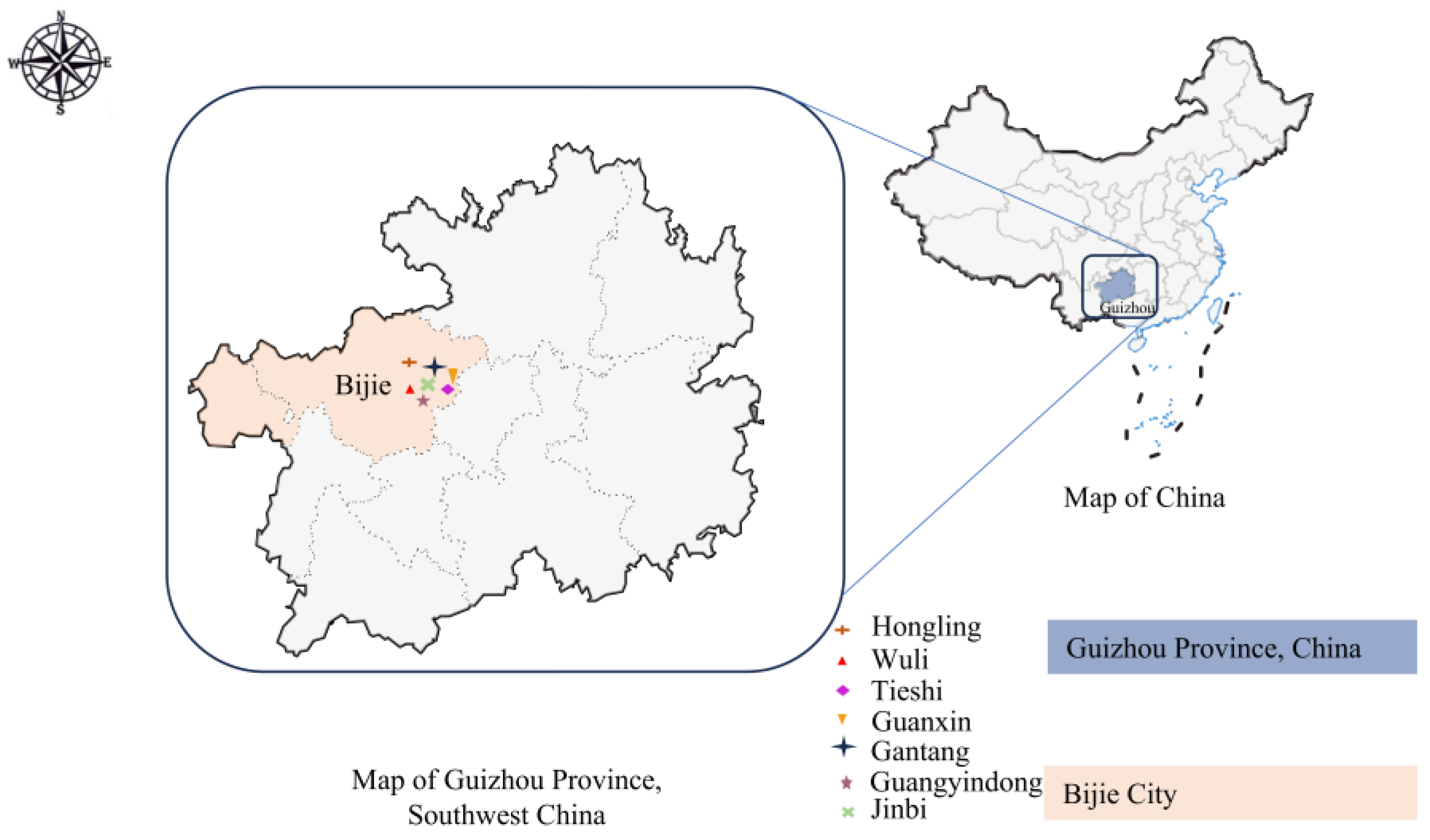
2.1. Sample Collection

In our study, a total of 817 Angus calves’ fecal samples were collected from Bijie City, Guizhou Province (26°21′~27°46′ N and 103°36′~106°43′ E), a closed fattening farm. Management: Corn-soybean diet (6:4 ratio), ad libitum feeding; biannual FMD/Influenza A vaccination; quarterly ivermectin deworming; semi-open cement-floor housing (4 m2/head). Key equipment: Hydraulic restraint stand and electronic scale (±0.5 kg accuracy).
In August 2023, we collected 79 and 75 fecal samples at Wuli and Hongling farms, respectively. In March 2024, we obtained 663 fecal samples from Angus calves across seven farms in the city, including Wuli (89), Hongling (99), Tieshi (102), Guanxin (93), Gantang (100), Guangyindong (97), and Jinbi (83) (Figure 1). Importantly, the samples collected in 2024 at these Wuli and Hongling farms were from cohorts, differing from the animals studied in 2023. Constrained by resources and funding, summer sampling was conceived as a preliminary investigation for seasonal comparison. Wuli and Hongling were guided by established collaborative partnerships. This study prioritized analyzing the impact of spring breeding protocols on prevalence rates, therefore concentrating resources on comprehensive spring data acquisition. Minimum sample sizes were calculated based on historical Cryptosporidium spp. 22.7% (35/154; 95% CI 16.4–30.2%), G. duodenalis 20.1% (31/154; 95% CI 14.1–27.3%), and E. bieneusi 12.3% (19/154; 95% CI 7.6–18.6%), with prevalence rates at 5% margin of error, yielding 270, 247, and 166 samples, respectively. The study’s 663 collected samples substantially exceeded these requirements. The study population consisted of 817 Angus calves aged 0–3 months, comprising 178 male and 639 female Angus calves, as well as 107 0–2-week age, 200 2–4-week age, 479 4–8-week age, 31 8–12-week age, 93 diarrhea, and 724 no-diarrhea fecal samples. Fecal samples were collected from Angus calves under 3 months of age using aseptic rectal swabbing techniques, with one sample per animal. Inclusion criteria comprised: No anthelmintic treatment within 30 days; absence of acute disease (core temperature ≤ 39.5 °C); no vaccination within 14 days prior; non-orphaned status. Through computer-generated randomization, 10% of eligible calves were selected for sampling. All sampled subjects met the predefined criteria irrespective of baseline health status. Detailed sampling data are presented in Table S1. Following collection, all samples underwent immediate storage at 4 °C. Subsequent transport to the laboratory utilized SF Express cold chain logistics, after which samples were maintained at 4 °C in refrigerated storage until DNA extraction. Each farm can accommodate up to 10,000 Angus cattle and is divided into three functional zones: the production area, the lactation area, and the fattening area. In the production area, each shed houses one Angus calf, while the other zones practice mixed grazing. Cows are free to move within any area, promoting natural behavior and reducing stress.

2.2. DNA Extraction and PCR Analysis

DNA extraction was performed using the TIANamp DNA stool Kit (TIANGEN, Beijing, China) following the manufacturer’s instructions. Briefly, 0.2 g of fecal material were homogenized using 1 mm grinding beads in the FastPrep®-24 Instrument (MP Biomedicals, Santa Ana, CA, USA) for 60 s at a frequency of 5.5 m/s, and this process was repeated. The extracted DNA was then eluted with 80 μL of molecular-grade water and stored at −20 °C for future analysis. In this study, Cryptosporidium spp. was detected using a nested PCR method targeting the 830 bp SSU rRNA gene. The PCR amplification was performed using the DNA polymerase and 2 × SanTaq PCR Master Mix (Sangon Biotech, Shanghai, China), following the reagent and condition protocols established in a previous study [33] and subtyping of C. bovis test as mentioned in the article [34]. G. duodenalis was identified and assemblaged based on sequence analysis of the glutamate dehydrogenase (gdh) gene, β-galactosidase (bg) gene, and triosephosphate isomerase (tpi) gene, respectively [35]. Detection of E. bieneusi used internal transcribed spacer (ITS), and the method from the prior study [36]. Negative controls, consisting of molecular-grade water, were included in each PCR analysis to ensure accuracy. The secondary PCR products were examined by electrophoresis on a 1.5% agarose gel and visualized following ethidium bromide staining. The primers and annealing temperatures employed in this study are listed in Table 1

2.3. Sequence Analysis

Positive PCR amplicons were sequenced by Qingke Biology (Beijing, China). The DNA sequences obtained in this study were checked using TBtools-II v. 2.125, then blasted against reference sequences from GenBank, and finally analyzed using the Neighbor-Joining analysed by MEGA v.11.0.13 software.

2.4. Statistical Analysis

All statistical analyses were performed using SPSS v. 27.0.1 software. The generalized linear mixed-effects model was used to evaluate the statistical differences in Cryptosporidium spp., G. duodenalis, and E. bieneusi infections among pre-weaned Angus calves, stratified by location, season, age, diarrhea, and gender. Statistical significance was determined with 95% confidence intervals. The p < 0.05 indicates statistical significance.

3. Results

3.1. Cryptosporidium bovis Dominance: 89.6% Prevalence with Geographic Clustering

In this study, of the 817 fecal samples collected from Bijie City, Guizhou Province, 192 tested positive for Cryptosporidium spp., via nested PCR targeting the SSU rRNA gene. The overall prevalence of Cryptosporidium species infection was 23.5% (192/817; 95% CI 28.1–34.6%), with the following distribution among the Cryptosporidium species: C. andersoni 2.1% (4/192; 95% CI 0.6–5.2%), C. bovis 89.6% (172/192; 95% CI 84.4–93.5%), and C. ryanae 7.8% (15/192; 95% CI 4.4–12.6%) (Table 2, Figure S1). Geographical variations in Cryptosporidium species prevalence were particularly notable across the 7 sampled farms. The highest infection rate was observed in Tieshi, 33.3% (34/102; 95% CI 24.3–43.3%), while Hongling showed the lowest prevalence, 12.1% (21/174; 95% CI 7.7–17.9%). Intermediate prevalence rates were observed in Wuli, 27.9% (47/168; 95% CI 21.3–35.3%), Guanxin (18/93; 95% CI 11.9–28.9%), Gantang, 26.0% (26/100; 95% CI 17.7–35.7%), Guanyindong, 28.9% (28/97; 95% CI 20.1–39.0%), and Jinbi, 21.7% (18/83; 95% CI 13.4–32.1%) (Table 2). Statistical analysis revealed significant differences in Cryptosporidium spp. positive rates across the 7 sites (p < 0.001). One of the Angus cattle from Hongling was found to be co-infected with C. bovis and C. ryanae. This study reveals significant differences in the dominant subtypes of C. bovis across various farms. The sample positive rate was 23.4% (155/663; 95% CI 20.2–26.8%) in spring and 24.0% (37/154; 95% CI 17.5–31.6%) in summer, with no significant difference observed between the seasons (p = 0.963 > 0.05). Furthermore, no significant interaction effect was found between location and season (p = 0.531 > 0.05), indicating their independent influences (Table 2).
Statistical analysis revealed significant variations in Cryptosporidium species prevalence across different age groups of Angus calves (p < 0.01). The highest positive rate was detected in 4–8-week-old Angus calves, 29.9% (143/479; 95% CI 25.8–34.1%), followed by Angus calves aged 2–4 weeks, with 21.5% (43/200; 95% CI 16.0–27.8%). Positive rates decreased to12.9% (4/31; 95% CI 3.6–29.8%) for Angus calves aged 8–12 weeks and were lowest, 4.7% (2/107; 95% CI 1.5–10.6%) in those less than 2 weeks old. Of the total samples, 22.6% (21/93; 95% CI 14.6–32.4%) of diarrhea cases tested positive, compared to 23.6% (171/724; 95% CI 20.6–26.9%) of non-diarrhea cases. There was no significant difference in the positive rates between diarrhea samples and non-diarrhea samples (p = 0.817 > 0.05). Males exhibited a lower positive rate, 17.9% (32/178; 95% CI 12.6–24.4%), compared to females, 25.0% (160/639; 95% CI 21.7–28.6%), with this difference being statistically significant (p = 0.032 < 0.05) (Table 2).
In this study, 6 subtypes of C. bovis were identified. On Hongling, Tieshi, and Guanxin farms, the subtypes XXVIf and XXVIe were commonly identified. On Hongling Farm, XXVIf was the predominant subtype. On Wuli Farm, four subtypes, XXVIb, XXVIc, XXVId, and XXVIe, were identified with relatively equal distribution, and a unique case of dual infection with XXVIb and XXVIc was detected. Guanyindong and Gantang farms share three C. bovis subtypes, XXVIa, XXVId, and XXVIf, but Guanyindong also had an additional subtype, XXVIe, which is not found in Gantang. XXVIa and XXVId were the most common subtypes in Guanyindong. On Jinbi Farm, the subtypes XXVIb and XXVIc were found, with XXVIc being the dominant subtype (Table 3, Figure S2).

3.2. Giardia duodenalis Assemblage E Prevails: Low Zoonotic Risk but High Farm-Level Variability

The overall prevalence of G. duodenalis infection across all study sites reached 31.3% (256/817; 95% CI 28.1–34.6%). Marked geographical variations were observed: Hongling recorded 18.9% (33/174; 95% CI 13.4–25.6%), Wuli 26.2% (44/168; 95% CI 19.7–33.5%), Tieshi 4.9% (5/102; 95% CI 1.6–11.1%), Guanxin 39.8% (37/93; 95% CI 29.8–50.5%), Gantan 55.0% (55/100; 95% CI 44.7–65.0%), Guanyindong 51.5% (50/97; 95% CI 41.2–61.8%), and Jinbi 38.6% (32/83; 95% CI 28.1–49.9%) (Table 4, Figure S1, Figure S2 and Figure S3). These regional differences demonstrated statistical significance (p < 0.001). The sample positive rate was 33.8% (224/663; 95% CI 30.2–37.5%) in spring and 20.8% (32/154; 95% CI 14.7–28.0%) in summer, with no significant difference observed between the seasons (p = 0.569 > 0.05). Furthermore, no significant interaction effect was found between location and season (p = 0.505 > 0.05), indicating their independent influences (Table 4). While zoonotic type A of G. duodenalis was detected in samples from Hongling, Wuli, and Guanxin farms, the infection rate was strikingly low, representing only 0.7% (6/817; 95% CI 0.3–1.6%) of total samples, among all the positive samples, the proportion of type A was 2.3% (6/256; 95% CI 0.9–5.0%). In contrast, type E was more prevalent in the Angus cattle population, 97.6% (250/256; 95% CI 95–99.1%). (Table 4). Infection rates varied significantly among different age Angus calves, with a statistically significant difference (p < 0.001). The highest infection rate, 37.2% (178/479; 95% CI 32.8–41.7%), was in 4–8-week-old Angus calves. Subsequent rates declined to 32.3% (10/31; 95% CI 16.7–51.4%) for 8–12 weeks, and 24.5% (49/200; 95% CI 18.7–31.1%) for 2–4 weeks. The lowest infection rate, 17.7% (19/107; 95% CI 11.0–26.3%), was found in Angus calves under 2 weeks. Diarrheal samples had a positive rate of 24.7% (23/93; 95% CI 16.3–34.8.3%), whereas non-diarrheal samples showed a higher positive rate of 32.2% (33/724; 95% CI 28.8–35.7%). However, the difference in positive rates between the diarrheal and non-diarrheal samples was not statistically significant (p = 0.166 > 0.05). Males exhibited a lower positive rate, 29.8% (53/178; 95% CI 23.2–37.1%), compared to females, 31.8% (203/639; 95% CI 28.2–35.5%), with this difference being no statistically significant (p = 0.629 > 0.05) (Table 4).

3.3. Enterocytozoon bieneusi Zoonotypes: BEB4/I Co-Circulation and Diarrhea Association

The overall prevalence of E. bieneusi was 19.7% (161/817; 95% CI 17.0–22.6%) (Table 5, Figure S6), with significant inter-location variation (p < 0.001). Notably, no positive cases were detected in Hongling and Guanxin farms. In contrast, positive cases were detected in other farms with positive rates ranging from 18.5% to 39.2%, showing the highest rate at 39.2% (38/97; 95% CI 29.4–49.6%) in Guanyindong and Gantang at 39.0% (39/100; 95% CI 29.4–49.3%), followed by Jinbi at 34.9% (29/83; 95% CI 24.8–46.2%), Tieshi at 23.5% (24/102; 95% CI 15.7–32.9%), and Wuli at 18.5% (31/168; 95% CI 12.9–25.2%). ITS sequencing analysis revealed four genotypes, including BEB4 49.7% (80/161; 95% CI 41.7–57.7%; 80/161), CHPM1 1.9% (3/161; 95% CI 0.4–5.3%), J 8.1% (13/161; 95% CI 4.4–13.4%), and I 40.4% (65/161; 95% CI 32.7–48.4%). The first three genotypes belong to Group 2 and are of the animal-adapted type, meaning they primarily infect animals but have also been detected in humans. The last genotype belongs to Group 1 and is a zoonotic genotype, which can be transmitted from animals to humans. Genotype J was only found in Wuli, and genotype CHPM1 was identified exclusively in Gantang. No new genotypes were detected. The sample positive rate was 21.4% (142/663; 95% CI 18.4–24.7%) in spring and 12.0% (19/154; 95% CI 7.6–18.6%) in summer, with no significant difference observed between the seasons (p = 0.843 > 0.05). Furthermore, no significant interaction effect was found between location and season (p = 0.843 > 0.05), indicating their independent influences (Table 5). The infection rates among Angus calves varied significantly by age (p < 0.001); the rates were as follows: the positivity rate increased progressively from 6.5% (7/107; 95% CI 2.7–13.0%) at 0–2 weeks to a peak of 26.1% (125/479; 95% CI 22.2–30.3%;) at 4–8 weeks of age; the positive rates were similar at 2–4 weeks, 12.5% (25/200; 95% CI 8.3–17.9%), and at 8–12 weeks, 12.9% (4/31; 95% CI 3.6–29.8%). The positive rate for diarrheal samples was 35.5% (33/93; 95% CI 25.8–46.1%), while for non-diarrheal samples, it was 17.7% (128/724; 95% CI 15.0–20.7%). Our study found a significant association between E. bieneusi infection and diarrhea in pre-weaned Angus calves (p < 0.001). Males exhibited a lower positive rate, 12.4% (22/178; 95% CI 7.9–18.1%), compared to females, 21.8% (139/639; 95% CI 18.6–25.2%), with this difference not being statistically significant (p = 0.065 > 0.05) (Table 5).

4. Discussion

4.1. Why Guizhou’s Infection Rates Surpass Sichuan and Yunnan: A Climate Lens?

The infection rate of Cryptosporidium species observed in our study was 23.5%, which aligns closely with the 22.5% prevalence reported in calves under 12 months of age across China from 2008 to 2018 [37]. This similarity extends to the prevalence of C. bovis, C. ryanae, and C. andersoni in pre-weaned calves [38]. However, infection rates can vary due to differing regional factor. In China, the regions with the highest infection rates are Taiwan, Inner Mongolia, Shandong, Hunan, and Qinghai; the regions with the lowest infection rates are Shanxi, Guangxi, Sichuan, Ningxia, and Gansu [39]. For example, in the southwestern region, infection rates in pre-weaned calves were 14.4% in Sichuan and 14.7% in Yunnan [20,40]. Regarding G. duodenalis, an infection rate of 31.3% was recorded, with the zoonotic assemblage A present at 0.7%. In contrast, the average G. duodenalis infection rate in Chinese cattle was reported to be 8.0% in 2022, with significant variation across provinces. For instance, in North China, the infection rate in Beijing was 1.7% [15]; in Northwest China, rates in Gansu, Ningxia, and Qinghai were 1.96% [24], 3.5% [41], and 8.1% [42], respectively; in South China, the rate in Guangdong was 74.2% [21]; in Central China, the rate in Hubei was 22.7% [17]; and in Southwest China, rates in Yunnan and Sichuan were 10.5% and 13.4%, respectively [20,26]. In pre-weaned Angus calves from Guizhou, an E. bieneusi infection rate of 19.7% was found, comparable to the global infection rate of 12.9%. Infection rates vary across regions, with 17.3% in South America, 11.5% in Asia, 10.4% in Oceania, 15.4% in Europe, 12.9% in North America, and 6.5% in Africa [43]. However, the infection rate in Yunnan was only 0.59% [44].
This study demonstrates that the infection rates of Cryptosporidium spp., G. duodenalis, and E. bieneusi in Guizhou are higher than those in Sichuan and Yunnan. Guizhou has a subtropical humid monsoon climate, with an annual temperature of 9 °C to 18 °C and an average annual precipitation of 665–1159 mm, with humidity ranging from 68.0% to 87.0% (The data is sourced from the China Meteorological Data Network, https://data.cma.cn/site/index.html, accessed on 8 July 2025). The average annual temperature in Sichuan is 16–18 °C in the basin, 4–12 °C in the western plateau, with an average annual precipitation of 800–1200 mm in the basin area and 400–600 mm in the western plateau, and humidity ranging from 70.0% to 80.0% in the basin and 50.0% to 60.0% in the western plateau (the data is sourced from the Sichuan Meteorological Bureau, http://sc.cma.gov.cn, accessed on 10 March 2025). Yunnan has an average annual temperature of 10 °C to 20 °C, with precipitation ranging from 800 to 2000 mm and humidity of 60.0–80.0% (The data are sourced from the Yunnan Meteorological Bureau, http://yn.cma.gov.cn, accessed on 10 March 2025). In contrast, Sichuan has lower humidity and temperature, which is less favorable for parasite prevalence. The humid environment in Guizhou provides stable conditions for the survival of Cryptosporidium spp. oocysts, G. duodenalis cysts, and E. bieneusi spores. In some areas of Sichuan and Yunnan, the long drought season and strong ultraviolet rays are not conducive to parasite survival. Guizhou’s karst landform also contributes to the contamination of drinking water sources [45,46,47].

4.2. From Animal-Adapted to Zoonotic: Evolutionary Risks of G. duodenalis Assemblage E

The zoonotic G. duodenalis assemblage A, detected in this study, is occasionally found in ruminants. Among all G. duodenalis isolates detected in Chinese cattle, assemblage A constitutes 8.5%, a relatively small proportion. Assemblage A has been detected in cattle in regions, including Shaanxi [14], Shanghai [16], Hebei [18], Guangdong [21], Inner Mongolia [28], Shanxi [30], Xinjiang [48], Henan [49], Sichuan [50], Qinghai [51], Jiangxi [52], and Yunnan [53]. Despite the low prevalence of zoonotic G. duodenalis assemblage A in cattle, its potential for zoonotic transmission cannot be overlooked. Among the detected G. duodenalis isolates, assemblage E was predominant, consistent with the prevalence pattern in cattle. Although no large-scale human infections with assemblage E have been reported, the frequent chromosomal rearrangements in G. duodenalis suggest the possibility of assemblage E adapting to human hosts in the future [54,55]. Therefore, strengthening epidemiological surveillance and implementing prevention and control measures are essential.

4.3. BEB4/I Co-Prevalence: Waterborne Transmission in Karst Hydrology

Among the positive samples of E. bieneusi, three genotypes were identified: I, BEB4, and J, with I and BEB4 being the dominant genotypes. This finding is similar to reports from Shanxi, where genotype I was predominant [56]. Although genotype J was also detected in this study, it was not the dominant genotype, differing from reports in Xinjiang [57], Shandong, Guangdong, and Gansu [58], where genotype J and I were the dominant genotypes. Additionally, this differs from the northwest region, where genotype BEB6 is the dominant genotype and was not detected in this study [59].
In this study, the co-dominant genotypes I and BEB4 were identified for the first time. Possible reasons include: Guizhou’s complex groundwater and humid environment favor the survival and transmission of microsporidia oocysts. Surface runoff may carry pathogens from animal or human feces into water sources, increasing co-infection risks. High humidity may extend the survival of oocysts, promoting the spread of different genotypes. Cattle frequently contact wild animals and farms in remote mountainous areas, which may make them natural hosts of E. bieneusi. Genotype BEB4 is common in pigs and cattle, while genotype I is often linked to human infections, suggesting a zoonotic transmission chain. In free-range systems, poor feces management (e.g., open-air stacking) worsens cross-transmission among hosts. The regional specificity of microbial genotypes means that the first detection of BEB4 and I in Guizhou may reflect unique local ecological pressures affecting pathogen evolution. The co-prevalence of BEB4 and I may complicate treatment, as different genotypes may respond differently to anti-microsporidia drugs (e.g., albendazole). Guizhou’s rural areas have weak sanitation and medical resources, and the pathogen monitoring system is incomplete, which may delay the identification and intervention of co-prevalence. Future studies should increase sample sizes, clarify the distribution of BEB4 and I in humans, livestock, and the environment, analyze the link between water pollution and genotype co-prevalence considering Guizhou’s karst topography, promote harmless feces treatment (e.g., bio-fermentation beds), reduce environmental oocyst pollution, and strengthen farm hygiene to break the zoonotic transmission chain.

4.4. Implications for Sustainable Disease Control and Prevention

Our findings highlight two critical leverage points for parasite control: the high infection burden in 4–8-week-old Angus calves and the co-circulation of zoonotic E. bieneusi assemblages. We propose a tiered intervention framework: (1) Oral administration of a formulated tebufenozide-albendazole premix significantly reduces the intestinal parasitic protozoan burden in Angus calves aged 4–8 weeks [60,61]. (2) Priority segregation of Angus calves aged 4–8 weeks, coupled with bio-fermentation to disrupt oocyst environmental persistence, temperature ≥ 55 °C for 3 days [62]. (3) Protection of karst water sources through filtration and UV disinfection, 1 mJ/cm2, due to the high humidity and hydrological vulnerability of Guizhou [63,64,65]. (4) Perform antibody-based screening for Cryptosporidium spp., G. duodenalis, and E. bieneusi among occupationally exposed groups, notably agricultural and abattoir personnel. Such integrated strategies could reduce parasite transmission while aligning with local agricultural practices.

5. Conclusions

This study represents the first systematic investigation of the prevalence and genotype distribution of three zoonotic parasites (Cryptosporidium spp., G. duodenalis, and E. bieneusi) in Angus calves in Guizhou Province, China. It was found that Cryptosporidium species had a 23.5% infection rate, predominantly C. bovis (89.6%), with significant geographical variation and higher rates in Angus calves aged 4–8 weeks (30.1%), but no association with diarrhea (p = 0.357). The geographical distribution characteristics of C. bovis subtypes (XXVIa-XXVIf) were discovered, providing a molecular basis for tracking the transmission path. Additionally, G. duodenalis showed a 31.3% infection rate, dominated by animal-adapted assemblage E (97.6%), suggesting that continuous monitoring of the adaptive evolution of G. duodenalis assemblage E is necessary to prevent future zoonotic risks. Moreover, E. bieneusi had a 19.7% infection rate, exclusively zoonotic genotypes (BEB4: 49.7%; type I: 40.4%), and was strongly associated with diarrhea (p < 0.001), highlighting its clinical significance and potential public health risks. It is recommended to strengthen prevention and control measures for Angus calves aged 4–8 weeks and optimize farm management (such as group feeding) to reduce cross-infection.

Supplementary Materials

The following supporting information can be downloaded at: https://www.mdpi.com/article/10.3390/microorganisms13081735/s1, Figure S1. Phylogenetic analysis of representative sequences for the 18S rRNA locus of Cryptosporidium species in this study with referenced sequences, obtained via Neighbor—Joining analysis; Figure S2. Phylogenetic analysis of representative sequences for the gp60 locus of Cryptosporidium bovis in this study with referenced sequences, obtained via Neighbor—Joining analysis; Figure S3. Phylogenetic analysis of representative sequences for the bg locus of Giardia duodenalis in this study with referenced sequences, obtained via Neighbor—Joining analysis; Figure S4. Phylogenetic analysis of representative sequences for the tpi locus of Giardia duodenalis in this study with referenced sequences, obtained via Neighbor—Joining analysis; Figure S5. Phylogenetic analysis of representative sequences for the gdh locus of Giardia duodenalis in this study with referenced sequences, obtained via Neighbor—Joining analysis; Figure S6. Phylogenetic analysis of representative sequences for the ITS locus of Enterocytozoon bieneusi in this study with referenced sequences, obtained via Neighbor—Joining analysis; Table S1. Detailed sampling data; Table S2. Homology analysis of SSU rRNA, gp60, ITS, bg, tpi and gdh sequences of Cryptosporidium spp., Enterocytozoon bieneusi and Giardia duodenalis.

Author Contributions

P.Q.: Investigation, Methodology, Writing—original draft; Z.T.: Investigation, Writing—review and editing; K.S., J.Z. and B.H.: Investigation, Writing—review and editing; H.L., C.W., J.Y., G.Z. and S.M.C.: Writing—review and editing; M.H.: Funding acquisition, Project administration, Resources, Supervision, Visualization, Writing—review and editing. All authors have read and agreed to the published version of the manuscript.

Funding

This research was supported by the National Key Research and Development Program of China (project number 2022YFE0114500).

Institutional Review Board Statement

This study obtained approval from the Ethics Committee of Huazhong Agricultural University on 14 August 2023 (ID Number: HZAUHU-2023-0025). With the permission of the farmers, fecal samples were collected directly from the Angus calves at their farms.

Informed Consent Statement

Not applicable.

Data Availability Statement

Sequence data from this study have been submitted to NCBI (Cryptosporidium spp. SSU rRNA: PV740224-PV740233; gp60: PV764543-PV764551. Enterocytozoon bieneusi ITS: PV747187-PV747192; Giardia duodenalis bg: PV763447-PV763459; tpi: PV763460-PV763471; gdh: PV764534-PV764542. The detailed information of the sequence was described in Supplementary Table S2.

Acknowledgments

We sincerely thank the farm owners and staff for their assistance with sample collection. We thank Kaizhi Shi (Guizhou Academy of Agricultural Sciences; Huazhong Agricultural University) for coordinating the fieldwork.

Conflicts of Interest

The authors declare no conflicts of interest.

References

  1. Guérin, A.; Striepen, B. The Biology of the Intestinal Intracellular Parasite Cryptosporidium. Cell Host Microbe 2020, 28, 509–515. [Google Scholar] [CrossRef] [PubMed]
  2. Rojas-López, L.; Marques, R.C.; Svärd, S.G. Giardia duodenalis. Trends Parasitol. 2022, 38, 605–606. [Google Scholar] [CrossRef] [PubMed]
  3. Nourrisson, C.; Lavergne, R.A.; Moniot, M.; Morio, F.; Poirier, P. Enterocytozoon bieneusi, a human pathogen. Emerg. Microbes Infect. 2024, 13, 2406276. [Google Scholar] [CrossRef] [PubMed]
  4. Xiao, L.; Fayer, R.; Ryan, U.; Upton, S.J. Cryptosporidium taxonomy: Recent advances and implications for public health. Clin. Microbiol. Rev. 2004, 17, 72–97. [Google Scholar] [CrossRef] [PubMed]
  5. Guo, Y.; Ryan, U.; Feng, Y.; Xiao, L. Emergence of zoonotic Cryptosporidium parvum in China. Trends Parasitol. 2022, 38, 335–343. [Google Scholar] [CrossRef] [PubMed]
  6. Buchanan, R.; Wieckowski, P.; Matechou, E.; Katzer, F.; Tsaousis, A.D.; Farré, M. Global prevalence of Cryptosporidium infections in cattle: A meta-analysis. Curr. Res. Parasitol. Vector Borne Dis. 2025, 7, 100264. [Google Scholar] [CrossRef] [PubMed]
  7. Einarsson, E.; Ma’ayeh, S.; Svärd, S.G. An up-date on Giardia and giardiasis. Curr. Opin. Microbiol. 2016, 34, 47–52. [Google Scholar] [CrossRef] [PubMed]
  8. Ryan, U.; Hijjawi, N.; Feng, Y.; Xiao, L. Giardia: An under-reported foodborne parasite. Int. J. Parasitol. 2019, 49, 1–11. [Google Scholar] [CrossRef] [PubMed]
  9. Li, W.; Feng, Y.; Santin, M. Host specificity of Enterocytozoon bieneusi and public health implications. Trends Parasitol. 2019, 35, 436–451. [Google Scholar] [CrossRef] [PubMed]
  10. Chen, Y.; Huang, J.; Qin, H.; Wang, L.; Li, J.; Zhang, L. Cryptosporidium parvum and gp60 genotype prevalence in dairy calves worldwide: A systematic review and meta-analysis. Acta Trop. 2023, 240, 106843. [Google Scholar] [CrossRef] [PubMed]
  11. de Aquino, M.C.C.; Inácio, S.V.; Rodrigues, F.S.; de Barros, L.D.; Garcia, J.L.; Headley, S.A.; Gomes, J.F.; Bresciani, K.D.S. Cryptosporidiosis and giardiasis in buffaloes (Bubalus bubalis). Front. Vet. Sci. 2020, 7, 557967. [Google Scholar] [CrossRef] [PubMed]
  12. Ryan, U.M.; Feng, Y.; Fayer, R.; Xiao, L. Taxonomy and molecular epidemiology of Cryptosporidium and Giardia—A 50 year perspective (1971–2021). Int. J. Parasitol. 2021, 51, 1099–1119. [Google Scholar] [CrossRef] [PubMed]
  13. Wang, R.; Wang, H.; Sun, Y.; Zhang, L.; Jian, F.; Qi, M.; Ning, C.; Xiao, L. Characteristics of Cryptosporidium transmission in preweaned dairy cattle in Henan, China. J. Clin. Microbiol. 2011, 49, 1077–1082. [Google Scholar] [CrossRef] [PubMed]
  14. Wang, X.T.; Wang, R.J.; Ren, G.J.; Yu, Z.Q.; Zhang, L.X.; Zhang, S.Y.; Lu, H.; Peng, X.Q.; Zhao, G.H. Multilocus genotyping of Giardia duodenalis and Enterocytozoon bieneusi in dairy and native beef (Qinchuan) calves in Shaanxi province, northwestern China. Parasitol. Res. 2016, 115, 1355–1361. [Google Scholar] [CrossRef] [PubMed]
  15. Li, F.; Wang, H.; Zhang, Z.; Li, J.; Wang, C.; Zhao, J.; Hu, S.; Wang, R.; Zhang, L.; Wang, M. Prevalence and molecular characterization of Cryptosporidium spp. and Giardia duodenalis in dairy cattle in Beijing, China. Vet. Parasitol. 2016, 219, 61–65. [Google Scholar] [CrossRef] [PubMed]
  16. Wang, X.; Cai, M.; Jiang, W.; Wang, Y.; Jin, Y.; Li, N.; Guo, Y.; Feng, Y.; Xiao, L. High genetic diversity of Giardia duodenalis assemblage E in pre-weaned dairy calves in Shanghai, China, revealed by multilocus genotyping. Parasitol. Res. 2017, 116, 2101–2110. [Google Scholar] [CrossRef] [PubMed]
  17. Fan, Y.; Wang, T.; Koehler, A.V.; Hu, M.; Gasser, R.B. Molecular investigation of Cryptosporidium and Giardia in pre- and post-weaned calves in Hubei Province, China. Parasit. Vectors 2017, 10, 519. [Google Scholar] [CrossRef] [PubMed]
  18. Hu, S.; Liu, Z.; Yan, F.; Zhang, Z.; Zhang, G.; Zhang, L.; Jian, F.; Zhang, S.; Ning, C.; Wang, R. Zoonotic and host-adapted genotypes of Cryptosporidium spp., Giardia duodenalis and Enterocytozoon bieneusi in dairy cattle in Hebei and Tianjin, China. Vet. Parasitol. 2017, 248, 68–73. [Google Scholar] [CrossRef] [PubMed]
  19. Wang, G.; Wang, G.; Li, X.; Zhang, X.; Karanis, G.; Jian, Y.; Ma, L.; Karanis, P. Prevalence and molecular characterization of Cryptosporidium spp. and Giardia duodenalis in 1–2-month-old highland yaks in Qinghai Province, China. Parasitol. Res. 2018, 117, 1793–1800. [Google Scholar] [CrossRef] [PubMed]
  20. Zhong, Z.; Dan, J.; Yan, G.; Tu, R.; Tian, Y.; Cao, S.; Shen, L.; Deng, J.; Yu, S.; Geng, Y.; et al. Occurrence and genotyping of Giardia duodenalis and Cryptosporidium in pre-weaned dairy calves in central Sichuan province, China. Parasite 2018, 25, 45. [Google Scholar] [CrossRef] [PubMed]
  21. Feng, Y.; Gong, X.; Zhu, K.; Li, N.; Yu, Z.; Guo, Y.; Weng, Y.; Kváč, M.; Feng, Y.; Xiao, L. Prevalence and genotypic identification of Cryptosporidium spp., Giardia duodenalis and Enterocytozoon bieneusi in pre-weaned dairy calves in Guangdong, China. Parasit. Vectors 2019, 12, 41. [Google Scholar] [CrossRef] [PubMed]
  22. Wang, R.; Li, N.; Jiang, W.; Guo, Y.; Wang, X.; Jin, Y.; Feng, Y.; Xiao, L. Infection patterns, clinical significance, and genetic characteristics of Enterocytozoon bieneusi and Giardia duodenalis in dairy cattle in Jiangsu, China. Parasitol. Res. 2019, 118, 3053–3060. [Google Scholar] [CrossRef] [PubMed]
  23. Wu, Y.; Chang, Y.; Zhang, X.; Chen, Y.; Li, D.; Wang, L.; Zheng, S.; Wang, R.; Zhang, S.; Jian, F.; et al. Molecular characterization and distribution of Cryptosporidium spp., Giardia duodenalis, and Enterocytozoon bieneusi from yaks in Tibet, China. BMC Vet. Res. 2019, 15, 417. [Google Scholar] [CrossRef] [PubMed]
  24. Wang, Y.; Cao, J.; Chang, Y.; Yu, F.; Zhang, S.; Wang, R.; Zhang, L. Prevalence and molecular characterization of Cryptosporidium spp. and Giardia duodenalis in dairy cattle in Gansu, northwest China. Parasite 2020, 27, 62. [Google Scholar] [CrossRef] [PubMed]
  25. Wei, X.; Wang, W.; Dong, Z.; Cheng, F.; Zhou, X.; Li, B.; Zhang, J. Detection of Infectious agents causing neonatal calf diarrhea on two large dairy farms in Yangxin County, Shandong Province, China. Front. Vet. Sci. 2020, 7, 589126. [Google Scholar] [CrossRef] [PubMed]
  26. Liang, X.X.; Zou, Y.; Li, T.S.; Chen, H.; Wang, S.S.; Cao, F.Q.; Yang, J.F.; Sun, X.L.; Zhu, X.Q.; Zou, F.C. First report of the prevalence and genetic characterization of Giardia duodenalis and Cryptosporidium spp. in Yunling cattle in Yunnan Province, southwestern China. Microb. Pathog. 2021, 158, 105025. [Google Scholar] [CrossRef] [PubMed]
  27. Liu, X.; Tang, L.; Li, W.; Li, C.; Gu, Y. Prevalence and molecular characterization of Cryptosporidium spp. and Enterocytozoon bieneusi from large-scale cattle farms in Anhui Province, China. J. Vet. Med. Sci. 2022, 84, 40–47. [Google Scholar] [CrossRef] [PubMed]
  28. Zhao, L.; Zhang, Z.S.; Han, W.X.; Yang, B.; Chai, H.L.; Wang, M.Y.; Wang, Y.; Zhang, S.; Zhao, W.H.; Ma, Y.M.; et al. Prevalence and molecular characterization of Giardia duodenalis in dairy cattle in Central Inner Mongolia, Northern China. Sci. Rep. 2023, 13, 13960. [Google Scholar] [CrossRef] [PubMed]
  29. Zhao, Q.; Yang, B.; Huang, M.; Qi, M.; Xu, C.; Jing, B.; Zhang, Z. Molecular detection and genetic characteristics of Giardia duodenalis in dairy cattle from large-scale breeding farms in Xinjiang, China. Parasitol. Res. 2024, 123, 106. [Google Scholar] [CrossRef] [PubMed]
  30. Zhao, L.; Wang, Y.; Wang, M.; Zhang, S.; Wang, L.; Zhang, Z.; Chai, H.; Yi, C.; Fan, W.; Liu, Y. First report of Giardia duodenalis in dairy cattle and beef cattle in Shanxi, China. Mol. Biol. Rep. 2024, 51, 403. [Google Scholar] [CrossRef] [PubMed]
  31. Hao, Y.; Liu, A.; Li, H.; Zhao, Y.; Yao, L.; Yang, B.; Zhang, W.; Yang, F. Molecular characterization and zoonotic potential of Cryptosporidium spp. and Giardia duodenalis in humans and domestic animals in Heilongjiang Province, China. Parasit. Vectors 2024, 17, 155. [Google Scholar] [CrossRef] [PubMed]
  32. Wang, M.Y.; Zhang, S.; Zhang, Z.S.; Qian, X.Y.; Chai, H.L.; Wang, Y.; Fan, W.J.; Yi, C.; Ding, Y.L.; Han, W.X.; et al. Prevalence and molecular characterization of Cryptosporidium spp., Enterocytozoon bieneusi, and Giardia duodenalis in dairy cattle in Ningxia, northwestern China. Vet. Res. Commun. 2024, 48, 2629–2643. [Google Scholar] [CrossRef] [PubMed]
  33. Xiao, L.; Alderisio, K.; Limor, J.; Royer, M.; Lal, A.A. Identification of species and sources of Cryptosporidium oocysts in storm waters with a small-subunit rRNA-based diagnostic and genotyping tool. Appl. Environ. Microbiol. 2000, 66, 5492–5498. [Google Scholar] [CrossRef] [PubMed]
  34. Wang, W.; Wan, M.; Yang, F.; Li, N.; Xiao, L.; Feng, Y.; Guo, Y. Development and application of a gp60-based subtyping tool for Cryptosporidium bovis. Microorganisms 2021, 9, 67. [Google Scholar] [CrossRef] [PubMed]
  35. Cacciò, S.M.; Beck, R.; Lalle, M.; Marinculic, A.; Pozio, E. Multilocus genotyping of Giardia duodenalis reveals striking differences between assemblages A and B. Int. J. Parasitol. 2008, 38, 1523–1531. [Google Scholar] [CrossRef] [PubMed]
  36. Sulaiman, I.M.; Fayer, R.; Lal, A.A.; Trout, J.M.; Schaefer, F.W., 3rd; Xiao, L. Molecular characterization of microsporidia indicates that wild mammals Harbor host-adapted Enterocytozoon spp. as well as human-pathogenic Enterocytozoon bieneusi. Appl. Environ. Microbiol. 2003, 69, 4495–4501. [Google Scholar] [CrossRef] [PubMed]
  37. Wang, R.; Zhao, G.; Gong, Y.; Zhang, L. Advances and perspectives on the epidemiology of bovine Cryptosporidium in China in the past 30 Years. Front. Microbiol. 2017, 8, 1823. [Google Scholar] [CrossRef] [PubMed]
  38. Cai, Y.; Zhang, N.Z.; Gong, Q.L.; Zhao, Q.; Zhang, X.X. Prevalence of Cryptosporidium in dairy cattle in China during 2008-2018: A systematic review and meta-analysis. Microb. Pathog. 2019, 132, 193–200. [Google Scholar] [CrossRef] [PubMed]
  39. Gong, C.; Cao, X.F.; Deng, L.; Li, W.; Huang, X.M.; Lan, J.C.; Xiao, Q.C.; Zhong, Z.J.; Feng, F.; Zhang, Y.; et al. Epidemiology of Cryptosporidium infection in cattle in China: A review. Parasite 2017, 24, 1. [Google Scholar] [CrossRef] [PubMed]
  40. Meng, Y.W.; Shu, F.F.; Pu, L.H.; Zou, Y.; Yang, J.F.; Zou, F.C.; Zhu, X.Q.; Li, Z.; He, J.J. Occurrence and molecular characterization of Cryptosporidium spp. in dairy cattle and dairy buffalo in Yunnan Province, Southwest China. Animals 2022, 12, 1031. [Google Scholar] [CrossRef] [PubMed]
  41. Huang, J.; Yue, D.; Qi, M.; Wang, R.; Zhao, J.; Li, J.; Shi, K.; Wang, M.; Zhang, L. Prevalence and molecular characterization of Cryptosporidium spp. and Giardia duodenalis in dairy cattle in Ningxia, northwestern China. BMC Vet. Res. 2014, 10, 292. [Google Scholar] [CrossRef] [PubMed]
  42. Jian, Y.; Zhang, X.; Li, X.; Karanis, G.; Ma, L.; Karanis, P. Prevalence and molecular characterization of Giardia duodenalis in cattle and sheep from the Qinghai-Tibetan Plateau Area (QTPA), northwestern China. Vet. Parasitol. 2018, 250, 40–44. [Google Scholar] [CrossRef] [PubMed]
  43. Qin, Y.; Chen, C.; Qin, Y.F.; Yang, X.B.; Li, M.H.; Meng, X.Z.; Zhao, Z.Y.; Ma, N.; Cai, Y.; Zhang, Y.; et al. Prevalence and related factors of Enterocytozoon bieneusi in cattle: A global systematic review and meta-analysis. Prev. Vet. Med. 2022, 208, 105775. [Google Scholar] [CrossRef] [PubMed]
  44. Song, H.Y.; Wang, K.S.; Yang, J.F.; Mao, H.M.; Pu, L.H.; Zou, Y.; Ma, J.; Zhu, X.Q.; Zou, F.C.; He, J.J. Prevalence and novel genotypes identification of Enterocytozoon bieneusi in dairy cattle in Yunnan Province, China. Animals 2021, 11, 3014. [Google Scholar] [CrossRef] [PubMed]
  45. Shi, X.; Zhang, W. Characteristics of an underground stope channel supplied by atmospheric precipitation and its water disaster prevention in the karst mining areas of Guizhou. Sci. Rep. 2023, 13, 15892. [Google Scholar] [CrossRef] [PubMed]
  46. Meng, Y.; Liu, G.; Xiang, Q.; Liu, Y. Spatio-temporal variation characteristics of stable isotopes of tap water and its potential as a proxy for surface water in Sichuan, China. Sci. Total Environ. 2024, 912, 168755. [Google Scholar] [CrossRef] [PubMed]
  47. Duan, H.; Shang, C.; Yang, K.; Luo, Y. Dynamic response of surface water temperature in urban lakes under different climate scenarios-a case study in Dianchi lake, China. Int. J. Environ. Res. Public Health 2022, 19, 12142. [Google Scholar] [CrossRef] [PubMed]
  48. Qi, M.; Wang, H.; Jing, B.; Wang, R.; Jian, F.; Ning, C.; Zhang, L. Prevalence and multilocus genotyping of Giardia duodenalis in dairy calves in Xinjiang, Northwestern China. Parasit. Vectors 2016, 9, 546. [Google Scholar] [CrossRef] [PubMed]
  49. Wang, H.; Qi, M.; Zhang, K.; Li, J.; Huang, J.; Ning, C.; Zhang, L. Prevalence and genotyping of Giardia duodenalis isolated from sheep in Henan Province, central China. Infect. Genet. Evol. 2016, 39, 330–335. [Google Scholar] [CrossRef] [PubMed]
  50. Dan, J.; Zhang, X.; Ren, Z.; Wang, L.; Cao, S.; Shen, L.; Deng, J.; Zuo, Z.; Yu, S.; Wang, Y.; et al. Occurrence and multilocus genotyping of Giardia duodenalis from post-weaned dairy calves in Sichuan province, China. PLoS ONE 2019, 14, e0224627. [Google Scholar] [CrossRef] [PubMed]
  51. Jin, Y.; Fei, J.; Cai, J.; Wang, X.; Li, N.; Guo, Y.; Feng, Y.; Xiao, L. Multilocus genotyping of Giardia duodenalis in Tibetan sheep and yaks in Qinghai, China. Vet. Parasitol. 2017, 247, 70–76. [Google Scholar] [CrossRef] [PubMed]
  52. Li, S.; Zou, Y.; Zhang, X.L.; Wang, P.; Chen, X.Q.; Zhu, X.Q. Prevalence and multilocus genotyping of Giardia lamblia in cattle in Jiangxi Province, China: Novel assemblage E subtypes identified. Korean J. Parasitol. 2020, 58, 681–687. [Google Scholar] [CrossRef] [PubMed]
  53. Heng, Z.J.; Yang, J.F.; Xie, X.Y.; Xu, C.R.; Chen, J.R.; Ma, J.; He, J.J.; Mao, H.M. Prevalence and multilocus genotyping of Giardia duodenalis in Holstein cattle in Yunnan, China. Front. Vet. Sci. 2022, 9, 949462. [Google Scholar] [CrossRef] [PubMed]
  54. Feng, Y.; Xiao, L. Zoonotic potential and molecular epidemiology of Giardia species and giardiasis. Clin. Microbiol. Rev. 2011, 24, 110–140. [Google Scholar] [CrossRef] [PubMed]
  55. Adam, R.D. Biology of Giardia lamblia. Clin. Microbiol. Rev. 2001, 14, 447–475. [Google Scholar] [CrossRef] [PubMed]
  56. Liu, Y.Y.; Qin, R.L.; Mei, J.J.; Zou, Y.; Zhang, Z.H.; Zheng, W.B.; Liu, Q.; Zhu, X.Q.; Gao, W.W.; Xie, S.C. Molecular detection and genotyping of Enterocytozoon bieneusi in beef cattle in Shanxi Province, North China. Animals 2022, 12, 2961. [Google Scholar] [CrossRef] [PubMed]
  57. Qi, M.; Jing, B.; Jian, F.; Wang, R.; Zhang, S.; Wang, H.; Ning, C.; Zhang, L. Dominance of Enterocytozoon bieneusi genotype J in dairy calves in Xinjiang, Northwest China. Parasitol. Int. 2017, 66, 960–963. [Google Scholar] [CrossRef] [PubMed]
  58. Wang, H.Y.; Qi, M.; Sun, M.F.; Li, D.F.; Wang, R.J.; Zhang, S.M.; Zhao, J.F.; Li, J.Q.; Cui, Z.H.; Chen, Y.C.; et al. Prevalence and population genetics analysis of Enterocytozoon bieneusi in dairy cattle in China. Front. Microbiol. 2019, 10, 1399. [Google Scholar] [CrossRef] [PubMed]
  59. Dong, H.; Zhao, Z.; Zhao, J.; Fu, Y.; Lang, J.; Zhang, J.; Liang, G.; Zhang, L.; Li, J.; Zhao, G. Molecular characterization and zoonotic potential of Enterocytozoon bieneusi in ruminants in northwest China. Acta Trop. 2022, 234, 106622. [Google Scholar] [CrossRef] [PubMed]
  60. Speich, B.; Marti, H.; Ame, S.M.; Ali, S.M.; Bogoch, I.I.; Utzinger, J.; Albonico, M.; Keiser, J. Prevalence of intestinal protozoa infection among school-aged children on Pemba Island, Tanzania, and effect of single-dose albendazole, nitazoxanide and albendazole-nitazoxanide. Parasit. Vectors 2013, 6, 3. [Google Scholar] [CrossRef] [PubMed]
  61. Wanke, C.A.; DeGirolami, P.; Federman, M. Enterocytozoon bieneusi infection and diarrheal disease in patients who were not infected with human immunodeficiency virus: Case report and review. Clin. Infect. Dis. 1996, 23, 816–818. [Google Scholar] [CrossRef] [PubMed]
  62. Ma, G.; Chen, Y.; Ndegwa, P. Anaerobic digestion process deactivates major pathogens in biowaste: A meta-analysis. Renew. Sust. Energy Rev. 2022, 153, 111752. [Google Scholar] [CrossRef]
  63. Zhou, J.; Wu, Q.; Gao, S.; Zhang, X.; Wang, Z.; Wu, P.; Zeng, J. Coupled controls of the infiltration of rivers, urban activities and carbonate on trace elements in a karst groundwater system from Guiyang, Southwest China. Ecotoxicol. Environ. Saf. 2023, 249, 114424. [Google Scholar] [CrossRef] [PubMed]
  64. Ahmed, W.; Toze, S.; Veal, C.; Fisher, P.; Zhang, Q.; Zhu, Z.; Staley, C.; Sadowsky, M.J. Comparative decay of culturable faecal indicator bacteria, microbial source tracking marker genes, and enteric pathogens in laboratory microcosms that mimic a sub-tropical environment. Sci. Total Environ. 2021, 751, 141475. [Google Scholar] [CrossRef] [PubMed]
  65. Betancourt, W.Q.; Rose, J.B. Drinking water treatment processes for removal of Cryptosporidium and Giardia. Vet. Parasitol. 2004, 126, 219–234. [Google Scholar] [CrossRef] [PubMed]
Figure 1. Geographic distribution of Angus cattle sampling sites in Guizhou Province, China.
Microorganisms 13 01735 g001
Table 1. Sequence information of the primers used for PCRs in this study.
TargetsPrimer NamesSequence (5′-3′)Annealing Temperature
18rRNA [33]18S-F1TTCTAGAGCTAATACATGCG55 °C
18S-R1CCCATTTCCTTCGAAACAGGA
18S-F2GGAAGGGTTGTATTTATTAGATAAAG
18S-R2CTCATAAGGTGCTGAAGGAGTA
gp60 [34]Bovis-gp60-F1ATGCGACTTACGCTCTACATTACTCT
Bovis-gp60-R1GACAAAATGAAGGCTGAGATAGATGGGA
Bovis-gp60-F2CCTCTCGGCATTTATTGCCCT
Bovis-gp60-R2ATACCTAAGGCCAAATGCTGATGAA
bg [35]bg-F1AAGCCCGACGACCTCACCCGCAGTGC65 °C
bg-R1GAGGCCGCCCTGGATCTTCGAGACGAC
bg-F2GAACGAGATCGAGGTCCG55 °C
bg-R2CTCGACGAGCTTCGTGTT
gdh [35]gdh-F1TTCCGTGTCCAGTACAACTC50 °C
gdh-R1GCCAGCTTCTCCTCGTTGAA
gdh-F2CGCTTCCACCCCTCTGTCAAT
gdh-R2TGTTGTCCTTGCACATCTC
tpi [35]tpi-F1AATAAATIATGCCTGCTCGTCG54 °C
tpi-R1ATGGACITCCTCTGCCTGCTC
tpi-F2CCCTTCATCGGIGGTAACTTCAA58 °C
tpi-R2GTGGCCACCACICCCGTGCC
ITS [36]Eb-ITS-F1GATGGTCATAGGGATGAAGAGCTT55 °C
Eb-ITS-R1TATGCTTAAGTCCAGGGAG
Eb-ITS-F2AGGGATGAAGAGCTTCGGCTCTG
Eb-ITS-R2AGTGATCCTGTATTAGGGATATT
Table 2. Geographical distribution of Cryptosporidium spp. prevalence.
FactorsNo. TestedNo. Positivep ValueSpecies (n)
(%, 95% CI)
Location
Hongling17421 (12.1, 7.7–17.9)<0.001C. bovis (19), C. ryanae (1), C. bovis & C. ryanae (1)
Wuli16847 (27.9, 21.3–35.3)C. bovis (42), C. ryanae (2), C. andersoni (3)
Tieshi10234 (33.3, 24.3–43.3)C. bovis (28), C. ryanae (6)
Guanxin9318 (19.4, 11.9–28.9)C. bovis (18)
Gantang10026 (26.0, 17.7–35.7)C. bovis (23), C. ryanae (2), C. andersoni (1)
Guangyindong9728 (28.9, 20.1–39.0)C. bovis (24), C. ryanae (4)
Jinbi8318 (21.7, 13.4–32.1)C. bovis (18)
Season
spring663155 (23.4, 20.2–26.8)0.963C. bovis (141), C. ryanae (12), C. andersoni (1), C. bovis & C. ryanae (1)
summer15437 (24.0, 17.5–31.6)C. bovis (31), C. ryanae (3), C. andersoni (3)
Age (week)
0–21072 (4.7, 1.5–10.6)<0.01C. bovis (2)
2–420043 (21.5, 16.0–27.8)C. bovis (41), C. ryanae (2)
4–8479143 (29.9, 25.8–34.1)C. bovis (126), C. ryanae (13), C. andersoni (3), C. bovis & C. ryanae (1)
8–12314 (12.9, 3.6–29.8)C. bovis (3), C. andersoni (1)
Diarrhea
Yes9321 (22.6, 14.6–32.4)0.817C. bovis (18), C. andersoni (3)
No724171 (23.6, 20.6–26.9)C. bovis (154), C. ryanae (12), C. andersoni (4), C. bovis & C. ryanae (1)
Gender
Male17832 (17.9, 12.6–24.4)0.032C. bovis (26), C. ryanae (4), C. andersoni (2)
Female639160 (25.0, 21.7–28.6)C. bovis (146), C. ryanae (11), C. andersoni (2), C. bovis & C. ryanae (1)
Total817192 (23.5, 20.6–26.6) C. bovis (172), C. ryanae (15), C. andersoni (4), C. bovis & C. ryanae (1)
Table 3. The distribution of subtypes of Cryptosporidium bovis.
FarmSubtypes (n)
HonglingXXVIf (18)\XXVIe (1)
WuliXXVIb (13)\XXVIc (5)\XXVId (11)\XXVIe (12)\mixed infection(XXVIb and XXVIc) (1)
TieshiXXVIe (23)\XXVIf (5)
GuanxinXXVIe (13)\XXVIf (5)
GantangXXVIa (1)\XXVId (21)\XXVIf (2)
GuangyindongXXVIa (12)\XXVId (10)\XXVIe (1)\XXVIf (1)
JinbiXXVIb (1)\XXVIc (17)
Table 4. Assemblage detected and positive rates of Giardia duodenalis in different farms in Guizhou.
FactorsNo. TestedNo. Positivep ValueAssemblages (n)
(%, 95% CI)
Locationbg gene (n)gdh gene (n)tpi gene (n)
Hongling17433 (18.9, 13.4–25.6)<0.001A (2), E (22)E (18)A (2), E (15)
Wuli16844 (26.2, 19.7–33.5)A (2), E (34)A (3), E (23)A (1), E (18)
Tieshi1025 (4.9, 1.6–11.1)E (5)E (4)E (3)
Guanxin9337 (39.8, 29.8–50.5)A (1), E (30)A (1), E (23)E (21)
Gantang10055 (55.0, 44.7–65.0)E (42)E (34)E (18)
Guangyindong9750 (51.6, 41.2–61.8)E (37)E (26)E (16)
Jinbi8332 (38.5, 28.1–49.9)E (23)E (12)E (11)
Season
spring663224 (33.8, 30.2–37.5)0.569E (219), A (5)
summer15432 (20.8, 14.7–28.0)E (31), A (1)
Age (week)Assemblage
0–210719 (17.7, 11.0–26.3)<0.001E (19)
2–420049 (24.5, 18.7–31.1)E (48), A (1)
4–8479178 (37.2, 32.8–41.7)E (173), A (5)
8–123110 (32.2, 16.7–51.4)E (10)
DiarrheaAssemblage
Yes9323 (24.7, 16.3–34.8)0.166E (23)
No724233 (32.2, 28.8–35.7)E (227), A (6)
GenderAssemblage
Male17853 (29.8, 23.2–37.1)0.629E (52), A (1)
Female639203 (31.8, 28.2–35.5)E (198), A (5)
Total817256 (31.3, 28.1–34.6) E (250), A (6)
Table 5. Genotypes and positive rates of Enterocytozoon bieneusi in different farms in Guizhou.
FactorsNo. TestedNo. Positivep ValueGenotypes (n)
(%, 95% CI)
Location
Hongling1740 (0.0, 0.0–2.1)<0.001-
Wuli16831 (18.5, 12.9–25.2)I (11), BEB4 (7), J (13)
Tieshi10224 (23.5, 15.7–32.9)BEB4 (24)
Guanxin930 (0.0, 0.0–3.9)-
Gantang10039 (39.0, 29.4–49.3)I (27), BEB4 (9), CHPM1 (3)
Guangyindong9738 (39.2, 29.4–49.6)I (13), BEB4 (25)
Jinbi8329 (34.9, 24.8–46.2)I (14), BEB4 (15)
Season
spring663142 (21.4, 18.4–24.7)0.843I (59), BEB4 (80), CHPM1 (3)
summer15419 (12.0, 7.6–18.6)I (6), J (13)
Age (week)
0–21077 (6.5, 2.7–13.0)<0.001I (5), BEB4 (2)
2–420025 (12.5, 8.3–17.9)I (11), BEB4 (12), CHPM1 (1), J (1)
4–8479125 (26.1, 22.2–30.3)I (48), BEB4 (64), CHPM1 (2), J (11)
8–12314 (12.9, 3.6–29.8)I (1), BEB4 (2), J (1)
Diarrhea
Yes9333 (35.5, 25.8–46.1)<0.01I (13), BEB4 (20)
No724128 (17.7, 15.0–20.7)I (52), BEB4 (60), CHPM1 (3), J (13)
Gender
Male17822 (12.4, 7.9–18.1)0.065I (8), BEB4 (11), J (3)
Female639139 (21.8, 18.6–25.2)I (57), BEB4 (69), CHPM1 (3), J (10)
Total817161 (19.7, 17.0–22.6) I (65), BEB4 (80), CHPM1 (3), J (13)
Disclaimer/Publisher’s Note: The statements, opinions and data contained in all publications are solely those of the individual author(s) and contributor(s) and not of MDPI and/or the editor(s). MDPI and/or the editor(s) disclaim responsibility for any injury to people or property resulting from any ideas, methods, instructions or products referred to in the content.

