Article

Development of a Strain-Specific Detection and Quantification Method for Bifidobacterium animalis subsp. lactis HN019 Using WGS-SNP Analysis and qPCR

1
Division of Chemical Metrology and Analytical Science, National Institute of Metrology, Beijing 100029, China
2
Key Laboratory of Milk and Dairy Products Detection and Monitoring Technology, State Administration for Market Regulation, Shanghai Institute of Quality Inspection and Technical Research, Shanghai 200233, China
*
Author to whom correspondence should be addressed.
These authors contributed equally to this work.
Microorganisms 2025, 13(7), 1596; https://doi.org/10.3390/microorganisms13071596
Submission received: 15 April 2025 / Revised: 24 June 2025 / Accepted: 3 July 2025 / Published: 7 July 2025
(This article belongs to the Special Issue Microbial Safety and Beneficial Microorganisms in Foods)

Abstract

Accurate quantification of Bifidobacterium animalis subsp. lactis HN019, a clinically validated probiotic strain conferring immune modulation, gastrointestinal health, and gut barrier integrity benefits, is essential for diverse applications. To address the critical need for strain-specific detection, we developed a quantitative PCR (qPCR) assay targeting a unique single-nucleotide polymorphism (SNP) within the galK gene, identified through comparative whole-genome sequencing (WGS) analysis of 31 B. animalis subsp. lactis strains. The assay exhibited exceptional specificity, distinguishing HN019 from 19 other Bifidobacterium strains. Sensitivity tests indicated a detection limit of 0.5 pg of DNA and 103 CFU/mL of bacterial cells, making it suitable for industrial-scale applications. Additionally, the method exhibited strong repeatability, reproducibility across different qPCR platforms, and resistance to interference from high cell density of B. animalis subsp. lactis DSMZ 10140. Successful quantification of HN019 in complex multi-strain probiotic powders confirmed its practical reliability. This work establishes a rapid, robust, and scalable tool for precise probiotic strain tracking, addressing critical quality control and regulatory compliance needs within the rapidly expanding probiotic industry.

1. Introduction

Probiotics are live microorganisms that confer health benefits when administered in adequate quantities and have become integral to modern therapeutic strategies, particularly in the context of precision medicine and microbiome-targeted interventions [1]. Recent insights into host–microbiome interactions, such as the “gut–brain axis” and “gut–liver axis”, have expanded their therapeutic potential beyond gastrointestinal health to encompass neurological, metabolic, and immune-related disorders [2,3,4]. These advancements underscore the need for precise strain-level characterization to ensure consistency in probiotic efficacy and safety.
Among probiotics, Bifidobacterium animalis subsp. lactis HN019 exemplifies translational promise due to its exceptional gastrointestinal persistence [5] and biological stability [6]. Extensive research has demonstrated its ability to enhance intestinal barrier function, exclude pathogens, and reduce the severity of pediatric infections [7,8]. Systemically, HN019 modulates immune-metabolic pathways by balancing the COX-2/COX-1 ratio [9], suppressing inflammatory cascades [10], and ameliorating conditions like radiation-induced colitis [11]. Clinically, it shortens the duration of respiratory infection-induced fever and improves bowel motility [12,13]. Notably, HN019 has been shown to reduce the risk of viral infections in infants [14] and enhance cellular immune function in elderly populations [15], highlighting its broad applicability across age groups and supporting its use in infant formulas, functional foods, and therapeutics [16].
The efficacy and safety of probiotics are inherently strain-specific, necessitating accurate identification and quantification for product development, quality control, and regulatory compliance. However, existing methods for strain identification and quantification are limited by significant challenges. Colony morphology, while historically used, requires 3–5 days for analysis and lacks resolution for species differentiation, with phenotypes often influenced by environmental factors such as nutrient availability and incubation conditions [17]. Genetic markers like 16S rRNA sequencing enabled species-level identification but failed to resolve subspecies distinctions, such as differentiating B. animalis subsp. lactis from B. animalis [18]. The atpD gene (ATP synthase subunit beta) improved subspecies resolution, but it could not distinguish between strains due to high sequence conservation [19]. Molecular fingerprinting techniques like pulsed-field gel electrophoresis (PFGE) and random amplified polymorphic DNA (RAPD) have been employed for strain typing [20]; these techniques are often complex, equipment-intensive, and unsuitable for large-scale industrial applications. To address these challenges, whole-genome sequencing (WGS) has emerged as a transformative tool. WGS-based single-nucleotide polymorphism (SNP) analysis helps identify strain-specific genetic markers with unprecedented precision [21]. When combined with fluorescence quantitative PCR (qPCR), WGS-SNP analysis offers a practical and scalable approach for quality control and regulatory applications in probiotic products.
In this study, we focus on B. animalis subsp. lactis HN019, employing WGS-SNP analysis to identify strain-specific genetic markers. These markers are integrated with fluorescence quantitative PCR to develop a highly efficient, specific, and stable method for the quantification of HN019. Through the validation of the method’s specificity, sensitivity, repeatability, reproducibility, and resistance to interference, we establish a framework for strain-level detection that can be applied both in research settings and across industrial applications. This study provides technical support for the market regulation of probiotic foods and the healthy development of the industry.

2. Materials and Methods

2.1. SNP Analysis

SNPs were analyzed in 31 B. animalis subsp. lactis strains, including BB-12, Bi07, and others (Table 1), using whole-genome sequences retrieved from the NCBI database. The B. animalis subsp. lactis HN019 genome served as the reference. Sequence alignments were performed with MUMmer (version 3.23, https://mummer.sourceforge.net/, accessed on 8 February 2025) [22]. Initially, the nucmer utility [23] included with the MUMmer (version 3.23, https://mummer.sourceforge.net/, accessed on 8 February 2025) was used for global pairwise alignment of the genomes from the 30 strains against the HN019 genome, generating a comprehensive map of genomic similarities and differences. To improve alignment accuracy, the delta-filter program (MUMmer version 3.23, https://mummer.sourceforge.net/, accessed on 10 February 2025) was applied to remove low-confidence alignments and redundant genomic regions, retaining only high-quality data [24]. SNPs were identified using the show-snps tool in MUMmer (version 3.23, https://mummer.sourceforge.net/, accessed on 12 February 2025), extracting variant positions between the HN019 strain and the other 30 strains. The analyses were conducted by Majorbio (Shanghai, China).

2.2. Primer and Probe Design

A total of 10,357 SNPs were identified in the B. animalis subsp. lactis HN019 genome (Table S1). Among these, six variant sites were shared with the other 30 strains, and five of these differential SNPs, located within four single-copy genes, were selected for primer and probe design. Sequences were designed using Primer Express 3.0 (Applied Biosystems, Foster City, CA, USA) [25]. Comparative evaluation revealed that the TaqMan assay targeting the galK SNP exhibited superior specificity for HN019 detection versus assays for other loci, showing no cross-reactivity with phylogenetically related subspecies strains. The sequences of the primers and probe used in this study are listed in Table 2 and were synthesized by Sangon Biotech (Shanghai, China).

2.3. DNA Extraction

Genomic DNA was extracted from the bacterial strains and samples using the Bacterial Genomic DNA Extraction Kit (No. DP302; Tiangen, Beijing, China), following the manufacturer’s instructions [26]. The extracted DNA was assessed by electrophoresis on a 1.0% agarose gel, and its concentration, 260/280 ratio, and 260/230 ratio were determined using a DS-11 spectrophotometer (DeNovix Inc., Wilmington, DE, USA) [27]. The DNA samples were then stored at −20 °C for further use.

2.4. Specificity

Primer and probe specificity were assessed using 20 different Bifidobacterium strains (Table 3). The qPCR reaction mixture consisted of 10 µL of Takara Prober Taq mix, 1 µL of upstream primer (5 µM), 1 µL of downstream primer (5 µM), 1 µL of probe (10 µM), 1 µL of DNA template, and double-distilled water (Beyotime Biotechnology, Beijing, China) to a final volume of 20 µL. The qPCR amplification was performed on the Roche LightCycler® 480 platform (Roche Diagnostics GmbH, Mannheim, Germany) [28]. The thermal cycling conditions were as follows: Stage 1, 95 °C for 2 min; Stage 2, 95 °C for 5 s, 64 °C for 35 s, for a total of 40 cycles. Sterile double-distilled water (Beyotime Biotechnology, Beijing, China) was used as the blank control.

2.5. Stability During Passage

B. animalis subsp. lactis HN019 was passaged from 1 to 35 generations. DNA was extracted from bacterial suspensions at the 5th, 15th, 25th, and 35th generations, each at a cell density of 108 CFU/mL. The DNA template was adjusted to a concentration of 50 ng/µL for qPCR amplification. Sterile double-distilled water (Beyotime Biotechnology, Beijing, China) was used as the blank control. The qPCR amplification was performed on the Roche LightCycler® 480 platform (Roche Diagnostics GmbH, Mannheim, Germany). The differences in Cq values were analyzed to assess passage stability.

2.6. Sensitivity and Efficiency

Sensitivity was evaluated for both DNA concentration and cell density. For DNA concentration sensitivity, a 50 ng/µL DNA solution was serially diluted in 10-fold steps, followed by qPCR amplification. For bacterial cell density sensitivity, DNA was extracted from bacterial suspensions with concentrations ranging from 108 to 103 CFU/mL, followed by qPCR amplification. The bacterial cell densities of B. animalis subsp. lactis HN019 were measured using the Chinese National Standard GB 4789.34-2016 [29]. The qPCR amplification was performed on the Roche LightCycler® 480 platform. Standard curves were constructed by plotting the logarithm of DNA concentration (Log [ng/µL]) or cell density (Log [CFU/mL]) against the Cq value using GraphPad Prism 9 [30]. The slope and R2 values were calculated, and amplification efficiency was determined using the qPCR Efficiency Calculator (Thermo Fisher Scientific, Waltham, MA, USA, https://www.thermofisher.cn/) [31].

2.7. Repeatability and Reproducibility

Repeatability was evaluated by conducting the experiment three times to test the consistency of the method. Reproducibility was assessed by performing experiments on two different qPCR platforms, Roche LightCycler® 480 and ABI 7500 (Applied Biosystems, Foster City, CA, USA) [32]. The tests were conducted at three different DNA concentrations (50 ng/µL, 5 ng/µL, and 0.5 ng/µL), with each experiment performed in triplicate, using double-distilled water as the blank control.

2.8. Interference Resistance

The anti-interference experiment was designed to validate the specificity and sensitivity of the qPCR assay for quantifying B. animalis subsp. lactis HN019 in complex microbial matrices containing genetically similar strains. This validation is critical for ensuring accurate quantification in real-world scenarios, such as probiotic products or gut microbiota samples, where closely related subspecies may coexist at high concentrations. Bacterial cell densities of B. animalis subsp. lactis HN019 at concentrations ranging from 108 to 103 CFU/mL were mixed with an equal volume of a 108 CFU/mL of B. animalis subsp. lactis DSMZ 10140. DNA was extracted from these mixtures, followed by qPCR amplification. The qPCR amplification was performed on the Roche LightCycler® 480 platform. Each experiment was conducted in triplicate, with double-distilled water as the blank control.

2.9. Actual Sample Test

The developed quantification method for the HN019 strain was applied to analyze actual bacterial powder samples. The powder, containing B. animalis subsp. lactis HN019, Lactobacillus acidophilus NCFM, Lacticaseibacillus rhamnosus GG, and oligosaccharides, was prepared according to the Chinese National Standard GB 4789.34-2016 [29]. A 25 g sample was dissolved in 225 mL sterile physiological saline, followed by a 10-fold serial dilution, and the dilution factor was recorded. DNA was extracted from bacterial suspensions (103 to 108 CFU/mL) following the method described in Section 2.3. The qPCR amplification was performed on the Roche LightCycler® 480 platform, with sterile physiological saline as the blank control. Standard curves were constructed by plotting the logarithm of cell density (Log [CFU/mL]) against the Cq value. In accordance with the Professional Standard of the People’s Republic of China for Entry-Exit Inspection and Quarantine SN/T 5642.3-2023 [33], the single-copy gene was used to establish equivalence between genomic copies and CFU, enabling direct reporting of results as CFU/g. The bacterial content of the product was calculated using the following formula:
M = 10x × (A + B)/A × C
where
M = B. animalis subsp. lactis HN019 content in the bacterial powder (CFU/g);
A = mass of the sample taken for analysis (g);
B = volume of dilution solution (mL);
C = dilution factor of the sample suspension.

2.10. Statistical Analysis

Each experiment was performed thrice. The results were subjected to one-way ANOVA analysis and Tukey’s multiple comparisons test by Graphpad Prism (version 9.0), with p < 0.05 considered statistically significant.

3. Results

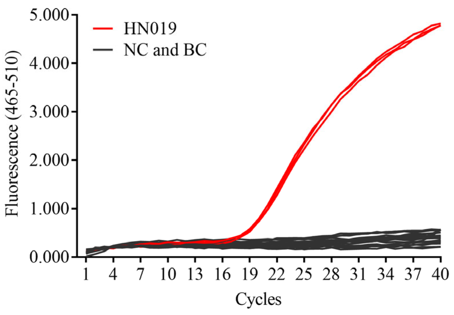
3.1. SNP Analysis and Specificity Test

Using the whole genome sequence of B. animalis subsp. lactis HN019 as a reference, we compared it with the whole-genome sequences of 30 other B. animalis subsp. lactis strains from the NCBI database. A total of 6 SNPs specific to the HN019 strain were identified. Among them, the SNP located in the galK gene was selected for the design of primers and probes. After performing qPCR amplification, this SNP-based primer/probe pair was able to effectively differentiate B. animalis subsp. lactis HN019 from 19 other Bifidobacterium strains. As shown in Figure 1, the Cq values for the HN019 strain were 19.25, 19.27, and 19.07, while no significant amplification was observed for the other strains or the blank control. Based on the criteria for interpreting qPCR results outlined by Wang et al. [34], a Cq value below 35 was considered positive.

3.2. Passage Stability Test

As shown in Table 4, the Cq value standard deviation for the stability test of the HN019 strain, from the 5th to the 35th generation, was 0.24, with a relative standard deviation of 1.31%. In addition, the investigation revealed no statistically significant disparities in the outcomes of the evaluation between the 5th, 15th, 25th, and 35th generations. This indicates that the SNP site located in the galK gene exhibits good stability throughout the passage process and can be used for the detection of B. animalis subsp. lactis HN019 in limited generations.

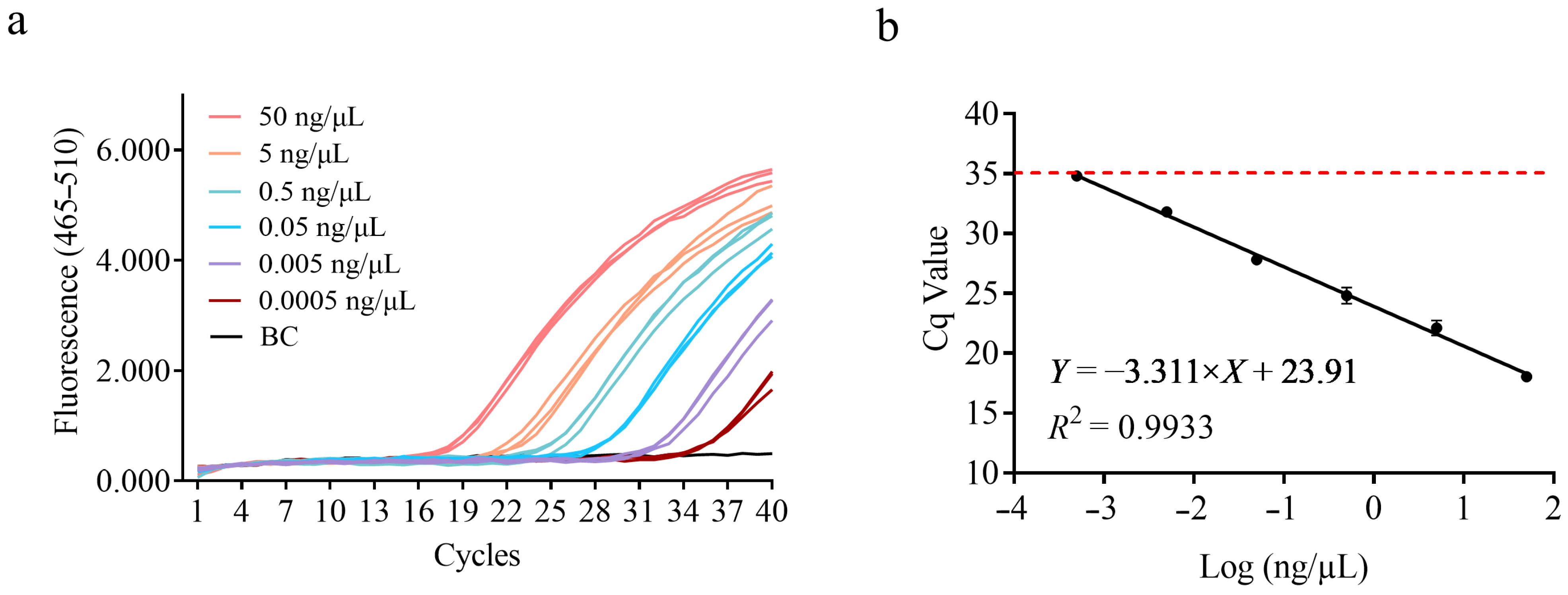
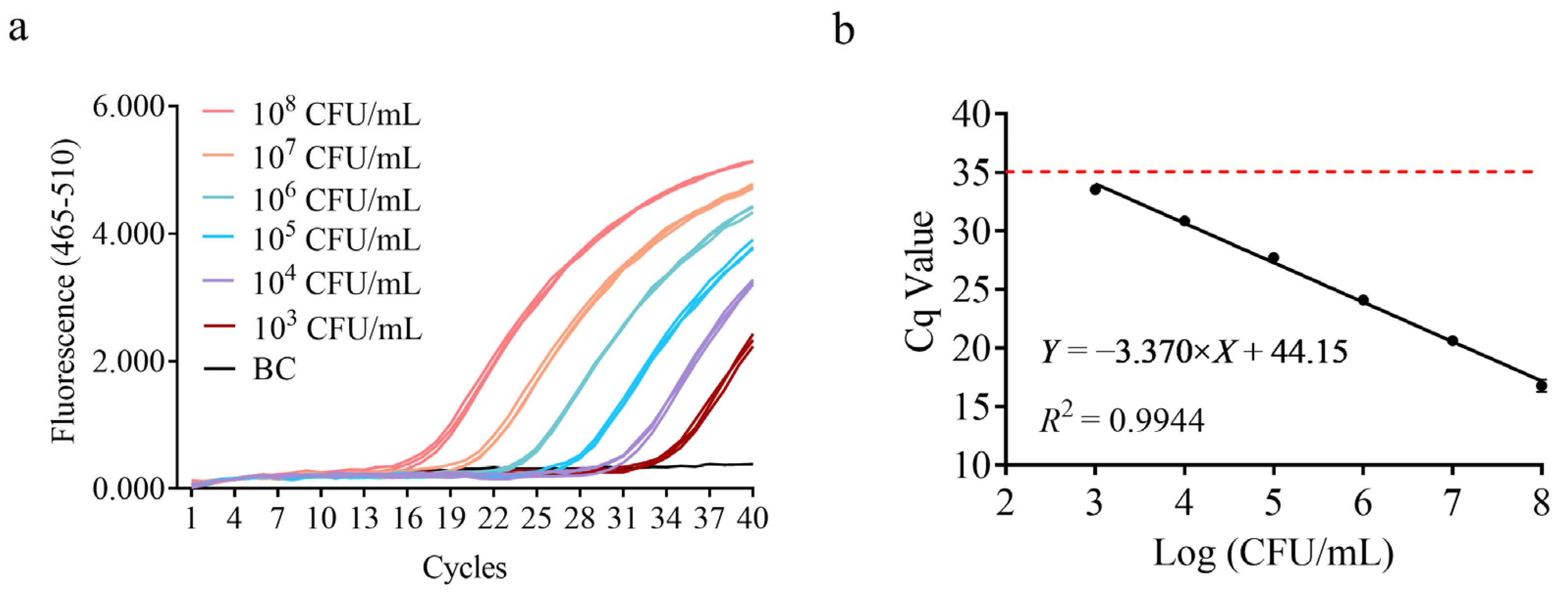
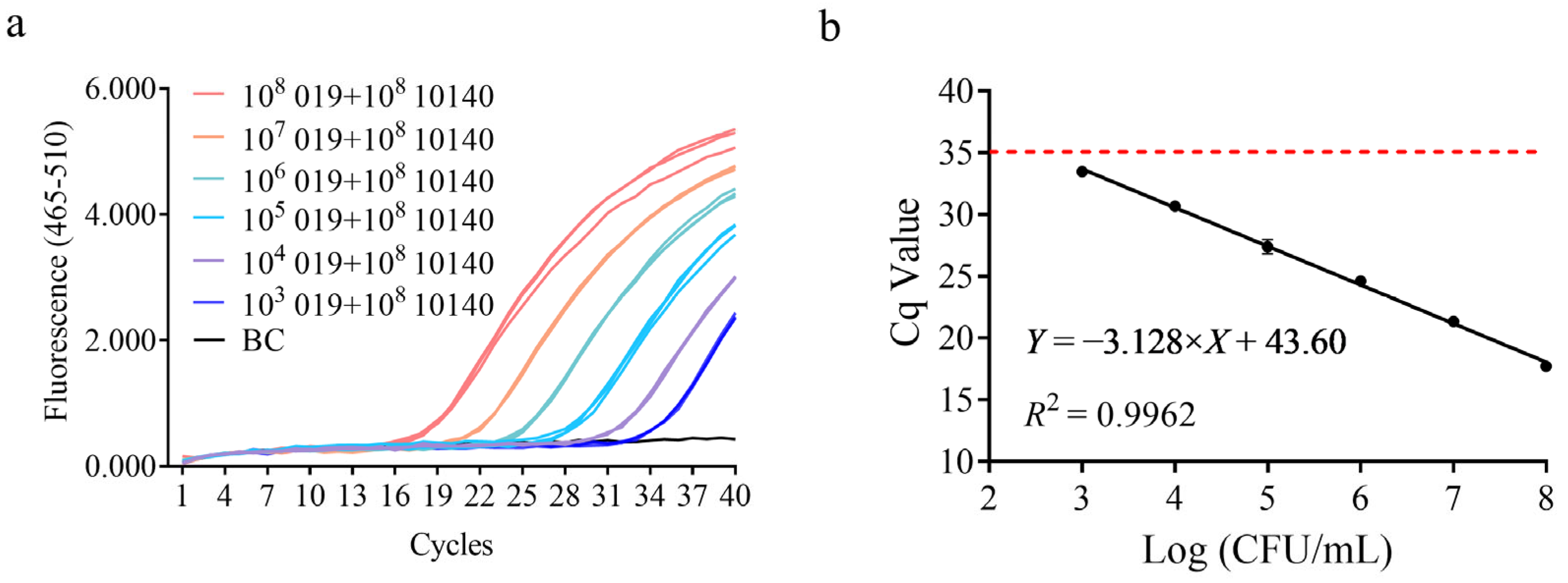
3.3. Sensitivity Test

To determine the sensitivity of this detection method, a DNA solution with a concentration of 50 ng/μL was serially diluted in 10-fold steps and subjected to qPCR amplification, as shown in Figure 2a. A standard curve was constructed with the logarithmic value of DNA concentration on the x-axis and the mean Cq value on the y-axis. From Figure 2b, the slope of the standard curve was −3.311, with an amplification efficiency of 100.46% and an R2 value of 0.9933 (Figure 2b). Based on the data in Figure 2, the detection limit for DNA concentration in this amplification system is approximately 0.5 pg, indicating that the detection method is highly sensitive.
To meet the needs of dairy companies for detecting lactic acid bacterial colony counts, quantitative results were correlated with bacterial colony-forming units (CFUs). A gradient dilution of B. animalis subsp. lactis HN019 bacterial suspension was prepared, DNA was extracted as the template, and qPCR amplification was performed. The amplification results showed that when the bacterial cell density was ≥103 CFU/mL, the detection was positive, as shown in Figure 3. A standard curve was constructed with the logarithmic value of bacterial concentration on the x-axis and the average Cq value on the y-axis. The slope was −3.370, with an amplification efficiency of 98.03% and an R2 value of 0.9944. These results demonstrate that the sensitivity of this method for bacterial suspension concentration is 103 CFU/mL, which meets the daily detection needs of dairy companies for 106 CFU/mL [35].

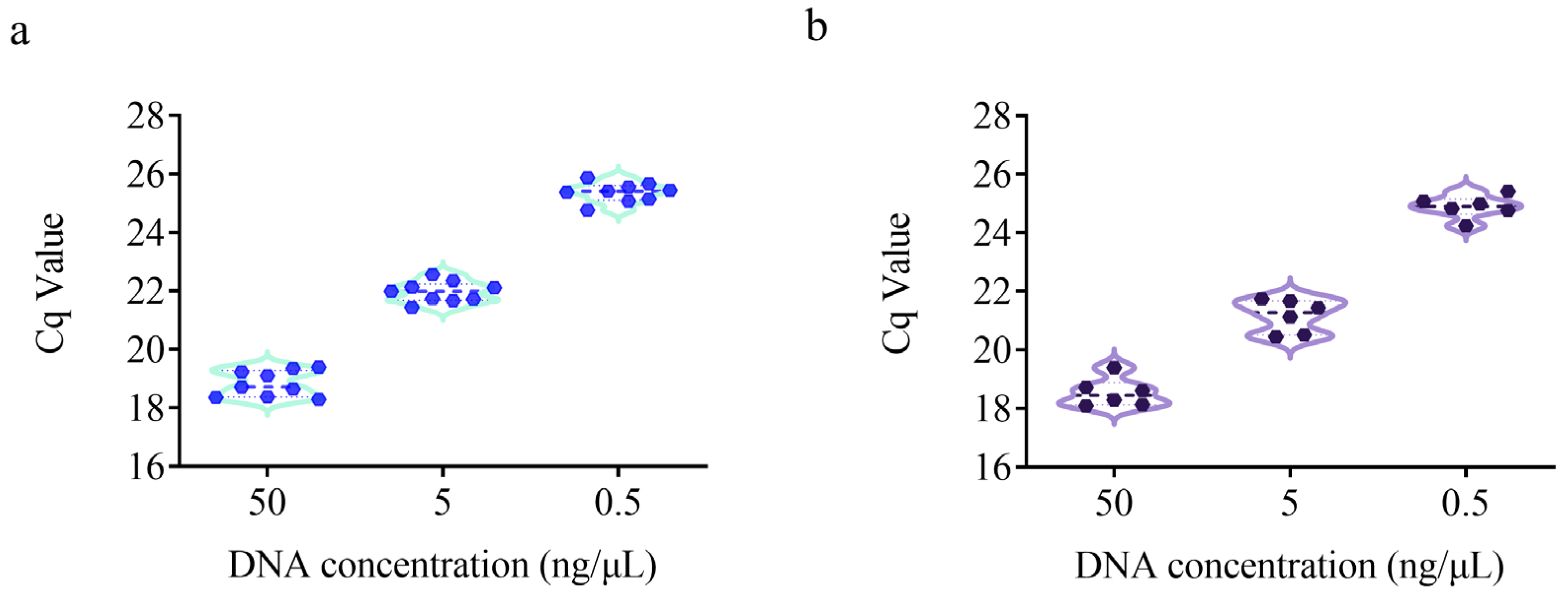
3.4. Repeatability and Reproducibility Test

The repeatability and reproducibility of the detection method were evaluated using DNA samples at three different concentrations (50, 5, and 0.5 ng/μL). As shown in Figure 4a, the RSD for repeatability over time ranged from 1.30% to 2.38%. For reproducibility testing across two different qPCR platforms (Roche LightCycler® 480 and ABI 7500), the RSD ranged from 1.55% to 2.65% (Figure 4b), which is well below the requirement that qPCR detection results should have an RSD value of less than 25% [36]. In the context of repeatability testing, no statistically significant variations were observed among the three test results at 50 ng/µL, 5 ng/µL, and 0.5 ng/µL. In the context of reproducibility testing, no statistically significant discrepancies were observed between the results obtained from two distinct platforms at concentrations of 50 ng/µL, 5 ng/µL, and 0.5 ng/µL. Therefore, the method demonstrated good repeatability and reproducibility and is suitable for use across different qPCR platforms.

3.5. Interference Resistance Test

To enhance the efficacy of probiotic products, a combination of target strains and other strains is often added to the products. Therefore, it is essential to verify the interference resistance of the HN019 strain detection method in the presence of closely related strains within the same subspecies. As shown in Figure 5, under the condition of adding 108 CFU/mL of DSMZ 10140, the established detection method successfully quantified the HN019 strain, maintaining a sensitivity of 103 CFU/mL. These results demonstrate that the method has strong resistance to interference, providing valuable data support for subsequent actual sample testing.

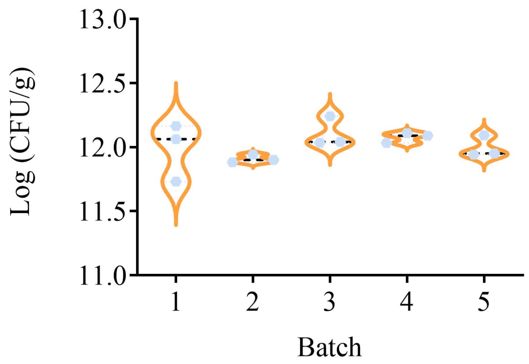
3.6. Quantification of HN019 Strain in Probiotic Powder Samples

The strain-specific detection method was used to precisely quantify the HN019 strain in five batches of actual composite probiotic powder samples. Using the standard curve equation Y = −3.347 × X + 44.17, R2 = 0.9944, and the formula 2.9, the HN019 strain content in the five batches of samples was calculated to range from (11.91 ± 0.03) to (12.11 ± 0.12) Log (CFU/g), as shown in Figure 6. A subsequent investigation revealed that there was no significant difference in the content of HN019 in the five batches of probiotic powder samples. The results indicate that the method is equally applicable to composite probiotic powder samples containing large amounts of oligosaccharides and excipients.

4. Discussion

The selection of B. animalis subsp. lactis HN019 as the focal strain for this study stems from its well-established yet uniquely complex clinical profile and its prominence in commercially significant probiotic formulations. Originally isolated from dairy sources [37], HN019 has transcended its industrial origins to become a benchmark probiotic strain with documented, dose-dependent health benefits in humans. Critically, robust clinical trials have demonstrated their efficacy in enhancing innate immune responses, such as natural killer cell activity, and reducing the incidence of antibiotic-associated diarrhea. These specific immunomodulatory properties, coupled with their documented stability and persistence in the gut, have positioned HN019 as a cornerstone strain in precision probiotic interventions, particularly for vulnerable populations including infants and the elderly experiencing immunosenescence [7]. However, the very characteristic that makes HN019 valuable also presents a major challenge: its clinical outcomes are demonstrably formulation-dependent and inconsistent across studies, such as null effects observed in constipation trials [38]. This inconsistency underscores a fundamental limitation in the field: the inability to reliably confirm and quantify the presence and dose of the specific, clinically validated HN019 strain within complex probiotic products. Consequently, rigorous strain-specific quantification is not merely beneficial but essential for elucidating HN019’s true characteristics in vivo, ensuring product fidelity, validating clinical efficacy claims, and ultimately advancing evidence-based probiotic development.
While traditional culture-based methods provide total viable counts, they fundamentally lack the specificity to distinguish HN019 from closely related strains within the B. animalis subsp. lactis subspecies, especially in multi-strain products or those containing low target populations or complex matrices. This gap highlights a persistent problem: the lack of a truly strain-specific, sensitive, and robust quantification tool for HN019. Molecular approaches targeting genes like tuf, Bal-23S, or Tal have been successfully applied to quantify the related BB-12 strain [39,40,41] but critically fail to differentiate between HN019 and BB-12 due to insufficient genetic resolution. This gap highlights a persistent problem: the lack of a truly strain-specific, sensitive, and robust quantification tool for HN019.
With the rapid advancement of high-throughput sequencing technologies, the complete genome sequences of more B. animalis subsp. lactis strains have been completed, and genomic data are available through public databases, providing a solid foundation for the application of WGS-SNP analysis. By designing primers and probes for SNPs in the galK gene, we successfully achieved specific detection of the HN019 strain. The selected SNP in galK exhibited robust genetic stability across 35 serial passages, a critical validation for industrial scalability that ensures consistent detectability throughout product lifecycles. According to the criterion of Cq values below 35 for positive results in qPCR, the DNA sensitivity and cell density sensitivity of our quantitative detection method are 0.5 pg and 103 CFU/mL, respectively. Additionally, the amplification efficiency exceeds 98%, falling within the optimal range (80–120%) [36], with an R2 value greater than 0.99, indicating excellent linearity. Evaluation of the method’s repeatability and reproducibility showed good consistency across different time intervals and platforms, with RSD values below 3%, confirming its high reliability and adaptability. To further assess the method’s resistance to interference, we conducted interference experiments by adding high concentrations of homologous strains. The results indicated that even with interference from other homologous strains and excipients, the method accurately quantifies the HN019 strain at 103 CFU/mL, while traditional plate counting methods fail to achieve such specific quantification in multi-strain samples.
The accuracy and reliability of quality control for the HN019 strain, a key probiotic, directly affect consumer health. The quantitative analysis method proposed in this study, based on WGS-SNP analysis and qPCR, offers high specificity, strong repeatability, genetic stability, resistance to interference, and good equipment adaptability, providing regulators and manufacturers with a gold-standard tool to verify HN019 strain identity, viability, and dose claims in commercial products, safeguarding against mislabeling and subpotency. Moreover, the SNP-based design paradigm establishes a blueprint for developing strain-specific assays for other high-value probiotics, advancing the field toward truly personalized microbial therapeutics.

5. Conclusions

As the probiotic market rapidly expands, consumers’ demands for the quality and safety of probiotic products are increasing. Accurate and reliable strain detection methods have become one of the key technologies driving the sustainable development of the probiotic industry. This study proposes a high-precision HN019 strain quantification method based on WGS-SNP analysis and quantitative PCR technology, providing scientific and reliable technical support for quality control of probiotic products. Our WGS-SNP-qPCR platform not only unlocks rigorous research into HN019’s bioactivity but also sets a new standard for quality control in the rapidly evolving probiotic industry, ensuring that evidence-based health claims translate into reliable consumer benefits.

Supplementary Materials

The following supporting information can be downloaded at: https://www.mdpi.com/article/10.3390/microorganisms13071596/s1. Table S1: Strains used for genomic comparison and specific loci information.

Author Contributions

Conceptualization, B.Z.; methodology, H.X.; software, L.Z.; validation, L.Z. and D.M.; formal analysis, L.Z. and D.M.; investigation, Q.Z.; resources, H.X. and B.Z.; data curation, L.Z.; writing—original draft preparation, L.Z.; writing—review and editing, H.X. and B.Z.; visualization, L.Z. and D.M.; supervision, H.X. and B.Z.; project administration, D.M. and L.Z.; funding acquisition, D.M. and L.Z. All authors have read and agreed to the published version of the manuscript.

Funding

This research was funded by the National Key Research and Development Program of China (grant number 2019YFC1604803) and the Science and Technology Project of the Shanghai Institute of Quality Inspection and Technical Research (grant number KY-2023-6-SP).

Institutional Review Board Statement

Not applicable.

Informed Consent Statement

Not applicable.

Data Availability Statement

The full genome data of 31 B. animalis subsp. lactis can be found in NCBI GenBank under the assembly number listed in Table 1.

Acknowledgments

We thank Shanghai majorbio Bio-pharm technology Co., Ltd. for helping in high-throughput data analysis.

Conflicts of Interest

The authors declare no conflicts of interest.

References

  1. Das, T.K.; Pradhan, S.; Chakrabarti, S.; Mondal, K.C.; Ghosh, K. Current status of probiotic and related health benefits. Appl. Food Res. 2022, 2, 100185. [Google Scholar] [CrossRef]
  2. Yan, M.; Man, S.; Sun, B.; Ma, L.; Guo, L.; Huang, L.; Gao, W. Gut liver brain axis in diseases: The implications for therapeutic interventions. Signal Transduct. Target. Ther. 2023, 8, 443. [Google Scholar] [CrossRef]
  3. Loh, J.S.; Mak, W.Q.; Tan, L.K.S.; Ng, C.X.; Chan, H.H.; Yeow, S.H.; Foo, J.B.; Ong, Y.S.; How, C.W.; Khaw, K.Y. Microbiota–gut–brain axis and its therapeutic applications in neurodegenerative diseases. Signal Transduct. Target. Ther. 2024, 9, 37. [Google Scholar]
  4. Kearns, R. Gut–brain axis and neuroinflammation: The role of gut permeability and the kynurenine pathway in neurological disorders. Cell. Mol. Neurobiol. 2024, 44, 64. [Google Scholar] [CrossRef] [PubMed]
  5. Lugli, G.A.; Argentini, C.; Tarracchini, C.; Mancabelli, L.; Viappiani, A.; Anzalone, R.; Angelini, L.; Alessandri, G.; Longhi, G.; Bianchi, M.G.; et al. Characterization of a Bifidobacterium animalis subsp. lactis reference strain based on ecology and transcriptomics. Appl. Environ. Microbiol. 2024, 90, e01080-24. [Google Scholar] [CrossRef]
  6. D’Ambrosio, S.; Dabous, A.; Sadiq, S.; Casillo, A.; Schiraldi, C.; Cassese, E.; Bedini, E.; Corsaro, M.M.; Cimini, D. Bifidobacterium animalis subsp. lactis HN019 live probiotics and postbiotics: Production strategies and bioactivity evaluation for potential therapeutic properties. Front. Bioeng. Biotechnol. 2024, 12, 1379574. [Google Scholar] [CrossRef] [PubMed]
  7. Cheng, J.; Laitila, A.; Ouwehand, A.C. Bifidobacterium animalis subsp. lactis HN019 effects on gut health: A review. Front. Nutr. 2021, 8, 790561. [Google Scholar] [CrossRef]
  8. Sharma, M.; Wasan, A.; Sharma, R.K. Recent developments in probiotics: An emphasis on Bifidobacterium. Food Biosci. 2021, 41, 100993. [Google Scholar] [CrossRef]
  9. Putaala, H.; Salusjärvi, T.; Nordström, M.; Saarinen, M.; Ouwehand, A.C.; Bech Hansen, E.; Rautonen, N. Effect of four probiotic strains and Escherichia coli O157:H7 on tight junction integrity and cyclo-oxygenase expression. Res. Microbiol. 2008, 159, 692–698. [Google Scholar] [CrossRef] [PubMed]
  10. Bernini, L.J.; Simão, A.N.C.; de Souza, C.H.B.; Alfieri, D.F.; Segura, L.G.; Costa, G.N.; Dichi, I. Effect of Bifidobacterium lactis HN019 on inflammatory markers and oxidative stress in subjects with and without the metabolic syndrome. Br. J. Nutr. 2018, 120, 645–652. [Google Scholar] [CrossRef]
  11. Invernici, M.M.; Furlaneto, F.A.C.; Salvador, S.L.; Ouwehand, A.C.; Salminen, S.; Mantziari, A.; Vinderola, G.; Ervolino, E.; Santana, S.I.; Silva, P.H.F.; et al. Bifidobacterium animalis subsp lactis HN019 presents antimicrobial potential against periodontopathogens and modulates the immunological response of oral mucosa in periodontitis patients. PLoS ONE 2020, 15, e0238425. [Google Scholar] [CrossRef] [PubMed]
  12. Bettocchi, S.; Comotti, A.; Elli, M.; De Cosmi, V.; Berti, C.; Alberti, I.; Mazzocchi, A.; Rosazza, C.; Agostoni, C.; Milani, G.P. Probiotics and Fever Duration in Children with Upper Respiratory Tract Infections: A Randomized Clinical Trial. JAMA Netw. Open 2025, 8, e250669. [Google Scholar] [CrossRef]
  13. Ala-Jaakkola, R.; Forssten, S.D.; Cheng, J.; Griffon, F.; Metreau, I.; Sturm, Y.; Lecerf, J.M.; Donazzolo, Y.; Junnila, J.; Nordlund, A.; et al. Effect of an 8-week Bifidobacterium lactis HN019 supplementation on functional constipation: A multi-center, triple-blind, randomized, placebo-controlled trial. Mol. Nutr. Food Res. 2025, e70081. [Google Scholar] [CrossRef]
  14. Jena, R.; Choudhury, P.K. Bifidobacteria in fermented dairy foods: A health beneficial outlook. Probiotics Antimicrob. Prot. 2025, 17, 1–22. [Google Scholar] [CrossRef]
  15. Waller, P.A.; Gopal, P.K.; Leyer, G.J.; Ouwehand, A.C.; Reifer, C.; Stewart, M.E.; Miller, L.E. Dose-response effect of Bifidobacterium lactis HN019 on whole gut transit time and functional gastrointestinal symptoms in adults. Scand. J. Gastroenterol. 2011, 46, 1057–1064. [Google Scholar] [CrossRef] [PubMed]
  16. Chen, J.; Chen, X.; Ho, C.L. Recent Development of probiotic Bifidobacteria for treating human diseases. Front. Bioeng. Biotechnol. 2021, 9, 770248. [Google Scholar] [CrossRef]
  17. Huang, C.H.; Li, S.W.; Huang, L.; Watanabe, K. Identification and classification for the Lactobacillus casei group. Front. Microbiol. 2018, 9, 1974. [Google Scholar] [CrossRef] [PubMed]
  18. Li, M.N.; Han, Q.; Wang, N.; Wang, T.; You, X.M.; Zhang, S.; Zhang, C.C.; Shi, Y.Q.; Qiao, P.Z.; Man, C.L.; et al. 16S rRNA gene sequencing for bacterial identification and infectious disease diagnosis. Biochem. Biophys. Res. Commun. 2024, 739, 150974. [Google Scholar] [CrossRef]
  19. Ruiz-Moyano, S.; Tao, N.; Underwood, M.A.; Mills, D.A. Rapid discrimination of Bifidobacterium animalis subspecies by matrix-assisted laser desorption ionization-time of flight mass spectrometry. Food Microbiol. 2012, 30, 432–437. [Google Scholar] [CrossRef]
  20. Huang, C.H.; Liou, J.S.; Huang, L.; Watanabe, K. Developing novel species-specific DNA markers for PCR-based species identification of the Lactobacillus sakei group. Lett. Appl. Microbiol. 2018, 66, 138–144. [Google Scholar] [CrossRef]
  21. Schürch, A.C.; Arredondo-Alonso, S.; Willems, R.J.L.; Goering, R.V. Whole genome sequencing options for bacterial strain typing and epidemiologic analysis based on single nucleotide polymorphism versus gene by gene—Based approaches. Clin. Microbiol. Infect. 2018, 24, 350–354. [Google Scholar] [CrossRef] [PubMed]
  22. Deschamps, S.; Zhang, Y.; Llaca, V.; Ye, L.; Sanyal, A.; King, M.; May, G.; Lin, H. A chromosome-scale assembly of the sorghum genome using nanopore sequencing and optical mapping. Nat. Commun. 2018, 9, 4844. [Google Scholar] [CrossRef]
  23. Lo, K.J.; Lin, S.S.; Lu, C.W.; Kuo, C.H.; Liu, C.T. Whole-genome sequencing and comparative analysis of two plant-associated strains of Rhodopseudomonas palustris (PS3 and YSC3). Sci. Rep. 2018, 8, 12769. [Google Scholar] [CrossRef] [PubMed]
  24. Leekitcharoensophon, P.; Kaas, R.S.; Thomsen, M.C.; Friis, C.; Rasmussen, S.; Aarestrup, F.M. snpTree a web server to identify and construct SNP trees from whole genome sequence data. BMC Genom. 2012, 13, S6. [Google Scholar]
  25. Shao, N.; Li, F.; Nie, K.; Fu, S.H.; Zhang, W.J.; He, Y.; Lei, W.W.; Wang, Q.Y.; Liang, G.D.; Cao, Y.X.; et al. TaqMan real-time rt-pcr assay for detecting and differentiating Japanese encephalitis virus. Biomed. Environ. Sci. 2018, 31, 208–214. [Google Scholar]
  26. Huang, S.; Wang, X.; Chen, X.; Liu, X.; Xu, Q.; Zhang, L.; Huang, G.; Wu, J. Rapid and sensitive detection of Pseudomonas aeruginosa by isothermal amplification combined with Cas12a-mediated detection. Sci. Rep. 2023, 13, 19199. [Google Scholar] [CrossRef] [PubMed]
  27. Sound, J.K.; Peters, A.; Bellamy-Carter, J.; Rad-Menéndez, C.; MacKechnie, K.; Green, D.H.; Leney, A.C. Rapid cyanobacteria species identification with high sensitivity using native mass spectrometry. Anal. Chem. 2021, 93, 14293–14299. [Google Scholar] [CrossRef]
  28. Hsieh, A.Y.Y.; Saberi, S.; Ajaykumar, A.; Hukezalie, K.; Gadawski, I.; Sattha, B.; Côté, H.C.F. Optimization of a relative telomere length assay by monochromatic multiplex real-time quantitative pcr on the LightCycler 480: Sources of variability and quality control considerations. J. Mol. Diagn. 2016, 18, 425–437. [Google Scholar] [CrossRef]
  29. GB 4789.34–2016; National Food Safety Standard–Food Microbiological Examination: Examination of Bifidobacteria. China Standards Press: Beijing, China, 2016.
  30. Yao, Y.T.; Zhang, X.; Wang, C.Y.; Zhang, Y.H.; Li, D.W.; Yang, W.D.; Li, H.Y.; Zou, L.G. Optimizing longifolene production in Yarrowia lipolytica via metabolic and protein engineering. Synth. Syst. Biotechnol. 2025, in press. [Google Scholar] [CrossRef]
  31. Svec, D.; Tichopad, A.; Novosadova, V.; Pfaffl, M.W.; Kubista, M. How good is a PCR efficiency estimate: Recommendations for precise and robust qPCR efficiency assessments. Biomol. Detect. Quantif. 2015, 3, 9–16. [Google Scholar] [CrossRef]
  32. Von Hertwig, A.M.; Sant’Ana, A.S.; Sartori, D.; da Silva, J.J.; Nascimento, M.S.; Iamanaka, B.T.; Pelegrinelli Fungaro, M.H.; Taniwaki, M.H. Real-time PCR-based method for rapid detection of Aspergillus niger and Aspergillus welwitschiae isolated from coffee. J. Microbiol. Methods 2018, 148, 87–92. [Google Scholar] [CrossRef] [PubMed]
  33. SN/T 5642.3-2023; Professional Standard of the People’s Republic of China for Entry-Exit Inspection and Quarantine–Detection of lactic acid bacteria in dairy products for export–Enumeration method of digital PCR–Part3: Bifidobacterium animalis. China Standards Press: Beijing, China, 2023.
  34. Wang, R.; Zhang, W.; Ye, R.; Pan, Z.; Li, G.; Su, S. One-step multiplex TaqMan probe-based method for real-time PCR detection of four canine diarrhea viruses. Mol. Cell. Probes 2020, 53, 101618. [Google Scholar] [CrossRef]
  35. Herbel, S.R.; Lauzat, B.; Von Nickisch-Rosenegk, M.; Kuhn, M.; Murugaiyan, J.; Wieler, L.H.; Guenther, S. Species-specific quantification of probiotic lactobacilli in yoghurt by quantitative real-time PCR. J. Appl. Microbiol. 2013, 115, 1402–1410. [Google Scholar] [CrossRef]
  36. Broeders, S.; Huber, I.; Grohmann, L.; Berben, G.; Taverniers, I.; Mazzara, M.; Roosens, N.; Morisset, D. Guidelines for validation of qualitative real-time PCR methods. Trends Food Sci. Technol. 2014, 37, 115–126. [Google Scholar] [CrossRef]
  37. Dekker, J.W.; Wickens, K.; Black, P.N.; Stanley, T.V.; Mitchell, E.A.; Fitzharris, P. Safety aspects of probiotic bacterial strains Lactobacillus rhamnosus HN001 and Bifidobacterium animalis subsp. lactis HN019 in human infants aged 0–2 years. Int. Dairy J. 2009, 19, 149–154. [Google Scholar]
  38. Cheng, J.; Gao, C.; Ala-Jaakkola, R. Eight-Week Supplementation with Bifidobacterium lactis HN019 and Functional Constipation: A Randomized Clinical Trial. JAMA Netw. Open 2024, 7, e2436888. [Google Scholar] [CrossRef] [PubMed]
  39. Solano-Aguilar, G.; Dawson, H.; Restrepo, M.; Andrews, K.; Vinyard, B.; Urban, J.F., Jr. Detection of Bifidobacterium animalis subsp. lactis (Bb12) in the intestine after feeding of sows and their piglets. Appl. Environ. Microbiol. 2008, 74, 6338–6347. [Google Scholar]
  40. Taipale, T.; Pieniähäkkänen, K.; Isolauri, E.; Larsen, C.; Brockmann, E.; Alanen, P.; Jokela, J.; Söderling, E. Bifidobacterium animalis subsp. lactis BB-12 in reducing the risk of infections in infancy. Br. J. Nutr. 2011, 105, 409–416. [Google Scholar]
  41. Requena, T.; Burton, J.; Matsuki, T.; Munro, K.; Simon, M.A.; Tanaka, R.; Watanabe, K.; Tannock, G.W. Identification, detection, and enumeration of human bifidobacterium species by PCR targeting the transaldolase gene. Appl. Environ. Microbiol. 2002, 68, 2420–2427. [Google Scholar] [CrossRef]
Figure 1. Primer specificity test for the B. animalis subsp. lactis HN019 strain. NC: Negative Control (strains other than HN019); BC: Blank Control (sterile double-distilled water).
Microorganisms 13 01596 g001
Figure 2. Sensitivity of the specific detection method for B. animalis subsp. lactis HN019 strain at varying DNA concentrations. (a) Amplification curve obtained using the Roche LightCycler® 480 platform for the DNA concentration sensitivity test. (b) Standard curve constructed by plotting Cq values against DNA concentration.
Microorganisms 13 01596 g002
Figure 3. Cell density sensitivity test for specific detection method of B. animalis subsp. lactis HN019 strain. (a) Amplification curve obtained using the Roche LightCycler® 480 platform for the bacterial cell density sensitivity test, BC: Blank Control (sterile double-distilled water). (b) Standard curve constructed by plotting Cq values against cell density.
Microorganisms 13 01596 g003
Figure 4. Repeatability (a) and reproducibility (b) test for specific detection method of B. animalis subsp. lactis HN019 strain. Violin plots were generated using GraphPad Prism 9, Median (dashed line), kernel density distribution (blue line or purple line) are shown. (a) Repeatability test, showing three trials with three replicates (dark blue dots) in each trial, for a total of 9 measurements. (b) Reproducibility test, showing experiments conducted on two different qPCR platforms (Roche LightCycler® 480 and ABI 7500), for a total of 6 measurements (dark purple dots).
Microorganisms 13 01596 g004
Figure 5. Interference resistance test for specific detection method of B. animalis subsp. lactis HN019 strain. (a) Amplification curve obtained using the Roche LightCycler® 480 platform for the interference resistance test, BC: Blank Control (sterile double-distilled water). (b) Standard curve constructed by plotting Cq values against cell density.
Microorganisms 13 01596 g005
Figure 6. Validation of the specific detection method using five batches of multi-strain bacterial powder samples, including B. animalis subsp. lactis HN019, L. acidophilus NCFM, L. rhamnosus GG, and oligosaccharides. Violin plots were generated using GraphPad Prism 9, Median (dashed line), kernel density distribution (orange line) are shown. Each batch was tested three times, with the results of B. animalis subsp. lactis HN019 represented by light blue dots.
Microorganisms 13 01596 g006
Table 1. Strains used for WGS-SNP analysis.
SpeciesStrainAssembly NumberScaffold NumberGenome Size (Mb)
B. animalis subsp. lactisHN019 *GCA_003606305.111.935423
19-D-1GCA_020309945.111.963057
AD011GCA_000021425.111.933695
ATCC 27673GCA_000471945.111.963012
B420GCA_000277325.111.938595
BAMA-B06GCA_028463865.111.944252
BB-12GCA_000025245.211.944152
BF052GCA_000818055.111.938624
BGI-N3GCA_040084745.111.944238
BI040GCA_038086955.111.944141
Bi-07GCA_000277345.111.938822
Bl-04GCA_000022705.111.938709
Bl12GCA_000414215.211.944036
BLC1GCA_000224965.211.938583
CGMCC1.15623GCA_044866785.111.944145
CNCM I-2494GCA_000220885.111.943113
DSM 10140GCA_000022965.111.938483
DSM 15954GCA_021018785.111.944152
GOLDGUT-BB18GCA_034561975.111.944144
H1GCA_016835115.111.944352
H3GCA_016835135.111.944351
HOM2120GCA_044502405.111.946900
i797GCA_019576095.111.943538
IDCC4301GCA_003428375.111.944141
KLDS 2.0603GCA_000816205.111.946899
LPL-RHGCA_030284625.111.944223
MH-02GCA_029167605.111.944290
S7GCA_003390755.111.944072
SFGCA_029542645.111.944374
TCI604GCA_036923305.111.944134
V9GCA_000092765.111.944050
*: reference genome.
Table 2. The sequences of primers and probe.
GeneThe Sequences of Primers and Probe Product Length (bp)
galKTGATATGGCGAATGCTTGCA61
CGGCTTGTGTGTCGTCATG
FAM-ACCCTTTTCATTTCCCTGC-MGB
Table 3. Strains used for specificity test.
No.SpeciesStrain
1B. animalis subsp. lactisHN019
2Bi-07
3Bl-04
4BB12
5DSMZ 10140
6CICC 21718
7ATCC 25527
8ATCC 27673
9B. animalisATCC 27672
10CICC 6165
11CICC 6250
12B. adolescentisATCC 15703
13B. longum subsp. infantis ATCC 15697
14B. breveATCC 15700
15CICC 6185
16CICC 6181
17B. bifidumCICC 6173
18B. longumCICC 6207
19CICC 6199
20Bl05
Table 4. Stability of specific amplification during serial passages.
Number of GenerationsCq ValueMean Cq ValueSDRSD (%)
518.8818.7818.9818.880.241.31
1518.9218.8618.2718.68
2518.7318.3818.8918.67
3517.9818.418.518.29
Disclaimer/Publisher’s Note: The statements, opinions and data contained in all publications are solely those of the individual author(s) and contributor(s) and not of MDPI and/or the editor(s). MDPI and/or the editor(s) disclaim responsibility for any injury to people or property resulting from any ideas, methods, instructions or products referred to in the content.

