Article

Molecular Survey and Genetic Analysis of Ehrlichia canis in Rhipicephalus sanguineus Ticks Infesting Dogs in Northern Taiwan

1
Graduate Institute of Medicine, College of Medicine, Kaohsiung Medical University, Kaohsiung 807, Taiwan
2
Graduate Institute of Pathology and Parasitology, National Defense Medical Center, Taipei 114, Taiwan
3
Department of Medical Research, Kaohsiung Medical University Hospital, Kaohsiung 807, Taiwan
*
Author to whom correspondence should be addressed.
Microorganisms 2025, 13(6), 1372; https://doi.org/10.3390/microorganisms13061372
Submission received: 2 May 2025 / Revised: 5 June 2025 / Accepted: 11 June 2025 / Published: 12 June 2025
(This article belongs to the Special Issue Ticks, Tick Microbiome and Tick-Borne Diseases)

Abstract

Ehrlichia canis is a tick-transmitted zoonotic pathogen in dogs. We conducted a molecular survey for screening of E. canis infection in Rhipicephalus sanguineus ticks infesting dogs and identified its genetic identity in Taiwan. A total of 1195 R. sanguineus ticks were collected and examined for Ehrlichia infection by nested-PCR assay targeting the 16S ribosomal RNA (rRNA) gene. In general, Ehrlichia infection was detected in 1.5% of examined ticks, and was detected in nymph, male and female stages with infection rates of 0.6%, 1.31% and 2.76%, respectively. The highest monthly prevalence was observed in August with an infection rate of 5.91%. Genetic identity was analyzed by comparing the 16S rRNA gene sequences obtained from 11 Taiwan strains and 15 other strains representing five genospecies of Ehrlichia spp., including two outgroups (Anaplasma phagocytophilum and Rickettsia rickettsii). Results revealed that all Taiwan strains were genetically affiliated to the same clade within various E. canis strains documented in GenBank with a high sequence similarity (99.7–100%) and that they can be clearly distinguished from other genospecies of Ehrlichia. This study provides the first evidence of E. canis identified in R. sanguineus ticks and highlights the potential threat for human infections in Taiwan.

1. Introduction

The genus Ehrlichia includes five species of gram-negative obligate intracellular bacteria infecting monocytes, and E. canis is recognized as the primary causative agent for canine monocytic ehrlichiosis (CME), a potentially fatal disease in dogs [1,2]. This Ehrlichia infection was described for the first time in Algeria in 1935 [3], and it has been reported in the southern regions of the USA [4] and was recorded in some western and southern regions of Europe [5]. Currently, this infection is widely distributed around the world, and the prevalence of E. canis depends on the distribution of its vector ticks. Although E. canis had been reported in asymptomatic dogs of central Taiwan [6], there is no confirming evidence for the existence of E. canis in its vector tick. Thus, a molecular survey on tick-borne E. canis in Rhipicephalus sanguineus ticks is crucial to understand the potential threat of emerging tick-borne E. canis infections in Taiwan.
The R. sanguineus tick is a haematophagous arthropod and is commonly parasitic on canine hosts throughout the world [7,8]. Previous studies revealed that R. sanguineus has been recognized as the major vector for the transmission of various tick-borne zoonotic pathogens, such as Babesia, Ehrlichia, Anaplasma, Hepatozoon and Rickettsia, among animals and humans [9,10,11]. Because of the increasing detection of H. canis, B. vogeli, B. gibsoni, A. platys and various Rickettsia spp. in R. sanguineus ticks of Taiwan [12,13,14,15,16,17], this tick species has become the focus of research on medical and veterinary importance. Although the R. sanguineus ticks had been recognized as the incriminated vector ticks for a variety of zoonotic pathogens, there has been no investigation confirming the prevalence and genetic identity of E. canis, a zoonotic pathogen for human infection, in this tick species in Taiwan.
Molecular detection targeting the 16S rRNA gene of Ehrlichia has made the feasibility for identifying the genetic identity within the vector ticks. Indeed, this molecular tool can be used to differentiate the genetic variance at the individual base-pair level and gives a direct method for measuring the genetic diversity between and within species of Ehrlichia [18,19]. Previous studies based on the molecular marker of 16S rRNA genes of Ehrlichia have demonstrated that it is sufficiently informative for the genetic analysis of phylogenetic relationships between the genetic diversity of Ehrlichia species among various vectors and hosts [20,21,22,23,24,25,26]. Thus, molecular detection and genetic analysis based on the genetic comparison of 16S rRNA genes have made it feasible to facilitate the identification and discrimination of Ehrlichia species within ticks.
The objectives of this study are to investigate the prevalence of Ehrlichia infection in R. sanguineus ticks collected from Taiwan and to determine the genetic identity of Ehrlichia spp. in R. sanguineus ticks. The genetic affiliation of Ehrlichia strains detected in R. sanguineus ticks of Taiwan was analyzed by comparing their nucleotide composition with other Ehrlichia strains identified from various biological origins and geographical sources documented in GenBank.

2. Materials and Methods

2.1. Tick Collection and Species Identification

A total of 1195 R. sanguineus ticks used in this study were collected from 236 dogs in twelve districts of Taipei City in northern Taiwan, and only 3 districts (Shihlin, Wanhua and Sinyi) were detected with positive ticks. All these dogs were handled by a veterinary practitioner for collecting the attached ticks. All these ticks were subsequently cleaned and stored in separate glass vials containing 75% ethanol. All tick specimens of R. sanguineus were identified to the species level on the basis of their morphological characteristics [16], and the external features of the R. sanguineus ticks were recorded using a stereo-microscope (SMZ 1500, Nikon, Tokyo, Japan) equipped with a fiber lamp and photographed for species identification. The genetic identity was also verified according to the mitochondrial 16S rRNA gene, as described previously [12].

2.2. DNA Extraction from Tick Specimens

Total genomic DNA was extracted from individual tick specimens used in this study. Briefly, tick specimens were cleaned by sonication for 3–5 min in 75% ethanol solution and then washed twice in sterile distilled water. Then, the individual tick specimen was immersed in a microcentrifuge tube filled with 180 μL lysing buffer solution supplied by the DNeasy Blood & Tissue Kit (catalogue no. 69506, Qiagen, Taipei, Taiwan) and homogenized with a TissueLyser II apparatus (catalogue no. 85300, Qiagen, Germany), as instructed by the manufacturer. The homogenate was centrifuged at room temperature, and the supernatant fluid was further processed by the DNeasy Blood & Tissue Kit, as instructed by the manufacturer. After filtration with the kit, the filtrated fluid was collected for quantifying the DNA concentration with a spectrophotometer (Epoch, Biotek, Winooski, VT, USA), and the extracted DNA was stored at −80 °C for further investigations [15].

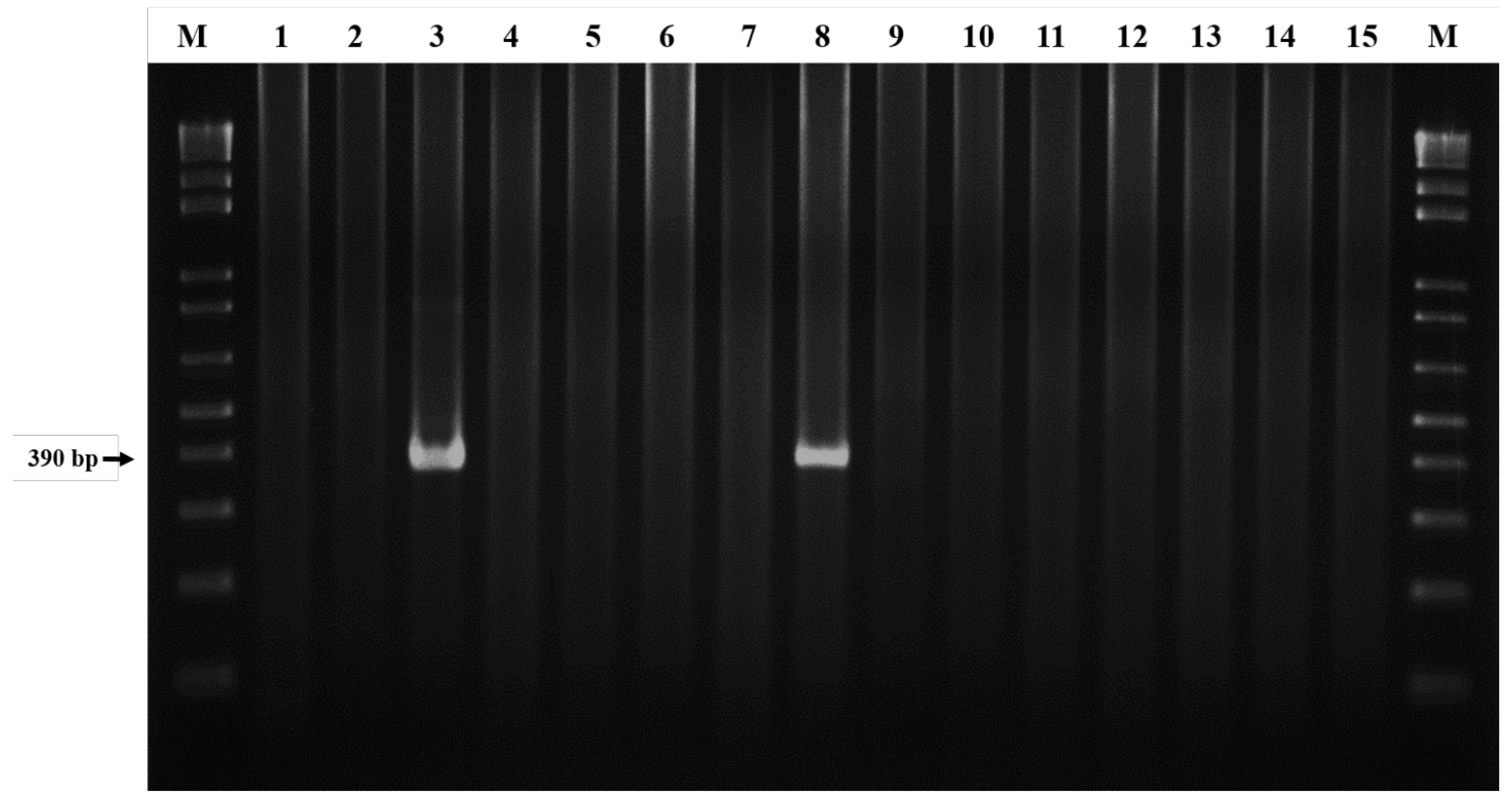
2.3. DNA Amplification by Nested Polymerase Chain Reaction

Extracted DNA samples from each tick specimen were used as a template for PCR amplification. Two primer sets targeting the 16S rRNA gene were used for amplification. Initially, the primer set ECC (5′-AGAACGAACGCTGGCGGCAAGC-3′) and ECB (5′-CGTATTACCGCGGCTGCTGGCA-3′) was used to amplify all Ehrlichia spp., which produced a PCR product approximately 450 bp [18]). Then, nested PCR was performed using the species-specific primer set ECAN5 (5′-CAATTATTTATAGCCTCTGGCTATAGGA-3′) and HE3-R (5′-TATAGGTACCGTCATTATCTTCCCTAT-3′) for E. canis-specific amplification, which produced an amplicon of approximately 390 bp [18,19]. All PCR reagents and Taq polymerase were obtained and used as recommended by the supplier (Takara Shuzo Co., Ltd., Kyoto, Japan). Briefly, each 25 μL reaction mixture contained 1.5 μL of forward and reverse primers, 2.5 μL of 10× PCR buffer (Mg2+), 2 μL of dNTP mixture (10 mM each), 1 unit of Taq DNA polymerase, 3 μL of DNA template and was filled up with an adequate volume of ddH2O. In contrast, an adequate amount of sterile distilled water was added to serve as a negative control. PCR amplification was performed with a thermocycler (Veriti, Applied Biosystems, Taipei, Taiwan) and was denatured at 94 °C for 3 min, amplified for 35 cycles with the conditions of denaturation at 94 °C for 1 min, annealing at 55 °C for 2 min, and extension at 72 °C for 2 min. For the nested PCR, the following conditions were used: denaturation at 94 °C for 1 min and then amplified for 35 cycles with the conditions of denaturation at 94 °C for 1 min, annealing at 55 °C for 2 min and extension at 72 °C for 90 s, followed by a final extension step at 72 °C for 5 min.
All amplified PCR products were electrophoresed on 1.5% agarose gels in Tris-Borate-EDTA (TBE) buffer and visualized under ultraviolet (UV) light after staining with ethidium bromide. A 100-bp DNA ladder (GeneRuler, Thermo Scientific, Taipei City, Taiwan) was used as the standard marker for comparison. A negative control of distilled water was included in parallel with each PCR amplification.

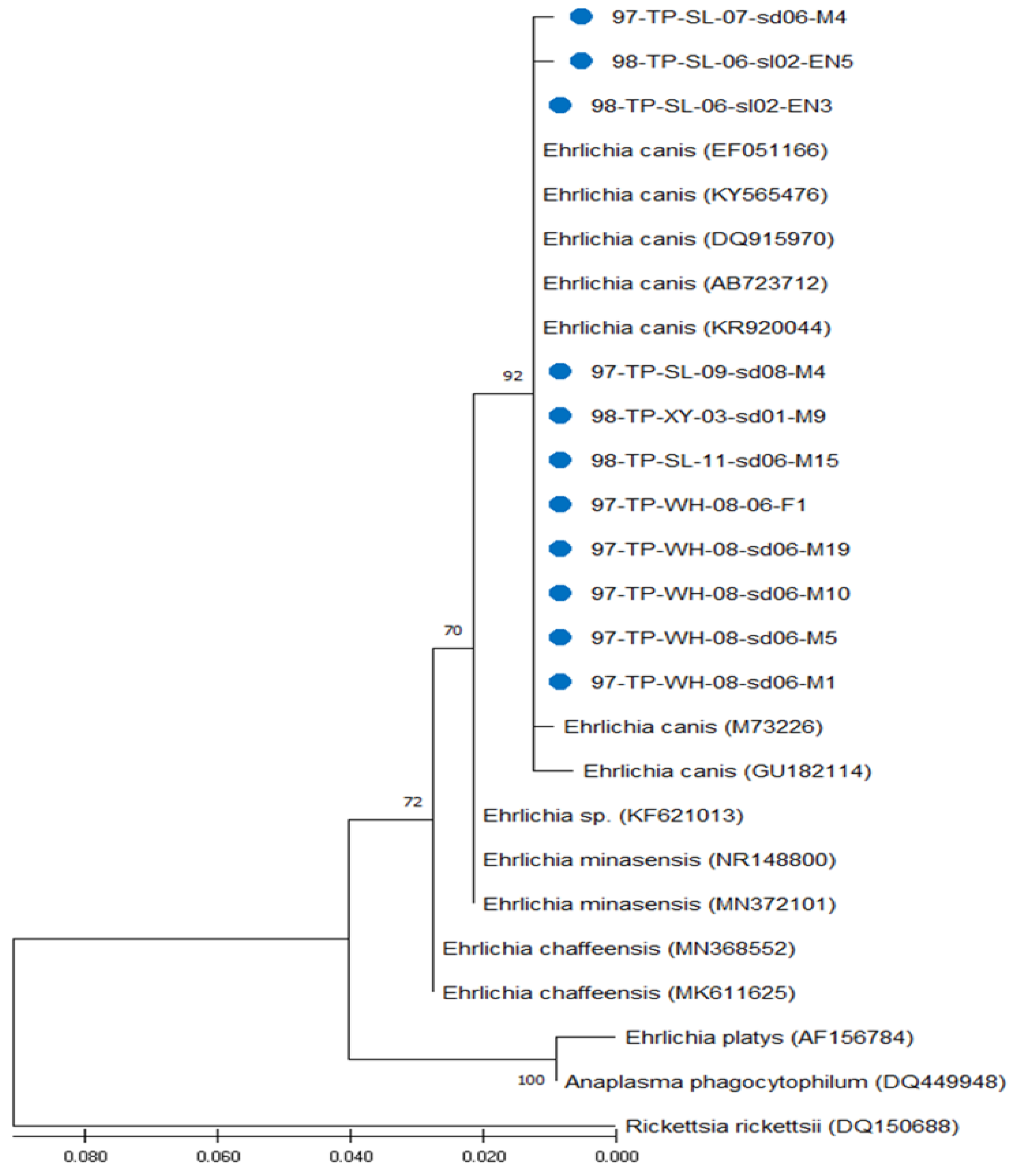
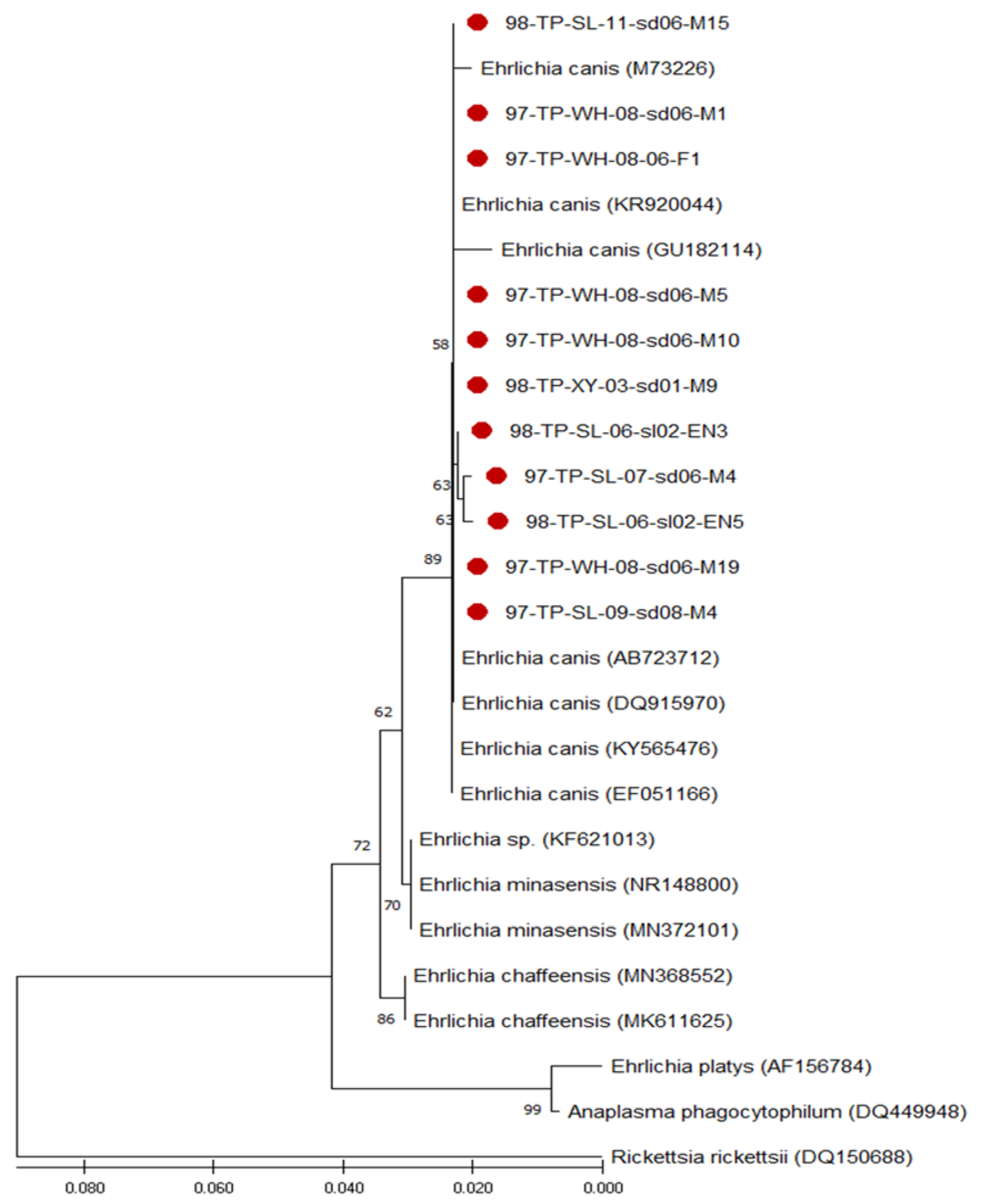
2.4. Gene Sequencing and Phylogenetic Analysis

In general, 10 μL of each selected sample with a clear band on the agarose gel was submitted for sequencing (Mission Biotech Co., Ltd., Taipei City, Taiwan). The sequencing reaction was performed with 25 cycles under the same conditions and the same primer set of nested amplification by the dye-deoxy terminator reaction method using the Big Dye Terminator Cycle Sequencing Kit in an ABI Prism 377-96 DNA Sequencer (Applied Biosystems, Foster City, CA, USA). The resulting sequences were initially edited by BioEdit software (V5.3) and aligned with the CLUSTAL W software (Version 2.0) [27]. Thereafter, the aligned sequences of Ehrlichia gene from 11 Taiwanese strains were analyzed by comparing them with other Ehrlichia and outgroup strains (Anaplasma phagocytophilum and Rickettsia rickettsii) identified from various geographical and biological origins that are documented in GenBank (Table 1). Phylogenetic analysis based on the 16S rRNA genes was performed to indicate the genetic relationships among 24 strains of Ehrlichia and 2 outgroup strains (A. phagocytophilum and R. rickettsii) analyzed in this study. Phylogenetic trees constructed by maximum likelihood (ML) and neighbour-joining (NJ) methods were performed to estimate the phylogeny of the entire alignment using the MEGA X software package [28]. The genetic distance values of intra- and interspecies variations were also analyzed by the Kimura two-parameter model [29]. All phylogenetic trees were constructed and performed with 1000 bootstrap replications to evaluate the reliability of the construction, as described previously [30].

2.5. Nucleotide Sequence Accession Numbers

In this study, the nucleotide sequences of the PCR-amplified 16S rRNA gene of 11 E. canis from R. sanguineus ticks of Taiwan were registered with GenBank and assigned the following accession numbers: 97-TP-SL-07-sd06-M4 (OP389160), 97-TP-SL-09-sd08-M4 (OP389165), 98-TP-SL-06-sl02-EN3 (OP389212), 98-TP-SL-06-sl02-EN5 (OP389213), 97-TP-WH-08-sd06-M10 (OP392572), 97-TP-WH-08-sd06-M19 (OP392573), 97-TP-WH-08-sd06-M1 (OP392574), 97-TP-WH-08-06-F1 (OP392575), 97-TP-WH-08-sd06-M5 (OP392578), 98-TP-XY-03-sd01-M9 (OP392580) and 98-TP-SL-11-sd06-M15 (OP392581). For phylogenetic analysis, the nucleotide sequences of 16S rRNA genes from other 13 Ehrlichia strains and 2 outgroup strains were included for comparison, and their GenBank accession numbers are shown in Table 1.

3. Results

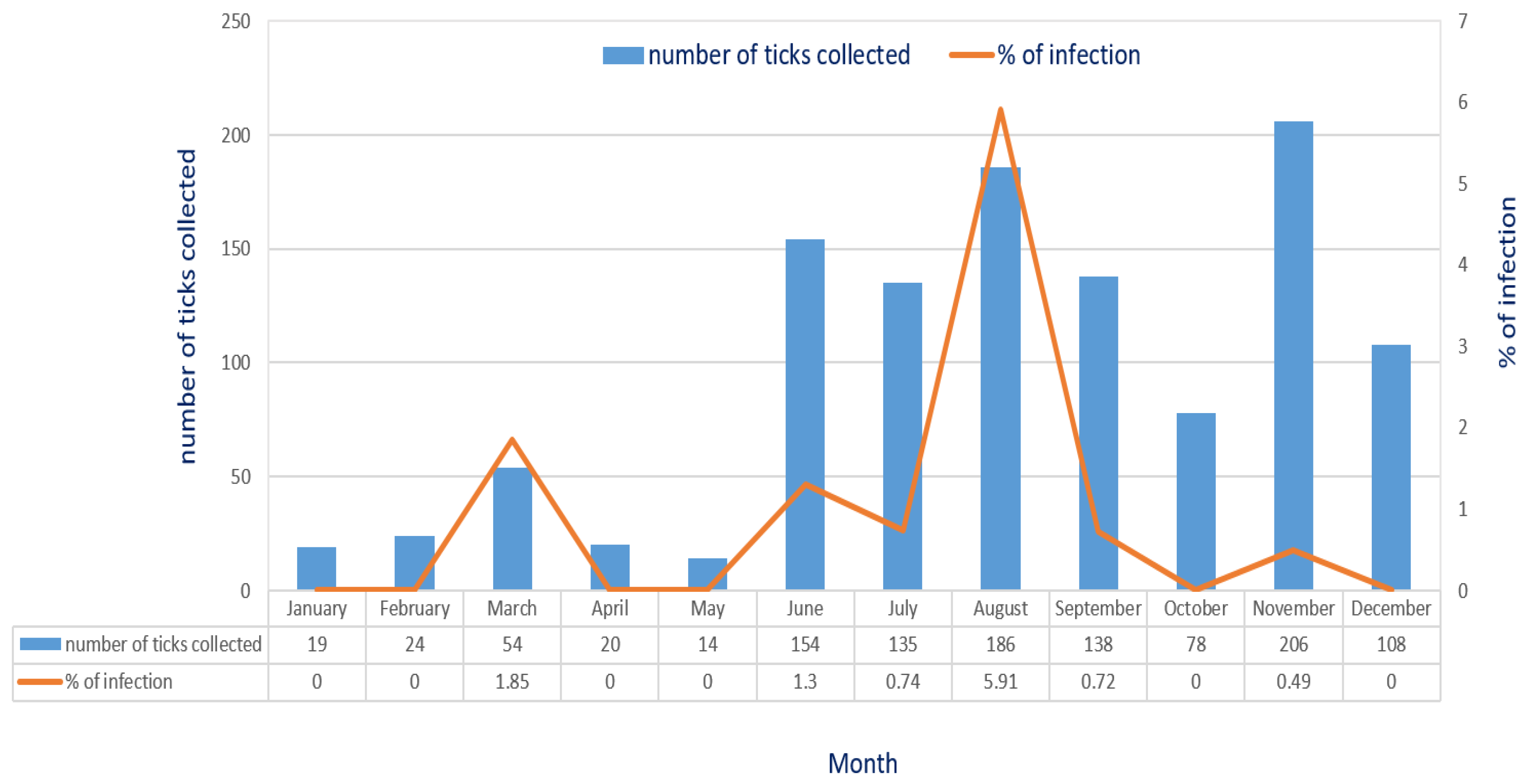
3.1. Detection of E. canis Infection in R. sanguineus Ticks of Taiwan

Molecular detection of E. canis in R. sanguineus ticks was conducted using a nested PCR assay targeting the 16S rRNA gene (Figure 1). In general, a total of 1.42% (17/1195) of R. sanguineus ticks were detected with E. canis infection. Based on the life stage of ticks, Ehrlichia canis infection was detected in nymphs, males and females of R. sanguineus ticks with an infection rates of 0.60% (2/331), 1.31% (8/610) and 2.76%, (7/254), respectively (Table 2). The highest monthly prevalence of E. canis infection was observed in August with an infection rate of 5.91%, followed by the months of March, June, July, September and November with infection rates of 1.85%, 1.3%, 0.74%, 0.72% and 0.49%, respectively (Figure 2).

3.2. Genetic Analysis of E. canis Detected in R. sanguineus Ticks

To clarify the genetic identity of Ehrlichia in R. sanguineus ticks of Taiwan, the sequences of 16S rRNA gene fragments from eleven Taiwan Ehrlichia strains analyzed in this study were compared with the downloaded sequences of thirteen other Ehrlichia strains and two outgroup strains from different geographical and biological origins documented in GenBank. Results reveal that all these Ehrlichia strains detected in R. sanguineus ticks of Taiwan were genetically affiliated with the genospecies E. canis with a high sequence similarity of 99.7–100% and that can be clearly discriminated from other genospecies of Ehrlichia and outgroup strains (Table 3). In addition, intra- and interspecies analysis based on the genetic distance (GD) values of the 16S rRNA gene indicated a lower level of genetic variation (GD < 0.003 for E. canis) within the E. canis strains and a high level of genetic variation from other Ehrlichia spp. (GD > 0.009) and outgroup species (GD > 0.054) (Table 3).

3.3. Phylogenetic Analysis of Ehrlichia Strains Detected in R. sanguineus Ticks of Taiwan

Bootstrap analysis was used to analyze the repeatability of the clustering of specimens represented in phylogenetic trees. Results indicated congruent basal topologies with three major clades of Ehrlichia that can be easily distinguished by ML analysis (Figure 3) and were congruent by NJ analysis (Figure 4). In general, all these Ehrlichia strains from Taiwan constitute a phylogenetic clade closely affiliated with the genospecies E. canis and can be discriminated from other Ehrlichia and outgroup species.

4. Discussion

This study provides the first molecular screening and genetic identification of E. canis in R. sanguineus ticks of Taiwan. In previous studies, E. canis was first identified as the causative agent of canine ehrlichiosis [3], and the first suspected human case was identified in asymptomatic patients from Venezuela [31]. Afterwards, E. canis was detected in the blood of patients with clinical signs suggestive of human ehrlichiosis [32]. Moreover, the comparison of ehrlichia 16S rRNA gene sequences revealed that the sequence profiles from the patients were identical to those from naturally infected dogs and R. sanguineus ticks [33]. In this study, Ehrlichia species detected in R. sanguineus ticks from Taiwan are genetically affiliated with the genospecies of E. canis with high sequence similarity (99.7–100%) to various E. canis strains identified from different biological and geographical origins (Table 3). Thus, our study provides the first molecular evidence and confirms sequences based on 16S rRNA genes of E. canis detected in R. sanguineus ticks of Taiwan.
The prevalence of E. canis infection in R. sanguineus ticks infesting dogs of Taiwan needs to be further investigated. In this study, the highest monthly prevalence of E. canis infection was observed in August (5.91%), and a higher prevalence was detected in early spring (March) and the summer season (June to August) (Figure 2). These observations are consistent with the zootiological survey of the seasonal abundance of tick populations infesting dogs described in our previous study [34]. Indeed, the warm climate from early Spring (March) to the hot summer season may enhance the searching activity of R. sanguineus ticks for feeding on dogs in the natural environment [35]. Indeed, the previous study also described that the higher prevalence of A. platys infection in dogs was observed in a heavily tick-infested kennel [36]. Thus, the seasonal abundance of R. sanguineus ticks is highly associated with the prevalence of E. canis infection in vector ticks and the geographical survey for the seasonal prevalence of E. canis infection in R. sanguineus ticks is essential for estimating the potential risk for human infection in Taiwan. Because of the close contact of dogs with humans, these observations demonstrate the importance of dogs serving as carrier hosts and highlight the potential risk for E. canis transmission to human populations.
Phylogenetic relationships among Ehrlichia spp. detected in R. sanguineus ticks can be determined by analyzing the sequence homogeneity of the 16S rRNA genes of Ehrlichia strains. Indeed, sequence analysis based on the 16S rRNA genes of Ehrlichia strains identified from different biological and geographical origins has been shown to be useful for evaluating the genetic relatedness of Ehrlichia strains among various hosts and tick species [18,19,20,21,22,23,24,25,26,32,33]. In this study, the phylogenetic analysis based on the 16S rRNA gene sequences of Ehrlichia strains detected in R. sanguineus ticks of Taiwan reveals identical similarity (100%) within these Taiwan strains and high genetic homogenity (99.7–100% similarity) affiliated with the genospecies of E. canis (Figure 3 and Figure 4). Indeed, phylogenetic analysis of this study also demonstrates that the genetic relatedness of E. canis detected in R. sanguineus ticks of Taiwan is mainly affiliated with the E. canis strains identified from dog blood in Malaysia, India, Taiwan and Portugal, respectively (GenBank accession no. KR920044, GU182114, KY565476 and EF051166) (Table 1, Figure 3 and Figure 4). In addition, genetic analysis based on 16S rRNA gene also revealed the discrimination of E. canis from the clades composed of other Ehrlichia and outergroup pathogens (A. phagocytophilum and R. rickettsii). The phylogenetic trees constructed by either ML or NJ analysis strongly support its discrimination (Figure 3 and Figure 4). Thus, the genetic identities of Ehrlichia strains detected in R. sanguineus ticks of Taiwan were verified as a monophyletic group affiliated with the genospecies of E. canis.
Although the biological mechanism for the transmission of tick-borne E. canis in R. sanguineus ticks remains controversial, R. sanguineus has been recognized as a main vector tick for the transmission of E. canis [37,38,39]. In previous studies, only ticks exposed to E. canis during the immature stages have been reported to transstadially transmit E. canis between dogs [40,41,42], and intrastadial transmission of E. canis by male R. sanguineus ticks has been experimentally proved in the absence of female ticks [43]. The co-feeding mechanism may account for another possible mode of transmission by ticks feeding closely to another infected tick on the same host, which may enhance pathogen transmission from an infected tick to a new tick [44,45]. In addition, co-infection with other tick-borne pathogens (such as Anaplasma spp., Ehrlichia spp., Babesia spp. and Rickettsia spp.) in R. sanguineus ticks has been described [20,46], and human infections will occur through the infective bite of R. sanguineus ticks.
The warming climate may be linked to the expansion of the geographical distribution of vector ticks, which will facilitate the transmission of tick-borne pathogens. Indeed, a previous study discovered that the I. ricinus tick is reported to have spread into previously unidentified northern areas of Sweden, Finland and Norway [47,48], and the warmer weather may enhance the attack/feeding activity of R. sanguineus ticks to dogs/humans [35]. Thus, it is possible that E. canis within R. sanguineus ticks can be transmitted to humans. Thus, further investigations focused on the prevalence of E. canis infection in relation to the geographical distribution of R. sanguineus ticks in Taiwan may help to illustrate the risk for transmitting of E. canis infection in the human population of Taiwan. In addition, epidemiological surveys regarding tick-borne zoonotic diseases in Taiwan needs to be further explored.

5. Conclusions

This study provides the first molecular evidence of E. canis in R. sanguineus ticks infesting dogs in Taiwan. The genetic relatedness based on phylogenetic analyses of 16S rRNA genes reveals genetic affiliation with the genospecies of E. canis. Because dogs serving as companion animals to humans, the discovery of E. canis in R. sanguineus ticks may draw attention to the potential transmission of tick-borne E. canis to humans in Taiwan.

Author Contributions

Conceptualization, C.-M.S. and L.-L.C.; formal analysis, C.-M.S. and L.-L.C.; investigation, P.-Y.K.; resources, C.-M.S.; methodology, L.-L.C. and P.-Y.K.; validation, C.-M.S. and L.-L.C.; visualization, P.-Y.K. and L.-L.C.; funding acquisition, C.-M.S.; writing—original draft, C.-M.S. and L.-L.C.; writing—review and editing, C.-M.S. All authors have read and agreed to the published version of the manuscript.

Funding

This work was supported in part by grants from the National Science and Technology Council (NSTC113-2320-B-037-010; NSTC114-2923-B-037-001), Taipei, Taiwan, R.O.C.

Institutional Review Board Statement

The collection of ticks from dogs was assisted by veterinary practitioners and approved by the Institutional Animal Care and Use Committee (IACUC) of the National Defense Medical Center (IACUC-11-169) and Kaohsiung Medical University (IACUC Approval No: 106142, 25 December 2017).

Informed Consent Statement

Not applicable.

Data Availability Statement

All data generated and analyzed in this study are included in this manuscript, and all submitted GenBank sequences will be available from GenBank after publication.

Acknowledgments

We would like to appreciate the sincere help for the collection of ticks from the veterinary practitioners and pet clinics of Taipei City, Taiwan.

Conflicts of Interest

The authors declare no conflicts of interest.

References

  1. Rikihisa, Y. The tribe Ehrlichieae and ehrlichial diseases. Clin. Microbiol. Rev. 1991, 4, 286–308. [Google Scholar] [CrossRef]
  2. Rar, V.; Golovljova, I. Anaplasma, Ehrlichia, and “Candidatus Neoehrlichia” bacteria: Pathogenicity, biodiversity, and molecular genetic characteristics, a review. Infec. Genet. Evolu. 2011, 11, 1842–1861. [Google Scholar] [CrossRef] [PubMed]
  3. Donatien, A.; Lestoquard, F. Existence en Algerie d’une Rickettsia du chien. Bull. Soc. Pathol. Exot. 1935, 28, 418–419. [Google Scholar]
  4. Skotarczak, B. Canine ehrlichiosis. Ann. Agric. Environ. Med. 2003, 10, 137–141. [Google Scholar] [PubMed]
  5. Cardoso, L.; Tuna, J.; Vieira, L.; Yisaschar-Mekuzas, Y.; Baneth, G. Molecular detection of Anaplasma platys and Ehrlichia canis in dogs from the north of Portugal. Vet. J. 2010, 183, 232–233. [Google Scholar] [CrossRef]
  6. Yuasa, Y.; Tsai, Y.L.; Chang, C.C.; Hsu, T.H.; Chou, C.C. The prevalence of Anaplasma platys and a potential novel Anaplasma species exceed that of Ehrlichia canis in asymptomatic dogs and Rhipicephalus sanguineus in Taiwan. J. Vet. Med. Sci. 2017, 79, 1494–1502. [Google Scholar] [CrossRef]
  7. Dantas-Torres, F. Biology and ecology of the brown dog tick, Rhipicephalus sanguineus. Parasit. Vectors 2010, 3, 26. [Google Scholar] [CrossRef]
  8. Walker, J.B.; Keirans, J.E.; Horak, I.G. The Genus Rhipicephalus (Acari, Ixodidae): A Guide to the Brown Ticks of the World; Cambridge University Press: Cambridge, UK, 2000. [Google Scholar]
  9. Shaw, S.E.; Day, M.J.; Birtles, R.J.; Breitschwerdt, E.B. Tick-borne infectious diseases of dogs. Trends Parasitol. 2001, 17, 74–80. [Google Scholar] [CrossRef]
  10. Otranto, D.; Dantas-Torres, F.; Breitschwerdt, E.B. Managing canine vector-borne diseases of zoonotic concern: Part one. Trends Parasitol. 2009, 25, 157–163. [Google Scholar] [CrossRef]
  11. Eremeeva, M.E.; Zambrano, M.L.; Anaya, L.; Beati, L.; Karpathy, S.E.; Santos-Silva, M.M.; Salceda, B.; MacBeth, D.; Olguin, H.; Dasch, G.A.; et al. Rickettsia rickettsii in Rhipicephalus ticks, Mexicali, Mexico. J. Med. Entomol. 2011, 48, 418–421. [Google Scholar] [CrossRef]
  12. Chao, L.L.; Shih, C.M. Molecular analysis of Rhipicephalus sanguineus (Acari: Ixodidae), an incriminated vector tick for Babesia vogeli in Taiwan. Exp. Appl. Acarol. 2016, 70, 469–481. [Google Scholar] [CrossRef] [PubMed]
  13. Chao, L.L.; Liao, H.T.; Ho, T.Y.; Shih, C.M. First detection and molecular identification of Babesia gibsoni from Rhipicephalus sanguineus ticks. Acta Trop. 2017, 166, 356–362. [Google Scholar] [CrossRef] [PubMed]
  14. Chao, L.L.; Liao, H.T.; Shih, C.M. First detection and genetic identification of Hepatozoon canis in Rhipicephalus sanguineus sensu lato ticks collected from dogs of Taiwan. Ticks Tick-Borne Dis. 2019, 10, 929–934. [Google Scholar] [CrossRef]
  15. Chao, L.L.; Ko, P.Y.; Shih, C.M. Molecular screening and genetic identification of Anaplasma platys in brown dog tick (Rhipicephalus sanguineus s. l.) infested on stray dogs in Taiwan. Microorganisms 2024, 12, 1779. [Google Scholar] [CrossRef]
  16. Shih, C.M.; Chao, L.L. Molecular Detection and Genetic Identification of Rickettsia Infection in Rhipicephalus sanguineus (Acari: Ixodidae) ticks collected from Southern Taiwan. Exp. Appl. Acarol. 2021, 85, 291–304. [Google Scholar] [CrossRef]
  17. Chao, L.L.; Robinson, M.; Liang, Y.F.; Shih, C.M. First detection and molecular identification of Rickettsia massiliae, a human pathogen, in Rhipicephalus sanguineus ticks collected from Southern Taiwan. PLoS Negl. Trop. Dis. 2022, 16, e0010917. [Google Scholar] [CrossRef] [PubMed]
  18. Anderson, B.E.; Summer, J.W.; Dawson, J.E.; Tzianabos, T.; Greene, C.R.; Olson, J.G.; Fishbein, D.B.; Olsen-Rasmuseen, M.; Holloway, B.P.; George, E.H.; et al. Detection of the etiological agent of human ehrlichiosis by polymerase chain reaction. J. Clin. Microbiol. 1992, 30, 775–780. [Google Scholar] [CrossRef]
  19. Dawson, J.E.; Biggie, K.L.; Warner, C.K.; Cookson, K.; Jenkins, S.; Levine, J.F.; Olson, J.G. Polymerase chain reaction evidence of Ehrlichia chaffeensis, an etiologic agent of human disease, in dogs from southeast Virginia. Am. J. Vet. Res. 1996, 57, 1175–1179. [Google Scholar] [CrossRef]
  20. Murphy, G.L.; Ewing, S.A.; Whitworth, L.C.; Fox, J.C.; Kocan, A.A. A molecular and serologic survey of Ehrlichia canis, E. chaffeensis and E. ewingii in dogs and ticks from Oklahoma. Vet. Parasitol. 1998, 79, 325–339. [Google Scholar] [CrossRef]
  21. Kim, C.M.; Yi, Y.H.; Yu, D.H.; Lee, M.R.; Cho, M.R.; Desai, A.R.; Shringi, S.; Klein, T.A.; Kim, H.C.; Song, J.W.; et al. Tick-borne rickettsial pathogens in ticks and small mammals in Korea. App. Envirn. Microbiol. 2006, 72, 5766–5776. [Google Scholar] [CrossRef]
  22. Maekawa, N.; Konnai, S.; Balbin, M.M.; Mingala, C.N.; Gicana, K.R.B.; Bernando, F.A.E.M.; Murata, S.; Obashi, K. Molecular detection and phylogenetic analysis of Ehrlichia canis in a Philippine dog. Ticks Tick-Borne Dis. 2018, 9, 266–269. [Google Scholar] [CrossRef] [PubMed]
  23. Munoz-Leal, S.; Clemes, Y.S.; Lopes, M.G.; Acosta, I.C.L.; Serpa, M.C.A.; Mayorga, L.F.S.P.; Gennari, S.M.; Gonzalez-Acuna, D.; Labruna, M.B. Novel Ehrlichia sp. detected in magellanic prnguins (Sphenicus magellanicus) and in the seabird tick Ixodes uriae from Magdalena Island, southern Chile. Ticks Tick-Borne Dis. 2019, 10, 101256. [Google Scholar] [CrossRef] [PubMed]
  24. Alhassan, A.; Hove, P.; Sharma, B.; Mattew-Belmar, V.; Karasek, I.; Lanza-Perea, M.; Werners, A.H.; Wilkerson, M.J.; Ganta, R.R. Molecular detection and characterization of Anaolasma platys and Ehrlichia canis in dogs from Caribbean. Ticks Tick-Borne Dis. 2021, 12, 101727. [Google Scholar] [CrossRef]
  25. Muraro, L.S.; Nogueira, M.F.; Borges, A.M.C.M.; de Oliveira Souza, A.; Vieira, T.S.W.J.; de Aguiar, D.M. Detection of Ehrlichia sp. in Amblyomma sculptum parasitizing horses from Brazilian Pantanal wetland. Ticks Tick-Borne Dis. 2021, 12, 101658. [Google Scholar] [CrossRef]
  26. Su, H.; Onoda, E.; Tai, H.; Fugita, H.; Sakabe, S.; Azuma, K.; Akachi, S.; Oishi, S.; Abe, F.; Ando, S.; et al. Diversity unearthed by the estimated molecular phylogeny and ecologically quantitative characteristics of uncultured Ehrlichia bacteria in Haemaphysalis ticks, Japan. Sci. Rep. 2022, 11, 687. [Google Scholar] [CrossRef]
  27. Larkin, M.A.; Blackshields, G.; Brown, N.P.; Chenna, R.; McGettigan, P.A.; McWilliam, H.; Valentin, F.; Wallace, I.M.; Wilm, A.; Lopez, R.; et al. Clustal W and Clustal X version 2.0. Bioinformatics 2007, 23, 2947–2948. [Google Scholar] [CrossRef]
  28. Kumar, S.; Stecher, G.; Li, M.; Knyaz, C.; Tamura, K. MEGA X: Molecular evolutionary genetics analysis across computing platforms. Mol. Bio. Evol. 2018, 35, 1547–1549. [Google Scholar] [CrossRef]
  29. Kimura, M. A simple method for estimating evolutionary rate of base substitutions through comparative studies of nucleotide sequences. J. Mol. Evol. 1980, 16, 111–120. [Google Scholar] [CrossRef]
  30. Felsenstein, J. Confidence limits on phylogenies: An approach using the bootstrap. Evolution 1985, 52, 1119–1134. [Google Scholar]
  31. Peres, M.; Rikihisa, Y.; Wen, B. Ehrlichia canis-like agent isolated from a man in Venezuela: Antigenic and genetic characterization. J. Clin. Microbiol. 1996, 34, 2133–2139. [Google Scholar] [CrossRef]
  32. Peres, M.; Bodor, M.; Zhang, C.; Xiong, Q.; Rikihisa, Y. Human infection with Ehrlichia canis accompanied by clinical signs in Venezuela. Ann. N. Y. Acad. Sci. 2006, 1078, 110–117. [Google Scholar] [CrossRef] [PubMed]
  33. Unver, A.; Peres, M.; Orellana, N.; Huang, H.; Rikihisa, Y. Molecular and antigenic comparison of Ehrlichia canis isolated from dogs, ticks, and a human in Venezuela. J. Clin. Microbiol. 2001, 39, 2788–2793. [Google Scholar] [CrossRef] [PubMed]
  34. Chao, L.L.; Hsieh, C.K.; Ho, T.Y.; Shih, C.M. First zootiological survey of hard ticks (Acari: Ixodidae) infesting dogs in northern Taiwan. Exp. Appl. Acarol. 2019, 77, 105–115. [Google Scholar] [CrossRef]
  35. Parola, P.; Socolovschi, C.; Jeanjean, L.; Bitam, I.; Fournier, P.E.; Sotto, A.; Labauge, P.; Raoult, D. Warmer weather linked to tick attack and emergence of severe rickettsioses. PLoS Negl. Trop. Dis. 2008, 2, e338. [Google Scholar] [CrossRef]
  36. Chang, A.C.H.; Chang, W.L.; Lin, C.T.; Pan, M.J.; Lee, S.C. Canine infectious cyclic thrombocytopenia found in Taiwan. J. Vet. Med. Sci. 1996, 58, 473–476. [Google Scholar] [CrossRef]
  37. Ewing, S.A. Canine ehrliciosis. Adv. Vet. Sci. Comp. Med. 1969, 13, 331–353. [Google Scholar]
  38. Groves, M.G.; Dennis, G.L.; Amys, H.L.; Huxsoll, D.L. Transmission of Ehrlichia canis to dogs by ticks (Rhipicephalus sanguineus). Am. J. Vet. Res. 1975, 36, 937–940. [Google Scholar] [CrossRef]
  39. Harvey, J.W.; Simpson, C.F.; Gaskin, J.M.; Sameck, J.H. Ehrlichiosis in wolves, dogs, and wolf-dog crosses. J. Am. Vet. Med. Assoc. 1979, 175, 901–905. [Google Scholar] [CrossRef]
  40. Lewis, G.E., Jr.; Ristic, M.; Smith, R.D.; Lincoln, T.; Stephenson, E.H. The brown dog tick Rhipicephalus sanguineus and the dog as experimental hosts of Ehrlichia canis. Am. J. Vet. Res. 1977, 38, 1953–1955. [Google Scholar] [CrossRef]
  41. Mathew, J.S.; Ewing, S.A.; Barker, R.W.; Fox, J.C.; Dawson, J.E.; Warner, C.K.; Murphy, G.L.; Kocan, K.M. Attempted transmission of Ehrlichia canis by Rhipicephalus sanguineus after passage in cell culture. Am. J. Vet. Res. 1996, 57, 1594–1598. [Google Scholar] [CrossRef]
  42. Johnson, E.M.; Ewing, S.A.; Barker, R.W.; Fox, J.C.; Crow, D.W.; Kocan, K.M. Experimental transmission of Ehrlichia canis (Rickettsiales: Ehrlichieae) by Dermacentor variabilis (Acari: Ixodidae). Vet. Parasitol. 1998, 74, 277–288. [Google Scholar] [CrossRef]
  43. Bremer, W.G.; Schaefer, J.J.; Wagner, E.R.; Ewing, S.A.; Rikihisa, Y.; Needham, G.R.; Jittapalapong, S.; Moore, D.L.; Stich, R.W. Transstadial and intrastadial experimental transmission of Ehrlichia canis by male Rhipicephalus sanguineus. Vet. Parasitol. 2005, 131, 95–105. [Google Scholar] [CrossRef] [PubMed]
  44. Snellgrove, A.N.; Krapiunaya, I.; Ford, S.L.; Stanley, H.M.; Wickson, A.G.; Hartzer, K.L.; Levin, M.L. Vector competence of Rhipicephalus sanguineus sensu lato for Anaplasma platys. Ticks Tick-Borne Dis. 2020, 11, 101517. [Google Scholar] [CrossRef] [PubMed]
  45. Randolph, S.E.; Gern, L.; Nuttall, P.A. Co-feeding ticks epidemiological significance for tick-borne pathogen transmission. Parasitol. Today 1996, 12, 472–479. [Google Scholar] [CrossRef] [PubMed]
  46. Laatamna, A.; Strube, C.; Bakkes, D.K.; Schaper, S.; Aziza, F.Z.; Chelef, H.B.; Amrane, N.E.H.; Bedraoui, R.; Dobler, G.; Chitimia Dobler, L. Molecular detection of tick-borne pathogens in Rhipicephalus sanguineus sensu stricto collected from dogs in the steppe and high plateau regions of Algeria. Acta Trop. 2022, 234, 106582. [Google Scholar] [CrossRef]
  47. Zemtova, G.E.; Apanaskevich, D.A.; Reeves, W.K.; Hahn, M.; Snellgrove, A.; Levin, M.L. Phylogeographical of Rhipicephalus sanguineus sensu lato and its relationships with climatic factors. Exp. Appl. Acarol. 2016, 69, 191–203. [Google Scholar] [CrossRef]
  48. EI-Sayed, A.; Kamel, M. Climate changes and their role in emergence and re-emergence of diseases. Environ Sci. Pollut. Res. 2020, 27, 22336–22352. [Google Scholar] [CrossRef]
Figure 1. Molecular detection of E. canis infection in Rhipicephalus sanguineus ticks infesting dogs of Taiwan by a nested PCR assay targeting the 16S rRNA gene. M, 100 bp DNA marker; 1–14, sample numbers; 15, negative control. The expected PCR product is 390 bp for the 16S rRNA gene.
Microorganisms 13 01372 g001
Figure 2. Monthly prevalence of E. canis infection and the number of R. sanguineus ticks collected from dogs in northern Taiwan.
Microorganisms 13 01372 g002
Figure 3. Phylogenetic tree based on the 16S rRNA gene. The aligned sequences of eleven Taiwan strains (indicated as ) detected in R. sanguineus ticks of Taiwan were compared with available sequences from GenBank, including thirteen strains of Ehrlichia spp. and two outgroup strains identified from different biological and geographical origins. The constructed tree was analyzed by the Maximum Likelihood (ML) method using 1000 bootstrap replicates. Branch length is drawn proportional to the estimated sequence divergence. Numbers at the nodes indicate the percentage reliability of the tree.
Microorganisms 13 01372 g003
Figure 4. Phylogenetic tree based on the 16S rRNA gene. The aligned sequences of eleven Taiwan strains (indicated as ) detected in R. sanguineus ticks of Taiwan were compared with available sequences from GenBank, including thirteen strains of Ehrlichia spp. and two outgroup strains identified from different biological and geographical origins. The constructed tree was analyzed by the neighbor-joining (NJ) method using 1000 bootstrap replicates. Branch length is drawn proportional to the estimated sequence divergence. Numbers at the nodes indicate the percentage reliability of the tree.
Microorganisms 13 01372 g004
Table 1. Ehrlichia, Anaplasma and Rickettsia strains used for phylogenetic analysis in this study.
StrainOrigin of Bacterial Strain16S rRNA Gene
Accession Number a
BiologicalGeographic
Taiwan strain
97-TP-SL-07-sd06-M4Rhipicephalus sanguineusTaiwanOP389160
97-TP-SL-09-sd08-M4Rhipicephalus sanguineusTaiwanOP389165
98-TP-SL-06-sl02-EN3Rhipicephalus sanguineusTaiwanOP389212
98-TP-SL-06-sl02-EN5Rhipicephalus sanguineusTaiwanOP389213
97-TP-WH-08-sd06-M10Rhipicephalus sanguineusTaiwanOP392572
97-TP-WH-08-sd06-M19Rhipicephalus sanguineusTaiwanOP392573
97-TP-WH-08-sd06-M1Rhipicephalus sanguineusTaiwanOP392574
97-TP-WH-08-06-F1Rhipicephalus sanguineusTaiwanOP392575
97-TP-WH-08-sd06-M5Rhipicephalus sanguineusTaiwanOP392578
98-TP-XY-03-sd01-M9Rhipicephalus sanguineusTaiwanOP392580
98-TP-SL-11-sd06-M15Rhipicephalus sanguineusTaiwanOP392581
Ehrlichia canisUnknownUSAM73226
Ehrlichia canisDog bloodMalaysiaKR920044
Ehrlichia canisDog bloodIndiaGU182114
Ehrlichia canisLeopard catJapanAB723712
Ehrlichia canisUnknownUSADQ915970
Ehrlichia canisDog bloodTaiwanKY565476
Ehrlichia canisDogPortugalEF051166
Ehrlichia sp.Cattle bloodBrazilKF621013
Ehrlichia minasensisRhipicephalus microplusCzech RepublicNR148800
Ehrlichia minasensisHyalomma tickEgyptMN372101
Ehrlichia chaffeensisHyalomma tickEgyptMN368552
Ehrlichia chaffeensisDeer bloodUSAMK611625
Ehrlichia platysUnknownChinaAF156784
Anaplasma phagocytophilum
Rickettsia rickettsii
Dermacentor silvarum
Dog
China
USA
DQ449948
DQ150688
a Bold GenBank accession numbers were submitted by this study.
Table 2. Molecular detection of Ehrlichia canis in various life stages of Rhipicephalus sanguineus ticks parasitizing dogs by nested PCR assay targeting the 16S ribosomal RNA gene.
Life Stage of TickE. canis Infection Detected by Nested PCR% of E. canis Infection
Number of Ticks PositiveNumber of Ticks Examined
Nymph23310.60
Male86101.31
Female72542.76
Total1711951.42
Table 3. Intra- and intergroup analysis of genetic distance values a based on the 16S rRNA gene sequences between the Ehrlichia strains of Taiwan and other Ehrlichia, Anaplasma and Rickettsia strains documented in GenBank.
Bacterial Strains b1234567891011121314151617
1. 97-TP-WH-08-sd06-M1 (Taiwan)
2. 97-TP-WH-08-sd06-M5 (Taiwan)0.000
3. 97-TP-WH-08-sd06-M10 (Taiwan)0.0000.000
4. 97-TP-WH-08-sd06-M19 (Taiwan)0.0000.0000.000
5. 97-TP-WH-08-06-F1 (Taiwan)0.0000.0000.0000.000
6. 98-TP-SL-11-sd06-M15 (Taiwan)0.0000.0000.0000.0000.000
7. 98-TP-XY-03-sd01-M9 (Taiwan)0.0000.0000.0000.0000.0000.000
8. 97-TP-SL-09-sd08-M4 (Taiwan)0.0000.0000.0000.0000.0000.0000.000
9. 98-TP-SL-06-sl02-EN3 (Taiwan)0.0000.0000.0000.0000.0000.0000.0000.000
10. Ehrlichia canis, Malaysia (KR920044)0.0000.0000.0000.0000.0000.0000.0000.0000.000
11. Ehrlichia canis, Japan (AB723712)0.0000.0000.0000.0000.0000.0000.0000.0000.0000.000
12. Ehrlichia canis, Portugal (EF051166)0.0000.0000.0000.0000.0000.0000.0000.0000.0000.0000.000
13. Ehrlichia canis, USA (M73226)0.0030.0030.0030.0030.0030.0030.0030.0030.0030.0030.0030.003
14. Ehrlichia minasensis (MN372101)0.0090.0090.0090.0090.0090.0090.0090.0090.0090.0090.0090.0100.012
15. Ehrlichia chafeensis (MN368552)0.0150.0150.0150.0150.0150.0150.0150.0150.0150.0150.0150.0160.0180.006
16. Anaplasma phagocytophilum (DQ449948)0.0540.0540.0540.0540.0540.0540.0540.0540.0540.0540.0540.0530.0570.0500.044
17. Rickettsia rickettsii (DQ150688)0.1570.1570.1570.1570.1570.1570.1570.1570.1590.1570.1570.1530.1610.1530.1530.172
a The pairwise distance calculation was performed by the Kimura two-parameter method, as implemented in MEGA X [28]. b Strains 10–15, 16 and 17 are the Ehrlichia, Anaplasma and Rickettsia strains documented in GenBank, respectively.
Disclaimer/Publisher’s Note: The statements, opinions and data contained in all publications are solely those of the individual author(s) and contributor(s) and not of MDPI and/or the editor(s). MDPI and/or the editor(s) disclaim responsibility for any injury to people or property resulting from any ideas, methods, instructions or products referred to in the content.

