Article

Impact of Temperature on the Biochemical Potential of Five Newly Isolated Strains of Microalgae Cultured in a Stirred Tank Reactor

Department of Biology, School of Natural Sciences, University of Patras, 26500 Patras, Greece
*
Author to whom correspondence should be addressed.
Microorganisms 2025, 13(5), 1155; https://doi.org/10.3390/microorganisms13051155
Submission received: 29 April 2025 / Revised: 15 May 2025 / Accepted: 16 May 2025 / Published: 18 May 2025
(This article belongs to the Section Microbial Biotechnology)

Abstract

:
The microalgal strains Picochlorum costavermella VAS2.5, Picochlorum oklahomense SAG4.4, Picochlorum oklahomense PAT3.2B, Microchloropsis gaditana VON5.3, and Nephroselmis pyriformis PAT2.7 were cultured in a Stirred Tank Reactor at 25 °C or 20 °C in modified artificial seawater and their biotechnological potential was assessed. VAS2.5, VON5.3, and PAT2.7 were high in biomass production at both temperatures (i.e., 438.8–671.3 mg/L and 418.4–546.7 mg/L at 25 °C and 20 °C, respectively), though P. oklahomense strains grew only at 25 °C. The highest lipid percentage was recorded for the cultures of VAS2.5 (19.3 ± 0.7%) and VON5.3 (16.4 ± 1.5%) at 25 °C, notably rich in Δ5,8,11,14,17C20:5, while PAT2.7 proved a major producer of Δ9C16:1. The predominant lipid fraction was glycolipids and sphingolipids (41.3–57.4%) for VAS2.5, PAT2.7 at 25 °C and VON5.3 at 20 °C and neutral lipids (55.6–63.5%) in the other cultures, indicating the different effect of temperature on lipid synthesis of the various microalgae. Additionally, almost all strains stood out for their high protein content, exceeding 50% in the culture of PAT3.2B, but polysaccharide and pigment content were not high. The biochemical profiles of the isolates showcased their suitability for use primarily as feed additives in the aquaculture sector.

1. Introduction

Microalgae are photosynthetic microorganisms with a global distribution, found not only in all types of water systems but in terrestrial ecosystems and the atmosphere as well [1,2,3]. Besides their obvious high ecological value, microalgae, through the conversion of solar energy and CO2, biosynthesize cellular materials of high nutritional interest (e.g., lipids, proteins, carbohydrates, pigments, etc.) for aquatic and terrestrial organisms in significant quantities [4,5]. Typically, green microalgae, rather than other groups of photosynthetic microorganisms, are considered as a superior oil source compared to conventional plants for a number of reasons. The major ones are related to their higher growth rates and their ability to grow under a wider range of pH and temperature values and valorize CO2 in a more efficient way comparing to plants [6,7,8].
More importantly, some microalgal species, e.g., representatives of the genera Picochlorum and Microchloropsis, are of additional interest due to their ability to synthesize polyunsaturated fatty acids (PUFAs), such as the omega-3 fatty acids α-linolenic (Δ9,12,15C18:3, ALA), eicosapentaenoic (Δ5,8,11,14,17C20:5, EPA), and docosahexaenoic (Δ4,7,10,13,16,19C22:6, DHA). Such fatty acids are sought after by the food, feed, and pharmaceutical industries because of their proven beneficial effects against cardiovascular and neurodegenerative diseases, various forms of cancer, etc. [9,10,11,12]. On the other hand, microalgal lipids that contain more saturated fatty acids, like those that belong to the Nephroselmis genus, are used as feedstock for biodiesel production, because they are resistant to oxidation and yield more energy during combustion [13,14,15].
In general, the major advantage of microalgal oils against the fish oils, which are the main source of omega-3 fatty acids, is that they are free of undesirable odors and pollutants such as heavy metals and microplastics [16,17]. Additionally, the extensive use of microalgal oils will contribute decisively to the context of controlling and reducing intensive fishing globally. After all, microalgae farming incurs low production costs and, thus, can offer economic benefits by reducing dependency on imported fish oil in countries of need [18,19]. Yet even though >50,000 species of microalgae are described, only a few are cultivated on a large scale due to their high production costs [20,21,22].
Therefore, the need to use new strains with high efficiency rates when used in large-scale applications is of outmost importance. However, a crucial challenge that is associated with the yield of lipid production is the design of proper culture conditions. Environmental factors, such as temperature, irradiance, and nutrient availability majorly affect lipid content and fatty acid composition in microalgae cells [23,24,25]. For instance, at low temperatures, one might expect increased synthesis of unsaturated fatty acids, including PUFAs, as cells need to maintain the fluidity of their membranes, increasing their PUFA content. Evidently, in low temperatures, higher yields of these molecules of high biotechnological significance can be achieved [26,27].
The aim of the present study was to investigate the effect of temperature on the physiology and high added-value metabolite synthesis capability of five strains of microalgae, previously isolated and characterized with molecular techniques [13], that belong to genera Picochlorum, Microchloropsis, and Nephroselmis, due to the attractive features of representatives of the aforementioned genera and the relatively limited available literature for some of these strains [13,28,29,30,31,32], when cultured in a Stirred Tank Reactor (STR). One of the main reasons for using these strains to serve the cultures in the current investigation relates to the relatively limited literature available on most of these strains and the lack of data on the potential utilization of such microorganisms on a commercial scale. Even though the main focus was on lipid production, the fatty acid composition of total lipids and their lipid fractions, and the content of these strains in proteins, polysaccharides, and pigments were also analyzed. As a result of the aforementioned analyses, the biotechnological potential of the used strains when cultured in the STR was assessed.

2. Materials and Methods

2.1. Biological Material and Culture Conditions

The microalgal strains Picochlorum costavermella VAS2.5, Picochlorum oklahomense SAG4.4, Picochlorum oklahomense PAT3.2B, Microchloropsis (Nannochloropsis) gaditana VON5.3, and Nephroselmis pyriformis PAT2.7, which were previously isolated from coastal areas of the Ionian Sea of Greece and identified by the use of molecular techniques [13], were used as biological material.
The strains were maintained in 0.25 L Erlenmeyer flasks containing 0.05 L of modified Artificial Sea Water (mASW) and were regularly sub-cultured. The composition of mASW is presented in Table 1. The flasks containing mASW were sterilized at 121 °C for 20 min, inoculated with 105 cells/mL of a fresh inoculum, derived from a 7-day pre-culture grown in sterilized mASW, and incubated at T = 25 ± 1 °C. Illumination was constant with an intensity of 300 μmol/m2∙s provided by linear fluorescent day light tubes (T5, 8 W, 6500 K). Before sterilization, the pH of the growth medium was adjusted to 8.5 ± 0.5 by adding 2.5 M NaOH (Sigma-Aldrich) solution and using an Orion 420A digital pHmeter (Thermo Scientific, Osterode, Germany) equipped with a glass electrode (HANNA Instruments, Athens, Greece). The pH of mASW was also measured after sterilization.
The microalgal strains were cultured in monoalgal but non-axenic batch cultures for approximately 250 h in a closed system bioreactor, type Ralf Plus-System from Bioengineering (Wald, Switzerland), which is a Stirred Tank Reactor (STR), with a capacity of 3.7 L and a working volume (Vw) = 2 L. The STR was converted into a light bioreactor after 8 fluorescent lamps (T5, 8 W, 6500 K) were placed circumferentially to the culture vessel, providing continuous illumination of 1071 μmol/m2∙s intensity (Figure 1). Prior to culture initiation, the culture vessel was cleaned with 70% ethanol (vol/vol), approximately 1.8 L of sterile mASW was added, and inoculation was performed with approximately 0.2 L of pre-cultured microalgae so that the initial cell density was 1.5 ∙ 106 cells/mL. The cultures were maintained at T = 25 ± 1 °C or T = 20 ± 1 °C under 100 rpm agitation and natural air was supplied at a rate of 0.5 vvm, which was passed through a Whatman-type bacteriological filter with a pore size of 0.2 μm. Both the selected temperatures correspond to the sea temperatures from which the original samples were collected. The initial pH was 8.5 ± 0.3 and was maintained at this value by the addition of 2.5 M HCl (Sigma-Aldrich) automatically added to the medium. At the end of each culture, the biomass was collected for quantification and further chemical analyses. Cultures were carried out in duplicate. The monitoring of the culture conditions and the physicochemical parameters of the system was possible through a computer equipped with Wonderware InTouch 9.5.001 software (Bioengineering, Wald, Switzerland).

2.2. Cell Growth and Biomass Determination

Daily cell counting in a Neubauer improved cell counting chamber (Poly-Optik, Bad Blankenburg, Germany) was performed, similar to other studies [33,34], so as to assess microalgae growth. In brief, 1 mL of microalgae culture was subtracted from the culture vessel daily, diluted properly, and in each slot of the Neubauer chamber, 10 μL of microalgae sample was placed. Then, under an optical microscope Carl Zeiss Axiostar (GmBH, Gottingen, Germany), at total magnification of 400×, cell density was calculated following the equation provided below (Equation (1)):
Nav. per squareDF (if any) ∙ 25 ∙ 104 cells/mL
where Nav. per square is the average number of cells per square of the Neubauer chamber and DF is the dilution factor.
The cell counts were expressed as microalgal cell density (cells/mL). In addition, the integrated version of Verhulst’s model (i.e., Sigmoidal Logistic function) (Equation (2)) was applied in order to estimate the parameters of growth:
Nt = Nmax/(1 + b ∙ e1/μ∙t)
where N represents the number of cells/mL∙106 at time t, b is a positive constant equal to (Nmax − N0)/N0 (where N0 is the initial number of cells/mL∙106), μ is the specific growth rate (1/d), and Nmax is the carrying capacity of the system. The parameter values were determined by fitting Equation (1) to the experimental data, using the Levenberg–Marquardt method for parameter value optimization. The minimization of the residual root means square error between the experimental and model-predicted data, quantified by the values of the coefficient of determination R2, was used as a criterion for parameter optimization.
Biomass determination was held by gravimetric determination of dry biomass. Specifically, prior to the gravimetric determination, harvesting of the microalgal cells by centrifugation at 7455× g for 10 min at 4 °C (NÜVE NF 800R, Ankara, Turkey) occurred. Then, washing of the cells with deionized water and centrifugation under the aforementioned conditions occurred in duplicate and then the biomass was collected and dried at 80 °C until constant weight and expressed as g/L.

2.3. Lipid Extraction and Purification

Total lipids from the microalgal cells were extracted in chloroform (PENTA)/methanol (Fisher Chemical, Hampton, NY, USA) 2:1 vol/vol, in accordance with a modified version of the Folch et al. (1957) [35] method [36]. Then, the extracts were filtered through Whatman No. 1 paper, washed with a 0.88% (wt/vol) KCl (Merck) solution in order to remove non-lipid components (i.e., lipoproteins, pigments), and dried over anhydrous Na2SO4 (Sigma-Aldrich). Consequently, solvent evaporation under vacuum, using a Rotavapor R-210 evaporator (BUCHI, Flawil, Switzerland), occurred and the total cellular lipids were gravimetrically determined and expressed as the percentage of lipids in the dry biomass (L/x%, wt/wt).

2.4. Lipid Fractionation

A column (25 × 100 mm) containing 1 g silicic acid (Fluka), which was activated by heating at 80 °C overnight, was used for the fractionation of approximately 100 mg of microalgal lipids which were priorly dissolved in 1 mL chloroform. The column was washed successively with 100 mL dichloromethane (Sigma-Aldrich) to obtain neutral lipids (N), 100 mL acetone (Fluka) to obtain glycolipids plus sphingolipids (G + S), and 50 mL methanol (Sigma-Aldrich) to obtain phospholipids (P) [33]. Following the evaporation under vacuum of the respective solvent of each fraction, all fractions (N, G + S, and P) were quantified gravimetrically and expressed as a percentage of total lipids.

2.5. Fatty Acid Composition of Cellular Lipids

The identification of total lipids and their lipid fraction composition in fatty acids was made attainable after their conversion into their fatty acid methyl-esters (FAMEs) and analysis with Gas Chromatography (GC). Specifically, an Agilent 7890A GC device (Agilent Technologies, Shanghai, China) equipped with a flame ionization detector at 280 °C and an HP-88 (J&W Scientific, Folsom, CA, USA) column (60 m × 0.32 mm) was used. The carrier gas was helium, at a flow rate 1 mL/min, and the analysis was run at 200 °C. Peaks of FAMEs were identified through comparison to authentic standards.

2.6. Polysaccharide Determination

Approximately 20 mg of fat-free biomass (xf) was hydrolyzed with 5 mL HCl (Sigma-Aldrich) 2.5 M at 100 °C for 60 min. When the hydrolysate was cooled at room temperature, it was neutralized with 2.5 M KOH (Sigma-Aldrich) and filtered through Whatman No. 1 paper so as to remove cell debris. The reducing sugars, expressed as glucose, were determined in the solution in accordance with the DNS method [37]. Intracellular polysaccharides, including storage and structural polysaccharides, were expressed (as glucose equivalents) as a percentage of dry biomass (S/x%, wt/wt).

2.7. Protein Determination

Total cellular protein was determined using approximately 10 mg of xf following the biuret method, as described elsewhere [33]. Cellular protein was expressed (as albumin equivalents) as a percentage of dry biomass (P/x%, wt/wt).

2.8. Pigment Estimation

Approximately 0.5 g of wet biomass was solubilized with 10 mL of ethanol 95%, vol/vol (Fisher Chemical) solved with Milli Q water (Honeywell, Charlotte, NC, USA). The mixture was centrifuged (Hettich Mikro 200R, Föhrenstr, Germany) at 13,300× g for 15 min at 4 °C. Then, 0.5 mL of the supernatant was collected and mixed with 4.5 mL of the ethanol solution [38]. The resulting solution was spectrophotometrically analyzed for Chlorophyll-a, Chlorophyll-b, and carotenoids in a quartz cuvette (1 cm2), while ethanol solvent was used as a blank.
The Equations (3)–(5) used for the pigment quantification (in µg/mL) were the following [22]:
Ca = (13.36∙A664) − (5.19∙A649)
Cb = (27.43∙A649) − (8.12∙A664)
Cx+c = ((1000∙A470) − (2.13∙Ca) − (97.63∙Cb))/209
where A represents Absorbance and Ca, Cb, and Cx+c stand for Chlorophyll-a, Chlorophyll-b, and carotenoids, respectively. Pigments, evaluated as Total Chlorophylls (TCh) and Total Carotenoids (TC), were expressed as percentage in the dry biomass (TCh/x%, wt/wt, and TC/x%, wt/wt).

2.9. Data Treatment and Statistical Analysis

The graphing and analysis software OriginPro 2021 9.8.0.200 ®, 1991–2020 (OriginLab Corp., Northampton, MA, USA) was used for the treatment of the experimental data from microalgal cultures and growth kinetics.

3. Results

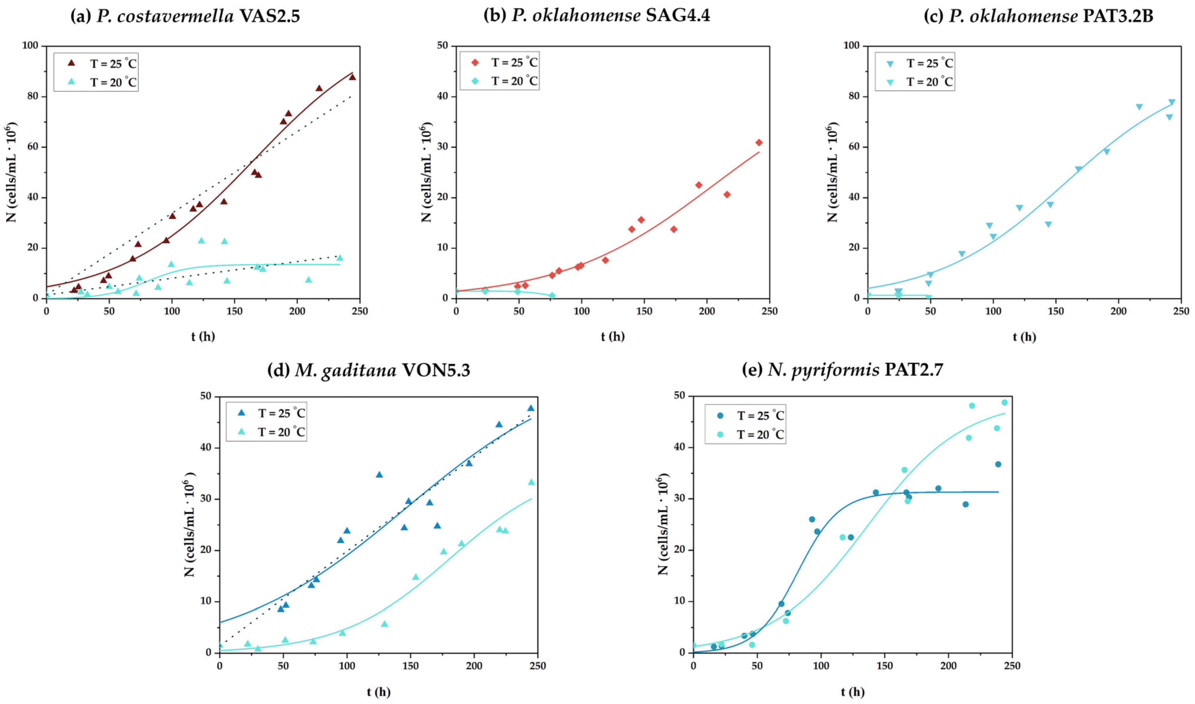
3.1. Cell Growth and Biomass Production

The growth curves of all strains under the examined growth conditions are collectively depicted in Figure 2, while the results regarding growth parameters are compiled in Table 2.
The strain P. costavermella VAS2.5 grew satisfactorily at both incubation temperatures tested, especially at T = 25 °C (Figure 2a), leading to higher biomass production as well (x = 671.3 ± 17.2 mg/L with px = 67.1 ± 1.7 mg/L∙d and μ = 0.45 ± 0.05 1/d), compared to 418.4 ± 17.7 mg/L of the other culture condition (Table 2). It is worth mentioning that due to the similarity of the conceivable curve that could express the trend of the cytometry points with a straight line when this strain was cultured at T = 25 °C, the linear model was also applied to the data, showing a very good fit (R2 = 0.98) (see respective dοtted line plot in Figure 2a). Similar observations were made regarding the culture at T = 20 °C (see respective dotted line plot in Figure 2a) as well as for the other two strains that belong to the Picochlorum genus and were used in the current study. Intriguingly, though, the other two Picochlorum isolates, namely, P. oklahomense SAG4.4 and P. oklahomense PAT3.2B, showed growth capacity only when incubated at T = 25 °C (Figure 2b,c). Under these culture conditions, both strains reached similar biomass production levels. Specifically, P. oklahomense SAG4.4 biomass production reached 224.8 ± 5.9 mg/L (px = 22.5 ± 0.7 mg/L∙d and μ = 0.39 ± 0.08 1/d) and P. oklahomense PAT3.2B produced 242.7 ± 13.6 mg/L (px = 24.3 ± 1.4 mg/ L∙d and μ = 0.47 ± 0.07 1/d) (Table 2).
Regarding the cultures of M. gaditana VON5.3, this strain grew satisfactorily at both examined temperatures, but clearly more efficiently when incubated at T = 25 °C (Figure 2d). As mentioned before for P. costavermella VAS2.5 cultured at T = 25 °C, the growth curve of the M. gaditana VON5.3 culture at the same temperature was also well-fitted to the linear model as shown by the dotted line (Figure 2d) (R2 = 0.90 and R2 = 0.88 for linear model and the integrated version of Verhulst’s model, respectively). As regards the biomass production, M. gaditana VON5.3 gained practically the same level of biomass at both incubation temperatures (Table 2). In particular, it was x = 527.4 ± 15.0 mg/L (px = 52.7 ± 1.5 mg/L∙d and μ = 0.35 ± 0.11 1/d) and x = 509.0 ± 77.8 mg/L (px = 50.9 ± 7.8 mg/L∙d and μ = 0.56 ± 0.12 1/d) for the culture at T = 25 °C and T = 20 °C, respectively.
The strain N. pyriformis PAT2.7 also grew satisfactorily at both incubation temperatures (Figure 2e). Interestingly, in this set of experiments, the biomass produced was lower when the microalga grew at T = 25 °C, i.e., x = 438.8 ± 89.3 mg/L (with px = 43.9 ± 8.9 mg/L d and μ = 1.47 ± 0.34 1/d) compared to x = 546.7 ± 1.2 mg/L (with px = 54.7 ± 0.1 mg/L∙d and μ = 0.64 ± 0.12 1/d) at T = 20 °C (Table 2).

3.2. Accumulation of Storage Materials

The results of storage material accumulation levels at the end of each culture performed are compiled in Table 2.
Regarding P. costavermella VAS2.5, it was observed that the lipid content was clearly higher at T = 25 °C (L/x% = 19.3 ± 0.7%, wt/wt), whereas at T = 20 °C, the lipid yield was relatively low (L/x% = 5.2 ± 0.3%, wt/wt). The other two Picochlorum strains, which were able to grow only at T = 25 °C, synthesized fewer lipids, i.e., L/x% = 4.1 ± 0.4%, wt/wt and L/x% = 11.5 ± 0.3%, wt/wt, for P. oklahomense SAG4.4 and P. oklahomense PAT3.2B, respectively (Table 2). Additionally, the percentages (%, wt/wt) of lipid fractions (N—neutral lipids, G + S, glycolipids, and sphingolipids, P—phospholipids) to total lipids (TLs) are also presented in Table 2. Herein, though, such investigation occurred only for P. costavermella VAS2.5 cultured at T = 25 °C, as it was not possible to investigate the contribution of individual lipid fractions to total lipids of the biomass collected from the lower temperature, due to the small amount of lipids produced. Likewise, the aforementioned results apply to the two P. oklahomense strains as well. In this respect, the predominant lipid fraction of P. costavermella VAS2.5 was that of G + S (57.4 ± 5.4%, wt/wt), followed by the N fraction (29.6 ± 3.4%, wt/wt) and P (13.1 ± 2.0%, wt/wt). M. gaditana VON5.3 produced biomass in which the reserve lipid content was slightly higher at T = 25 °C (L/x% = 16.4 ± 2.1%, wt/wt) compared to the culture at T = 20 °C (L/x% = 13.8 ± 0.0%, wt/wt). The dominant lipid fraction was that of N (55.6 ± 1.6%, wt/wt), followed by the G + S fraction (24.7 ± 1.4%, wt/wt) and P (19.8 ± 0.3%, wt/wt) when the microalgae was grown at T = 25 °C. On the contrary, the highest percentage was recorded for the G + S fraction (41, 3 ± 2.0%, wt/wt) after culture at T = 20 °C, with P also following at high percentages (36.3 ± 2.1%, wt/wt), and finally, the N fraction (22.5 ± 0.2%, wt/wt) (Table 2). Lastly, as regards the biomass content of N. pyriformis PAT2.7 to reserve lipids in the two incubation conditions, a higher percentage was observed in the culture at T = 20 °C (L/x% = 9.3 ± 6.1%, wt/wt) compared to that at T = 25 °C (L/x% = 3.3 ± 1.7%, wt/wt) (Table 2). When this strain was grown at T = 25 °C, the highest percentage belonged to the lipid fraction G + S (48.6 ± 5.0%, wt/wt) which was marginally higher than the N fraction (45.8 ± 4.3%, wt/wt), and the last one was the P fraction (6.0 ± 1.0%, wt/wt). In culture at 20 °C, there was a clear dominance of the N fraction (63.5 ± 6.0%, wt/wt), followed by G + S (34.8 ± 4.8%, wt/wt) and P (2.3 ± 1.2%, wt/wt) (Table 2).
Regarding the ability of P. costavermella VAS2.5 to store polysaccharides and proteins under the examined culture conditions, a significant difference was recorded in the accumulation of polysaccharides (S/x% = 13.5 ± 0.8 and S/x% = 6.7 ± 0.1, for cultures at T = 25 °C and T = 20 °C, respectively), but not in protein content (P/x% ranged in the 25.0–26.7%, wt/wt range) (Table 2). Similar levels of polysaccharides and protein synthesis were recorded for P. oklahomense SAG4.4 (S/x% = 11.3 ± 0.4%, wt/wt and P/x% = 28.8 ± 1.0%, wt/wt) cultured at T = 25 °C (Table 2). On the other hand, despite polysaccharide accumulation being similar to the above for P. oklahomense PAT3.2B (i.e., S/x% = 11.2 ± 1.2%, wt/wt), the exhibited protein content of the biomass was significantly higher (P/x% = 50.6 ± 3.0%, wt/wt) (Table 2). Regarding the ability of M. gaditana VON5.3 to store polysaccharides and proteins under the two culture conditions, similar percentages with little variation were also recorded (S/x% = 8.5–9.4% and P/x% = 16.4–17.5%) (Table 2). In the same manner, small variations were recorded in the case of N. pyriformis PAT2.7, though the recorded protein content was high (S/x% = 8.0–10.7% and P/x% = 41.0–45.2%) (Table 2).
Finally, the values of total chlorophyll and carotenoids in dry biomass were recorded (Table 2). The obtained results presented similar levels of chlorophyll biosynthesis (the percentage of which ranged between 2.1 and 2.6%, wt/wt) and slightly higher levels of carotenoids when P. costavermella was grown at T = 20 °C (0.8% at t = 20 °C versus 0.2%, wt/wt at T = 25 °C). On the contrary, the cultures of both P. oklahomense strains at T = 25 °C led to higher total chlorophyll (TCh/x% = 4.9 ± 0.1%, wt/wt and TCh/x% = 6.1 ± 0.7%, wt/wt, for SAG4.4 and PAT3.2B, respectively) and carotenoid (TC/x% = 0.8 ± 0.0%, wt/wt and TC/x% = 1.1 ± 0.1%, wt/wt, for SAG4.4 and PAT3.2B, respectively) content of the cells. Respective analyses on M. gaditana VON5.3 biomass showed higher levels of chlorophyll biosynthesis when the microalga was grown at T = 20 °C (TCh/x = 3.2% vs. 1.4%) and similar rates of carotenoid biosynthesis when grown at T = 25 °C (i.e., TCh/x = 0.3–0.9%, wt/wt and TC/x = 0.1–0.3%, wt/wt). Finally, total chlorophyll and carotenoid contents were at low levels in both culture conditions tested for N. pyriformis PAT2.7 (in the range of TCh/x% = 1.0–1.7%, wt/wt, and TC/x% = 0.3%, wt/wt).

3.3. Fatty Acid Composition of Total Lipids and Lipid Fractions

The composition of total lipids and lipid fractions in fatty acids at the end of the cultures under both culture conditions are listed in Table 3, Table 4 and Table 5.
Regarding the culture of P. costavermella VAS2.5 at T = 25 °C, it is observed that in TLs the predominant fatty acids are C16:0 (29.0 ± 0.2%, wt/wt) and Δ9C16:1 (28.6 ± 0.3%, wt/wt), followed by Δ5,8,11,14,17C20:5 presenting in a significant proportion (17.0 ± 0.2%, wt/wt). The N lipid fraction showed a similar fatty acid composition to that of the TLs, except that the proportion of Δ5,8,11,14,17C20:5 in it was significantly lower (5.4 ± 0.1%, wt/wt). The percentages of fatty acids within the G lipid fraction were similar to those of TLs. With regard to the P fraction, a decrease in the percentage of C16:0 and Δ9C16:1 was observed, which was accompanied by a significant increase in the percentage of Δ5,8,11,14,17C20:5 to 30.3 ± 4.9% (wt/wt), compared to TLs. The three aforementioned fatty acids were also dominant in the TL cultures at T = 20 °C; however, in this case, limited biosynthesis of C16:0 was observed (14.3 ± 0.6%, wt/wt), in contrast to Δ5,8,11,14,17C20:5, whose concentration was higher (35.9 ± 0.7%, wt/wt), compared to that of lipids produced at T = 25 °C.
Regarding the fatty acid composition of total lipids produced by P. oklahomense SAG4.4, the predominant fatty acid is the rare fatty acid Δ6,9,12,15C18:4, (18.9 ± 3.6%, wt/wt), followed at similar levels by Δ9C18:1 (18.4 ± 2.4%, wt/wt), while C16:0 (12.2 ± 2.1%, wt/wt) came in third place (Table 3). Also, Table 3 shows that the fatty acid Δ9,12,15C18:3 was also present at remarkable levels (6.3 ± 0.6%, wt/wt). On the other hand, in the case of P. oklahomense PAT3.2B, the fatty acid detected in the highest percentage was Δ9,12C18:2 (25.3 ± 0.0%, wt/wt), followed by the fatty acids Δ9,12,15C18:3 (17.9 ± 1.0%, wt/wt), C16:0 (15.2 ± 0.1%, wt/wt), and Δ9C18:1 (14.4 ± 0.8%, wt/wt) (Table 3). Other fatty acids detected, but at significantly lower percentages, were Δ9C14:1 (9.3 ± 0.3, wt/wt), C18:0 (6.4 ± 0.7, wt/wt), Δ9C16:1 (4.4 ± 0.4, wt/wt), and C14:0 (2.3 ± 0.2, wt/wt) (Table 3).
In the case of M. gaditana VON5.3, it was observed for the culture at T = 25 °C that in TLs the dominant fatty acids were Δ9C16:1 (30.5 ± 0.8%, wt/wt) and C16:0 (24.1 ± 1.3%, wt/wt), followed by Δ5,8,11,14,17C20:5 (19.9 ± 0.1%, wt/wt) (Table 4). The lipid N fraction showed a similar pattern to that of TLs, but contained Δ5,8,11,14,17C20:5 at a significantly lower percentage (10.4 ± 1.9%, wt/wt). The lipid fraction G showed a similar fatty acid composition to TLs, while the P fraction showed a small percentage of Δ9C16:1 and a large percentage for C16:0 (36.3 ± 1.0%, wt/wt) and C18:0 (17.6 ± 0.9%, wt/wt) fatty acids, while Δ5,8,11,14,17C20:5 was detected at the levels of the N fraction. The three fatty acids that dominated at 25 °C were also those with the highest percentages when M. gaditana was grown at 20 °C, and at similar levels to those determined in the culture incubated at T = 25 °C (Table 4). As shown in Table 4, there is a correspondence with the 25 °C growth case in terms of the fatty acid composition of the lipid fractions, with the only difference being the high percentage of Δ5,8,11,14,17C20:5 (29.5 ± 3.3%, wt/wt) in the lipid fraction of P.
Lastly, the predominant fatty acids of N. pyriformis PAT2.7 were Δ9C16:1 (40.1 ± 0.1%, wt/wt) and C14:0 (31.0 ± 0.8%, wt/wt) in TLs when this strain was grown at T = 25 °C, and the N and G lipid fractions showed a similar pattern to that of TLs (Table 5). However, the presence of C14:0 at lower amounts (15.5 ± 0.7%, wt/wt) was recorded for the P lipid fraction and a remarkable percentage for Δ9C18:1 (19.4 ± 2.3%, wt/wt), compared to TLs and the two aforementioned lipid fractions. When N. pyriformis was grown at T = 20 °C, a similar pattern to the 25 °C growth was observed in terms of the composition and fatty acid content of total lipids and their lipid fractions (Table 5).

4. Discussion

Microalgae are used in a plethora of biotechnological applications and several species are being used to produce several tons of biomass and metabolites of high biotechnological value [4,5,21]. Concurrently, microalgae are an important biological material for basic research [39]. The main cellular components that determine the caloric value and quality of biomass of photosynthetic microorganisms intended for feeding terrestrial and aquatic organisms are lipids, proteins, and polysaccharides [3,4,21]. Some species are of additional interest because of their ability to synthesize lipids that contain PUFAs in high concentrations. PUFAs are quite popular in the food, feed, and pharmaceutical industries due to their proven beneficial effects against various threats to human and animal health [9,10,11]. On the other hand, species which contain more saturated fatty acids can be used in biodiesel production [7,21,40].
All isolates used in the present study were previously characterized biochemically and cultured under laboratory conditions, exhibiting satisfactory yields regarding growth, biomass, and high added-value metabolite production [13]. Herein, special emphasis was given to the ability of the selected microalgal strains to accumulate significant amounts of lipids rich in PUFAs (i.e., Picochlora and M. gaditana VON5.3), or more saturated lipids suitable for biodiesel (i.e., N. pyriformis PAT2.7) and, secondly, to polysaccharide, protein, and pigment production. The predominant reserve material in all microalgae was proteins, followed by lipids and polysaccharides. Another factor for the selection of these strains was the limited literature available on species of the genera Picochlorum and Nephroselmis; thus, the opportunity to offer useful data regarding exploring the exploitation potential of such microorganisms has arisen.
The STR was chosen for the cultures performed since it offers better control on culture conditions, e.g., regarding temperature, aeration, agitation, etc. Another crucial factor was the ability of considerable high illumination supply (i.e., 1071 μmol/m2∙s). Moreover, Sorokin and Krauss (1958) [41], in early studies, after examination of several strains of the genus Chlorella (related to Picochlorum as both genera belong to the order Chlorellales), under light intensities of about 300 μmol/m2∙s and higher, concluded that in green microalgae the maximum growth rate is achieved under continuous illumination at intensities up to saturation of the photosynthetic capacity of the microalgae. The aforementioned conclusions are also confirmed by the later study of Sukenin and Carmeli (1989) [42] who, apart from biomass production, linked the synthesis of lipids and polysaccharides to the intensity of light provided to a Nannochloropsis sp. strain grown in F/2 nutrient medium on turbidostat at three different light intensities (35 μmol/m2∙s, 290 μmol/m2∙s, 550 μmol/m2∙s). De la Vega et al. (2011) [43], at supplied light intensities of 100 and 1,200 μE/m2∙s, recorded a small increase in the specific growth rate (from 0.031 1/h to 0.034 1/h) of Picochlorum sp. HM1, concluding that its growth is nearly saturated at relatively low light intensities, while no photoinhibition was observed for light intensities of 1,200 μE/m2∙s. In any case, it has to be noted that the findings of this research may suggest the existence of two limiting factors for growth, which were probably the dissolved CO2 concentration, as atmospheric air, containing 0.03% CO2, was provided to the cultures, and lighting, which is often a limiting factor in high volume cultures.
Another parameter to be considered regarding the light energy supply, which can potentially influence biomass growth, is ‘mutual shading’ [44,45,46]. Specifically, an increase in cell concentration can be associated with an increase in the ‘mutual shading’ caused by preventing light from penetrating deeper into the culture. This phenomenon is also observed in the natural environment of microalgae, which can cause shading of either cells of the same species or cells of other species, affecting the balance of the ecosystem. In this respect, the lateral light supply in the agitated cultures carried out in the STR contributed positively to a better uptake of light energy by the cells. In this context, a safe strategy to increase the synthesis of pigments, especially carotenoids, is considered to be the provision of high light intensity or a gradual increase in temperature in the culture environment, as both are parameters that stress the cells and activate the photoprotection mechanisms of microalgae [47]. However, in the present study, regardless of culture conditions, a generally similar, but not significantly high, production of chlorophylls was observed when comparing to other studies [29,48,49,50,51,52]. Of note, the pigment content of M. gaditana VON5.3 was higher than other strains, e.g., Nannochloropsis sp., used in their study by Magpusao et al. (2024) [53]. In any case, the synthesis of chlorophyll-a was higher than chlorophyll-b (i.e., results are given as total chlorophyll values) in all measurements, which is in agreement with the findings other studies [48,49]. However, since all the examined strains are known producers of carotenoids, rich in lutein and zeaxanthin beta-carotene, siphonaxanthin, neoxanthin, etc. [20,43,53,54,55,56], which are of high biotechnological value due to their proven antioxidant and anti-inflammatory activity, culture conditions should be modified when the desired product is these molecules.
Herein, the conclusion drawn from the cultures in the STR of the three Picochlora strains was that they all exhibited relatively high specific growth rates and gained satisfying amounts of biomass. The results were comparable or better to the cultures previously performed in Erlenmeyer flasks [13]. Specifically, P. costavermella VAS2.5 produced biomass that exceeded 670 mg/L at T = 25 °C, i.e., almost twice the size compared to the experiments in Erlenmeyer flasks [13], while even at T = 20 °C, biomass production reached 418 mg/L. On the other hand, both P. oklahomense strains, which were able to grow only at T = 25 °C, reached similar biomass values between them and when compared to their respective cultures in Erlenmeyer flasks [13]. The preference of Picochlora for relatively high temperatures has been recorded elsewhere as well [47,57]; thus, the response of the P. oklahomense strains came as no big surprise. In any case, taking into consideration that the cultures of this investigation lasted half the time of cultures in Erlenmeyer flasks, it is highlighted that, for these strains, light supply played a key role in increased productivities. Notably, the potential of Picochlora to gain high biomass yields is confirmed by other studies as well. For example, Zhu and Dunford (2013) [58] who cultured a P. oklahomense strain in a 2 L bioreactor at medium F/2 and even applied a 12:12 photoperiod, recorded biomass production at a significantly higher level (x = 2.1 g/L). Intriguingly, strains such as Picochlorum sp. HM1 [43] and Picochlorum celeri [59], which present short generation times and similar growth rates with the strains used herein, are directly competitive to commercial strains belonging to genera such as Dunaliella and Nannochloropsis. However, despite the aforementioned studies, it must be taken into account that, to date, Picochlora have not been used extensively in commercial/industrial scale applications. Specifically, to the best of our knowledge, the only study involving the culture of a Picochlorum oculatum strain at pilot (Vw = 150 L) and commercial scale (Vw = 2000 L) in an open horizontal bioreactor outdoors belongs to Dogaris et al. (2019) [60]. The researchers concluded that this strain can be a good candidate for use in the biodiesel production industry. From the above study, it can be concluded that strains of the genus Picochlorum could pose as attractive candidates for large-scale applications.
M. gaditana VON5.3 grown in an STR showed a higher specific growth rate and higher biomass production, similarly to Picochlora strains, compared to its culture in Erlenmeyer flasks [13]. However, it is worth mentioning that in this study a similar amount of biomass was also produced when this strain was cultured at T = 20 °C, demonstrating its very good adaptation even in lower temperature environments. In agreement with the observations on the positive effect of incubation under high light intensity, another study on N. salina also shows a positive correlation of the increase in biomass production with the intensity of the light energy provided [61]. On the other hand, N. salina grown in mASW in what resembles an open-pond system reactor at 120 μE/m2∙s, produced biomass of equivalent amount to that of M. gaditana VON5.3 [33]. Simionato et al. (2011) [62] supported that within a certain light energy threshold little change in biomass production occurs, while only under extreme light intensities can significant changes in biomass production be observed. However, when N. oculata was grown in 2 L containers on growth medium F/2, with a light output of 56 ± 4 μmol/m2∙s and photoperiod of 12:12, it produced biomass that reached 1.2 g/L, though in a culture that lasted almost as long [58].
N. pyriformis PAT2.7 showed high specific rate values. Even though N. pyriformis PAT2.7 gained more biomass when grown in Erlenmeyer flasks [13], significant biomass production was recorded in STR at both examined temperatures, as well, especially taking into consideration that these cultures lasted half the time compared to the ones in Erlenmeyer flasks. Noteworthily, in this case, growth appeared to be more efficient when N. pyriformis was cultured at T = 20 °C. The results of the research by Hotos and Avramidou (2021) [63] seem to verify the above observation, since they recorded satisfactory growth and remarkable biomass production from another Nephroselmis strain when cultured at T = 20–21.5 °C. Nevertheless, it should be mentioned that other studies recorded the production of equivalent or even more biomass when representatives of the genus were grown at higher temperatures (e.g., 25–27 °C) [29,40,64]. From the above observations, it can be concluded that species of this genus exhibit plasticity responsive to different temperature values, with the optimal growth temperature varying between species or even strains. This observation suggests that the intensity of the light energy provided, although clearly high, had neither a positive nor a negative effect on the growth of the microalgae. However, it should be mentioned that the intershading from the very day of the cultures played an important role since the cultures showed a dark green color, indicating the insufficient availability of the supplied light in the inner part of the culture volume.
With regard to lipid biosynthesis, the lowest yields for P. costavermella VAS2.5 were observed when grown at 20 °C, which can be attributed to the environmental stress conditions and the need to maintain cell homeostasis. All other values regarding Picochlora strains used in this study were comparable to their respective cultures in Erlenmeyer flasks [13]. Lipid accumulation of the P. oklahomense strain used by Zhu and Dunford (2013) [58] was similar to the one presented by P. costavermella VAS2.5 at T = 25 °C. However, Tran et al. (2014) [65] characterized a strain of the genus that gained up to L/x% = 48.6% (wt/wt) under stress conditions and low temperature (T = 15 °C), while its growth rate was not severely affected. The aforementioned observations confirm the plasticity of the representatives of this genus. Higher lipid content was observed for M. gaditana VON5.3 compared to when it was grown in Erlenmeyer flasks (i.e., L/x% = 9.3 ± 3.0%, wt/wt) [13] at both culture temperatures, while no significant effect of temperature was recorded for the amount of lipids synthesized between the two cultures. The increased yields compared to the cultures in Erlenmeyer flasks can be accounted for by the high light energy supplied to the cultures grown in this type of bioreactor, in accordance with the findings of Mohammady (2014) [61]. In any case, lipid content of this strain was comparable to that of the commercial strain Nannochloropsis sp. of Reed Mariculture (USA), which though it exceeded 17% (wt/wt), was significantly lower than N. oceanica F&M-M24, which approached 30% (wt/wt) [53,66]. With N. pyriformis PAT2.7 cultures, lipid synthesis was low at T = 25 °C, while it was considered adequate under culture at T = 20 °C. The available literature on the ability of Nephroselmis to accumulate lipids is quite limited, yet the results of the present study are comparable to those of another Nephroselmis strain (L/x% = 10.5%, wt/wt), which was grown for about one week in F nutrient medium at 25 °C, with a salinity of 28‰ and a light intensity of 80–100 μmol/m2∙s [67]. However, the Nephroselmis sp. that Hotos et al. (2023) [68] cultured in Walne medium accumulated lipids at 15%, wt/wt. Even higher lipid accumulation was exhibited by Nephroselmis sp. KGE1, also grown on BBM, with L/x% = 33.0 ± 0.06% (wt/wt) [64]. BBM, taking into account its composition, is a growth medium more suitable for freshwater microalgae, highlighting the adaptability of Nephroselmis in various aquatic systems, which has been confirmed elsewhere (e.g., Hotos et al. (2020) [69]). Yet, it could also be noted that BBM contains KNO3 = 0.25 g/L as a nitrogen source, compared to 1 g/L contained in mASW, implying that in Nephroselmis lipid production is be significantly influenced by the availability of nitrogen rather than the temperature in the growth environment.
Regarding the content of total lipids in their lipid fractions, it must be taken into consideration that a common feature for a plethora of microalgae is the dominance of the G + S fraction over total lipids, as has been reported for strains of various genera, like Chlorella, Nannochloropsis, and Tetraselmis [33,34,49,70]. The above is answered by the fact that G + S are the main components (in percentages up to 80–90%) of lipids of chloroplast and thylakoid membranes [71,72,73,74]. Glycolipids of microalgae are considered an important source of omega-3 fatty acids, while they can also play the role of storage material [75,76]. Sphingolipids are a diverse group of membrane lipids that were highly conserved during evolution and play an important role in cell communication with its environment and other cells as well as in several important cell functions, such as the control of cell division [77,78]. The P fraction also belongs to the predominant structural lipids in microalgae, as phospholipids are a key component of all cell membranes. Biosynthesis of the polar lipids is strongly affected by several factors, such as the composition of the nutrient medium, the provided light intensity and photoperiod, and the growth phase of the culture [79]. However, one of the most influential factors is environmental temperature. In low temperature, the presence of saturated lipids in cell membranes reduces their fluidity and negatively affects cellular metabolism. Consequently, an increased synthesis of unsaturated fatty acids is expected, which could increase survival under such adverse conditions [26,27].
The main lipid fraction, for P. costavermella VAS2.5 at T = 25 °C, the sole case that was possible to perform such analyses among the cultures performed using the Picochlora strains, was that of G + S, followed by N lipid fraction and then P. In the same manner, from the cultures of M. gaditana VON5.3, the fraction G + S was also expected to be the dominant one, as documented elsewhere [33,34]. However, when this strain was cultured at T = 25 °C, the N fraction dominated; albeit, this could be attributed to the high light intensity [80]. N. gaditana 1049 was also shown to synthesize mainly N lipids, though after the 16th day of cultivation [81]. The authors interpreted this phenomenon as a consequence of a shift in the biochemical pathways of lipid biosynthesis of an aged culture from chloroplasts and other cell membranes mainly towards N lipid production. Another study denoted growth of Nannochloropsis sp. SCSIO-45217 under phosphorus-limiting conditions as favorable for synthesis of N lipids [82]. In contrast, when was M. gaditana VON5.3 cultured at T = 20 °C, it presented high G + S content, aligning with the aforementioned observations regarding the need of cells to remodel their cell wall and photosynthetic system in response to low temperature. Overall, since G + S and P are more abundant in PUFAs, the M. gaditana strain VON5.3, especially under relatively low temperatures, appears to be a particularly attractive candidate for production of PUFAs in large-scale applications. In contrast to the other microalgae, N. pyriformis PAT2.7 proved to be a major producer of N lipids and, secondarily, of G + S. The very high content in N lipid reserves is another indication of the ability of this microalga to survive at T = 20 °C.
In any case, the examined microalgal strains presented a notable fatty acid composition. P. costavermella VAS2.5 synthesized Δ5,8,11,14,17C20:5 at high concentrations, exceeding the P fraction, where the PUFAs are biosynthesized, 30% (wt/wt) at 25 °C, or even 35% (wt/wt) at 20 °C in total lipids. The Δ5,8,11,14,17C20:5 content of this strain can explain the very low percentages or even the occasional absence of Δ9,12,15C18:3 and Δ6,9,12,15C18:4. According to the mechanisms described by Bellou et al. (2014) [4] and Jesionowska et al. (2023) [83], very long chain PUFAs (VLC-PUFAs) are synthesized by the catalytic action of desaturases and elongases on Δ9C18:1 via the ω-3 and/or ω-6 pathways. In brief, C16:0 is initially synthesized within chloroplasts through the enzymatic action of the fatty acid synthase (FAS) complex and is then converted to C18:0 by the addition of two carbon atoms derived from acetyl-CoA. The latter one is then desaturated to Δ9C18:1 which in its activated form is transferred to the endoplasmic reticulum where it is further desaturated by Δ12 and Δ15 desaturases and produces Δ9,12C18:2 and Δ9,12,15C18:3. The last ones serve as precursors for the ω-6 and ω-3 pathways, respectively, for the biosynthesis of VLC-PUFA. In the ω-3 biosynthetic pathway, Δ9,12,15C18:3 is converted to Δ6,9,12,15C18:4, Δ8,11,14,17C20:4, and then to Δ5,8,11,14,17C20:5 by desaturase Δ6, elongase Δ6, and desaturase Δ5, respectively. Δ5,8,11,14,17C20:5 is a PUFA of high biotechnological significance, since it is a biosynthetic precursor of prostaglandins-3 which inhibit platelet aggregation, thromboxane-3, and eicosanoids [83]. The presence of this fatty acid appears to have been recorded in only one other representative of the genus, though at much lower levels [65].
The main fatty acids produced by M. gaditana VON5.3 were the same as when grown in Erlenmeyer flasks [13], namely, Δ9C16:1 and Δ5,8,11,14,17C20:5. Total lipids and lipid fractions of M. gaditana VON5.3 were generally similar in composition, and the Δ5,8,11,14,17C20:5 was detected in high proportions, in polar lipids. At the same time, Δ9C16:1 was also high in polar lipids, especially at T = 20 °C. A similar pattern in total lipids and their lipid fractions was also recorded for other Microchloropsis and Nannochloropsis strains elsewhere [33,34,53]. More importantly, M. gaditana VON5.3 showed a significantly higher Δ5,8,11,14,17C20:5 content compared to the nine Nannochloropsis and Microchloropsis strains used by Ma et al. (2014) [84]. In their study, the most productive strain in terms of Δ5,8,11,14,17C20:5 did not exceed 13% (wt/wt) on total lipids. Also, Nannochloropsis sp. SCSIO-45217 presented significantly lower Δ5,8,11,14,17C20:5 content (i.e., 2.1–7.3%, wt/wt) than M. gaditana VON5.3 when grown under phosphorus-limiting conditions (at concentrations of 4–40 mg/L), albeit it synthesized Δ9C16:1 in significant amounts (35.2–37.1%, wt/wt) [82]. Even lower was the lipid content of the aforementioned fatty acids in the case of N. oceanica F&M-M24 [66]. The above observations suggest the ability and suitability of M. gaditana VON5.3 as a source of lipid production of high nutritional and medicinal value, especially cultured at T = 20 °C, which presented further enhanced yields in PUFA synthesis.
N. pyriformis PAT2.7 synthesized mainly saturated fatty acids and an absence of PUFAs was recorded, with the exception of traces of Δ6,9,12C18:3 detected in some cases. These features highlight N. pyriformis PAT2.7 as a remarkable candidate for the biodiesel production industry. The predominant fatty acid in all cases was Δ9C16:1 at high percentages of total lipids. Moreover, Δ9C16:1 occurred in similar proportions in the N and G + S lipid fractions, while its proportion was reduced to about half in the P fraction. The significant production of Δ9C16:1 is considered to be of outstanding importance and possibly opens up new prospects for the exploitation of this microalga, since Δ9C16:1 is attributed with anti-inflammatory properties and the ability to reprogram the intestinal microflora. Other strains of the genus synthesized much smaller amounts of Δ9C16:1 (3.9–6.1%, wt/wt) [40,67] but also linolenic acids (Δ6,9,12C18:3 and Δ9,12,15C18:3), which in total approached 15% (wt/wt) of total lipids. Even higher yields were recorded in other studies regarding Δ6,9,12C18:3 content of total lipids (i.e., 37.7–56.4%) [51,64]. The above studies further support the intra-species variability and the need to select the proper species/strain in correspondence to the desired final product.
Regarding polysaccharide accumulation in Picochlora strains, their content was almost double in comparison with their respective cultures in Erlenmeyer flasks [13], yet not in considerable amounts. Similarly, M. gaditana VON5.3 synthesized polysaccharides at slightly higher levels compared to Dritsas et al. (2023) [13]’s respective culture and at comparable amounts to the N. salina used by Bellou and Aggelis (2012) [33]. However, strains that belong to Microchloropsis and Nannochloropsis can accumulate polysaccharides in higher levels (i.e., 20–25%, wt/wt) [34,53,66]. N. pyriformis PAT2.7 produced polysaccharides in the same range of values with other strains of the genus [13,51,67]. According to Bellou and Aggelis (2012) [33], the amount of intracellular reserve materials is linked with the growth phase of microalgae, while the authors also considered reduced photosynthesis and CO2 availability as crucial factors. Specifically, they concluded that the passage of light through the culture and CO2 captured at high cell densities can lead to not covering the metabolic needs of the cells in the culture. Consequently, the cells meet the need for energy and metabolic precursors at the expense of the reserve materials. In addition, it is worth noting that Mastropetros et al. (2023) [52] suggest high salinity and light intensity as environmental stressors for increased polysaccharide production in Nephroselmis.
Regarding the protein content of Picochlorum strains, high accumulation rates were recorded independent of culture temperature, especially for P. oklahomense PAT3.2B, in which protein content exceeding 50% (wt/wt). The protein synthesis was comparable to the cultures of these strains in Erlenmeyer flasks [13]. The above did not come as a surprise, as generally the ability of Picochlora to accumulate proteins at high levels has been documented elsewhere [48,49,58,85]. Interestingly, the existence of amino acid profiles of high commercial and biotechnological interest has been reported for other strains of the genus [56,65,86]. When M. gaditana VON5.3 was grown in the STR, the protein content of the biomass was about half compared to the experiments in Erlenmeyer flasks [13], which may possibly be explained by the increased metabolic activity of the cells and the high biomass production in a relatively short incubation period. In each case, the protein content under these conditions was comparable to that of other Microchloropsis or Nannochloropsis strains [33,34,58], while temperature practically did not affect protein synthesis. On the other hand, a particularly high content was observed in N. pyriformis PAT2.7 cultures in both temperatures, slightly higher when this microalga was cultured at T = 20 °C. Protein synthesis was comparable to the cultures of this strain previously performed in Erlenmeyer flasks [13]. The high protein content of N. pyriformis PAT2.7 grown at 20 °C demonstrates its good adaptation to these conditions and suggests the suitability of the strain for large-scale outdoor cultivation where temperatures are low during the winter months. Moreover, the very high protein content of Nephroselmis highlights it as capable of being utilized in the diet of fish (directly or indirectly) or even humans. It appears that under conditions of environmental stress the microalgae channel energy towards energy conservation and/or growth rather than towards protein synthesis.
Taking into consideration the above observations, it reasonably follows the deduction and the ambition that this investigation can contribute to the design of the proper culture conditions, taking into account parameters such as temperature, adapted to the species or even strains of the same species, with the aim to optimize yields of the desired products. This would offer the possibility and the prospect for the development of various applications of microalgae, both in the aquaculture sector and in the production of high added-value products through technologies compatible with green growth.

5. Conclusions

Cultures of the microalgal strains Picochlorum costavermella VAS2.5, Picochlorum oklahomense SAG4.4, Picochlorum oklahomense PAT3.2B, Microchloropsis gaditana VON5.3, and Nephroselmis pyriformis PAT2.7 at T = 25 °C led to significant biomass production in most cases, especially taking into account that they all lasted for almost 10 days. Notably, P. costavermella VAS2.5, M. gaditana VON5.3, and N. pyriformis PAT2.7 grew efficiently even at T = 20 °C. Except for the genetic plasticity of these strains, the above results suggest that, in addition to CO2 possibly being a limiting factor, the high and lateral, which confined ‘cross-shading’, light supply in the STR played a decisive role in the increase in cell number regardless of culture temperature. The most favorable condition for lipid accumulation proved to be incubation at 25 °C for P. costavermella VAS2.5 and M. gaditana VON5.3, though N. pyriformis PAT2.7 increased its lipid synthesis capacity in culture under T = 20 °C. The predominant lipid fraction was glycolipids and sphingolipids (G + S) for P. costavermella VAS2.5 and N. pyriformis PAT2.7 at 25 °C and M. gaditana VON5.3 at 20 °C, while neutral lipids (N) were higher in the other cultures. P. costavermella VAS2.5 and M. gaditana VON5.3 proved attractive Δ5,8,11,14,17C20:5 producers. Furthermore, the content in Δ5,8,11,14,17C20:5 regarding M. gaditana VON5.3 further increased when cultured under T = 20 °C. Also, N. pyriformis PAT2.7 stood out as a very promising candidate for Δ9C16:1 production, which was present mostly in the N fraction. On the other hand, polysaccharide content seemed to not be affected by the culture temperature, while the high protein content at low incubation temperatures (e.g., T = 20 °C) is an indicator of the microalga’s good adaptation to these conditions and suggests its suitability for large-scale outdoor cultivation where temperatures are low during the winter months. Taking into account the findings of the present research, it is evident that all strains under the proper culture conditions can be exploited on a commercial scale, mostly due to their remarkable lipid profiles and protein content, especially in the aquaculture industry. At the same time, the lipid content of N. pyriformis makes it an alternative candidate for the biodiesel industry.

Author Contributions

Conceptualization, P.D. and G.A.; methodology, P.D. and G.A.; formal analysis, P.D.; investigation, P.D.; data curation, P.D.; writing—original draft preparation, P.D.; writing—review and editing, G.A.; supervision, G.A.; project administration, G.A.; funding acquisition, P.D. and G.A. All authors have read and agreed to the published version of the manuscript.

Funding

This research was funded by the project ‘Isolation of microalgae native of the Ionian Sea and their use in the production of high added value products IonianAlgae’ (MIS 5045862, FK 80967). The project ‘IonianAlgae’ was co-funded by the Greek State (Greek General Secretariat for Research and Technology) and the European Union. The research work was also supported by the Hellenic Foundation for Research and Innovation (HFRI) under the 3rd Call for HFRI PhD Fellowships (Fellowship Number: 6587).

Institutional Review Board Statement

Not applicable.

Informed Consent Statement

Not applicable.

Data Availability Statement

The original contributions presented in this study are included in the article. Further inquiries can be directed to the corresponding author.

Conflicts of Interest

The authors declare no conflicts of interest.

Abbreviations

The following abbreviations are used in this manuscript:
PUFAspolyunsaturated fatty acids
ALAα-linolenic acid
EPAEicosapentaenoic acid
DHADocosahexaenoic acid
STRStirred Tank Reactor
mASWmodified Artificial Sea Water

References

  1. Chapman, R.L. Algae: The world’s most important “plants”-an introduction. Mitig. Adapt. Strateg. Glob. Change 2013, 18, 5–12. [Google Scholar] [CrossRef]
  2. Raven, J.A. The possible roles of algae in restricting the increase in atmospheric CO2 and global temperature. Eur. J. Phycol. 2017, 52, 506–522. [Google Scholar] [CrossRef]
  3. Jassey, V.E.J.; Walcker, R.; Kardol, P.; Geisen, S.; Heger, T.; Lamentowicz, M.; Hamard, S.; Lara, E. Contribution of soil algae to the global carbon cycle. New Phytol. 2022, 234, 64–76. [Google Scholar] [CrossRef]
  4. Bellou, S.; Baeshen, M.N.; Elazzazy, A.M.; Aggeli, D.; Sayegh, F.; Aggelis, G. Microalgal lipids biochemistry and biotechnological perspectives. Biotechnol. Adv. 2014, 32, 1476–1493. [Google Scholar] [CrossRef]
  5. Korozi, E.; Tsagou, V.; Kefalogianni, I.; Markou, G.; Antonopoulos, D.; Chakalis, L.; Kotzamanis, Y.; Chatzipavlidis, I. Continuous culture of Auxenochlorella protothecoides on biodiesel derived glycerol under mixotrophic and heterotrophic conditions: Growth parameters and biochemical composition. Microorganisms 2022, 10, 541. [Google Scholar] [CrossRef]
  6. Chew, K.W.; Yap, J.Y.; Show, P.L.; Suan, N.H.; Juan, J.C.; Ling, T.C.; Lee, D.J.; Chang, J.S. Microalgae biorefinery: High value products perspectives. Bioresour. Technol. 2017, 229, 53–62. [Google Scholar] [CrossRef]
  7. Chisti, Y. Biodiesel from microalgae. Biotechnol. Adv. 2007, 25, 294–306. [Google Scholar] [CrossRef]
  8. Damiani, M.C.; Popovich, C.A.; Constenla, D.; Leonardi, P.I. Lipid analysis in Haematococcus pluvialis to assess its potential use as a biodiesel feedstock. Bioresour. Technol. 2010, 101, 3801–3807. [Google Scholar] [CrossRef]
  9. Alhattab, M.; Moorthy, L.S.; Patel, D.; Franco, C.M.M.; Puri, M. Oleaginous microbial lipids’ potential in the prevention and treatment of neurological disorders. Mar. Drugs 2024, 22, 80. [Google Scholar] [CrossRef] [PubMed]
  10. Kalampounias, G.; Dritsas, P.; Karayannis, D.; Androutsopoulou, T.; Gardeli, C.; Papanikolaou, S.; Aggelis, G.; Katsoris, P. Poly-unsaturated fatty acids from Thamnidium elegans and Mortierella alpina suppress prostate cancer cells proliferation and migration. Fermentation 2024, 10, 578. [Google Scholar] [CrossRef]
  11. Karageorgou, D.; Rova, U.; Christakopoulos, P.; Katapodis, P.; Matsakas, L.; Patel, A. Benefits of supplementation with microbial omega-3 fatty acids on human health and the current market scenario for fish-free omega-3 fatty acid. Trends Food Sci. Technol. 2023, 136, 169–180. [Google Scholar] [CrossRef]
  12. Sayegh, F.; Elazzazy, A.; Bellou, S.; Moustogianni, A.; Elkady, A.I.; Baeshen, M.N.; Aggelis, G. Production of polyunsaturated single cell oils possessing antimicrobial and anticancer properties. Ann. Microbiol. 2016, 66, 937–948. [Google Scholar] [CrossRef]
  13. Dritsas, P.; Asimakis, E.; Lianou, A.; Efstratiou, M.; Tsiamis, G.; Aggelis, G. Microalgae from the Ionian Sea (Greece): Isolation, molecular identification and biochemical features of biotechnological interest. Algal Res. 2023, 74, 103210. [Google Scholar] [CrossRef]
  14. Pereira, H.; Barreira, L.; Custódio, L.; Alrokayan, S.; Mouffouk, F.; Varela, J.; Abu-Salah, K.M.; Ben-Hamadou, R. Isolation and fatty acid profile of selected microalgae strains from the Red Sea for biofuel production. Energies 2013, 6, 2773–2783. [Google Scholar] [CrossRef]
  15. Sakarika, M.; Kornaros, M. Chlorella vulgaris as a green biofuel factory: Comparison between biodiesel, biogas and combustible biomass production. Bioresour. Technol. 2019, 273, 237–243. [Google Scholar] [CrossRef]
  16. Ryan, L.; Symington, A.M. Algal-oil supplements are a viable alternative to fish-oil supplements in terms of docosahexaenoic acid (22:6n-3; DHA). J. Funct. Foods 2015, 19, 852–858. [Google Scholar] [CrossRef]
  17. Chen, W.; Li, T.; Du, S.; Chen, H.; Wang, Q. Microalgal polyunsaturated fatty acids: Hotspots and production techniques. Front. Bioeng. Biotechnol. 2023, 11, 1146881. [Google Scholar] [CrossRef]
  18. Ansari, F.A.; Guldhe, A.; Gupta, S.K.; Rawat, I.; Bux, F. Improving the feasibility of aquaculture feed by using microalgae. Environ. Sci. Pollut. Res. Int. 2021, 28, 43234–43257. [Google Scholar] [CrossRef]
  19. Gao, S.; Chen, W.; Cao, S.; Sun, P.; Gao, X. Microalgae as fishmeal alternatives in aquaculture: Current status, existing problems, and possible solutions. Environ. Sci. Pollut. Res. Int. 2024, 31, 16113–16130. [Google Scholar] [CrossRef]
  20. Di Lena, G.; Casini, I.; Lucarini, M.; Sanchez del Pulgar, J.; Aguzzi, A.; Caproni, R.; Gabrielli, P.; Lombardi-Boccia, G. Chemical characterization and nutritional evaluation of microalgal biomass from large-scale production: A comparative study of five species. Eur. Food Res. Technol. 2020, 246, 323–332. [Google Scholar] [CrossRef]
  21. Dourou, M.; Dritsas, P.; Baeshen, M.N.; Elazzazy, A.; Al-Farga, A.; Aggelis, G. High-added value products from microalgae and prospects of aquaculture wastewaters as microalgae growth media. FEMS Microbiol. Lett. 2021, 367, fnaa081. [Google Scholar] [CrossRef]
  22. Sarker, N.K.; Kaparaju, P. A critical review on the status and progress of microalgae cultivation in outdoor photobioreactors conducted over 35 years (1986–2021). Energies 2023, 16, 3105. [Google Scholar] [CrossRef]
  23. Markou, G.; Nerantzis, E. Microalgae for high-value compounds and biofuels production: A review with focus on cultivation under stress conditions. Biotechnol. Adv. 2013, 31, 1532–1542. [Google Scholar] [CrossRef]
  24. Renaud, S.M.; Thinh, L.-V.; Lambrinidis, G.; Parry, D.L. Effect of temperature on growth, chemical composition and fatty acid composition of tropical Australian microalgae grown in batch cultures. Aquaculture 2002, 211, 195–214. [Google Scholar] [CrossRef]
  25. Sarat Chandra, T.; Deepak, R.S.; Maneesh Kumar, M.; Mukherji, S.; Chauhan, V.S.; Sarada, R.; Mudliar, S.N. Evaluation of indigenous fresh water microalga Scenedesmus obtusus for feed and fuel applications: Effect of carbon dioxide, light and nutrient sources on growth and biochemical characteristics. Bioresour. Technol. 2016, 207, 430–439. [Google Scholar] [CrossRef]
  26. Van Wagenen, J.; Miller, T.W.; Hobbs, S.; Hook, P.; Crowe, B.; Huesemann, M. Effects of light and temperature on fatty acid production in Nannochloropsis salina. Energies 2012, 5, 731–740. [Google Scholar] [CrossRef]
  27. Venkata Subhash, G.; Rohit, M.V.; Devi, M.P.; Swamy, Y.V.; Venkata Mohan, S. Temperature induced stress influence on biodiesel productivity during mixotrophic microalgae cultivation with wastewater. Bioresour. Technol. 2014, 169, 789–793. [Google Scholar] [CrossRef]
  28. Eker-Develi, E. First record of chlorophyte Nephroselmis pyriformis from the north-eastern Mediterranean Sea coast. Mar. Biodivers. Rec. 2015, 8, e65. [Google Scholar] [CrossRef]
  29. Coulombier, N.; Nicolau, E.; Le Déan, L.; Antheaume, C.; Jauffrais, T.; Lebouvier, N. Impact of light intensity on antioxidant activity of tropical microalgae. Mar. Drugs 2020, 18, 122. [Google Scholar] [CrossRef]
  30. Thoré, E.S.J.; Schoeters, F.; Spit, J.; Van Miert, S. Real-time monitoring of microalgal biomass in pilot-scale photobioreactors using nephelometry. Processes 2021, 9, 1530. [Google Scholar] [CrossRef]
  31. Karapanagiotidis, I.T.; Metsoviti, M.N.; Gkalogianni, E.Z.; Psofakis, P.; Asimaki, A.; Katsoulas, N.; Papapolymerou, G.; Zarkadas, I. The effects of replacing fishmeal by Chlorella vulgaris and fish oil by Schizochytrium sp. and Microchloropsis gaditana blend on growth performance, feed efficiency, muscle fatty acid composition and liver histology of gilthead seabream (Sparus aurata). Aquaculture 2022, 561, 738709. [Google Scholar] [CrossRef]
  32. Krishnan, A.; Dahlin, L.R.; Guarnieri, M.T.; Weissman, J.C.; Posewitz, M.C. Small cells with big photosynthetic productivities: Biotechnological potential of the Picochlorum genus. Trends Biotechnol. 2024, 43, 759–772. [Google Scholar] [CrossRef]
  33. Bellou, S.; Aggelis, G. Biochemical activities in Chlorella sp. and Nannochloropsis salina during lipid and sugar synthesis in a lab-scale open pond simulating reactor. J. Biotechnol. 2012, 164, 318–329. [Google Scholar] [CrossRef]
  34. Dourou, M.; Tsolcha, O.N.; Tekerlekopoulou, A.G.; Bokas, D.; Aggelis, G. Fish farm effluents are suitable growth media for Nannochloropsis gaditana, a polyunsaturated fatty acid producing microalga. Eng. Life Sci. 2018, 18, 851–860. [Google Scholar] [CrossRef]
  35. Folch, J.; Lees, M.; Sloane Stanley, G.H. A simple method for the isolation and purification of total lipides from animal tissues. J. Biol. Chem. 1957, 226, 497–509. [Google Scholar] [CrossRef]
  36. Dourou, M.; Mizerakis, P.; Papanikolaou, S.; Aggelis, G. Storage lipid and polysaccharide metabolism in Yarrowia lipolytica and Umbelopsis isabellina. Appl. Microbiol. Biotechnol. 2017, 101, 7213–7226. [Google Scholar] [CrossRef]
  37. Miller, G.L. Use of dinitrosalicylic acid reagent for determination of reducing sugar. Anal. Chem. 1959, 31, 426–428. [Google Scholar] [CrossRef]
  38. Sumanta, N.; Imranul Haque, C.; Nishika, J.; Suprakash, R. Spectrophotometric analysis of chlorophylls and carotenoids from commonly grown fern species by using various extracting solvents. Res. J. Chem. Sci. 2014, 4, 63–69. [Google Scholar]
  39. Dolganyuk, V.; Belova, D.; Babich, O.; Prosekov, A.; Ivanova, S.; Katserov, D.; Patyukov, N.; Sukhikh, S. Microalgae: A promising source of valuable bioproducts. Biomolecules 2020, 10, 1153. [Google Scholar] [CrossRef]
  40. Ahn, Y.; Park, S.; Ji, M.K.; Ha, G.S.; Jeon, B.H.; Choi, J. Biodiesel production potential of microalgae, cultivated in acid mine drainage and livestock wastewater. J. Environ. Manag. 2022, 314, 115031. [Google Scholar] [CrossRef]
  41. Sorokin, C.; Krauss, R.W. The effects of light intensity on the growth rates of green algae. Plant Physiol. 1958, 33, 109–113. [Google Scholar] [CrossRef] [PubMed]
  42. Sukenin, A.; Carmeli, Y. Regulation of fatty acid composition by irradiance level in the Eustigmatophyte Nannochloropsis sp. J. Phycol. 1989, 25, 686–692. [Google Scholar] [CrossRef]
  43. de la Vega, M.; Díaz, E.; Vila, M.; León, R. Isolation of a new strain of Picochlorum sp. and characterization of its potential biotechnological applications. Biotechnol. Prog. 2011, 27, 1535–1543. [Google Scholar] [CrossRef] [PubMed]
  44. Carvalho, A.P.; Silva, S.O.; Baptista, J.M.; Malcata, F.X. Light requirements in microalgal photobioreactors: An overview of biophotonic aspects. Appl. Microbiol. Biotechnol. 2011, 89, 1275–1288. [Google Scholar] [CrossRef]
  45. Richmond, A. Microalgal biotechnology at the turn of the millennium: A personal view. J. Appl. Phycol. 2000, 12, 441–451. [Google Scholar] [CrossRef]
  46. Timm, U.; Klinck, J.M.; Okubo, A. Self-and mutual shading and competition effect on competing algal distributions: Biological implications of the model. Ecol. Model. 1991, 59, 11–36. [Google Scholar] [CrossRef]
  47. Barten, R.; Kleisman, M.; D’Ermo, G.; Nijveen, H.; Wijffels, R.H.; Barbosa, M.J. Short-term physiologic response of the green microalga Picochlorum sp. (BPE23) to supra-optimal temperature. Sci. Rep. 2022, 12, 3290. [Google Scholar] [CrossRef]
  48. Dahmen, I.; Chtourou, H.; Jebali, A.; Daassi, D.; Karray, F.; Hassairi, I.; Sayadi, S.; Abdelkafi, S.; Dhouib, A. Optimisation of the critical medium components for better growth of Picochlorum sp. and the role of stressful environments for higher lipid production. J. Sci. Food Agric. 2014, 94, 1628–1638. [Google Scholar] [CrossRef] [PubMed]
  49. Grubišić, M.; Šantek, B.; Zorić, Z.; Čošić, Z.; Vrana, I.; Gašparović, B.; Čož-Rakovac, R.; Šantek, M.I. Bioprospecting of microalgae isolated from the Adriatic Sea: Characterization of biomass, pigment, lipid and fatty acid composition, and antioxidant and antimicrobial activity. Molecules 2022, 27, 1248. [Google Scholar] [CrossRef]
  50. Hotos, G.N.; Bekiari, V. Absorption spectra as predictors of algal biomass and pigment content of the cultured microalgae Amphidinium carterae, Isochrysis galbana, Nephroselmis sp., and Anabaena sp. Int. J. Plant Biol. 2023, 14, 879–895. [Google Scholar] [CrossRef]
  51. Ji, M.K.; Yun, H.S.; Hwang, B.S.; Kabra, A.N.; Jeon, B.H.; Choi, J. Mixotrophic cultivation of Nephroselmis sp. using industrial wastewater for enhanced microalgal biomass production. Ecol. Eng. 2016, 95, 527–533. [Google Scholar] [CrossRef]
  52. Mastropetros, S.G.; Tsigkou, K.; Cladas, Y.; Priya, A.K.; Kornaros, M. Effect of nitrogen, salinity, and light intensity on the biomass composition of Nephroselmis sp.: Optimization of lipids accumulation (including EPA). Mar. Drugs 2023, 21, 331. [Google Scholar] [CrossRef] [PubMed]
  53. Magpusao, J.; Oey, I.; Kebede, B. Chemical, rheological, and volatile profiling of microalgae Arthrospira, Isochrysis, Nannochloropsis, and Tetraselmis species. Food Innov. Adv. 2024, 3, 75–87. [Google Scholar] [CrossRef]
  54. Dambeck, M.; Sandmann, G. Antioxidative activities of algal keto carotenoids acting as antioxidative protectants in the chloroplast. Photochem. Photobiol. 2014, 90, 814–819. [Google Scholar] [CrossRef]
  55. Manabe, Y.; Takii, Y.; Sugawara, T. Siphonaxanthin, a carotenoid from green algae, suppresses advanced glycation end product-induced inflammatory responses. J. Nat. Med. 2020, 74, 127–134. [Google Scholar] [CrossRef]
  56. Watanabe, K.; Fujii, K. Isolation of high-level-CO2-preferring Picochlorum sp. strains and their biotechnological potential. Algal Res. 2016, 18, 135–143. [Google Scholar] [CrossRef]
  57. Mucko, M.; Padisák, J.; Gligora Udovič, M.; Pálmai, T.; Novak, T.; Medić, N.; Gašparović, B.; Peharec Štefanić, P.; Orlić, S.; Ljubešić, Z. Characterization of a high lipid-producing thermotolerant marine photosynthetic pico alga from genus Picochlorum (Trebouxiophyceae). Eur. J. Phycol. 2020, 55, 384–399. [Google Scholar] [CrossRef]
  58. Zhu, Y.; Dunford, N.T. Growth and biomass characteristics of Picochlorum oklahomensis and Nannochloropsis oculata. JAOCS 2013, 90, 841–849. [Google Scholar] [CrossRef]
  59. Weissman, J.C.; Likhogrud, M.; Thomas, D.C.; Fang, W.; Karns, D.A.J.; Chung, J.W.; Nielsen, R.; Posewitz, M.C. High-light selection produces a fast-growing Picochlorum celeri. Algal Res. 2018, 36, 17–28. [Google Scholar] [CrossRef]
  60. Dogaris, I.; Loya, B.; Cox, J.; Philippidis, G. Study of landfill leachate as a sustainable source of water and nutrients for algal biofuels and bioproducts using the microalga Picochlorum oculatum in a novel scalable bioreactor. Bioresour. Technol. 2019, 282, 18–27. [Google Scholar] [CrossRef]
  61. Mohammady, N. Growth and oil production of Nannochloropsis salina cultivated under multiple stressors. J. Pure Appl. Microbiol. 2014, 8, 2761–2772. [Google Scholar]
  62. Simionato, D.; Sforza, E.; Corteggiani Carpinelli, E.; Bertucco, A.; Giacometti, G.M.; Morosinotto, T. Acclimation of Nannochloropsis gaditana to different illumination regimes: Effects on lipids accumulation. Bioresour. Technol. 2011, 102, 6026–6032. [Google Scholar] [CrossRef]
  63. Hotos, G.N.; Avramidou, D. The effect of various salinities and light intensities on the growth performance of five locally isolated microalgae [Amphidinium carterae, Nephroselmis sp., Tetraselmis sp. (var. Red Pappas), Asteromonas gracilis and Dunaliella sp.] in laboratory batch cultures. J. Mar. Sci. Eng. 2021, 9, 1275. [Google Scholar] [CrossRef]
  64. Yun, H.S.; Lee, H.; Park, Y.T.; Ji, M.K.; Kabra, A.N.; Jeon, C.; Jeon, B.H.; Choi, J. Isolation of novel microalgae from acid mine drainage and its potential application for biodiesel production. Appl. Biochem. Biotechnol. 2014, 173, 2054–2064. [Google Scholar] [CrossRef]
  65. Tran, D.; Giordano, M.; Louime, C.; Tran, N.; Vo, T.; Nguyen, D.; Hoang, T. An isolated Picochlorum species for aquaculture, food, and biofuel. N. Am. J. Aquac. 2014, 76, 305–311. [Google Scholar] [CrossRef]
  66. Niccolai, A.; Chini Zittelli, G.; Rodolfi, L.; Biondi, N.; Tredici, M.R. Microalgae of interest as food source: Biochemical composition and digestibility. Algal Res. 2019, 42, 101617. [Google Scholar] [CrossRef]
  67. Thinh, L.-V.; Renaud, S.M.; Parry, D.L. Evaluation of recently isolated Australian tropical microalgae for the enrichment of the dietary value of brine shrimp, Artemia nauplii. Aquaculture 1999, 170, 161–173. [Google Scholar] [CrossRef]
  68. Hotos, G.; Avramidou, D.; Mastropetros, S.G.; Tsigkou, K.; Kouvara, K.; Makridis, P.; Kornaros, M. Isolation, identification, and chemical composition analysis of nine microalgal and cyanobacterial species isolated in lagoons of western Greece. Algal Res. 2023, 69, 102935. [Google Scholar] [CrossRef]
  69. Hotos, G.N.; Avramidou, D.; Bekiari, V. Calibration curves of culture density assessed by spectrophotometer for three microalgae (Nephroselmis sp., Amphidinium carterae and Phormidium sp.). Eur. J. Biol. Biotechnol. 2020, 1. [Google Scholar] [CrossRef]
  70. Makri, A.; Bellou, S.; Birkou, M.; Papatrehas, K.; Dolapsakis, N.P.; Bokas, D.; Papanikolaou, S.; Aggelis, G. Lipid synthesized by micro-algae grown in laboratory- and industrial-scale bioreactors. Eng. Life Sci. 2011, 11, 52–58. [Google Scholar] [CrossRef]
  71. Guschina, I.A.; Harwood, J.L. Lipids and lipid metabolism in eukaryotic algae. Prog. Lipid Res. 2006, 45, 160–186. [Google Scholar] [CrossRef] [PubMed]
  72. Harwood, J.L.; Guschina, I.A. The versatility of algae and their lipid metabolism. Biochimie 2009, 91, 679–684. [Google Scholar] [CrossRef] [PubMed]
  73. Hoffmann, M.; Marxen, K.; Schulz, R.; Vanselow, K.H. TFA and EPA productivities of Nannochloropsis salina influenced by temperature and nitrate stimuli in turbidostatic controlled experiments. Mar. Drugs 2010, 8, 2526–2545. [Google Scholar] [CrossRef]
  74. Zhong, Y.; Li, Y.; Xu, J.; Cao, J.; Zhou, C.; Yan, X. Isolation of chloroplasts from marine microalga Isochrysis galbana Parke for their lipid composition analysis. J. Ocean Univ. China 2022, 21, 225–235. [Google Scholar] [CrossRef]
  75. Fakas, S.; Papanikolaou, S.; Galiotou-Panayotou, M.; Komaitis, M.; Aggelis, G. Lipids of Cunninghamella echinulata with emphasis to γ-linolenic acid distribution among lipid classes. Appl. Microbiol. Biotechnol. 2006, 73, 676–683. [Google Scholar] [CrossRef] [PubMed]
  76. Khozin-Goldberg, I.; Cohen, Z. Unraveling algal lipid metabolism: Recent advances in gene identification. Biochimie 2011, 93, 91–100. [Google Scholar] [CrossRef]
  77. Li, Y.; Lou, Y.; Mu, T.; Ke, A.; Ran, Z.; Xu, J.; Chen, J.; Zhou, C.; Yan, X.; Xu, Q.; et al. Sphingolipids in marine microalgae: Development and application of a mass spectrometric method for global structural characterization of ceramides and glycosphingolipids in three major phyla. Anal. Chim. Acta 2017, 986, 82–94. [Google Scholar] [CrossRef]
  78. Miazek, K.; Lebecque, S.; Hamaidia, M.; Paul, A.; Danthine, S.; Willems, L.; Frederich, M.; De Pauw, E.; Deleu, M.; Richel, A.; et al. Sphingolipids: Promising lipid-class molecules with potential applications for industry. A review. BASE 2016, 20, 321–336. [Google Scholar] [CrossRef]
  79. Fidalgo, J.P.; Cid, A.; Torres, E.; Sukenik, A.; Herrero, C. Effects of nitrogen source and growth phase on proximate biochemical composition, lipid classes and fatty acid profile of the marine microalga Isochrysis galbana. Aquaculture 1998, 166, 105–116. [Google Scholar] [CrossRef]
  80. Alboresi, A.; Perin, G.; Vitulo, N.; Diretto, G.; Block, M.; Jouhet, J.; Meneghesso, A.; Valle, G.; Giuliano, G.; Maréchal, E.; et al. Light remodels lipid biosynthesis in Nannochloropsis gaditana by modulating carbon partitioning between organelles. Plant Physiol. 2016, 171, 2468–2482. [Google Scholar] [CrossRef]
  81. Hu, Q.; Xiang, W.; Dai, S.; Li, T.; Yang, F.; Jia, Q.; Wang, G.; Wu, H. The influence of cultivation period on growth and biodiesel properties of microalga Nannochloropsis gaditana 1049. Bioresour. Technol. 2015, 192, 157–164. [Google Scholar] [CrossRef] [PubMed]
  82. Wu, J.; Chen, C.; Wu, H.; Li, T.; Chen, X.; Wu, H.; Xiang, W. Enhancement in lipid productivity of the marine microalga Nannochloropsis sp. SCSIO-45217 through phosphate adjustment strategies. J. Appl. Phycol. 2023, 35, 1023–1035. [Google Scholar] [CrossRef]
  83. Jesionowska, M.; Ovadia, J.; Hockemeyer, K.; Clews, A.C.; Xu, Y. EPA and DHA in microalgae: Health benefits, biosynthesis, and metabolic engineering advances. J. Am. Oil Chem. Soc. 2023, 100, 831–842. [Google Scholar] [CrossRef]
  84. Ma, Y.; Wang, Z.; Yu, C.; Yin, Y.; Zhou, G. Evaluation of the potential of 9 Nannochloropsis strains for biodiesel production. Bioresour. Technol. 2014, 167, 503–509. [Google Scholar] [CrossRef] [PubMed]
  85. El-Kassas, H.Y. Growth and fatty acid profile of the marine microalga Picochlorum sp. grown under nutrient stress conditions. Egypt. J. Aquat. Res. 2013, 39, 233–239. [Google Scholar] [CrossRef]
  86. Bach, T.M.H.; Takagi, H. Properties, metabolisms, and applications of l-proline analogues. Appl. Microbiol. Biotechnol. 2013, 97, 6623–6634. [Google Scholar] [CrossRef]
Figure 1. Cultures of selected microalgae in a Stirred Tank Reactor (STR) (Vw = 2 L). Culture conditions: modified artificial seawater (mASW), pH: 8.5 ± 0.3, temperature: 25 ± 1 °C or 20 ± 1 °C, photoperiod: 24:0, light intensity: 1071 μmol/m2∙s, agitation: 100 rpm, aeration: 0.5 vvm.
Microorganisms 13 01155 g001
Figure 2. Growth curves of (a) Picochlorum costavermella VAS2.5, (b) Picochlorum oklahomense SAG4.4, (c) Picochlorum oklahomense PAT3.2B, (d) Microchloropsis gaditana VON5.3, and (e) Nephroselmis pyriformis PAT2.7 grown in mASW in a 3.7 L (Vw = 2 L) capacity Stirred Tank Reactor (STR) (two biological replicates). Each point is an average of two measurements and the curves were obtained by fitting the experimental data to the integrated form of Verhulst’s model. The dotted lines refer to the application of the linear growth model to the experimental results. Culture conditions: modified artificial seawater (mASW), pH = 8.5 ± 0.3; temperature: 25 ± 1 °C or 20 ± 1 °C; photoperiod: 24:0; light intensity: 1071 μmol/m2∙s; agitation rate: 100 rpm; aeration: 0.5 vvm.
Microorganisms 13 01155 g002
Table 1. The composition of the modified Artificial Sea Water (mASW) used as a growth medium for all cultures performed in this study.
CompoundSupplierConcentration (g/L)
NaClPENTA (Prague, Czech Republic)27.0
MgSO4·7H2OPanReac AppliChem (Darmstadt, Germany)6.6
CaCl2PENTA1.5
KNO3Scharlau (Barcelona, Spain)1.0
KH2PO4Himedia (Mumbai, India)0.07
FeCl3·6H2OBDH (Poole, England)0.014
Na2EDTAMerck (Darmstadt, Germany)0.019
Microelement solution
ZnSO4·7H2OMerck40.0
H3BO3Fluka (Steinheim, Germany)600.0
CoCl2·6H2OSigma-Aldrich (St. Louis, MO, USA)1.5
CuSO4·5H2OBDH40.0
MnCl2Sigma-Aldrich400.0
(NH4)6MO7O24·4H2OSigma-Aldrich370.0
Table 2. Biomass production, reserve material accumulation, and growth parameters of Picochlorum costavermella strain VAS2.5, Picochlorum oklahomense SAG4.4, P. oklahomense PAT3.2B, Microchloropsis gaditana VON5.3, and Nephroselmis pyriformis PAT2.7 grown in mASW in a 3.7 L (Vw = 2.0 L) capacity Stirred Tank Reactor (STR). Culture conditions: pH = 8.5 ± 0.3, temperature: 25 ± 1 °C or 20 ± 1 °C, photoperiod: 24:0, light intensity: 1071 μmol/m2∙s, agitation rate: 100 rpm, aeration: 0.5 vvm. Analyses were carried out at least in duplicate.
StrainT (°C)Biomass
(x)
Lipids
(L)
Polysaccharides (S)Proteins
(P)
PigmentsGrowth Parameters
xpxL/x
(%)
Lipid Fractions (%)S/x
(%)
P/x
(%)
TCh/x
(%)
TC/x (%)NfNmaxμR2
(mg/L)(mg/L∙d)NG + SP(cells/mL∙106)(1/d)
P. costavermella VAS2.525671.3 ± 17.267.1 ±
1.7
19.3 ± 0.729.6 ± 3.457.4 ± 5.413.1 ± 2.013.5 ±
0.8
25.0 ±
0.2
2.6 ± 0.10.2 ± 0.087.5110.2 ±12.90.45 ± 0.050.98
20418.4 ± 17.741.8 ±
2.5
5.2 ± 0.3UNDUNDUND6.7 ±
0.1
26.7 ±
0.0
2.1 ± 0.20.8 ± 0.115.913.6 ± 2.21.41 ± 1.040.51
P. oklahomense SAG4.425224.8 ± 5.922.5 ±
0.6
4.1 ± 0.4UNDUNDUND11.3 ±
0.4
28.8 ±
1.0
4.9 ± 0.10.8 ± 0.030.945.3 ± 15.60.39 ± 0.080.95
P. oklahomense PAT3.2B25242.7 ± 13.624.3 ±
1.4
11.5 ± 0.3UNDUNDUND11.2 ±
1.2
50.6 ±
3.0
6.1 ± 0.71.1 ± 0.120.691.8 ± 10.80.47 ± 0.070.97
M. gaditana VON5.325527.4 ± 15.052.7 ±
1.5
16.4 ± 2.155.6 ± 1.624.7 ± 1.419.8 ± 0.38.5 ±
0.0
16.4 ±
2.6
1.4 ± 0.01.4 ± 0.047.756.7 ± 15.70.35 ± 0.110.88
20509.0 ± 77.850.9 ±
7.8
13.8 ± 0.022.5 ± 0.241.3 ± 2.036.3 ± 2.19.4 ±
2.1
17.5 ±
1.2
3.2 ± 0.31.1 ± 0.133.336.8 ± 6.70.56 ± 0.120.97
N. pyriformis PAT2.725438.8 ± 89.343.9 ±
8.9
3.3 ± 1.745.8 ± 4.348.6 ± 5.06.0 ± 1.08.0 ±
0.2
45.2 ± 13.11.7 ± 0.30.3 ± 0.036.731.3 ± 1.31.47 ± 0.340.95
20546.7 ± 1.254.7 ±
0.1
9.3 ± 6.163.5 ± 6.034.8 ± 4.82.3 ± 1.210.7 ±
3.1
41.0 ± 10.41.0 ± 0.00.3 ± 0.048.849.3 ± 3.50.64 ± 0.120.98
Abbreviations: T (°C), temperature; x (mg/L), dry biomass; px (mg/L∙d), volumetric biomass productivity; L/x (%), lipids on dry biomass; N (%); neutral lipid fraction on total lipids; G + S (%); glycolipids and sphingolipid fraction on total lipids; P (%), phospholipids fraction on total lipids; S/x (%), intracellular polysaccharides on dry biomass; P/x (%), intracellular proteins on dry biomass; TCh/x (%), total chlorophyll (chlorophyll-a and -b) on dry biomass; TC/x (%), total carotenoids on dry biomass; Nf (cells/mL∙106), number of cells at the end of culture; Nmax (cells/mL∙106), carrying capacity of the system; μ (1/d), maximum specific growth rate; R2, R-squared statistical measure; and UND, undetermined.
Table 3. Fatty acid composition of total lipids (TLs) and their lipid fractions (neutral lipids—N, glycolipids—G, and phospholipids—P) of the microalgal strains that belong to the genus Picochlorum that were cultured in a Stirred Tank Reactor (Vw = 2 L) after approximately 250 h of culture. Culture conditions: modified artificial seawater (mASW), pH: 8.5 ± 0.3, temperature: 25 ± 1 °C or 20 ± 1 °C, photoperiod: 24:0, illumination: 1071 μmol/m2∙s, agitation: 100 rpm, aeration: 0.5 vvm. Analyses were carried out at least in duplicate.
StrainT (°C)Lipid FractionComposition of Total Lipids and Lipid Fractions in Fatty Acids (%, wt/wt)
C14:0Δ9C14:1C16:0Δ9C16:1C17:0C18:0Δ9C18:1Δ9,12C18:2Δ9,12,15C18:3Δ6,9,12,15C18:4Δ13C20:1Δ5,8,11,14,17C20:5* Others
P. costavermella
VAS2.5
25TLs6.8 ± 0.33.5 ± 0.429.0 ± 0.228.6 ± 0.3<0.5<0.57.0 ± 0.61.1 ± 0.1<0.5<0.53.8 ±
0.1
17.0 ±
0.2
1.8 ±
0.0
N4.5 ± 0.22.8 ± 1.234.4 ± 2.429.0 ± 3.2<0.51.6 ± 0.315.8 ± 5.71.4. ± 0.7<0.50.8 ± 0.21.8 ±
0.1
5.4 ±
0.1
1.2 ±
0.3
G9.1 ± 0.34.3 ± 0.626.7 ± 2.229.6 ± 1.8<0.5<0.54.9 ± 0.01.1 ± 0.1<0.5ND3.1 ±
0.0
18.6 ±
4.0
0.8 ±
0.4
P3.0 ± 0.30.7 ± 0.021.5 ± 1.020.1 ± 1.40.7 ± 0.1<0.510.1 ± 0.13.2 ± 1.31.3 ±
0.1
ND7.9 ±
1.0
30.3 ±
4.9
1.0 ±
0.2
20TLs6.3 ± 0.54.1 ± 0.114.3 ± 0.626.8 ± 0.9<0.5<0.54.7 ± 0.31.4 ± 0.3NDND3.7 ±
0.7
35.9 ±
0.7
2.3 ±
0.1
P. oklahomense SAG4.425TLs2.4 ± 0.28.5 ± 0.512.2 ± 2.14.7 ± 0.63.7 ± 0.27.2 ± 0.018.4 ± 2.48.2 ± 0.26.3 ±
0.6
18.9 ± 3.6NDND9.5 ±
0.3
P. oklahomense PAT3.2B25TLs2.3 ± 0.29.3 ± 0.315.2 ± 0.14.4 ± 0.4ND6.4 ± 0.714.4 ± 0.825.3 ± 0.017.9 ± 1.0NDNDND4.8 ±
0.0
Abbreviations: ND: not detected. * Others: mainly C10:0, C12:0, and in some cases, Δ6,9,12C18:3. Note: only glycolipids (i.e., G fraction) are mentioned as the amide bond of sphingolipids resists methanolysis during methyl esterification.
Table 4. Fatty acid composition of total lipids (TLs) and their lipid fractions (neutral lipids—N, glycolipids—G, and phospholipids—P) of the microalgal strain Microchloropsis gaditana VON5.3 that was cultured in a Stirred Tank Reactor (Vw = 2 L) after approximately 250 h of culture. Culture conditions: modified artificial seawater (mASW), pH: 8.5 ± 0.3, temperature: 25 ± 1 °C or 20 ± 1 °C, photoperiod: 24:0, illumination: 1071 μmol/m2∙s, agitation: 100 rpm, aeration: 0.5 vvm. Analyses were carried out at least in duplicate.
StrainT (°C)Lipid FractionComposition of Total Lipids and Lipid Fractions in Fatty Acids (%, wt/wt)
C14:0Δ9C14:1C16:0Δ9C16:1C17:0C18:0Δ9C18:1Δ9,12C18:2Δ9,12,15C18:3Δ6,9,12,15C18:4Δ13C20:1Δ5,8,11,14,17C20:5* Others
Microchloropsis gaditana
VON5.3
25TLs7.2 ± 0.63.7 ± 0.724.1 ± 1.330.5 ± 0.8<0.5<0.55.5 ± 1.32.0 ± 0.0<0.5ND3.4 ± 0.519.9 ±
0.1
2.9 ±
0.7
N6.6 ± 0.93.5 ± 0.527.5 ± 1.134.3 ± 0.60.6 ± 0.10.7 ± 0.16.7 ± 1.41.7 ± 0.3<0.5ND3.1 ± 0.610.4 ±
1.9
2.1 ±
0.2
G10.1 ± 1.05.3 ± 0.322.6 ± 1.828.7 ± 0.6<0.5<0.55.0 ± 1.42.0 ± 0.70.6 ± 0.0ND2.5 ± 0.519.6 ±
2.7
2.4 ±
0.1
P2.8 ± 0.01.7 ± 0.136.3 ± 1.08.4 ± 0.1<0.517.6 ± 0.913.0 ± 0.61.6 ± 0.60.6 ± 0.0ND5.9 ± 1.410.9 ±
0.2
1.1 ±
0.1
20TLs7.0 ± 0.53.4 ± 0.527.1 ± 1.730.4 ± 0.3ND1.0 ± 0.46.0 ± 2.81.8 ± 0.52.2 ± 1.40.6 ±
0.0
2.8 ± 0.916.0 ±
6.9
1.8 ±
0.1
N5.5 ± 0.32.5 ± 1.332.4 ± 1.237.8 ± 1.5<0.50.9 ± 0.15.9 ± 0.41.1 ± 0.10.7 ± 0.01.5 ±
0.6
2.0 ± 0.08.2 ±
0.1
0.9 ±
0.2
G9.0 ± 0.25.6 ± 0.824.3 ± 0.728.1 ± 0.7<0.5<0.52.4 ± 0.31.2 ± 0.0<0.5<0.51.4 ± 0.118.7 ±
2.3
2.2 ±
0.2
P2.6 ± 0.01.1 ± 0.117.6 ± 0.828.9 ± 1.7<0.5<0.55.1 ± 0.22.6 ± 0.01.0 ± 0.1ND7.5 ± 1.029.5 ±
3.3
0.8 ±
0.1
Abbreviations: ND: not detected. * Others: mainly C10:0, C12:0, and in some cases, Δ6,9,12C18:3. Note: only glycolipids (i.e., G fraction) are mentioned as the amide bond of sphingolipids resists methanolysis during methyl esterification.
Table 5. Fatty acid composition of total lipids (TLs) and their lipid fractions (neutral lipids—N, glycolipids—G, and phospholipids—P) of the microalgal strain Nephroselmis pyriformis PAT2.7 that was cultured in a Stirred Tank Reactor (Vw = 2 L) after approximately 250 h of culture. Culture conditions: modified artificial seawater (mASW), pH: 8.5 ± 0.3, temperature: 25 ± 1 °C or 20 ± 1 °C, photoperiod: 24:0, illumination: 1071 μmol/m2∙s, agitation: 100 rpm, aeration: 0.5 vvm. Analyses were carried out at least in duplicate.
StrainT
(°C)
Lipid FractionComposition of Total Lipids and Lipid Fractions in Fatty Acids (%, wt/wt)
C14:0Δ9C14:1C16:0Δ9C16:1C18:0Δ9C18:1Δ9,12C18:2* Others
Nephroselmis pyriformis PAT2.725TLs31.0 ± 0.87.2 ± 1.29.2 ± 0.640.1 ± 0.12.0 ± 0.73.7 ± 2.02.2 ± 0.34.8 ± 3.2
N36.0 ± 1.46.0 ± 0.39.4 ± 0.643.0 ± 1.90.7 ± 0.31.6 ± 0.81.4 ± 0.01.6 ± 0.5
G31.9 ± 0.25.4 ± 0.78.2 ± 0.439.8 ± 0.42.2 ± 1.13.2 ± 0.7<0.58.8 ± 1.9
P15.5 ± 0.74.4 ± 3.914.3 ± 0.629.8 ± 0.25.8 ± 1.119.4 ± 2.31.9 ± 0.59.1 ± 0.1
20TLs30.8 ± 1.05.9 ± 0.18.4 ± 0.239.2 ± 0.91.2 ± 0.11.4 ± 0.3ND13.1 ± 1.2
N37.4 ± 1.86.3 ± 0.28.8 ± 0.844.8 ± 0.9<0.50.8 ± 0.0ND0.9 ± 0.2
G30.3 ± 0.417.3 ± 0.07.6 ± 0.422.1 ± 0.02.5 ± 0.14.7 ± 0.5ND7.8 ± 1.2
P20.3 ± 3.41.5 ± 0.214.5 ± 0.515.1 ± 1.38.7 ± 0.220.2 ± 5.7ND10.5 ± 0.1
Abbreviations: ND: not detected. * Others: mainly C10:0, C12:0, and in some cases Δ6,9,12C18:3. Note: only glycolipids (i.e., G fraction) are mentioned as the amide bond of sphingolipids resists methanolysis during methyl esterification.
Disclaimer/Publisher’s Note: The statements, opinions and data contained in all publications are solely those of the individual author(s) and contributor(s) and not of MDPI and/or the editor(s). MDPI and/or the editor(s) disclaim responsibility for any injury to people or property resulting from any ideas, methods, instructions or products referred to in the content.

