Article

Formation of Mono-Organismal and Mixed Staphylococcus aureus and Streptococcus mutans Biofilms in the Presence of NaCl

1
Department of Pediatric Dentistry/Special Needs Dentistry, Division of Oral Health Sciences, Graduate School of Medical and Dental Sciences, Institute of Science Tokyo, Tokyo 113-8549, Japan
2
Department of Orthodontics, Nihon University Dental School at Matsudo, Chiba 271-8587, Japan
3
Department of Microbiology and Immunology, Nihon University Dental School at Matsudo, Chiba 271-8587, Japan
*
Author to whom correspondence should be addressed.
Microorganisms 2025, 13(5), 1118; https://doi.org/10.3390/microorganisms13051118
Submission received: 26 March 2025 / Revised: 28 April 2025 / Accepted: 8 May 2025 / Published: 13 May 2025
(This article belongs to the Collection Feature Paper in Biofilm)

Abstract

:
Staphylococcus aureus, an opportunistic bacterium found in the oral cavity, has been reported as a causative agent of infective endocarditis and pneumonia. Salt is an essential mineral for cell maintenance in the human body. This study was conducted to clarify how salt affects the formation of biofilms by S. aureus and Streptococcus mutans, pathogens implicated in dental caries. Bacteria were cultivated with various concentrations of NaCl on a 96-well microtiter plate in tryptic soy broth with 0.25% sucrose or 0.25% glucose (TSBs and TSBg, respectively) for 16 h. The effects of glucosyltransferase in S. mutans membrane vesicles (MVs) and extracellular DNA during biofilm formation were also analyzed. S. aureus biofilms were induced by 0.004–0.25 M NaCl but not by NaCl at concentrations greater than 0.25 M in TSBs. The mixed S. aureus and S. mutans biofilms gradually grew and were constructed by dead cells in a NaCl concentration-dependent manner in both TSBs and TSBg. Moreover, biofilms were slightly induced by glucan generation mediated by the glucosyltransferases in MVs under high-salinity conditions. The formation of mixed-species S. aureus and S. mutans biofilms increased in the presence of both extracellular DNA and MVs. Therefore, extracellular DNA, MVs, and dead cells are factors that promote S. aureus biofilm formation under harsh conditions containing NaCl. The sugar (sucrose and glucose) ingestion-induced S. mutans biofilm may be a risk factor for infection by opportunistic pathogens such as S. aureus in individuals who consume food and drinks containing high concentrations of salt.

1. Introduction

Oral biofilms on the tooth surface are biological communities of oral microorganisms that cause oral conditions such as dental caries and periodontal disease. Oral microorganisms metabolize the nutrients remaining in the oral cavity after eating, and biofilms are formed by their adhesion, proliferation, and aggregation on the tooth surface [1,2]. When a biofilm forms, the microorganisms within it are resistant to antibacterial substances, giving them an opportunity to survive in the oral cavity for a long time [3,4]. In recent years, the incidence of death due to aspiration pneumonia associated with oral biofilms has exceeded that of cerebrovascular diseases [5]. Oral and pharyngeal microorganisms are considered causative agents of pneumonia [6]. In developed countries, the proportion of elderly individuals is increasing due to declining birth rates and aging, and the number of elderly people that are bedridden is also increasing [7]. These bedridden elderly individuals have oral biofilms with more opportunistic pathogens, such as Staphylococcus aureus and Candida albicans [7,8]. S. aureus, which has also been detected in the oral cavity, has been reported as a causative agent of infective endocarditis and pneumonia in institutionalized elderly people [9,10]. The microorganisms that cause aspiration pneumonia are bacteria residing in the oral biofilm and oral mucosa [7,8]. Oral bacteria infect the lungs through saliva. In addition, S. aureus-infected immunocompromised hosts, such as those with diabetes and HIV, are at risk of developing systemic diseases. Notably, S. aureus, producing Panton–Valentine leukocidin (PVL), has been recognized as a causative agent of necrotizing pneumonia and severe musculoskeletal infections [11,12]. PVL is a potent cytolytic molecule with a unique ability to create pores in the cell membranes of human neutrophils and to induce the release of chemotactic factors [13]. In addition to pore-forming toxins, S. aureus expresses several superantigens that activate T cells by crosslinking the T-cell receptor with MHC class II molecules to disrupt the physiological immune response [14]. In co-occurring S. aureus lung infection and influenza, the proteases derived from S. aureus can cleave hemagglutinin, promote viral infection, and cause severe influenza [15].
Salt (NaCl and KCl, 0.9%) is used to flavor food and is an essential mineral necessary for the maintenance of cells in the human body. Certain ethnic groups in many countries effectively utilize salt in their diets. The concentration of salt is about 0.16% in human saliva from the oral cavity. However, salty foods (more than 3%) can also aggravate and increase dry mouth symptoms. When the mouth is dry, the normal commensal oral flora become a community that harbors many pathogens that cause oral diseases [16,17]. NaCl, at certain concentrations, exhibits bactericidal properties. Before toothpaste was used, NaCl was applied in the brushing of teeth. Additionally, salt is sometimes used in oral rinses and toothpastes to remove accumulated oral biofilms [18]. However, it has been reported that biofilm formation is promoted in media containing certain concentrations of NaCl [19,20]. This phenomenon is considered to be dependent on the salt concentration, but the detailed mechanism has not been elucidated. This phenomenon may be associated with the ability of S. aureus to survive under harsh conditions, including high salinity, as S. aureus is tolerant of high-salt concentrations, or with communication between S. aureus and the oral commensal bacteria in biofilms.
Streptococcus mutans, a pathogen of dental caries, forms biofilms composed of insoluble and soluble glucans induced by the principal enzymes glucosyltransferase B (GtfB) and glucosyltransferase C (GtfC) in the presence of sucrose [21]. Insoluble glucans synthesized by GtfB and GtfC are important virulence factors that strongly promote bacterial adhesion to tooth surfaces, their aggregation within the biofilm, and the development of an acidic environment [22,23]. Generally, Gram-negative and Gram-positive bacteria produce membrane vesicles (MVs) as they grow. Nucleotides, proteins, lipids, lipopolysaccharides, peptide glycans, lipoproteins, and enzymes, such as the toxic factor (toxins) GTFs, are associated with MVs [24,25]. MVs are produced abundantly by bacteria in nature and promote biofilm formation and interactions between bacteria and their growth environment [26]. Therefore, MVs play important roles in the communication between bacterial cells and are responsible for microbial interactions in host cells [25,26,27,28]. GtfC-containing MVs promote glucan-dependent biofilm formation by the initial oral colonizers on the tooth surface and are employed during cell-to-cell communication and the transition of bacteria from a non-virulent to a pathogenic state [23,29,30,31].
Extracellular DNA (eDNA) contributes to the development of biofilms and plays a number of important roles in bacterial adhesion to, and aggregation on, surfaces during the initial stage of biofilm formation [32,33]. Previous reports have shown that the absence of glucan and the presence of eDNA induce significant S. mutans biofilm formation on human saliva-coated hydroxyapatite disks upon raffinose supplementation [23,34]. eDNA may be required for S. aureus to initially attach to the surface, colonize it, and form biofilms with other bacterial species.
There are various factors, such as MVs with glucans and eDNA from dead cells from S. mutans [23,31], that may contribute to and affect the formation of S. aureus single- and mixed-species biofilms with S. mutans under high-salinity conditions. The purpose of this study was to clarify how these factors contribute to the formation of S. aureus single-species biofilms and S. mutans and S. aureus mixed-species biofilms and to identify the roles of these factors under harsh conditions, including those with high concentrations of NaCl. Studying how these factors affect biofilm formation under high-salinity conditions is important in the treatment of elderly immunocompromised patients with S. aureus infections.

2. Materials and Methods

2.1. Bacterial Strains and Culture Conditions

The bacterial strains S. aureus cowan I, S. mutans UA159 [29], and S. mutans UA159, as well as the gtfBC deletion mutant (gtfBC) [30], were cultured and maintained in brain–heart infusion (BHI) broth supplemented with 0.086 M NaCl (Becton Dickinson and Company, Franklin Lakes, NJ, USA) at 37 °C in an atmosphere with 5% CO2 (gas pack: Mitsubishi Gas Chemical Co., Inc., Tokyo, Japan) for experimental use. The growth of S. aureus and S. mutans UA159 gtfBC was evaluated as the turbidity in BHI with and without various concentrations of NaCl by measuring the absorbance at 600 nm (OD600).

2.2. Collection of Human Saliva

Whole samples of saliva, stimulated by chewing paraffin gum, were collected over 5 min from 3 healthy human volunteers aged 25–31 years and pooled into ice-chilled sterile bottles. The samples were clarified via centrifugation at 10,000× g and 4 °C for 10 min, sterilized by filtering through 0.22 µm and 0.45 µm Millex-GP filter units (Merck kGaA, Darmstadt, Germany), and added to 96-well polystyrene microtiter plates (Sumitomo Bakelite, Tokyo, Japan) for biofilm formation assays. The use of human saliva to coat the plates in the study was approved by the Ethics Committee of Nihon University of Dental School (IRB approval number: EC21-029). Prior to enrolment, written informed consent was obtained from each subject according to the code of ethics of the World Medical Association (Declaration of Helsinki).

2.3. Extraction of MVs

S. mutans was cultivated in 1000 mL of BHI broth (pH 7.4) at 37 °C overnight in an atmosphere with 5% CO2. The initial cell density was 1.5 × 10⁸ in 1000 mL, and the final cell density was 1.5 × 10¹¹ in 1000 mL. Additionally, separate BHI broth solutions (pH 6.0) were prepared with HCl, lactic acid, and acetic acid. Moreover, BHI broth solutions (pH 8.0) were prepared via the addition of NaOH and cultivated at 37 °C overnight in an atmosphere with 5% CO2. The MVs were prepared as described previously, with some modifications [21]. The culture supernatants were separated via centrifugation (6000× g for 20 min) and concentrated to >50 kDa via centrifugal filtration (Amicon Ultra 4, Merck kGaA, Darmstadt, Germany or VIVASPIN 20, Sartorius, Stonehouse, United Kingdom). Briefly, the concentrated MVs were filtered through polyvinylidene difluoride filter membranes (Merck kGaA) with pore sizes of 0.45 and 0.22 μm. The MV samples were centrifuged (150,000× g at 4 °C for 2 h) with a Beckman SW 41 Ti swinging bucket rotor and a Beckman optima L-90k ultracentrifuge (Beckman Coulter, South Kraemer Boulevard, Brea, CA, USA), and the pellets were suspended in sterile phosphate-buffered saline (PBS) (pH 7.2). The samples were also ultracentrifuged (150,000× g at 4 °C for 2 h), and the pellets were resuspended in 200 μL of sterile PBS for sample generation. The suspended samples were considered as MVs and used for the following experiments. The MV protein concentration was determined using a Bio-Rad protein assay kit (Bio-Rad Laboratories, Inc., Hercules, CA, USA).

2.4. Biofilm Formation Assay

Biofilms from each strain were developed in 96-well polystyrene microtiter plates (Sumitomo Bakelite) precoated with sterile human saliva at 4 °C overnight [23,34,35]. After washing with sterile PBS, the biofilm formation assays were performed according to a modified procedure [35]. Overnight cultures of S. aureus cowan I, S. mutans UA159, and S. mutans UA159.gtfBC in BHI broth were inoculated at a ratio of 1:100 in 200 µL of tryptic soy broth (TSB), supplemented with 0.086 M NaCl and either 0.25% sucrose (TSBs) or 0.25% glucose (TSBg), with or without 0.25 mg/mL MVs, in the presence of various concentrations of NaCl. MVs was confirmed via TEM and SEM in previous reports [23,31,36]. Three wells in each experiment were used for each concentration of NaCl, MVs, or mixture of salt and MVs. The concentration of MVs was confirmed to positively affect biofilm formation in a previous study [37]. Single cultures of S. aureus and S. mutans and mixed S. aureus and S. mutans UA159 or UA159.gtfBC bacterial cultures were tested in the presence of various concentrations of NaCl. TSBs and TSBg were used to observe glucan-dependent and glucan-independent biofilm formation, respectively. To determine how eDNA affects MV-dependent biofilm formation, 50 units/mL of DNase I (Roche Applied Science, Mannheim, Germany), which destroys DNA, was added to the biofilm formation assay components with 0.25 M NaCl. The plates were then incubated at 37 °C under aerobic conditions with 5% CO2 for 16 h. After incubation, the planktonic cells were removed by washing with distilled water (DW), and the adherent cells were stained with 0.25% safranin for 15 min to determine the extent of biofilm formation [35]. After washing twice with DW, the safranin was eluted from the biofilms with 70% (vol/vol) ethanol; then, the color intensity of the extracted safranin solution was used to determine the extent of biofilm formation. Biofilm formation was quantified by measuring the absorbance of the stained biofilms at 492 nm.

2.5. Observing Live Cells and Glucans During Biofilm Formation

The biofilms were stained with the FilmTracer Live/Dead Biofilm Viability Kit (Molecular Probes, Inc., Eugene, OR, USA), and SYTO 9 and propidium iodide were added to the biofilms, each at a final concentration of 5 µM. The glucans were labeled with an Alexa Fluor 647-dextran conjugate (Molecular Probes, Invitrogen Corp., Carlsbad, CA, USA) [22], which emits red fluorescence, whereas the nucleic acids in the bacterial cells were labeled with SYTO 9, which emits green fluorescence. A live/dead staining assay was performed in TSBg and TSBs, where red indicates dead cells, green indicates live cells, and yellow indicates a mix of live and dead cells.
The biofilms were incubated with the dyes at room temperature for 20–40 min before being imaged via confocal laser scanning microscopy (CLSM) (LSM700 Meta NLO, Carl Zeiss Inc., Jena, Germany). Two-dimensional (2D) images were acquired with a Plan-Apochromat 10×/0.45M27 objective lens. The confocal images of the biofilms were evaluated using ZEN (Carl Zeiss, ZEN2008) analysis software.

2.6. Statistical Analysis

The biofilm formation extents are expressed as the means ± standard deviations (SDs). In the biofilm assay, the significance of the differences between the bacteria with and without various concentrations of NaCl was determined via Quade non-parametric analysis of covariance (ANCOVA) and one-way analysis of variance (ANOVA) with Bonferroni correction (IBM SPSS statistics 28, IBM Corporation, Armonk, NY, USA). The biofilm comparisons between two conditions (NaCl vs. no NaCl, MV vs. no MV, and DNase I vs. no DNase I) were performed using Student’s t-test. A p-value of less than 0.05 was considered to indicate statistical significance. All experiments were repeated independently three times.

3. Results

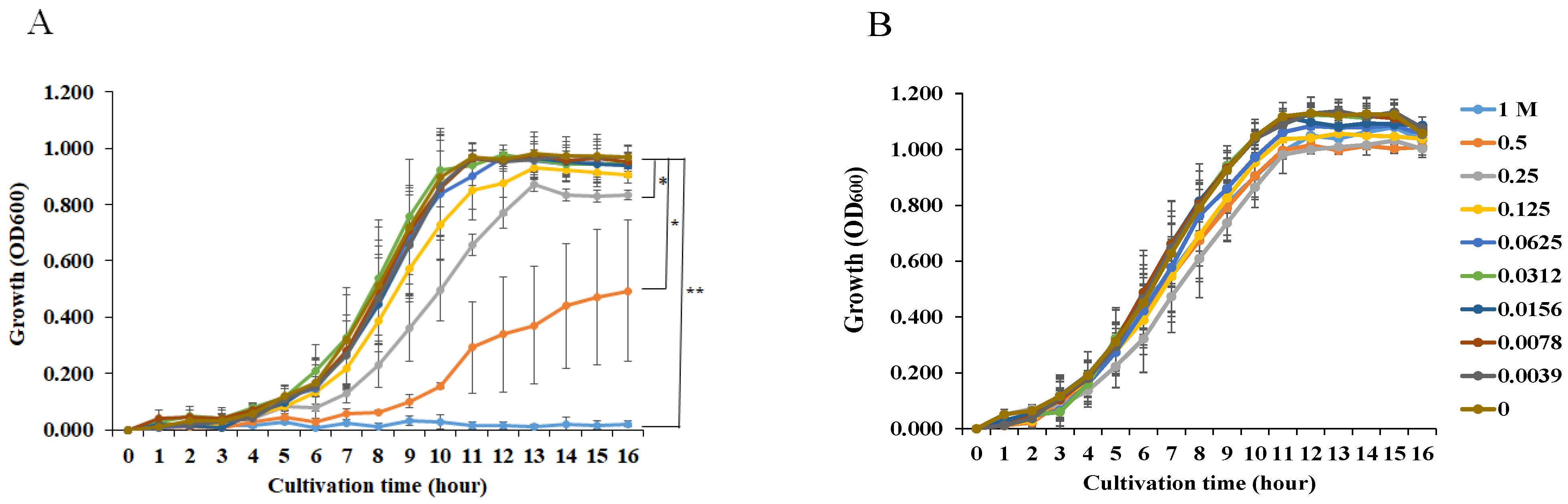
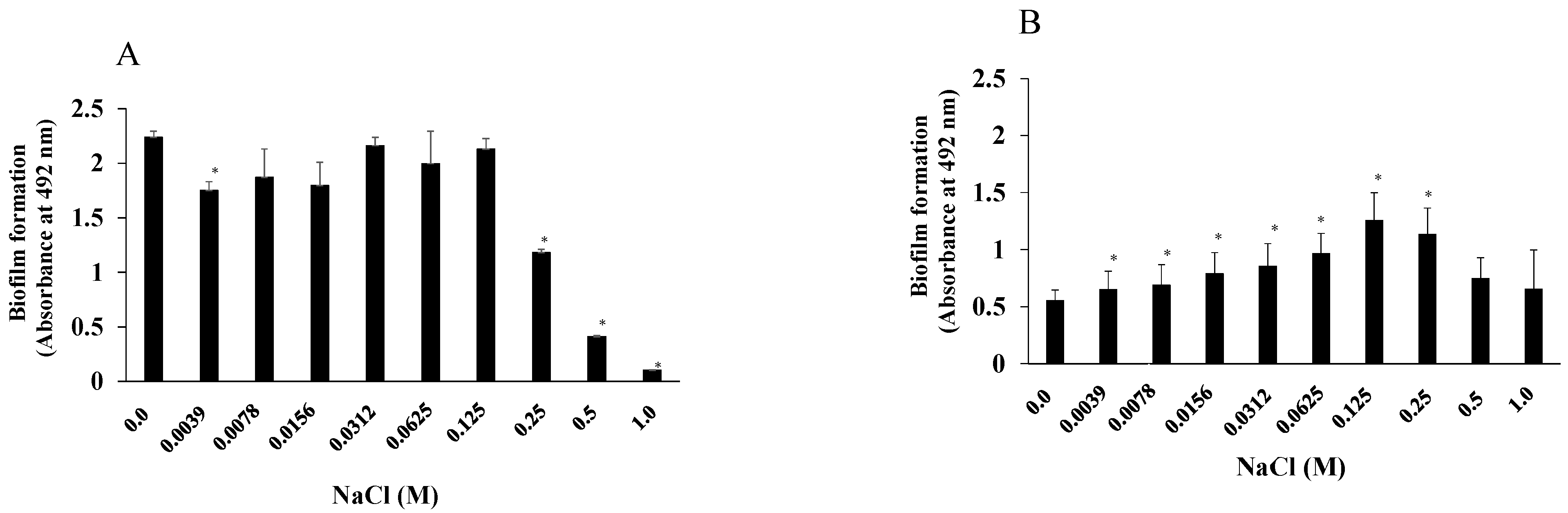
3.1. Effects of NaCl Concentration on S. aureus and S. mutans Growth and Biofilm Formation

We examined the effects of NaCl on the growth of S. mutans UA159.gtfBC and S. aureus and found that more than 0.25 M NaCl significantly inhibited S. mutans UA159.gtfBC growth (Figure 1A). In contrast, the growth of S. aureus was not inhibited by any concentration of NaCl. To observe how NaCl affects the formation of a single-species biofilm, various concentrations of NaCl were applied in the biofilm formation assay using S. mutans and S. aureus. More than 0.25 M NaCl significantly inhibited the formation of S. mutans biofilms in TSBs (Figure 2A), and this was dependent on the inhibition of cell growth (Figure 1A). In contrast, 0.0039–0.25 M NaCl significantly induced S. aureus biofilm formation compared with that in the absence of NaCl (Figure 2B), and biofilm formation peaked at 0.125 M NaCl (Figure 2B).

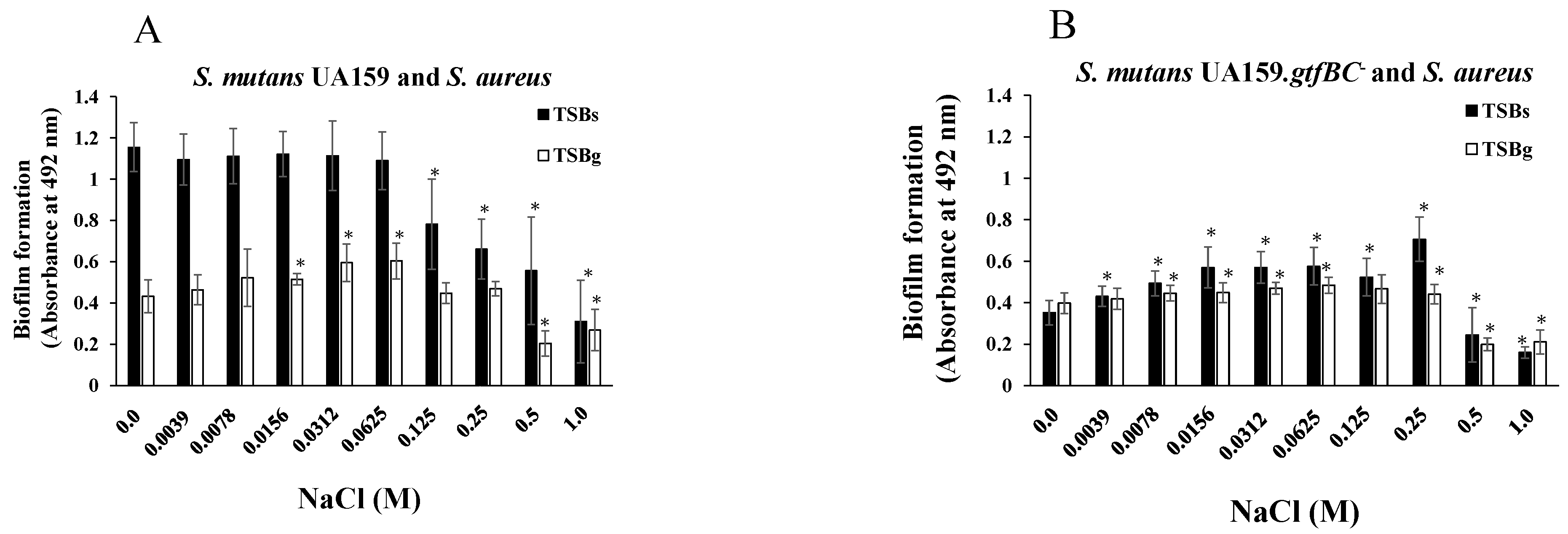
3.2. Effects of NaCl on the Formation of Mixed-Species Biofilms

Next, mixtures of S. aureus and S. mutans were used in the biofilm formation assay in the presence of NaCl. TSBs and TSBg were used separately for cultivation to determine the effects of NaCl on S. aureus and S. mutans mixed-species biofilms in the presence and absence of glucan. S. aureus was mixed with S. mutans or S. mutans UA159.gtfBC, lacking the insoluble glucan-synthesizing enzymes GtfB and GtfC, and added to 96-well microtiter plates precoated with human saliva with TSBs or TSBg containing various concentrations of NaCl. More than 0.125 M NaCl significantly inhibited the formation of the S. mutans and S. aureus mixed biofilms in TSBs (Figure 3A). In contrast, biofilm formation was significantly better in the presence of 0.0156 M, 0.0312 M, and 0.0625 M NaCl than in the absence of NaCl in TSBg (Figure 3A). More than 0.5 M NaCl significantly decreased the formation of mixed biofilms in TSBg. To completely eliminate water-insoluble glucan synthesis, S. mutans UA159.gtfBC was used instead of S. mutans UA159. The S. aureus and S. mutans UA159.gtfBC mixed biofilm gradually formed in a NaCl concentration-dependent manner, peaking at 0.25 M NaCl in TSBs (Figure 3B). However, biofilm formation was significantly inhibited with more than 0.5 M NaCl in comparison to 0 M NaCl. These results indicate that the formation of S. aureus and S. mutans UA159.gtfBC mixed biofilms in TSBs and TSBg was similar to that of S. aureus and S. mutans UA159 in TSBg. Therefore, in the absence of glucan, 0.125 M and 0.25 M NaCl significantly induced the formation of single-species S. aureus and mixed-species S. aureus and S. mutans UA159.gtfBC biofilms (Figure 2B and Figure 3B).
To observe the living and dead cells in the glucan-independent S. aureus and S. mutans mixed-species biofilms, a live/dead staining assay was performed in TSBg and TSBs. Dead cells were observed in the mixed-species biofilms in the presence of 0–0.25 M NaCl (Figure 4). Both living and dead cells were observed in the mixed-species biofilms with NaCl in the range of 0.0156–0.25 M. However, mainly live cells were observed with 0.5 M NaCl, but the amount of biofilm formation was small. Neither live nor dead cells were observed with 1 M NaCl. These results indicate that 0.0156–0.25 M NaCl, which significantly increased biofilm formation (Figure 3), induced dead and live cell-dependent mixed-species biofilms in the absence of glucan (Figure 4). Excess concentrations (>0.5 M) of NaCl inhibited the growth of S. mutans (Figure 1A) and did not induce dead cell-dependent biofilm formation in mixtures of S. aureus and S. mutans. Dead cells from S. mutans may support the biofilm formation of S. aureus in the range of 0–0.125 M NaCl in TSBs and 0–0.25 M NaCl in TSBg. A mix of live and dead cells was mainly observed at 0.25 M NaCl in TSBs.

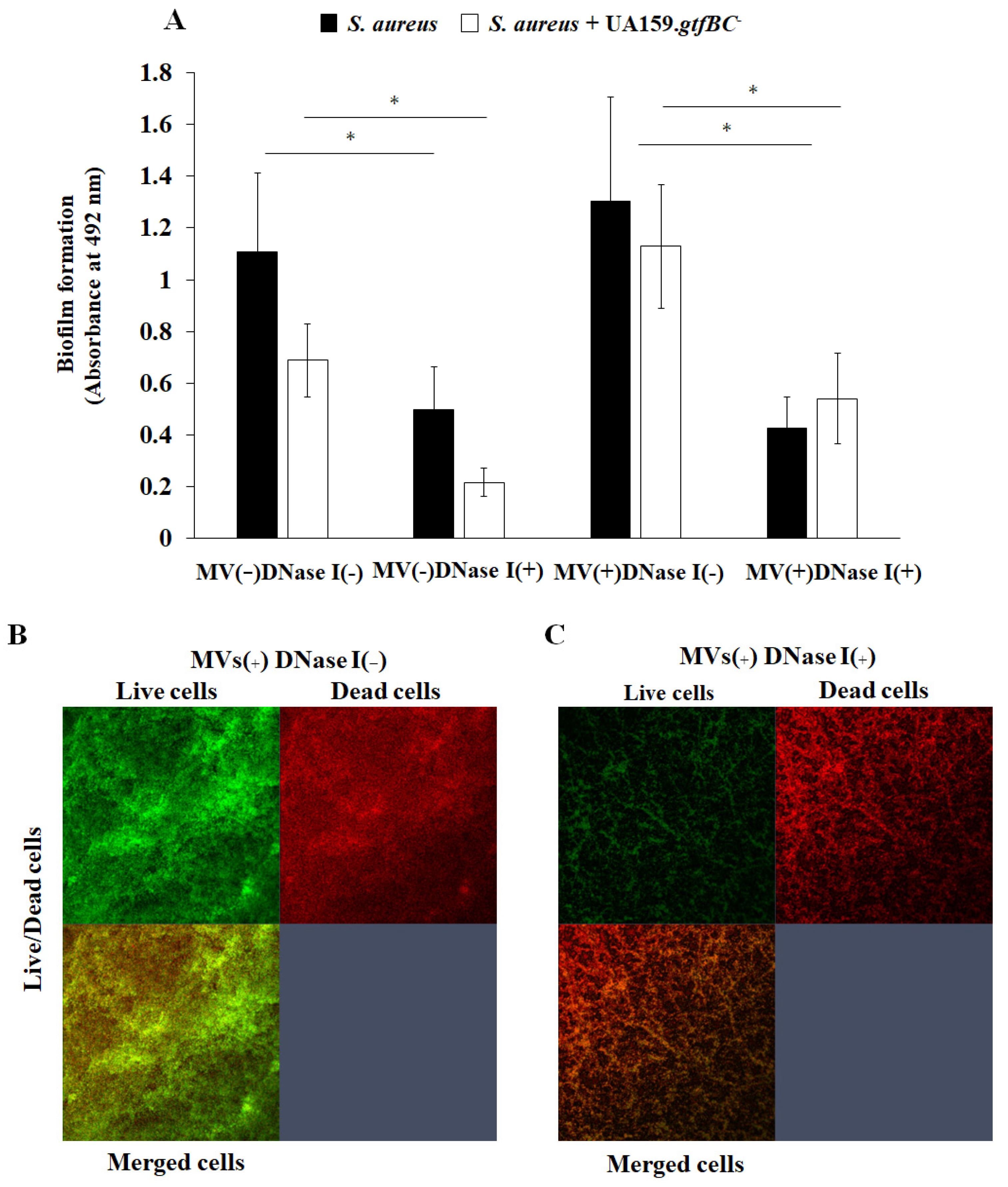
3.3. Effects of MVs on Biofilm Formation

MVs are released upon Gram-positive bacteria cell lysis [38]. MVs are important for the development of S. mutans biofilms because they are associated with GTFs and induce soluble and insoluble glucan-dependent biofilm formation of other bacteria [23]. To determine whether MVs containing GTFs affect the formation of S. aureus biofilms, 0.25 M NaCl was added to S. aureus to assess biofilm formation in the presence of 0.25 mg/mL S. mutans MVs, as this concentration induced the greatest biofilm formation in TSBs (Figure 3B). S. aureus biofilm formation was significantly increased by the addition of 0.25 NaCl. Moreover, the addition of MVs slightly enhanced biofilm formation in 0.25 M NaCl, but the difference was not significant (Figure 5A). To observe glucan generation during S. aureus biofilm formation with MVs in TSB with 0.25 M NaCl, Alexa Fluor 647-dextran conjugates were added to the assay mixture, and the results were compared with those obtained without MVs. Glucan generation was clearly observed upon the addition of MVs in the presence of 0.25 M NaCl (Figure 5B). Therefore, the slight improvement in S. aureus biofilm formation with MVs might be dependent on the presence of glucan. Compared with the dead cells in the absence of 0.25 M NaCl, more dead cells were found in the biofilms in the presence of 0.25 M NaCl (Figure 5B). This may be because glucan can combine with dead cells to induce biofilm formation.
To investigate different culture conditions, BHI media with initial pH values were prepared via the addition of various acids or NaOH, to which S. mutans UA159 was inoculated and cultivated overnight [34]. The pH values decreased in a time-dependent manner during culture. The MVs acquired from S. mutans cultures with an initial pH of 8.0 increased the protein volumes of GtfB and GtfC, which were comparable to the protein levels observed in the control (pH 7.2) [34]. The pH 6.0 media prepared with acetic acid produced MVs with greater protein volume of GtfC than those produced in pH 6.0 media prepared with HCl and lactic acid. The expression of gtfB and gtfC in S. mutans cultures was inhibited at pH 6.0 [36]. To observe the effects of different volumes of GtfB and GtfC in MVs, various MVs were applied during biofilm formation assays using S. mutans UA159.gtfBC and S. aureus with 0 and 0.25 M NaCl. The addition of NaCl inhibited the formation of S. mutans UA159.gtfBC biofilms in the presence of MVs (Figure 6A) but enhanced the formation of S. aureus biofilms in the presence of MVs (Figure 6B). The effect of NaCl on S. aureus biofilm formation was greater than the effects of different contents of Gtfs, as the volumes of GtfB and GtfC in the MVs did not affect S. aureus biofilm formation.

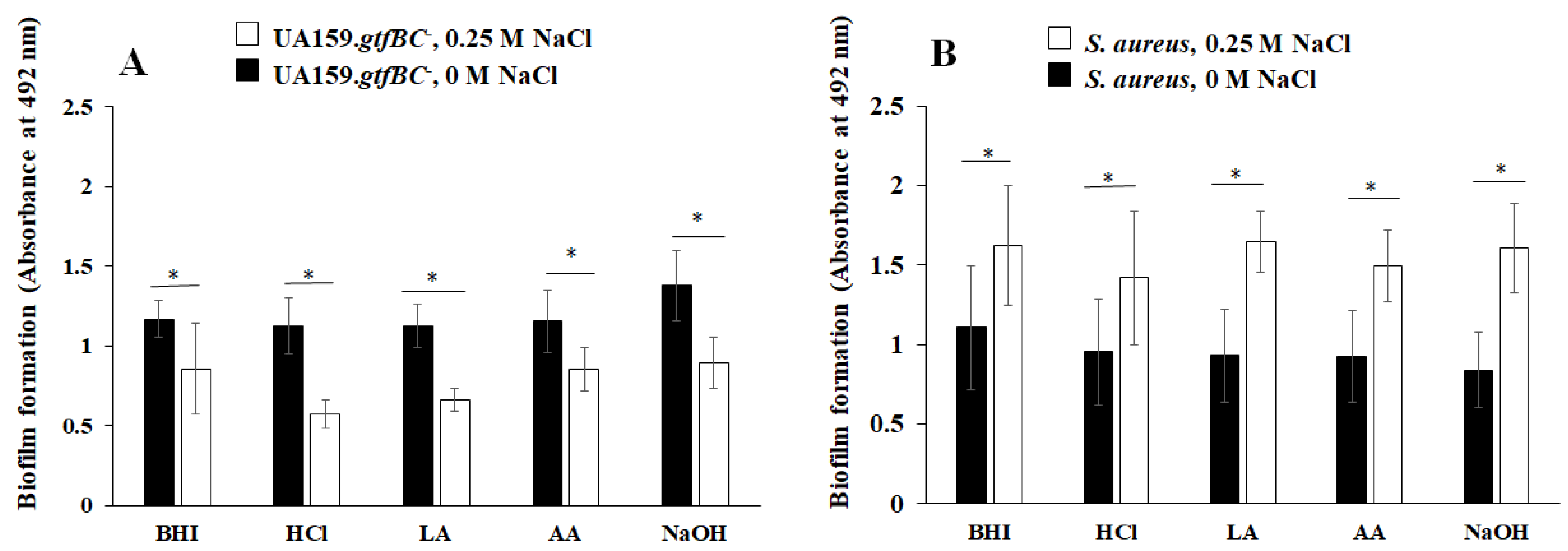
3.4. Effects of eDNA on Biofilm Formation

To elucidate how eDNA affects biofilm formation, DNase I was added to assess the formation of S. aureus single-species biofilms and S. aureus and S. mutans UA159.gtfBC mixed-species biofilms with and without S. mutans MVs in the presence of 0.25 M NaCl (Figure 7). The formation of both the single- and mixed-species biofilms was significantly inhibited by DNase I. Notably, the formation of the mixed-species biofilms was enhanced by the addition of MVs, and the bacteria in the biofilm were significantly inhibited by DNase I (Figure 7A). To confirm glucan generation in the mixed-species biofilm, an Alexa Fluor 647-dextran conjugate was added during the biofilm formation assay along with MVs and 0.25 M NaCl. Glucan generation was detected, and the live cells in the biofilms were inhibited by the addition of DNase I, as observed via confocal microscopy (Figure 7B). Therefore, eDNA principally contributes to the formation of mixed-species biofilms in the presence of salt.

4. Discussion

The formation of both single- and mixed-species S. aureus biofilms, which are tolerant to high salt concentrations, increased with the addition of 0.25 M NaCl (Figure 3). When 0.25 M NaCl was added to TSBs containing 0.086 M NaCl, the final concentration of NaCl in the media was 0.336 M. The physiological salt concentration in the human body is 0.154 M NaCl [39], and this concentration of NaCl is used during fluid infusion. Increasing the NaCl concentration in food to the physiological concentration might stimulate eDNA-dependent S. aureus biofilm formation. Additionally, high salinity might increase the density and viscosity of bacterial cell suspensions [34], which may be a result of the ability of these cells to survive under high-salinity conditions. The high viscosity and density of hypersaline solutions result in suspended particles, such as MVs, having a reduced settling velocity [40]. The slower settling of the MVs may result in increased turbidity for S. aureus and increased efficiency of aggregation transport during mixed-species biofilm formation. Moreover, fructan, which is produced by S. mutans in the presence of sucrose, may affect biofilm formation because it enhances the viscosity of eDNA solutions and is necessary for Bacillus subtilis [41] and S. mutans biofilm formation [34]. It was thus hypothesized that the bacterial cells aggregate due to the increased density and viscosity of the cell suspension in the presence of eDNA and fructan under high-salinity conditions. When S. mutans is inoculated in a high-salinity environment with high osmotic pressure, the difference in osmotic pressure between the inside and the outside of the bacterial cell causes plasmolysis and death, which reduces the amount of biofilm that forms [42]. In contrast, S. aureus, which is salt-tolerant, increases its intercellular osmolality by storing glycine betaine intracellularly and incorporating osmolytes such as proline and taurine [43]. As a result, S. aureus survives by maintaining an equilibrium between the osmotic pressure inside and outside the cell and subsequently forms biofilms under the favorable high-salinity conditions mentioned above.
S. mutans produces three types of GTFs: GTF-I, encoded by gtfB; GTF-SI, encoded by gtfC; and GTF-S, encoded by gtfD [44,45]. In the presence of sucrose, the polysaccharide synthesized by GTF-I on the bacterial surface is composed of insoluble glucan rich in α1.3 linkages, and insoluble glucan is thought to be derived from bacterial aggregation [19,20]. Furthermore, GTF-SI, which is attached to the salivary pellicle-covered tooth surface, produces insoluble and soluble glucan rich in α1.3 and α1.6 linkages [21]. Aggregated bacteria, guided by insoluble glucan, attach to the tooth surface and fuse with glucan. S. aureus is an opportunistic pathogen that is associated with oral biofilms in the oral cavities of elderly people requiring care [7,8]. However, S. aureus does not express glucan-binding proteins (gbps), such as the gbps of S. mutans [21,46]. Soluble and insoluble glucans might not affect the adherence and aggregation of S. aureus because S. aureus differs from S. mutans in various ways. However, MVs containing GTFs slightly enhanced the formation of single-species S. aureus biofilms and mixed-species S. aureus and S. mutans UA159.gtfBC biofilms under high-salinity conditions. S. mutans MVs are associated mainly with GtfC [32], and S. mutans biofilms are enhanced by both the soluble and insoluble glucans in MVs. To investigate whether differences in the abundance of Gtfs in MVs affect S. aureus biofilm formation under high-salinity conditions, a biofilm formation assay with S. aureus and MVs with different amounts of Gtfs, as identified in a previous study [36], was performed. More Gtfs were loaded in MVs acquired in pH 8.0 media prepared via the addition of NaOH [36]. The pH of this medium did not decrease to less than 5.5 after 16 h of culture, and the expression of gtfB and gtfC was not inhibited by low pH. In pH 6.0 prepared via the addition of acetic acid, the protein volume of GtfC in the MVs was lower than that in pH 8.0 media but greater than that in pH 6.0 media prepared via the addition of HCl or lactic acid [36]. The pH decreases to below 5.0 after culture because lactic acid is produced, and the expression of gtfB and gtfC was inhibited by low pH. However, weak acids, such as acetic acid, can permeate bacterial membranes more readily than strong acids such as HCl and lactic acid. The addition of acetic acid induced a greater increase in the GtfC volume than the addition of HCl and lactic acid did, resulting in oral biofilms with different activities [36]. However, different volumes of GtfB and GtfC in the MVs did not affect S. aureus biofilm formation (Figure 6). Therefore, the presence of both soluble and insoluble glucans might support the physical adherence and aggregation of S. aureus stimulated by excess salt (0.25 M NaCl), whereas the difference in the volumes of soluble and insoluble glucans did not affect the volume of the biofilm formed.
Mature S. aureus biofilms are sensitive to the external addition of DNase I, indicating that eDNA is a structural component of the S. aureus biofilm matrix [47]. Owing to the negative charge of DNA, eDNA may participate in the early adhesion and maturation stages of biofilm formation by participating in electrostatic interactions and playing a basic role in the structural integrity of the biofilms [48]. In this study, biofilm formation by live bacterial cells was induced mainly by the presence of eDNA and was not influenced by glucan under high-salinity conditions in TSBg. The presence of NaCl preferentially induces eDNA-dependent live S. aureus biofilm formation via different mechanisms than those of glucan generation by S. mutans MVs. The opportunities and mechanisms by which S. aureus produces DNA are unknown, but DNA synthesis is required when a single S. aureus cell initially attaches to the surface in the absence of glucan.
Cyclic di-adenosine monophosphate (c-di-AMP) is a recently discovered secondary messenger that is produced predominantly by Gram-positive bacteria [49,50,51,52]. c-di-AMP production is critical for the growth of S. aureus in chemically defined media and in rich media supplemented with additional sodium or potassium chloride [53]. The influence of c-di-AMP on biofilms has been shown and may play an important role in the persistence of S. aureus biofilms under high-salinity conditions [54]. The production of c-di-AMP promotes K+ and cell wall homeostasis, biofilm formation, and virulence but sensitizes the cells to osmotic stress [55,56,57]. Elevated c-di-AMP concentrations may promote S. aureus biofilm formation under high-salinity conditions, as previously shown in several streptococci [58,59].
Compared with single-species S. aureus biofilms, whole-cell S. mutans biofilms reduced the formation of mixed-species biofilms (Figure 7A). In contrast, S. mutans MVs slightly enhanced dead cell-dependent biofilm formation (Figure 5B). Glucans in MVs might enhance the associations between live and dead S. aureus cells under high-salinity conditions. In contrast, in the presence of glucose, S. aureus may be able to attach to and colonize old biofilms containing dead bacteria because dead cells do not produce antibacterial substances such as acids or bacteriocin [7]. When the commensal bacteria in the oral cavity are alive and colonizing, opportunistic pathogens such as S. aureus are less likely to colonize [60]. This is also why young people with sufficient normal oral flora have a low incidence of S. aureus infection.
Thus, MVs, eDNA, and dead cells from S. mutans enhance S. aureus biofilm formation in the presence of high concentrations of salt. The sugar (sucrose and glucose) ingestion-induced S. mutans biofilm may be a risk factor for dental caries with S. aureus in the elderly and bedridden, who typically have high physiological concentrations of NaCl. Therefore, to avoid dental caries via infection by opportunistic pathogens, such as S. aureus, and to maintain oral and systemic health, sucrose, glucose, and NaCl intake should be limited.

5. Conclusions

The presence of S. mutans might be a risk factor for S. aureus infection in the oral cavity because the formation of single-species S. aureus biofilms and S. aureus mixed-species biofilms is enhanced by MVs, extracellular DNA, and dead S. mutans cells in tryptic soy broth with sucrose, glucose, and high concentrations of salt, including NaCl. The glucosyltransferases in S. mutans MVs slightly enhanced the association between S. aureus and dead cells under high-salinity conditions in TSBs. Biofilm formation by live bacterial cells was induced mainly by the presence of extracellular DNA. Therefore, to avoid dental caries via infection by opportunistic pathogens, such as S. aureus, and to maintain oral and systemic health, sucrose, glucose, and NaCl intake should be limited.

Author Contributions

Conceptualization, H.S. and Y.I.; data curation, Y.I., H.Y., T.U. and S.K.; funding acquisition, H.S.; investigation, Y.I., H.S., T.U. and M.S.; methodology, H.S., H.Y., M.S. and Y.I.; resources, Y.I., S.K. and H.S.; supervision, H.S.; writing—original draft, Y.I. and H.S.; writing—review and editing, H.S.; All authors have read and agreed to the published version of the manuscript.

Funding

This work was supported, in part, by a Grant-in-Aid for the Development of Scientific Research (20K10286 and 24K02661) from the Ministry of Education, Culture, Sports, Science, and Technology of Japan and the Research Grant of the SoltScience Program (2017).

Institutional Review Board Statement

The study was conducted in accordance with the Declaration of Helsinki, and approved by the Ethics Committee of Nihon University School of Dentistry at Matsudo (IRB approval number: EC21-029), approval date: 29 March 2022.

Informed Consent Statement

Informed consent was obtained from all subjects involved in the study.

Data Availability Statement

The original contributions presented in this study are included in the article. Further inquiries can be directed to the corresponding author.

Acknowledgments

We thank Kaori Ochiai who supported the secretary works in purchasing experimental items.

Conflicts of Interest

The authors declare no conflict of interest.

References

  1. Loesche, W.J. Role of Streptococcus mutans in human dental decay. Microbiol. Rev. 1986, 50, 353–380. [Google Scholar] [CrossRef]
  2. Hamada, S.; Slade, H.D. Biology, immunology, and cariogenicity of Streptococcus mutans. Microbiol. Rev. 1980, 44, 331–384. [Google Scholar] [CrossRef]
  3. Costerton, J.W.; Stewart, P.S.; Greenberg, E.P. Bacterial biofilms: A common cause of persistent infections. Science 1999, 284, 1318–1322. [Google Scholar] [CrossRef]
  4. Mirghani, R.; Saba, T.; Khaliq, H.; Mitchell, J.; Do, L.; Chambi, L.; Diaz, K.; Kennedy, T.; Alkassab, K.; Huynh, T.; et al. Biofilms: Formation, drug resistance and alternatives to conventional approaches. AIMS Microbiol. 2022, 8, 239–277. [Google Scholar] [CrossRef]
  5. Mandell, L.A.; Niederman, M.S. Aspiration pneumonia. N. Engl. J. Med. 2019, 380, 651–663. [Google Scholar] [CrossRef]
  6. Yoneyama, T.; Yoshida, M.; Matsui, T.; Sasaki, H. Oral care and pneumonia. Oral Care Working Group. Lancet 1999, 354, 515. [Google Scholar] [CrossRef]
  7. Senpuku, H.; Sogame, A.; Inoshita, E.; Tsuha, Y.; Miyazaki, H.; Hanada, N. Systemic diseases in association with microbial species in oral biofilm from elderly requiring care. Gerontology 2003, 49, 301–309. [Google Scholar] [CrossRef]
  8. Tada, A.; Hanada, N. Opportunistic respiratory pathogens in the oral cavity of the elderly. FEMS Immunol. Med. Microbiol. 2010, 60, 1–17. [Google Scholar] [CrossRef]
  9. El-Solh, A.A.; Pietrantoni, C.; Bhat, A.; Aquilina, A.T.; Okada, M.; Grover, V.; Gifford, N. Microbiology of severe aspiration pneumonia in institutionalized elderly. Am. J. Respir. Crit. Care Med. 2003, 167, 1650–1654. [Google Scholar] [CrossRef]
  10. Koukos, G.; Sakellari, D.; Arsenakis, M.; Tsalikis, L.; Slini, T.; Konstantinidis, A. Prevalence of Staphylococcus aureus and methicillin resistant Staphylococcus aureus (MRSA) in the oral cavity. Arch. Oral Biol. 2015, 60, 1410–1415. [Google Scholar] [CrossRef]
  11. Saeed, K.; Gould, I.; Esposito, S.; Ahmad-Saeed, N.; Ahmed, S.S.; Alp, E.; Bal, A.M.; Bassetti, M.; Bonnet, E.; Chan, M.; et al. Panton-valentine leukocidin-positive Staphylococcus aureus: A position statement from the International Society of Chemotherapy. Int. J. Antimicrob. Agents 2018, 51, 16–25. [Google Scholar] [CrossRef]
  12. Shallcross, L.J.; Fragaszy, E.; Johnson, A.M.; Hayward, A.C. The role of the panton valentine leucocidin toxin in staphylococcal disease: A systematic review and meta-analysis. Lancet Infect. Dis. 2013, 13, 43–54. [Google Scholar] [CrossRef]
  13. Miles, G.; Movileanu, L.; Bayley, H. Subunit composition of a bicomponent toxin: Staphylococcal leukocidin forms an octameric transmembrane pore. Protein Sci. 2002, 11, 894–902. [Google Scholar] [CrossRef]
  14. Tashiro, M.; Ciborowski, P.; Reinacher, M.; Pulverer, G.; Klenk, H.-D.; Rott, R. Synergistic role of staphylococcal proteases in the induction of influenza virus pathogenicity. Virology 1987, 157, 421–430. [Google Scholar] [CrossRef]
  15. McCormick, J.K.; Yarwood, J.M.; Schlievert, P.M. Toxic shock syndrome and bacterial superantigens: An update. Annu. Rev. Microbiol. 2001, 55, 77–104. [Google Scholar] [CrossRef]
  16. Scannapieco, F.A. Role of oral bacteria in respiratory infection. J. Periodontol. 1999, 70, 793–802. [Google Scholar] [CrossRef]
  17. Desai, J.P.; Nair, R.U. Oral health factors related to rapid oral health deterioration among older adults: A narrative review. J. Clin. Med. 2023, 12, 3202. [Google Scholar] [CrossRef]
  18. Hoover, J.; Tovar, E.; Zlatnik, T.; Karunanayake, C. Efficacy of a rinse containing sea salt and lysozyme on biofilm and gingival health in a group of young adults: A pilot study. Int. J. Dent. 2017, 2017, 4056708. [Google Scholar] [CrossRef]
  19. Lee, S.; Choi, K.-H.; Yoon, Y. Effect of NaCl on biofilm formation of the isolate from Staphylococcus aureus outbreak linked to ham. Kor. J. Food Sci. Anim. Resour. 2014, 34, 257–261. [Google Scholar] [CrossRef]
  20. O’Neill, E.; Pozzi, C.; Houston, P.; Smyth, D.; Humphreys, H.; Robinson, D.A.; O’Gara, J.P. Association between methicillin susceptibility and biofilm regulation in Staphylococcus aureus isolates from device-related infections. J. Clin. Microbiol. 2017, 45, 1379–1388. [Google Scholar] [CrossRef]
  21. Bowen, W.; Koo, H. Biology of Streptococcus mutans-derived glucosyltransferases: Role in extracellular matrix formation of cariogenic biofilms. Caries Res. 2011, 45, 69–86. [Google Scholar] [CrossRef]
  22. Koo, H.; Xiao, J.; Klein, M.I.; Jeon, J.G. Exopolysaccharides produced by Streptococcus mutans glucosyltransferases modulate the establishment of microcolonies within multispecies biofilms. J. Bacteriol. 2010, 192, 3024–3032. [Google Scholar] [CrossRef]
  23. Senpuku, H.; Nakamura, T.; Iwabuchi, Y.; Hirayama, S.; Nakao, R.; Ohnishi, M. Effects of complex DNA and MVs with GTF extracted from Streptococcus mutans on the oral biofilm. Molecules 2019, 24, 3131. [Google Scholar] [CrossRef]
  24. Bonnington, K.E.; Kuehn, M.J. Protein selection and export via outer membrane vesicles. Biochim. Biophys. Acta 2014, 1843, 1612–1619. [Google Scholar] [CrossRef]
  25. Ellis, T.N.; Kuehn, M.J. Virulence and immunomodulatory roles of bacterial outer membrane vesicles. Microbiol. Mol. Biol. Rev. 2010, 74, 81–94. [Google Scholar] [CrossRef]
  26. Biller, S.J.; Schubotz, F.; Roggensack, S.E.; Thompson, A.W.; Summons, R.E.; Chisholm, S.W. Bacterial vesicles in marine ecosystems. Science 2014, 343, 183–186. [Google Scholar] [CrossRef]
  27. Berleman, J.; Auer, M. The role of bacterial outer membrane vesicles for intra- and interspecies delivery. Environ. Microbiol. 2013, 15, 347–354. [Google Scholar] [CrossRef]
  28. Kuehn, M.J.; Kesty, N.C. Bacterial outer membrane vesicles and the host-pathogen interaction. Genes Dev. 2005, 19, 2645–2655. [Google Scholar] [CrossRef]
  29. Lai, H.; White, J.; Roe, B.A.; Ferretti, J.J. Genome sequence of Streptococcus mutans UA159, a cariogenic dental pathogen. Proc. Natl. Acad. Sci. USA 2002, 99, 14434–14439. [Google Scholar]
  30. Suzuki, Y.; Nagasawa, R.; Senpuku, H. Inhibiting effects of fructanase on competence-stimulating peptide-dependent quorum sensing system in Streptococcus mutans. J. Infect. Chemother. 2017, 23, 634–641. [Google Scholar] [CrossRef]
  31. Nakamura, T.; Iwabuchi, Y.; Hirayama, S.; Narisawa, N.; Takenaga, F.; Nakao, R.; Senpuku, H. Roles of membrane vesicles from Streptococcus mutans for the induction of antibodies to glucosyltransferase in mucosal immunity. Microb. Pathog. 2020, 149, 104260. [Google Scholar] [CrossRef] [PubMed]
  32. Das, T.; Sharma, P.K.; Busscher, H.J.; Van Der Mei, H.C.; Krom, B.P. Role of extracellular DNA in initial bacterial adhesion and surface aggregation. Appl. Environ. Microbiol. 2010, 76, 3405–3408. [Google Scholar] [CrossRef] [PubMed]
  33. Das, T.; Sehar, S.; Manefield, M. The roles of extracellular DNA in the structural integrity of extracellular polymeric substance and bacterial biofilm development. Environ. Microbiol. Rep. 2013, 5, 778–786. [Google Scholar] [CrossRef]
  34. Nagasawa, R.; Sato, T.; Senpuku, H. Raffinose induces biofilm formation by Streptococcus mutans in low concentrations of sucrose by increasing production of extracellular DNA and fructan. Appl. Environ. Microbiol. 2017, 83, e00869-17. [Google Scholar] [CrossRef]
  35. Motegi, M.; Takagi, Y.; Yonezawa, H.; Kanada, N.; Terajima, J.; Watanabe, H.; Senpuku, H. Assessment of genes associated with Streptococcus mutans biofilm morphology. Appl. Environ. Microbiol. 2006, 72, 6277–6287. [Google Scholar] [CrossRef]
  36. Liao, S.; Klein, M.I.; Heim, K.P.; Fan, Y.; Bitoun, J.P.; Ahn, S.-J.; Burne, R.A.; Koo, H.; Brady, L.J.; Wen, Z.T. Streptococcus mutans extracellular DNA is upregulated during growth in biofilms, actively released via membrane vesicles, and influenced by components of the protein secretion machinery. J. Bacteriol. 2014, 196, 2355–2366. [Google Scholar] [CrossRef] [PubMed]
  37. Iwabuchi, Y.; Nakamura, T.; Kusumoto, Y.; Nakao, R.; Iwamoto, T.; Shinozuka, O.; Senpuku, H. Effects of pH on the properties of membrane vesicles including glucosyltransferase in Streptococcus mutans. Microorganisms 2021, 9, 2308. [Google Scholar] [CrossRef]
  38. Toyofuku, M.; Cárcamo-Oyarce, G.; Yamamoto, T.; Eisenstein, F.; Hsiao, C.-C.; Kurosawa, M.; Gademann, K.; Pilhofer, M.; Nomura, N.; Eberl, L. Prophage-triggered membrane vesicle formation through peptidoglycan damage in Bacillus subtilis. Nat. Commun. 2017, 8, 481. [Google Scholar] [CrossRef]
  39. Groessl, M.; Hartinger, C.G.; Polec-Pawlak, K.; Jarosz, M.; Keppler, B.K. Capillary electrophoresis hyphenated to inductively coupled plasma-mass spectrometry: A novel approach for the analysis of anticancer metallodrugs in human serum and plasma. Electrophoresis 2008, 29, 2224–2232. [Google Scholar] [CrossRef]
  40. Weisbrod, N.; Yechieli, Y.; Shandalov, S.; Lensky, N. On the viscosity of natural hyper-saline solutions and its importance: The dead sea brines. J. Hydrol. 2016, 532, 46–51. [Google Scholar] [CrossRef]
  41. Dogsa, I.; Brloznik, M.; Stopar, D.; Mandic-Mulec, I. Exopolymer diversity and the role of levan in Bacillus subtilis biofilms. PLoS ONE 2013, 8, e62044. [Google Scholar] [CrossRef] [PubMed]
  42. He, H.; Chen, Y.; Li, X.; Cheng, Y.; Yang, C.; Zeng, G. Influence of salinity on microorganisms in activated sludge processes: A review. Int. Biodeter. Biodeg. 2017, 119, 520–527. [Google Scholar] [CrossRef]
  43. Graham, J.E.; Wilkinson, B.J. Staphylococcus aureus osmoregulation: Roles for choline, glycine betaine, proline, and taurine. J. Bacteriol. 1992, 174, 2711–2716. [Google Scholar] [CrossRef]
  44. Wexler, D.L.; Hudson, M.C.; Burne, R.A. Streptococcus mutans fructosyltransferase (ftf) and glucosyltransferase (gtfBC) operon fusion strains in continuous culture. Infect. Immun. 1993, 61, 1259–1267. [Google Scholar] [CrossRef] [PubMed]
  45. Fujiwara, T.; Terao, Y.; Hoshino, T.; Kawabata, S.; Ooshima, T.; Sobue, S.; Kimura, S.; Hamada, S. Molecular analyses of glucosyltransferase genes among strains of Streptococcus mutans. FEMS Microbiol. Lett. 1998, 161, 331–336. [Google Scholar] [CrossRef]
  46. Smith, D.J.; Akita, H.; King, W.F.; Taubman, M.A. Purification and antigenicity of a novel glucan-binding protein of Streptococcus mutans. Infect. Immunity 1994, 62, 2545–2552. [Google Scholar] [CrossRef]
  47. Mann, E.E.; Rice, K.C.; Boles, B.R.; Endres, J.L.; Ranjit, D.; Chandramohan, L.; Tsang, L.H.; Smeltzer, M.S.; Horswill, A.R.; Bavles, K.W. Modulation of eDNA release and degradation affects Staphylococcus aureus biofilm maturation. PLoS ONE 2009, 4, e5822. [Google Scholar] [CrossRef]
  48. Campoccia, D.; Montanaro, L.; Arciola, C.R. Extracellular DNA (eDNA). A major ubiquitous element of the bacterial biofilm architecture. Int. J. Mol. Sci. 2021, 22, 9100. [Google Scholar] [CrossRef]
  49. Corrigan, R.M.; Abbott, J.C.; Burhenne, H.; Kaever, V.; Gründling, A. c-di-AMP is a new second messenger in Staphylococcus aureus with a role in controlling cell size and envelope stress. PLoS Pathog. 2011, 7, e1002217. [Google Scholar] [CrossRef]
  50. Corrigan, R.M.; Gründling, A. Cyclic di-AMP: Another second messenger enters the fray. Nat. Rev. Microbiol. 2013, 11, 513–524. [Google Scholar] [CrossRef]
  51. Römling, U. Great times for small molecules: c-di-AMP, a second messenger candidate in bacteria and archaea. Sci. Signal. 2008, 1, 39. [Google Scholar] [CrossRef]
  52. Witte, G.; Hartung, S.; Büttner, K.; Hopfner, K.P. Structural biochemistry of a bacterial checkpoint protein reveals diadenylate cyclase activity regulated by DNA recombination intermediates. Mol. Cell 2008, 30, 167–178. [Google Scholar] [CrossRef] [PubMed]
  53. Zeden, M.S.; Schuster, C.F.; Bowman, L.; Zhong, Q.; Williams, H.D.; Gründling, A. Cyclic di-adenosine monophosphate (c-di-AMP) is required for osmotic regulation in Staphylococcus aureus but dispensable for viability in anaerobic conditions. J. Biolog. Chem. 2018, 293, 3180–3200. [Google Scholar] [CrossRef] [PubMed]
  54. Huynh, N.; Luo, S.; Pensinger, D.; Sauer, J.D.; Tong, L.; Woodward, J.J. An HD-domain phosphodiesterase mediates cooperative hydrolysis of c-di-AMP to affect bacterial growth and virulence. Proc. Natl. Acad. Sci. USA 2015, 112, E747–E756. [Google Scholar] [CrossRef]
  55. Luo, Y.; Helmann, J.D. Analysis of the role of Bacillus subtilis σ(M) in β-lactam resistance reveals an essential role for c-di-AMP in peptidoglycan homeostasis. Mol. Microbiol. 2012, 83, 623–639. [Google Scholar] [CrossRef] [PubMed]
  56. Bai, Y.; Yang, J.; Eisele, L.; Underwood, A.J.; Koestler, B.J.; Waters, C.M.; Metzger, D.W.; Bai, G. Two DHH subfamily 1 proteins in Streptococcus pneumoniae possess cyclic di-AMP phosphodiesterase activity and affect bacterial growth and virulence. J. Bacteriol. 2013, 195, 5123–5132. [Google Scholar] [CrossRef]
  57. Jackson-Litteken, C.D.; Ratliff, C.T.; Kneubehl, A.R.; Siletti, C.; Pack, L.; RLan, R.; Huynh, T.N.; Lopez, J.E.; Blevins, J.S. The diadenylate cyclase CdaA is critical for Borrelia turicatae virulence and physiology. Infect. Immun. 2021, 89, e00787-2089. [Google Scholar] [CrossRef]
  58. Du, B.; Ji, W.; An, H.; Shi, Y.; Huang, Q.; Cheng, Y.; Fu, Q.; Wang, H.; Yan, Y.; Sun, J. Functional analysis of c-di-AMP phosphodiesterase, GdpP, in Streptococcus suis serotype 2. Microbiol. Res. 2014, 169, 749–758. [Google Scholar] [CrossRef]
  59. Peng, X.; Zhang, Y.; Bai, G.; Zhou, X.; Wu, H. Cyclic di-AMP mediates biofilm formation. Mol. Microbiol. 2016, 99, 945–959. [Google Scholar] [CrossRef]
  60. Tada, A.; Senpuku, H.; Motozawa, Y.; Yoshihara, A.; Hanada, N.; Tanzawa, H. Association between commensal bacteria and opportunistic pathogens in the dental plaque of elderly individuals. Clin. Microbiol. Infect. 2006, 12, 776–781. [Google Scholar] [CrossRef]
Figure 1. Effects of NaCl on the growth of S. mutans UA159.gtfBC and S. aureus. S. mutans UA159.gtfBC (A) and S. aureus cowan I (B) were cultivated at 0, 1/28, 1/27, 1/26, 1/25, 1/24, 1/23, 1/22, 1/21, and 1/20 M NaCl dilutions in BHI. The OD600 of the bacterial cell suspension was measured after growth. The data are presented as the means ± SDs of three independent experiments. An asterisk indicates that there is a significant difference between the two groups in ANCOVA (*: p < 0.05, **: p < 0.01; various concentrations of NaCl vs. no NaCl).
Microorganisms 13 01118 g001
Figure 2. Effects of NaCl on the formation of single-species biofilms. S. mutans UA159 (A) and S. aureus cowan I (B) were cultivated at 0, 1/28, 1/27, 1/26, 1/25, 1/24, 1/23, 1/22, 1/21, and 1/20 M NaCl dilutions in TSBs to assess biofilm formation. The data are presented as the means ± SDs of three independent experiments at 16 h after culture. An asterisk indicates that there is a significant difference between the two conditions in ANOVA with Bonferroni correction (*: p < 0.05; various concentrations of NaCl vs. no NaCl).
Microorganisms 13 01118 g002
Figure 3. Effects of NaCl on the formation of mixed-species biofilms. S. aureus was inoculated with S. mutans UA159 (A) or S. mutans UA159.gtfBC (B) in TSBs or TSBg with various concentrations of NaCl to assess biofilm formation. The data are presented as the means ± SDs of three independent experiments at 16 h after culture starts. An asterisk indicates that there is a significant difference between the two conditions in TSBs or TSBg in ANOVA with the Bonferroni correction (*: p < 0.05, various concentrations of NaCl vs. no NaCl).
Microorganisms 13 01118 g003
Figure 4. Effects of NaCl on the formation of mixed-species biofilms. S. aureus was inoculated with S. mutans UA159 in TSBs and TSBg with various concentrations of NaCl to assess biofilm formation. The biofilms were treated via live/dead staining and observed via confocal microscopy. Representative data from more than three independent experiments are presented in the images.
Microorganisms 13 01118 g004
Figure 5. Effects of MVs on the formation of S. aureus biofilms with and without NaCl. S. aureus was inoculated in TSBs with and without 0.25 M NaCl to assess biofilm formation with and without S. mutans MVs (A). The data are presented as the means ± SDs of three independent experiments at 16 h after culture starts. An asterisk indicates that there is a significant difference between the two conditions in 0 M NaCl or 0.25 M NaCl in Student’ t-test (MVs vs. no MVs). The biofilms were treated by live/glucan and live/dead staining and observed by confocal microscopy (B). Upper row: upper left square indicates live cells biofilm. The upper right square indicates glucan or dead cells. The lower left square indicates biofilm merged with live cells and glucan or dead cells. Representative data from more than three independent experiments are presented in the images.
Microorganisms 13 01118 g005
Figure 6. The effects of S. mutans MVs acquired after cultivation in media with various initial pH values. S. mutans UA159 gtfBC (A) and S. aureus (B) biofilms were cultivated in TSBs with and without MVs in media with an initial pH value of 6.0 prepared via the addition of HCl, lactic acid (LA), and acetic acid (AA), and in media with an initial pH value of 8.0 prepared via the addition of NaOH supplemented with 0 M NaCl and 0.25 M NaCl. The data are presented as the means ± SDs of three independent experiments at 16 h after culture starts. An asterisk indicates that there is a significant difference between the two conditions in the Student’ t-test (*: p < 0.05; NaCl vs. no NaCl).
Microorganisms 13 01118 g006
Figure 7. S. aureus or a mixture of S. aureus and S. mutans gtfBC was inoculated with and without MVs in TSBs with and without DNase I. The data represent the means ± SDs of three independent experiments at 16 h after start (A). An asterisk indicates that there are significant differences among the four conditions in the Student’ t-test (*: p < 0.05; DNase I vs. no DNase I). The MV-dependent S. aureus and S. mutans gtfBC mixed-species biofilms formed both with and without DNase I (B,C). The biofilms were treated through live/dead staining and observed via confocal microscopy. Upper row: upper left square indicates live cells in biofilm. The upper right square indicates dead cells. The lower left square indicates biofilm merged with live cells and dead cells. Representative data from more than three independent experiments are presented in the images.
Microorganisms 13 01118 g007
Disclaimer/Publisher’s Note: The statements, opinions and data contained in all publications are solely those of the individual author(s) and contributor(s) and not of MDPI and/or the editor(s). MDPI and/or the editor(s) disclaim responsibility for any injury to people or property resulting from any ideas, methods, instructions or products referred to in the content.

