Article

Diversity and Pathogenicity of Colletotrichum Species Causing Coffee Anthracnose in China

1
Environment and Plant Protection Institute, Chinese Academy of Tropical Agricultural Sciences, Key Laboratory of Integrated Pest Management on Tropical Crop, Ministry of Agriculture and Rural Affairs, Haikou 571101, China
2
Hainan Key Laboratory for Monitoring and Control of Tropical Agricultural Pests, Haikou 571101, China
3
School of Tropical Agricultural and Forestry, Hainan University, Danzhou 571737, China
4
College of Plant Science and Technology, Huazhong Agricultural University, Wuhan 430070, China
5
Sanya Research Institute, Chinese Academy of Tropical Agricultural Sciences, Sanya 572025, China
*
Author to whom correspondence should be addressed.
Microorganisms 2025, 13(3), 512; https://doi.org/10.3390/microorganisms13030512
Submission received: 11 February 2025 / Revised: 24 February 2025 / Accepted: 24 February 2025 / Published: 26 February 2025

Abstract

:
Coffee is a significant traded commodity for developing countries. Among the various diseases affecting coffee, anthracnose caused by Colletotrichum spp. has re-emerged as a major constraint on global coffee production. To better understand the Colletotrichum species complex associated with coffee anthracnose, we characterized Colletotrichum spp. using a combination of phenotypic traits, MAT1-2 (ApMat) gene analysis, multi-locus phylogenetic (ITS, ACT, CHS-1, and GAPDH), and pathogenicity assays. A total of 74 Colletotrichum isolates were collected from coffee plants exhibiting anthracnose symptoms across nine coffee plantations in China. Among these, 55 isolates were identified as the C. gloeosporioides species complex using the ApMat locus, while the remaining 19 isolates were identified through multi-locus phylogenetic analyses. The isolates represented seven Colletotrichum species from five species complexes: C. gloeosporioides (including C. siamense, C. nupharicola, and C. theobromicola), C. boninens (C. karstii), C. gigasporum (C. gigasporum), C. orchidearum (C. cliviicola), and C. magnum (C. brevisporum). This is the first report of C. nupharicola and C. cliviicola causing coffee anthracnose worldwide, and the first report of C. nupharicola in China. Pathogenicity tests confirmed that all seven species were capable of infecting coffee leaves. This research enhances our understanding of the Colletotrichum species responsible for coffee anthracnose, and provides valuable insights for developing effective disease management strategies.

1. Introduction

Coffee is a major traded commodity in the developing world [1]. Three primary commercial varieties are cultivated globally: Coffea arabica (arabica), C. canephora var. robusta (robusta), and C. liberica (liberica) [2]. Among these, arabica and robusta are the dominant species, accounting for over 99% of global exports. Coffee was introduced to China more than a century ago, and has since become a significant cash crop in Yunnan and Hainan provinces. Arabica coffee is predominantly cultivated in Yunnan, while robusta coffee is mainly grown in Hainan [1,3].
Diseases are among the most critical factors limiting coffee production. The most prevalent and economically significant diseases include coffee rust (Hemileia vastatrix), Cercospora leaf spot (Cercospora coffeicola), Phoma leaf spot (Phoma spp.), and anthracnose and blister spot caused by Colletotrichum spp. Coffee anthracnose, caused by species of the genus Colletotrichum, is a major issue in Southeast Asia, India, and China [4,5,6]. Anthracnose is characterized by irregular, large brown-to-gray patches that often appear along the edges of leaves. Other symptoms may include dieback, brown blight, and blister spots, which manifest as light-green, oily-looking lesions on both leaves and fruit [6,7]. The genus Colletotrichum is a significant group within the fungal family Glomerellaceae, order Glomerellales, class Sordariomycetes, and phylum Ascomycota. It is recognized as one of the top 10 most important genera of plant pathogenic fungi globally, with a wide range of impacts on agriculture and ecosystems [8]. Colletotrichum species are notorious for causing anthracnose diseases in a wide variety of plants, leading to significant agricultural losses [9,10,11,12,13,14]. The genus Colletotrichum is highly diverse and complex, with its classification system continually evolving as new species are discovered and molecular techniques improve. A species complex (or ‘aggregate’) in Colletotrichum is defined as a monophyletic group of species that share common characteristics, such as similar conidial morphology. The current classification system recognizes 15 species complexes: C. acutatum, C. agaves, C. boninense, C. caudatum, C. dematium, C. destructivum, C. dracaenophilum, C. gigasporum, C. gloeosporioides, C. graminicola, C. magnum, C. orbiculare, C. orchidearum, C. spaethianum, and C. truncatum. Additionally, there are several singleton species that do not belong to any of these complexes. The C. caudatum species complex was initially considered a sub-aggregate within the C. graminicola complex, but is now often treated as a separate complex. Recently, Bhunjun et al. suggested merging the two into a single C. graminicolacaudatum species complex [15]. The phylogenetic backbone of Colletotrichum has been continuously updated as new species have been described: Cannon et al. [16]: 119 species; Jayawardena et al. [17] and Marin-Felix et al. [18]: 189 species; Jayawardena et al. [19]: 247 species; Bhunjun et al. [15]: 248 species. The discovery of new species highlights the high species diversity within the genus. Twenty Colletotrichum species from six species complexes have been reported to be associated with coffee worldwide, including C. acutatum, C. costarricense, C. cuscutae, and C. walleri from the C. acutatum species complex; C. endophytica, C. ledongense, C. tropicale, C. asianum, C. fructicola, C. gloeosporioides, C. kahamae subsp. kahawae, C. queeslandicum, C. theobromicola (syn. C. fragariae), and C. siamense from the C. gloeosporioides species complex; C. boninense and C. karsitii from the C. boninense species complex; C. truncatum (syn. C. capsici) from the C. truncatum species complex; C. gigasporum and C. vietnamense from the C. gigasporum species complex; and one singleton species (C. brevisporum) [13,20,21,22,23,24,25,26].
The exact Colletotrichum species causing diseases in coffee varies among regions. For example, C. asianum, C. fructicola, and C. siamense have been reported in northern Thailand [22]; C. acutatum, C. boninense, C. truncatum, C. gigasporum, C. gloeosporioides, C. karstii, C. vietnamense, and C. walleri in Vietnam [13,21]; C. gigasporum, C. gloeosporioides, C. karstii, C. siamense, and C. theobromicola in Mexico [27]; and C. endophytica, C. fructicola, C. ledongense, C. siamense, C. tropicale, C. karstii, C. gigasporum, and C. brevisporum in Hainan, China [26].
Sutton et al. suggested that relationships within the genus Colletotrichum were unlikely to be resolved using morphology alone [28]. Morphological plasticity and overlapping phenotypes make traditional taxonomic criteria unreliable for the accurate delineation of Colletotrichum species [29,30]. The adoption and use of multi-locus phylogenetic analysis, a polyphasic approach combining the application of molecular methods with morphological and pathogenicity data, significantly changed the classification and species concepts in Colletotrichum [20,25,31,32,33,34]. Therefore, this study aimed to identify Colletotrichum species on diseased coffee leaves in Hainan and Yunnan based on multi-gene phylogenetic analyses and morphological characteristics. The pathogenicity of different Colletotrichum species on coffee leaves was then assessed.

2. Materials and Methods

2.1. Sample Collection, Isolation, and Purification

In 2019, coffee leaves with typical anthracnose symptoms were collected from nine coffee plantations in Hainan and Yunnan, China. Seven Yunnan coffee plantations were arabica, and two plantations of Hainan were mixed plantations with both arabica and robusta. A total of 120 diseased coffee samples were obtained for fungal isolation (Table 1). The fungus was isolated from diseased samples utilizing the method delineated by Huang et al. [35]. Diseased tissues were surface-sterilized in 70% ethanol for 30 s, then in 1% NaClO for 1 min, before being rinsed in sterile distilled water for 30 s and dried on sterile paper (one piece of paper for each sample). Three 5 × 5 mm pieces of tissue taken from the margin of diseased tissues from each sample were plated onto potato dextrose agar (PDA; 200 g potato, 20 g glucose, 20 g agar, distilled water to 1 L), and incubated at 25 °C, with a 12 h photoperiod, for 2 weeks. After 20 days, colonies with asexual conidia developed on the edge of the leaf disk. Single-spore isolates were produced by diluting the conidia and spreading them on water agar, and later transferring single germination conidia to PDA. A total of 74 pure cultures were secured and were stored in sterilized water in microtubes at 4 °C, and stock cultures were stored in PDA slants at 4 °C in the dark; they were then deposited at the Environment and Plant Protection Institute, Chinese Academy of Tropical Agricultural Sciences.

2.2. DNA Extraction, PCR Amplification, and Sequencing

All isolates were grown on PDA for 7 days, at 25 °C, with a 12 h photoperiod. A small amount of aerial mycelium was scraped with a sterile 10 µL pipette tip from the colony surface. Genomic DNA of all isolates was extracted using the E.Z.N.A. Fungal DNA Mini Kit (Omega Bio-tek, Dusseldorf, Germany), following the manufacturer’s instructions.
ITS, actin (ACT), chitin synthase 1 (CHS-1), and the glycolytic enzyme glyceraldehyde 3-phosphate dehydrogenase (GAPDH) and the mating type locus MAT1-2 (ApMat) regions were amplified using the primer pairs in Table 2. PCR was performed using the Premix TaqTM (TaKaRa TaqTM Version 2.0 plus dye) on a Heal Force thermal cycler T960 (Thermo Fisher Scientific, Waltham, MA, USA) in a 25 μL reaction volume. The PCR mixtures contained 1 μL of DNA template, 12.5 μL of the Premix TaqTM (TaKaRa TaqTM Version 2.0 plus dye), and 0.5 μL of 20 μM of each primer. PCR reactions for ITS were performed using the following conditions: initial denaturation at 95 °C for 5 min, followed by 35 cycles, each consisting of 45 s at 95 °C, 30 s at 55 °C, and then extension for 1 min at 72 °C, with a final extension step at 72 °C for 10 min. PCR conditions for other loci were the same, except for the annealing temperatures: ACT, CHS-1, and GAPDH at 58 °C, GS at 60 °C, and ApMat at 62 °C. PCR products were examined by electrophoresis in 1.2% agarose gels stained with GoodView Nucleic Acid Stain (Beijin SBS Genetech, Beijing, China) in 1 × Tris-acetate acid EDTA (TAE) buffer, and photographed under UV light. The amplicons were purified using the E.Z.N.A MicroElute Cycle Pure Kit and then sequenced by Invitrogen Company, Waltham, MA, USA.

2.3. Phylogenetic Analysis

Fungal sequences with high similarities to the gene/region sequences (ITS, ACT, CHS-1, GAPDH, and ApMat) of Colletotrichum species were identified and retrieved from the NCBI database (https://www.ncbi.nlm.nih.gov/). For each gene/region, sequences from pathogens belonging to the same species complex were aligned by the MAFFT v.7 online server [39]. The aligned sequences were manually edited using MEGA v.10 to improve the alignment [40]. All gaps were treated as missing data. Phylogenetic analyses were conducted based on concatenated loci for Colletotrichum species in MEGA v.10, using the maximum-likelihood method with the Tanmura-Nei model and 1000 bootstrap replicates, and figures of trees were created. The sequences derived in this study were deposited in GenBank (https://www.ncbi.nlm.nih.gov/) and NGDC (https://ngdc.cncb.ac.cn/) (Supplementary File: Tables S1 and S2).

2.4. Morphological Analysis

A morphological study of one isolate randomly selected from each Colletotrichum species (BSC4-1, BSC1-2, BSC1-3, Bai1, BEC191A, BEC92, and FS4-2) was carried out, following the procedures of Wire et al. [25]. Mycelial blocks (6 mm in diameter) were placed in the center of the plates. Each mycelial block was transferred onto four PDA plates that were subsequently incubated at 25 °C in the dark. Colony diameters were measured daily for 5 days to calculate the mycelial growth rates (mm/day). Characteristics of fungal colonies were recorded and colony colors were identified. Fungal structures that developed on the plant tissues or agar plates, Appressoria were produced using the glass culture technique, using a glass slide. The sizes and shapes of conidia, appressoria, and setae of acervuli were measured in >30 samples using an Olympus BX51 compound light microscope (Olympus America, Center Valley, PA, USA) fitted with a QImaging Retiga 2000R camera (Q Imaging, Surrey, BC, Canada).

2.5. Pathogenicity Assay

Twelve Colletotrichum isolates were identified based on phylogenetic analyses of multiple loci, and selected for pathogenicity testing by inoculating conidial suspension onto detached leaves. These comprised two C. brevisporum (BSC92, BSC15-2), C. cliviicola (FS4-2), one isolate from C. gigasporum (BEC191A), C. karstii (Bai 1, BEC26C), two C. nupharicola (BSC1-2, BEC106B), two C. siamense (BSC4-1, HG 9), and C. theobromicola (BSC1-3, IG3). For in vitro inoculation, asymptomatic young leaves of coffee were collected from 1-year-old coffee plants. Detached leaves were surface-sterilized with 75% ethanol, washed three times with sterile water, and air-dried on sterile filter paper. The leaves were wounded with a sterile needle (insect pin, 0.5 mm diameter); three wounds one either side of the midrib of each leaf were made. A 6 µL drop of conidial suspension (106 conidia mL−1) was placed onto each wound on the left side of the leaf; similarly, sterile water was placed onto the wounds on the right side of the same leaf as a control. Treated leaves were then put in plastic trays, covered with a piece of plastic wrap to maintain a relative humidity, and incubated at 25 °C with a 12 h photoperiod, and monitored daily for lesion development. Symptoms in the coffee leaves were observed for 7 days after inoculation. The experiment followed a completely randomized design with three replicates per isolate, each replicate consisting of three leaves. The incidence of infection was calculated as (IP/NP) × 100, where IP was the number of infected points on the leaves, and NP was the number of inoculated points on the leaves. Pathogens were reisolated from the resulting lesions and identified as described above.

3. Results

3.1. Phylogenetic Analyses

We obtained 74 isolates of Colletotrichum spp. from diseased leaves of coffee plants from the main coffee growing region of China, and identified them based on phylogeny and morphological characteristics. Based on the BLAST (https://blast.ncbi.nlm.nih.gov/Blast.cgi, accessed on 15 December 2024) search results on the NCBI database with the ITS sequences, all Colletotrichum isolates in this study were preliminarily allocated to the following species complexes: the C. gloeosporioides, C. gigasporum, C. orchidearum, C. magnum, and C. boninense species complexes.
The ApMat gene alignment contained 56 taxa, including boninense (CBS 123755) and C. javanense (CBS144963) as outgroups. The ML/BI phylogenetic analyses showed that 24 isolates were clustered into the same clade as C. siamense, 22 isolates were clustered into a clade with C. nupharicola, and 9 isolates were clustered with C. theobromicola (Figure 1).
Figure 2 demonstrates the phylogenetic relationship isolates in the C. boninense, C. orchidearum, C. magnum, and C. gigasporum species complexes. The concatenated alignment (ITS, ACT, CHS-1, and GAPDH) contained 19 isolates, with C. gloesporioides (CBS:119204) and C. destructivum (CBS:136228) as outgroups. The dataset comprised 1067 characters, including the alignment gaps. The gene boundaries in the alignment were ITS: 1-503, ACT: 504-739, CHS-1: 740-847, and GAPDH: 848-1067. A maximum of 1000 equally most parsimonious trees were retained. The trees generated from the PAUP and RAXML analyses were similar to that from the Bayesian analysis (Figure 2). Fifteen tested isolates belonging to C. karstii were clustered in the C. boninense species complex, one isolate (FS4-2) belonging to C. cliviicola was clustered in the C. orchidearum species complex, two isolates (BSC15-2 and BEC92) belonging to C. brevisporum were grouped in the C. magnum species complex, and one isolate (BEC191A) belonging to C. gigasporum was clustered in the C. gigasporum species complex (Figure 2).

3.2. Taxonomy

Colletotrichum siamense Prihastuti, L. Cai and K.D. Hyde, 2009 [29].
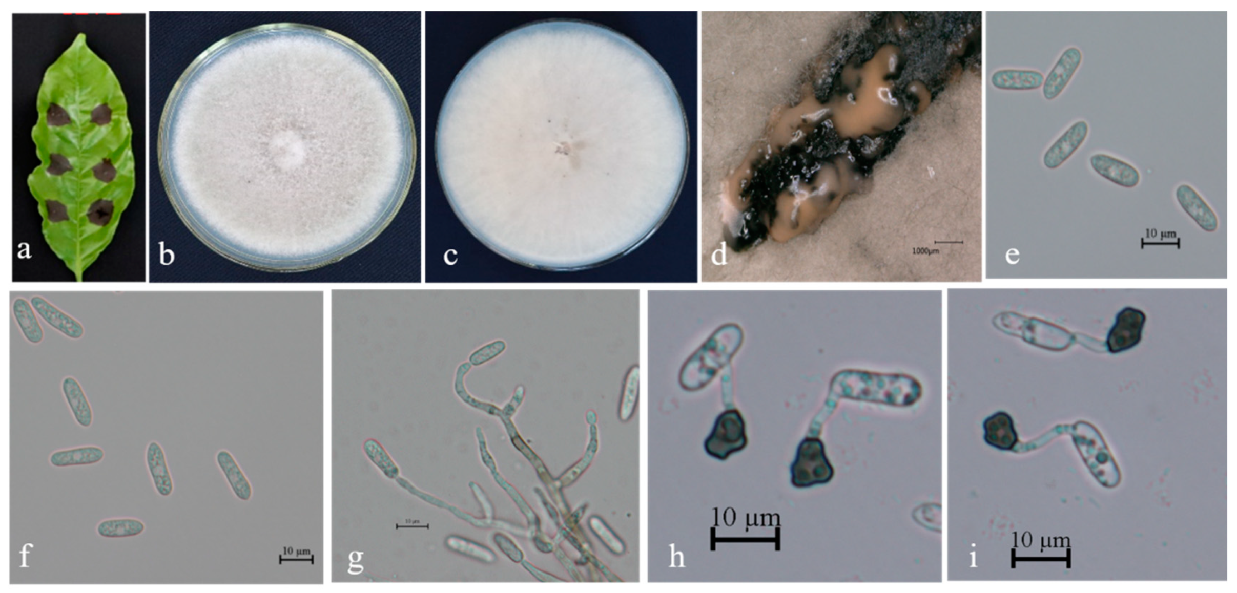
Description. Colonies 63–72 mm in diameter after 7 days at 25 °C on PDA. Aerial mycelium is white and cottony, surface of colony has numerous orange conidiomata; reverse is white at first, then red at center. Conidia are hyaline, aseptate, smooth-walled, and ovoid to cylindrical, with both ends bluntly round, (11.9–) 14.1–19.7 (–23.3) × 3.8–4.9 μm, mean ± SD = 16.3 ± 1.4 × 4.1 ± 0.4 μm, L/W ratio = 3.9. Appressoria formed in slide culture: (8.5-) 10.1–12.9 (–14.5) × 4.1–6.0 μm, mean ± SD = 7.9 ± 1.7 × 5.1 ± 0.9 μm, L/W ratio = 1.8, formed from branched mycelia, terminal, brown-to-dark-brown, variable in shape, irregular. Sexual state not observed in culture (Figure 3).
Specimens examined. Diseased leaves on cultures BSC2-1, BSC2-2, BSC4-1, BSC7-1, BSC7-2, BSC9-1, BSC9-3, and BSC10-2 from Hainan, Baisha, China coffee planting base; on cultures BD15, BD18, and BD23 from Yunnan, Mangshi, Bandong coffee planting base; on cultures HG6 and HG9 from Yunnan, Mangshi, Hougu coffee base; on cultures BEC70A, BEC75A, BEC76B, and BEC77D from Nandao river, Yunnan province coffee planting base; on cultures BEC59A and BEC41B from Yunnan tropical crop institute demonstration base; on cultures BEC91B amd BEC176B from Yunnan agricultural germplasm resources nursery; on culture RBEC193B from Yunnan, Xinghua farm; and on cultures F3-1 and CF2 from Hainan, Fushan coffee planting base.
Colletotrichum nupharicola D.A. Johnson, Carris and J.D. Rogers, 1997 [41].
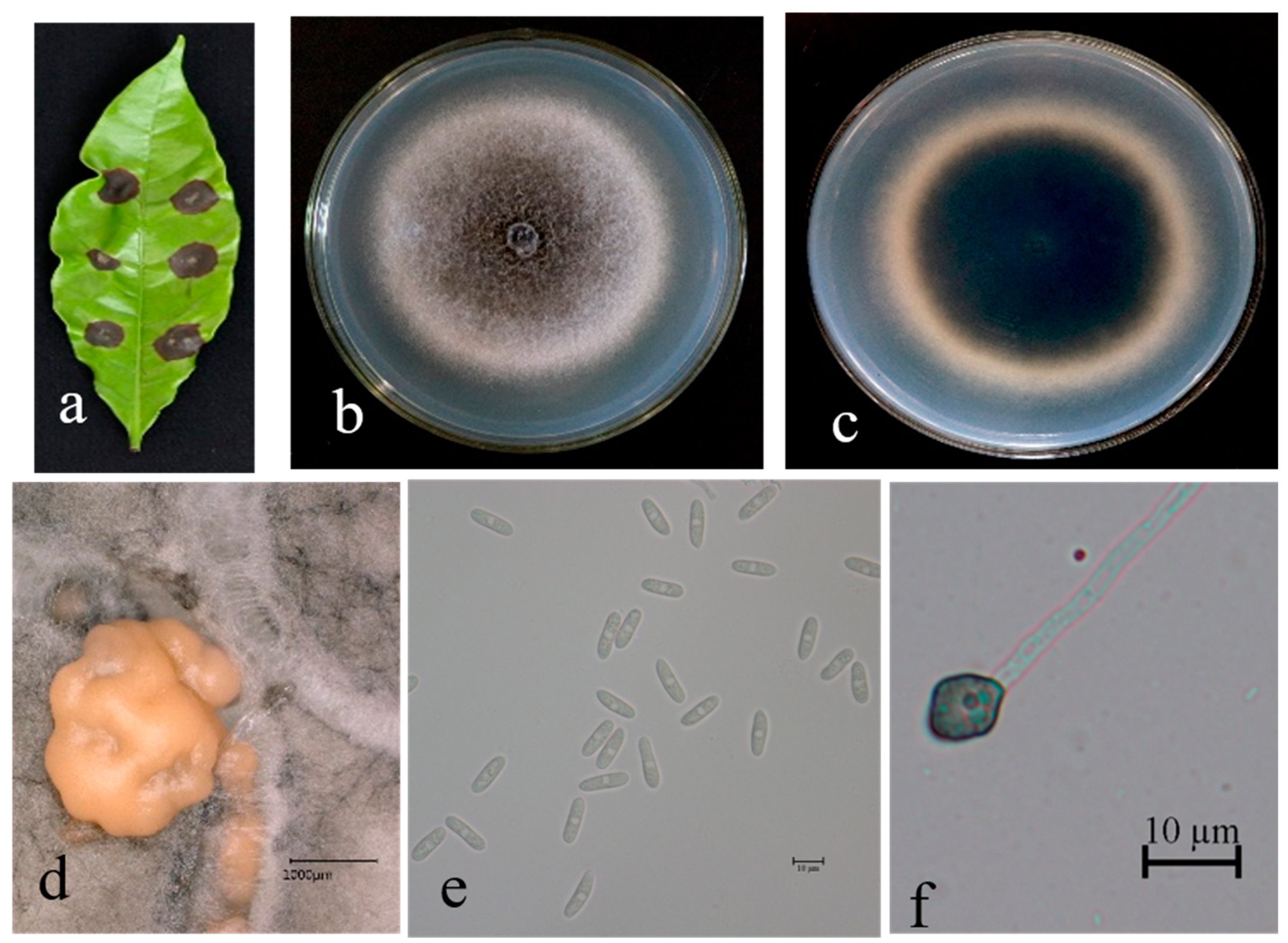
Description. On PDA. Colonies 61–67 mm in diameter after 5 days at 25 °C. Aerial mycelium is dense, cottony, and white; reverse has a white, cottony surface. Conidiomata are apricot and black; vegetative hyphae are hyaline and medium-brown, usually smooth-walled, septate, and branched; chlamydospores and setae are not observed. Conidiophores are either directly formed from hyphae or from cushion of spherical, hyaline cells, septate and sometimes branched. Conidiogenous cells are hyaline-to-pale-brown, cylindrical, and straight to flexuous. Conidia are hyaline, usually aseptate, sometimes become 1–2 septate with age, are smooth-walled, are ovoid to cylindrical or clavate, with both ends rounded, or one end rounded and one end acute, are guttulate, and are granular; (12.5–) 14.0–19.7 (–21.3) × 3.8–4.9 μm, mean ± SD = 16.5 ± 1.2 × 4.3 ± 0.8 μm, L/W ratio = 3.7. Appressoria formed in slide culture: (5.3–) 6.2–8.5 (–9.3) × 4.0–6.1 μm, mean ± SD = 7.8 ± 0.8 × 5.2 ± 0.8 μm, L/W ratio = 1.2, formed from branched mycelia, terminal, brown-to-dark brown, variable in shape, irregular. Sexual state not observed in culture (Figure 4).
Specimens examined. Diseased leaves on cultures BSC1-2, BSC14-1, and BSC15-1 from China, Hainan, Baisha coffee planting base; on cultures HG7, HG8, and HG10 from Yunnan, Mangshi, Hougu coffee base; on cultures RL24, RL25, RL28, RL29, RL30, RL31, RL33, BEC80A, and BEC156A from Yunnan agricultural germplasm resources nursery; on cultures BEC76A and BEC77A from Nandao river, Yunnan province coffee planting base; on cultures BEC10A and BEC14B from Yunnan tropical crop institute demonstration base; and on cultures BEC106B, BEC126B, and BEC127B from Malipo, Wenshan, Yunnan coffee planting base.
Colletotrichum theobromicola Delacr. 1905.
Weir BS, Johnston PR, Damm U. 2012 [25].
Description. On PDA. Colonies raised, white-to-gray, dense aerial mycelium; reverse: gray-to-black, cottony surface of colony with numerous small orange/apricot conidiomata; colonies 62–69 mm in diameter after 5 days at 25 °C. Vegetative hyphae are hyaline-to-medium-brown, usually smooth-walled, septate, and branched; chlamydospores and setae not observed. Conidiophores are either directly formed from hyphae or from cushion of spherical, hyaline cells, septate, and sometimes branched. Conidiogenous cells are hyaline-to-pale-brown, cylindrical, and straight-to-flexuous. Conidia are hyaline, usually aseptate, smooth-walled, ovoid-to-cylindrical or clavate, with both ends rounded, or one end rounded and one end acute, and are guttulate and granular; (11.3–) 13.9–18.6 (–20.6) × 3.5–4.4 μm, mean ± SD = 16.3 ± 0.9 × 4.0 ± 0.5 μm, L/W ratio = 3.9. Appressoria formed in slide culture: (8.9–) 10.2–13.2 (–15.2) × 4.1-6.1 μm, mean ± SD = 7.6 ± 1.3 × 5.1 ± 0.4 μm, L/W ratio = 2.2, formed from branched mycelia, terminal, brown-to-dark-brown, variable in shape, and irregular. Sexual state not observed in culture (Figure 5).
Specimens examined. Diseased leaves on cultures BSC1-3, BSC6-1, BSC8-1, BSC8-3, BSC13-1, and BSC14-2 from China, Hainan, Baisha coffee planting base; on culture IG3 from Hainan, Fushan coffee planting base; on culture RL32 from Yunnan agricultural germplasm resources nursery; and on BEC108A from Malipo, Wenshan, Yunnan coffee planting base.
Colletotrichum karstii Y.L. Yang, Zuo Y. Liu, K.D. Hyde and L. Cai, 2012 [42].
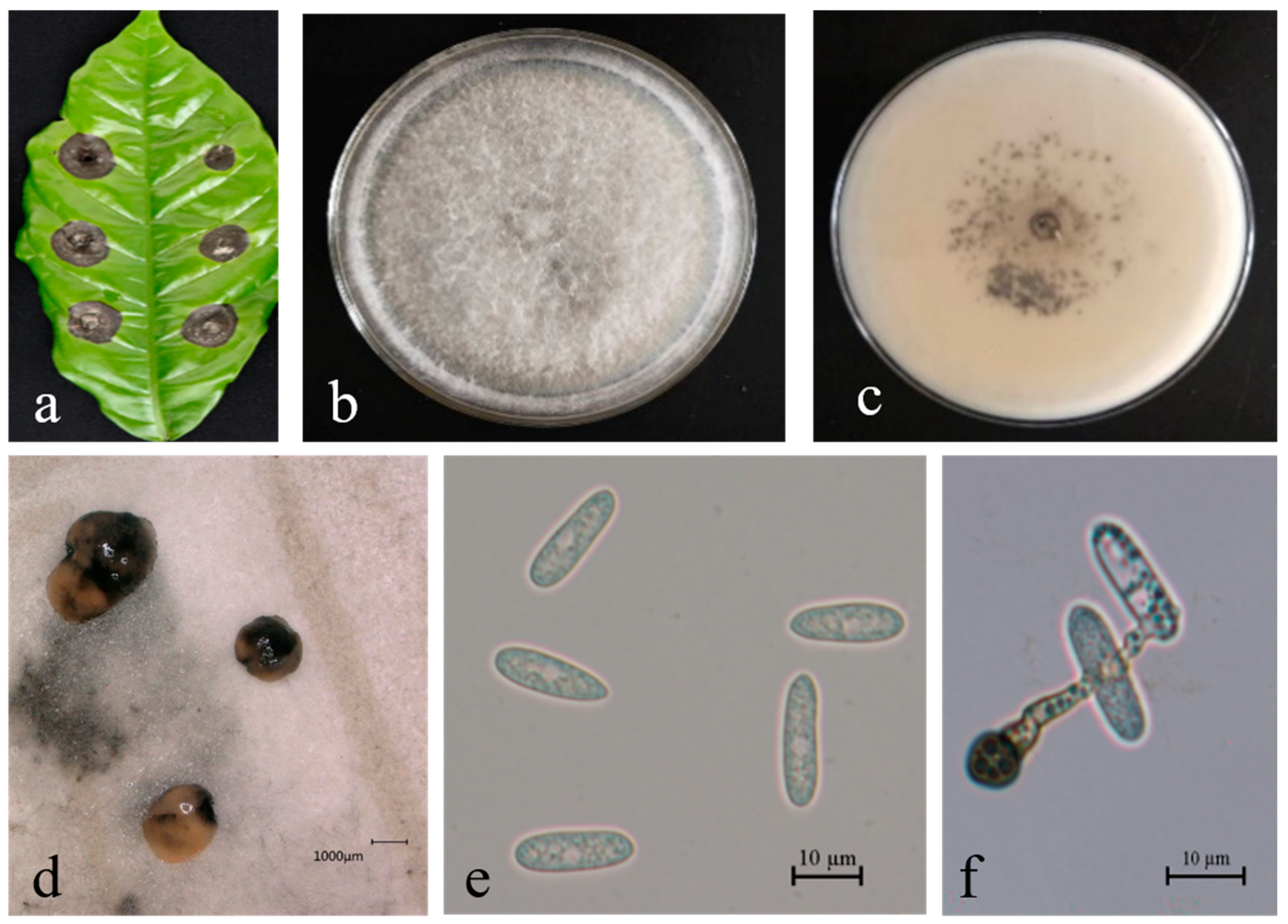
Description. On PDA. Colonies raised, white-to-gray, dense aerial mycelium; reverse: red, cottony surface of colony with numerous small orange/apricot conidiomata; colonies 40–52 mm in diameter after 5 days at 25 °C. Vegetative hyphae hyaline-to-medium-brown, usually smooth-walled, septate, and branched; chlamydospores and setae not observed. Conidiophores are either directly formed from hyphae or from cushion of spherical, hyaline cells, septate and sometimes branched. Conidiogenous cells are hyaline-to-pale-brown, cylindrical, and straight-to-flexuous. Conidia are hyaline, usually aseptate, smooth-walled, and ovoid-to-cylindrical or clavate, with both ends rounded, or one end rounded and one end acute, and are guttulate, granular; (10.5–) 12.1–13.5 (–17.5) × 4.0–5.5 μm, mean ± SD = 13.4 ± 1.7 × 4.5 ± 0.4 μm, L/W ratio = 2.2. Appressoria formed in slide culture: (6.9–) 8.8-8.1 (–9.5) × 8.1–10.0 μm, mean ± SD = 11.6 ± 1.0 × 8.9 ± 0.6 μm, L/W ratio = 1.0, formed from branched mycelia, terminal, brown-to-dark-brown, variable in shape, and irregular. Sexual state not observed in culture (Figure 6).
Specimens examined. Diseased leaves on cultures Bai1, Bai2, Bai4, and Bai5 from Hainan, Baisha coffee planting base; on cultures HG11, HG12, and HG14 from Yunnan, Mangshi, Hougu coffee base; on cultures BEC26C, BEC32A, BEC33B, BEC38A, and RBEC151 from Yunnan tropical crop institute demonstration base; and on cultures BEC52A, BEC107A, and BEC117B from Malipo, Wenshan, Yunnan coffee planting base.
Colletotrichum cliviicola Damm and Crous, 2019, nom. nov. [43].
Description. On PDA. Colonies 58–65 mm in diameter after 5 days at 25 °C. Edge is flat; aetial mycelium is dense, cottony, and gray-to-dark-gray at center; reverse is grayish-green with white halo. Conidiomata are apricot/orange; vegetative hyphae are hyaline-to-medium-brown, usually smooth-walled, septate, and branched. Chlamydospores and setae not observed. Conidiophores are either directly formed from hyphae or from cushion of spherical, hyaline cells, septate and sometimes branched. Conidiogenous cells are hyaline-to-pale-brown, cylindrical, and straight-to-flexuous. Conidia are hyaline and usually aseptate, sometimes becoming 1–2 septate with age; are smooth-walled and ovoid-to-cylindrical or clavate, with both ends rounded, or one end rounded and one end acute; and are guttulate and granular; (13.9–) 15.2–20.1 (–23.4) × 4.8–5.6 μm, mean ± SD = 17.7 ± 1.0 × 5.2 ± 0.7 μm, L/W ratio = 3.4. Appressoria formed in slide culture: (5.2–) 6.2–11.3 (–13.8) × 5.8–9.6 μm, mean ± SD = 8.7 ± 0.5 × 7.5 ± 0.9 μm, L/W ratio = 1.2, formed from branched mycelia, terminal, brown-to-dark-brown, variable in shape, and irregular. Sexual state not observed in culture (Figure 7).
Specimens examined. Diseased leaves on culture FS4-2 from Hainan, Fushan coffee planting base.
Colletotrichum brevisporum Phoulivong, Noireung, L. Cai and K.D. Hyde 2012 [44].
Description. On PDA. Colonies raised, white-to-gray, with dense aerial mycelium; reverse is white; conidiomata are apricot and black. Colonies 58–67 mm in diameter after 5 days at 25 °C. Vegetative hyphae are hyaline-to-medium brown, usually smooth-walled, septate, and branched; chlamydospores and setae not observed. Conidiophores either directly formed from hyphae or from cushion of spherical, hyaline cells, septate and sometimes branched. Conidiogenous cells are hyaline-to-pale-brown, cylindrical, and straight-to-flexuous. Conidia are hyaline and usually aseptate, sometimes becoming 1–2 septate with age; are smooth-walled and ovoid-to-cylindrical or clavate, with both ends rounded, or one end rounded and one end acute; and are guttulate and granular; (13.4–) 17.0–19.5 (−21.7) × 4.8–6.1 μm, mean ± SD = 18.3 ± 1.2 × 5.6 ± 0.8 μm, L/W ratio = 3.3. Appressoria formed in slide culture: (6.5–) 7.5–14.3 (−15.3) × 6.0–11.0 μm, mean ± SD = 10.8 ± 1.1 × 8.2 ± 1.0 μm, L/W ratio = 1.1, formed from branched mycelia, terminal, brown-to-dark-brown, variable in shape, and irregular. Sexual state not observed in culture (Figure 8).
Specimens examined. Diseased leaves on culture BSC15-2 from Hainan, Baisha coffee planting base; and on BEC92 from Yunnan agricultural germplasm resources nursery.
Colletotrichum gigasporum E.F. Rakotoniriana and Munaut, 2013 [45].
Description. On PDA. Colonies 42–56 mm in diameter after 7 days at 25 °C. Aerial mycelium are white and cottony; surface of colony has numerous small orange conidiomata; reverse is white at first, then gray-to-pale-olive at center. Conidia are hyaline, aseptate, smooth-walled, and ovoid-to-cylindrical, with both ends bluntly round; (17.3–) 22.2–28.5 (−31.8) × 8.2–9.8 μm, mean ± SD = 25.7 ± 1.1 × 8.9 ± 0.5 μm, L/W ratio = 2.7. Appressoria formed in slide culture: (8.8–) 10.0–19.1 (−21.3) × 6.5–11.3 μm, mean ± SD = 13.5 ± 1.0 × 8.9 ± 0.8 μm, L/W ratio = 1.2, formed from branched mycelia, terminal, brown-to-dark-brown, variable in shape, and irregular. Sexual state not observed in culture (Figure 9).
Specimen examined. Diseased leaves on culture BEC191A from Yunnan, Xinghua farm.

3.3. Species Diversity of Colletotrichum in China

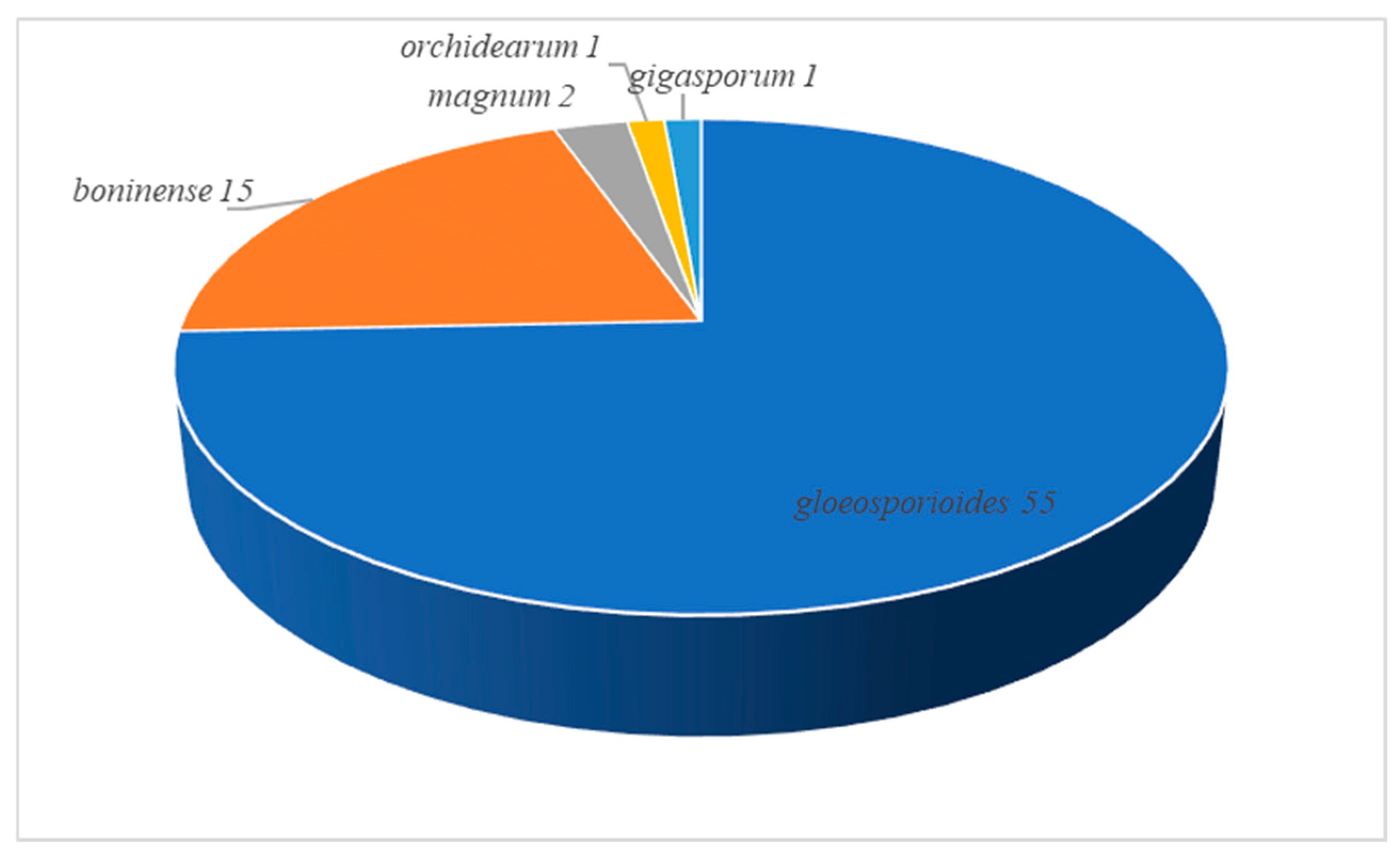
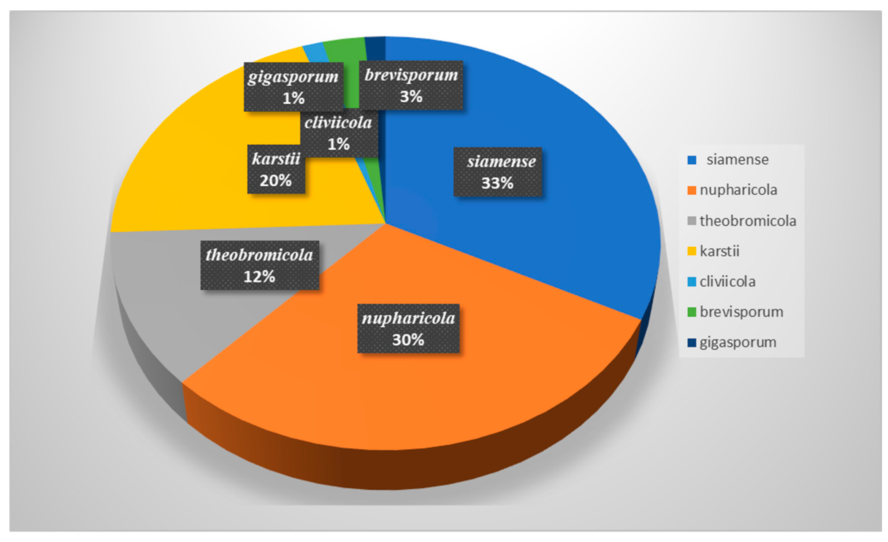
Based on BLASTn searches and phylogenetic analyses of single and multi-locus sequences, 74 strains were assigned to seven species, belonging to five species complexes. The majority of the analyzed strains belonged to the C. gloeosporioides species complex, followed by the C. boninense species complex, the C. magnum species complex, the C. orchidearum species complex, and the C. gigasporum species complex (Figure 10). Among all the identified species, C. siamense was the most common taxon, accounting for 32.43% of the total isolates obtained, followed by C. nupharicola, accounting for 29.73%, and C. theobromicola, accounting for 12.16%. C. karstii accounted for 20.27%, while C. cliviicola, C. brevisporum, and C. gigasporum had an incidence of <5% (Figure 11).

3.4. Pathogenicity Assay

Pathogenicity was tested on detached coffee leaves in vitro for two C. brevisporum (BSC92, BSC15-2), C. cliviicola (FS4-2), one isolate from C. gigasporum (BEC191A), C. karstii (Bai 1, BEC26C), two C. nupharicola (BSC1-2, BEC106B), two C. siamense (BSC4-1, HG 9), and C. theobromicola (BSC1-3, IG3) At 7 dpi, all the tested isolates were pathogenic to leaves of arabica; the incidence of infection ranged from 8.5 to 100%. All isolates except for BEC92 (C. brevisporum) and IG3 (C. theobromicola) were pathogenic to robusta, with the incidence of infection ranging from 33.3 to 100%. The incidence of infection of both young and mature leaves varied among species/isolates (Table 3).

4. Discussion

Effective management of plant diseases relies on a comprehensive understanding of the host, pathogen, and environmental factors. However, in the case of Colletotrichum spp., each of these aspects is highly complex and multi-dimensional. Accurate identification of the pathogen is crucial for developing appropriate and effective disease management strategies. This study represents the first effort to utilize a polyphasic approach to characterize Colletotrichum spp. associated with coffee anthracnose in China.
The polyphasic approach, which integrates phenotypic characteristics and multi-locus phylogeny, has previously been successfully employed to resolve taxonomic complexities within the Colletotrichum genus. This method has proven effective in clarifying species delineations and addressing challenges related to the identification and classification of Colletotrichum species [20,25,46]. In this study, a thorough phenotypic and molecular analysis of 74 isolates revealed that seven species of Colletotrichum are associated as causal agents of coffee anthracnose in China. Specifically, C. siamense, C. nupharicola, and C. theobromicol, belonging to the C. gloeosporioides species complex, and C. karstii, C. cliviicola, C. brevisporum, and C. gigasporum. This finding highlights the diversity and complexity of Colletotrichum species involved in coffee anthracnose pathology. Among the species complexes, the C. gloeosporioides species complexes contain more species than the other species complexes. Nearly 75% of all the isolates obtained from the coffee plantations samples were from the C. gloeosporioides species complex. The majority of Colletotrichum isolates associated with coffee anthracnose in Vietnam belonged to the C. gloeosporioides species complex [21]. Studies also indicate that the C. gloeosporioides species complex is the dominant species complex in rubber trees and coffee trees in Hainan [26]. The composition of Colletotrichum species differ between different regions and coffee varieties; a large-scale survey is needed to confirm this differential distribution.
C. siamense is reported to originate from coffee berries in Thailand [22], and has a wide range of hosts and geographical distributions. C. siamense is reported to infect plants in Australia and Mexico [27,47]. A study found that most isolates from the C. gloeosporioides species complex that caused leaf anthracnose of rubber trees in Hainan were C. siamense [26]. In this study, 32.43% of isolates from coffee plants were identified to be this species.
Although morphological characteristics alone are insufficient to distinguish Colletotrichum species at the individual species level, they remain important taxonomic tools for identifying species within broader species complexes [16]. For instance, conidia of species in the C. gigasporum complex are notably larger than those of other species complexes [13], while typical conidia of the C. boninense complex are cylindrical, with a prominent basal scar [32]. However, many species across various complexes, such as the C. dracaenophilum, C. magnum, and C. orchidearum species complexes, as well as singleton species, produce cylindrical conidia with rounded ends, a feature commonly associated with the C. gloeosporioides complex [43]. In general, species within the C. acutatum, C. bambusicola, C. boninense, C. dracaenophilum, C. gigasporum, C. gloeosporioides, C. magnum, C. orbiculare, and C. orchidearum species complexes produce straight conidia. In contrast, species in the C. caudatum, C. dematium, C. graminicola, C. spaethianum, and C. truncatum complexes produce curved conidia [17,20,30,32,33,34,43,48,49,50,51,52,53]. Notably, species complexes with curved conidia are distributed throughout the phylogenetic tree, suggesting that the evolution of curved spores may have occurred multiple times independently within the genus. This highlights the complexity and diversity of morphological traits in the Colletotrichum taxonomy.
Many Colletotrichum species lack consistent and reliable diagnostic morphological features, making their identification challenging [25]. As a result, molecular methods were employed in this study to accurately identify Colletotrichum species. Both single-locus ApMat and multi-locus sequences (ITS, ACT, CHS-1, GAPDH) were utilized for phylogenetic analysis. The phylogenetic trees generated from both approaches yielded similar results, demonstrating that the ApMat marker provides superior phylogenetic resolution compared to other loci, and is capable of differentiating most species within the C. gloeosporioides species complex. These findings align with previous studies by Silva et al. [24] and Vieira et al. [54]. The ApMat marker shows potential for development as a tool for the rapid diagnosis of species within the C. gloeosporioides complex associated with anthracnose on coffee in China. This could significantly enhance the efficiency and accuracy of species identification in the future.
Research on the relationships between hosts and the pathogenicity of pathogens provides an important theoretical foundation for the control of plant diseases. By uncovering the molecular mechanisms of host–pathogen interactions, more precise and sustainable control strategies can be developed, such as disease-resistant breeding, biological control, and gene-editing technologies [55,56]. In this study, pathogenicity assay confirmed that all tested isolates of Colletotrichum species were pathogenic to Arabica coffee leaves, causing infections with varying degrees of severity. The incidence of infection ranged from 8.5% to 100%, indicating significant variability in the ability of different isolates to infect arabica leaves. Most isolates were also pathogenic to Robusta coffee leaves, except for two isolates: BSC92 (C. brevisporum) and IG3 (C. theobromicola). The incidence of infection on Robusta leaves ranged from 33.3% to 100%, suggesting that Robusta may exhibit some level of resistance or reduced susceptibility to certain isolates compared to Arabica. The study revealed significant differences in pathogenicity among the Colletotrichum species or isolates; C. siamense (isolates BSC4-1 and HG 9) and C. karstii (isolates Bai 1 and BEC26C) showed high pathogenicity, with infection incidences reaching up to 100% on both Arabica and Robusta leaves. C. brevisporum (isolate BSC92) and C. theobromicola (isolate IG3) exhibited lower or no pathogenicity on Robusta leaves, suggesting potential host-specific interactions or reduced virulence in these isolates. Even within the same species, isolates demonstrated variability in their ability to infect coffee leaves. For instance, C. nupharicola isolates (BSC1-2 and BEC106B) showed differing infection incidences, highlighting the importance of genetic or phenotypic variation among isolates. The high pathogenicity of certain isolates, such as those from C. siamense and C. karstii, underscores the need for targeted disease management strategies, particularly for Arabica coffee, which is more susceptible. The reduced susceptibility of Robusta coffee to some isolates (e.g., BSC92 and IG3) suggests potential genetic resistance traits that could be explored for breeding programs to develop more resistant coffee varieties. Variability in pathogenicity among isolates and species highlights the importance of monitoring Colletotrichum populations in coffee plantations, as shifts in species or isolate composition could impact disease severity and spread.

5. Conclusions

In this study, five Colletotrichum species were obtained from coffee leaves with anthracnose symptoms in China. Two species were reported to be associated with coffee for the first time. It is also encouraging that C. kahawae, the main pathogen threatening coffee production in Africa, has not been observed in China. The results of this study can be valuable for developing sustained management strategies for anthracnose of coffee. Further large-scale surveys and pathogenicity testing are necessary before effective disease management strategies can be formulated and implemented.

Supplementary Materials

The following supporting information can be downloaded at https://www.mdpi.com/article/10.3390/microorganisms13030512/s1. Tables S1 and S2. Species and GenBank accession numbers of DNA sequences used in this study with new sequences in bold.

Author Contributions

Y.L. (Ying Lu) and K.Y. conceptualized and designed the experiments; W.W., C.H., Y.L. (Yanqiong Liang) and X.H. (Xing Huang) collected the samples; Y.L. (Ying Lu), W.Z. and X.H. (Xiaoli Hu) conducted the experiments and analyzed the results; Y.L. (Ying Lu) wrote the original draft; W.W., C.H., Y.L. (Yanqiong Liang) and K.Y. revised and corrected the manuscript. All authors have read and agreed to the published version of the manuscript.

Funding

This research was financially supported by the National Key R&D Program of China (2024YFD1400600), the Yunnan International Innovation Platform ‘Yunnan International Joint Laboratory for Coffee Research’ (202403AP140038), and the Innovational Fund for Scientific and Technological Personnel of Hainan Province (KJRC2023B18).

Institutional Review Board Statement

Not applicable.

Informed Consent Statement

Not applicable.

Data Availability Statement

The original contributions presented in this study are included in the article. Further inquiries can be directed to the corresponding author.

Conflicts of Interest

The authors declare no competing interests.

References

  1. Dong, W.J.; Zhao, J.P.; Hu, R.S.; Dong, Y.P.; Tan, L.H. Differentiation of Chinese robusta coffees according to species, using a combined electronic nose and tongue, with the aid of chemometrics. Food Chem. 2017, 229, 743–751. [Google Scholar] [CrossRef] [PubMed]
  2. Avelino, J.; Allinne, C.; Cerda, R.; Willocquet, L.; Savary, S. Multiple-disease system in coffee: From crop loss assessment to sustainable management. Annu. Rev. Phytopathol. 2018, 56, 611–635. [Google Scholar] [CrossRef] [PubMed]
  3. Chu, R.; Wan, L.S.; Peng, X.R.; Yu, Y.M.; Zhang, Z.R.; Zhou, L.; Li, Z.R.; Qiu, M.H. Characterization of new Ent-kaurane diterpenoids of Yunnan Arabica coffee beans. Nat. Prod. Bioprospect. 2016, 6, 217–223. [Google Scholar] [CrossRef] [PubMed]
  4. Vossen, H.A.M.V.D.; Walyaro, D.J. Additional evidence for oligogenic inheritance of durable host resistance to coffee berry disease (Colletotrichum kahawae) in arabica coffee (Coffea arabica L.). Euphytica 2009, 165, 105–111. [Google Scholar] [CrossRef]
  5. Nguyen, T.H.P.; Säll, T.; Bryngelsson, T.; Liljeroth, E. Variation among Colletotrichum gloeosporioides isolates from infected coffee berries at different locations in Vietnam. Plant Pathol. 2009, 58, 898–909. [Google Scholar] [CrossRef]
  6. Zheng, X.L.; He, C.P.; Gao, Y.N.; Zhang, G.N.; Xi, J.G. Biological characteristics and toxicity determination of the Colletotrichum gloeosporioides Penz from Coffea arabica Linn. Chin. J. Trop. Agric. 2015, 35, 94–98. [Google Scholar]
  7. Abreu, M.S.; Ferreira, J.B.; Martins, F.G. Mancha manteigosa no context do complex Colletotrichum em cafeeiros. In Simposio de Manejo de Plantas: Manejo Fitossanitário do Cafeerio; UFLA: Lavras, Brazil, 2008; pp. 105–126. [Google Scholar]
  8. Dean, R.; Van Kan, J.A.; Pretotius, Z.A.; Hammond-Kosack, K.E.; Pietro, A.D.; Spanu, P.D.; Rudd, J.J.; Dickman, M.; Kahmann, R.; Ellis, J. The Top 10 fungal pathogens in molecular plant pathology. Mol. Plant Pathol. 2012, 13, 414–430. [Google Scholar] [CrossRef]
  9. Guarro, J.; Svidzinski, T.E.; Zaror, L.; Foriaz, M.H.; Gené, J.; Fischman, O. Subcutaneous hyalohyphomycosis caused by Colletotrichum gloeosporioides. J. Clin. Microbiol. 1998, 36, 3060–3065. [Google Scholar] [CrossRef]
  10. Damm, U.; Woudenberg, J.H.C.; Cannon, P.F.; Crous, P.W. Colletotrichum species with curved conidia from herbaceous hosts. Fungal Divers. 2009, 39, 45–87. [Google Scholar]
  11. Shiraishi, A.; Araki-Sasaki, A.; Mitani, A.; Miyamoto, H.; Ohashi, Y.C. Clinical characteristics of keratitis due to Colletotrichum gloeosporioides. J. Ocul. Pharmacol. Ther. 2011, 27, 487–491. [Google Scholar] [CrossRef]
  12. Shivaprakash, M.R.; Appannanavar, S.B.; Dhaliwal, M.; Gupta, A.; Gupta, S.; Gupta, A.; Chakrabarti, K. Colletotrichum truncatum: An unusual pathogen causing mycotic keratitis and endophthalmitis. J. Clin. Microbiol. 2011, 49, 2894–2898. [Google Scholar] [CrossRef]
  13. Liu, F.; Cai, L.; Crous, P.W.; Damm, U. The Colletotrichum gigasporum species complex. Persoonia 2014, 33, 83–97. [Google Scholar] [CrossRef] [PubMed]
  14. Buchta, V.; Nekolová, J.; Jirásková, N.; Bolehovská, R.; Wipler, J.; Hubka, V. Fungal keratitis caused by Colletotrichum dematium: Case study and review. Mycopathologia 2019, 184, 441–453. [Google Scholar] [CrossRef] [PubMed]
  15. Bhunjun, C.S.; Phukhamsakda, C.; Jayawardena, R.; Jeewon, R.; Promputtha, I.; Hyde, K.D. Investigating species boundaries in Colletotrichum. Fungal Divers. 2021, 107, 107–127. [Google Scholar] [CrossRef]
  16. Cannon, P.F.; Damm, U.; Johnston, P.R.; Weir, B.S. Colletotrichum: Current status and future directions. Stud. Mycol. 2012, 73, 181–213. [Google Scholar] [CrossRef]
  17. Jayawardena, R.S.; Hyde, K.D.; Damm, U.; Cai, L.; Yan, J.Y. Notes on currently accepted species of Colletotrichum. Mycosphere 2016, 7, 1192–1260. [Google Scholar] [CrossRef]
  18. Marin-Felix, Y.; Groenewald, J.Z.; Cai, L.; Chen, Q.; Crous, P.W. Genera of phytopathogenic fungi: GOPHY 1. Stud. Mycol. 2017, 86, 99–216. [Google Scholar] [CrossRef]
  19. Jayawardena, R.S.; Hyde, K.D.; Chen, Y.J.; Papp, V.; Palla, B.; Papp, D.; Bhunjun, C.S.; Hurdeal, V.G.; Senwanna, C.; Manawasinghe, I.S. One stop shop IV: Taxonomic update with molecular phylogeny for important phytopathogenic genera: 76–100. Fungal Divers. 2020, 103, 87–218. [Google Scholar] [CrossRef]
  20. Damm, U.; Cannon, P.F.; Woudenberg, J.H.C.; Crous, P.W. The Colletotrichum acutatum species complex. Stud. Mycol. 2012, 73, 37–114. [Google Scholar] [CrossRef]
  21. Nguyen, P.T.H.; Pettersson, O.V.; Olsson, P.; Liljeroth, E. Identification of Colletotrichum species associated with anthracnose disease of coffee in Vietnam. Eur. J. Plant Pathol. 2010, 127, 73–87. [Google Scholar] [CrossRef]
  22. Prihastuti, H.; Cai, L.; Chen, H.; Mckenzie, E.H.C.; Hyde, K.D. Characterization of Colletotrichum species associated with coffee berries in northern Thailand. Fungal Divers. 2009, 39, 89–109. [Google Scholar]
  23. Liu, F.; Ma, Z.Y.; Hou, L.W.; Diao, Y.Z.; Wu, W.P.; Damm, U.; Song, S.; Cai, L. Updating species diversity of Colletotrichum, with a phylogenomic overview. Stud. Mycol. 2022, 101, 1–56. [Google Scholar] [CrossRef] [PubMed]
  24. Silva, D.N.; Talhinhas, P.; Várzea, V.; Cai, L.; Paulo, O.S.; Batista, D. Application of the Apn2/MAT locus to improve the systematics of the Colletotrichum gloeosporioides complex: An example from coffee (Coffea spp.) hosts. Mycologia 2012, 104, 396–409. [Google Scholar] [CrossRef] [PubMed]
  25. Weir, B.S.; Johnston, P.R.; Damm, U. The Colletotrichum gloeosporioides species complex. Stud. Mycol. 2012, 73, 115–180. [Google Scholar] [CrossRef]
  26. Cao, X.R.; Xu, X.M.; Che, H.Y.; Swet, J.T.; Luo, D.Q. Characteristics and distribution of Colletotrichum species in coffee plantations in Hainan, China. Plant Pathol. 2019, 6, 1146–1156. [Google Scholar] [CrossRef]
  27. Cristóbal-Martínez, A.L.; de Jesús Yáǹez-Morales, M.; Solano-Vidal, R. Diversity of Colletotrichum species in coffee (Coffea arabica) plantations in Mexico. Eur. J. Plant Pathol. 2017, 147, 605–614. [Google Scholar] [CrossRef]
  28. Sutton, B.C.; Bailey, J.A.; Jeger, M.J. The genus Glomerella and its anamorph Colletotrichum. Colletotrichum Biol. Pathol. Control 1992, 78, 1–26. [Google Scholar]
  29. Cai, L.; Hyde, K.D.; Taylor, P.W.J.; Weir, B.; Waller, J.; Abang, M.M.; Zhang, J.Z.; Yang, Y.L.; Phoulivong, S.; Liu, Z.Y.; et al. A polyphasic approach for studying Colletotrichum. Fungal Divers. 2009, 39, 183–204. [Google Scholar]
  30. Liu, F.; Wang, M.; Damm, U.; Crous, P.W.; Cai, L. Species boundaries in plant pathogenic fungi: A Colletotrichum case study. BMC Evol. Biol. 2016, 16, 81. [Google Scholar] [CrossRef]
  31. Samson, R.A.; Noonim, P.; Meijer, M.; Houbraken, J.A.M.P.; Frisvad, J.C.; Varga, J. Diagnostic tools to identify black aspergilli. Stud. Mycol. 2007, 59, 129–145. [Google Scholar] [CrossRef]
  32. Damm, U.; Cannon, P.F.; Woudenberg, J.H.C.; Johnston, P.R.; Weir, B.S.; Tan, Y.P.; Shivas, R.G.; Crous, P.W. The Colletotrichum boninense species complex. Stud. Mycol. 2012, 73, 75–88. [Google Scholar] [CrossRef] [PubMed]
  33. Damm, U.; Cannon, P.F.; Liu, F.; Barreto, R.W.; Guatimosim, E.; Crous, P.W. The Colletotrichum orbiculare species complex: Important pathogens of field crops and weeds. Fungal Divers. 2013, 61, 29–59. [Google Scholar] [CrossRef]
  34. Damm, U.; O’Connell, R.J.; Groenewald, J.Z.; Crous, P.W. The Colletotrichum destructivum species complex-hemibiotrophic pathogens of forage and field crops. Stud. Mycol. 2014, 79, 49–84. [Google Scholar] [CrossRef] [PubMed]
  35. Huang, L.; Li, Q.C.; Zhang, Y.; Li, D.W.; Ye, J.R. Colletotrichum gloeosporioides sensu stricto is a pathogen of leaf anthracnose on evergreen spindle tree (Euonymus japonicus). Plant Dis. 2016, 100, 672–678. [Google Scholar] [CrossRef]
  36. White, T.J.; Bruns, S.; Lee, S.; Taylor, J. Amplification and direct sequencing of fungal ribosomal RNA genes for phylogenetics. In PCR Protocols a Guide to Methods & Applications; Academic Press: New York, NY, USA, 1990; Volume 18, pp. 315–322. [Google Scholar]
  37. Carbone, I.; Kohn, L.M. A method for designing primer sets for speciation studies in filamentous as conycetes. Mycologia 1999, 91, 553–556. [Google Scholar] [CrossRef]
  38. Templeton, M.D.; Rikkerink, E.H.A.; Solon, S.L.; Crowhurst, W.N. Cloning and molecular characterization of the glyceraldehyde-3-phosphate dehydrogenase-encoding gene and cDNA from the plant pathogenic fungus Glomerella cingulata. Gene 1992, 122, 225–230. [Google Scholar] [CrossRef]
  39. Katoh, K.; Standley, D.M. MAFFT multiple sequence alignment software version 7: Improvements in performance and usability. Mol. Biol. Evol. 2013, 30, 772–780. [Google Scholar] [CrossRef]
  40. Kumar, S.; Stecher, G.; Li, M.; Knyaz, C.; Tamura, K. MEGA X: Molecular evolutionary genetic analysis across computing platforms. Mol. Biol. Evol. 2018, 35, 1547–1549. [Google Scholar] [CrossRef]
  41. Johnson, D.A.; Carris, L.M.; Rogers, J.D. Morphological and molecular characterization of Colletotrichum nymphaeae and C. nupharicola sp. nov. on water-lilies (Nymphaea and Nuphar). Mycol. Res. 1997, 101, 641–649. [Google Scholar] [CrossRef]
  42. Yang, Y.; Liu, Z.; Cai, L.; Hyde, K.D. New species and notes of Colletotrichum on daylilies (Hemerocallis spp.). Trop. Plant Path. 2012, 37, 165–174. [Google Scholar] [CrossRef]
  43. Damm, U.; Sato, T.; Alizadeh, A.; Groenewald, J.Z.; Crous, P.W. The Colletotrichum dracaenophilum, C. magnum and C. orchidearum species complexes. Stud. Mycol. 2019, 92, 1–46. [Google Scholar] [CrossRef]
  44. Noireung, P.; Phoulivong, S.; Liu, F.; Cai, L.; Hyde, K.D. Novel species of Colletotrichum revealed by morphology and molecular analysis. Cryptogam. Mycol. 2012, 33, 347–362. [Google Scholar] [CrossRef]
  45. Rakotoniriana, E.F.; Rafamantanana, M.; Randriamampionona, D.; Rabemanantsoa, C.; Urveg-Ratsimamanga, S.; Jaziri, M.E. Study in vitro of the impact of endophytic bacteria isolated from Centella asiatica on the disease incidence caused by the hemibiotrophic fungus Colletotrichum higginsianum. Antonie Leeuwenhoek 2013, 103, 121–133. [Google Scholar] [CrossRef] [PubMed]
  46. Prathibha, V.H.; Rajesh, M.K.; Dinesh, A.; Patil, B.; Daliyamol; Nagaraja, N.R.; Rajkumar; Sabana, A.A.; Gangaraj, K.P.; Thejasri, K.P.; et al. Multi-gene phylogeny and phenotypic analyses revealed an association of different Colletotrichum species with inflorescence dieback and leaf spot of arecanut in India. Physiol. Mol. Plant Pathol. 2024, 134, 102416. [Google Scholar] [CrossRef]
  47. James, R.S.; Ray, J.; Tan, Y.P.; Shivas, G.R. Colletotrichum siamense, C. theobromicola and C. queenslandicum from several plant species and the identification of C. asianum in the Northern Territory, Australia. Australas. Plant Dis. Notes 2014, 9, 138. [Google Scholar] [CrossRef]
  48. Damm, U.; Sun, Y.C.; Huang, C.J. Colletotrichum eriobotryae sp. nov. and C. nymphaeae, the anthracnose pathogens of loquat fruit in central Taiwan, and their sensitivity to azoxystrobin. Mycol. Prog. 2020, 19, 367–380. [Google Scholar] [CrossRef]
  49. Crouch, J.A. Colletotrichum caudatum s.l. is a species complex. IMA Fungus 2014, 5, 17–30. [Google Scholar] [CrossRef]
  50. Liu, F. Taxonomy and Biodiversity of Colletotrichum. Ph.D. Thesis, University Utrecht, Utrecht, The Netherlands, 2016. [Google Scholar]
  51. Fu, M.; Crous, P.W.; Bai, Q. Colletotrichum species associated with anthracnose of Pyrus spp. in China. Persoonia 2019, 42, 1–35. [Google Scholar] [CrossRef]
  52. Zhang, W.; Damm, U.; Crous, P.W.; Groenewald, J.Z.; Li, Y. Anthracnose disease of carpetgrass (Axonopus compressus) caused by Colletotrichum hainanense sp. nov. Plant Dis. 2020, 104, 1744–1750. [Google Scholar] [CrossRef]
  53. Liu, F.; Weir, B.S.; Damm, U.; Crous, P.W.; Wang, Y.; Liu, B.; Meng, Z.; Cai, L. Unravelling Colletotrichum species associated with Camellia: Employing ApMat and GS loci to resolve species in the C. gloeosporioides complex. Persoonia 2015, 35, 63–86. [Google Scholar] [CrossRef]
  54. Vieira, W.A.S.; Bezerra, P.A.; da Silva, A.C.; Veloso, J.S.; Câmara, M.P.S.; Doyle, V.P. Optimal markers for the identification of Colletotrichum species. Mol. Phylogenet. Evol. 2020, 143, 10664. [Google Scholar] [CrossRef] [PubMed]
  55. Takata, Y.; Komine, M.; Uchikawa, K.; Nozawa, S.; Watanabe, K. Pathogenicity of Colletotrichum species isolated from rotten fruit and asymptomatic flowers of loquat in Nagasaki Prefecture, Japan and characterization of C. nagasakiense Takata & Kyoko Watan. sp. nov. Fungal Dis. 2024, 90, 69–81. [Google Scholar]
  56. Wang, C.B.; Jiang, N.; Xue, H.; Piao, C.G.; Li, Y. Colletotrichum chinense sp. nov. from Yucca gloriosa and C. quercicola sp. nov. from Quercus variabilis in China. MycoKeys 2022, 93, 1–21. [Google Scholar] [CrossRef] [PubMed]
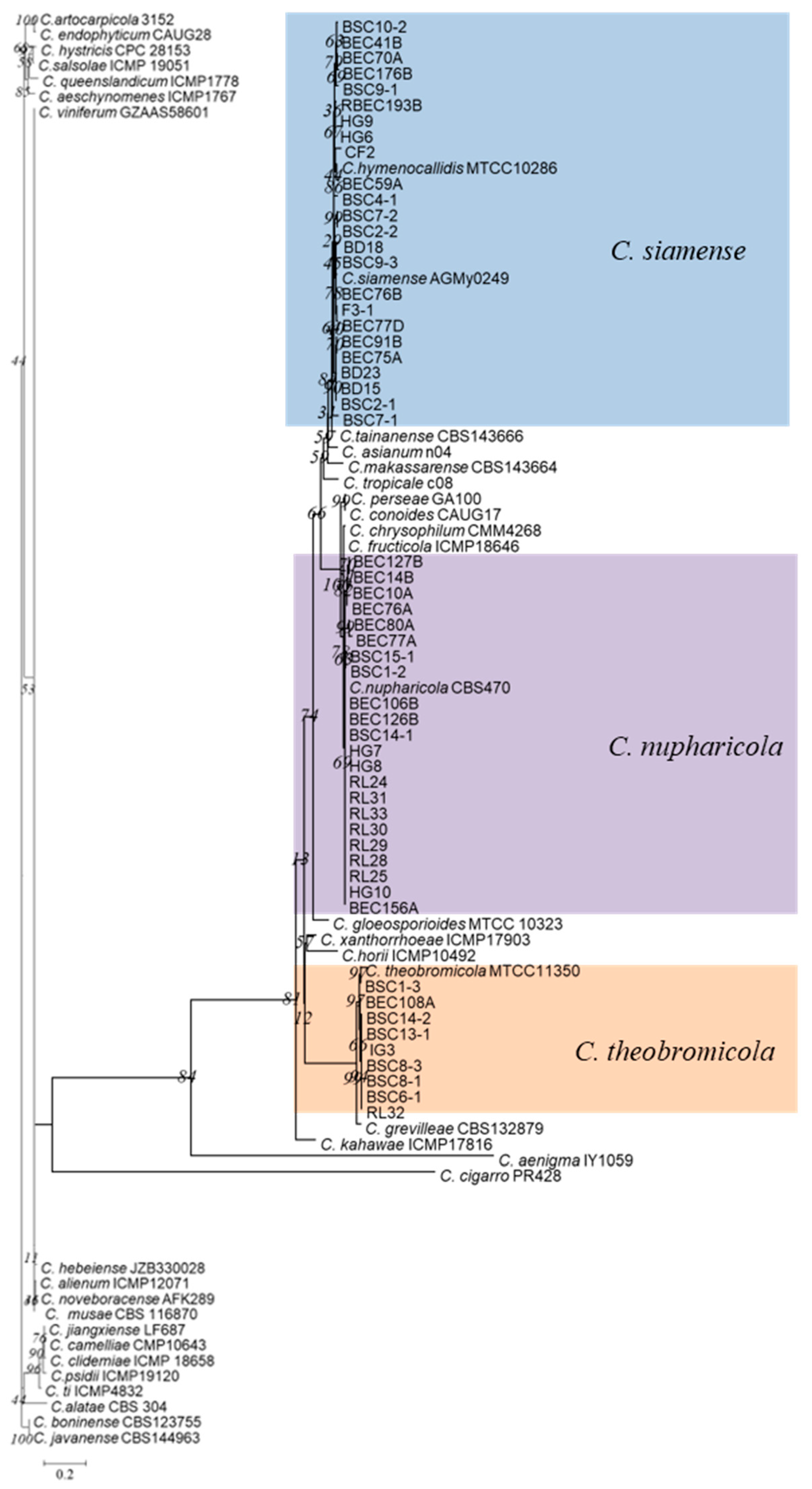
Figure 1. Phylogenetic tree generated by maximum likelihood analysis based on ApMat gene sequences from Colletotrichum gloeosporioides species complexes. Phylogeny is rooted with C. boninense (CBS 123755) and C. javanense (CBS144963) as outgroups.
Microorganisms 13 00512 g001
Figure 2. Phylogenetic tree generated by maximum likelihood analysis based on combined ITS, ACT, CHS-1, and GAPDH gene sequences. Tree displays phylogenetic relationships between Colletotrichum species isolated from coffee plants in China. C. gloesporioides (CBS:119204) and C. destructivum (CBS:136228) were used as outgroups.
Microorganisms 13 00512 g002
Figure 3. Colletotrichum siamense (BSC4-1) on leaf of host plant (a); surface (b) and reverse (c) sides of colony after incubation for 7 days on PDA; (d) conidiomata; (e,f) conidia; (g) conidiophore, conidiogenous cells, and conidia; (h,i) conidial appressoria. Scale bars: 10 µm (ei).
Microorganisms 13 00512 g003
Figure 4. Colletotrichum nupharicola (BSC1-2) on leaf of host plant (a); surface (b) and reverse (c) sides of colony after incubation for 5 days on PDA; (d) conidiomata; (e,f) conidia; (g) conidiophore, conidiogenous cells, and conidia; (h,i) conidial appressoria. Scale bars: 10 μm (ei).
Microorganisms 13 00512 g004
Figure 5. Colletotrichum theobromicola (BSC1-3) on leaf of host plant (a); surface (b) and reverse (c) sides of colony after incubation for 5 days on PDA; (d) conidiomata; (e) conidiophore, conidiogenous cells, and conidia; (f) conidia; (g,h) conidial appressoria. Scale bars: 10 μm (eh).
Microorganisms 13 00512 g005
Figure 6. Colletotrichum karstii (Bai1) on leave of host plant (a); surface (b) and reverse (c) sides of colony after incubation for 5 days on PDA; (d) conidiomata; (e,f) conidia; (g) conidial appressoria. Scale bars: 10 μm (eg).
Microorganisms 13 00512 g006
Figure 7. Colletotrichum cliviicola (FS4-2) on leaf of host plant (a); surface (b) and reverse (c) sides of colony after incubation for 5 days on PDA; (d) conidiomata; (e) conidia; (f) conidial appressoria. Scale bars: 10 μm (e,f).
Microorganisms 13 00512 g007
Figure 8. Colletotrichum brevisporum (BEC92) leaf of host plant (a); surface (b) and reverse (c) sides of colony after incubation for 5 days on PDA; (d) conidiomata; (e) conidia; (f) conidial appressoria. Scale bars: 10 μm (e,f).
Microorganisms 13 00512 g008
Figure 9. Colletotrichum gigasporum (BEC191A) on leaf of host plant (a); surface (b) and reverse (c) sides of colony after incubation for 7 days on PDA; (d) conidiophore, conidiogenous cells, and conidia; (e) conidiomata; (f) conidia; (g) conidial appressoria. Scale bars: 10 μm (f,g).
Microorganisms 13 00512 g009
Figure 10. Statistics of Colletotrichum species complexes in this study.
Microorganisms 13 00512 g010
Figure 11. Statistics of Colletotrichum species in this study.
Microorganisms 13 00512 g011
Table 1. Sampling location of night coffee plantation in Yunan and Hainan, China.
LocationHost PlantGPS CoordinatesNo. of SamplesNo. of Isolates
Yunnan tropical crop institute demonstration baseArabica22°47′47″; 100°58′59″129
Nandao River, Yunnan province coffee planting baseArabica22°36′52″; 101°0′38″96
Malipo, Wenshan, Yunnan coffee planting baseArabica23°12′47″; 104°54′1″107
Yunnan agricultural germplasm resources nurseryArabica22°37′37″; 100°59′47″1713
Yunnan, Xinghua farmArabica24°11′41″; 98°10′11″42
Yunnan, Mangshi, Bandong coffee planting baseArabica24°15′7″; 98°7′28″163
Yunnan, Mangshi, Hougu coffee baseArabica24°21′38″; 98°27′44″108
Hainan, Fushan coffee planting baseRobusta/Arabica19°49′55″; 109°55′33″84
Hainan, Baisha coffee planting baseRobusta/Arabica19°9′55″; 109°28′39″3422
Table 2. Gene regions and PCR primers used in this study.
LocusGenePrimersSequence (5′-3′)Reference
Internal transcribed spacer regions with intervening 5.8S nrRNA geneITSITS1
ITS4
TCCGTAGGTGAACCTGCGG
TCCTCCGCTTATTGATATGC
White et al., 1990 [36]
Partial actin geneACTACT-512F
ACT-783R
ATGTGCAAGGCCGGTTTCGC
TACGAGTCCTTCTGGCCCAT
Carbone et al., 1999 [37]
Partial chitin synthase 1 geneCHS-1CHS-79F
CHS-354R
TGGGGCAAGGATGCTTGGTTGAAG
TGGAAGAACCATCTGTGAGAGTTG
Carbone et al., 1999 [37]
Partial glyceraldehyde-3-phosphate dehydrogenase geneGAPDHGDF
GDR
GCCGTCAACGACCCCTTCATTGA
GGGTGGAGTCGTACTTGAGCATGT
Templeton et al., 1992 [38]
Partial mating type protein 1-2-1 geneApMatAMF
AMR
TCATTCTACGTATGTGCCCG
CCAGAAATACACCGAACTTGC
Silva et al., 2012 [24]
Table 3. The incidence of infection of Colletotrichum spp. inoculated on leaves of arabica and robusta a.
SpeciesIsolatesVarietyArabicaRobusta
Young LeafMature LeafYoung LeafMature Leaf
C. brevisporumBEC92Arabica16.78.500
BSC15-2Robusta33.3255033.3
C. cliviicolaFS4-2Robusta2516.75016.7
C. gigasporumBEC191AArabica7566.75050
C. karstiiBai1Robusta83.37510066.7
BEC26CArabica33.316.75033.3
C. nupharicolaBSC1-2Robusta100507575
BEC106BArabica33.333.35033.3
C. siamenseBSC4-1Robusta507575100
HG 9Arabica75505050
C. theobromicolaBSC1-3Robusta75100100100
IG3Arabica33.316.700
a infection incidence (%) = (number of infected points on leaves/number of inoculated points on leaves) × 100; twelve inoculation points for each leaf.
Disclaimer/Publisher’s Note: The statements, opinions and data contained in all publications are solely those of the individual author(s) and contributor(s) and not of MDPI and/or the editor(s). MDPI and/or the editor(s) disclaim responsibility for any injury to people or property resulting from any ideas, methods, instructions or products referred to in the content.