Case Report

Chlamydia pecorum Infection Associated with Ocular Disease in Goats in Alabama, USA

by
Jenna Workman Stockler
1,
Thomas Passler
1,
Anna-Catherine Bowden
1,
Subarna Barua
2,
Kelly Chenoweth
2,3 and
Chengming Wang
2,3,*
1
Department of Clinical Sciences, College of Veterinary Medicine, Auburn University, Auburn, AL 36849, USA
2
Department of Pathobiology, College of Veterinary Medicine, Auburn University, Auburn, AL 36849, USA
3
Molecular Diagnostic Laboratory, College of Veterinary Medicine, Auburn University, Auburn, AL 36849, USA
*
Author to whom correspondence should be addressed.
Microorganisms 2025, 13(12), 2715; https://doi.org/10.3390/microorganisms13122715
Submission received: 5 November 2025 / Revised: 17 November 2025 / Accepted: 24 November 2025 / Published: 28 November 2025
(This article belongs to the Section Veterinary Microbiology)

Abstract

A herd of approximately 300 Spanish meat goats in central Alabama experienced sporadic ocular, respiratory, and reproductive diseases over two years, prompting diagnostic investigation at Auburn University’s JT Vaughan Large Animal Teaching Hospital. Five representative doelings exhibiting ocular lesions were examined. Clinical signs included conjunctivitis, corneal opacity, uveitis, and, in one severe case, systemic illness. Initial treatment with topical and systemic antibiotics provided incomplete resolution, raising suspicion of infectious keratoconjunctivitis of atypical etiology. Comprehensive diagnostic testing was performed, including aerobic and Mycoplasma cultures, Giemsa staining, and molecular assays. Moraxella bovoculi was cultured; however, Giemsa staining revealed Chlamydia elementary bodies, and a FRET-qPCR with DNA sequencing confirmed high Chlamydia pecorum loads (up to 1.1 × 10⁷ copies/swab). Mycoplasma testing was negative. Extended treatment with systemic and topical oxytetracycline led to gradual clinical improvement, with C. pecorum DNA declining over 22,000-fold and becoming undetectable after five weeks. This case represents the first documented report of C. pecorum–associated keratoconjunctivitis in goats in the United States. The findings underscore the diagnostic importance of molecular assays for detecting intracellular pathogens that may be missed by culture. The protracted treatment course highlights the therapeutic challenges posed by chlamydial infections due to their intracellular persistence. Additionally, the concurrent detection of M. bovoculi suggests the potential for mixed infections influencing disease severity. These results emphasize C. pecorum as an emerging pathogen of caprine ocular disease with implications for herd health and management.

1. Introduction

Chlamydia pecorum is an obligate intracellular bacterium that infects a wide range of ruminants worldwide, including cattle, sheep, and goats [1]. It has been implicated in polyarthritis, pneumonia, enteritis, conjunctivitis, and reproductive disorders such as abortion and infertility [1,2,3]. Although C. pecorum is well recognized as a pathogen of small ruminants in Australia, Europe and Asia [4,5,6,7,8,9], its prevalence and clinical impact in goats in the United States remain poorly defined. Ocular disease caused by C. pecorum is underreported, and uveitis attributable to this pathogen has rarely been described in small ruminants. C. pecorum traditionally causes surface ocular disease rather than intraocular disease with mucoid to mucopurulent ocular discharge, conjunctivitis characterized by significant chemosis, hyperemia, and follicle formation [10,11], and keratitis with corneal neovascularization and rare corneal ulceration. Most ocular cases are bilateral and symmetric [11].
Infectious keratoconjunctivitis (IKC) in goats is commonly attributed to Mycoplasma spp., especially Mycoplasma conjunctivae, with Moraxella ovis possibly contributing to ocular disease [12]. Treatment of these, typically extracellular, pathogens with standard topical or systemic antimicrobial therapy often results in rapid resolution. In contrast, the intracellular lifestyle of C. pecorum can complicate medical management, require extended treatment, and pose challenges to animal health and productivity.
This report describes an outbreak of ocular disease in Spanish goats in Alabama, with clinical manifestations ranging from conjunctivitis and keratitis to severe uveitis. Molecular diagnostics confirmed C. pecorum as the etiologic agent. One doeling required more than five weeks of systemic and topical antimicrobial treatment before bacterial resolution, underscoring the chronicity and therapeutic challenges of this infection. To our knowledge, this represents one of the first documented cases of C. pecorum-associated uveitis in goats in the United States.

2. Case Description

Auburn University’s JT Vaughan Large Animal Teaching Hospital (AULATH) was contacted by the herd owner of approximately 300 Spanish meat goats in central Alabama. Over two years, the owner had observed sporadic cases of ocular, respiratory, and reproductive disease. The goats were maintained on mixed summer grass pasture across two farms. FAMACHA scores were assessed monthly, and animals were dewormed as needed; no vaccines had been administered within the last four years. The owner reported recurrent episodes of “pinkeye,” characterized by epiphora to mucopurulent discharge, blepharospasm, and corneal opacity. Previous treatment attempts with topical triple antibiotic ointment (neomycin sulfate, polymyxin B, bacitracin) and systemic oxytetracycline provided incomplete resolution, although affected animals eventually improved. The owner also noted late-term abortions in breeding-age does and pneumonia in goats of various ages, although veterinary evaluation had not previously been sought.
Five approximately 10-month-old doelings with representative clinical signs were presented to AULATH. On admission, all animals were bright, alert, and responsive. Doelings #1 and #5 exhibited bilateral ocular discharge and conjunctivitis. Doeling #3 was febrile and had corneal edema with a pinpoint fluorescein-positive ulcer in the right eye. Doeling #2 had no ocular abnormalities. Doeling #4 was systemically ill, with dull mentation, poor body condition (1/5), tachypnea, increased bronchovesicular sounds, pale mucous membranes, and severe ocular lesions—specifically, a melting corneal ulcer in the right eye and uveitis in the left eye.
Based on the clinical presentation, infectious keratoconjunctivitis was suspected. Initial therapy included topical oxytetracycline/polymyxin B ointment (Terramycin®, Zoetis, Kalamazoo, MI, USA) every six hours. Doelings #3 and #4 also received IV flunixin meglumine (1.1 mg/kg q12h for 3 days) and topical autologous serum every six hours. Tulathromycin (5 mg/kg SQ, Draxxin®, Zoetis) was administered to all goats on day 3 due to persistence or progression of ocular lesions. Four doelings (#1, #2, #3, #5) improved sufficiently for discharge on day 8 with continued topical therapy.
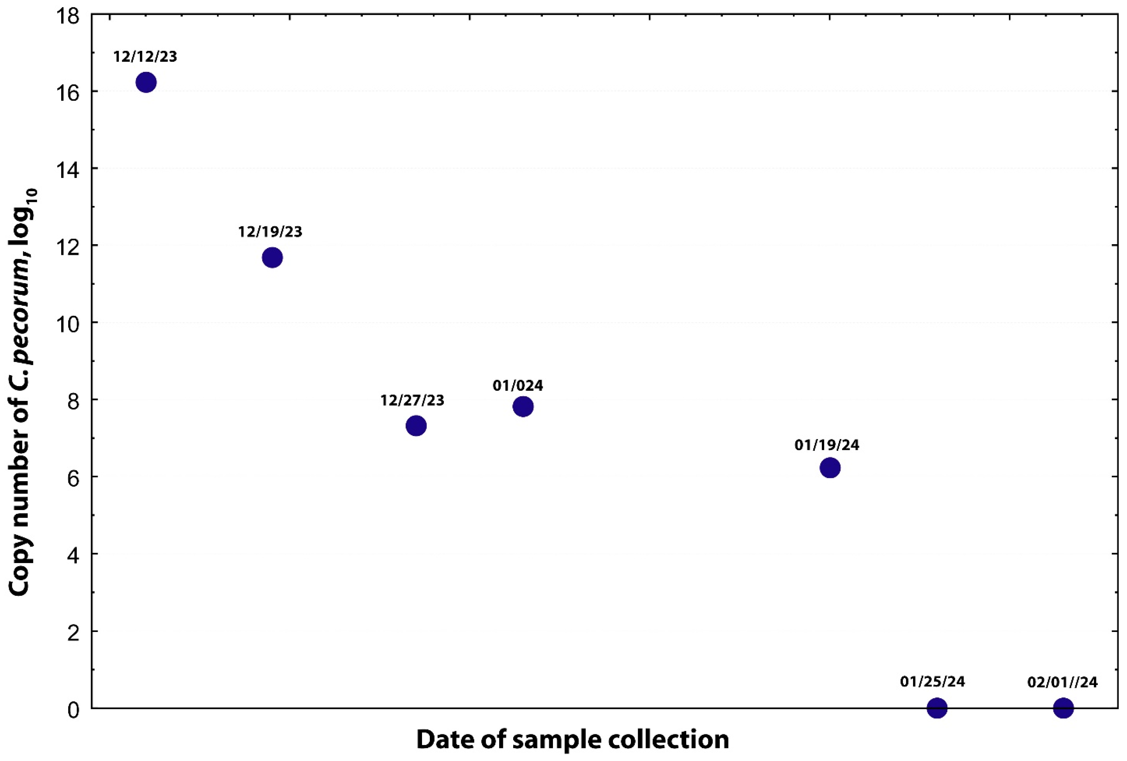
Doeling #4 exhibited the most severe ocular involvement and was donated to AULATH for further diagnostic evaluation and treatment. Conjunctival swabs were submitted for aerobic culture after administration of topical anesthesia (proparacaine hydrochloride USP 0.5%, Alcon, Fort Worth, TX, USA), Mycoplasma culture, and PCR testing for Chlamydia and Mycoplasma at a commercial laboratory (Newport Laboratories—Vaxxinova), as well as for Chlamydia-specific PCR at the Molecular Diagnostic Laboratory, Auburn University College of Veterinary Medicine. Moraxella bovoculi was detected by the Newport Laboratory, while Mycoplasma testing was negative. Giemsa staining revealed abundant Chlamydia elementary bodies in conjunctival samples (Figure 1). FRET-qPCR followed by DNA sequencing and BLASTn as described [9,13], confirmed a high C. pecorum load, reaching up to 11,255,000 (1016.2) copies per swab (Figure 2).
Systemic oxytetracycline (22 mg/kg SQ q48h, Zoetis, Kalamazoo, MI, USA) was administered for 11 doses and discontinued due to potential for toxicity. Topical oxytetracycline therapy was continued for an additional 27 days. Extra-label drug use was performed with the consent of AULATH clinicians and complied with provisions of AMDUCA and 21 CFR 530. To confirm infection clearance, all antimicrobials were later withdrawn, and serial conjunctival PCR testing was performed (Figure 1). C. pecorum DNA levels decreased 22,510-fold following five weeks of treatment, and PCR results ultimately became negative. The doeling responded well and was discharged 120 days after initial hospitalization.

3. Discussion

This case highlights several clinically important aspects of Chlamydia pecorum infection in goats. Although C. pecorum has been well documented in ruminants globally, ocular disease—particularly keratoconjunctivitis—has rarely been reported in goats, and, to our knowledge, not previously in the United States. These findings emphasize the importance of considering C. pecorum in the differential diagnosis of caprine keratoconjunctivitis, alongside Moraxella and Mycoplasma spp. [14].
The protracted treatment required in this case illustrates the therapeutic challenges associated with chlamydial infections. The affected doeling required more than five weeks of systemic and topical antimicrobial therapy—considerably longer than typical regimens for infectious keratoconjunctivitis of other etiologies. This prolonged course likely reflects the intracellular persistence of C. pecorum, which limits antimicrobial penetration and complicates pathogen clearance. The use of molecular diagnostics was critical for confirming the etiologic agent and monitoring therapeutic response, as standard bacterial cultures would not have identified this intracellular organism. The elementary bodies on conjunctival cytology, while helpful to confirm suspicions in this case, may not be reliable in every case and appear in only one-third of cases [11].
At the herd level, the owner’s reports of abortions and respiratory disease may also be consistent with systemic or subclinical C. pecorum infections, given this pathogen’s broad tissue tropism. Although both Moraxella bovoculi and C. pecorum were detected, the clinical presentation, high chlamydial load, and treatment response strongly support C. pecorum as the primary cause of keratoconjunctivitis and uveitis in this case. Uveitis is a previously undocumented ocular sign of C. pecorum and may reflect breakdown of the blood-ocular barrier secondary to systemic inflammation. Nonetheless, the concurrent detection of M. bovoculi raises the possibility of mixed infections influencing disease expression or severity. Reports indicate that goats may be asymptomatic carriers of Chlamydia infections [6], and a 2024 study indicated Mycoplasma conjunctivae, a common co-infection with Chlamydia, can be cross-infected between livestock and wild ungulates [15]. Both wildlife and asymptomatic herd members could be sources of continued outbreaks. Future research is warranted to investigate the interactions between M. bovoculi and C. pecorum. Determination of whether co-infection exacerbates ocular inflammation or alters disease progression and treatment outcomes in goats and other ruminants is important for future directions.
Tulathromycin treatment appeared to be effective against C. pecorum infection in this study, likely due to its large volume of distribution and resulting high intracellular drug concentrations. In contrast, topically administered oxytetracycline achieves therapeutic levels only briefly and does not adequately penetrate the corneal or conjunctival tissues. Moreover, the cellular penetration of oxytetracycline is substantially lower than that of most macrolides and some fluoroquinolones. Overuse of oxytetracycline has also been associated with the development of tetracycline resistance in Chlamydia suis [16].

4. Conclusions

This case represents one of the first documented reports of Chlamydia pecorum–associated keratoconjunctivitis and uveitis in goats in the United States. It highlights the diagnostic value of molecular assays for accurate pathogen identification, the potential need for extended therapy, and the broader significance of C. pecorum as an emerging pathogen affecting the health and productivity of caprine.

Author Contributions

Conceptualization: J.W.S., T.P. and C.W.; Methodology: J.W.S., T.P., A.-C.B., S.B., K.C. and C.W. Writing: J.W.S., T.P. and C.W. All authors have read and agreed to the published version of the manuscript.

Funding

This study was supported, in part, by the Molecular Diagnostic Laboratory at Auburn University College of Veterinary Medicine.

Institutional Review Board Statement

The samples used in this case report were convenience samples obtained through routine diagnostic services. The use of such samples for research purposes is exempt from IACUC review.

Informed Consent Statement

The samples used in this case report were convenience samples obtained through routine diagnostic services. The use of such samples for research purposes is exempt from consent statements from the animal owners.

Data Availability Statement

All data related to this work are included in the manuscript.

Conflicts of Interest

The authors declare no conflicts of interest.

References

  1. Nietfeld, J.C. Chlamydial infections in small ruminants. Vet. Clin. N. Am. Food Anim. Pract. 2001, 17, 301–314. [Google Scholar] [CrossRef] [PubMed]
  2. Bommana, S.; Walker, E.; Desclozeaux, M.; Jelocnik, M.; Timms, P.; Polkinghorne, A.; Carver, S. Molecular and serological dynamics of Chlamydia pecorum infection in a longitudinal study of prime lamb production. PeerJ 2018, 6, e4296. [Google Scholar] [CrossRef] [PubMed]
  3. Giannitti, F.; Anderson, M.; Miller, M.; Rowe, J.; Sverlow, K.; Vasquez, M.; Canton, G. Chlamydia pecorum: Fetal and placental lesions in sporadic caprine abortion. J. Vet. Diagn. Investig. 2016, 28, 184–189. [Google Scholar] [CrossRef] [PubMed]
  4. Dias-Alves, A.; Cabezon, O.; Borel, N.; Lopez-Olvera, J.R.; Mentaberre, G.; Lavin, S.; Fernandez Aguilar, X. Molecular Detection and Identification of Chlamydiaceae in the Eyes of Wild and Domestic Ruminant Hosts from Northern Spain. Pathogens 2021, 10, 383. [Google Scholar] [CrossRef] [PubMed]
  5. Fernandez-Aguilar, X.; Rossi, L.; Cabezon, O.; Giorgino, A.; Victoriano Llopis, I.; Frey, J.; Lopez-Olvera, J.R. Infectious keratoconjunctivitis and occurrence of Mycoplasma conjunctivae and Chlamydiaceae in small domestic ruminants from Central Karakoram, Pakistan. Vet. Rec. 2017, 181, 237. [Google Scholar] [CrossRef] [PubMed]
  6. Jayasekara, P.P.; Jenkins, C.; Gerber, P.F.; Olmo, L.; Xaikhue, T.; Theppangna, W.; Walkden-Brown, S.W. Case-control study to identify the causative agents of ophthalmia and conjunctivitis in goats in Savannakhet province of Lao PDR. Vet. Microbiol. 2024, 296, 110195. [Google Scholar] [CrossRef] [PubMed]
  7. Jelocnik, M.; Forshaw, D.; Cotter, J.; Roberts, D.; Timms, P.; Polkinghorne, A. Molecular and pathological insights into Chlamydia pecorum-associated sporadic bovine encephalomyelitis (SBE) in Western Australia. BMC Vet. Res. 2014, 10, 121. [Google Scholar] [CrossRef] [PubMed]
  8. Jelocnik, M.; Self, R.; Timms, P.; Borel, N.; Polkinghorne, A. Novel sequence types of Chlamydia pecorum infect free-ranging Alpine ibex (Capra ibex) and red deer (Cervus elaphus) in Switzerland. J. Wildl. Dis. 2015, 51, 479–483. [Google Scholar] [CrossRef] [PubMed]
  9. Li, J.; Guo, W.; Kaltenboeck, B.; Sachse, K.; Yang, Y.; Lu, G.; Zhang, J.; Luan, L.; You, J.; Huang, K.; et al. Chlamydia pecorum is the endemic intestinal species in cattle while C. gallinacea, C. psittaci and C. pneumoniae associate with sporadic systemic infection. Vet. Microbiol. 2016, 193, 93–99. [Google Scholar] [CrossRef] [PubMed]
  10. Wilsmore, A.J.; Dagnall, G.J.; Woodland, R.M. Experimental conjunctival infection of lambs with a strain of Chlamydia psittaci isolated from the eyes of a sheep naturally affected with keratoconjunctivitis. Vet. Rec. 1990, 127, 229–231. [Google Scholar] [PubMed]
  11. Hopkins, J.B.; Stephenson, E.H.; Storz, J.; Pierson, R.E. Conjunctivitis associated with chlamydial polyarthritis in lambs. J. Am. Vet. Med. Assoc. 1973, 163, 1157–1160. [Google Scholar] [CrossRef] [PubMed]
  12. Gulaydin, A.; Gulaydin, O.; Akgul, M.B.; Sindak, N.; Yildirim, O. Investigation of the presence of Chlamydia spp., Mycoplasma spp. and Moraxella ovis in infectious keratoconjunctivitis cases in sheep and goats in Siirt province and evaluation of clinical findings. Pol. J. Vet. Sci. 2024, 27, 183–191. [Google Scholar] [CrossRef] [PubMed]
  13. Guo, W.; Li, J.; Kaltenboeck, B.; Gong, J.; Fan, W.; Wang, C. Chlamydia gallinacea, not C. psittaci, is the endemic chlamydial species in chicken (Gallus gallus). Sci. Rep. 2016, 6, 19638. [Google Scholar] [CrossRef] [PubMed]
  14. Meekins, J.M.; Apley, M.D.; Lubbers, B.; Peddireddi, L.; Rankin, A.J. Evaluation of conjunctival bacterial flora in a herd of goats in the Midwestern United States. Vet. Ophthalmol. 2017, 20, 40–45. [Google Scholar] [CrossRef] [PubMed]
  15. Lopez-Olvera, J.R.; Ramirez, E.; Martinez-Carrasco, C.; Granados, J.E. Wildlife-Livestock Host Community Maintains Simultaneous Epidemiologic Cycles of Mycoplasma conjunctivae in a Mountain Ecosystem. Vet. Sci. 2024, 11, 217. [Google Scholar] [CrossRef] [PubMed]
  16. Unterweger, C.; Schwarz, L.; Jelocnik, M.; Borel, N.; Brunthaler, R.; Inic-Kanada, A.; Marti, H. Isolation of Tetracycline-Resistant Chlamydia suis from a Pig Herd Affected by Reproductive Disorders and Conjunctivitis. Antibiotics 2020, 9, 187. [Google Scholar] [CrossRef] [PubMed]
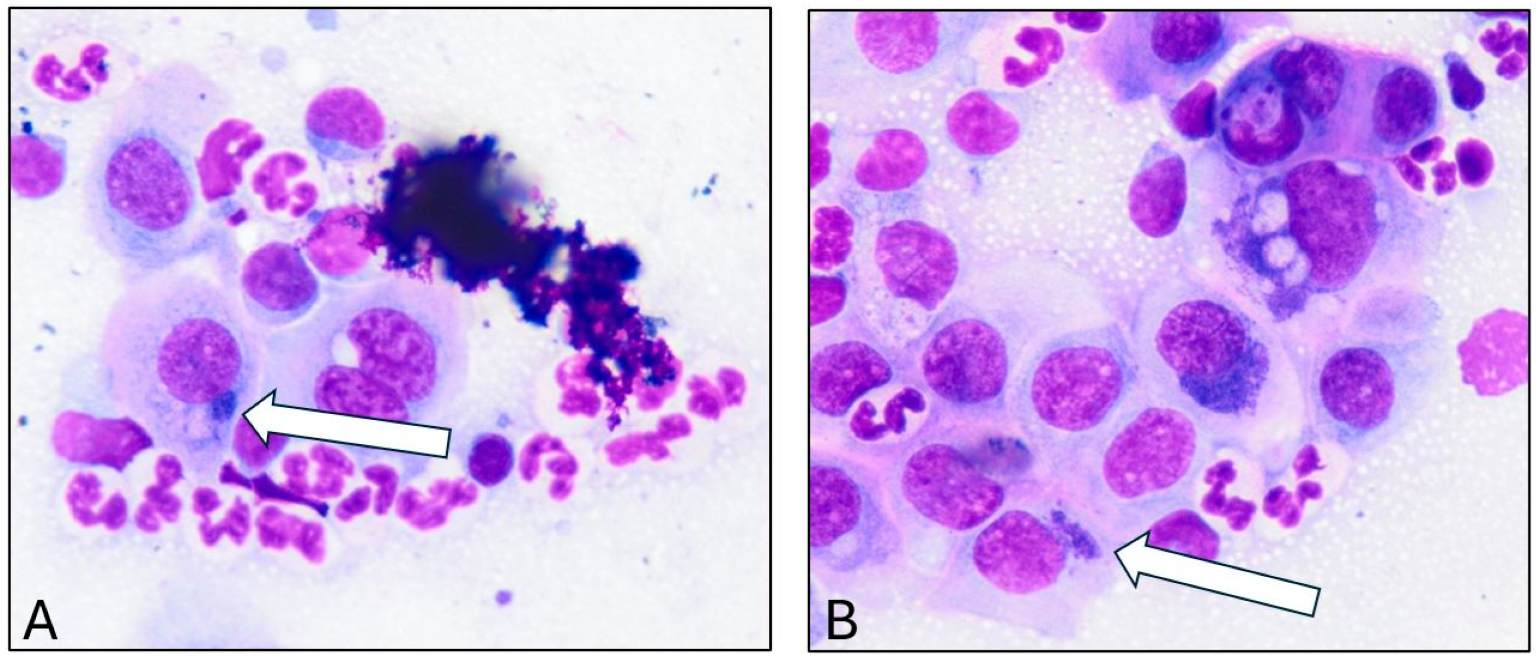
Figure 1. (A,B) Elementary bodies within conjunctival epithelial cells after Giemsa staining. The white arrows indicate the juxtanuclear, basophilic, intracytoplasmic inclusions typical of Chlamydia infection.
Microorganisms 13 02715 g001
Figure 2. Clearance of C. pecorum infection in a goat following prolonged oxytetracycline therapy. Systemic oxytetracycline (22 mg/kg, subcutaneous, every 48 h) was administered for 11 doses (extra-label use) and discontinued to prevent potential toxicity. Topical oxytetracycline therapy was continued for an additional 27 days. To confirm microbial clearance, all antimicrobial treatments were withdrawn, and serial conjunctival swabs were analyzed by means of PCR. The initial C. pecorum load was 1016.2 copies per swab, which decreased by approximately 22,510-fold after five weeks of treatment.
Microorganisms 13 02715 g002
Disclaimer/Publisher’s Note: The statements, opinions and data contained in all publications are solely those of the individual author(s) and contributor(s) and not of MDPI and/or the editor(s). MDPI and/or the editor(s) disclaim responsibility for any injury to people or property resulting from any ideas, methods, instructions or products referred to in the content.