Article

Virome and Microbiome of Florida Bats Illuminate Viral Co-Infections, Dietary Viral Signals, and Gut Microbiome Shifts

by
Julia E. Paoli
1,2,3,4,
Thanaporn Thongthum
1,2,†,
Maclean Bassett
1,3,†,
Jakob Beardsley
1,3,
Massimiliano S. Tagliamonte
1,3,5,
Melanie N. Cash
1,3,
Jason Spertus Newman
1,
Lisa M. Smith
6,
Benjamin D. Anderson
1,2,
Marco Salemi
1,3,
Kuttichantran Subramaniam
1,7,
Michael E. von Fricken
1,2,4,8,
Elizabeth Braun de Torrez
6,
Verity Mathis
9 and
Carla N. Mavian
1,3,4,8,*
1
Emerging Pathogens Institute, University of Florida, Gainesville, FL 32610, USA
2
Department of Environmental and Global Health, College of Public Health and Health Professions, University of Florida, Gainesville, FL 32610, USA
3
Department of Pathology, Immunology and Laboratory Medicine, College of Medicine, University of Florida, Gainesville, FL 32610, USA
4
One Health Center of Excellence, College of Public Health and Health Professions, University of Florida, Gainesville, FL 32610, USA
5
Bioinformatics Core, Interdisciplinary Center for Biotechnology Research, University of Florida, Gainesville, FL 32610, USA
6
Terrestrial Mammal Research, Florida Fish and Wildlife Research Institute, Florida Fish and Wildlife Conservation Commission, Gainesville, FL 32601, USA
7
Department of Infectious Diseases and Immunology, College of Veterinary Medicine, University of Florida, Gainesville, FL 32610, USA
8
Global Health Program Smithsonian’s, National Zoo & Conservation Biology, Washington, DC 20008, USA
9
Florida Museum of Natural History, University of Florida, Gainesville, FL 32611, USA
*
Author to whom correspondence should be addressed.
These authors contributed equally to this work.
Microorganisms 2025, 13(11), 2625; https://doi.org/10.3390/microorganisms13112625
Submission received: 12 September 2025 / Revised: 12 November 2025 / Accepted: 18 November 2025 / Published: 19 November 2025
(This article belongs to the Special Issue Advances in Viral Metagenomics)

Abstract

Florida’s bat virome remains poorly characterized despite the state’s high bat species diversity and conservation importance. We characterized viral metagenomes from rectal tissues, anal swabs, and feces of Myotis austroriparius and Tadarida brasiliensis sampled across north Florida. We recovered a near-complete Hubei virga-like virus 2 (HVLV2) genome from T. brasiliensis feces, a finding consistent with an arthropod-derived dietary signal rather than active bat infection. An Alphacoronavirus (AlphaCoV) was detected in two M. austroriparius specimens, including one with a putative co-infection involving an Astrovirus (AstV), the first detection of AstV in Florida bats to date. Parallel profiling of the M. austroriparius gut microbiome highlighted compositional differences in the co-infected individual relative to AlphaCoV-only and virus-negative bats, suggestive of potential associations between viral detection and gut microbial shifts. Our study expands the known viral diversity in Florida bat populations, and demonstrates how metagenomics can simultaneously illuminate host diet, viral exposure, and gut microbial ecology. This approach provides a scalable framework for monitoring how diet, microbiome composition, and environmental pressures shape the bat virome, and inform conservation and zoonotic risk assessments.

1. Introduction

Bats (order Chiroptera) are known natural reservoirs for several emerging viruses with pandemic potential including filoviruses, paramyxoviruses, and coronaviruses [1]. Compared to other mammals, bats host the greatest number of zoonotic pathogens owing in part to their remarkable tolerance for viral infection [2]. Since the occurrence of several high-profile outbreaks linked to spillover from bats, research has primarily focused on investigating the virome composition of bats as a surveillance tool [3,4,5,6,7,8]. The bat gut microbiome may play a key role in regulating host immunity and antiviral responses, potentially contributing to bats’ increased tolerance to viral infections and their role as reservoirs for zoonotic pathogens [9,10,11]. However, the mechanisms underlying these interactions remain poorly understood [11,12,13]. Further studies are needed to determine how the gut microbiome of bats may be affecting pathogen exposure, evolution of tolerance, and spillover mechanisms [11,14].
Bats play a crucial role in maintaining healthy ecosystems and supporting agricultural production by controlling insect populations through their insectivorous diets [15,16]. In the United States, bats frequently consume agricultural pests responsible for significant economic losses, providing natural pest control services valued at billions of dollars in annual crop savings [15,17]. Bat feeding habits have important effects on the composition and function of their gut microbiomes, with insectivorous bats found to have greater microbial community diversity than frugivorous and hematophagous species [10,18,19]. In addition to diet, the gut microbial composition is influenced by host factors such as species, age, and sex, as well as environmental variables including season and geographic location [18,20,21,22,23,24,25].
The use of metagenomic next-generation sequencing (mNGS) has greatly expanded knowledge of the diversity of bat-borne viruses worldwide [5,7,26,27,28,29]. The agnostic nature of mNGS allows for detection of diet-associated viruses from insects, such as members of the Dicistroviridae and Iflaviridae families, in addition to mammalian viruses [30,31]. Previous research has emphasized the need to characterize viral diversity among North American bat species, as most studies of bat-borne viruses have focused on Asia, Europe, and Africa [32,33,34]. In North America, several bat-borne alphacoronaviruses (AlphaCoVs) have been identified in at least six species of bats, including those from Brazilian free-tailed bats (Tadarida brasiliensis) in Florida and other states [32,35]. Astroviruses (AstVs), which belong to the genus Mamastrovirus in mammals and Avastrovirus in birds, infect a wide range of hosts, including humans, and generally exhibit low virus–host specificity [36]. Despite their zoonotic relevance, only one complete AstV genome from North American bats is publicly available; it was sequenced from bat feces collected in a mixed-species bat roost in California [37].
Florida harbors thirteen native species of bats year-round, including the Southeastern myotis (Myotis austroriparius) and the Brazilian free-tailed bat (T. brasiliensis) [38]. Both species are insectivorous and presumed to be largely confined within the state, as the warm climate and consistent food availability reduce the need for long-distance migrations [38]. Despite this diversity of bat species, the virome, microbiome and diet of Florida bats remain poorly studied. Published work has only documented AlphaCoV [35], with no AstV found in Florida bat populations to date. Similarly, few studies have used DNA metabarcoding to characterize the diet of Florida bats, though the research that has been conducted highlights their ecological and economic importance, particularly through consumption of agricultural pests [39,40,41]. Research on the microbiome of Florida’s bats is limited, and to our knowledge, no published studies have examined the microbiome of the state’s bat population.
To better understand the viruses circulating among two of Florida’s common native bat species, we used mNGS to examine the virome of M. austroriparius and T. brasiliensis sampled across north Florida. We detected one M. austroriparius specimen harboring a potential co-infection of AstV and AlphaCoV, as well as another M. austroriparius specimen with only AlphaCoV. In addition, we recovered a near-full-length HVLV2 genome from T. brasiliensis feces, consistent with dietary intake of mosquitoes. Finally, we characterized the composition and diversity of the gut microbiota in the M. austroriparius museum specimens to explore potential associations between viral presence and microbial composition shifts. Our findings provide insight on the understudied virome of native Florida bats, which will aid bat-conservation efforts and public health preparedness.

2. Materials and Methods

2.1. Sample Preparation from Whole Frozen Bat Specimens

We obtained adult, whole specimens of T. brasiliensis (N = 5) and M. austroriparius (N = 5) from the Florida Museum of Natural History (FLMNH) (Table 1, Figure 1). These bats belonged to a mixed-species roost under the roof of a fire station in Gilchrist County, Florida during December 2021. The specimens were donated to the FLMNH after bats were killed accidentally during a power washing event. Following death, a wildlife rehab center brought the frozen carcasses to the FLMNH where they were weighed, sexed, and stored at −20 °C for preservation (Table S1). From the FLMNH archives, we additionally obtained frozen T. brasiliensis adult whole specimens collected from Escambia (N = 1) and Volusia (N = 1) counties, and one specimen from an unknown Florida county (Table 1, Figure 1). These three specimens were given to FLMNH from wildlife rehab centers, and the causes of death were unknown at the time of donation. The whole animal specimen was dissected, and the rectum was removed from each bat using a sterile scalpel (Thermo Fisher Scientific, Waltham, MA, USA). The rectum was then transferred into a sterile Petri dish, and another sterile scalpel was used to excise appropriately sized fragments (10 mm diameter) for RNA extraction. Excised rectal tissues were placed in RNAlater (Thermo Fisher Scientific, Waltham, MA, USA), then homogenized using a motorized pestle (Thermo Fisher Scientific, Waltham, MA, USA) and used immediately for RNA extraction. The samples were donated to the FLMNH and were not collected for a university-based project. Therefore, an Institutional Animal Care and Use Committee (IACUC) protocol was not required by the University of Florida.

2.2. Anal Swab and Fecal Sample Collection

We obtained opportunistic samples from adult T. brasiliensis that were actively and passively sampled on 21 November 2022, at one occupied roost (bat house) at Camp Blanding Joint Training Center in Clay County, Florida (Table 1, Figure 1). Fecal and anal swab samples were actively collected by capturing T. brasiliensis emerging from the roost using a 2 m × 2 m harp trap (Bat Conservation and Management, Carlisle, PA, USA). The harp trap was placed 1 m in front of the roost entrance one hour before sunset. Captured bats were immediately removed from the harp trap and placed individually in clean cloth bags for up to one hour or until they defecated, whichever occurred first. The sex, age, reproductive condition, mass, and forearm length for each bat were recorded. Feces were collected from the bag of each bat when present using ethanol flame-sterilized forceps. Anal swab samples were collected from each bat by first dipping a small sterile swab (VWR International, Radnor, PA, USA) in DNA/RNA Shield (Zymo Research, Irvine, CA, USA), then gently inserting the swab into the rectum and rotating 3–5 times. Bats were released at the site of capture immediately after processing. Fecal samples were passively collected at the same bat house by placing four 8 L buckets directly beneath the bat house. Approximately 1 h after the bats emerged, the buckets were removed and sterile forceps used to collect individual fecal pellets. Swabs and feces were immediately placed into 2 mL screw-cap microtubes (Sarstedt, Nümbrecht, Germany) containing DNA/RNA Shield (Zymo Research, Irvine, CA, USA) and stored at −80° C until analyzed. All live-capture and handling activities followed American Society of Mammalogists (ASM) guidelines [42] and were completed by experienced bat biologists with the Florida Fish and Wildlife Conservation Commission (FWC) under safe and humane protocols approved by the FWC.
In addition, on 10 April 2024, we obtained guano pellets (N = 12) from a T. brasiliensis colony roosting at the University of Florida’s bat houses in Alachua County using a passive sampling approach that did not require handling of bats (Table 1, Figure 1). Collection buckets were placed beneath the roosts to allow feces to accumulate naturally one hour prior to sunset to avoid interference with bat emergence from the house. After approximately 20 min, the buckets were retrieved, and individual guano pellets were collected using sterile forceps. Samples were pooled together for preservation in DNA/RNA Shield (Zymo Research, Irvine, CA, USA) and stored at −80 °C until processing. The fecal samples were collected non-invasively, without animal handling, manipulation, or roost disturbance; therefore, no IACUC protocol was required by the University of Florida.

2.3. RNA Extraction and NGS Library Preparation of Museum Samples and Anal Swabs

RNA was extracted from the museum specimen rectum tissue homogenate using the AllPrep DNA/RNA Mini Kit (Qiagen, Hilden, Germany) following the manufacturer’s protocol for total RNA isolation from animal tissues. For the anal swab samples, the ZymoBIOMICS DNA/RNA Miniprep kit was used following manufacturer’s protocol for RNA extraction (Zymo Research, Irvine, CA, USA). Subsequently, for both the museum tissue samples and anal swab samples, eluted RNA was treated with ezDNase (Thermo Fisher Scientific, Waltham, MA, USA) to remove residual genomic DNA, following the manufacturer’s protocol. First-strand cDNA was synthesized using SuperScript IV Reverse Transcriptase (Thermo Fisher Scientific, Waltham, MA, USA). Second-strand cDNA was synthesized using the Second Strand cDNA Synthesis Kit (Thermo Fisher Scientific, Waltham, MA, USA). The resulting double-stranded cDNA was purified using the Monarch PCR DNA Cleanup Kit (New England Biolabs, Ipswich, MA, USA).
Next-generation sequencing (NGS) libraries were prepared using the NEBNext Ultra II FS DNA Library Prep Kit for Illumina (New England Biolabs, Ipswich, MA, USA), following the low-input protocol (≤100 ng) with NEBNext Multiplex Oligos (Dual Index Primers Set 1) for Illumina (New England Biolabs, Ipswich, MA, USA). Libraries were amplified and bead-purified using AMPure XP beads (Beckman Coulter, Brea, CA, USA) at a 0.9× ratio. Library quality and fragment size distribution were assessed using the Agilent 4150 TapeStation system (Agilent Technologies, Santa Clara, CA, USA). Libraries containing primer-dimer peaks (<150 bp) were subjected to additional rounds of bead purification (0.9×) and re-analyzed until satisfactory quality was achieved. Final libraries were size-selected on the SageELF electrophoresis system (Sage Science, Beverly, MA, USA) to isolate fragments between 200 and 500 bp. Quantification of libraries was determined with the Qubit dsDNA HS Assay Kit (Thermo Fisher Scientific, Waltham, MA, USA) on the Qubit 4 Fluorometer (Thermo Fisher Scientific, Waltham, MA, USA). The final pooled library was sequenced on a NovaSeq 6000 system (Illumina, San Diego, CA, USA) using the NovaSeq 6000 S1 Reagent Kit v1.5 (paired-end, 2 × 150) at the University of Florida Interdisciplinary Center for Biotechnology Research (UF ICBR). Fastq files were deposited in SRA under BioProject accession PRJNA1305506.

2.4. RNA Extraction and NGS Library Preparation of Fecal Samples

For RNA extraction, guano pellets were thawed and homogenized in DNA/RNA Shield (Zymo Research, Irvine, CA, USA) using a motorized pestle. The homogenate was centrifuged at 2000 rpm for 5 min, and the resulting supernatant was used for RNA extraction with the MagMAX Viral RNA Isolation Kit (Thermo Fisher Scientific, Waltham, MA, USA). To remove potential PCR inhibitors, RNA samples were further purified using the OneStep PCR Inhibitor Removal Kit (Zymo Research, Irvine, CA, USA). RNA was extracted from guano pellets according to the previously published RAPIDprep assay [43] for the following steps: gDNA removal, rRNA depletion, first-strand cDNA synthesis, second-strand cDNA synthesis, and double-stranded cDNA cleanup. Two in-house modifications were made to the original RAPIDprep protocol to reduce reagent usage. First, the QIAseq FastSelect (Qiagen, Hilden, Germany) dilution mix was prepared at a 1:10 ratio by combining 1 µL of 5S/16S/23S rRNA removal reagent, 1 µL of HMR (human/mouse/rat) depletion reagent, and 8 µL of nuclease-free water. Second, the Sequenase dilution was modified to use 1 unit per reaction, corresponding to 0.08 µL of Sequenase Version 2.0 DNA Polymerase (Thermo Fisher Scientific, Waltham, MA, USA) mixed with 0.92 µL of nuclease-free water per reaction.
NGS libraries were prepared from the purified cDNA with the NEBNext Ultra II FS DNA Library Prep Kit for Illumina (New England Biolabs, Ipswich, MA, USA), following the low-input protocol (≤100 ng) with NEBNext Multiplex Oligos (Dual Index Primers Set 1) for Illumina (New England Biolabs, Ipswich, MA, USA). Libraries were amplified and bead-purified (0.9×) using Omega Bio-tek Mag-Bind Total Pure NGS beads (Omega Bio-tek, Norcross, GA, USA). Library fragment size distribution was assessed using the Agilent 4150 TapeStation system (Agilent Technologies, Santa Clara, CA, USA). Libraries containing primer-dimer peaks (<150 bp) were subjected to additional rounds of bead purification (0.9×) and re-analyzed until satisfactory quality was achieved. Quantification of libraries was determined with the Qubit dsDNA HS Assay Kit (Thermo Fisher Scientific, Waltham, MA, USA) on the Qubit 4 Fluorometer (Thermo Fisher Scientific, Waltham, MA, USA). Final libraries were normalized and pooled for sequencing with the NextSeq 1000 platform (Illumina, San Diego, CA, USA) using the NextSeq 1000/2000 P1 Reagents cartridge (paired-end, 2 × 150 bp). Fastq files were deposited in SRA under BioProject accession PRJNA1305506.

2.5. Metagenomic Data Analysis

Fastq files were trimmed with Trimmomatic v.0.39 [44] to remove adapters and poor quality regions. De novo assembly was performed with MEGAHIT v.1.2.9 [45] to generate contigs which were then assessed with NCBI’s VecScreen to remove potential vector contamination (https://www.ncbi.nlm.nih.gov/tools/vecscreen; accessed 17 October 2025). Cleaned contigs were compared to the BLAST nt/nr v2.17.0 [46] and InterPro v101.0 [47,48] databases for taxonomic identification and genome annotation. We obtained single, contiguous contigs for all samples with one exception (Table 2): for sample Ma_Frozen_3, two non-overlapping AlphaCoV contigs were recovered (k119_6311 and k119_670). Fastq files were deposited in SRA under BioProject accession PRJNA1305506. Viral contigs were submitted to GenBank with the following accessions: PX462113 (contig k119_6311); PX470470 (contig k119_670); PX470471 (contig k119_1119); PX482536 (contig k119_11258); PX462114 (contig k141_4344) (Table S2).

2.6. Recombination Analysis

For each virus of interest, representative complete genomes and their associated metadata were compiled from NCBI Virus to generate background datasets [49]. Multiple sequence alignments of these full-genome datasets were produced with MAFFT v.7.520 [50] and manually polished in AliView v1.28 [51]. In the AlphaCoV dataset, two Tadarida brasiliensis bat alphacoronavirus 2 reference sequences (OP700657.1 and OP715780.1) from the same study were included and hereafter referred to by the abbreviated virus name ACoV-2-Tb. Because only partial genomes were obtained in this study, recombination analysis was restricted to the full-genome background datasets, which were assessed using RDP5 with default settings for linear sequences [52]. Recombinant sequences were defined as those with significant evidence of recombination signal supported by at least six of the seven following methods: RDP, GENECONV, Chimaera, MaxChi, BootScan, SiScan, and 3Seq [52]. Identified recombinant regions were removed.

2.7. Phylogenetic Analysis

Following recombination analysis, we obtained several recombinant-free alignments for the publicly available genomes and produced several alignments with our viral contigs for phylogenetic analysis using MAFFT v.7.520 [50]. All alignments were constructed using single, contiguous de novo-assembled viral contigs, with one exception: to retain all AlphaCoV genomic signal from sample Ma_Frozen_3 in the phylogeny, a partial genome was constructed by inserting Ns between the two contigs (k119_6311 and k119_670), followed by masking these ambiguous regions during multiple sequence alignment in AliView v1.28 [51]. This resulted in a concatenated alignment consisting only of confidently assembled positions, for phylogenetic purposes only, referred to as ACoV-Ma3-FL-2021. The other recovered contigs used for phylogenetic analysis are hereafter referred to by the virus name followed by host specimen, location, and year as follows: (1) AstV-Ma4-FL-2021 (contig k119_11258) and (2) HVLV2-Tb15-FL-2024 (contig k141_4344). Maximum likelihood (ML) trees were obtained from (1) alignments based on the full contigs and (2) alignments based on individual genes: for AstV, the RNA-dependent RNA polymerase (RdRp) and open reading frame 2 (ORF2) genes; and for AlphaCoV, the spike, envelope, membrane, and nucleocapsid genes. ML phylogenetic tree reconstruction was performed with IQ-TREE v.2.2.2.7 [53] using the Bayesian information criteria (BIC) to infer the best nucleotide substitution model based on the data and 2000 ultrafast bootstrap replicates for support [54]. ML trees were outgroup rooted with closely related viruses and visualized in R using the ggtree v3.2.1 package [55]. Evidence of sufficiently robust phylogenetic signal of sequence data was assessed by likelihood mapping implemented in IQ-TREE v.2.2.2.7 per recommended guidelines: side/center areas of the likelihood mapping must include <40% of the unresolved quartets [56,57]. Pairwise genetic distance analysis was conducted with Molecular Evolutionary Genetics Analysis (MEGA) v.11 [58] using the models for the number of nucleotide differences and p-distance with the following parameters: Gamma distribution (shape parameter = 4.0), 100 bootstrap replicates for variance estimation, and gaps treated as complete deletions. The 1st, 2nd, 3rd, and non-coding sites were included.

2.8. DNA Extraction and 16S Library Preparation

From the M. austroriparius museum specimen rectum tissue homogenate, DNA was extracted using the AllPrep DNA/RNA Mini Kit (Qiagen, Hilden, Germany) following the manufacturer’s protocol for genomic DNA isolation from animal tissues. To assess the initial presence of bacterial DNA in the extracts, the V3–V4 hypervariable region of the bacterial 16S rRNA gene was amplified by PCR. Reactions were performed in a total volume of 25 μL using Q5 High-Fidelity 2X Master Mix (New England BioLabs, Ipswich, MA, USA). Each reaction contained 8 μL of input DNA, 12.5 μL of Q5 2X Master Mix, 2 μL of nuclease-free water, and 1.25 μL each of forward and reverse primers (10 μM): S-D-Bact-0341-b-S-17 (5′-CCTACGGGNGGCWGCAG-3′) and S-D-Bact-0785-a-A-21 (5′-GACTACHVGGGTATCTAATCC-3′) [59]. PCR cycling conditions were as follows: initial denaturation at 98 °C for 30 s; 35 cycles of 98 °C for 10 s, 55 °C for 30 s, and 72 °C for 30 s; followed by a final extension at 72 °C for 2 min. Amplicons were visualized by electrophoresis on a 1% agarose gel stained with ethidium bromide and compared against a 50–2000 bp DNA ladder (Bio-Rad, Hercules, CA, USA).
16S rRNA libraries were prepared using the Quick-16S Library Prep Kit (Zymo Research, Irvine, CA, USA) according to the manufacturer’s protocol for low microbial DNA samples. Library amplification was confirmed by nucleic acid quantification using the 4150 TapeStation System (Agilent Technologies, Santa Clara, CA, USA). Confirmed libraries were pooled according to the Quick-16S protocol and the final pooled library was quantified using both a Qubit fluorometer (Thermo Fisher Scientific, Waltham, MA, USA) and TapeStation analysis. Final libraries were diluted to 4 nM, then denatured and diluted to a final concentration of 10 pM with a 15% PhiX (Illumina, San Diego, CA, USA) spike-in. Libraries were sequenced on the MiSeq platform (Illumina, San Diego, CA, USA) using the MiSeq Reagent Kit v3 (paired-end, 2 × 300 bp). Fastq files were deposited in SRA under BioProject accession PRJNA1305506.

2.9. 16S rRNA Reads Processing and Statistical Analysis

Raw paired-end 16S rRNA gene sequences were processed using Quantitative Insights Into Microbial Ecology 2 (QIIME2) v2024.10 [60]. Sequences were quality filtered, joined, and denoised using the Deblur plugin [61] with a trim length of 400 base pairs, a mean per-nucleotide error threshold of 0.00125, and a minimum read count of 10 per sample. These parameters generated amplicon sequence variants (ASVs) and removed chimeric and low-abundance sequences. A rooted phylogenetic tree was constructed with the Fasttree pipeline [62] for downstream diversity analyses. For taxonomic assignment, a Naive Bayes classifier was trained on SILVA [63,64,65] 138 reference sequences trimmed in silico to the V3–V4 region using primers 341F (CCTACGGGNGGCWGCAG) and 806R (GACTACHVGGGTATCTAATCC) [65,66]. Core diversity metrics were computed using the core-metrics-phylogenetic pipeline with a rarefaction depth of 6000 sequences per sample, based on alpha rarefaction curves showing a plateau in observed feature richness. Alpha diversity was assessed using Shannon diversity index, implemented in QIIME2’s v2024.10 alpha-group-significance plugin. Beta diversity was evaluated using Bray–Curtis dissimilarity [67], weighted UniFrac [68], and unweighted UniFrac [69] distances. The results were visualized using principal coordinate analysis (PCoA). Differences in community composition across sample types were assessed using Kruskal–Wallis tests [70,71] and Permutational Multivariate Analysis of Variance (PERMANOVA) with 999 permutations [72]. We did not perform Kruskal–Wallis tests or PERMANOVA to compare bacterial composition across samples because each group comprised a single sample (no biological replicates).

2.10. Geospatial Mapping

Maps were generated with QGIS v.3.28.1 using GPS coordinates collected during sampling events (QGIS Association, 2024). Shapefiles for the state and counties of Florida were obtained from https://geodata.floridagio.gov/ (accessed 8 July 2025).

3. Results

3.1. First Detection of Astrovirus Detection in Florida Bats

We performed mNGS on RNA from rectal tissue dissected from five adult M. austroriparius and T. brasiliensis whole frozen specimens housed at FLMNH to assess viromes (Table 1, Figure 1). We did not detect any viral contigs (mammalian nor insect) in the five T. brasiliensis frozen museum specimens tested, nor in M. austroriparius specimens: Ma_Frozen_1, Ma_Frozen_2, and Ma_Frozen_5 (Table S1). In M. austroriparius specimen Ma_Frozen_4 we recovered a single Astrovirus (AstV) contig (k119_11258) that was 3292 bp long (accounting for 51% of the AstV genome) (Table 2). Hereafter, the contig will be referred to as AstV-Ma4-FL-2021 (Table S2). BLASTN v2.17.0 analysis showed the highest nucleotide identity to bat AstV from Myotis daubentoniid sampled in Denmark in 2015 (MZ218054.1; 80% nucleotide identity) and from Myotis chinensis sampled in China in 2020 (OR951016.1; 80% nucleotide identity). AstV-Ma4-FL-2021 spans 54% of the ORF1b and 100% of ORF2 plus the 3′ untranslated region (Figure S1); within ORF1b, it covers 69% of the RdRp gene (Figure S1).
According to current International Committee on Taxonomy of Viruses (ICTV) guidelines, the Astroviridae family comprises two genera: Avastrovirus, which infect birds, and Mamastrovirus, which infect mammals [73]. We therefore compiled a reference dataset including all Mamastrovirus reference sequences from GenBank, along with the sequences showing the highest nucleotide identity to AstV-Ma4-FL-2021 based on BLAST v2.17.0 analysis (Table S3). Recombination analysis indicated evidence of recombination in reference datasets for the full genome and ORF2; however, there was no evidence of recombination occurring in the RdRp gene. Using recombination free alignments, we calculated pairwise genetic distance which showed that AstV-Ma4-FL-2021 shares 70–71% nucleotide identity across the ORF1b-ORF2 region with three Myotis-associated bat AstVs (MZ218054.1, MZ218053.1, and OR951016.1) (Table S4), all currently unclassified within Astroviridae. These three genomes showed no evidence of recombination. Within RdRp, sequence identity was 72–73% (p-distance of 0.27–0.28; standard error = 0.02). Across ORF2, identity was 68% (p-distance of 0.33; standard error = 0.01), increasing to 81% (p-distance of 0.19; standard error = 0.01) within the conserved N-terminal region of the capsid protein.
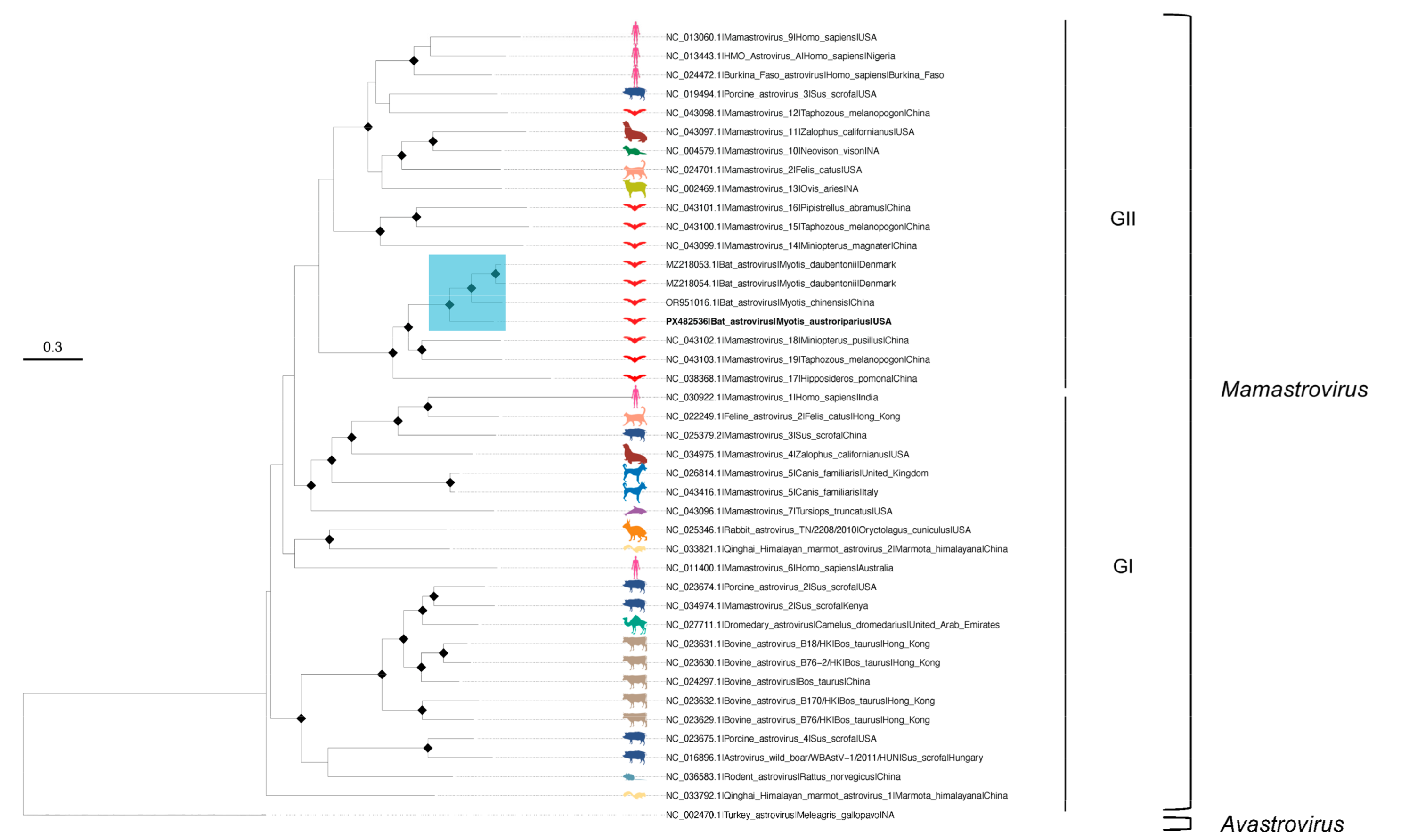
The ML phylogenies were inferred for AstV-Ma4-FL-2021 and individual gene (RdRp, ORF2) alignments, rooted with an avian outgroup (Turkey astrovirus; NC_002470.1). BIC selected GTR+F+R5 (AstV-Ma4-FL-2021), GTR+F+I+G4 (RdRp), and TVMe+R5 (ORF2). Likelihood mapping analysis indicated robust phylogenetic signal for all phylogenies (Figure S2). The full contig tree resolved two Mamastrovirus genogroups (I and II) [74,75] with all bat-derived sequences—including AstV-Ma4-FL-2021—clustering in genogroup II (Figure 2). Within this genogroup, AstV-Ma4-FL-2021 formed a well-supported monophyletic subclade composed exclusively of bat AstVs, grouping most closely with the unclassified Myotis-associated sequences (MZ218054.1, MZ218053.1, OR951016.1) and with Mamastrovirus 17 (NC_038368.1), Mamastrovirus 18 (NC_043102.1), and Mamastrovirus 19 (NC_043103.1). In gene-specific trees, AstV-Ma4-FL-2021 showed congruent placement within genogroup II (Figures S3 and S4); Mamastrovirus 17 clustered with the subclade in the ORF2 tree (Figure S3) but was excluded from the RdRp tree (Figure S4) due to insufficient coverage. Notably, in the RdRp tree the AstV-Ma4-FL-2021 subclade shared a more recent common ancestor with a neighboring clade (Mamastrovirus 14, 15, 16)—a relationship not recovered in the concatenated or ORF2 trees—potentially reflecting differing evolutionary constraints on the conserved RdRp versus the more variable capsid.

3.2. Co-Infection of an M. austroriparius with Astrovirus and a Bat Alphacoronavirus

In M. austroriparius specimen Ma_Frozen_4 we also detected an AlphaCoV contig (k119_1119) that was 1327 bp long. This finding suggests that Ma_Frozen_4 was co-infected with AlphaCoV and AstV (Table 2, Table S1). Contig k119_1119 shared 84% nucleotide identity with an unclassified AlphaCoV strain Tadarida brasiliensis bat alphacoronavirus 2 (ACoV-2-Tb; OP700657.1) (Table 2). Relative to OP700657.1, k119_1119 covered 51% of nucleocapsid gene and 100% of ORF7, ORF8, and the 3′ genomic region (Figure S5). From a second M. austroriparius specimen, Ma_Frozen_3, we recovered two additional AlphaCoV contigs: k119_6311 (925 bp; 81% identity to OP700657.1) and k119_670 (5309 bp; 81% identity to OP700657.1) (Table 2). Relative to OP700657.1, k119_6311 covered the 5′ genomic region and 5% of ORF1a, whereas k119_670 covered 42% of spike, 100% of ORF3, envelope, membrane, nucleocapsid, ORF7, ORF8, and the 3′ region (Table 2, Figure S5). For analytical purposes, a draft AlphaCoV genome was reconstructed by concatenating contigs k119_6311 and k119_670 (hereafter ACoV-Ma3-FL-2021) to facilitate phylogenetic placement. The AlphaCoV contig k119_1119 from sample Ma_Frozen_4 was short and identical to the corresponding region in k119_670 from sample Ma_Frozen_3; therefore, only contigs from Ma_Frozen_3 were used for downstream analyses.
To assess relatedness, we assembled a background dataset representing the 15 recognized AlphaCoV subgenera plus unclassified bat AlphaCoVs closely related to study contigs (Table S5). As expected for coronaviruses, recombination was detected in the reference dataset; recombinant regions were removed prior to analysis. No recombination was detected in ACoV-2-Tb, the closest lineage. Gene-level pairwise comparison of study contigs with reference genomes indicated that the genes for ORF1a, envelope, membrane, and nucleocapsid shared greatest identity with ACoV-2-Tb strains (Table S6). In the spike gene, our sample shared greatest identity (p-distance 0.29; standard error 0.01) with a Myotis siligorensis bat coronavirus sampled in China in 2019 (OQ175040.1) and was less similar to ACoV-2-Tb strains sampled from T. brasiliensis in Argentina in 2016–2017 (p-distance 0.36; standard error 0.01) (Table S6).
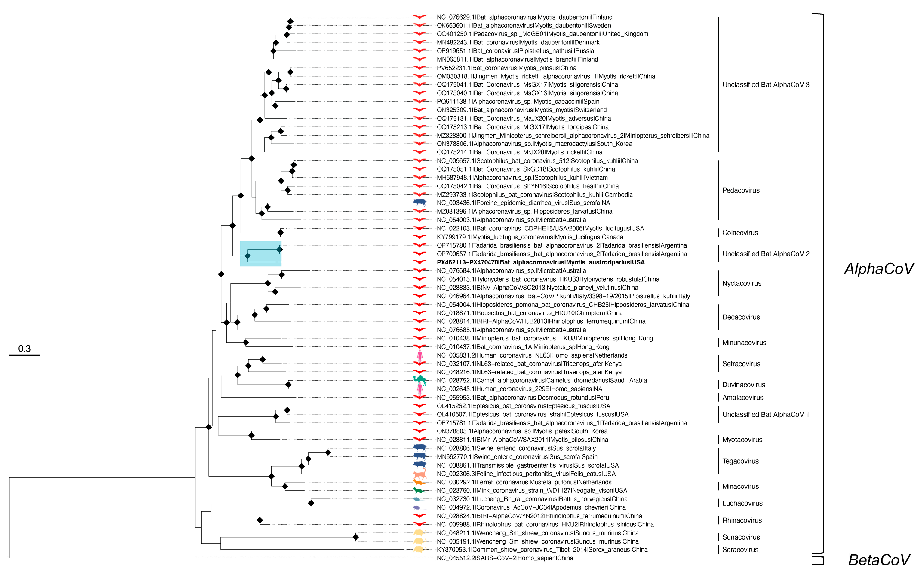
Based on the concatenated alignment, in the ML tree ACoV-Ma3-FL-2021 clustered in a well-supported subclade with ACoV-2-Tb strains from Argentina (OP700657.1, OP715780.1), sharing 81% nucleotide identity, respectively (Figure 3). ACoV-Ma3-FL-2021 occupied a basal, longer branch relative to the Argentine strains, consistent with a distinct lineage and/or unsampled diversity in Florida bat populations, geographic separation, different hosts and sampling times (Argentine sequences were sampled in 2016 and in 2017). A second putative species, named Tadarida brasiliensis bat alphacoronavirus 1 (ACoV-1-Tb; OP715781.1), forms a separate lineage that clusters with unclassified AlphaCoVs from Eptesicus bats collected in the USA and South Korea (here termed ‘Unclassified Bat AlphaCoV 1′) [76]. The most closely related classified subgenera to ACoV-Ma3-FL-2021 were Pedacovirus and Colacovirus, the latter currently comprising Myotis lucifugus coronaviruses collected in Colorado in 2006 (NC_022103.1) and in Canada in 2010 (KY799179.1) (Figure 3). Gene-specific phylogenies for envelope, membrane, and nucleocapsid recapitulated the concatenated-tree placement, with ACoV-Ma3-FL-2021 forming a well-supported subclade on a long branch basal to the Argentine ACoV-2-Tb lineage (Figure S6).
Phylogenetic analysis of the partial spike gene revealed the ACoV-2-Tb strains to not cluster with ACoV-Ma3-FL-2021 in a subclade and instead, they grouped in a distantly related, well-supported subclade with the Decacovirus Alphacoronavirus sp. WA3607 (NC_076685.1) (Figure S6). ACoV-Ma3-FL-2021 remained closely related to members of Colacovirus, Pedacovirus, as well as unclassified Myotis-associated coronaviruses in the spike phylogeny.

3.3. Detection of Hubei Virga-like Virus 2 from T. brasiliensis Feces

Although no viruses were detected in T. brasiliensis anal swabs or fecal samples from Clay County, we identified the unclassified RNA virus HVLV2 in fecal samples from T. brasiliensis roosting in Alachua County (Table 1, Figure 1). De novo assembly yielded a single viral contig of 9829 bp (k141_4344) with 99% nucleotide identity to publicly available HVLV2 sequences (Table 2). Contig k141_4344 spans 90% of the complete HVLV2 genome, including 99% of the polyprotein region; however none of the coat protein was recovered (Figure S7). Hereafter, contig k141_4344 will be referred to as HVLV2-Tb15-FL-2024.
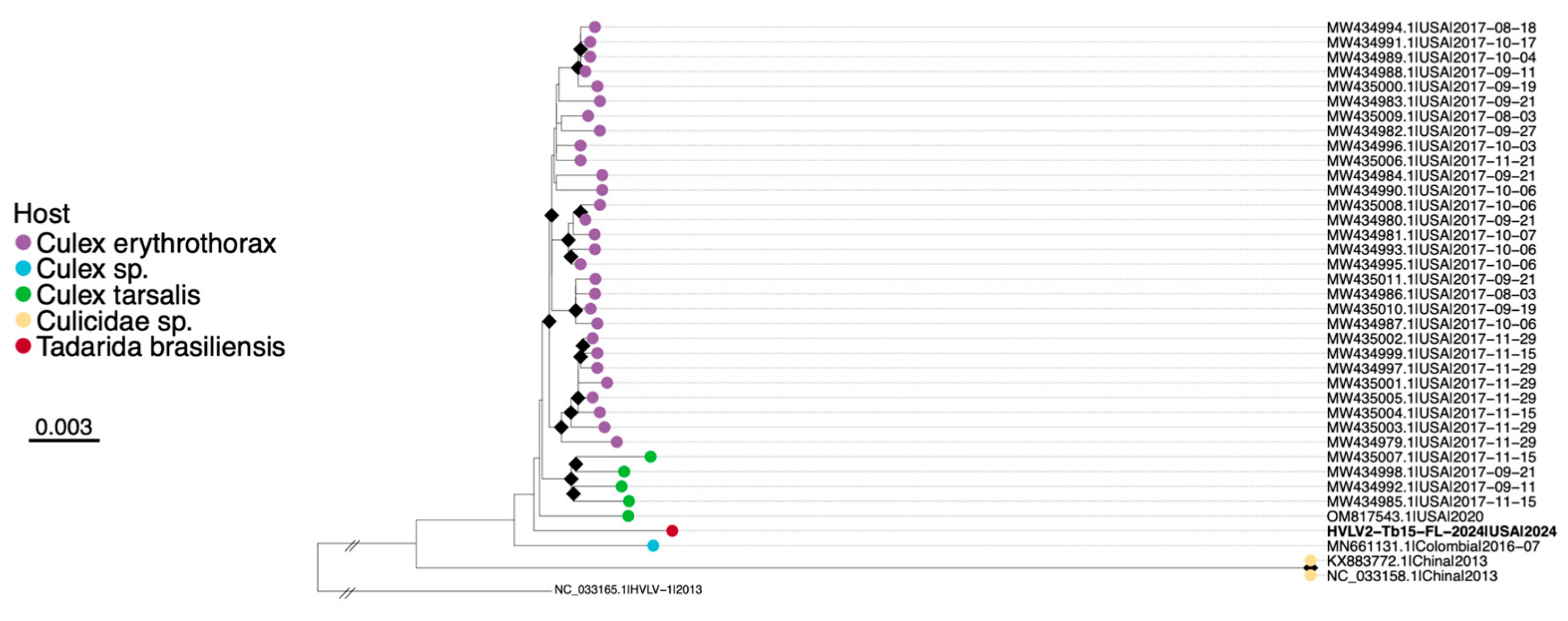
A total of 37 HVLV2 genomes were retrieved from NCBI Virus predominately detected in mosquitoes of the Culex genus (Table S6). Recombination analysis showed no evidence of recombination between included sequences in the dataset. Pairwise distance analysis of the polyprotein revealed high nucleotide similarity between HVLV2-Tb15-FL-2024 and all included HVLV2 sequences (96–99.5%) (Table S7). The closest genomes to our sample were from Culex erythrothorax collected in California, USA, in 2017 (MW434980.1; MW434995.1; p-distance 0.005, SE 0.001) and Culex tarsalis sampled in the USA in 2020 and in California, USA, in 2017 (OM817543.1; MW434992.1; p-distance 0.006, SE 0.001). ML tree placed HVLV2-Tb15-FL-2024 within the clade of genomes obtained from Culex sp. mosquitoes, in between sequences from Colombia and U.S., although bootstrap support was low (Figure 4). HVLV2-Tb15-FL-2024 resides on a relatively long branch basal to U.S. sequences, consistent with geographic structure and/or unsampled diversity.

3.4. Microbiome Shifts in AlphaCoV and AstV Co-Infected Bat

We performed 16S rRNA gene sequencing on M. austroriparius samples and compared the gut microbiota of Ma_Frozen_3 (AlphaCoV positive) and Ma_Frozen_4 (AlphaCoV and AstV positive) with those of Ma_Frozen_1, Ma_Frozen_2, and Ma_Frozen_5 (virus-negative). For Ma_Frozen_4 and Ma_Frozen_5, we processed two preparation types (secondary-prepared and concentrated) and evaluated whether they could be combined for downstream analyses. Microbial community composition was highly similar between preparation types at both the genus and species levels (Figures S8 and S9). Shannon diversity did not differ (Kruskal–Wallis: H = 0.59, df = 1, p = 0.438; Figure S10). Beta diversity patterns based on Bray–Curtis dissimilarities showed minimal visual differences in community structure (Figure S11), and PERMANOVA detected no effect of preparation type (F = 0.1679, R2 = 0.077, p = 1.000; 23 permutations). Accordingly, we merged the two preparation types for each sample in subsequent analyses.
Visual presentation of alpha and beta diversity suggested alpha diversity (Shannon index) varied across specimens, with Ma_Frozen_4 exhibiting the highest diversity, followed by Ma_Frozen_2, Ma_Frozen_1, Ma_Frozen_3, and Ma_Frozen_5 (Figure S12). Beta diversity based on Bray–Curtis dissimilarity (PCoA) plot grouped Ma_Frozen_1 with Ma_Frozen_2 and Ma_Frozen_3 with Ma_Frozen_4, whereas Ma_Frozen_5 formed an outlier cluster (Figure S13). Weighted UniFrac PCoA, which incorporates phylogenetic relatedness and abundance, grouped Ma_Frozen_3 with Ma_Frozen_1/2 and resolved Ma_Frozen_4 separately; Ma_Frozen_5 again appeared as an outlier (Figure S14). Overall, these observations also indicated a possible trend toward higher alpha diversity and a phylogenetically distinct community in the AlphaCoV+AstV co-infected Ma_Frozen_4 relative to the AlphaCoV-only individual (Ma_Frozen_3) and virus-negative bats (Ma_Frozen_1/2); however, these patterns are purely descriptive and were not tested statistically.
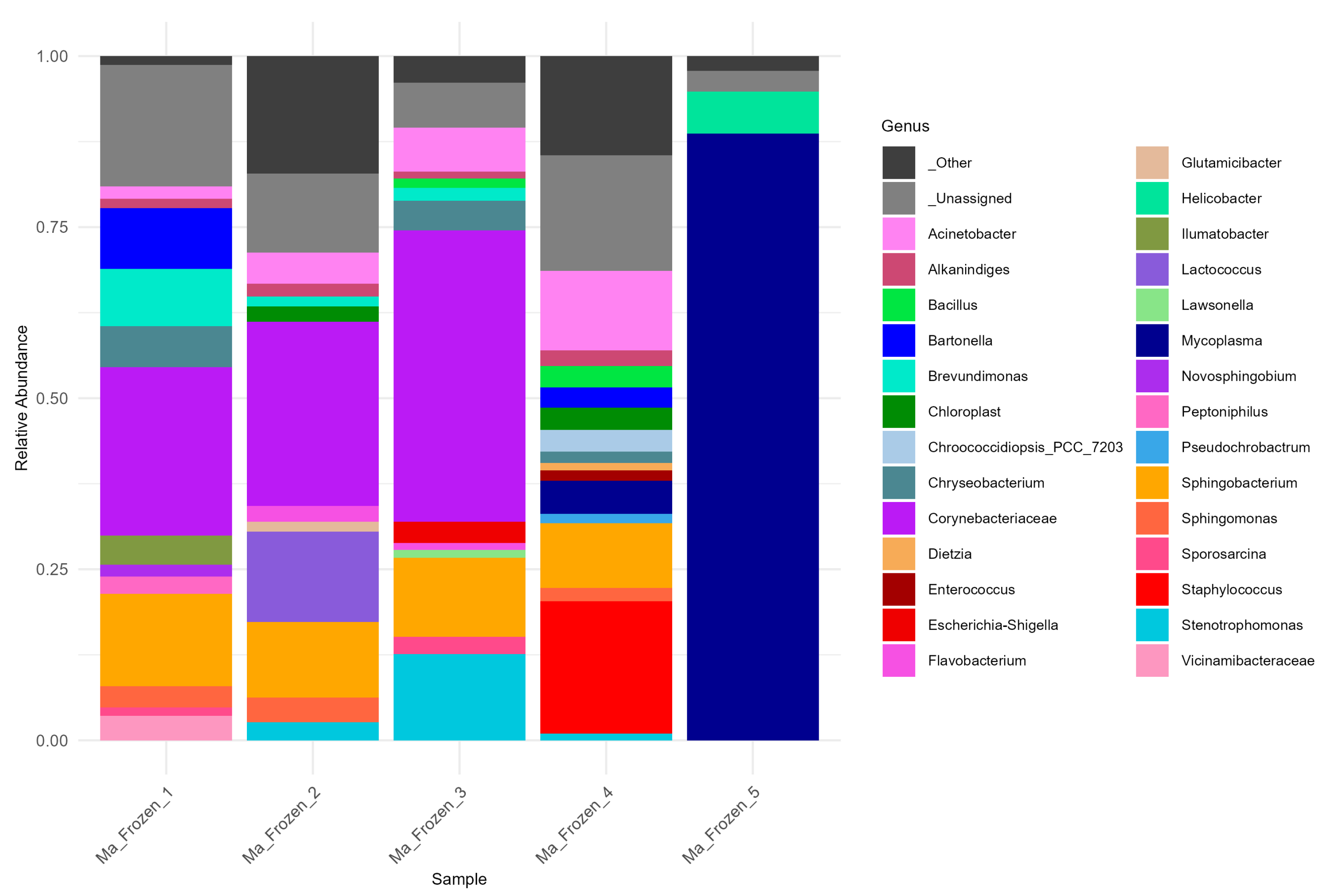
At the genus level, communities were compositionally distinct (Figure 5). The family Corynebacteriaceae (dominated by Corynebacterium) prevailed in Ma_Frozen_1/2/3 but was absent from Ma_Frozen_4, consistent with altered gut microbiota under co-infection. Acinetobacter and Sphingobacterium were prevalent across Ma_Frozen_1–4, with Acinetobacter relatively enriched in Ma_Frozen_3/4 and Sphingobacterium enriched in Ma_Frozen_1/2. In Ma_Frozen_4, no single genus dominated; notable taxa included Mycoplasma, Acinetobacter, Sphingobacterium, Bartonella, and Staphylococcus. By contrast, Ma_Frozen_5 was overwhelmingly dominated by Mycoplasma with minor Helicobacter. The Bartonella genus, present in Ma_Frozen_1 and Ma_Frozen_4, were taxonomically consistent with genuine Bartonella rather than typical mitochondrial (Rickettsiales) or chloroplast (Cyanobacteria/Chloroplast) origins, suggesting they are unlikely to represent organellar contamination. However, given the low-biomass and single-sample context, we cannot entirely exclude the possibility of Alphaproteobacterial background signals. At the species level, virus positive samples Ma_Frozen_3/4 showed higher abundance of Acinetobacter iwoffi compared to virus-negative samples (Figure S15).

4. Discussion

In this study, we analyzed the viromes and gut microbiomes of native north Florida bats. As the state of Florida experiences one of the highest human population growth rates in the United States, many bats in Florida are likely experiencing population declines due to loss of natural habitat from deforestation and human disturbance or destruction of roosting sites [77,78]. Bat habitat loss is strongly linked to poor bat health outcomes and increased human–bat interactions, which elevate the risk of zoonotic spillover [79,80]. We aimed to characterize the viral diversity circulating in two common bat species, T. brasiliensis and M. austroriparius, given the paucity of studies on Florida’s resident bat populations and their tendency to roost in urban areas, including in buildings, bridges, and culverts, making them particularly vulnerable to human interference [81,82,83,84]. We report here the first detection of AstV and AlphaCoV in M. austroriparius in Florida. Moreover, we found evidence of co-infection with both viruses in one individual. Such co-infections are plausible, as infection with one virus may weaken the host’s immune system and increase susceptibility to a secondary infection [85]. The AstV identified in this study clustered within Mamastrovirus genogroup II with Myotis-associated AstV collected in Denmark and in China [7,86]. As Florida M. austroriparius are not known to undertake long-distance migrations, cross-continental contact is unlikely; the relatively low nucleotide identity to available references more plausibly reflects undersampling of New World Myotis astroviruses and potential host-associated divergence. Collectively, these findings suggest AstV-Ma4-FL-2021 may represent a novel Mamastrovirus lineage associated with native M. austroriparius.
The AlphaCoV found in two of five M. austroriparius was most similar to ACoV-2-Tb, previously detected in fecal samples from T. brasiliensis collected in Argentina in 2017–2018 [76]. In the phylogeny of the concatenated alignment, ACoV-Ma3-FL-2021 formed a well-supported monophyletic subclade with ACoV-2-Tb strains, distinct from classified subgenera. Our results are consistent with prior observation that the ACoV-2-Tb lineage clusters with Myotis-associated AlphaCoVs from the USA [76]. Similar detections of closely related AlphaCoVs in geographically distant bat hosts have been reported and may reflect historical cross-species transmission [33]. The relatively long branch leading to our AlphaCoV and its nucleotide divergence from ACoV-2-Tb are consistent with host-associated diversification and/or unsampled regional diversity, raising the possibility of a distinct, M. austroriparius-associated lineage. The only other documented AlphaCoV from native Florida bats is a partial RdRp sequence detected in T. brasiliensis collected in Gilchrist County in May 2016 [35]. Because our AlphaCoV assembly does not include RdRp, direct comparison with these viruses was not possible. However, RdRp phylogenies place these viruses in a lineage distinct from the AlphaCoV from Argentina closely related to ours, suggesting co-circulation of separate two AlphaCoV lineages in different bat species within Florida [35,76]. In the spike gene phylogeny, ACoV-2-Tb strains clustered in a distant and separate lineage from ACoV-Ma3-FL-2021, while our study sample maintained its close association with Colacovirus and Pedacovirus. This pattern suggests the ACoV-2-Tb strains may be recombinant in the spike region, which is consistent with Cerri et al.’s finding that ACoV-2-Tb is closely related to the Colacovirus subgenus, except for in the spike gene [76]. Our analysis of the spike phylogeny is limited by partial recovery of the spike gene from our sample; therefore future sampling of M. austroriparius in North Florida is warranted to better understand the complete spike evolutionary history.
Neither AlphaCoV nor AstV were detected in samples obtained from T. brasiliensis collected across multiple Florida counties. Because RNA viruses are routinely recovered by metagenomic sequencing from bat intestinal/rectal tissues and feces, sample type alone is unlikely to explain this non-detection. This absence may reflect limited sample size and/or species-specific differences in viral shedding dynamics of bat species in north Florida [85]. There is strong evidence in the literature that poor bat health stemming from nutritional stress and/or loss of habitat is associated with increased viral shedding [85,87].
Although we cannot determine whether the bats with evidence of AlphaCoV and AstV infection were sick, previous reports of AlphaCoV [88,89,90] and AstV [74,91] in bats did not describe clinical signs of disease, consistent with the asymptomatic presentation commonly observed in reservoir hosts [92,93,94]. Overall, our metagenomic analyses seem to suggest that the bats sampled in north Florida are generally healthy, and while viral detection was limited to individuals roosting in an urban fire station, with no infections observed in bats from designated rural bat houses, we cannot exclude that this was due to the limited sampling in this study. Urban roosting may expose bats to factors such as habitat disturbance, noise, light pollution, and altered food availability, all of which may modulate host immunity, impair health status, and influence viral shedding [34,95,96,97]. Therefore, we should investigate further the hypothesis that this pattern may reflect anthropogenic stressors associated with urban, high-human activity environments compared to more natural settings. Maintaining the health of Florida’s native bat populations is important both for conservation and for mitigating viral shedding-related risks. Expanded, longitudinal sampling of co-roosting species in urban and natural environments, complemented by targeted assays, will be necessary to clarify whether AlphaCoV or AstV transmission occurs between species in north Florida.
The mammalian gut microbiome modulates host susceptibility to infection via effects on physiology, immunity, nutrition, and behavior [10,11,98]. In bats, microbial composition and diversity vary among species, likely reflecting differences in diet, habitat, and seasonal behavior [10,13,18,25,99]. Emerging evidence indicates that gut microbes can modulate antiviral responses, which may contribute to bats’ notable tolerance to viral pathogens [12,13]. Although several studies characterized gut microbiota in members of the genus Myotis, this is, to our knowledge, the first investigation of the gut microbial community in M. austroriparius and the first microbiome study of bats from Florida. Across bat species, Corynebacterium is generally regarded as a commensal genus, [10,100,101,102,103,104,105] so the absence of Corynebacterium in the co-infected individual (Ma_Frozen_4) suggests a disruption of the gut microbiome. In this individual, Staphylococcus and Mycoplasma emerged as notable taxa. Both genera have been reported to display negative associations with other gut bacteria, implying competitive interactions and microbial imbalance under conditions of viral co-infection [106]. Additional indication of altered gut microbiota comes from the presence of Acinetobacter iwoffi, which is an opportunistic human pathogen associated with gastroenteritis [107,108,109] and may be in bats as well. This individual exhibited the greatest within-sample (alpha) diversity and a phylogenetically distinct community composition relative to AlphaCoV-only and virus-negative conspecifics. This pattern could reflect a potential synergistic effect of co-infection on microbiome structure, or alternatively, with AstV—an enteric virus—being the primary driver of community shifts [110,111,112,113,114]. Although AstV infections are generally considered asymptomatic in bats, they may nonetheless perturb gut communities [115,116]. Wasimuddin et al. reported age-dependent effects in Artibeus jamaicensis, with infected adults showing increased diversity and enrichment of potentially pathogenic genera such as Mycoplasma and Acinetobacter [116]. Consistent with this pattern, the AstV co-infected M. austroriparius displayed higher alpha diversity and greater between-sample dissimilarity (Bray–Curtis) than the AlphaCoV-only and virus-negative individuals, along with increased relative abundances of Mycoplasma, Staphylococcus, and Acinetobacter. While these preliminary data are consistent with possible effects of viral co-infection on microbiome structure, they are based on a single individual and therefore warrant further sampling to test the effects of co-infection on gut microbial shifts in M. austroriparius. The divergence in microbial community structure observed in one of the healthy individuals (Ma_Frozen_5) is unlikely to be virus-associated as no viral sequences were detected from this specimen. Instead, the predominance of Mycoplasma together with reduced alpha diversity suggests a potential enteric Mycoplasma infection, consistent with previous associations between decreased diversity and diseased states [106]. Alternatively, we cannot exclude that variation in decomposition time or sample handling prior to deposition at the FLMNH may have contributed to the observed pattern. The causal directionality between viral infection and microbiome state remains unresolved; viruses may reshape gut communities, or particular community configurations may predispose bats to infection [11].
Beyond crop protection, bat predation contributes a clear public-health value in mosquito control [17,117]. Detection of mosquito-associated viruses in bat feces can provide insight into the breadth of mosquito consumption across bat species and habitats. Here, we report the first detection of insect virus HVLV2 in guano of Floridian T. brasiliensis likely reflecting dietary intake of mosquitoes, as insectivorous bat fecal viromes are often dominated by diet-derived insect viruses [31]. HVLV2 has been detected in several mNGS studies of multiple Culex species and may therefore serve as a useful marker of Culex consumption when identified in bat feces [118,119,120,121]. In our study, the clustering of HVLV2 with viruses detected in arbovirus vectors of public health importance, Cx. erythrothorax and Cx. tarsalis [122,123,124], warrants further investigation to assess the potential role of bat predation in suppressing endemic arbovirus vector populations [40,117,125]. This finding is interesting for several reasons. First, it suggests that Florida bats are predators of Culex mosquitoes, the globally widespread vector for several human pathogens, including West Nile virus, St. Louis encephalitis virus, and Japanese encephalitis virus [126]. Second, neither of these species has been reported in Florida; therefore, this finding highlights the importance of studying insect-virus profiles in bat feces, as such data may provide early indicators of geographic expansion by non-native mosquito species. Last, it could suggest association of this virus with other Culex species present in north Florida such as Cx. quinquefasciatus and Cx. nigripalpus, both epidemiologically and ecologically important mosquito species [123,127,128]. Detecting Culex-associated viral sequences (such as HVLV2) in bat feces may therefore not only reflect dietary exposure but also provide sentinel evidence of local vector activity and virus circulation within overlapping ecological niches.
Although mammalian viruses often dominate zoonotic discussions, insect-associated viruses captured via mNGS illuminate predator–prey interactions and ecological drivers shaping bat viromes [30]. From a conservation perspective, diet monitoring informs population health, potential shedding risks linked to nutritional stress, and the impacts of anthropogenic disturbance [79,129]. Manual inspection of guano can underestimate mosquito consumption due to the difficulty of identifying digested remains [40,130]. In contrast, passive guano collection coupled with mNGS offers a sensitive, convenient, and cost-effective approach that avoids trapping individuals and circumvents morphological identification [131]. Accordingly, fecal-virome mNGS can provide actionable insights into insect-associated viruses of agricultural and public-health concern and position bats as sentinels for the circulation of insect-borne viruses [132].
This study is constrained by a small sample size and a cross-sectional design, owing to the opportunistic nature of sample acquisition, which limits statistical power, generalizability, and causal inference regarding links between viral detection and microbiome composition. We did not include extraction blanks or other negative controls; thus, prevalence-based contaminant removal (e.g., decontam in R) was not possible, and potential low-biomass contaminants cannot be distinguished from true community members. In addition, each group comprised a single sample without biological replication, precluding statistical comparisons (e.g., Kruskal–Wallis, PERMANOVA, differential abundance tests). Accordingly, taxonomic and diversity results are reported descriptively and should be interpreted as exploratory. Future studies will include appropriate controls and replicates to enable formal decontamination and inference. The use of archived museum specimens introduces heterogeneity in preservation history and RNA integrity, and host metadata (e.g., age, sex, reproductive status, diet) were incomplete, leaving potential confounders unaccounted for. Metagenomic assemblies generally resulting in partial genomes and read-based detection cannot always distinguish transient dietary/environmental exposure from active infection (as considered for HVLV2). A general lack of existing data from Florida bats restricts comparison between the data presented here and other studies.
Despite these constraints, our data provide a proof-of-concept for future viral surveillance studies in bat populations in Florida. We report the first detection of HVLV2 in Florida’s T. brasiliensis, the first evidence of AstV and AlphaCoV co-infection in Florida’s M. austroriparius, along with coherent shifts in gut microbial community structure in the co-infected bat, and the first occurrence of AstV in Florida bats overall. Our results validate a museum-enabled, integrated virome–microbiome approach and motivate larger, longitudinal sampling with targeted assays to confirm infection status and dissect mechanistic links between viral presence, microbiome state, and host ecology. We highlight the value of using museum specimens as an ethical and convenient resource for investigating the bat virome and microbiome without the need to sacrifice live animals [133]. This approach allows for a comparison of historical trends in virus circulation when compared to newly collected wildlife samples. Our aim is not to bring negative attention to Florida’s bat population, but rather to highlight the worthiness of viral surveillance from a conservation and public health standpoint.

Supplementary Materials

The following supporting information can be downloaded at: https://www.mdpi.com/article/10.3390/microorganisms13112625/s1. Figure S1: Genome scheme of AstV contig; Figure S2: Likelihood mapping analysis results; Figure S3: Maximum likelihood tree of AstV RdRp gene; Figure S4 Maximum likelihood tree of AstV ORF2 gene; Figure S5: Genome scheme of AlphaCoV contigs; Figure S6: Maximum likelihood trees of AlphaCoV spike, envelope, membrane, and nucleocapsid genes; Figure S7: Genome scheme of HVLV2 contig; Figure S8: Microbial composition of Ma_Frozen_4 and Ma_Frozen_5 at the genus level, comparing sample preparations; Figure S9: Microbial composition of Ma_Frozen_4 and Ma_Frozen_5 at the species level, comparing sample preparations; Figure S10: Shannon alpha diversity comparison of Ma_Frozen_4 and Ma_Frozen_5 sample preparations; Figure S11: Bray–Curtis PCoA of Ma_Frozen_4 and Ma_Frozen_5 comparing secondary-prepared and concentrated samples; Figure S12: Shannon alpha diversity indices for Ma_Frozen samples; Figure S13: Bray–Curtis PCoA of Ma_Frozen samples; Figure S14: PCoA of Ma_Frozen samples based on Weighted UniFrac distances; Figure S15: Species-level microbial community composition of Ma_Frozen samples. Table S1: Complete description of bats sampled in this study; Table S2: Complete description of contigs obtained in this study; Table S3: AstV accessions; Table S4: Comparative Genetic Identity of AstV-Ma4-FL-2021; Table S5: AlphaCoV accessions; Table S6: Comparative Genetic Identity of AlphaCoV; Table S7: HVLV2 accessions; Table S8: Comparative Genetic Identity of HVLV2.

Author Contributions

Conceptualization, C.N.M.; methodology, J.E.P., T.T., M.B., J.B., M.S.T., M.N.C., J.S.N., L.M.S., K.S., E.B.d.T., V.M. and C.N.M.; software, J.E.P., T.T., M.S.T. and C.N.M.; validation, J.E.P. and C.N.M.; formal analysis, J.E.P., T.T., M.B., J.B., M.S.T., L.M.S., K.S. and E.B.d.T.; investigation, J.E.P., T.T., M.B. and C.N.M.; resources, M.S., B.D.A., M.E.v.F. and C.N.M.; data curation, J.E.P. and T.T.; writing—original draft preparation, J.E.P.; writing—review and editing, J.E.P. and C.N.M.; visualization, J.E.P., T.T. and C.N.M.; supervision, C.N.M.; project administration, C.N.M.; funding acquisition, C.N.M. All authors have read and agreed to the published version of the manuscript.

Funding

This research was supported by the University of Florida Department of Pathology EPIG RAS 2021–2022; the University of Florida Informatics Institute SEED 2022–2023; and the University of Florida Emerging Pathogens Institute funds. Mavian and Mathis were supported by the University of Florida Biodiversity Institute SEED 2022–2023.

Institutional Review Board Statement

The museum bat samples analyzed in this study were donated to the FLMNH and therefore, an IACUC protocol was not required by the University of Florida. All live-capture and handling activities were completed under safe and humane protocols approved by the FWC and followed American Society of Mammalogists guidelines (2016). The bat fecal samples collected at the University of Florida bat houses were collected without animal handling or manipulation and thus an IACUC protocol was not required by the University of Florida.

Informed Consent Statement

Not applicable.

Data Availability Statement

The metagenomic and 16S sequence reads have been deposited at NCBI Sequence Read Archive (SRA) under BioProject identifier (ID) PRJNA1305506. The contigs have been deposited to GenBank under accessions listed in Table 2. Data and code repository available at: https://github.com/paolij/FL_Bat_Virome_Microbiome (accessed on 12 November 2025).

Acknowledgments

We thank David J. Nolan for carefully reading the manuscript and providing valuable suggestions. Bioinformatic analyses were in part performed by the Bioinformatics Core of the Interdisciplinary Center for Biotechnology Research, University of Florida (RRID:SCR_019120).

Conflicts of Interest

The authors declare no conflicts of interest.

Abbreviations

The following abbreviations are used in this manuscript:
AlphaCoVAlphacoronavirus
AstVAstrovirus
HVLV2Hubei virga-like virus 2
mNGSMetagenomic next-generation sequencing
RdRpRNA-dependent RNA polymerase
FLMNHFlorida Museum of Natural History
FWCFlorida Fish and Wildlife Conservation Commission
ICTVInternational Committee on Taxonomy of Viruses
NGSNext-generation sequencing
ACoV-2-TbTadarida brasiliensis bat alphacoronavirus 2

References

  1. Brook, C.E.; Dobson, A.P. Bats as “special” Reservoirs for Emerging Zoonotic Pathogens. Trends Microbiol. 2015, 23, 172–180. [Google Scholar] [CrossRef] [PubMed]
  2. Irving, A.T.; Ahn, M.; Goh, G.; Anderson, D.E.; Wang, L.-F. Lessons from the Host Defences of Bats, a Unique Viral Reservoir. Nature 2021, 589, 363–370. [Google Scholar] [CrossRef] [PubMed]
  3. Khan, S.A.; Imtiaz, M.A.; Islam, M.M.; Tanzin, A.Z.; Islam, A.; Hassan, M.M. Major Bat-Borne Zoonotic Viral Epidemics in Asia and Africa: A Systematic Review and Meta-Analysis. Vet. Med. Sci. 2022, 8, 1787–1801. [Google Scholar] [CrossRef]
  4. Sparrer, M.N.; Hodges, N.F.; Sherman, T.; VandeWoude, S.; Bosco-Lauth, A.M.; Mayo, C.E. Role of Spillover and Spillback in SARS-CoV-2 Transmission and the Importance of One Health in Understanding the Dynamics of the COVID-19 Pandemic. J. Clin. Microbiol. 2023, 61, e0161022. [Google Scholar] [CrossRef]
  5. Van Brussel, K.; Holmes, E.C. Zoonotic Disease and Virome Diversity in Bats. Curr. Opin. Virol. 2022, 52, 192–202. [Google Scholar] [CrossRef]
  6. Wang, J.; Pan, Y.-F.; Yang, L.-F.; Yang, W.-H.; Lv, K.; Luo, C.-M.; Wang, J.; Kuang, G.-P.; Wu, W.-C.; Gou, Q.-Y.; et al. Individual Bat Virome Analysis Reveals Co-Infection and Spillover among Bats and Virus Zoonotic Potential. Nat. Commun. 2023, 14, 4079. [Google Scholar] [CrossRef]
  7. Wang, Y.; Xu, P.; Han, Y.; Zhao, W.; Zhao, L.; Li, R.; Zhang, J.; Zhang, S.; Lu, J.; Daszak, P.; et al. Unveiling Bat-Borne Viruses: A Comprehensive Classification and Analysis of Virome Evolution. Microbiome 2024, 12, 235. [Google Scholar] [CrossRef]
  8. Tan, C.W.; Yang, X.; Anderson, D.E.; Wang, L.-F. Bat Virome Research: The Past, the Present and the Future. Curr. Opin. Virol. 2021, 49, 68–80. [Google Scholar] [CrossRef]
  9. Bazzoni, E.; Cacciotto, C.; Zobba, R.; Pittau, M.; Martella, V.; Alberti, A. Bat Ecology and Microbiome of the Gut: A Narrative Review of Associated Potentials in Emerging and Zoonotic Diseases. Animals 2024, 14, 3043. [Google Scholar] [CrossRef]
  10. Dhivahar, J.; Parthasarathy, A.; Krishnan, K.; Kovi, B.S.; Pandian, G.N. Bat-Associated Microbes: Opportunities and Perils, an Overview. Heliyon 2023, 9, e22351. [Google Scholar] [CrossRef]
  11. Jones, D.N.; Ravelomanantsoa, N.A.F.; Yeoman, C.J.; Plowright, R.K.; Brook, C.E. Do Gastrointestinal Microbiomes Play a Role in Bats’ Unique Viral Hosting Capacity? Trends Microbiol. 2022, 30, 632–642. [Google Scholar] [CrossRef]
  12. Luo, J.; Liang, S.; Jin, F. Gut Microbiota in Antiviral Strategy from Bats to Humans: A Missing Link in COVID-19. Sci. China Life Sci. 2021, 64, 942–956. [Google Scholar] [CrossRef] [PubMed]
  13. Popov, I.V.; Mazanko, M.S.; Kulaeva, E.D.; Golovin, S.N.; Malinovkin, A.V.; Aleshukina, I.S.; Aleshukina, A.V.; Prazdnova, E.V.; Tverdokhlebova, T.I.; Chikindas, M.L.; et al. Gut Microbiota of Bats: Pro-Mutagenic Properties and Possible Frontiers in Preventing Emerging Disease. Sci. Rep. 2021, 11, 21075. [Google Scholar] [CrossRef] [PubMed]
  14. Dimkić, I.; Fira, D.; Janakiev, T.; Kabić, J.; Stupar, M.; Nenadić, M.; Unković, N.; Grbić, M.L. The Microbiome of Bat Guano: For What Is This Knowledge Important? Appl. Microbiol. Biotechnol. 2021, 105, 1407–1419. [Google Scholar] [CrossRef] [PubMed]
  15. Boyles, J.G.; Cryan, P.M.; McCracken, G.F.; Kunz, T.H. Conservation. Economic Importance of Bats in Agriculture. Science 2011, 332, 41–42. [Google Scholar] [CrossRef]
  16. Long, R.F.; Simpson, T.; Ding, T.-S.; Heydon, S.; Reil, W. Bats Feed on Crop Pests in Sacramento Valley. Calif. Agric. 1998, 52, 8–10. [Google Scholar] [CrossRef]
  17. Kunz, T.H.; Braun de Torrez, E.; Bauer, D.; Lobova, T.; Fleming, T.H. Ecosystem Services Provided by Bats. Ann. N. Y. Acad. Sci. 2011, 1223, 1–38. [Google Scholar] [CrossRef]
  18. Dai, W.; Leng, H.; Li, J.; Li, A.; Li, Z.; Zhu, Y.; Li, X.; Jin, L.; Sun, K.; Feng, J. The Role of Host Traits and Geography in Shaping the Gut Microbiome of Insectivorous Bats. mSphere 2024, 9, e0008724. [Google Scholar] [CrossRef]
  19. Mena Canata, D.A.; Benfato, M.S.; Pereira, F.D.; Ramos Pereira, M.J.; Hackenhaar, F.S.; Mann, M.B.; Frazzon, A.P.G.; Rampelotto, P.H. Comparative Analysis of the Gut Microbiota of Bat Species with Different Feeding Habits. Biology 2024, 13, 363. [Google Scholar] [CrossRef]
  20. Carrillo-Araujo, M.; Taş, N.; Alcántara-Hernández, R.J.; Gaona, O.; Schondube, J.E.; Medellín, R.A.; Jansson, J.K.; Falcón, L.I. Phyllostomid Bat Microbiome Composition Is Associated to Host Phylogeny and Feeding Strategies. Front. Microbiol. 2015, 6, 447. [Google Scholar] [CrossRef]
  21. Dietrich, M.; Kearney, T.; Seamark, E.C.J.; Paweska, J.T.; Markotter, W. Synchronized Shift of Oral, Faecal and Urinary Microbiotas in Bats and Natural Infection Dynamics during Seasonal Reproduction. R. Soc. Open Sci. 2018, 5, 180041. [Google Scholar] [CrossRef]
  22. Federici, L.; Masulli, M.; De Laurenzi, V.; Allocati, N. An Overview of Bats Microbiota and Its Implication in Transmissible Diseases. Front. Microbiol. 2022, 13, 1012189. [Google Scholar] [CrossRef]
  23. Lebeuf-Taylor, E.; Cosby, A.; Webber, Q.; Cottenie, K. Social Structuring of the Gut Microbiome in Communally Roosting Bats. PLoS ONE 2025, 20, e0325710. [Google Scholar] [CrossRef]
  24. Lutz, H.L.; Jackson, E.W.; Webala, P.W.; Babyesiza, W.S.; Kerbis Peterhans, J.C.; Demos, T.C.; Patterson, B.D.; Gilbert, J.A. Ecology and Host Identity Outweigh Evolutionary History in Shaping the Bat Microbiome. mSystems 2019, 4. [Google Scholar] [CrossRef] [PubMed]
  25. Xiao, G.; Liu, S.; Xiao, Y.; Zhu, Y.; Zhao, H.; Li, A.; Li, Z.; Feng, J. Seasonal Changes in Gut Microbiota Diversity and Composition in the Greater Horseshoe Bat. Front. Microbiol. 2019, 10, 2247. [Google Scholar] [CrossRef] [PubMed]
  26. Bassi, C.; Guerriero, P.; Pierantoni, M.; Callegari, E.; Sabbioni, S. Novel Virus Identification through Metagenomics: A Systematic Review. Life 2022, 12, 2048. [Google Scholar] [CrossRef]
  27. Buddle, S.; Forrest, L.; Akinsuyi, N.; Martin Bernal, L.M.; Brooks, T.; Venturini, C.; Miller, C.; Brown, J.R.; Storey, N.; Atkinson, L.; et al. Evaluating Metagenomics and Targeted Approaches for Diagnosis and Surveillance of Viruses. Genome Med. 2024, 16, 111. [Google Scholar] [CrossRef] [PubMed]
  28. Ge, X.; Li, Y.; Yang, X.; Zhang, H.; Zhou, P.; Zhang, Y.; Shi, Z. Metagenomic Analysis of Viruses from Bat Fecal Samples Reveals Many Novel Viruses in Insectivorous Bats in China. J. Virol. 2012, 86, 4620–4630. [Google Scholar] [CrossRef]
  29. Mishra, N.; Fagbo, S.F.; Alagaili, A.N.; Nitido, A.; Williams, S.H.; Ng, J.; Lee, B.; Durosinlorun, A.; Garcia, J.A.; Jain, K.; et al. A Viral Metagenomic Survey Identifies Known and Novel Mammalian Viruses in Bats from Saudi Arabia. PLoS ONE 2019, 14, e0214227. [Google Scholar] [CrossRef]
  30. Huang, Z.; Wang, Z.; Liu, Y.; Ke, C.; Feng, J.; He, B.; Jiang, T. The Links between Dietary Diversity and RNA Virus Diversity Harbored by the Great Evening Bat (Ia Io). Microbiome 2024, 12, 246. [Google Scholar] [CrossRef]
  31. Li, L.; Victoria, J.G.; Wang, C.; Jones, M.; Fellers, G.M.; Kunz, T.H.; Delwart, E. Bat Guano Virome: Predominance of Dietary Viruses from Insects and Plants plus Novel Mammalian Viruses. J. Virol. 2010, 84, 6955–6965. [Google Scholar] [CrossRef] [PubMed]
  32. Hernández-Aguilar, I.; Lorenzo, C.; Santos-Moreno, A.; Naranjo, E.J.; Navarrete-Gutiérrez, D. Coronaviruses in Bats: A Review for the Americas. Viruses 2021, 13, 1226. [Google Scholar] [CrossRef] [PubMed]
  33. Kotwa, J.D.; Bhuinya, A.; Yim, W.; Gallo, G.; Singh, P.; Liu, Q.; Kumar, A.; Chien, E.; Hou, A.; Chee, H.-Y.; et al. High Host Specificity of Alphacoronaviruses in Nearctic, Insectivorous Bats. NPJ Viruses 2025, 3, 38. [Google Scholar] [CrossRef]
  34. Warmuth, V.M.; Metzler, D.; Zamora-Gutierrez, V. Human Disturbance Increases Coronavirus Prevalence in Bats. Sci. Adv. 2023, 9, eadd0688. [Google Scholar] [CrossRef]
  35. Bonny, T.S.; Driver, J.P.; Paisie, T.; Salemi, M.; Morris, J.G.; Shender, L.A.; Smith, L.; Enloe, C.; Oxenrider, K.; Gore, J.A.; et al. Detection of Alphacoronavirus vRNA in the Feces of Brazilian Free-Tailed Bats (Tadarida brasiliensis) from a Colony in Florida, USA. Diseases 2017, 5, 7. [Google Scholar] [CrossRef]
  36. Leong, R.; Hoarau, A.O.G.; Carcauzon, V.; Köster, M.; Dietrich, M.; Tortosa, P.; Lebarbenchon, C. High Astrovirus Diversity in an Endemic Bat Species Suggests Multiple Spillovers from Synanthropic Rodents and Birds. J. Virol. 2025, 99, e0135724. [Google Scholar] [CrossRef]
  37. Li, Y.; Altan, E.; Reyes, G.; Halstead, B.; Deng, X.; Delwart, E. Virome of Bat Guano from Nine Northern California Roosts. J. Virol. 2021, 95. [Google Scholar] [CrossRef]
  38. Marks, C.S.; Marks, G.E. Bats of Florida; University Press of Florida: Gainesville, FL, USA, 2006; ISBN 978-0-8130-2985-6. [Google Scholar]
  39. Hughes, M.J.; Braun de Torrez, E.C.; Ober, H.K. Big Bats Binge Bad Bugs: Variation in Crop Pest Consumption by Common Bat Species. Agric. Ecosyst. Environ. 2021, 314, 107414. [Google Scholar] [CrossRef]
  40. Hughes, M.J.; Braun de Torrez, E.C.; Buckner, E.A.; Ober, H.K. Consumption of Endemic Arbovirus Mosquito Vectors by Bats in the Southeastern United States. J. Vector Ecol. 2022, 47, 153–165. [Google Scholar] [CrossRef]
  41. Webb, E.N.; Ober, H.K.; Braun de Torrez, E.C. Moth Munchers: Limited Intraspecific Variation in Summer Diet of the Endangered Florida Bonneted Bat (Eumops floridanus). J. Mammal. 2025, 106, 1139–1150. [Google Scholar] [CrossRef]
  42. Sikes, R. Guidelines of the American Society of Mammalogists for the Use of Wild Mammals in Research and Education. J. Mammal. 2016, 97, 663–688. [Google Scholar] [CrossRef] [PubMed]
  43. Kim, K.; Eden, J.-S. RAPIDprep: A Simple, Fast Protocol for RNA Metagenomic Sequencing of Clinical Samples, V2; Protocols.io: Berkeley, CA, USA, 2022. [Google Scholar] [CrossRef]
  44. Bolger, A.M.; Lohse, M.; Usadel, B. Trimmomatic: A Flexible Trimmer for Illumina Sequence Data. Bioinformatics 2014, 30, 2114–2120. [Google Scholar] [CrossRef] [PubMed]
  45. Li, D.; Liu, C.-M.; Luo, R.; Sadakane, K.; Lam, T.-W. MEGAHIT: An Ultra-Fast Single-Node Solution for Large and Complex Metagenomics Assembly via Succinct de Bruijn Graph. Bioinformatics 2015, 31, 1674–1676. [Google Scholar] [CrossRef] [PubMed]
  46. Altschul, S.F.; Gish, W.; Miller, W.; Myers, E.W.; Lipman, D.J. Basic Local Alignment Search Tool. J. Mol. Biol. 1990, 215, 403–410. [Google Scholar] [CrossRef]
  47. Jones, P.; Binns, D.; Chang, H.-Y.; Fraser, M.; Li, W.; McAnulla, C.; McWilliam, H.; Maslen, J.; Mitchell, A.; Nuka, G.; et al. InterProScan 5: Genome-Scale Protein Function Classification. Bioinformatics 2014, 30, 1236–1240. [Google Scholar] [CrossRef]
  48. Blum, M.; Andreeva, A.; Florentino, L.C.; Chuguransky, S.R.; Grego, T.; Hobbs, E.; Pinto, B.L.; Orr, A.; Paysan-Lafosse, T.; Ponamareva, I.; et al. InterPro: The Protein Sequence Classification Resource in 2025. Nucleic Acids Res. 2025, 53, D444–D456. [Google Scholar] [CrossRef]
  49. Brister, J.R.; Ako-Adjei, D.; Bao, Y.; Blinkova, O. NCBI Viral Genomes Resource. Nucleic Acids Res. 2015, 43, D571–D577. [Google Scholar] [CrossRef]
  50. Katoh, K.; Standley, D.M. MAFFT Multiple Sequence Alignment Software Version 7: Improvements in Performance and Usability. Mol. Biol. Evol. 2013, 30, 772–780. [Google Scholar] [CrossRef]
  51. Larsson, A. AliView: A Fast and Lightweight Alignment Viewer and Editor for Large Datasets. Bioinformatics 2014, 30, 3276–3278. [Google Scholar] [CrossRef]
  52. Martin, D.P.; Varsani, A.; Roumagnac, P.; Botha, G.; Maslamoney, S.; Schwab, T.; Kelz, Z.; Kumar, V.; Murrell, B. RDP5: A Computer Program for Analyzing Recombination in, and Removing Signals of Recombination from, Nucleotide Sequence Datasets. Virus Evol. 2021, 7, veaa087. [Google Scholar] [CrossRef]
  53. Minh, B.Q.; Schmidt, H.A.; Chernomor, O.; Schrempf, D.; Woodhams, M.D.; Von Haeseler, A.; Lanfear, R. IQ-TREE 2: New Models and Efficient Methods for Phylogenetic Inference in the Genomic Era. Mol. Biol. Evol. 2020, 37, 1530–1534. [Google Scholar] [CrossRef]
  54. Hoang, D.T.; Chernomor, O.; Von Haeseler, A.; Minh, B.Q.; Vinh, L.S. UFBoot2: Improving the Ultrafast Bootstrap Approximation. Mol. Biol. Evol. 2018, 35, 518–522. [Google Scholar] [CrossRef]
  55. Yu, G. Using Ggtree to Visualize Data on Tree-Like Structures. Curr. Protoc. Bioinform. 2020, 69, e96. [Google Scholar] [CrossRef]
  56. Strimmer, K.; von Haeseler, A. Likelihood-Mapping: A Simple Method to Visualize Phylogenetic Content of a Sequence Alignment. Proc. Natl. Acad. Sci. USA 1997, 94, 6815–6819. [Google Scholar] [CrossRef]
  57. Mavian, C.; Marini, S.; Prosperi, M.; Salemi, M. A Snapshot of SARS-CoV-2 Genome Availability up to April 2020 and Its Implications: Data Analysis. JMIR Public. Health Surveill. 2020, 6, e19170. [Google Scholar] [CrossRef] [PubMed]
  58. Tamura, K.; Stecher, G.; Kumar, S. MEGA11: Molecular Evolutionary Genetics Analysis Version 11. Mol. Biol. Evol. 2021, 38, 3022–3027. [Google Scholar] [CrossRef] [PubMed]
  59. Klindworth, A.; Pruesse, E.; Schweer, T.; Peplies, J.; Quast, C.; Horn, M.; Glöckner, F.O. Evaluation of General 16S Ribosomal RNA Gene PCR Primers for Classical and Next-Generation Sequencing-Based Diversity Studies. Nucleic Acids Res. 2013, 41, e1. [Google Scholar] [CrossRef] [PubMed]
  60. Bolyen, E.; Rideout, J.R.; Dillon, M.R.; Bokulich, N.A.; Abnet, C.C.; Al-Ghalith, G.A.; Alexander, H.; Alm, E.J.; Arumugam, M.; Asnicar, F.; et al. Reproducible, Interactive, Scalable and Extensible Microbiome Data Science Using QIIME 2. Nat. Biotechnol. 2019, 37, 852–857, Erratum in Nat. Biotechnol. 2019, 37, 1091. [Google Scholar] [CrossRef]
  61. Amir, A.; McDonald, D.; Navas-Molina, J.A.; Kopylova, E.; Morton, J.T.; Zech Xu, Z.; Kightley, E.P.; Thompson, L.R.; Hyde, E.R.; Gonzalez, A.; et al. Deblur Rapidly Resolves Single-Nucleotide Community Sequence Patterns. mSystems 2017, 2, e00191-16. [Google Scholar] [CrossRef]
  62. Price, M.N.; Dehal, P.S.; Arkin, A.P. FastTree 2—Approximately Maximum-Likelihood Trees for Large Alignments. PLoS ONE 2010, 5, e9490. [Google Scholar] [CrossRef]
  63. Yilmaz, P.; Parfrey, L.W.; Yarza, P.; Gerken, J.; Pruesse, E.; Quast, C.; Schweer, T.; Peplies, J.; Ludwig, W.; Glöckner, F.O. The SILVA and “All-Species Living Tree Project (LTP)” Taxonomic Frameworks. Nucleic Acids Res. 2014, 42, D643–D648. [Google Scholar] [CrossRef]
  64. Glöckner, F.O.; Yilmaz, P.; Quast, C.; Gerken, J.; Beccati, A.; Ciuprina, A.; Bruns, G.; Yarza, P.; Peplies, J.; Westram, R.; et al. 25 Years of Serving the Community with Ribosomal RNA Gene Reference Databases and Tools. J. Biotechnol. 2017, 261, 169–176. [Google Scholar] [CrossRef] [PubMed]
  65. Quast, C.; Pruesse, E.; Yilmaz, P.; Gerken, J.; Schweer, T.; Yarza, P.; Peplies, J.; Glöckner, F.O. The SILVA Ribosomal RNA Gene Database Project: Improved Data Processing and Web-Based Tools. Nucleic Acids Res. 2013, 41, D590–D596. [Google Scholar] [CrossRef] [PubMed]
  66. Bokulich, N.A.; Kaehler, B.D.; Rideout, J.R.; Dillon, M.; Bolyen, E.; Knight, R.; Huttley, G.A.; Gregory Caporaso, J. Optimizing Taxonomic Classification of Marker-Gene Amplicon Sequences with QIIME 2’s Q2-Feature-Classifier Plugin. Microbiome 2018, 6, 90. [Google Scholar] [CrossRef] [PubMed]
  67. An Ordination of the Upland Forest Communities of Southern Wisconsin—Bray—1957—Ecological Monographs—Wiley Online Library. Available online: https://esajournals.onlinelibrary.wiley.com/doi/10.2307/1942268 (accessed on 11 September 2025).
  68. Lozupone, C.A.; Hamady, M.; Kelley, S.T.; Knight, R. Quantitative and Qualitative Beta Diversity Measures Lead to Different Insights into Factors That Structure Microbial Communities. Appl. Environ. Microbiol. 2007, 73, 1576–1585. [Google Scholar] [CrossRef]
  69. Lozupone, C.; Knight, R. UniFrac: A New Phylogenetic Method for Comparing Microbial Communities. Appl. Environ. Microbiol. 2005, 71, 8228–8235. [Google Scholar] [CrossRef]
  70. Kruskal, W.H.; Wallis, W.A. Use of Ranks in One-Criterion Variance Analysis. J. Am. Stat. Assoc. 1952, 47, 583–621. [Google Scholar] [CrossRef]
  71. McKight, P.E.; Najab, J. Kruskal-Wallis Test. In The Corsini Encyclopedia of Psychology; Weiner, I.B., Craighead, W.E., Eds.; John Wiley & Sons, Inc.: Hoboken, NJ, USA, 2010; ISBN 978-0-470-47921-6. [Google Scholar]
  72. Anderson, M.J. Permutational Multivariate Analysis of Variance (PERMANOVA). In Wiley Statsref: Statistics Reference Online; Balakrishnan, N., Colton, T., Everitt, B., Piegorsch, W., Ruggeri, F., Teugels, J.L., Eds.; John Wiley & Sons, Ltd: Chichester, UK, 2014; pp. 1–15. ISBN 978-1-118-44511-2. [Google Scholar]
  73. Simmonds, P.; Adriaenssens, E.M.; Lefkowitz, E.J.; Oksanen, H.M.; Siddell, S.G.; Zerbini, F.M.; Alfenas-Zerbini, P.; Aylward, F.O.; Dempsey, D.M.; Dutilh, B.E.; et al. Changes to Virus Taxonomy and the ICTV Statutes Ratified by the International Committee on Taxonomy of Viruses (2024). Arch. Virol. 2024, 169, 236. [Google Scholar] [CrossRef]
  74. Donato, C.; Vijaykrishna, D. The Broad Host Range and Genetic Diversity of Mammalian and Avian Astroviruses. Viruses 2017, 9, 102. [Google Scholar] [CrossRef]
  75. King, A.M.; Lefkowitz, E.; Adams, M.J.; Carstens, E.B. Virus Taxonomy: Ninth Report of the International Committee on Taxonomy of Viruses, 1st ed.; Elsevier: London, UK, 2011; ISBN 978-0-12-384684-6. [Google Scholar]
  76. Cerri, A.; Bolatti, E.M.; Zorec, T.M.; Montani, M.E.; Rimondi, A.; Hosnjak, L.; Casal, P.E.; Di Domenica, V.; Barquez, R.M.; Poljak, M.; et al. Identification and Characterization of Novel Alphacoronaviruses in Tadarida brasiliensis (Chiroptera, Molossidae) from Argentina: Insights into Recombination as a Mechanism Favoring Bat Coronavirus Cross-Species Transmission. Microbiol. Spectr. 2023, 11, e0204723. [Google Scholar] [CrossRef]
  77. Ober, H.K.; Mazzotti, F.J. Conservation of Bats in Florida. EDIS 2024, 2008. [Google Scholar] [CrossRef]
  78. Perry, M.; Rogers, L.; Wilder, K. Florida Fastest-Growing State for First Time Since 1957. Available online: https://www.census.gov/library/stories/2022/12/florida-fastest-growing-state.html (accessed on 7 July 2025).
  79. Eby, P.; Peel, A.J.; Hoegh, A.; Madden, W.; Giles, J.R.; Hudson, P.J.; Plowright, R.K. Pathogen Spillover Driven by Rapid Changes in Bat Ecology. Nature 2023, 613, 340–344. [Google Scholar] [CrossRef] [PubMed]
  80. Plowright, R.K.; Eby, P.; Hudson, P.J.; Smith, I.L.; Westcott, D.; Bryden, W.L.; Middleton, D.; Reid, P.A.; McFarlane, R.A.; Martin, G.; et al. Ecological Dynamics of Emerging Bat Virus Spillover. Proc. Biol. Sci. 2015, 282, 20142124. [Google Scholar] [CrossRef] [PubMed]
  81. Gore, J.A.; Studenroth, K.R. Status and Management of Bats Roosting in Bridges in Florida; Florida Department of Transportation: Tallahassee, FL, USA, 2005. [Google Scholar]
  82. Li, H.; Wilkins, K.T. Selection of Building Roosts by Mexican Free-Tailed Bats (Tadarida brasiliensis) in an Urban Area. Acta Chiropterologica 2015, 17, 321–330. [Google Scholar] [CrossRef]
  83. Rice, D.W. Life History and Ecology of Myotis Austroriparius in Florida. J. Mammal. 1957, 38, 15. [Google Scholar] [CrossRef]
  84. Smith, L.M.; Doonan, T.J.; Gore, J.A. Bats Roost in Culverts during Hibernation and Maternity Season in North Florida. J. N. Am. Bat Res. 2024, 2, 1–18. [Google Scholar]
  85. Seltmann, A.; Corman, V.M.; Rasche, A.; Drosten, C.; Czirják, G.Á.; Bernard, H.; Struebig, M.J.; Voigt, C.C. Seasonal Fluctuations of Astrovirus, but Not Coronavirus Shedding in Bats Inhabiting Human-Modified Tropical Forests. Ecohealth 2017, 14, 272–284. [Google Scholar] [CrossRef]
  86. Lazov, C.M.; Belsham, G.J.; Bøtner, A.; Rasmussen, T.B. Full-Genome Sequences of Alphacoronaviruses and Astroviruses from Myotis and Pipistrelle Bats in Denmark. Viruses 2021, 13, 1073. [Google Scholar] [CrossRef]
  87. Niu, Y.; McKee, C.D. Bat Viral Shedding: A Review of Seasonal Patterns and Risk Factors. Vector Borne Zoonotic Dis. 2025, 25, 229–239. [Google Scholar] [CrossRef]
  88. Figueiroa, T.; Galvão Bueno, M.; Bento Moura, P.E.; de Oliveira, M.B.; Passos Cordeiro, J.L.; Santos-Cavalcante, N.; Camacho Antevere Mazzarotto, G.A.; Wallau, G.L.; Corrêa da Silva Junior, L.; Resende, P.C.; et al. Alpha and Betacoronavirus Detection in Neotropical Bats from Northeast Brazil Suggests Wide Geographical Distribution and Persistence in Natural Populations. Animals 2025, 15, 332. [Google Scholar] [CrossRef]
  89. Moreira-Soto, A.; Taylor-Castillo, L.; Vargas-Vargas, N.; Rodríguez-Herrera, B.; Jiménez, C.; Corrales-Aguilar, E. Neotropical Bats from Costa Rica Harbour Diverse Coronaviruses. Zoonoses Public Health 2015, 62, 501–505. [Google Scholar] [CrossRef] [PubMed]
  90. Bittar, C.; Machado, R.R.G.; Comelis, M.T.; Bueno, L.M.; Beguelini, M.R.; Morielle-Versute, E.; Nogueira, M.L.; Rahal, P. Alphacoronavirus Detection in Lungs, Liver, and Intestines of Bats from Brazil. Microb. Ecol. 2020, 79, 203–212. [Google Scholar] [CrossRef] [PubMed]
  91. De Benedictis, P.; Schultz-Cherry, S.; Burnham, A.; Cattoli, G. Astrovirus Infections in Humans and Animals—Molecular Biology, Genetic Diversity, and Interspecies Transmissions. Infect. Genet. Evol. 2011, 11, 1529–1544. [Google Scholar] [CrossRef] [PubMed]
  92. Mackenzie, J.S.; Jeggo, M. Reservoirs and Vectors of Emerging Viruses. Curr. Opin. Virol. 2013, 3, 170–179. [Google Scholar] [CrossRef]
  93. Guito, J.C.; Prescott, J.B.; Arnold, C.E.; Amman, B.R.; Schuh, A.J.; Spengler, J.R.; Sealy, T.K.; Harmon, J.R.; Coleman-McCray, J.D.; Kulcsar, K.A.; et al. Asymptomatic Infection of Marburg Virus Reservoir Bats Is Explained by a Strategy of Immunoprotective Disease Tolerance. Curr. Biol. 2021, 31, 257–270.e5. [Google Scholar] [CrossRef]
  94. Banerjee, A.; Kulcsar, K.; Misra, V.; Frieman, M.; Mossman, K. Bats and Coronaviruses. Viruses 2019, 11, 41. [Google Scholar] [CrossRef]
  95. Seltmann, A.; Czirják, G.Á.; Courtiol, A.; Bernard, H.; Struebig, M.J.; Voigt, C.C. Habitat Disturbance Results in Chronic Stress and Impaired Health Status in Forest-Dwelling Paleotropical Bats. Conserv. Physiol. 2017, 5, cox020. [Google Scholar] [CrossRef]
  96. Fleischer, R.; Jones, C.; Ledezma-Campos, P.; Czirják, G.Á.; Sommer, S.; Gillespie, T.R.; Vicente-Santos, A. Gut Microbial Shifts in Vampire Bats Linked to Immunity Due to Changed Diet in Human Disturbed Landscapes. Sci. Total Environ. 2024, 907, 167815. [Google Scholar] [CrossRef]
  97. Cravens, Z.M.; Boyles, J.G. Illuminating the Physiological Implications of Artificial Light on an Insectivorous Bat Community. Oecologia 2019, 189, 69–77. [Google Scholar] [CrossRef]
  98. Ramadan, Y.N.; Alqifari, S.F.; Alshehri, K.; Alhowiti, A.; Mirghani, H.; Alrasheed, T.; Aljohani, F.; Alghamdi, A.; Hetta, H.F. Microbiome Gut-Brain-Axis: Impact on Brain Development and Mental Health. Mol. Neurobiol. 2025, 62, 10813–10833. [Google Scholar] [CrossRef]
  99. Zhou, J.; Liu, Y.; Gu, T.; Zhou, J.; Chen, F.; Li, S. Investigating the Gut Bacteria Structure and Function of Hibernating Bats through 16S rRNA High-Throughput Sequencing and Culturomics. mSystems 2025, 10, e0146324. [Google Scholar] [CrossRef] [PubMed]
  100. Corduneanu, A.; Wu-Chuang, A.; Maitre, A.; Obregon, D.; Sándor, A.D.; Cabezas-Cruz, A. Structural Differences in the Gut Microbiome of Bats Using Terrestrial vs. Aquatic Feeding Resources. BMC Microbiol. 2023, 23, 93. [Google Scholar] [CrossRef] [PubMed]
  101. De Leon, M.P.; Montecillo, A.D.; Pinili, D.S.; Siringan, M.A.T.; Park, D.-S. Bacterial Diversity of Bat Guano from Cabalyorisa Cave, Mabini, Pangasinan, Philippines: A First Report on the Metagenome of Philippine Bat Guano. PLoS ONE 2018, 13, e0200095. [Google Scholar] [CrossRef] [PubMed]
  102. Heard, D.J.; De Young, J.L.; Goodyear, B.; Ellis, G.A. Comparative Rectal Bacterial Flora of Four Species of Flying Fox (Pteropus sp.). J. Zoo. Wildl. Med. 1997, 28, 471–475. [Google Scholar]
  103. Selvin, J.; Lanong, S.; Syiem, D.; De Mandal, S.; Kayang, H.; Kumar, N.S.; Kiran, G.S. Culture-Dependent and Metagenomic Analysis of Lesser Horseshoe Bats’ Gut Microbiome Revealing Unique Bacterial Diversity and Signatures of Potential Human Pathogens. Microb. Pathog. 2019, 137, 103675. [Google Scholar] [CrossRef]
  104. Li, A.; Li, Z.; Dai, W.; Parise, K.L.; Leng, H.; Jin, L.; Liu, S.; Sun, K.; Hoyt, J.R.; Feng, J. Bacterial Community Dynamics on Bats and the Implications for Pathogen Resistance. Environ. Microbiol. 2022, 24, 1484–1498. [Google Scholar] [CrossRef]
  105. Li, Z.; Li, A.; Dai, W.; Leng, H.; Liu, S.; Jin, L.; Sun, K.; Feng, J. Skin Microbiota Variation among Bat Species in China and Their Potential Defense against Pathogens. Front. Microbiol. 2022, 13, 808788. [Google Scholar] [CrossRef]
  106. Banskar, S.; Mourya, D.T.; Shouche, Y.S. Bacterial Diversity Indicates Dietary Overlap among Bats of Different Feeding Habits. Microbiol. Res. 2016, 182, 99–108. [Google Scholar] [CrossRef]
  107. Darby, E.M.; Moran, R.A.; Holden, E.; Morris, T.; Harrison, F.; Clough, B.; McInnes, R.S.; Schneider, L.; Frickel, E.M.; Webber, M.A.; et al. Differential Development of Antibiotic Resistance and Virulence between Acinetobacter Species. mSphere 2024, 9, e0010924. [Google Scholar] [CrossRef]
  108. Regalado, N.G.; Martin, G.; Antony, S.J. Acinetobacter lwoffii: Bacteremia Associated with Acute Gastroenteritis. Travel. Med. Infect. Dis. 2009, 7, 316–317. [Google Scholar] [CrossRef]
  109. Rosa, R.; Mills, J.; Munoz-Price, L.S. Clinical and Microbiological Characteristics of Acinetobacter lwoffii Bacteremia Compared with Acinetobacter baumannii. Open Forum Infect. Dis. 2015, 2, 1780. [Google Scholar] [CrossRef]
  110. Brila, I.; Lavrinienko, A.; Tukalenko, E.; Kallio, E.R.; Mappes, T.; Watts, P.C. Idiosyncratic Effects of Coinfection on the Association between Systemic Pathogens and the Gut Microbiota of a Wild Rodent, the Bank Vole Myodes Glareolus. J. Anim. Ecol. 2023, 92, 826–837. [Google Scholar] [CrossRef] [PubMed]
  111. Cortez, V.; Margolis, E.; Schultz-Cherry, S. Astrovirus and the Microbiome. Curr. Opin. Virol. 2019, 37, 10–15. [Google Scholar] [CrossRef] [PubMed]
  112. Hoarau, A.O.G.; Mavingui, P.; Lebarbenchon, C. Coinfections in Wildlife: Focus on a Neglected Aspect of Infectious Disease Epidemiology. PLoS Pathog. 2020, 16, e1008790. [Google Scholar] [CrossRef] [PubMed]
  113. Schmid, D.W.; Risely, A. Co-Infections Mask Pathogen-Specific Associations with the Gut Microbiota in Wild Voles. J. Anim. Ecol. 2023, 92, 790–793. [Google Scholar] [CrossRef]
  114. Hetta, H.F.; Ramadan, Y.N.; Alharbi, A.A.; Alsharef, S.; Alkindy, T.T.; Alkhamali, A.; Albalawi, A.S.; El Amin, H. Gut Microbiome as a Target of Intervention in Inflammatory Bowel Disease Pathogenesis and Therapy. Immuno 2024, 4, 400–425. [Google Scholar] [CrossRef]
  115. Johnson, C.; Hargest, V.; Cortez, V.; Meliopoulos, V.A.; Schultz-Cherry, S. Astrovirus Pathogenesis. Viruses 2017, 9, 22. [Google Scholar] [CrossRef]
  116. Wasimuddin; Brändel, S.D.; Tschapka, M.; Page, R.; Rasche, A.; Corman, V.M.; Drosten, C.; Sommer, S. Astrovirus Infections Induce Age-Dependent Dysbiosis in Gut Microbiomes of Bats. ISME J. 2018, 12, 2883–2893. [Google Scholar] [CrossRef]
  117. Wray, A.K.; Jusino, M.A.; Banik, M.T.; Palmer, J.M.; Kaarakka, H.; White, J.P.; Lindner, D.L.; Gratton, C.; Peery, M.Z. Incidence and Taxonomic Richness of Mosquitoes in the Diets of Little Brown and Big Brown Bats. J. Mammal. 2018, 99, 668–674. [Google Scholar] [CrossRef]
  118. Kondo, H.; Chiba, S.; Maruyama, K.; Andika, I.B.; Suzuki, N. A Novel Insect-Infecting Virga/Nege-like Virus Group and Its Pervasive Endogenization into Insect Genomes. Virus Res. 2019, 262, 37–47. [Google Scholar] [CrossRef]
  119. Konstantinidis, K.; Dovrolis, N.; Kouvela, A.; Kassela, K.; Rosa Freitas, M.G.; Nearchou, A.; de Courcy Williams, M.; Veletza, S.; Karakasiliotis, I. Defining Virus-Carrier Networks That Shape the Composition of the Mosquito Core Virome of a Local Ecosystem. Virus Evol. 2022, 8, veac036. [Google Scholar] [CrossRef]
  120. Liu, Q.; Cui, F.; Liu, X.; Fu, Y.; Fang, W.; Kang, X.; Lu, H.; Li, S.; Liu, B.; Guo, W.; et al. Association of Virome Dynamics with Mosquito Species and Environmental Factors. Microbiome 2023, 11, 101. [Google Scholar] [CrossRef]
  121. Zhang, Y.-Z.; Shi, M.; Holmes, E.C. Using Metagenomics to Characterize an Expanding Virosphere. Cell 2018, 172, 1168–1172. [Google Scholar] [CrossRef] [PubMed]
  122. Esterly, A.T.; Alemayehu, D.; Rusmisel, B.; Busam, J.; Shelton, T.L.; Sebay, T.; Zahiri, N.; Huston, J.W.; Clausnitzer, R.J.; Haas-Stapleton, E.J. Culex Erythrothorax (Diptera: Culicidae): Activity Periods, Insecticide Susceptibility and Control in California (USA). PLoS ONE 2020, 15, e0228835. [Google Scholar] [CrossRef] [PubMed]
  123. Gorris, M.E.; Bartlow, A.W.; Temple, S.D.; Romero-Alvarez, D.; Shutt, D.P.; Fair, J.M.; Kaufeld, K.A.; Del Valle, S.Y.; Manore, C.A. Updated Distribution Maps of Predominant Culex Mosquitoes across the Americas. Parasit. Vectors 2021, 14, 547. [Google Scholar] [CrossRef] [PubMed]
  124. Madhav, M.; Blasdell, K.R.; Trewin, B.; Paradkar, P.N.; López-Denman, A.J. Culex-Transmitted Diseases: Mechanisms, Impact, and Future Control Strategies Using Wolbachia. Viruses 2024, 16, 1134. [Google Scholar] [CrossRef]
  125. Puig-Montserrat, X.; Flaquer, C.; Gómez-Aguilera, N.; Burgas, A.; Mas, M.; Tuneu, C.; Marquès, E.; López-Baucells, A. Bats Actively Prey on Mosquitoes and Other Deleterious Insects in Rice Paddies: Potential Impact on Human Health and Agriculture. Pest. Manag. Sci. 2020, 76, 3759–3769. [Google Scholar] [CrossRef]
  126. Moser, S.K.; Barnard, M.; Frantz, R.M.; Spencer, J.A.; Rodarte, K.A.; Crooker, I.K.; Bartlow, A.W.; Romero-Severson, E.; Manore, C.A. Scoping Review of Culex Mosquito Life History Trait Heterogeneity in Response to Temperature. Parasit. Vectors 2023, 16, 200. [Google Scholar] [CrossRef]
  127. Day, J.F. The Florida SLE Mosquito, Culex (Culex) Nigripalpus Theobald (Insecta: Diptera: Culicidae). EDIS 1997, 1997. [Google Scholar] [CrossRef]
  128. Tavares, Y.; Bauer, A.; Reeves, L.; Campbell, L. Fact Sheet: West Nile Virus. EDIS 2022, 2022. [Google Scholar] [CrossRef]
  129. Falvo, C.A.; Crowley, D.E.; Benson, E.; Hall, M.N.; Schwarz, B.; Bohrnsen, E.; Ruiz-Aravena, M.; Hebner, M.; Ma, W.; Schountz, T.; et al. Diet-Induced Changes in Metabolism Influence Immune Response and Viral Shedding in Jamaican Fruit Bats. Proc. Biol. Sci. 2025, 292, 20242482. [Google Scholar] [CrossRef]
  130. Wetzler, G.; Boyles, J. The Energetics of Mosquito Feeding by Insectivorous Bats. Can. J. Zool. 2017, 96, 373–377. [Google Scholar] [CrossRef]
  131. Perea, S.; Meinecke, C.D.; Larsen-Gray, A.L.; Greene, D.U.; Villari, C.; Gandhi, K.J.K.; Castleberry, S.B. Winter Diet of Bats in Working Forests of the Southeastern U.S. Coastal Plain. Sci. Rep. 2024, 14, 12778, Erratum in Sci. Rep. 2024, 14, 14313. [Google Scholar] [CrossRef] [PubMed]
  132. Bourgarel, M.; Noël, V.; Pfukenyi, D.; Michaux, J.; André, A.; Becquart, P.; Cerqueira, F.; Barrachina, C.; Boué, V.; Talignani, L.; et al. Next-Generation Sequencing on Insectivorous Bat Guano: An Accurate Tool to Identify Arthropod Viruses of Potential Agricultural Concern. Viruses 2019, 11, 1102. [Google Scholar] [CrossRef] [PubMed]
  133. Thompson, C.W.; Phelps, K.L.; Allard, M.W.; Cook, J.A.; Dunnum, J.L.; Ferguson, A.W.; Gelang, M.; Khan, F.A.A.; Paul, D.L.; Reeder, D.M.; et al. Preserve a Voucher Specimen! The Critical Need for Integrating Natural History Collections in Infectious Disease Studies. MBio 2021, 12. [Google Scholar] [CrossRef] [PubMed]
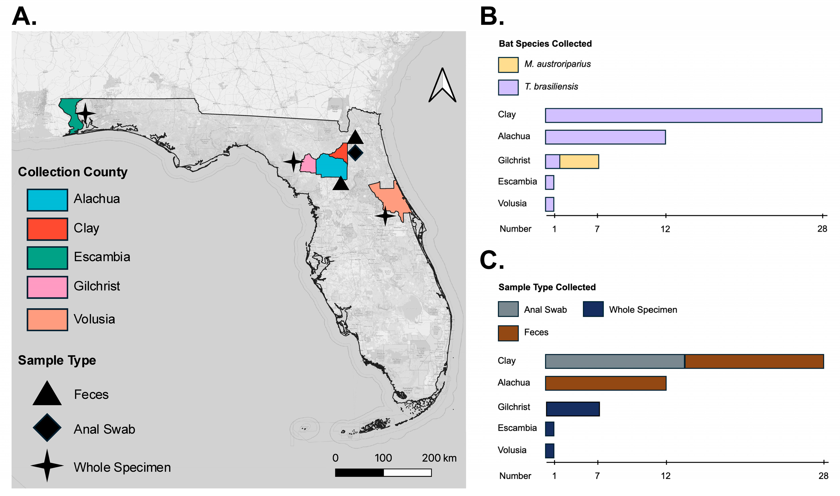
Figure 1. Map of sampling across Florida counties. The map indicates sampling sites where either whole specimens or fecal samples were collected: (A) map of the state of Florida with counties highlighted where bat samples were collected and the sample type collected by county; (B) distribution of bat species sampled by county; (C) distribution of sample type collected by county.
Microorganisms 13 02625 g001
Figure 2. ML tree of AstV found in M. austroriparius sampled in Gilchrist County, FL in 2021 (specimen Ma_Frozen_4). The tree is based on the concatenated alignment of Mamastrovirus genomes with the study sample (GTR+F+R5). Mamastrovirus genogroups are labeled as GI and GII. The subclade containing AstV-Ma4-FL-2021 (GenBank PX482536), indicated in bold, is highlighted in blue. Sequence hosts are depicted and colored by host species, with bats highlighted in red. Diamonds at nodes indicate ultrafast bootstrap values above 90. Viruses labeled by GenBank accession number, name, and host. Scale bar in nucleotide substitutions per site. Outgroup rooted by Avastrovirus Turkey Astrovirus (NC_002480.1).
Microorganisms 13 02625 g002
Figure 3. ML tree of AlphaCoV found in M. austroriparius sampled in Gilchrist County, FL in 2021 (specimen Ma_Frozen_3). The tree is based on concatenated alignment of ACoV-Ma3-FL-2021 (PX462113-PX470470) with alphacoronaviruses from all fifteen classified subgenera as well as unclassified sequences with close identity to ACoV-Ma3-FL-2021 (GTR+F+R6). The subclade containing ACoV-Ma3-FL-2021, indicated in bold, is highlighted in blue. Sequence hosts are depicted and colored by host species, with bats highlighted in red. Diamonds at nodes indicate ultrafast bootstrap values above 90. Viruses labeled by GenBank accession number, virus name, and host. Scale bar in nucleotide substitutions per site. Outgroup rooted by Betacoronavirus SARS-CoV-2 (NC_045512.2).
Microorganisms 13 02625 g003
Figure 4. ML tree of HVLV2 found in T. brasiliensis guano sampled in Alachua County, FL in 2024 (specimen Tb_Feces_15). The tree is based on the concatenated alignment of the study sample with HVLV2 reference genomes (TVM+F+I). HVLV2-Tb15-FL-2024 (PX462114) is indicated in bold. Black diamonds at nodes indicate ultrafast bootstrap values above 90. Viruses labeled by GenBank accession number, country, and date. Tips are colored according to host species. Scale bar in nucleotide substitutions per site. Outgroup rooted by Hubei virga-like virus 1 (NC_033165.1) and truncated for visualization purposes.
Microorganisms 13 02625 g004
Figure 5. Genus-level bacterial composition of M. austroriparius frozen museum samples. Only taxa with ≥1% relative abundance are shown. For taxonomic assignment, a Naive Bayes classifier was trained on SILVA 138 reference sequences trimmed in silico to the V3–V4 region using primers 341F (CCTACGGGNGGCWGCAG) and 806R (GACTACHVGGGTATCTAATCC).
Microorganisms 13 02625 g005
Table 1. Description of study bat samples.
Sample TypeBat Species (N)Collection YearCollection County
FecesT. brasiliensis (12)2024Alachua
T. brasiliensis (14)2022Clay
Anal SwabT. brasiliensis (14)2022Clay
Museum SpecimenM. austroriparius (5)2021Gilchrist
T. brasiliensis (5)2021Escambia, Gilchrist, Volusia
Table 2. Results of BLAST search of viral contigs.
VirusBat Species
(Bat ID)
Contig
(# bp)
Mean Coverage DepthBLAST Best Match
(Accession)
Percent IdentityGenome
Region
AlphaCoVM. austroriparius (Ma_Frozen_3)k119_6311
(925)
21ACoV-2-Tb (OP700657.1)815′, ORF1a
k119_670
(5309)
178ACoV-2-Tb (OP700657.1)81S, ORF3, E, M, N, ORF7, ORF8, 3′
M. austroriparius (Ma_Frozen_4)k119_1119
(1327)
12ACoV-2-Tb (OP700657.1)84N, ORF7, ORF8, 3′
AstVM. austroriparius (Ma_Frozen_4)k119_11258 (3292)209Bat AstV
(MZ218054.1)
80ORF1b, ORF2
HVLV2T. brasiliensis (Tb_Guano_15)k141_4344
(9829)
194HVLV2
(MW434995.1)
99Polyprotein
Disclaimer/Publisher’s Note: The statements, opinions and data contained in all publications are solely those of the individual author(s) and contributor(s) and not of MDPI and/or the editor(s). MDPI and/or the editor(s) disclaim responsibility for any injury to people or property resulting from any ideas, methods, instructions or products referred to in the content.