Article

Screening and Genomic Analysis of Bacillus velezensis R12 as a Biocontrol Agent Against Fusarium oxysporum Causing Wilt in Longya Lily (Lilium brownii var. viridulum)

1
Key Laboratory of Natural Microbial Medicine Research of Jiangxi Province, Jiangxi Science and Technology Normal University, Nanchang 330013, China
2
Key Laboratory of Microbial Resources and Metabolism of Nanchang City, Jiangxi Science and Technology Normal University, Nanchang 330013, China
3
National Key Laboratory of Uranium Resources Exploration-Mining and Nuclear Remote Sensing, East China University of Technology, Nanchang 330013, China
*
Authors to whom correspondence should be addressed.
These authors contributed equally to this work.
Microorganisms 2025, 13(11), 2430; https://doi.org/10.3390/microorganisms13112430
Submission received: 8 September 2025 / Revised: 9 October 2025 / Accepted: 21 October 2025 / Published: 23 October 2025
(This article belongs to the Section Microbial Biotechnology)

Abstract

Longya Lily (Lilium brownii var. viridulum) bulb rot, a devastating soil-borne disease caused by Fusarium oxysporum f. sp. lilii (Fol L1-1), severely compromises yield and quality of this economically significant crop. In this study, strain R12 was isolated from the rhizosphere soil of asymptomatic Longya lily plants and identified as Bacillus velezensis. The strain significantly disrupted the spore germination and hyphal morphology of Fol L1-1. In pot experiments, R12 not only effectively suppressed disease development but also promoted plant growth, a trait potentially linked to its indole-3-acetic acid (IAA) production capacity. Genomic analysis revealed a 4,015,523 bp circular chromosome (46.42% GC content) harboring gene clusters for the synthesis of diverse secondary metabolites, including surfactin, fengycin, difficidin, and bacillibactin. These findings highlight the potential of B. velezensis R12 as a biocontrol agent and provide insights into its mechanisms for suppressing phytopathogens and promoting plant growth.

1. Introduction

Longya lily (Lilium brownii var. viridulum), a commercially significant geophyte endemic to subtropical China, is intensively cultivated in Hunan and Jiangxi provinces for its economic and medicinal value. Its bulbs exhibit a distinctive morphological profile, characterized by large size, waxy epidermis, and viscous parenchyma tissue when cooked, enabling dual applications in culinary traditions and phytotherapy. Phytochemical analyses confirm the presence of bioactive steroidal saponins, polysaccharides, alkaloids, phenolics, and volatile oils [1,2,3] which synergistically confer demonstrated pharmacological activities, including: antioxidant, anti-inflammatory, immunomodulatory, anti-tumour and hypoglycaemic activities [3]. Owing to its high nutrient density and remarkable therapeutic potential, it is designated “Southern Ginseng” in traditional herbalism [1].
However, the rapid intensification of Longya lily monoculture faces mounting constraints from soil-borne diseases, particularly a destructive bulb rot and systemic wilt caused by Fusarium oxysporum f. sp. lilii (Fol-L1-1). This pathogen, capable of long-term soil survival and vascular colonization, leads to substantial yield losses through impaired bulb maturation and post-harvest deterioration [4,5,6,7,8]. Compounding this threat, the limited availability of registered fungicides and the accelerating development of pathogen resistance underscore the urgent need for sustainable biological control strategies.
Plant-growth-promoting rhizobacteria (PGPR) belonging to the genus Bacillus enhance plant growth and suppress Fusarium oxysporum (Fo) through multifaceted mechanisms [9,10,11,12]. Representative PGPR strains demonstrating this dual capacity include B. subtilis [9], B. velezensis [10], B. amyloliquefaciens [11], and B. cereus [12]. Their antagonistic activities include secreting antifungal lipopeptides (e.g., iturin, fengycin, surfactin) that disrupt fungal membrane integrity, siderophore-mediated iron competition, and the induction of systemic resistance in host plants. Critically, such PGPR strains demonstrate targeted efficacy against Fusarium pathogens in lily systems. For instance, B. velezensis strain E exhibited 86% inhibition of F. oxysporum mycelial growth in vitro through the synthesis of antifungal substances; simultaneously, rhizosphere application reduced root rot severity by 83% in potted Lilium plants. This biocontrol effect was synergized with growth promotion: strain E increased the bulb fresh weight through siderophore and IAA production, consequently promoting scale bulblet formation [13]. This dual synergy underscores the potential of B. velezensis in sustainable lily cultivation.
In this study, we isolated a novel strain, Bacillus velezensis R12, from the rhizosphere of asymptomatic Longya lily plants. It exhibited pronounced antagonism against Fol-L1-1 and demonstrated significant disease suppression alongside plant growth promotion in greenhouse trials. Genomic analysis revealed a comprehensive biosynthetic repertoire, including gene clusters for surfactin, fengycin, and the polyketide difficidin. This distinctive genomic potential, combined with its pronounced efficacy within the Longya lily system, distinguishes strain R12 from previously reported B. velezensis strains and highlights its promise as a tailored biocontrol agent.

2. Materials and Methods

2.1. Soil Sampling and Bacterial Isolation

Rhizosphere soil samples were collected from healthy Longya lily plants in Baishui Township, Yichun City, Jiangxi Province. The rhizosphere soil adhering to bulbs and roots was carefully collected using sterile brushes. Serial dilutions (10−1–10−6) of the soil samples were prepared in sterile saline (0.85% NaCl), plated on LB agar, and incubated at 30 °C for 24–48 h. Morphologically distinct colonies were purified through three rounds of streak-plate isolation.

2.2. Screening of Antagonistic Bacteria

Antagonistic bacteria were screened by the plate confrontation test. Fol was inoculated onto Potato Dextrose Agar (PDA) plates and cultured at 28 °C for 5–7 days. A mycelial plug (5 mm in diameter) of the pathogen was aseptically transferred to the center of a fresh PDA plate using a sterile cork borer (Biosharp, Langjieke Technology Co., Ltd., Hefei, China). After 1 day of culture at 28 °C, all the bacterial strains were spot-inoculated at equidistant positions 2.5 cm away from the central plug. Plates were re-incubated at 28 °C for 5 days, and inhibition zones were observed. The isolate exhibiting maximal inhibition was selected for further study. The isolation and identification of the pathogenic strain Fol L1-1 are described in detail in Supplementary Materials.

2.3. Inhibitory Effect of Strain R12 Bacterial Culture Filtrate (BCF) on Spore Morphology and Hyphal Growth of Fol L1-1

Fol L1-1 spore suspension preparation: fungal cultures grown in PDB at 28 °C/180 rpm for 5 days were filtered through sterile gauze. Harvested spores were resuspended in sterile distilled water and adjusted to 1 × 10⁶ spores/mL using a hemocytometer.
R12 BCF preparation: after reviving from −80 °C storage on LB agar (37 °C, 24 h), a single colony was inoculated into LB broth (37 °C, 200 rpm, 12 h). Subcultured broth (100 μL) was transferred to fresh LB medium (28 °C, 180 rpm, 48 h), followed by centrifugation (10,000× g, 10 min) and sterile filtration (0.22 μm) to obtain cell-free filtrate.
Inhibition assay: Fol L1-1 spores were exposed to 10% (v/v) BCF (treatment) or LB broth (control) at 28 °C/180 rpm. After 5 h of co-incubation, spore morphological alterations were assessed using scanning electron microscopy (SEM; Chengdu SCI-GO Research Service Co., Ltd., Chengdu, Sichuan, China). Following 72 h incubation, hyphal morphological changes were analyzed by light microscopy and SEM.

2.4. Scanning Electron Microscopy of Strain R12

The R12 suspension was centrifuged (10,000× g, 10 min), pelleted cells were fixed with 2.5% glutaraldehyde (4 °C, 24 h), washed thrice with 0.1 M phosphate buffer (pH 7.4), and processed for imaging using a Nova Nano SEM 450 microscope (FEI, Hillsboro, OR, USA).

2.5. Greenhouse Biocontrol Efficacy of Strain R12 Against Longya Lily Fusarium Wilt

Pathogen preparation: Fol L1-1 spore suspensions were prepared as previously described, with concentration adjusted to 1 × 10⁷ CFU/mL using sterile distilled water.
Biocontrol agent preparation: Strain R12 was cultivated according to the BCF preparation protocol detailed previously (28 °C, 180 rpm, 24 h in LB broth). Cultures were centrifuged (10,000× g, 10 min), washed, and resuspended in sterile water to 1 × 10⁷ CFU/mL.
Experimental design: Longya lily bulb scales were surface-sterilized in 75% (v/v) ethanol for 30 s, followed by three rinses with sterile water. After air-drying, each bulb scale was planted individually in a pot (16.3 cm in height, 20 cm in diameter) containing 500 g of sterilized soil mixture. Plants were cultivated in a greenhouse maintained at 25 °C under a 16 h light/8 h dark photoperiod. After 10 days, the seedlings were subjected to the following treatments: (1) drenching with 15 mL of strain R12 suspension; (2) drenching with 15 mL of strain R12 suspension followed 24 h later by 15 mL of Fol L1-1 spore suspension; (3) drenching with 15 mL of 0.1% hymexazol followed 24 h later by 15 mL of Fol L1-1 spore suspension; (4) drenching with 15 mL of sterile distilled water; or (5) drenching with 15 mL of sterile distilled water followed 24 h later by 15 mL of Fol L1-1 spore suspension. Each treatment was performed using 10 plants with 3 replicates. At 45 days post-inoculation with R12, shoot fresh and dry weights were measured. For plants inoculated with Fol L1-1, disease severity and disease index were recorded 45 days after R12 inoculation. Disease severity was rated on a 0–4 disease scale: 0 = no symptoms; 1 = leaf wilting affecting less than 1/4 of the seedling; 2 = leaf wilting affecting 1/4 to 1/2 of the seedling; 3 = leaf wilting affecting more than 1/2 of the seedling; 4 = complete wilting and death of the plant. The disease index (DI) was calculated as follows: DI = [[(0 × N0) + (1 × N1) + (2 × N2) + (3 × N3) + (4 × N4)]/T × 4] × 100, where N0–N4 represent the number of plants in each disease rating category, and T is the total number of plants assessed. Disease incidence was calculated as: [N1 + N2 + N3 + N4]/T × 100%. Control efficacy was determined as: (DI of control − DI of treatment)/DI of control × 100%.

2.6. Plant Growth-Promoting (PGP) Trait Analysis of Strain R12

The PGP capabilities of strain R12 were evaluated through quantitative assays for phosphate solubilization, siderophore production, and indole-3-acetic acid (IAA) synthesis. Phosphate solubilization was assessed per Nautiyal [14] via halo formation on Pikovskaya’s agar (7 d, 30 °C), siderophore production detected on CAS agar [15] through orange halo measurement after 72 h incubation, and IAA synthesis quantified colorimetrically [16] using Salkowski reagent (530 nm) against standards after culturing R12 in LB + 0.2% L-tryptophan (48 h, 30 °C). All assays were conducted with triplicate biological replicates with three technical repetitions each.

2.7. Identification of Antagonistic Bacteria R12

Morphological characterization: Morpho-physiological characterization was conducted following Bergey’s Manual of Systematic Bacteriology, including biochemical tests (starch hydrolysis, gelatin liquefaction, methyl red, nitrate reduction), enzymatic assays (oxidase, catalase, Voges-Proskauer), and metabolic trait evaluation (indole production, H2S generation).
16S rRNA gene identification: Genomic DNA was extracted from strain R12, followed by PCR amplification of the 16S rDNA using universal primers 27F (5′-AGAGTTTATCCTGGGCTCAG-3′) and 1492R (5′-GGTTACCTTGTTACGACTT-3′). The purified PCR products were subsequently sequenced by Sangon Biotech Co., Ltd. (Shanghai, China). The obtained sequences were subjected to BLAST (version 2.16.0) homology alignment against the NCBI database. Phylogenetic tree construction was performed using the neighbor-joining method implemented in MEGA 11.0 software.

2.8. Genome Sequencing and Assembly of Strain R12

Whole-genome sequencing of DNA samples was performed by Baimaike Biotechnology Co., Ltd. (Beijing, China). Following quality control, high-molecular-weight DNA was fragmented using g-TUBE devices (Covaris, LLC, Woburn, MA, USA). The sheared DNA was subjected to damage repair and end—repair procedures, followed by ligation with dumbbell—shaped adapters. The resulting products were treated with exonuclease to remove unligated fragments. Finally, target fragments were size—selected using the BluePippin system (software v.6.31, Sage Science, Beverly, MA, USA) to construct the sequencing library.
The filtered reads were assembled using Hifiasm v0.12. Circularization and origin positioning were then performed using Circlator v1.5.5. The genome was further polished using Pilon v1.22 with second—generation sequencing data to achieve a higher—accuracy genome assembly. A circular genome map of R12 was generated using Circos v0.66.

2.9. Genomic Functional Annotation of Strain R12

The predicted sequences of the genes underwent BLAST analysis against multiple databases, including Nr, KEGG, eggNOG, SwissProt, TrEMBL, TCDB, and PHI-base, to obtain functional annotation results. Based on the BLAST results from the Nr database, the genomic sequences were annotated against the Gene Ontology (GO) database using Blast2GO software (v2.5). Sequences of unknown function were compared with the Pfam database using HMMER software (v3.4) to complete Pfam database annotation. Additionally, the protein sequences were aligned with Hidden Markov Models (HMMs) of each family in the CAZy database using HMMER software to accomplish CAZy database annotation. The Resistance Gene Identifier (RGI) tool from the Comprehensive Antibiotic Resistance Database (CARD) was utilized to align the protein sequences with the database, generating CARD database annotation results. Finally, the protein sequences were subjected to BLAST alignment against the core dataset Sets A to obtain the Virulence Factors Database (VFDB) annotations for the genome.

2.10. Analysis of Gene Clusters Responsible for the Biosynthesis of Secondary Metabolites

Potential secondary-metabolite biosynthetic gene clusters were systematically mined and annotated with antiSMASH 8.0.3, following the pipeline described by Blin et al. [17].

2.11. Statistical Analyses

All data were processed and analyzed using Microsoft Excel and IBM SPSS Statistics (version 26), respectively. Statistical significance among groups was determined by one-way analysis of variance (ANOVA) followed by the Least Significant Difference (LSD) post hoc test for multiple comparisons. Data are presented as the mean ± standard error of the mean (SEM) from three independent biological replicates (n = 3). Differences were considered statistically significant at p < 0.05. All graphs were generated using Origin 2024 software.

3. Results

3.1. Isolation and Screening of Antagonistic Bacteria Against Fusarium oxysporum

A total of 110 bacterial strains exhibiting distinct colony morphologies were isolated and purified from rhizosphere soil of asymptomatic Longya lily plants. In dual-culture assays with Fusarium oxysporum f. sp. Lilii strain L1-1 on PDA medium, four strains significantly inhibited mycelial growth [7]. Among these, strain R12 exhibited the strongest antagonistic activity against Fol L1-1 (Figure 1A,B). This strain’s biocontrol activity is not limited to a single pathogen; our previous work has demonstrated its efficacy against a spectrum of fungi, including Fusarium sp. L3, Penicillium ochrochloron F5, and Penicillium janthinellum F7 [7].

3.2. Antagonistic Activity of Strain R12 BCF Against Fol L1-1

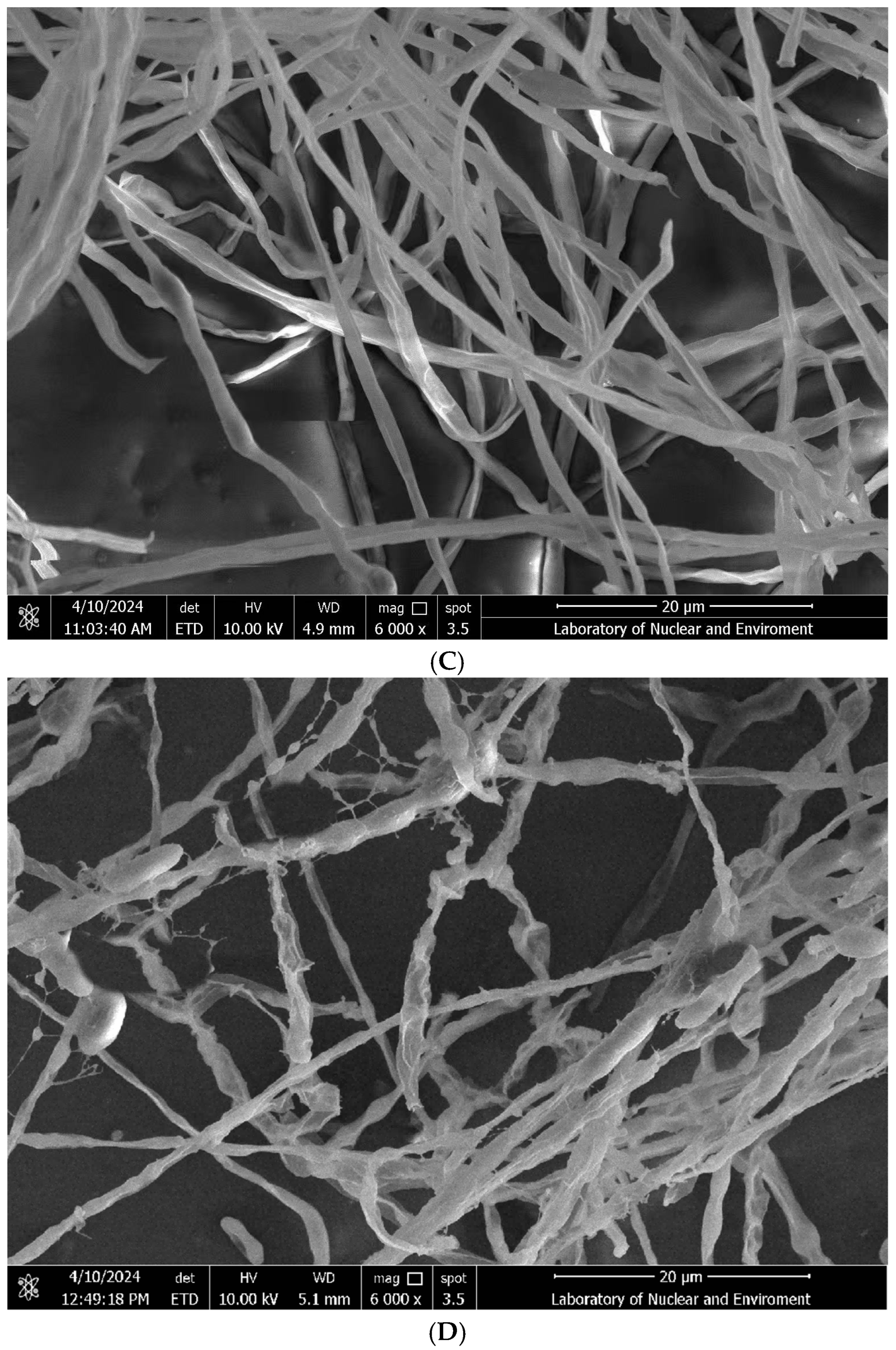
Morphological alterations in Fol L1-1 conidia following 5 h exposure to R12 BCF are shown in Figure 2. SEM analysis revealed that untreated conidia maintained turgidity with intact surfaces, whereas R12 BCF-treated conidia exhibited severe deformation, including surface invagination and cellular shrinkage. Additionally, R12 BCF induced aberrant hyphal development: control hyphae displayed robust growth with uniform diameter and smooth morphology, while co-cultured hyphae showed ultrastructural collapse, featuring irregular invaginations, localized disintegration, and fragmentation. These results demonstrate that R12 BCF directly compromises the structural integrity of Fol L1-1 conidia and hyphae, leading to growth suppression.

3.3. Physiology, Biochemistry, and 16S rRNA Gene Identification of Antagonistic Bacterial Strain R12

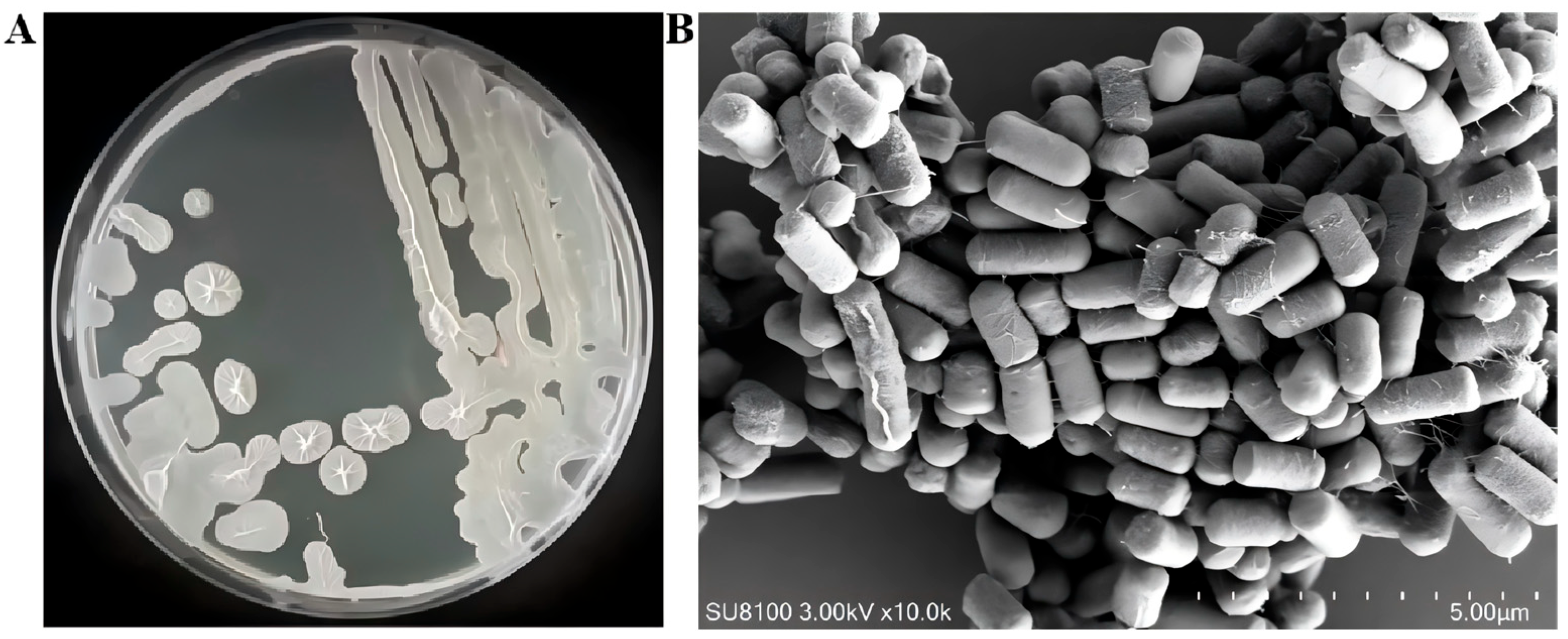
Morphological characterization: After 12 h of incubation on LB agar plates, colonies of strain R12 exhibited rough texture, desiccated surfaces, and wrinkled morphology. Scanning electron microscopy (SEM) revealed straight, flexible rod-shaped cells, confirming their bacillus morphology (Figure 3).
Physiological and biochemical characterization: Strain R12 is a Gram-positive bacterium. It yielded negative results for the Voges-Proskauer (V-P) reaction and mannitol utilization but was positive for all other tested traits (Table 1).
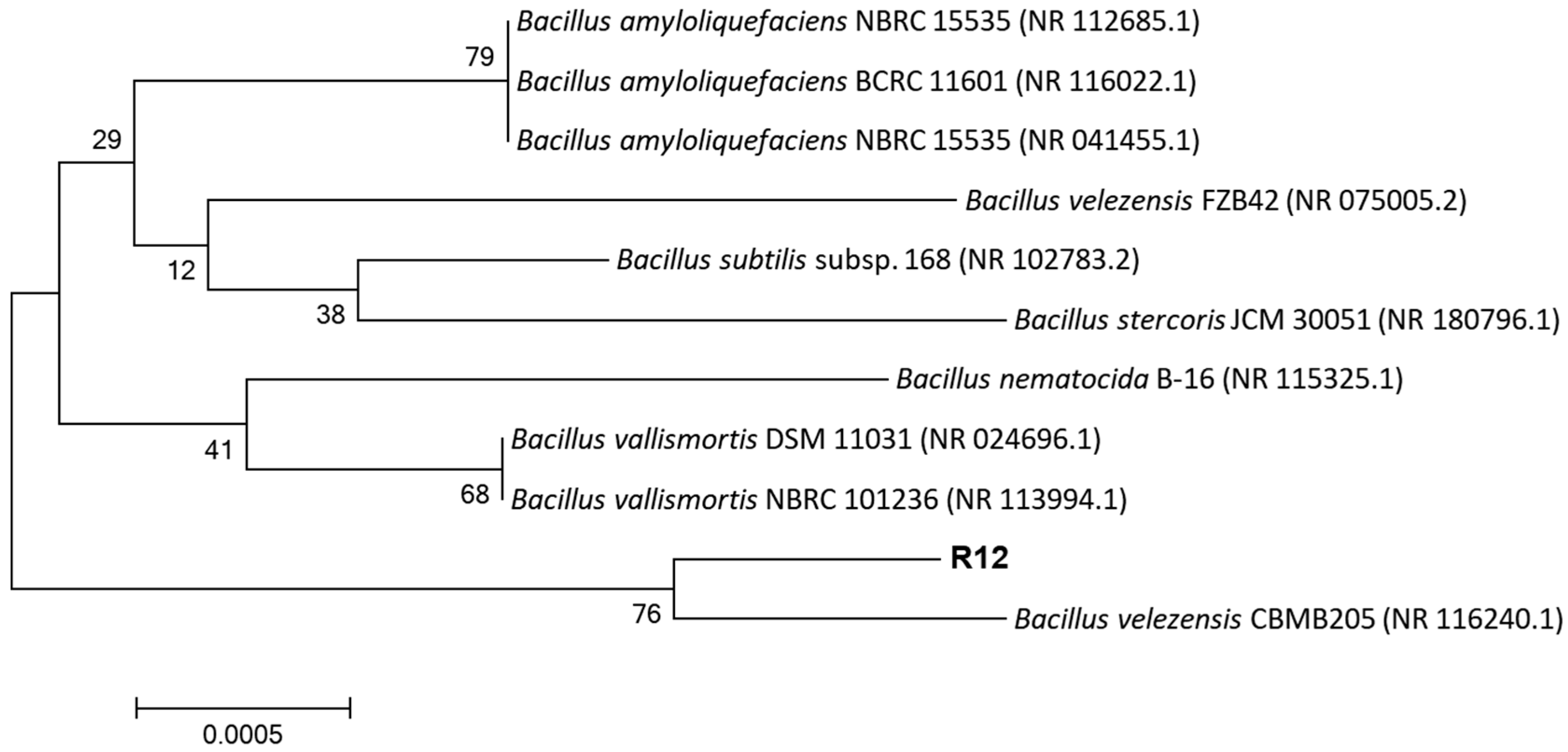
Molecular identification and phylogenetic analysis: Genomic DNA was extracted from strain R12, and the 16S rRNA gene was amplified using universal primers 27F/1492R. The sequenced PCR product was subjected to BLAST analysis, revealing 99% sequence similarity to Bacillus velezensis in the NCBI database. A phylogenetic tree constructed with MEGA 11.0 showed that strain R12 clusters with Bacillus velezensis CBMB205 on a distinct branch (Figure 4).
Integrating morphological, physiological, biochemical, and molecular evidence confirmed that antagonistic strain R12 is Bacillus velezensis.

3.4. Assessment of PGP Traits in B. velezensis R12

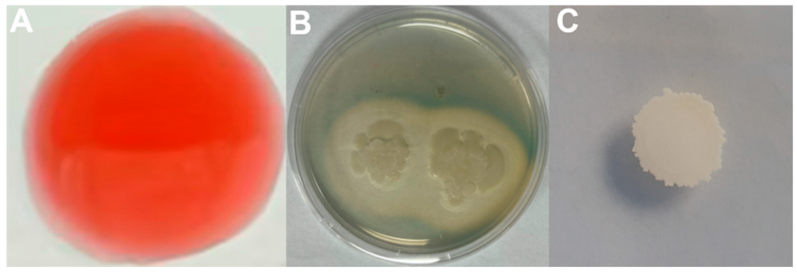
The PGP traits of B. velezensis R12, including IAA synthesis, siderophore production, and phosphate solubilization, were evaluated. The results indicated that strain R12 possesses the ability to produce IAA (Figure 5A), albeit at a relatively low concentration of 6.87 mg/L. In contrast, the strain demonstrates strong siderophore production capacity, with an A/Ar value of 0.388 and a yield reaching 32.56 mg/L (Figure 5B), but does not exhibit phosphate-solubilizing activity (Figure 5C). Therefore, the growth-promoting effects of R12 on Longya lily may be attributed to siderophore and IAA production, rather than to phosphate solubilization.

3.5. Effects of B. velezensis R12 on Fusarium Wilt Suppression and Growth Promotion in Longya Lily

B. velezensis R12, which exhibited the highest antagonistic activity against Fol L1-1 in vitro, was selected for further assessment of its biocontrol efficacy in greenhouse trials. The results indicated that R12 significantly promoted the growth of Longya lily seedlings both under Fol L1-1 challenge and in pathogen-free conditions (Figure 6). Furthermore, R12 effectively alleviated disease symptoms and reduced the severity of Fusarium wilt at 45 days after inoculation with Fol L1-1. Data on disease incidence, disease index, and biocontrol efficacy confirmed that R12 substantially suppressed wilt symptoms caused by Fol L1-1 (Table 2).

3.6. Genome Sequencing and Assembly of R12

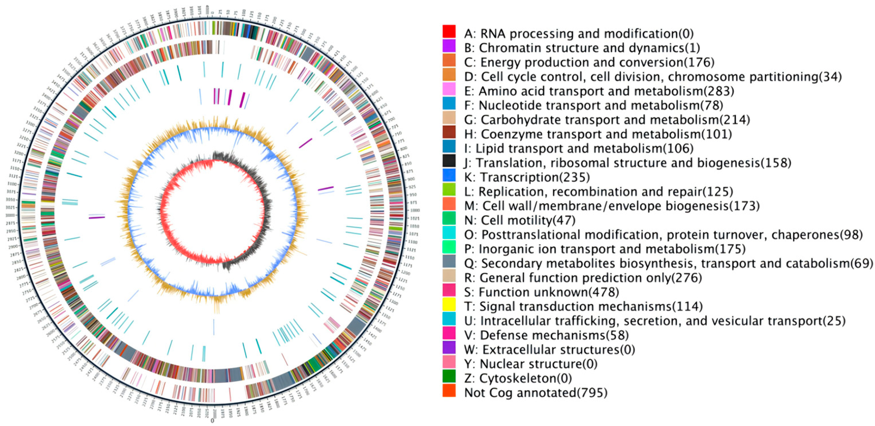
Whole-genome sequencing of strain R12 was sequenced and assembled using PacBio Sequel II platform single-molecule real-time (SMRT) technology. The genome map showed that 22,709 gene sequences were obtained. Sequencing yielded 210,960,009 bp of clean data, and the length of N50 was 10,170 bp. The final assembly revealed a complete circular chromosome of 4,015,523 bp with a GC content of 46.42% (Figure 7 and Table 3).

3.7. Functional Annotation of the Genome of R12

Comparative genomic annotation of strain R12 against seven functional databases revealed differential gene assignment patterns: 3803 protein-coding genes were mapped to NCBI non-redundant (Nr), 2993 to Gene Ontology (GO), 2171 to Kyoto Encyclopedia of Genes and Genomes (KEGG), 3024 to eggNOG, 3392 to Pfam, 2829 to Swiss-Prot, and 3798 to TrEMBL databases (Table S1).

3.8. Annotation Results Description of the Nr Database

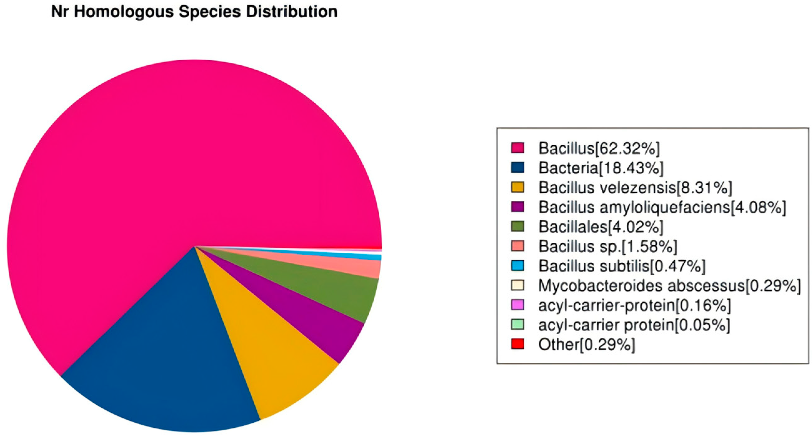
The Nr database (Non-Redundant Protein Database) consolidates experimentally validated and computationally predicted protein sequences with taxonomic classification. Comparative analysis indicated that the Nr database achieved the highest annotation rate among the evaluated repositories. In this study, 3803 genes in the R12 genome were functionally annotated via the Nr database, with 62.32% showing homology to characterized Bacillus species (Figure 8).

3.9. Description of GO Database Annotation Results

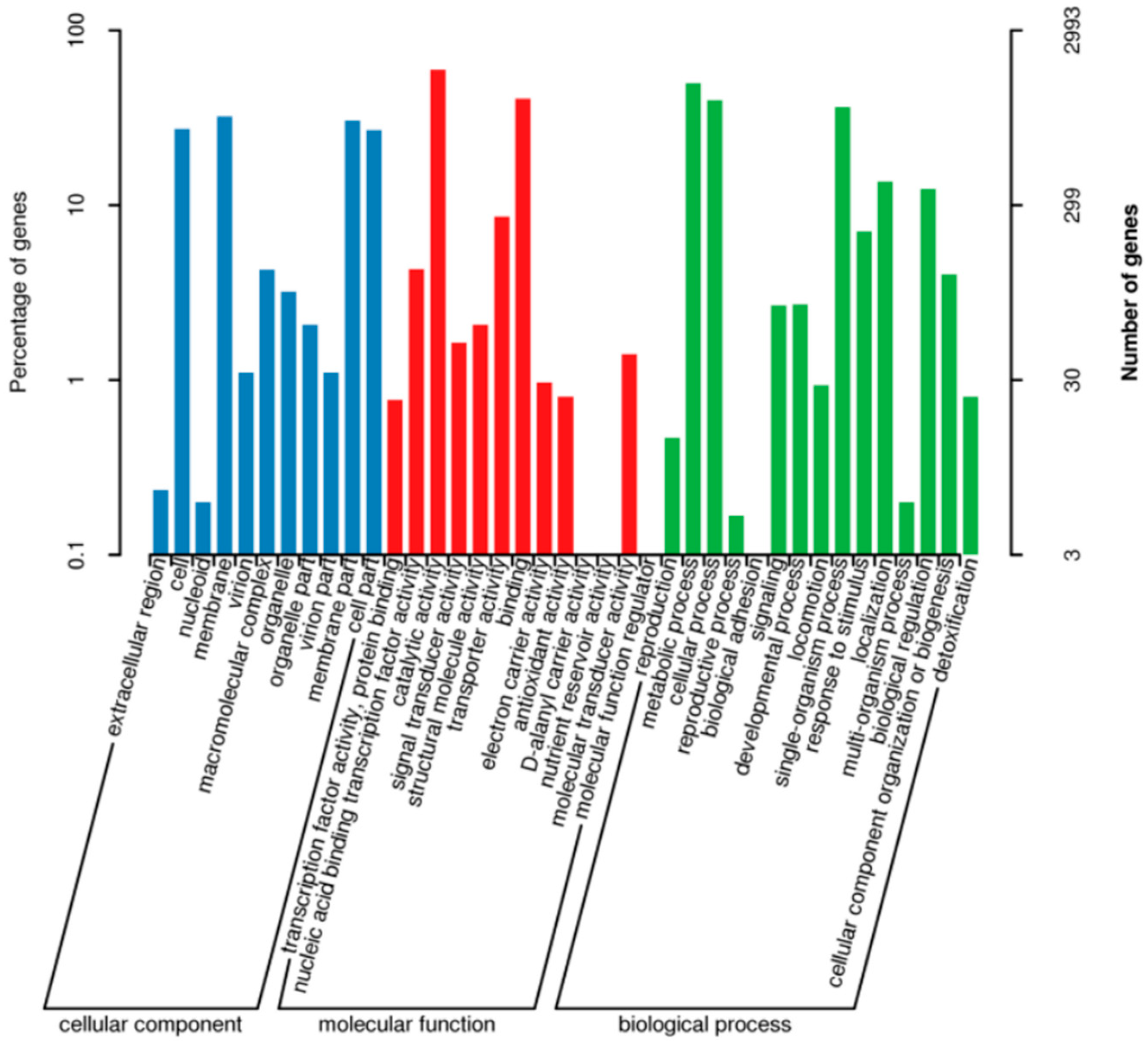
Gene Ontology (GO) annotation categorizes biological functions into three primary domains: cellular component, molecular function, and biological process. Functional annotation of strain R12 against the GO database assigned 2993 protein-coding genes, with hierarchical distribution as follows: 1488 genes annotated to cellular component organization, 2372 genes to molecular function (constituting 79.3% of total annotations), and 2200 genes to biological process regulation (Figure 9).

3.10. Annotation Results Description of the eggNOG Database

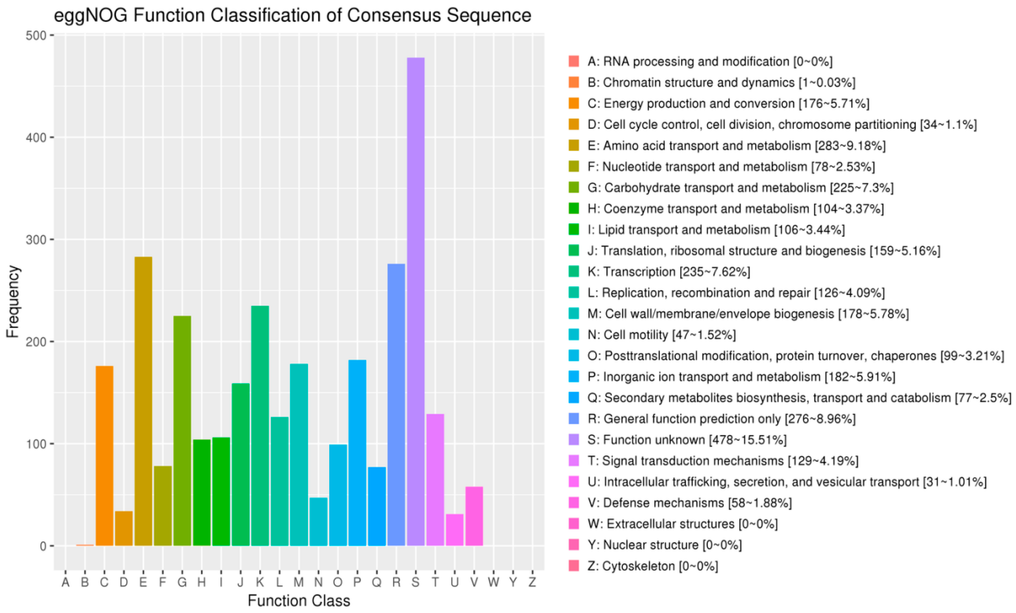
BLAST alignment of R12-encoded proteins against the eggNOG database annotated 3024 genes with functional orthologous groups. The predominant functional categories comprised: amino acid transport and metabolism (283 genes, 9.18%); general function prediction (276 genes, 8.96%); transcription regulation (235 genes, 7.62%), and carbohydrate transport and metabolism (225 genes, 7.30%). Chromatin structure and dynamics represented the least populated category (1 gene, 0.03%) (Figure 10).

3.11. Genomic Analysis of Antimicrobial Biosynthetic Potential in R12

Online prediction of the secondary metabolite biosynthesis gene clusters in B. velezensis R12 was performed using the antiSMASH 8.0.3 database, which revealed high sequence homology to known biosynthetic gene clusters. As shown in Table S2, a total of 15 secondary metabolite gene clusters were identified in the genome of strain R12. Among these, eight clusters exhibited high genetic similarity to those encoding known bioactive compounds, including surfactin, macrolactin H, bacillaene, fengycin, difficidin, bacillibactin, bacilysin, and mersacidin.
Genomic analysis revealed that R12 possesses complete biosynthetic gene clusters for key lipopeptides, including surfactin, fengycin, and difficidin. These compounds are well-established to act synergistically in membrane disruption, surfactin facilitating pore formation that enhances fengycin’s membrane permeabilization [8], while difficidin exhibits broad-spectrum antibacterial activity. Surfactin promotes biofilm matrix assembly in Bacillus spp. through quorum sensing modulation [18] and demonstrates dual biocontrol efficacy via induced systemic resistance (ISR) activation in plants and direct antifungal activity [19,20,21]. Macrolactin can inhibit protein synthesis in bacteria and possesses potent antibacterial activity [22]. Bacilysin, a dipeptide antibiotic, exerts activity through its C-terminal L-antipain domain, though its biosynthesis is strain-specific in Bacillus [23]. Bacillomycin lyses hyphal and conidial cell walls via surfactant activity [24] while inducing lethal reactive oxygen species (ROS) accumulation in fungal propagules [25].
We hypothesize that the synergistic action of surfactin and fengycin gene clusters may be responsible for the observed membrane damage, though this requires experimental validation through gene knockout studies. Further investigations are required to delineate the dominant lipopeptide governing L1-1 antagonism and its molecular mechanism.

4. Discussion

Lily bulb rot disease progressively induces tissue maceration and complete bulb decomposition. Our experiments demonstrated that both vegetative cells of B. velezensis R12 and its cell-free fermentation supernatant exhibited pronounced antagonism against Fusarium oxysporum L1-1. Previous studies have established B. velezensis’s biocontrol efficacy across plant-pathogen systems. Notably, Khan et al. [26] isolated B. velezensis Lle-9 from lily bulbs, demonstrating dual functionality through antifungal activity and phytohormone-mediated growth promotion. Genomic analyses by Li et al. [13] further corroborated B. velezensis’s genomic repertoire for integrated disease management, functioning simultaneously as a rhizosphere-competent biocontrol agent and nitrogen-fixing biofertilizer in lily cultivation systems.
The broad-spectrum biocontrol potential of Bacillus spp. against diverse phytopathogens is well-documented. Khanal et al. [27] isolated Bacillus sp. ST24 and Bacillus panaciterrae ST25 from rice seeds, demonstrating antagonistic activity against Marasmius graminum and Rhizoctonia solani AG11, causal agents of rice seedling blight. Degani et al. [28] confirmed the inhibitory effects of Bacillus subtilis R2 on Magnaporthiopsis maydis, the etiological agent of maize late wilt disease. Wei et al. [29] found that Bacillus mojavensis ZA1 can produce antimicrobial substances that inhibit Colletotrichum coccodes. Kamalanathan et al. [30] reported nematicidal activity of Bacillus velezensis VB7 against Meloidogyne incognita (root-knot nematode) in tomato. Shuang et al. [31] isolated Bacillus sp. K-9 from the interface between lesions and healthy tissue on potato tubers, and demonstrated its potent suppression of Rhizoctonia canker. Cui et al. [32] characterized Bacillus amyloliquefaciens 3–5 from potato tubers, revealing its efficacy against potato common scab.
Bacillus spp. show significant potential for controlling infectious diseases in Liliaceae plants. Wu et al. [33] established that Bacillus agavensis LFM-30 effectively suppresses Fusarium-mediated wilt in Liliaceae hosts. Tu et al. [34] reported biocontrol efficacies of 58.74% and 68.93% for Bacillus amyloliquefaciens BF1 and Bacillus subtilis Y37 against lily wilt disease, respectively. Li et al. [13] isolated Bacillus velezensis strain E from lily rhizospheric soils, which exhibited potent antagonism against Fusarium oxysporum and Botrytis cinerea. Liu et al. [35] elucidated that Bacillus cereus produces induced systemic resistance (ISR)-activating elicitors to suppress foliar pathogens in lilies through systemic acquired resistance pathways.
Bacillus velezensis has been widely recognized as a biocontrol agent with broad-spectrum antimicrobial activity. Yao et al. [36] reported its ovicidal and larvicidal efficacy against Meloidogyne incognita; R et al. [37] demonstrated in vitro and in silico inhibition of Fusarium oxysporum by B. velezensis CBMB205; Zhao et al. [38] validated B. velezensis JIN4’s suppression of Pseudomonas syringae-induced kiwifruit bacterial canker. Additional studies confirmed its biocontrol potential against rubber tree red root rot [39], apple anthracnose [40], postharvest chili anthracnose [41], rice sheath blight disease [42], and cereal diseases [43]. However, the antifungal mechanisms of B. velezensis [44] and its pan-genomic characteristics [45,46,47,48] remain inadequately characterized. This is particularly true for its antagonistic interactions with lily bulb rot pathogens, which are poorly understood.
Bacillus velezensis R12 possesses a unique complement of biosynthetic gene clusters (BGCs), including those for difficidin and bacillaene, which underpins its experimentally verified dual functionality—suppressing Fusarium wilt while promoting plant growth in Longya lily. Our genomic analysis revealed the presence of complete biosynthetic gene clusters for known antifungal lipopeptides such as surfactin, fengycin, and iturin. While the observed biocontrol efficacy is consistent with the activity of these compounds, future work will include liquid chromatography-mass spectrometry (LC-MS) analysis to experimentally validate the production and quantify the levels of these metabolites by strain R12 under various conditions. Functional annotation via the eggNOG database assigned 3024 genes to secondary metabolite biosynthesis, transport, and metabolic pathways, providing a genetic foundation for these observed activities. These findings establish a basis for the subsequent functional characterization of these BGCs and the antimicrobial metabolites they encode in B. velezensis R12.
While the results from this study are promising, the path to practical application requires further validation. Future research will focus on large-scale field trials to confirm efficacy under natural conditions, testing against a wider array of regionally relevant pathogen strains, and direct metabolite confirmation through advanced analytical chemistry.

5. Conclusions

The antagonistic bacterium Bacillus velezensis R12 demonstrated significant biocontrol potential against lily bulb rot disease, effectively inhibiting hyphal growth and spore germination of Fusarium oxysporum L1-1. Genomic analysis revealed a complete circular chromosome of 4,015,523 bp harboring diverse biosynthetic gene clusters for antimicrobial secondary metabolites, which underpin its efficacy and highlight its promise for development into a sustainable biocontrol agent. Future work will focus on validating the functional roles of specific biosynthetic gene clusters through gene expression studies and targeted mutagenesis.

Supplementary Materials

The following supporting information can be downloaded at https://www.mdpi.com/article/10.3390/microorganisms13112430/s1. Table S1. General database annotation statistics. Table S2. Prediction of secondary metabolite biosynthetic gene clusters in R12. Figure S1. Pathogenicity of Strain L1-1. Figure S2. (a) Colony Morphology of L1-1; (b) Spore Morphology of L1-1. Figure S3. Phylogenetic Tree of Pathogenic Strain L1-1.

Author Contributions

Conceptualization, H.G., Y.H., Z.C. and Q.Z.; methodology, H.G., Y.H., Z.C. and M.Z.; software, H.G., Y.H. and Q.L.; validation, H.G., Y.H. and Z.C.; formal analysis, H.G. and Y.H.; investigation, H.G., Y.H. and Z.C.; resources, H.G., Y.H. and Z.C.; data curation, H.G., Y.H. and Z.C.; writing—original draft preparation, H.G.; writing—review and editing, H.G., Y.H. and S.L.; visualization, H.G., Y.H. and Z.C.; supervision, K.Z., D.Z. and S.L.; project administration, H.J. and S.L.; funding acquisition, S.L. All authors have read and agreed to the published version of the manuscript.

Funding

This research was funded by National Natural Science Foundation of China (grant number 32360080) and Jiangxi Provincial Natural Science Foundation (grant numbers 20242BAB20286; 20252BAC240090).

Institutional Review Board Statement

Not applicable.

Informed Consent Statement

Informed consent was obtained from all individual participants included in the study.

Data Availability Statement

The original data presented in the study are openly available in China Center for Type Culture Collection (CCTCC) at CCTCC M 20241785.

Conflicts of Interest

The authors declare no conflict of interest.

References

  1. Li, X.; Wang, Z.; Hou, C.; Gong, X.; Liu, Z.; Shi, Y.; Yan, J.; Tong, Q. Identification and Characterization of Volatile Organic Compounds Based on GC-IMS Technology in Different Organs of Lilium brownii var. viridulum and After Bud-Removal and Non-Bud-Removal Treatments. Molecules 2025, 30, 1238. [Google Scholar] [CrossRef]
  2. He, D.; Zhang, H.; Li, S.; Zhou, X.; Zhang, S.H. Research progress on chemical constituents and pharmacological effects of Baihe (Lilii bulbus) and predictive analysis on quality markers. Chin. Arch. Tradit. Chin. Med. 2022, 40, 205–212. [Google Scholar]
  3. Zhou, J.; An, R.; Huang, X. Genus Lilium: A review on traditional uses, phytochemistry and pharmacology. J. Ethnopharmacol. 2021, 270, 113852. [Google Scholar] [CrossRef]
  4. Baayen, R.; Rijkenberg, F. Fine structure of the early interaction of lily roots with Fusarium oxysporum f. sp. lilii. Eur. J. Plant Pathol. 1999, 105, 431–443. [Google Scholar] [CrossRef]
  5. Liu, C.; Wang, Y.; Jin, L.; Wang, Y.; Liu, D. Morphological, molecular, and biological characterization of bulb rot pathogens in stored Lanzhou lily and the in vitro antifungal efficacy of three plant essential oils. Front. Microbiol. 2024, 15, 1307966. [Google Scholar] [CrossRef] [PubMed]
  6. Michielse, C.B.; Rep, M. Pathogen profile update: Fusarium oxysporum. Mol. Plant Pathol. 2009, 10, 311–324. [Google Scholar] [CrossRef]
  7. Liang, L.; Liu, S.; Zhu, D.; Huang, Y.; Zheng, Q.; Ji, H.; Zhan, M.; Chen, M. The identification of pathogens of bulb rot of Lilium browni var. viridulum and screening of antagonistic bacteria. J. Jiangxi Norm. Univ. 2024, 48, 491–498. [Google Scholar] [CrossRef]
  8. Al-Hazmi, N.E. Antibacterial and anticancer activities of rhizobacterial lipopeptides against carcinogenic bacteria. Antonie Van Leeuwenhoek 2025, 118, 118. [Google Scholar] [CrossRef] [PubMed]
  9. Xu, W.; Yang, Q.; Yang, F.; Xie, X.; Goodwin, P.H.; Deng, X.; Tian, B.; Yang, L. Evaluation and Genome Analysis of Bacillus subtilis YB-04 as a Potential Biocontrol Agent Against Fusarium Wilt and Growth Promotion Agent of Cucumber. Front. Microbiol. 2022, 13, 885430. [Google Scholar] [CrossRef]
  10. Cao, Y.; Pi, H.; Chandrangsu, P.; Li, Y.; Wang, Y.; Zhou, H.; Xiong, H.; Helmann, J.D.; Cai, Y. Antagonism of Two Plant-Growth Promoting Bacillus velezensis Isolates Against Ralstonia solanacearum and Fusarium oxysporum. Sci. Rep. 2018, 8, 4360. [Google Scholar] [CrossRef] [PubMed]
  11. Fan, B.; Chen, X.H.; Budiharjo, A.; Bleiss, W.; Vater, J.; Borriss, R. Efficient colonization of plant roots by the plant growth promoting bacterium Bacillus amyloliquefaciens FZB42, engineered to express green fluorescent protein. J. Biotechnol. 2011, 151, 303–311. [Google Scholar] [CrossRef] [PubMed]
  12. Ramírez, V.; Martínez, J.; Bustillos-Cristales, M.D.R.; Catañeda-Antonio, D.; Munive, J.A.; Baez, A. Bacillus cereus MH778713 elicits tomato plant protection against Fusarium oxysporum. J. Appl. Microbiol. 2022, 132, 470–482. [Google Scholar] [CrossRef]
  13. Li, B.; Wan, J.; Sha, J.; Tian, M.; Wang, M.; Zhang, X.; Sun, W.; Mao, Y.; Min, J.; Qin, Y.; et al. Genomics assisted functional characterization of Bacillus velezensis E as a biocontrol and growth promoting bacterium for lily. Front. Microbiol. 2022, 13, 976918. [Google Scholar] [CrossRef]
  14. Nautiyal, C.S. An efficient microbiological growth medium for screening phosphate solubilizing microorganisms. FEMS Microbiol. Lett. 1999, 170, 265–270. [Google Scholar] [CrossRef]
  15. Schwyn, B.; Neilands, J.B. Universal chemical assay for the detection and determination of siderophores. Anal. Biochem. 1987, 160, 47–56. [Google Scholar] [CrossRef]
  16. Glickmann, E.; Dessaux, Y. A critical examination of the specificity of the salkowski reagent for indolic compounds produced by phytopathogenic bacteria. Appl. Environ. Microbiol. 1995, 61, 793–796. [Google Scholar] [CrossRef] [PubMed]
  17. Blin, K.; Shaw, S.; Steinke, K.; Villebro, R.; Ziemert, N.; Lee, S.Y.; Medema, M.H.; Weber, T. antiSMASH 5.0: Updates to the secondary metabolite genome mining pipeline. Nucleic Acids Res. 2019, 47, W81–W87. [Google Scholar] [CrossRef] [PubMed]
  18. Stannius, R.O.; Fusco, S.; Cowled, M.S.; Kovács, Á.T. Surfactin accelerates Bacillus subtilis pellicle biofilm development. Biofilm 2025, 9, 100249. [Google Scholar] [CrossRef]
  19. Xiao, P.; Tian, X.; Zhu, P.; Xu, Y.; Zhou, C. The use of surfactin in inhibiting Botrytis cinerea and in protecting winter jujube from the gray mold. AMB Express 2023, 13, 37. [Google Scholar] [CrossRef]
  20. Krishnan, N.; Velramar, B.; Velu, R.K. Investigation of antifungal activity of surfactin against mycotoxigenic phytopathogenic fungus Fusarium moniliforme and its impact in seed germination and mycotoxicosis. Pestic. Biochem. Physiol. 2019, 155, 101–107. [Google Scholar] [CrossRef]
  21. Wang, Y.; Sun, Z.; Wang, S.; Wen, F.; Xia, Z. Surfactin: A novel Aphis gossypii killing surfactin produced by Bacillus australimaris TRM82479 of Taklamakan Desert origin. Front. Microbiol. 2025, 16, 1559495. [Google Scholar] [CrossRef]
  22. Vasilchenko, A.S.; Lukyanov, D.A.; Dilbaryan, D.S.; Usachev, K.S.; Poshvina, D.V.; Taldaev, A.K.; Nikandrova, A.A.; Imamutdinova, A.N.; Garaeva, N.S.; Bikmullin, A.G.; et al. Macrolactin a is an inhibitor of protein biosynthesis in bacteria. Biochimie 2025, 232, 25–34. [Google Scholar] [CrossRef]
  23. Islam, T.; Rabbee, M.F.; Choi, J.; Baek, K.H. Biosynthesis, Molecular Regulation, and Application of Bacilysin Produced by Bacillus Species. Metabolites 2022, 12, 397. [Google Scholar] [CrossRef]
  24. Jin, P.; Wang, H.; Tan, Z.; Xuan, Z.; Dahar, G.Y.; Li, Q.X.; Miao, W.; Liu, W. Antifungal mechanism of bacillomycin D from Bacillus velezensis HN-2 against Colletotrichum gloeosporioides Penz. Pestic. Biochem. Physiol. 2020, 163, 102–107. [Google Scholar] [CrossRef]
  25. Gu, Q.; Yang, Y.; Yuan, Q.; Shi, G.; Wu, L.; Lou, Z.; Huo, R.; Wu, H.; Borriss, R.; Gao, X. Bacillomycin D Produced by Bacillus amyloliquefaciens Is Involved in the Antagonistic Interaction with the Plant-Pathogenic Fungus Fusarium graminearum. Appl. Environ. Microbiol. 2017, 83, e01075-17. [Google Scholar] [CrossRef]
  26. Khan, M.S.; Gao, J.; Chen, X.; Zhang, M.; Yang, F.; Du, Y.; Moe, T.S.; Munir, I.; Xue, J.; Zhang, X. The Endophytic Bacteria Bacillus velezensis Lle-9, Isolated from Lilium leucanthum, Harbors Antifungal Activity and Plant Growth-Promoting Effects. J. Microbiol. Biotechnol. 2020, 30, 668–680. [Google Scholar] [CrossRef]
  27. Khanal, S.; Imran, M.; Zhou, X.G.; Antony-Babu, S. Characterization of differences in seed endophytic microbiome in conventional and organic rice by amplicon-based sequencing and culturing methods. Microbiol. Spectr. 2024, 12, e0366223. [Google Scholar] [CrossRef]
  28. Degani, O.; Ayoub, A.; Dimant, E.; Gordani, A. Antagonistic interactions between maize seeds microbiome species and the late wilt disease agent, Magnaporthiopsis maydis. Front. Fungal Biol. 2024, 5, 1436759. [Google Scholar] [CrossRef]
  29. Wei, L.; Fan, L.; Yang, C.; Jin, M.; Osei, R. Analysis of Bioactive Compounds Produced by Bacillus mojavensis ZA1 and Their Antagonistic Effect on Colletotrichum coccodes by GC-MS. Appl. Biochem. Biotechnol. 2024, 196, 4914–4933. [Google Scholar] [CrossRef]
  30. Kamalanathan, V.; Sevugapperumal, N.; Nallusamy, S. Antagonistic Bacteria Bacillus velezensis VB7 Possess Nematicidal Action and Induce an Immune Response to Suppress the Infection of Root-Knot Nematode (RKN) in Tomato. Genes 2023, 14, 1335. [Google Scholar] [CrossRef]
  31. Shuang, M.; Wang, Y.; Teng, W.; Jin, G. Isolation and identification of an endophytic bacteria Bacillus sp. K-9 exhibiting biocontrol activity against potato common scab. Arch. Microbiol. 2022, 204, 483. [Google Scholar] [CrossRef]
  32. Cui, L.; Yang, C.; Wang, Y.; Ma, T.; Cai, F.; Wei, L.; Jin, M.; Osei, R.; Zhang, J.; Tang, M. Potential of an endophytic bacteria Bacillus amyloliquefaciens 3–5 as biocontrol agent against potato scab. Microb. Pathog. 2022, 163, 105382. [Google Scholar] [CrossRef]
  33. Wu, H.; Lu, J.; Zhao, S.; Fei, J.; Qu, Z.; Zhao, M.; Yang, H. Microbial Community of Wilted Fritillaria ussuriensis and Biocontrol Effects of Bacillus tequilensis and Trichoderma koningiopsis. Biology 2024, 13, 940. [Google Scholar] [CrossRef] [PubMed]
  34. Tu, J.; Zhao, X.; Yang, Y.; Yi, Y.; Wang, H.; Wei, B.; Zeng, L. Two Bacillus spp. Strains Improve the Structure and Diversity of the Rhizosphere Soil Microbial Community of Lilium brownii var. viridulum. Microorganisms 2023, 11, 1229. [Google Scholar] [CrossRef]
  35. Liu, Y.H.; Huang, C.J.; Chen, C.Y. Evidence of induced systemic resistance against Botrytis elliptica in lily. Phytopathology 2008, 98, 830–836. [Google Scholar] [CrossRef] [PubMed]
  36. Yao, Y.; Wang, L.; Zhai, H.; Dong, H.; Wang, J.; Zhao, Z.; Xu, Y. Bacillus velezensis A-27 as a potential biocontrol agent against Meloidogyne incognita and effects on rhizosphere communities of celery in field. Sci. Rep. 2025, 15, 1057. [Google Scholar] [CrossRef]
  37. Vibha, R.; Granada, D.L.; Skariyachan, S.; Ujwal, P.; Sandesh, K. In vitro and In silico investigation deciphering novel antifungal activity of endophyte Bacillus velezensis CBMB205 against Fusarium oxysporum. Sci. Rep. 2025, 15, 684. [Google Scholar] [CrossRef]
  38. Zhao, X.; Zhai, Y.; Wei, L.; Xia, F.; Yang, Y.; Yi, Y.; Wang, H.; Qiu, C.; Wang, F.; Zeng, L. Isolation and identification of a novel Bacillus velezensis strain JIN4 and its potential for biocontrol of kiwifruit bacterial canker caused by Pseudomonas syringae pv. actinidiae. Front. Plant Sci. 2024, 15, 1513438. [Google Scholar] [CrossRef]
  39. Meng, X.; Cai, H.; Luo, Y.; Zhao, X.; Fu, Y.; Zou, L.; Zhou, Y.; Tu, M. Biocontrol Potential of Endophytic Bacillus velezensis LSR7 Against Rubber Red Root Rot Disease. J. Fungi 2024, 10, 849. [Google Scholar] [CrossRef]
  40. Park, H.; Lee, Y.; Balaraju, K.; Kim, J.; Jeon, Y. Characterization and Biocontrol Efficacy of Bacillus velezensis GYUN-1190 against Apple Bitter Rot. Plant Pathol. J. 2024, 40, 681–695. [Google Scholar] [CrossRef]
  41. Zhong, J.; Wu, X.; Guo, R.; Li, J.; Li, X.; Zhu, J. Biocontrol potential of Bacillus velezensis HG-8-2 against postharvest anthracnose on chili pepper caused by Colletotrichum scovillei. Food Microbiol. 2024, 124, 104613. [Google Scholar] [CrossRef]
  42. Liu, Y.; Dai, C.; Zuo, Y.; Qiao, J.; Shen, J.; Yin, X.; Liu, Y. Characterization of Siderophores Produced by Bacillus velezensis YL2021 and Its Application in Controlling Rice Sheath Blight and Rice Blast. Phytopathology 2024, 114, 2491–2501. [Google Scholar] [CrossRef] [PubMed]
  43. Dutilloy, E.; Arias, A.A.; Richet, N.; Guise, J.F.; Duban, M.; Leclere, V.; Selim, S.; Jacques, P.; Jacquard, C.; Clément, C.; et al. Bacillus velezensis BE2 controls wheat and barley diseases by direct antagonism and induced systemic resistance. Appl. Microbiol. Biotechnol. 2024, 108, 64. [Google Scholar] [CrossRef] [PubMed]
  44. Liu, F.; Chen, S.; Chen, X.; Yong, B.; He, B. Identification of chitinase from Bacillus velezensis strain S161 and its antifungal activity against Penicillium digitatum. Protein Expr. Purif. 2024, 223, 106562. [Google Scholar] [CrossRef] [PubMed]
  45. Teixeira, G.M.; Cordeiro Montanari, G.C.; Nicoletto, M.L.A.; da Silva, D.V.; Noriler, S.A.; de Oliveira, J.P.; da Silva Rodrigues, M.V.; Sipoli Sanches, D.; de Padua Pereira, U.; Nunes da Rocha, U.; et al. Draft genome of Bacillus velezensis CMRP6330, a suitable biocontrol agent for disease management in crops. Microbiol. Resour. Announc. 2024, 13, e0065724. [Google Scholar] [CrossRef]
  46. Liu, Y.; Yin, C.; Zhu, M.; Zhan, Y.; Lin, M.; Yan, Y. Comparative Genomic Analysis of Bacillus velezensis BRI3 Reveals Genes Potentially Associated with Efficient Antagonism of Sclerotinia sclerotiorum (Lib.) de Bary. Genes 2024, 15, 1588. [Google Scholar] [CrossRef]
  47. Zhang, Y.; Zhang, N.; Bi, X.; Bi, T.; Baloch, F.B.; Miao, J.; Zeng, N.; Li, B.; An, Y. Growth promotion on maize and whole-genome sequence analysis of Bacillus velezensis D103. Microbiol. Spectr. 2024, 12, e0114724. [Google Scholar] [CrossRef]
  48. Ercole, T.G.; Kava, V.M.; Petters-Vandresen, D.A.L.; Nassif Gomes, M.E.; Aluizio, R.; Ribeiro, R.A.; Hungria, M.; Galli, L.V. Unlocking the growth-promoting and antagonistic power: A comprehensive whole genome study on Bacillus velezensis strains. Gene 2024, 927, 148669. [Google Scholar] [CrossRef]
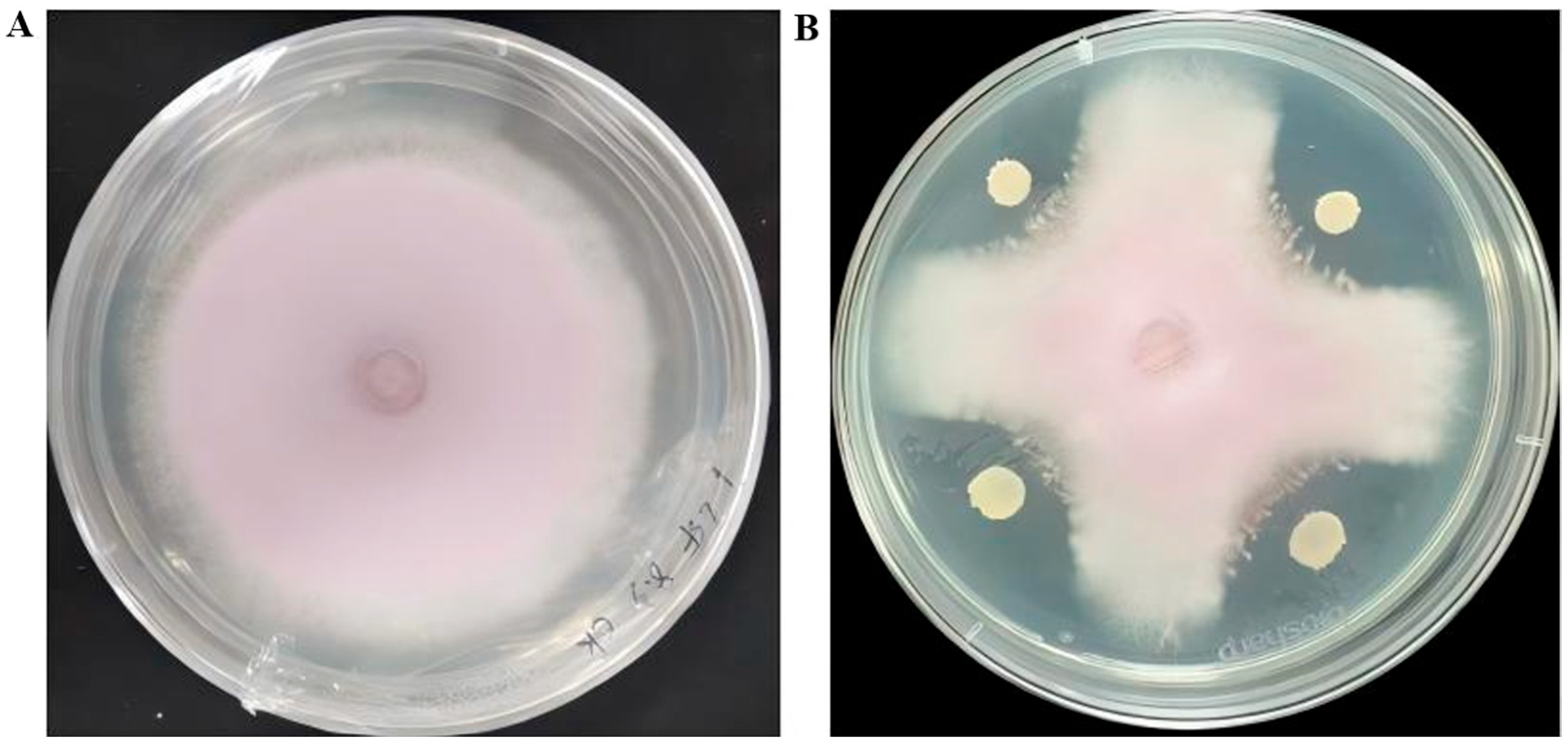
Figure 1. Antagonistic activity of strain R12 against Fusarium oxysporum f. sp. lilii L1-1. (A) Inhibition zone formed by strain R12; (B) Control plate without bacterial inoculation.
Microorganisms 13 02430 g001
Figure 2. SEM analysis of morphological alterations in Fol L1-1 conidia and hyphae following exposure to R12 BCF. (A) Untreated Fol L1-1 conidia; (B) R12 BCF-treated conidia; (C) Untreated Fol L1-1 hyphae; (D) R12 BCF-treated hyphae.
Microorganisms 13 02430 g002aMicroorganisms 13 02430 g002b
Figure 3. Morphology of strain R12. (A) Colony morphology of strain R12; (B) SEM analysis of strain R12 morphology.
Microorganisms 13 02430 g003
Figure 4. Phylogenetic Tree of Antagonistic Bacterium R12.
Microorganisms 13 02430 g004
Figure 5. PGP traits of B. velezensis R12. (A) IAA production; (B) siderophore production; (C) phosphate solubilization (n = 3 biological replicates).
Microorganisms 13 02430 g005
Figure 6. Biocontrol of Fusarium wilt and growth promotion of Longya lily seedlings by B. velezensis R12. (A) phenotypic representation of Longya lily seedlings across different treatments (n = 3 biological replicates); (B) Biomass of Longya lily seedlings under different treatments. Data represent mean ± SD (n = 3), different letters indicate significant differences at p < 0.05 level.
Microorganisms 13 02430 g006
Figure 7. Circular genome map of B. velezensis R12. The outermost circle indicates genome size, with each mark representing 5 kb. The 2nd and 3rd circles show genes on the positive and negative strands, respectively, where different colors indicate different COG functional categories. The 4th circle shows repeated sequences. The 5th circle shows tRNA (blue) and rRNA (purple). The 6th circle shows GC content; light yellow areas indicate GC content above the genome average, with higher peaks showing greater differences, while blue areas indicate GC content below the average. The innermost circle shows GC-skew, with dark gray showing regions where G content is higher than C, and red showing regions where C content is higher than G.
Microorganisms 13 02430 g007
Figure 8. Species distribution of sequences aligned to the Nr database. This figure reflects the species distribution of sequences aligned in the Nr database. Different colors indicate distinct species.
Microorganisms 13 02430 g008
Figure 9. GO functional annotation classification statistics. The x-axis indicates GO subcategories, while the left y-axis represents the percentage of annotated genes and the right y-axis denotes absolute gene counts. This visualization demonstrates the enrichment patterns of level 2 GO functional subcategories relative to the whole genomic background, highlighting the dominance hierarchy among these functional subdivisions.
Microorganisms 13 02430 g009
Figure 10. eggNOG functional gene categorization statistics. The x-axis denotes eggNOG subcategories, and the y-axis represents the relative abundance (%) of annotated genes within corresponding functional classifications.
Microorganisms 13 02430 g010
Table 1. Physiological and biochemical characteristics of antagonist R12.
CharacteristicsResults
Gram stain+
Starch hydrolysis experiment+
Glucose+
Arabinose+
Gelatine+
Hydrogen peroxide enzyme test+
Indole reaction+
V-P reaction
mannitol
Note: “+” indicates positive, “−” indicates negative.
Table 2. Disease incidence, disease index, and control efficacy of B. velezensis R12 against Longya lily Fusarium wilt.
TreatmentDisease Incidence (%)Disease IndexControl Efficacy (%)
Fol L1-197.26 ± 0.67 a94.57 ± 0.51 a
Fol L1-1 + 0.1% Hymexazol2.19 ± 0.14 b2.67 ± 0.13 b97.18 ± 0.13 a
Fol L1-1 + R122.68 ± 0.15 b3.77 ± 0.11 b96.01 ± 0.07 a
Note: The data are presented as mean ± standard deviation (SD). Within the same column, different letters (a,b) indicate significant differences at p < 0.05 level.
Table 3. Cleaned genomic sequence data of Bacillus velezensis R12.
GenomeGenome Assembly
Number of sequences22,709Scaffold length (bp)4,015,523
SumBase210,960,009Scaffold number1
N50 reads length (bp)10,170Scaffold N50 (bp)4,015,523
N90 reads length (bp)6141Scaffold N90 (bp)4,015,523
Mean reads length (bp)9289Contig Length (bp)4,015,523
Max length (bp)30,021GC content (%)46.42
Mean reads quality33.73Gaps number0
Disclaimer/Publisher’s Note: The statements, opinions and data contained in all publications are solely those of the individual author(s) and contributor(s) and not of MDPI and/or the editor(s). MDPI and/or the editor(s) disclaim responsibility for any injury to people or property resulting from any ideas, methods, instructions or products referred to in the content.