Article

Detection of Tick-Borne Microorganisms, Anaplasmataceae and Piroplasmida, in Sorex spp. in Hokkaido, Japan

1
Research Center for Biosafety, Laboratory Animal and Pathogen Bank, National Institute of Infectious Diseases (NIID), Japan Institute for Health Security (JIHS), Musashimurayama 208-0011, Tokyo, Japan
2
Environment Grasp Promotion Network-PEG, Nonprofit Organization, Kushiro 085-0806, Hokkaido, Japan
3
Department of Parasitology, Asahikawa Medical University, Asahikawa 078-8510, Hokkaido, Japan
4
Department of Veterinary Science, National Institute of Infectious Diseases (NIID), Japan Institute for Health Security (JIHS), Shinjuku 162-8640, Tokyo, Japan
*
Author to whom correspondence should be addressed.
Current address: Division of Veterinary Science, Department of Veterinary Medicine, Obihiro University of Agriculture and Veterinary Medicine, Obihiro 080-8555, Hokkaido, Japan.
Retired.
Microorganisms 2025, 13(10), 2288; https://doi.org/10.3390/microorganisms13102288
Submission received: 24 July 2025 / Revised: 12 September 2025 / Accepted: 19 September 2025 / Published: 1 October 2025
(This article belongs to the Special Issue Vector-Borne Zoonoses: Surveillance, Transmission and Interventions)

Abstract

The habitats of shrews substantially overlap with those of rodents, which are well known as reservoirs for many tick-borne diseases. However, the ecological role of shrews (Mammalia: Eulipotyphla: Soricidae) remains poorly understood. We examined 103 Sorex spp. (S. unguiculatus, S. gracillimus, S. caecutiens) from Kushiro, Hokkaido, Japan, to investigate their relationships with ticks and tick-borne microorganisms, including Piroplasmida and Anaplasmataceae. Pathogen screening revealed Babesia microti Hobetsu lineage (9.7%), Neoehrlichia mikurensis (26.2%), Ehrlichia japonica (13.6%), and E. muris (0.97%). These intracellular protozoa and bacteria, typically associated with rodents, are recognized zoonotic agents or have zoonotic potential. Detection rates were highest in S. caecutiens (62.5%, 10/16), followed by S. unguiculatus (45.3%, 24/53) and S. gracillimus (23.5%, 8/34). Co-infections were observed between N. mikurensis and B. microti (n = 3) and between N. mikurensis and E. japonica (n = 4). Immature stages of Ixodes ovatus and I. persulcatus were collected from the body surface of shrews, and transstadial transmission of N. mikurensis was suggested by its detection in a molted I. ovatus nymph. These results indicate that shrews act as feeding hosts for immature ticks and reservoirs for multiple tick-borne pathogens. Shrews should be considered important reservoirs for tick-borne diseases.

1. Introduction

Tick-borne zoonotic diseases occasionally emerge as public health threats, making it crucial to investigate potential infections in wildlife. Rodents play a crucial role as reservoir hosts for many tick-borne pathogens [1,2,3,4,5]. Recently, soricids (order Eulipotyphla), which occupy habitats similar to those of rodents, have been recognized as potentially important reservoirs for many tick-borne microorganisms, including Babesia microti, Anaplasma phagocytophilum, and various viruses [6,7,8]. Most shrews do not burrow, but inhabit areas beneath leaf litter and prey on surface-dwelling earthworms and small arthropods (insects and spiders) [9,10]. The semi-underground habitats of shrews not only overlap with the habitats of rodents but also make them a suitable host for blood meals of ticks that prefer higher humidity and lower temperatures within the leaf litter. If shrews have similar susceptibilities to infectious microorganisms to rodents, their role as reservoirs for tick-borne infectious diseases may have been overlooked [11]. Shrews are sometimes captured in traps used for small wild mammals, such as Sherman traps. However, the pitfall method, which is more labor-intensive, is required to efficiently capture shrews.
Ticks infesting shrews in Japan have been investigated in a few studies, which collected Ixodes ovatus and I. persulcatus ticks from the body surface of Sorex spp. [11,12]. Ixodes ovatus and I. persulcatus are the principal vectors for many tick-transmitting rodent-borne microorganisms, including Babesia microti, Neoehrlichia mikurensis, and Ehrlichia japonica.
B. microti is a complex of genetically diverse protozoan parasites (B. microti-group) of zoonotic or zoonotic potential. In the eastern part of Hokkaido, Japan (including Kushiro), previous field surveys have shown that two distinct lineages of the B. microti group, the Hobetsu and US lineages, were maintained by various rodent species, including Myodes rufocanus, and that I. ovatus and I. persulcatus ticks specifically transmitted the Hobetsu and US lineages, respectively [13,14]. The US lineage is genetically closely related to B. microti sensu stricto, the causative agent of human babesiosis in US. The Hobetsu lineage is widely distributed throughout Japan, and a retrospective survey suggested subclinical infection in the endemic areas of tick-borne infectious diseases [15]. In our previous study, one Hobetsu-lineage isolate was obtained from an S. unguiculatus sympatrically collected with the rodents in Hokkaido [16]; whether the shrew contributed to the parasite’s life cycle was unclear.
Neoehrlichia mikurensis was initially identified in wild rodents and ticks in Japan and is now recognized as an emerging human pathogen in Europe and Asia [3]. In Japan, the bacterium has been detected in rodents and their associated ticks (I. ovatus and I. persulcatus) with a prevalence of 9–47% [17,18]. Ehrlichia japonica, a newly recognized species formerly known as Ehrlichia sp. HF, was previously isolated from I. ovatus and is genetically closely related to E. chaffeensis, the agent of human monocytic ehrlichiosis (HME) [19]. However, no cases of infection with this organism have been reported.
The spleen is crucial for clearing blood-borne pathogens by removing damaged blood cells, capturing infected cells by macrophages, and immune activation [20]. Splenomegaly is a major clinical manifestation of B. microti and Ehrlichia infections [21,22,23]. In wild animals, the influence of these microorganism infections on the spleen has scarcely been investigated [13].
Owing to the recent emergence of infectious diseases, accumulating data on tick vectors and their reservoir hosts is critical for public health. In this study, we actively investigated the potential role of shrews as hosts for ticks and reservoirs for tick-borne obligate intracellular pathogens, including Babesia, Neoehrlichia, and Ehrlichia. Additionally, we investigated splenomegaly in relation to infection with these microorganisms.

2. Materials and Methods

2.1. Collection of Wild Shrew

Shrews were unintentionally collected during salamander surveys in Kushiro Wetland in Hokkaido, Japan, in October 2023 (Figure 1) and dead bodies were provided. In the survey, a set of 75 pitfall traps with drift fences were placed. Species were primarily identified based on their morphological characteristics [24]. The fifth unicuspid tooth (U5) of the upper jaw was examined to discriminate between S. caecutiens and S. gracillimus [25], as the appearance of immature S. caecutiens was similar to that of adult S. gracillimus [24]. Additionally, genetic examination was performed based on the cyt-b sequence (780 bp) [26] using DNA from the spleen (see below). Spleens were dissected using disposable tweezers and blades and stored in alcohol until use. To eliminate cross-contamination, the dissection instruments were changed for each individual. An apparently enlarged spleen (e.g., a spleen covering the entire abdominal cavity) was regarded as splenomegaly.

2.2. Collection of Ticks

Ticks were collected from the body surface of Sorex spp. before dissection. Additionally, ticks that remained on the shrew’s body were collected from the dissected body. The body was tightly enclosed in a plastic bag for a few hours at room temperature. The ticks that crawled into the plastic bag were collected. All collected ticks were stored in moistened plastic tubes at room temperature. Engorged ticks were kept alive in tightly sealed plastic tubes over a moistened filling of solidified plaster with activated charcoal at 25 °C until molting. Ticks were collected in 2020 at the same location in October using the same method described above. The ticks collected in 2020 were kept in alcohol until species identification.

2.3. DNA Extraction from Ticks and Spleen of Shrews

The spleens of wild shrews were cut into approximately 100 mg pieces and homogenized in tubes containing a ceramic ball (Ceramic Sphere, MP Biomedicals, Solon, OH, USA; catalog no. 6540412), garnet beads (Garnet Matrix A Bulk, MP Biomedicals; catalog no. 6540-427), and TNE buffer [50 mM Tris–HCl (pH 7.4), 100 mM NaCl, and 0.1 mM EDTA] by shaking at 4000 rpm for 30 s using a Micro Smash MS-100 (TOMY, Katsushika, Tokyo, Japan). Proteinase K (0.1 mg/mL) and sodium dodecyl sulfate (0.1%) were then added, and the samples were incubated overnight at 55 °C. DNA was extracted using the standard phenol/ethanol method, dissolved in 200 μL of TE buffer [10 mM Tris–HCl (pH 7.4) and 1 mM EDTA], and stored at −30 °C.
Ticks were homogenized with plastic pestles in TNE buffer containing Proteinase K (0.1 mg/mL) and sodium dodecyl sulfate (0.1%). After incubation at 55 °C for three hours, DNA was extracted from the ticks using a standard protocol based on the phenol/ethanol method [27].

2.4. Microorganism Detection in Ticks and Spleens

Ex Taq polymerase (Takara Bio, Otsu, Japan) was used according to the manufacturer’s instructions.
Piroplasmids were detected using nested polymerase chain reaction (PCR) targeting the 18S rRNA gene, as described by Tsuji et al. [13]. The 18S rRNA-positive samples were further examined for the presence of Babesia microti and lineage identification. Since three B. microti lineages are distributed in wild rodents in Japan, the lineage was discriminated using discriminative PCR based on the β-tubulin sequence [28].
Members of the Anaplasmataceae family were identified by dual gene sequencing based on the 16S rRNA and groEL genes [29]. Nested PCR and direct sequencing were performed using the primers listed in Table S1. BLAST (v.2.16.0) was used to search for high sequence similarity to known species. A positive result for Anaplasmataceae identification was confirmed by the high sequence similarity of both the 16S rRNA and groEL genes to those of a known species in the same specimen. In cases where different species (e.g., Neoehrlichia and Ehrlichia) were identified based on 16S rRNA and groEL sequences from the same specimen, further analyses were conducted. PCRs targeting Ehrlichia P28 [17] and Neoehrlichia mikurensis-specific 16S rRNA genes [18] were performed in a separate laboratory. Co-infection was confirmed when multiple pathogens were identified in both laboratories. All primers used in this study are listed in the Supplementary Table S1.

2.5. Phylogenetic Analysis

For phylogenetic analysis, the sequences determined in this study and those retrieved from GenBank were aligned using ClustalW. Evolutionary relationships were inferred using the neighbor-joining method implemented in MEGA X software (version 11.0.13) [30]. Bootstrap values were estimated for 1000 replicates and are indicated at the branch. An outgroup was chosen from species which are closely related to the group of interest.

2.6. Statistical Test

The chi-squared test was used to examine statistically significant differences between the observed and expected frequencies of splenomegaly. The test was performed for each microorganism-positive group individually, for the combined Anaplasmataceae-positive group (N. mikurensis, E. japonica, and E. muris), and for all microorganism-positive shrews (B. microti, N. mikurensis, E. japonica, and E. muris).

3. Results

3.1. Collection and Phylogeny of Sorex spp.

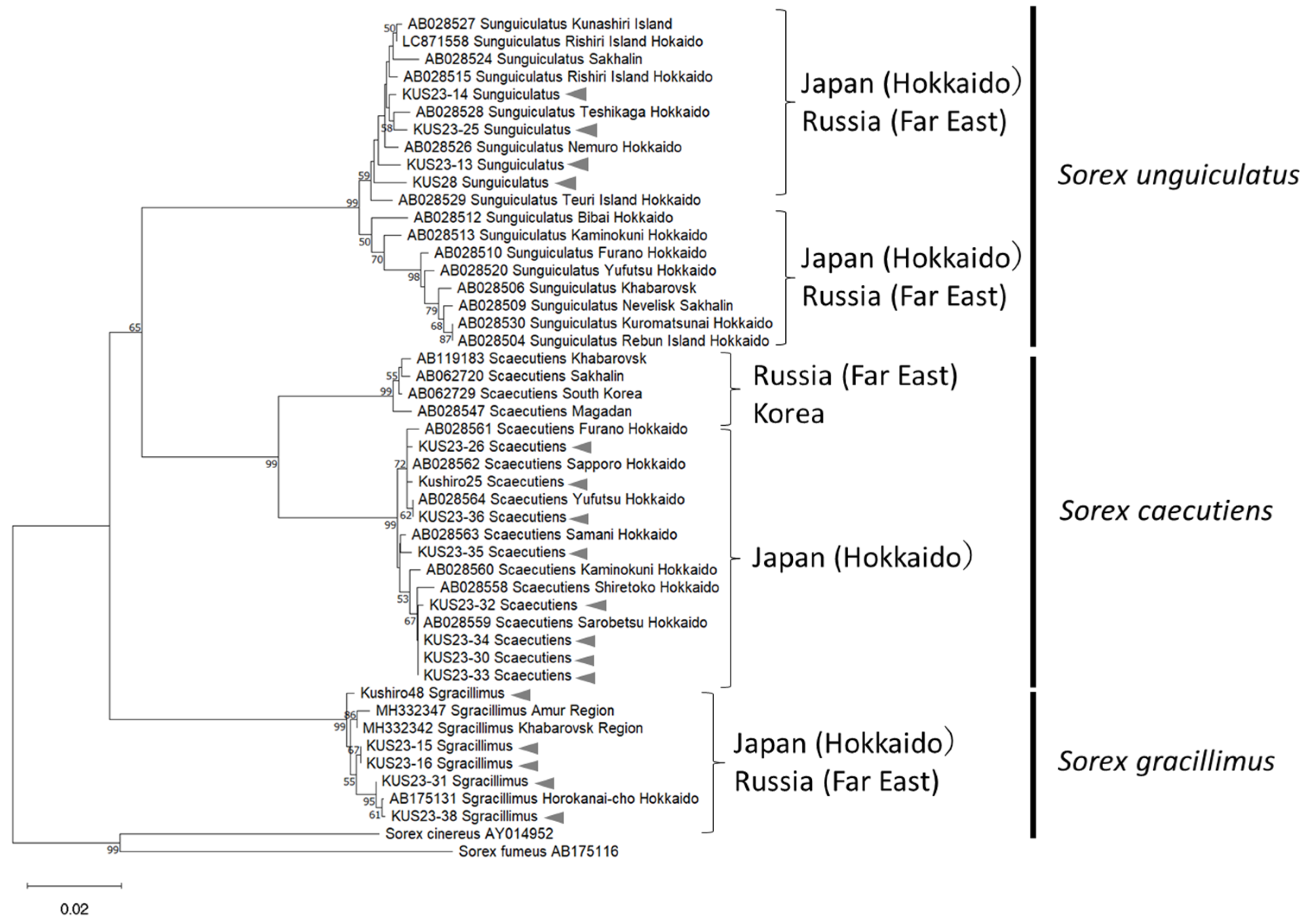
In total, 103 Sorex spp. specimens were collected, including S. unguiculatus (n = 53), S. gracillimus (n = 34), and S. caecutiens (n = 16). Two shrew specimens that were morphologically identified as S. caecutiens were molecularly identified as S. gracillimus, while one shrew specimen morphologically identified as S. gracillimus was molecularly identified as S. caecutiens, indicating that molecular identification is crucial for accurately differentiating these species. A phylogenetic tree of Sorex spp. based on the cytb showed that S. gracillimus and S. caecutiens collected in Kushiro in this study formed a distinct lineage with those previously reported individuals collected in Hokkaido, Japan, and were clearly distinguished from those found in Russia (Figure 2). In contrast, the S. unguiculatus consisted of two lineages, regardless of the collection area, including Hokkaido and Russia, consistent with previous findings by Odachi et al. [31]. Since S. unguiculatus is thought to have distributed across northeastern Eurasia more recently than S. gracillimus and S. caecutiens, insufficient nucleotide substitutions may have accumulated to reflect geographical isolation [31].

3.2. Immature Ixodes ovatus and I. persulcatus Feed on Sorex spp.

Sixty-four ticks were collected from 12 shrews (5.3 ticks/shrew) (Table 1). The collection included the immature stages of two ixodid ticks, I. ovatus (n = 28) and I. persulcatus (n = 36), but not the adult stage. Both tick species were collected from all three species of Sorex. I. persulcatus (56%) and I. ovatus (44%) were collected in relatively equal numbers. The mean numbers of I. ovatus and I. persulcatus collected from shrews were 2.3 ticks/shrew and 3 ticks/shrew, respectively. Owing to the small sample size, statistical analysis to assess the correlations between Sorex host species and bloodsucking tick species was not performed. As in the 2020 collection, all ticks (n = 148) were immature stages of I. ovatus (n = 101) and I. persulcatus (n = 47) (Table 1). The mean number of ticks collected from the shrews was 9.9 ticks/shrew.

3.3. Molecular Detection of Piroplasmida and Anaplasmataceae in Ticks

Ticks collected from the body surface of Sorex spp. were kept in containers for molting. After 1 month or more, a total of 21 nymphs molted from the larvae (Table 2). From four S. unguiculatus and two S. caecutiens, 15 and 6 nymphs were obtained, respectively. The tick species and numbers collected from each shrew are listed in Table 2.
PCR examination for microorganisms in the ticks revealed a Piroplasmida-positive I. persulcatus (designated as KUS23-33_m3) and an Anaplasmataceae-positive I. ovatus (designated as KUS23-33_m5). Both ticks were recovered from same S. unguiculatus (designated KUS23-33) in which N. mikurensis was detected (see below). Further PCR for lineage discrimination of B. microti in the piroplasmidapositive specimen (KUS23-33_m3) failed to identify the species, as amplification was not observed with any primer sets (Table S1). PCR amplicon of 18S rRNA obtained during the initial screening was subjected to sequencing. In the BLAST search, the resulting 18S rRNA (1519 bp) of KUS23-33_m3 was most closely related to that of the B. divergens Asia lineage (KC493555.1), with 99.87% identity. The I. persulcatus tick KUS23-33_m3 infected with the B. divergens Asia lineage most likely originated from an I. persulcatus female infected with this lineage, since B. divergens is both transstadially and transovarially transmitted through a multi-stage life cycle, and this lineage is primarily maintained by wild deer and I. persulcatus in Hokkaido [27].
GroEL sequence (290 bp) and 16S rRNA (756 bp) of the Anaplasmataceae-positive specimen (KUS23-33_m5) was identical to that of N. mikurensis Io3 from I. ovatus in Japan (LC385831) and N. mikurensis FIN686 (AB196304), respectively (Figure 3).

3.4. Detection of Babesia microti, Neoehrlichia mikurensis, Ehrlichia japonica, and E. muris in Sorex spp. Spleen

DNA extracted from the spleen was examined for the presence of tick-borne organisms, including the Piroplasmida and Anaplasmataceae species. The positivity rates for each microorganism in Sorex spp. are shown in Table 3. Piroplasmida 18S rRNA-positive samples were classified as belonging to the B. microti Hobetsu lineage using lineage-specific PCR. Sequencing of all Anaplasmataceae GroEL-positive samples (n = 38) revealed three distinct sequences (Table 4). One was identical to the groEL of N. mikurensis Io3 from I. ovatus in Japan (LC385831) (n = 23), and the others were identical to that of Ehrlichia sp. HF565 (E. japonica) isolated from I. ovatus (AB032712) (n = 14) and E. muris (n = 1). The N. mikurensis sequence was also identical to those of the IS58 strain from wild rats (AB074461) and the A113 strain from wild rodents (AB204865) in Japan (Figure 3). An identical sequence of N. mikurensis was also detected in sympatrically captured Myodes rufocanus (KUS75) (Figure 3). The 16S rRNA sequences were identified as those of N. mikurensis (n = 27), E. japonica (n = 10), or E. muris (n = 1), which corresponded to the results obtained from groEL, except for four samples (S. unguiculatus, n = 3; S. gracillimus, n = 1).
In the four spleen samples, groEL and 16SrRNA sequences were of different microorganisms: groEL sequences were identified as E. japonica, whereas 16S rRNA sequences were identified as N. mikurensis. Co-infection with these organisms was further confirmed in a separate laboratory using PCR assays targeting N. mikurensis 16S rRNA and Ehrlichia P28. The nearly full-length 16S rRNA sequences from all four samples were identical to each other and to that of N. mikurensis strain IS58 (AB074460). All four P28 sequences were identified as E. japonica, although the chromatograms of the central region were ambiguous. Co-infection with N. mikurensis and B. microti was observed in S. caecutiens (n = 2) and S. unguiculatus (n = 1). Co-infection data are summarized in Table 4.

3.5. Detection of Splenomegaly in Shrews

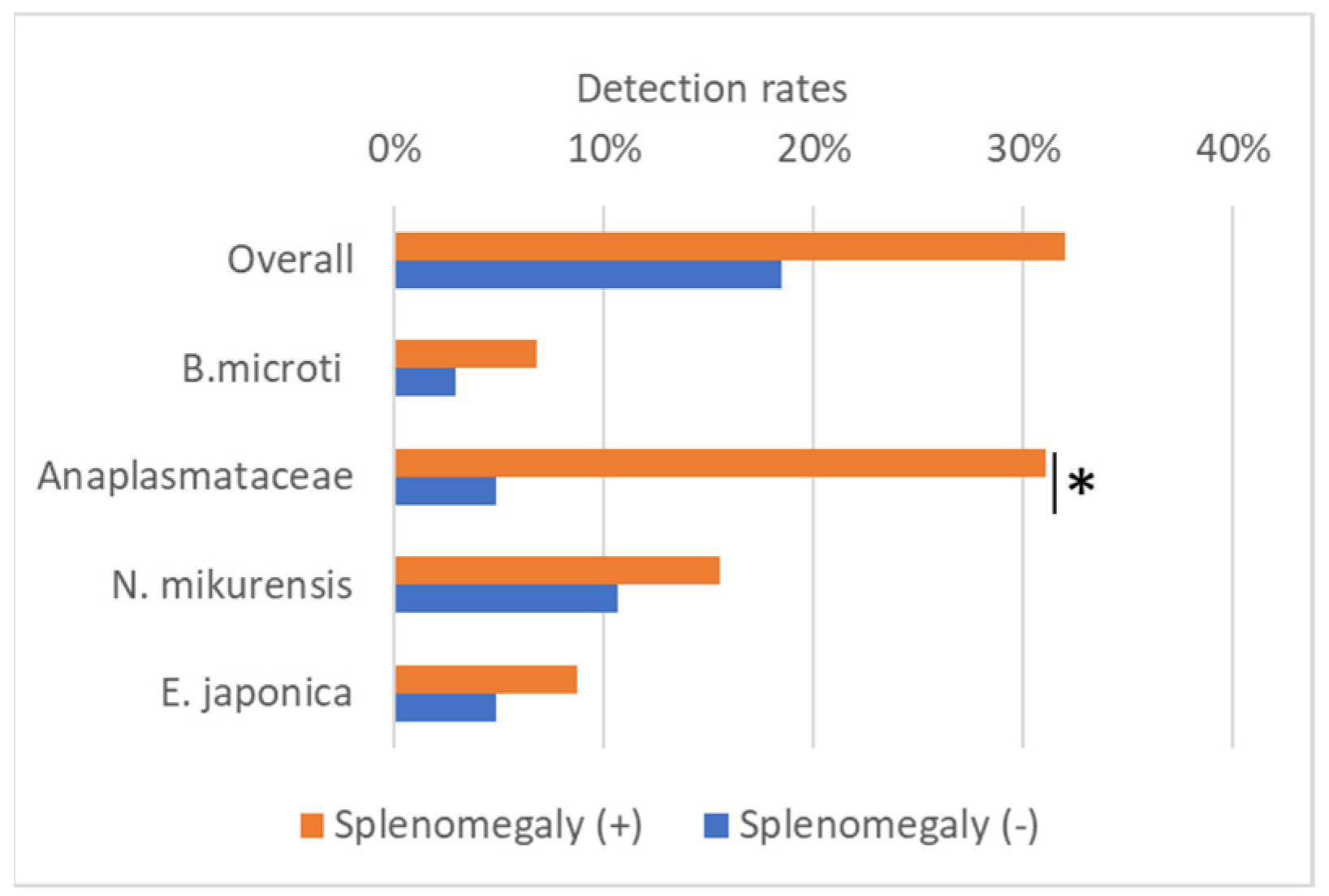
Splenomegaly was observed in 66 Sorex spp. examined (64.1%). Positive rates in each Sorex spp. were 90.6% (48/53) in S. unguiculatus, 35.3% (12/34) in S. gracillimus, and 37.5% (6/16) in S. caecutiens. Splenomegaly was highly frequent in S. unguiculatus. Necropsy revealed no visible nodules in the spleen. The number of splenomegaly-positive shrews was higher than that of splenomegaly-negative shrews for all infections (Figure 4). In particular, the number of splenomegaly-positive shrews was statistically higher in the Anaplasmataceae (N. mikurensis, E. japonica, and E. muris)-positive shrews (Chi-Square Test, p < 0.05).

4. Discussion

In this study, we investigated the presence of Sorex spp. in Hokkaido to explore their potential role as reservoirs of tick-borne microorganisms and the tick species involved in their transmission. Molecular screening targeting Piroplasmida and Anaplasmataceae in Sorex spp. spleen identified B. microti Hobetsu lineage, E. japonica, N. mikurensis, and E. muris (Table 3). Co-infection of N. mikurensis and B. microti, the most frequent co-infection case in wild rodents [28], was observed in Sorex spp. (Table 3). Ticks collected from the body of the shrew (Table 1) were of Ixodes ovatus and I. persulcatus, which are the principal vectors of B. microti Hobetsu lineage (I. ovatus) [14], N. mikurensis (I. ovatus and I. persulcatus) [17,19], E. japonica (I. ovatus) [32], and E. muris (I. persulcatus) [33]. These results indicate that both shrews and rodents serve as the reservoirs for tick-borne microorganisms, including B. microti Hobetsu lineage, E. japonica, N. mikurensis, and E. muris, and that I. ovatus and I. persulcatus ticks transmit the transmission of these microorganisms.
The results of this study showed that the infection rate of the Hobetsu lineage in shrews (9.7%, 10/103; Table 3) was comparable to that observed in rodents [16]. Furthermore, this lineage was detected in all three predominant species of Sorex spp. in Hokkaido (Table 4). Thus, we conclude that Sorex spp. play a major role in maintaining B. microti Hobetsu lineage reservoirs. We speculate that the extensive distribution of the Hobetsu lineage throughout Japan, from north (Hokkaido) to south (Shikoku), is due to divergent mammalian reservoirs, including Sorex spp., and the geographically wide distribution of the competent vector (I. ovatus). In the USA [34,35] and UK [6], Sorex spp. are regarded as competent US lineage (or B. microti sensu stricto) reservoirs. The reason why the US lineage has not yet been detected in Sorex spp. in this country is unclear. Although, I. persulcatus, the principal US-lineage vector, was collected from Sorex spp. as efficiently as I. ovatus (Table 2).
We found that Sorex spp. also harbored N. mikurensis, with particularly high prevalence in S. caecutiens (43.8%) and S. unguiculatus (30.2%; Table 3), comparable to rodent infection rates. Regarding the previous detection of N. mikurensis in S. unguiculatus (2/4) [36], our findings suggest that shrews are important reservoirs for the natural maintenance of N. mikurensis in Hokkaido. Notably, N. mikurensis undergoes vertical (transplacental) transmission in rodents, supporting its persistence in small mammalian populations, even in the absence of tick feeding [36]. Although vertical transmission of N. mikurensis has been confirmed in rodents, similar mechanisms in shrews remain speculative and require further investigation. Combined with the tick preference for shrews (Table 2) and confirmed transstadial transmission (Table 3), I. ovatus and I. persulcatus likely play pivotal roles in maintaining and bridging N. mikurensis among both the shrew and rodent populations in Hokkaido.
In this study, E. japonica was detected in S. unguiculatus and S. gracillimus, with the highest prevalence in S. unguiculatus (20.8%) (Table 3). Notably, co-infection with N. mikurensis was confirmed in several individuals using both groEL and P28 analyses. This co-infection may be due to sharing the same vector tick, I. ovatus [18,37], which feeds on Sorex spp. (Table 1). The strong association between E. japonica infection and splenomegaly (Figure 4) is consistent with the immunostimulatory effects observed in canine ehrlichiosis caused by E. canis [22,38], suggesting its potential pathogenicity. Considering the wide geographical distribution of I. ovatus, continued surveillance of E. japonica in wildlife reservoirs and tick populations is necessary.
We speculated that the observed splenomegaly was associated with infections by B. microti, Ehrlichia spp., and/or N. mikurensis [39], given that splenomegaly was present in as many as 64.1% of the examined shrews. However, our data did not provide statistical support for an association between B. microti and the detection of other microorganisms (Figure 4). The co-detection of Babesia, Ehrlichia, and Neoehrlichia in our samples suggests that a dynamic community of tick-borne pathogens is maintained within the shrew population. Therefore, the splenomegaly observed in this study may not be attributable to a single pathogen, but rather may reflect chronic immune stimulation driven by the diverse assemblage of tick-borne pathogens present in the spleen.
The collection of immature I. ovatus and I. persulcatus from shrews (Table 1) corresponds to a report in which S. unguiculatus and sympatrically captured A. speciosus were investigated in October in Furano (approximately 150 km away from Kushiro) [11]. October may restrict tick collection in terms of stage, as adult ticks that overwinter are active in the spring. Only immature I. ovatus and I. persulcatus were primarily shown to feed on rodents, Apodemus spp. and Myodes spp., through the tick-active period (5 months) in Kiyosato and Shimizucho (approximately 100 km away from Kushiro) [40]. Ongoing global warming, ecology, and the distribution of ticks may change, making it necessary to conduct continuous surveys for a comprehensive understanding of wild shrews as feeding hosts.
Shrews have been increasingly identified as an important reservoir of zoonotic viral infections [6,7]. The results of this study emphasize the involvement of Sorex spp. as the reservoir of protozoa and bacteria, including B. microti Hobetsu lineage [14], E. japonica [19,32], and N. mikurensis [32,37]. Although clinical cases of human babesiosis caused by B. microti Hobetsu lineage have not been documented, the presence of antibodies against this lineage in a high-risk group [15] suggests subclinical infection. Ongoing, geographically expanded surveys encompassing a wide range of pathogens are essential for clarifying the ecological role of shrews as reservoirs and preparing for emerging infectious disease outbreaks.

5. Conclusions

The examination of ticks attached to shrews and the detection of Babesia microti Hobetsu lineage, Neoehrlichia mikurensis, and Ehrlichia japonica in Sorex spp. in Kushiro, Hokkaido, Japan, indicate that shrews serve as feeding hosts for immature I. ovatus and I. persulcatus ticks and as predominant reservoirs for these tick-borne microorganisms. These findings indicate that Sorex spp. may play a pivotal role in the transmission dynamics of tick-borne pathogens. To clarify the ecological significance of shrews as potential risk factors for tick-borne diseases, continuous and geographically expanded surveillance, encompassing a broader spectrum of pathogens, will be required.

Supplementary Materials

The following supporting information can be downloaded at https://www.mdpi.com/article/10.3390/microorganisms13102288/s1, Table S1: Primers used for PCR in this study.

Author Contributions

Conceptualization, A.Z.-N.; methodology, A.Z.-N., S.T., M.S., and M.N.; investigation, A.Z.-N., S.T., M.S., and M.N.; resources, S.T., M.S., and M.N.; data curation, A.Z.-N.; writing—original draft preparation, A.Z.-N.; writing—review and editing, A.Z.-N., M.T., and K.-I.H.; funding acquisition, A.Z.-N. All authors have read and agreed to the published version of the manuscript.

Funding

This research was funded by the Osimo Foundation (grant year 2023, 2025), the Japan Agency for Medical Research and Development (AMED) (21fk0108097j0803), and JSPS KAKENHI (JP18K06398).

Institutional Review Board Statement

Hokkaido Prefectural Government Kushiro General Subprefectural Bureau kindly permitted capturing Soricidae [Reference No. 604, 2020 and 602, 2023 to Terui]. The experiment protocol was approved by Japan Institute for Health Security, National Institute of Infectious Diseases (NIID), Institutional Animal Care and Use Committee.The approval code was 125139. The approval date was 1 October 2025.

Informed Consent Statement

Not applicable.

Data Availability Statement

Representative sequences obtained in this study were deposited in the DNA Data Bank of Japan (DDBJ) under accession numbers LC877955–LC877961 (N. mikurensis, 16S rRNA), LC867973–LC867977 (N. mikurensis, GroEL), LC877954 (E. japonica, 16S rRNA), LC867978–LC867980 (E. japonica, GroEL), and LC871563–LC871578 (Sorex spp., Cyt-b).

Acknowledgments

We are grateful to Akiko Saigo for her technical assistance. During the preparation of this manuscript/study, the author(s) used Microsoft Copilot for the purposes of generating the graphical abstract. The authors have reviewed and edited the output and take full responsibility for the content of this publication.

Conflicts of Interest

The authors declare no conflicts of interest.

Abbreviation

The following abbreviation is used in this manuscript:
PCRPolymerase chain reaction

References

  1. Moraga-Fernández, A.; Muñoz-Hernández, C.; Sánchez-Sánchez, M.; Fernández de Mera, I.G.; de la Fuente, J. Exploring the diversity of tick-borne pathogens: The case of bacteria (Anaplasma, Rickettsia, Coxiella and Borrelia) protozoa (Babesia and Theileria) and viruses (Orthonairovirus, tick-borne encephalitis virus and louping ill virus) in the European continent. Vet. Microbiol. 2023, 286, 109892. [Google Scholar] [CrossRef]
  2. Xu, A.L.; Xue, H.; Li, Y.; Wang, X.; Zheng, J.X.; Shi, F.Y.; Cui, Q.X.; Lu, Y.; Cun, D.J.; Li, L.H. Comprehensive meta-analysis of severe fever with thrombocytopenia syndrome virus infections in humans, vertebrate hosts and questing ticks. Parasites Vectors 2024, 17, 265. [Google Scholar] [CrossRef]
  3. Wennerås, C. Infections with the tick-borne bacterium Candidatus Neoehrlichia mikurensis. Clin. Microbiol. Infect. 2015, 21, 621–630. [Google Scholar] [CrossRef]
  4. Eisen, L. Rodent-targeted approaches to reduce acarological risk of human exposure to pathogen-infected Ixodes ticks. Ticks Tick Borne Dis. 2023, 14, 102119. [Google Scholar] [CrossRef]
  5. Krawczyk, A.I.; van Duijvendijk, G.L.A.; Swart, A.; Heylen, D.; Jaarsma, R.I.; Jacobs, F.H.H.; Fonville, M.; Sprong, H.; Takken, W. Effect of rodent density on tick and tick-borne pathogen populations: Consequences for infectious disease risk. Parasites Vectors 2020, 13, 34. [Google Scholar] [CrossRef] [PubMed]
  6. Bown, K.J.; Lambin, X.; Telford, G.; Heyder-Bruckner, D.; Ogden, N.H.; Birtles, R.J. The common shrew (Sorex araneus): A neglected host of tick-borne infections? Vector Borne Zoonotic Dis. 2011, 11, 947–953. [Google Scholar] [CrossRef]
  7. Goethert, H.K.; Mather, T.N.; Johnson, R.W.; Telford, S.R., 3rd. Incrimination of shrews as a reservoir for Powassan virus. Commun. Biol. 2021, 4, 1319. [Google Scholar] [CrossRef]
  8. Li, H.; Huang, Z.Y.X.; Lan, J.; Hu, L.; Wei, X.; Wang, Y.; Li, X.; Li, Y.; Becker, D.J.; Wei, F.; et al. Diversity and transmission and zoonotic potential of microbes in true insectivores. Nat. Commun. 2025, 16, 6709. [Google Scholar] [CrossRef]
  9. Yajima, S.; Nakashima, Y.; Kawahara, A.; Ohdachi, S.D. Utilization of above-ground space in captivity by four sympatric shrew species from Hokkaido, Japan. Mammal Study 2024, 49, 137–143. [Google Scholar] [CrossRef]
  10. Ohdachi, S. Burrowing habits and earthworm preferences of three species of Sorex in Hokkaido. J. Mamm. Soc. Jpn. 1995, 20, 85–88. [Google Scholar]
  11. Nakao, M.; Miyamoto, K. Long-tailed shrew, Sorex unguiculatus, as a potential reservoir of the spirochetes transmitted by Ixodes ovatus in Hokkaido, Japan. Med. Entomol. Zool. 1993, 44, 237–245. [Google Scholar] [CrossRef]
  12. Kameyama, Y.; Sato, R.; Asakawa, M.; Ito, T.; Okimoto, K.; Shimoi, G.; Hashizume, R. Parasitic Fauna in Long-Clawed Shrew, Sorex unguiculatus, in Abashiri, Hokkaido. J. Agric. Sci. Tokyo Univ. Agric. 2015, 60, 10–17. [Google Scholar]
  13. Tsuji, M.; Zamoto, A.; Kawabuchi, T.; Kataoka, T.; Nakajima, R.; Asakawa, M.; Ishihara, C. Babesia microti-like parasites detected in Eurasian red squirrels (Sciurus vulgaris orientis) in Hokkaido, Japan. J. Vet. Med. Sci. 2006, 68, 643–646. [Google Scholar] [CrossRef]
  14. Zamoto-Niikura, A.; Tsuji, M.; Qiang, W.; Nakao, M.; Hirata, H.; Ishihara, C. Detection of two zoonotic Babesia microti lineages, the Hobetsu and U.S. lineages, in two sympatric tick species, Ixodes ovatus and Ixodes persulcatus, respectively, in Japan. Appl. Environ. Microbiol. 2012, 78, 3424–3430. [Google Scholar] [CrossRef]
  15. Arai, S.; Tsuji, M.; Kaiho, I.; Murayama, H.; Zamoto, A.; Wei, Q.; Okabe, N.; Kamiyama, T.; Ishihara, C. Retrospective seroepidemiological survey for human babesiosis in an area in Japan where a tick-borne disease is endemic. J. Vet. Med. Sci. 2003, 65, 335–340. [Google Scholar] [CrossRef]
  16. Zamoto, A.; Tsuji, M.; Wei, Q.; Cho, S.H.; Shin, E.H.; Kim, T.S.; Leonova, G.N.; Hagiwara, K.; Asakawa, M.; Kariwa, H.; et al. Epizootiologic survey for Babesia microti among small wild mammals in northeastern Eurasia and a geographic diversity in the beta-tubulin gene sequences. J. Vet. Med. Sci. 2004, 66, 785–792. [Google Scholar] [CrossRef]
  17. Inayoshi, M.; Naitou, H.; Kawamori, F.; Masuzawa, T.; Ohashi, N. Characterization of Ehrlichia species from Ixodes ovatus ticks at the foot of Mt. Fuji, Japan. Microbiol. Immunol. 2004, 48, 737–745. [Google Scholar] [CrossRef] [PubMed]
  18. Li, H.; Jiang, J.F.; Liu, W.; Zheng, Y.C.; Huo, Q.B.; Tang, K.; Zuo, S.Y.; Liu, K.; Jiang, B.G.; Yang, H.; et al. Human infection with Candidatus Neoehrlichia mikurensis, China. Emerg. Infect. Dis. 2012, 18, 1636–1639. [Google Scholar] [CrossRef] [PubMed]
  19. Shibata, S.; Kawahara, M.; Rikihisa, Y.; Fujita, H.; Watanabe, Y.; Suto, C.; Ito, T. New Ehrlichia species closely related to Ehrlichia chaffeensis isolated from Ixodes ovatus ticks in Japan. J. Clin. Microbiol. 2000, 38, 1331–1338. [Google Scholar] [CrossRef]
  20. Lewis, S.M.; Williams, A.; Eisenbarth, S.C. Structure and function of the immune system in the spleen. Sci. Immunol. 2019, 4, eaau6085. [Google Scholar] [CrossRef]
  21. Rocha, S.C.; Moustafa, M.A.M.; Velásquez, C.V.; Azuama, O.C.; Zafar, K.; Meyer, C.; Araujo, M.; Taylor, K.; Parveen, N. Long-term survival of Babesia microti and Borrelia burgdorferi in C3H/HeJ mice and their effect on Lyme arthritis and babesiosis manifestations. Microbiol. Spectr. 2025, 13, e0025225. [Google Scholar] [CrossRef] [PubMed]
  22. McGill, J.L.; Wang, Y.; Ganta, C.K.; Boorgula, G.D.Y.; Ganta, R.R. Antigen-specific CD4+CD8+ double-positive T cells are increased in the blood and spleen during Ehrlichia chaffeensis infection in the canine host. Front. Immunol. 2018, 9, 1585. [Google Scholar] [CrossRef] [PubMed]
  23. Gynthersen, R.M.M.; Stensvold, C.R.; Nielsen, S.L.; Møller, H.J.; Nielsen, H.V.; Lebech, A.M.; Christensen, J.R.; Mens, H.; El Fassi, D. Neoehrlichia mikurensis-An emerging opportunistic tick-borne infection in immunosuppressed patients. J. Intern. Med. 2023, 293, 782–790. [Google Scholar] [CrossRef]
  24. Abe, H.; Ishii, N.; Kaneko, Y.; Maeda, K.; Miura, S.; Yoneda, M. A Pictorial Guide to the Mammals of Japan; Tokai University Press: Tokyo, Japan, 1994. (In Japanese) [Google Scholar]
  25. Ohdachi, S.; Kawahara, A. The first record of Laxmann’s shrew (Sorex caecutiens) with completely white pelage, captured in Hokkaido, Japan. Mamm. Sci. 2018, 58, 63–66. [Google Scholar]
  26. Zamoto-Niikura, A.; Saigo, A.; Sato, M.; Kobayashi, H.; Sasaki, M.; Nakao, M.; Suzuki, T.; Morikawa, S. The presence of Ixodes pavlovskyi and I. pavlovskyi-borne microorganisms in Rishiri Island: An ecological survey. mSphere 2023, 8, e0021323. [Google Scholar] [CrossRef]
  27. Zamoto-Niikura, A.; Morikawa, S.; Hanaki, K.I.; Holman, P.J.; Ishihara, C. Ixodes persulcatus ticks as vectors for the Babesia microti U.S. lineage in Japan. Appl. Environ. Microbiol. 2016, 82, 6624–6632. [Google Scholar] [CrossRef]
  28. Zamoto, A.; Tsuji, M.; Kawabuchi, T.; Wei, Q.; Asakawa, M.; Ishihara, C. U.S.-type Babesia microti isolated from small wild mammals in Eastern Hokkaido, Japan. J. Vet. Med. Sci. 2004, 66, 919–926. [Google Scholar] [CrossRef]
  29. Tabara, K.; Arai, S.; Kawabuchi, T.; Itagaki, A.; Ishihara, C.; Satoh, H.; Okabe, N.; Tsuji, M. A molecular survey of Babesia microti, Ehrlichia species, and Candidatus Neoehrlichia mikurensis in wild rodents from Shimane Prefecture, Japan. Microbiol. Immunol. 2007, 51, 359–367. [Google Scholar] [CrossRef]
  30. Kumar, S.; Stecher, G.; Li, M.; Knyaz, C.; Tamura, K. MEGA X: Molecular Evolutionary Genetics Analysis across Computing Platforms. Mol. Biol. Evol. 2018, 35, 1547–1549. [Google Scholar] [CrossRef] [PubMed]
  31. Ohdachi, S.; Dokuchaev, N.E.; Hasegawa, M.; Masuda, R. Intraspecific phylogeny and geographical variation of six species of northeastern Asiatic Sorex shrews based on the mitochondrial cytochrome b sequences. Mol. Ecol. 2001, 10, 2199–2213. [Google Scholar] [CrossRef]
  32. Taira, M.; Ando, S.; Kawabata, H.; Fujita, H.; Kadosaka, T.; Sato, H.; Monma, N.; Ohashi, N.; Saijo, M. Isolation and molecular detection of Ehrlichia species from ticks in western, central, and eastern Japan. Ticks Tick Borne Dis. 2019, 10, 344–351. [Google Scholar] [CrossRef]
  33. Inokuma, H.; Ohashi, M.; Jilintai Tanabe, S.; Miyahara, K. Prevalence of tick-borne Rickettsia and Ehrlichia in Ixodes persulcatus and Ixodes ovatus in Tokachi district, Eastern Hokkaido, Japan. J. Vet. Med. Sci. 2007, 69, 661–664. [Google Scholar] [CrossRef]
  34. Goethert, H.K.; Lubelcyzk, C.; LaCombe, E.; Holman, M.; Rand, P.; Smith, R.P., Jr.; Telford, S.R., 3rd. Enzootic Babesia microti in Maine. J. Parasitol. 2003, 89, 1069–1071. [Google Scholar] [CrossRef]
  35. Goethert, H.K.; Cook, J.A.; Lance, E.W.; Telford, S.R. Fay and Rausch 1969 revisited: Babesia microti in Alaskan small mammals. J. Parasitol. 2006, 92, 826–831. [Google Scholar] [CrossRef] [PubMed]
  36. Moustafa, M.A.M.; Taylor, K.; Nakao, R.; Shimozuru, M.; Sashika, M.; Rosà, R.; Thu, M.J.; Rizzoli, A.; Tsubota, T. Dynamics, co-infections and characteristics of zoonotic tick-borne pathogens in Hokkaido small mammals, Japan. Ticks Tick Borne Dis. 2016, 7, 922–928. [Google Scholar] [CrossRef] [PubMed]
  37. Kawahara, M.; Rikihisa, Y.; Isogai, E.; Takahashi, M.; Misumi, H.; Suto, C.; Shibata, S.; Zhang, C.; Tsuji, M. Ultrastructure and phylogenetic analysis of ‘Candidatus Neoehrlichia mikurensis’ in the family Anaplasmataceae, isolated from wild rats and found in Ixodes ovatus ticks. Int. J. Syst. Evol. Microbiol. 2004, 54, 1837–1843. [Google Scholar] [CrossRef] [PubMed]
  38. McQuiston, J.H.; McCall, C.L.; Nicholson, W.L. Ehrlichiosis and related infections. J. Am. Vet. Med. Assoc. 2003, 223, 1750–1756. [Google Scholar] [CrossRef]
  39. Jackson, J.A.; Hall, A.J.; Friberg, I.M.; Ralli, C.; Lowe, A.; Zawadzka, M.; Turner, A.K.; Stewart, A.; Birtles, R.J.; Paterson, S.; et al. An immunological marker of tolerance to infection in wild rodents. PLoS Biol. 2014, 12, e1001901. [Google Scholar] [CrossRef]
  40. Taylor, K.R.; Takano, A.; Konnai, S.; Shimozuru, M.; Kawabata, H.; Tsubota, T. Differential tick burdens may explain differential Borrelia afzelii and Borrelia garinii infection rates among four, wild, rodent species in Hokkaido, Japan. J. Vet. Med. Sci. 2013, 75, 785–790. [Google Scholar] [CrossRef]
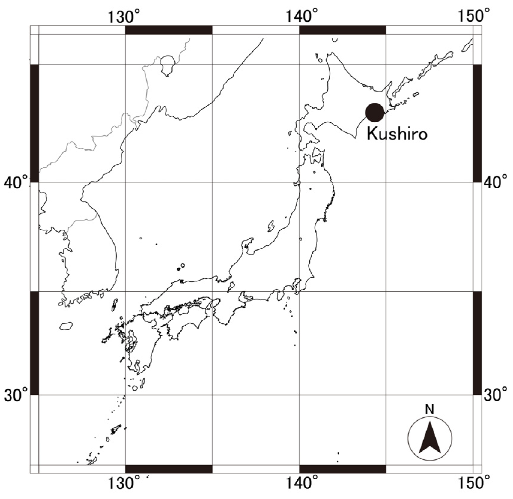
Figure 1. Map of the survey area (Kushiro, Hokkaido, Japan).
Microorganisms 13 02288 g001
Figure 2. Phylogenetic tree based on the mitochondrial cyt-b gene sequence (780 bp, NJ method). Bootstrap values (%) of 1000 times are shown at branching points. The representative sequences from Sorex spp. collected in Kushiro are indicated by arrows. The sequences from GenBank are shown with accession numbers.
Microorganisms 13 02288 g002
Figure 3. Phylogenetic tree based on the groEL sequence (282 bp) using the NJ method. Bootstrap values (%) of 1000 times are shown at branching points. The sequences determined in this study are indicated by the arrows.
Microorganisms 13 02288 g003
Figure 4. Detection of Piroplasmida and Anaplasmataceae in splenomegaly-positive (n = 66) and -negative (n = 37) Sorex spp. The Anaplasmataceae family includes Neoehrlichia and Ehrlichia. Significant differences between splenomegaly-positive and -negative groups are represented by an asterisk (tested by Chi-Square Test, * p < 0.05).
Microorganisms 13 02288 g004
Table 1. Tick collection from Sorex spp.
Sorex spp.Ixodes ovatusI. persulcautus
LarvaNymphLarvaNymphTotalTick/Shrew
2023S. unguiculatus (n = 10)1181118484.8
S. gracillimus (n = 1)010011
S. caecutiens (n = 1)35341515
2020S. unguiculatus (n = 10)83841013213.2
S. gracillimus (n = 5)8260163.2
Table 2. Detection of Piroplasmida and Anaplasmataceae in molted ticks.
Sorex spp. 1Resulted Nymph
I. ovatusI. persulcatus
No. ExaminedMicroorganism DetectedNo. ExaminedMicroorganism Detected
S. unguiculatus1- 20-
S. unguiculatus7-5Babesia divergens Asia lineage
S. unguiculatus1-0-
S. unguiculatus0-1-
S. caecutiens2Neoehrlichia mikurensis3-
S. caecutiens1-0-
1 Ticks were collected from each shrew (total of 6). 2 Not detected.
Table 3. Detection rate of Piroplasmida and Anaplasmataceae in the spleen of Sorex spp.
Sorex spp.No. ExaminedB. microtiN. mikurensisE. japonicaE. muris
S. unguiculatus533 1(5.7%)16 1,2(30.2%)11 2(20.8%)0(0%)
S. gracillimus343(8.8%)4 3(11.8%)3 3(8.8%)1(2.9%)
S. caecutiens164 4(25.0%)7 4(43.8%)0(0%)0(0%)
1 Containing mixed infection of B. microti and N. mikurensis (n = 1). 2 Containing mixed infections of N. mikurensis and E. japonica (n = 3). 3 Containing mixed infection of N. mikurensis and E. japonica (n = 1). 4 Containing mixed infection of B. microti and N. mikurensis (n = 2).
Table 4. Detection of multiple tick-borne microorganisms in Sorex spp.
Microorganisms Co-InfectedS. unguiculatusS. gracillimusS. caecutiensTotal
Neoehrlichia mikurensis and Ehrlichia japonica3104
N. mikurensis and Babesia microti1023
Disclaimer/Publisher’s Note: The statements, opinions and data contained in all publications are solely those of the individual author(s) and contributor(s) and not of MDPI and/or the editor(s). MDPI and/or the editor(s) disclaim responsibility for any injury to people or property resulting from any ideas, methods, instructions or products referred to in the content.