Article

LncRNA MEG3 Regulates Glaesserella parasuis-Induced Apoptosis of Porcine Alveolar Macrophages via Regulating ssc-miR-135/CASP8 Axis

1
Key Laboratory of Livestock Infectious Diseases, Ministry of Education, and Key Laboratory of Ruminant Infectious Disease Prevention and Control (East), Ministry of Agriculture and Rural Affairs, College of Animal Science and Veterinary Medicine, Shenyang Agricultural University, 120 Dongling Road, Shenyang 110866, China
2
State Key Laboratory for Diagnosis and Treatment of Severe Zoonotic Infectious Diseases, Key Laboratory of Zoonosis Research, Ministry of Education, Jilin University, Changchun 130012, China
*
Author to whom correspondence should be addressed.
These authors have contributed equally to this work.
Microorganisms 2025, 13(10), 2287; https://doi.org/10.3390/microorganisms13102287
Submission received: 28 August 2025 / Revised: 17 September 2025 / Accepted: 29 September 2025 / Published: 1 October 2025
(This article belongs to the Section Veterinary Microbiology)

Abstract

Glaesserella parasuis (G. parasuis), a common pathogenic bacterium in the porcine respiratory tract, can cause porcine polyserositis, arthritis, and meningitis. Alveolar macrophages are the first line of defense in the pulmonary innate immunity, and their abnormal apoptosis plays a critical role in the pathogenic process of G. parasuis. Long non-coding RNA maternally expressed gene 3 (MEG3) is associated with G. parasuis infection, but its mechanism remains incompletely unclear. This study aimed to investigate the role of MEG3 in G. parasuis-induced apoptosis of the porcine alveolar macrophage cell line 3D4/21 and its detailed molecular mechanism. Here, we found that MEG3 overexpression promoted G. parasuis-induced apoptosis and upregulated key extrinsic pathway proteins caspase-8 (CASP8) and caspase-3 (CASP3). Mechanistically, MEG3 functioned as a competing endogenous RNA by sponging ssc-miR-135, which directly targets and inhibits CASP8. Consequently, MEG3 overexpression alleviated ssc-miR-135-mediated repression of CASP8. Functional rescue experiments confirmed that either ssc-miR-135 mimic or CASP8 siRNA reversed the pro-apoptotic effect of MEG3. In conclusion, this study reveals that MEG3 relieves the inhibitory effect of ssc-miR-135 on CASP8 through competitively binding, thereby regulating G. parasuis-induced apoptosis of 3D4/21 cells. This study provides new insights into the pathogenic molecular mechanism of G. parasuis.

1. Introduction

Glaesserella parasuis (G. parasuis), a Gram-negative opportunistic pathogen, mainly infects the respiratory system of pigs and can cause Glässer’s disease characterized by fibrinous polyserositis, arthritis, and meningitis, resulting in severe economic losses to the global swine industry [1,2,3,4]. Recent studies have shown that the pathogenic mechanism of G. parasuis not only involves direct damage to host cells by virulence factors but is also closely related to abnormal biological behaviors of host cells [5]. Among them, the abnormal regulation of apoptosis is a key link in G. parasuis breaking the host immune barrier and promoting infection spread [6]. Li et al. demonstrated that G. parasuis can induce P53-dependent cell death [7]. In addition, Yan et al. reported that G. parasuis can induce more significant apoptosis by inhibiting the expression of the anti-cell death gene BCL2 [6]. A recent study indicated that G. parasuis can trigger endoplasmic reticulum (ER) stress through the PERK/eIF2α/ATF4/CHOP pathway, thereby leading to mitochondrial damage [8]. Although these studies have emphasized the pathways of G. parasuis-induced apoptosis, the specific molecular mechanisms and targets remain incompletely elucidated.
Long non-coding RNAs (lncRNAs), a class of non-coding RNA molecules longer than 200 nucleotides (nt), do not have protein-coding functions but are widely involved in biological processes such as cell apoptosis, proliferation, differentiation, and immune response through mechanisms including epigenetic regulation, post-transcriptional regulation, and acting as molecular sponges. To date, numerous studies have reported that lncRNAs can act as competitive endogenous RNAs (ceRNAs) to inhibit miRNA functions and regulate mRNA expression by competitively binding to miRNAs [9]. For example, lncRNA CASC2 can act as a “sponge” for miR-24 and miR-221, regulating caspase-3/8 (CASP3/8) to affect TNF-related apoptosis-inducing ligand (TRAIL) resistance in hepatocellular carcinoma [10]. LncRNA THUMPD3-AS1 inhibits apoptosis of ovarian cancer cells by regulating the miR-320d/ARF1 axis [11]. As a widely studied tumor suppressor-related lncRNA, lncRNA maternally expressed gene 3 (MEG3) has been confirmed to regulate cell apoptosis, proliferation, and other processes by targeting different miRNAs [12,13,14,15,16,17,18]. It has been reported that MEG3 affects the proliferation and apoptosis of psoriatic epidermal cells by targeting miR-21/CASP8 [19]. Silencing of MEG3 exacerbates lipopolysaccharide-stimulated apoptosis of human lung cells by regulating miR-4262 [20]. Previous studies have shown that MEG3 is involved in the inflammatory response and apoptosis of porcine alveolar macrophages infected with G. parasuis by regulating the miR-210/TLR4 axis [21]. However, the role and potential molecular mechanism of MEG3 in G. parasuis-induced apoptosis remain largely unknown. In this study, we constructed a novel MEG3-miRNA-mRNA ceRNA network regulatory axis to further explore the function and potential molecular mechanism of MEG3 in G. parasuis-induced apoptosis. The results of this finding enrich the research content on the pathogenic mechanism of G. parasuis.

2. Materials and Methods

2.1. Bacterial Strain and Cell Line

The highly virulent G. parasuis serovar 13 strain CY1201 was isolated from a diseased pig in Liaoning, China, and cultured at 37 °C on tryptic soy agar (TSA, Solarbio, Beijing, China) or in tryptic soy broth (TSB, Solarbio, Beijing, China) supplemented with 2% nicotinamide adenine dinucleotide (NAD, Solarbio, Beijing, China) and 5% fetal bovine serum (Sangon Biotech, Shanghai, China). The porcine alveolar macrophage cell line 3D4/21 (Newgainbio, Wuxi, China) was cultured in RPMI 1640 medium (Sangon Biotech, Shanghai, China) supplemented with 10% fetal bovine serum (Sangon Biotech, Shanghai, China), penicillin–streptomycin mixture (Servicebio, Wuhan, China), and non-essential amino acids (Sangon Biotech, Shanghai, China) at 37 °C in a 5% CO2 atmosphere.

2.2. Cell Transfection and Bacterial Infection

The MEG3 overexpression plasmid (OE-MEG3) was synthesized by BGI Genomics Co., Ltd. (Beijing, China), and the negative control pcDNA3.1 (+) was used as the control vector (OE-NC). ssc-miR-135 mimic (5′-UAUGGCUUUUUAUUCCUAUGUGA ACAUAGGAAUAAAAAGCCAUAUU-3′), mimic mock (Mimic NC, 5′-UUCUCCGAACGUGUCACGUTT ACGUGACACGUUCGGAGAATT-3′), ssc-miR-135 inhibitor (5′-UCACAUAGGAAUAAAAAGCCAUA-3′), inhibitor mock (Inhibitor NC inhibitor, 5′-CAGUACUUUUGUGUAGUACAA-3′), Silencer® CASP8 siRNA (si-CASP8, 5′-GAAAGUGCUUCGGAUGAAUTT AUUCAUCCGAAGCACUUUCTT-3′) and Silencer® Negative Control (si-NC, 5′-UUCUCCGAACGUGUCACGUTT ACGUGACACGUUCGGAGAATT-3′) were designed and synthesized by Genepharma (Shanghai, China). Plasmids were transfected using PolyFast Transfection Reagent (MCE, Monmouth Junction, NJ, USA), while miRNA mimics, miRNA inhibitors, and siRNAs were transfected using GP-transfect-Mate (Genepharma, Shanghai, China) according to the manufacturer’s instructions. Transient transfection was performed when cell confluency reached 70–90%. After 8 h of transfection, the medium was replaced, and cells were cultured for 24 or 36 h. Cells were infected with G. parasuis for which multiplicity of infection (MOI) was 20 for 24 h, followed by further detection or RNA/protein extraction.

2.3. Cell Viability Assay

Cell viability was detected using the Cell Counting Kit-8 (CCK-8, Jiangsu Cowin Biotech Co., Ltd., Taizhou, China) according to the manufacturer’s instructions. Briefly, cells were seeded into 96-well plates (Sangon Biotech, Shanghai, China) at a density of 2000 cells per well in 100 μL of medium. After 24 h of transfection, the cells were treated with G. parasuis for 24 h. Subsequently, 10 μL of CCK-8 solution was added to each well, and the cells were incubated for an additional 2 h. The absorbance at 450 nm was measured using a microplate reader (Servicebio, Wuhan, China).

2.4. Annexin V-FITC/PI Staining Assay

According to the manufacturer’s instructions, 5 × 10⁵ cells were incubated with Annexin V-FITC Reagent and PI Reagent (Annexin V-FITC/PI Cell Apoptosis Detection Kit, Elabscience Biotechnology, Wuhan, China) at room temperature in the dark for 20 min, and the apoptosis rate was detected using BD FACSAria III (Becton Dickinson, Franklin Lakes, NJ, USA).

2.5. Bioinformatics Analysis

RNAhybrid 2.1.2 predicts potential binding sites between lncRNA, miRNA, and mRNA. The prediction of miRNA target genes was performed via OmicStudio tools (LC-Bio Technologies Co., Ltd., Hangzhou, China; https://www.omicstudio.cn/tool/, accessed on 20 July 2025), which integrates two algorithms: TargetScan (v5.0) and miRanda (v3.3a). The screening thresholds were set as follows: TargetScan_score ≥ 50 and miRanda_Energy < −10. To improve the reliability of predictions, the target genes identified by both algorithms were combined, and their overlapping genes were retained for subsequent analysis. Gene Ontology (GO) terms/Kyoto Encyclopedia of Genes and Genomes (KEGG) enrichment analysis provides all GO terms/KEGG pathways that are significantly enriched in miRNA target genes compared to the genome background. All miRNA target genes were mapped to GO terms/KEGG pathways in the GO database (http://www.geneontology.org/, accessed on 20 July 2025) and KEGG database (https://www.kegg.jp/, accessed on 20 July 2025); gene numbers were calculated for every term/pathway; significantly enriched GO terms/KEGG pathways in miRNA target genes compared to the genome background were defined by hypergeometric test. Database for Annotation, Visualization and Integrated Discovery (DAVID, https://david.ncifcrf.gov/, accessed on 20 July 2025), Metascape (https://metascape.org/, accessed on 20 July 2025) and WebGestalt (https://www.webgestalt.org/, accessed on 20 July 2025) and “clusterProfiler” package were used for data analyses. GO database and KEGG database were used for enrichment analysis, with the species chosen being Sus scrofa. For the enrichment analysis, the criteria were set as follows: p value < 0.05 and enrichment fold (EF; ratio of target gene proportion in the pathway to background gene proportion in the pathway) > 1.5.

2.6. Dual-Luciferase Reporter Assay

RNAhybrid 2.1.2 was used to predict potential binding sites between MEG3 and ssc-miR-135, as well as between ssc-miR-135 and the 3′ untranslated region (UTR) of CASP8. The wild type MEG3 (MEG3-Wt), mutant type MEG3 (MEG3-Mut), wild type CASP8 3′ UTR (CASP8 3′ UTR-Wt), and mutant type CASP8 3′ UTR (CASP8 3′ UTR-Mut) sequences were cloned into the dual-luciferase reporter vector pmirGLO (Genepharma, Shanghai, China), which was synthesized by BGI Genomics Co., Ltd. (Shenzhen, China). The constructed dual-luciferase reporter vectors were co-transfected with miR-135 mimic into 3D4/21 cells. Luciferase activity was determined using the dual-Luciferase Kit (Jiangsu Cowin Biotech Co., Ltd., Taizhou, China) according to the manufacturer’s instructions.

2.7. RNA Extraction and Quantitative Real-Time PCR (qPCR) Detection

Total cellular RNA was extracted using the Trizol RNA Extraction Kit (Vazyme, Nanjing, China). First-strand cDNA was synthesized using a reverse transcription kit (Vazyme, Nanjing, China) according to the manufacturer’s instructions. cDNA for detecting the expression level of ssc-miR-135 was synthesized using the SuperStar miRNA First-Strand cDNA Synthesis Kit (by tailing A) (Jiangsu Cowin Biotech Co., Ltd., Taizhou, China). qPCR was performed to detect the expression levels of MEG3, ssc-miR-135, and mRNA. GAPDH and U6 were used as internal reference genes for expression normalization. 2−ΔΔCt method was used to analyze the experimental results. The primer sequences are shown in Table 1.

2.8. Western Blot

Total protein was extracted using RIPA lysis buffer (Epizyme, Shanghai, China) containing protease inhibitors and phosphatase inhibitors, and protein concentration was determined using a BCA protein assay kit (Epizyme, Shanghai, China). Equal amounts of protein were loaded onto 12.5% sodium dodecyl sulfate polyacrylamide gel electrophoresis (SDS-PAGE) (Epizyme, Shanghai, China) for separation, then transferred to 0.22 μm PVDF membranes (Abcholal, Wuhan, China). The membranes were blocked with 5% skimmed milk (Epizyme, Shanghai, China) at room temperature for 2 h, then incubated with primary antibodies anti-CASP3 (1:1000 dilution, #T40044, Abmart, Shanghai, China), anti-Cleaved-CASP3 (1:1000 dilution, #T40044, Abmart, Shanghai, China), anti-CASP8 (1:1000 dilution, #PK85346, Abmart, Shanghai, China), anti-Cleaved-CASP8 (1:1000 dilution, #PK85387, Abmart, Shanghai, China), and anti-GAPDH (1:10,000 dilution, #10494-1-AP, Proteintech Group, Wuhan, China) at 4 °C overnight. After washing, the membranes were incubated with horseradish peroxidase (HRP)-conjugated secondary antibody (1:8000 dilution, #LF108, Epizyme, Shanghai, China). Chemiluminescent signals were detected using an ECL kit (Ncmbio, Suzhou, China) and visualized using a gel imaging system (Servicebio, Wuhan, China). The expression of each protein was normalized to GAPDH, and semi-quantitative analysis was performed using ImageJ.

2.9. Statistical Analysis

Statistical analysis and visualization were conducted using SPSS 15.0 software (SPSS Inc., Chicago, IL, USA) and GraphPad Prism 8.0 software (GraphPad Software, San Diego, CA, USA). Data are presented as mean ± standard deviation (SD). Comparisons between two groups were made using an independent samples t-test, with a significance level set at p < 0.05, indicating a statistically significant difference.

3. Results

3.1. Overexpression of MEG3 Promotes G. parasuis-Induced Apoptosis of 3D4/21 Cells

To clarify the effect of MEG3 on G. parasuis-induced apoptosis of 3D4/21 cells, we constructed a MEG3 overexpression plasmid, transfected it into 3D4/21 cells, and then infected the cells with G. parasuis. First, qPCR was used to detect the overexpression efficiency. Compared with the control group, the expression level of MEG3 in the MEG3 overexpression group was significantly increased (p < 0.01), indicating the successful establishment of a MEG3 overexpressing cell model in G. parasuis-infected 3D4/21 cells (Figure 1A). The CCK-8 assay showed that overexpression of MEG3 significantly reduced cell viability (p < 0.01) (Figure 1B). Flow cytometry analysis revealed that overexpression of MEG3 significantly increased the apoptosis rate (p < 0.01) (Figure 1C,D). qPCR analysis of apoptosis-related genes showed that MEG3 overexpression promoted the mRNA expression of FASLG, TNF, CASP8, BAX, and CASP3 while inhibiting the mRNA expression of BCL2 (p < 0.05 or p < 0.01). In contrast, the mRNA levels of TP53, CYCS, CASP9, AIFM1, and ENDOG showed no significant changes (p > 0.05) (Figure 1E). Most of these differentially expressed genes are involved in the extrinsic apoptosis pathway, suggesting that MEG3 may be involved in the extrinsic apoptosis pathway. Furthermore, G. parasuis significantly increased the protein expression levels of Cleaved-CASP8/CASP8 and Cleaved-CASP3/CASP3 in 3D4/21 cells, and this change was exacerbated by MEG3 overexpression (p < 0.01) (Figure 1F).
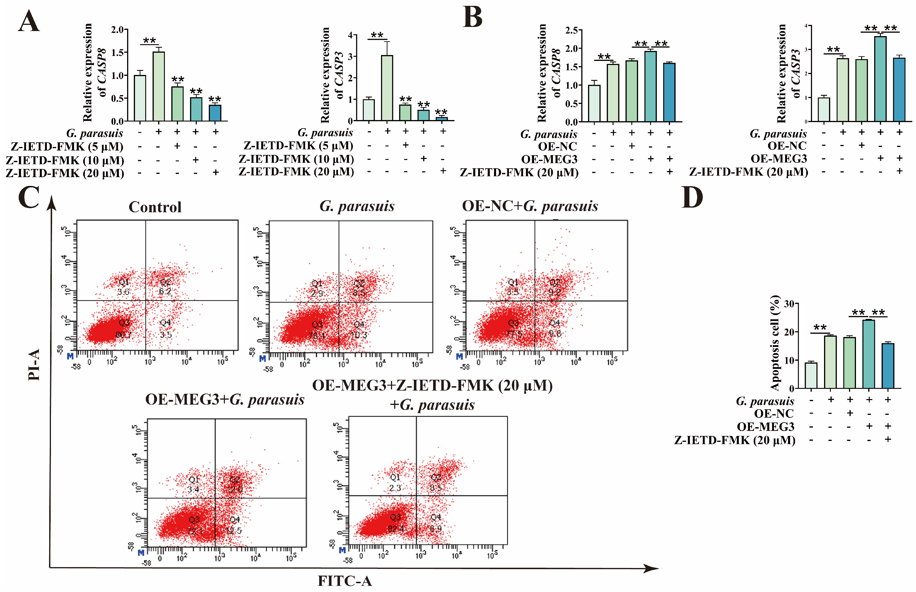
As CASP8 is a key initiator of the extrinsic apoptosis pathway, to further confirm that MEG3 exerts its function through the extrinsic apoptosis pathway, we used Z-IETD-FMK treatment to inhibit its expression, thereby suppressing the activation of extrinsic apoptosis. First, qPCR was performed to detect the effects of different concentrations of Z-IETD-FMK on mRNA levels of CASP8 and CASP3 in G. parasuis-infected 3D4/21 cells. The results showed that, after G. parasuis infection of 3D4/21 cells, the mRNA levels of CASP8 and CASP3 were significantly increased (p < 0.01). After treatment with different concentrations of Z-IETD-FMK, the mRNA levels of CASP8 and CASP3 gradually decreased with increasing inhibitor concentration (p < 0.01) (Figure 2A), indicating that Z-IETD-FMK can effectively inhibit the activation of the extrinsic apoptosis pathway after G. parasuis infection. Furthermore, after transfection with the MEG3 overexpression plasmid, 3D4/21 cells were treated with 20 μM of Z-IETD-FMK followed by G. parasuis infection. qPCR results showed that the increase in mRNA levels of CASP8 and CASP3 caused by MEG3 overexpression was inhibited after the addition of Z-IETD-FMK (p < 0.01) (Figure 2B). Flow cytometry analysis revealed that the increase in apoptosis rate induced by MEG3 overexpression was also suppressed after Z-IETD-FMK treatment (p < 0.01) (Figure 2C,D). These results indicate that overexpression of MEG3 promotes G. parasuis-induced apoptosis of 3D4/21 cells through the extrinsic apoptosis pathway.

3.2. MEG3 as a Molecular Sponge for ssc-miR-135

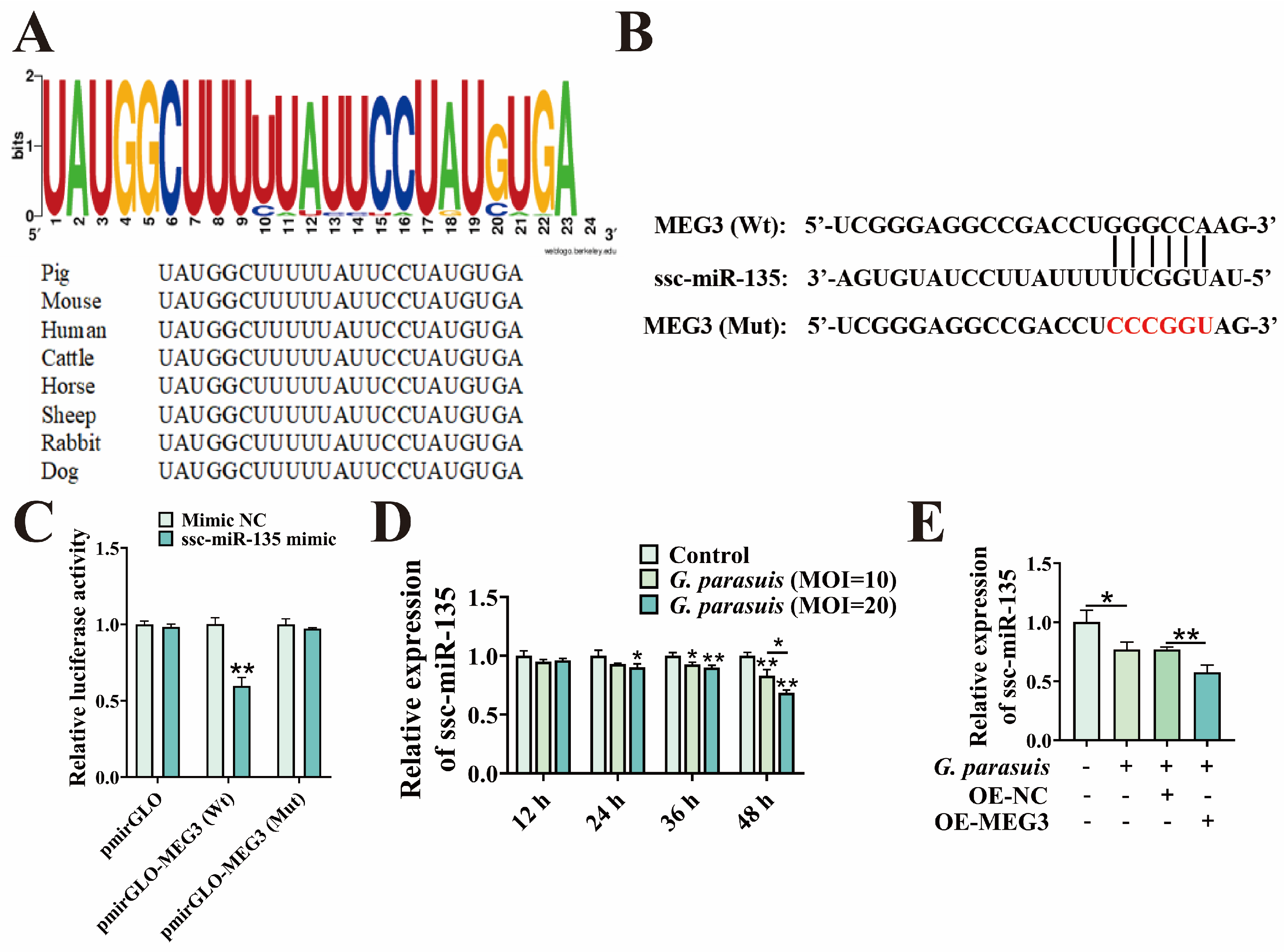
It has been reported that MEG3 can bind to miR-135 [22]. Therefore, we identified ssc-miR-135 as a potential target, as it is highly conserved in mammals (Figure 3A). RNAhybrid prediction revealed a binding site between MEG3 and ssc-miR-135 (Figure 3B). Following further mutation of the binding site, in 3D4/21 cells transfected with ssc-miR-135 mimic, the luciferase activity of MEG3-Wt decreased (p < 0.01), while that of MEG3-Mut showed no significant effect (p > 0.05) (Figure 3C). The results indicate that MEG3 targets ssc-miR-135. Subsequently, we measured the expression changes in ssc-miR-135 in 3D4/21 cells infected with G. parasuis. The results showed that ssc-miR-135 exhibited time and dose-dependent downregulation (p < 0.05 or p < 0.01) (Figure 3D). We further examined the expression of ssc-miR-135 in G. parasuis-infected 3D4/21 cells with MEG3 overexpression. The results showed that overexpression of MEG3 significantly reduced the expression of ssc-miR-135 (p < 0.01) (Figure 3E). In summary, these results indicate that MEG3 directly binds to ssc-miR-135 in 3D4/21 cells.

3.3. ssc-miR-135 Regulates G. parasuis-Induced Apoptosis in 3D4/21 Cells

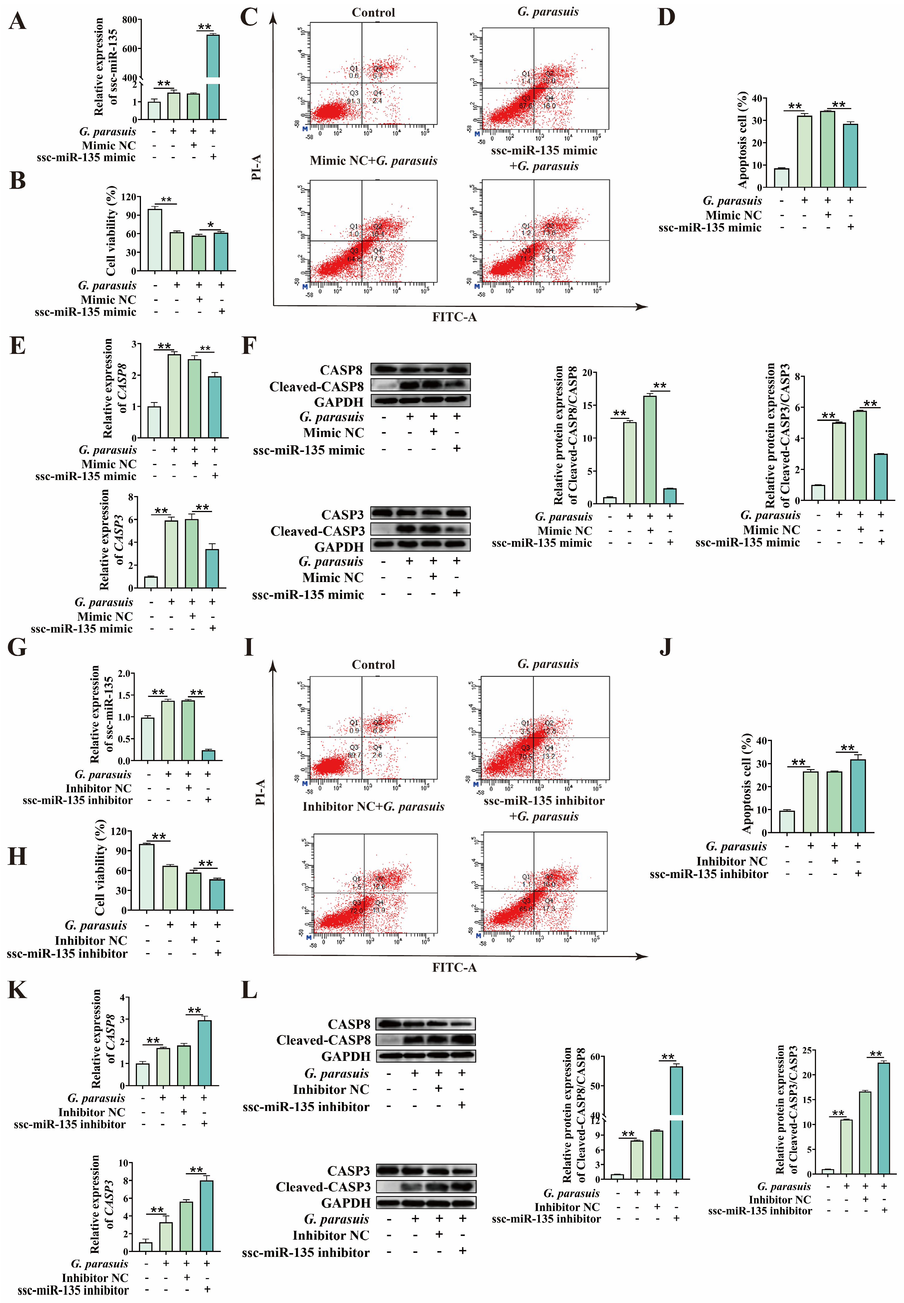
To clarify whether ssc-miR-135 plays a role in G. parasuis-induced apoptosis of 3D4/21 cells, we first transfected 3D4/21 cells with ssc-miR-135 mimic or ssc-miR-135 inhibitor, and verified the overexpression/knockdown efficiency (Figure 4A,G). Subsequently, G. parasuis infection was performed in 3D4/21 cells with ssc-miR-135 overexpression or knockdown. CCK-8 assay indicated that, compared with the control group, transfection with ssc-miR-135 mimic significantly increased cell viability, while transfection with ssc-miR-135 inhibitor significantly decreased cell viability (p < 0.01) (Figure 4B,H). To determine whether the reduction in cell viability was attributed to apoptosis, flow cytometry analysis was performed. The results revealed that transfection with ssc-miR-135 mimic significantly reduced the apoptosis rate of 3D4/21 cells induced by G. parasuis infection, whereas transfection with ssc-miR-135 inhibitor increased the apoptosis rate (p < 0.01) (Figure 4C,D,I,J). qPCR analysis revealed that transfection with ssc-miR-135 mimic significantly inhibited the expression of CASP8 and CASP3 mRNA levels induced by G. parasuis infection, while an opposite trend was observed in cells transfected with ssc-miR-135 inhibitor (p < 0.01) (Figure 4E,K). Western blot analysis indicated that, compared with the control group, the protein expression ratios of Cleaved-CASP8/CASP8 and Cleaved-CASP3/CASP3 were decreased by transfection with ssc-miR-135 mimic and increased by transfection with ssc-miR-135 inhibitor (p < 0.01) (Figure 4F,L). Collectively, these results indicate that ssc-miR-135 negatively regulates G. parasuis-induced apoptosis in 3D4/21 cells.

3.4. ssc-miR-135 Directly Interacts with CASP8

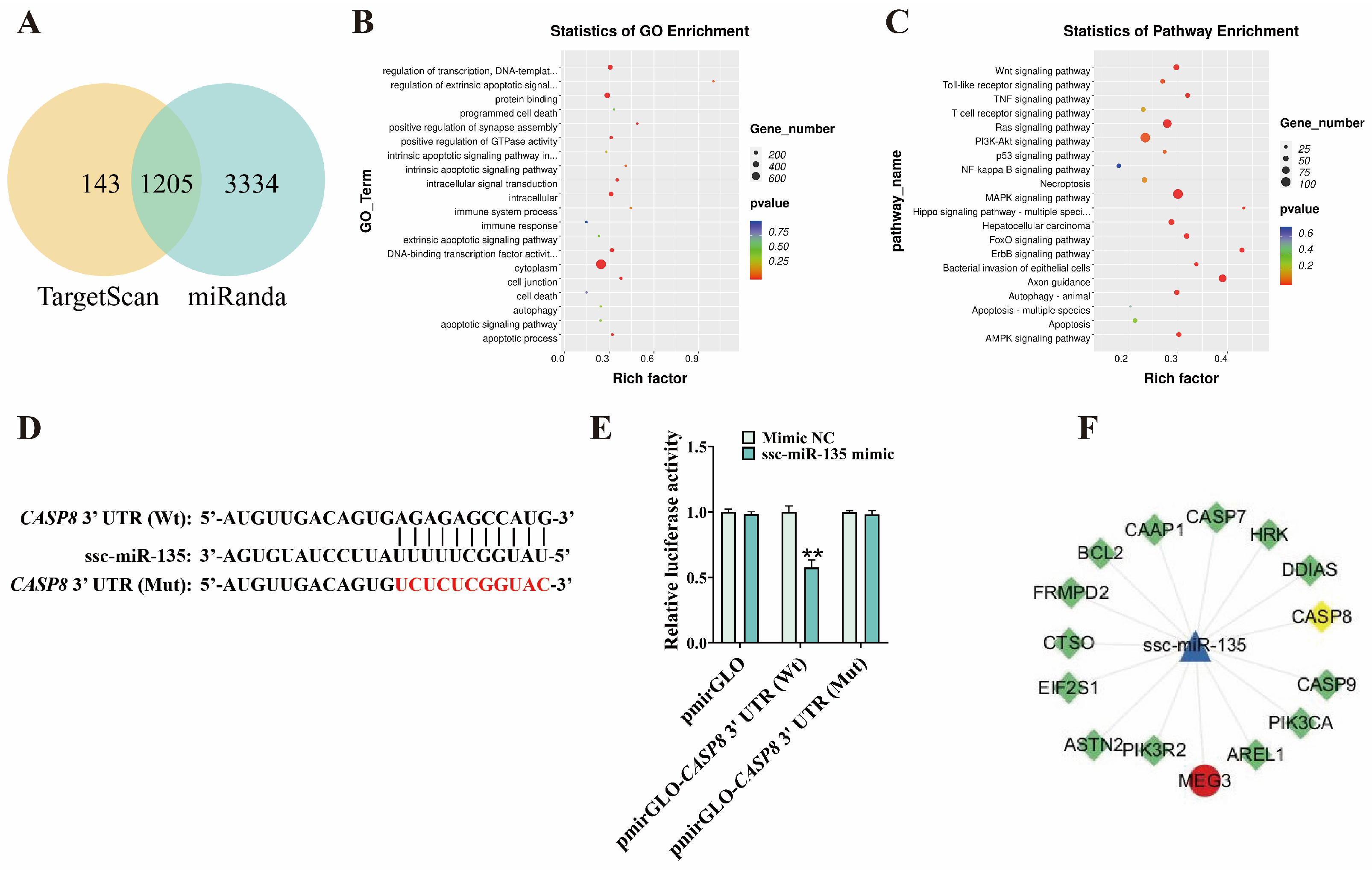
To further explore the interaction between miRNA and its downstream targets, we used TargetScan and miRanda to predict the target genes of ssc-miR-135 (Figure 5A) (prediction results are shown in Table S1). GO and KEGG enrichment analysis of the predicted target genes revealed enrichment in GO terms such as cell death, apoptotic signaling pathway, and apoptotic process, as well as the Apoptosis KEGG pathway (Figure 5B,C) (enrichment results are shown in Tables S2 and S3). RNAhybrid prediction identified a binding site between ssc-miR-135 and the 3′ UTR of CASP8 (Figure 5D). Subsequent dual-luciferase reporter assay results showed that, when ssc-miR-135 mimic were co-transfected with the CASP8 3′ UTR-Wt, luciferase activity was significantly reduced (p < 0.01), while there was no significant change in the CASP8 3′ UTR-Mut (p > 0.01) (Figure 5E). These findings indicate that ssc-miR-135 targets CASP8. Additionally, previous results showed that ssc-miR-135 mimic significantly reduced CASP8 mRNA levels and Cleaved-CASP8/CASP8 protein levels, while ssc-miR-135 interference had the opposite trend (Figure 4). Collectively, these results indicate that CASP8 is a target gene of ssc-miR-135.

3.5. MEG3 Regulates G. parasuis-Induced Apoptosis in 3D4/21 Cells via ssc-miR-135/CASP8 Axis

Given that MEG3 can regulate G. parasuis-induced apoptosis in 3D4/21 cells and interacts with ssc-miR-135, while ssc-miR-135 negatively regulates G. parasuis-induced apoptosis in 3D4/21 cells, we hypothesized that MEG3 regulates the expression of CASP8 via ssc-miR-135, forming a ceRNA regulatory relationship (Figure 5F). To verify this hypothesis, 3D4/21 cells were co-transfected with MEG3 overexpression plasmid and ssc-miR-135 mimic, followed by infection with G. parasuis. CCK-8 assays revealed that ssc-miR-135 mimic reversed the reduction in cell viability caused by MEG3 overexpression (p < 0.01) (Figure 6A). Additionally, we found that, in G. parasuis-infected 3D4/21 cells, co-transfection of MEG3 overexpression plasmid and ssc-miR-135 mimic significantly reduced apoptosis rates compared to transfection with MEG3 overexpression plasmid alone (p < 0.01) (Figure 6B,C). qPCR results showed that co-transfection of MEG3 overexpression plasmid and ssc-miR-135 mimic significantly increased ssc-miR-135 expression (p < 0.01) (Figure 6E) and decreased MEG3 and CASP8 expression (p < 0.01) (Figure 6D,F) compared to transfection with MEG3 overexpression plasmid alone. Western blot analysis indicated that, compared with transfection with MEG3 overexpression plasmid alone, co-transfection with MEG3 overexpression plasmid and ssc-miR-135 mimic significantly reduced the protein expression of Cleaved-CASP8/CASP8 (p < 0.01) (Figure 6G). These results indicate that MEG3 acts as a molecular sponge for ssc-miR-135 to regulate the expression of CASP8.
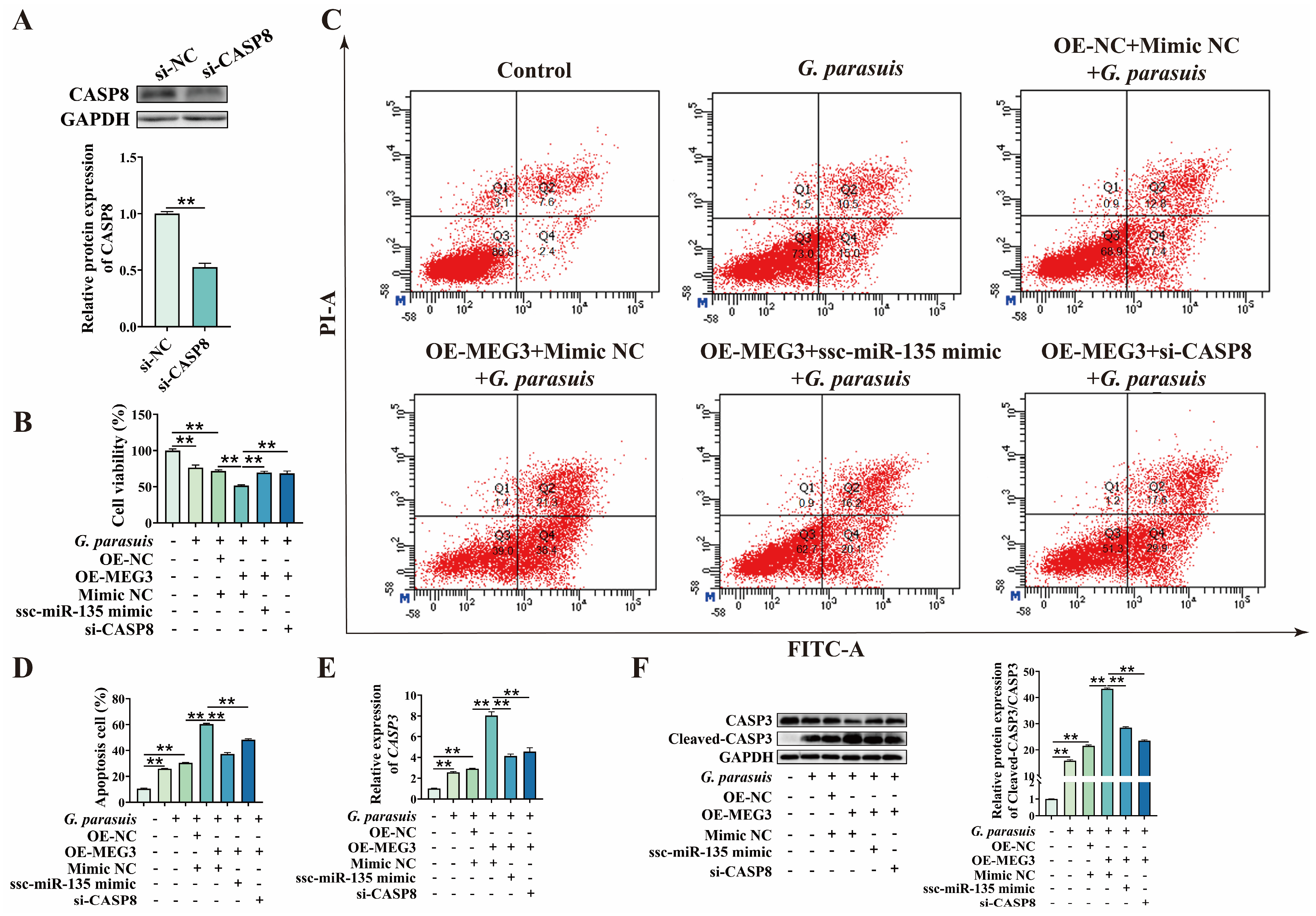
Finally, to clarify that MEG3 regulates 3D4/21 cell apoptosis via ssc-miR-135/CASP8 axis, we first designed siRNA targeting CASP8 and verified its knockdown efficiency (Figure 7A). Subsequently, 3D4/21 cells were co-transfected with MEG3 overexpression plasmid and ssc-miR-135 mimic, as well as with MEG3 overexpression plasmid and si-CASP8, respectively. CCK-8 assay results demonstrated that ssc-miR-135 mimic reversed the decrease in cell viability caused by MEG3 overexpression, and a similar reversing effect was observed upon CASP8 knockdown (p < 0.01) (Figure 7B). Additionally, we found that, in G. parasuis-infected 3D4/21 cells, compared to transfection with MEG3 overexpression plasmid alone, co-transfection with MEG3 overexpression plasmid and ssc-miR-135 mimic, as well as co-transfection with MEG3 overexpression plasmid and si-CASP8, significantly reduced the apoptosis rate (p < 0.01) (Figure 7C,D). qPCR and Western blot analysis results showed that, compared to transfection with MEG3 overexpression plasmid alone, co-transfection with MEG3 overexpression plasmid and ssc-miR-135 mimic, as well as co-transfection with MEG3 overexpression plasmid and si-CASP8, significantly reduced the expression of CASP3 (p < 0.01) (Figure 7E,F). In summary, these findings indicate that MEG3 regulates G. parasuis-induced apoptosis in 3D4/21 cells via ssc-miR-135/CASP8 axis.

4. Discussion

Glässer’s disease caused by G. parasuis has caused serious economic losses to the pig industry [23]. It has been reported that many lncRNAs with differential expression during G. parasuis infection have been identified, but their specific functions remain unclear [24]. Beyond its well-known role as a tumor-suppressive lncRNA, mounting evidence indicates that MEG3 plays crucial regulatory roles in the occurrence and development of various diseases. In this study, we found that overexpression of MEG3 can significantly reduce the viability of 3D4/21 cells infected with G. parasuis and increase the apoptosis rate, which is consistent with previous research findings [21]. At the same time, we further observed that overexpression of MEG3 leads to a significant increase in the mRNA and protein levels of apoptosis-initiating factors and apoptosis-effecting factors CASP8 and CASP3 in the extrinsic apoptosis pathway. These findings indicate that MEG3 may regulate G. parasuis-induced apoptosis in 3D4/21 cells through the extrinsic apoptotic pathway. Our results further clarify the role of MEG3 in G. parasuis-induced apoptosis in 3D4/21 cells. Accumulating research evidence has demonstrated that lncRNAs are associated with host–pathogen interactions [25,26,27,28,29]. Previous studies have confirmed that the cytolethal distending toxin (CDT), a virulence factor of G. parasuis, can enhance the adhesion and invasion of G. parasuis to host cells, activate the DNA damage response, and induce cell cycle arrest and host cell apoptosis [7,30,31]. However, whether the apoptosis regulated by MEG3 involves virulence factors of G. parasuis remains to be further studied.
Given that ssc-miR-135 is differentially expressed after G. parasuis infection [32], and MEG3 can promote bovine myoblast differentiation through the sponge miR-135 [22], we further explored the function and interaction of MEG3 and ssc-miR-135 in the process of G. parasuis-induced apoptosis in 3D4/21 cells. In our study, we again confirmed that ssc-miR-135 is a target miRNA of MEG3 using a dual-luciferase reporter assay. ssc-miR-135 is downregulated in 3D4/21 cells infected with G. parasuis, and knockdown of ssc-miR-135 promotes G. parasuis-induced apoptosis in 3D4/21 cells, which is consistent with the effect of MEG3 overexpression. Meanwhile, overexpression of ssc-miR-135 reverses the promotional effect of MEG3 overexpression on G. parasuis-induced apoptosis in 3D4/21 cells. Our study first revealed that MEG3 can regulate G. parasuis-induced apoptosis in 3D4/21 cells through competitively binding to ssc-miR-135.
MicroRNAs (miRNAs) exert their biological functions by post-transcriptionally regulating their downstream targets [33]. Yu et al. reported that miR-135 regulates NLRP3 inflammasome activation by inhibiting the expression of CaSR [34]. Previous studies have indicated that ssc-miR-135 may promote G. parasuis-induced inflammatory responses through the NF-κB signaling pathway [32]. However, whether ssc-miR-135 regulates the pathogenic process of G. parasuis infection by binding to downstream targets remains unknown. In this study, we first identified CASP8 as a downstream target of ssc-miR-135 and also as a downstream effector molecule of the MEG3/CASP8 axis. Cleavage of CASP8 is critical for the activation of the extrinsic apoptotic pathway, which ultimately induces the cleavage of CASP3 protein [35]. Our findings indicate that ssc-miR-135 participates in regulating G. parasuis-induced apoptosis in 3D4/21 cells by negatively regulating CASP8 expression. Additionally, LncRNA can act as a miRNA sponge and weaken mRNA regulation [36]. Similarly, we found that CASP8 expression is promoted by MEG3 overexpression, while ssc-miR-135 overexpression eliminates this effect. Furthermore, when CASP8 is inhibited, the promotion of downstream CASP3 expression caused by MEG3 overexpression is suppressed, thereby attenuating G. parasuis-induced apoptosis in 3D4/21 cells. These results reveal a novel regulatory network mechanism by which MEG3 regulates G. parasuis-induced apoptosis in 3D4/21 cells via the ssc-miR-135/CASP8 axis (Figure 8).
This study has several limitations. First, it remains unclear whether other miRNAs are involved in the process by which MEG3 regulates G. parasuis-induced apoptosis. Moreover, the in vitro porcine alveolar macrophage model can only simulate local infection, and the regulatory mechanism of the MEG3/ssc-miR-135/CASP8 axis during systemic infection requires further evaluation in animal models or using different cell lines.

5. Conclusions

This study demonstrates that MEG3 can regulate G. parasuis-induced apoptosis in 3D4/21 cells via ssc-miR-135/CASP8 axis. These findings provide new insights into the molecular mechanisms underlying the pathogenicity of G. parasuis.

Supplementary Materials

The following supporting information can be downloaded at: https://www.mdpi.com/article/10.3390/microorganisms13102287/s1, Table S1: TargetScan and miRanda to predict the target genes of ssc-miR-135; Table S2: GO enrichment analysis of the target genes of ssc-miR-135; Table S3: KEGG enrichment analysis of the target genes of ssc-miR-135.

Author Contributions

Conceptualization, Y.J. and M.Q.; methodology, Y.J. and X.S.; software, R.Y. (Ronglan Yin); validation, Y.J., N.L. and A.S.; formal analysis, H.W.; investigation, Y.J.; resources, R.Y. (Ronghuan Yin); data curation, F.Z. and Y.Z.; writing—original draft preparation, Y.J. and M.Q.; writing—review and editing, Y.J. and M.Q.; visualization, Y.Z.; supervision, R.Y. (Ronghuan Yin); project administration, R.Y. (Ronghuan Yin); funding acquisition, R.Y. (Ronghuan Yin). All authors have read and agreed to the published version of the manuscript.

Funding

This research was funded by the National Natural Science Foundation of China (No. 32172861), the Natural Science Foundation of Liaoning Province (No. 2021-MS-221), the Scientific Research Project of Liaoning Provincial Department of Education (No. LSNJC201917), and the Zoonosis Research Engineering Laboratory Project of Liaoning Province (No. 01072917001).

Institutional Review Board Statement

Not applicable.

Informed Consent Statement

Not applicable.

Data Availability Statement

The original contributions presented in this study are included in the article/Supplementary Materials. Further inquiries can be directed to the corresponding author.

Acknowledgments

The authors are grateful to the support and assistance in terms of instruments and facilities provided by Key Laboratory of Livestock Infectious Diseases, Ministry of Education, and Key Laboratory of Ruminant Infectious Disease Prevention and Control (East), Ministry of Agriculture and Rural Affairs, College of Animal Science and Veterinary Medicine, Shenyang Agricultural University, 120 Dongling Road, Shenyang 110866, China.

Conflicts of Interest

The authors declare no conflicts of interest.

Abbreviations

CCK-8Cell counting kit-8
CDTCytolethal distending toxin
ceRNACompetitive endogenous RNA
circRNAcircular RNA
EREndoplasmic reticulum
G. parasuisGlaesserella parasuis
LncRNALong non-coding RNA
MEG3Maternally expressed gene 3
miRNAmicroRNA
MOIMultiplicity of infection
NADNicotinamide adenine dinucleotide
ntNucleotide
qPCRQuantitative Real-Time PCR
SDStandard deviation
SDS-PAGESodium dodecyl sulfate polyacrylamide gel electrophoresis
snoRNAsmall nucleolar RNA
TRAILTNF-related apoptosis-inducing ligand
TSATryptic soy agar
TSBTryptic soy broth
UTRUntranslated region

References

  1. Xu, J.; Jin, X.; Li, X.; Yang, D. Epidemiology and pathogenicity of Haemophilus parasuis in eastern China. Front. Microbiol. 2025, 16, 1589975. [Google Scholar] [CrossRef]
  2. Guo, Z.; Zhou, Y.; Li, N.; Shen, A.; Jia, Y.; Yin, R.; Yang, J.; Yuan, J.; Yin, R. The Molecular Mechanism by Which miR-129a-3p Targets the TLR4/NF-κB Signaling Pathway to Regulate Inflammatory Damage in 3D4/21 Cells Infected with Glaesserella parasuis. Animals 2025, 15, 1355. [Google Scholar] [CrossRef] [PubMed]
  3. Nielsen, D.W.; Sarlo Davila, K.M.; Brockmeier, S.L.; Hau, S.J. Transcriptional profile of Glaesserella parasuis in swine serosal and joint fluids. Front. Vet. Sci. 2025, 12, 1452973. [Google Scholar] [CrossRef]
  4. Dai, L.; Wan, J.; Zhang, R.; Xie, T.; Jia, Y.; Lu, Z.; Zhang, F.; Ke, W.; Liu, F.; Lei, L. Multi-epitope vaccines Xlc and Ddc against Glaesserella parasuis infection in mice. Vet. Microbiol. 2025, 304, 110491. [Google Scholar] [CrossRef]
  5. Fu, S.; Liu, H.; Xu, L.; Qiu, Y.; Liu, Y.; Wu, Z.; Ye, C.; Hou, Y.; Hu, C.A. Baicalin modulates NF-κB and NLRP3 inflammasome signaling in porcine aortic vascular endothelial cells Infected by Haemophilus parasuis Causing Glässer’s disease. Sci. Rep. 2018, 8, 807. [Google Scholar] [CrossRef] [PubMed]
  6. Yan, P.; Jia, Y.C.; Zhang, X.L.; Zhou, Y.Y.; Guo, Y.; Yin, R.L.; Yuan, J.; Wang, L.X.; Guo, Z.B.; Wang, J.Y.; et al. Virulence assessment of four Glaesserella parasuis strains isolated in Liaoning province of China. Res. Vet. Sci. 2023, 158, 226–234. [Google Scholar] [CrossRef]
  7. Li, G.; Niu, H.; Zhang, Y.; Li, Y.; Xie, F.; Langford, P.R.; Liu, S.; Wang, C. Haemophilus parasuis cytolethal distending toxin induces cell cycle arrest and p53-dependent apoptosis. PLoS ONE 2017, 12, e0177199. [Google Scholar] [CrossRef]
  8. Feng, W.; Han, Y.; Zhang, W.; Wu, L.; Zhou, A.; Shi, L.; Zhang, J. Glaesserella parasuis infection triggers endoplasmic reticulum stress-mediated pyroptosis via PERK/eIF2α/ATF4 axis and metabolic reprogramming in porcine alveolar macrophages. Vet. Res. 2025, 56, 150. [Google Scholar] [CrossRef]
  9. Meng, J.; Ding, T.; Chen, Y.; Long, T.; Xu, Q.; Lian, W.; Liu, W. LncRNA-Meg3 promotes Nlrp3-mediated microglial inflammation by targeting miR-7a-5p. Int. Immunopharmacol. 2021, 90, 107141. [Google Scholar] [CrossRef] [PubMed]
  10. Jin, X.; Cai, L.; Wang, C.; Deng, X.; Yi, S.; Lei, Z.; Xiao, Q.; Xu, H.; Luo, H.; Sun, J. CASC2/miR-24/miR-221 modulates the TRAIL resistance of hepatocellular carcinoma cell through caspase-8/caspase-3. Cell Death Dis. 2018, 9, 318. [Google Scholar] [CrossRef]
  11. Mu, Q.; Wang, X.; Huang, K.; Xia, B.; Bi, S.; Kong, Y. THUMPD3-AS1 inhibits ovarian cancer cell apoptosis through the miR-320d/ARF1 axis. FASEB J. 2024, 38, e23772. [Google Scholar] [CrossRef] [PubMed]
  12. Zhang, Z.; Shi, S.; Li, J.; Costa, M. Long Non-Coding RNA MEG3 in Metal Carcinogenesis. Toxics 2023, 11, 157. [Google Scholar] [CrossRef]
  13. Li, J.; Liu, W.; Peng, F.; Cao, X.; Xie, X.; Peng, C. The multifaceted biology of lncR-Meg3 in cardio-cerebrovascular diseases. Front. Genet. 2023, 14, 1132884. [Google Scholar] [CrossRef]
  14. Zhao, Y.; Liu, Y.; Zhang, Q.; Liu, H.; Xu, J. The Mechanism Underlying the Regulation of Long Non-coding RNA MEG3 in Cerebral Ischemic Stroke. Cell Mol. Neurobiol. 2023, 43, 69–78. [Google Scholar] [CrossRef]
  15. Li, Z.; Gao, J.; Sun, D.; Jiao, Q.; Ma, J.; Cui, W.; Lou, Y.; Xu, F.; Li, S.; Li, H. LncRNA MEG3: Potential stock for precision treatment of cardiovascular diseases. Front. Pharmacol. 2022, 13, 1045501. [Google Scholar] [CrossRef] [PubMed]
  16. Xu, J.; Wang, X.; Zhu, C.; Wang, K. A review of current evidence about lncRNA MEG3: A tumor suppressor in multiple cancers. Front. Cell Dev. Biol. 2022, 10, 997633. [Google Scholar] [CrossRef]
  17. Jiang, X. Long noncoding RNA MEG3: An active player in fibrosis. Pharmacol. Rep. 2025, 77, 21–30. [Google Scholar] [CrossRef] [PubMed]
  18. Puri, B.; Majumder, S.; Gaikwad, A.B. The multifaceted role of lncRNA MEG3 in kidney disease: A focus on mechanisms, therapeutic and diagnostic potential. Drug Discov. Today 2025, 30, 104437. [Google Scholar] [CrossRef]
  19. Jia, H.Y.; Zhang, K.; Lu, W.J.; Xu, G.W.; Zhang, J.F.; Tang, Z.L. LncRNA MEG3 influences the proliferation and apoptosis of psoriasis epidermal cells by targeting miR-21/caspase-8. BMC Mol. Cell Biol. 2019, 20, 46. [Google Scholar] [CrossRef]
  20. Li, X.; Zhang, Q.; Yang, Z. Silence of MEG3 intensifies lipopolysaccharide-stimulated damage of human lung cells through modulating miR-4262. Artif. Cells Nanomed. Biotechnol. 2019, 47, 2369–2378. [Google Scholar] [CrossRef]
  21. Yin, R.H.; Guo, Z.B.; Zhou, Y.Y.; Wang, C.; Yin, R.L.; Bai, W.L. LncRNA-MEG3 Regulates the Inflammatory Responses and Apoptosis in Porcine Alveolar Macrophages Infected with Haemophilus parasuis Through Modulating the miR-210/TLR4 Axis. Curr. Microbiol. 2021, 78, 3152–3164. [Google Scholar] [CrossRef]
  22. Liu, M.; Li, B.; Peng, W.; Ma, Y.; Huang, Y.; Lan, X.; Lei, C.; Qi, X.; Liu, G.E.; Chen, H. LncRNA-MEG3 promotes bovine myoblast differentiation by sponging miR-135. J. Cell. Physiol. 2019, 234, 18361–18370. [Google Scholar] [CrossRef]
  23. Gong, X.; Cui, Q.; Zhang, W.; Shi, Y.; Zhang, P.; Zhang, C.; Hu, G.; Sahin, O.; Wang, L.; Shen, Z.; et al. Genomic insight into the diversity of Glaesserella parasuis isolates from 19 countries. mSphere 2024, 9, e0023124. [Google Scholar] [CrossRef] [PubMed]
  24. Zhou, Y.Y.; Yuan, J.; Jia, Y.C.; Guo, Y.; Yin, R.L.; Guo, Z.B.; Wang, J.Y.; Wang, C.; Yin, R.H. Transcriptomics analysis reveals key lncRNAs and genes related to the infection of porcine lung macrophages by Glaesserella parasuis. Microb. Pathog. 2022, 169, 105617. [Google Scholar] [CrossRef]
  25. Cheng, Y.; Liang, Y.; Tan, X.; Liu, L. Host long noncoding RNAs in bacterial infections. Front. Immunol. 2024, 15, 1419782. [Google Scholar] [CrossRef] [PubMed]
  26. Aznaourova, M.; Schmerer, N.; Schmeck, B.; Schulte, L.N. Disease-Causing Mutations and Rearrangements in Long Non-coding RNA Gene Loci. Front. Genet. 2020, 11, 527484. [Google Scholar] [CrossRef]
  27. Wang, B.; Liu, X.; Li, C.; Yang, N. LncRNA (BCO1-AS) regulate inflammatory responses in bacterial infection through caspase-1 in turbot (Scophthalmus maximus). Int. J. Biol. Macromol. 2024, 279, 135131. [Google Scholar] [CrossRef]
  28. Chu, S.; Zhao, T.; Li, M.; Sun, Y.; Yang, Y.; Yang, Z. Long non-coding RNA (CMR) involved in autoprotection in S. aureus mastitis in dairy cows by regulating miR-877/FOXM1. Ecotoxicol. Environ. Saf. 2024, 278, 116456. [Google Scholar] [CrossRef]
  29. Chen, Y.; Yang, J.; Huang, Z.; Jing, H.; Yin, B.; Guo, S.; Deng, G.; Guo, M. Exosomal lnc-AFTR as a novel translation regulator of FAS ameliorates Staphylococcus aureus-induced mastitis. Biofactors 2022, 48, 148–163. [Google Scholar] [CrossRef] [PubMed]
  30. Mao, W.; Wang, Z.; Wen, S.; Lin, Y.; Gu, J.; Sun, J.; Wang, H.; Cao, Q.; Xu, Y.; Xu, X.; et al. LRRC8A promotes Glaesserella parasuis cytolethal distending toxin-induced p53-dependent apoptosis in NPTr cells. Virulence 2023, 14, 2287339. [Google Scholar] [CrossRef]
  31. Zhang, B.; He, Y.; Xu, C.; Xu, L.; Feng, S.; Liao, M.; Ren, T. Cytolethal distending toxin (CDT) of the Haemophilus parasuis SC096 strain contributes to serum resistance and adherence to and invasion of PK-15 and PUVEC cells. Vet. Microbiol. 2012, 157, 237–242. [Google Scholar] [CrossRef] [PubMed]
  32. Wang, J.; Yan, P.; Jia, Y.; Guo, Z.; Guo, Y.; Yin, R.; Wang, L.; Fan, Z.; Zhou, Y.; Yuan, J.; et al. Expression profiles of miRNAs in the lung tissue of piglets infected with Glaesserella parasuis and the roles of ssc-miR-135 and ssc-miR-155-3p in the regulation of inflammation. Comp. Immunol. Microbiol. Infect. Dis. 2024, 111, 102214. [Google Scholar] [CrossRef]
  33. Wang, J.Y.; Yang, Y.; Ma, Y.; Wang, F.; Xue, A.; Zhu, J.; Yang, H.; Chen, Q.; Chen, M.; Ye, L.; et al. Potential regulatory role of lncRNA-miRNA-mRNA axis in osteosarcoma. Biomed. Pharmacother. 2020, 121, 109627. [Google Scholar] [CrossRef]
  34. Yu, Y.; Fan, Z.; Han, Y.; Sun, X.; Dong, C.; Liu, G.; Yin, X.; Liu, L.; Bai, Y.; Yang, B. miR-135 protects against atrial fibrillation by suppressing intracellular calcium-mediated NLRP3 inflammasome activation. J. Cell Commun. Signal. 2023, 17, 813–825. [Google Scholar] [CrossRef]
  35. Gupta, S.; Lopez, M.A.; Ektesabi, A.M.; Tsoporis, J.N.; Vaswani, C.M.; Gandhi, S.Y.; Fairn, G.D.; Dos Santos, C.C.; Marshall, J.C. Caspase-8: Arbitrating Life and Death in the Innate Immune System. Cells 2025, 14, 240. [Google Scholar] [CrossRef] [PubMed]
  36. Samidurai, A.; Olex, A.L.; Ockaili, R.; Kraskauskas, D.; Roh, S.K.; Kukreja, R.C.; Das, A. Integrated Analysis of lncRNA-miRNA-mRNA Regulatory Network in Rapamycin-Induced Cardioprotection against Ischemia/Reperfusion Injury in Diabetic Rabbits. Cells 2023, 12, 2820. [Google Scholar] [CrossRef] [PubMed]
Figure 1. Effects of MEG3 overexpression on G. parasuis-induced apoptosis of 3D4/21 cells. (A) After transfection with the MEG3 overexpression plasmid, the expression of MEG3 in G. parasuis-infected 3D4/21 cells was detected by qPCR. “**” indicates p < 0.01. (B) The effect of MEG3 overexpression on cell viability of G. parasuis-infected 3D4/21 cells was detected by the CCK-8 assay. “**” indicates p < 0.01. (C) After transfection with the MEG3 overexpression plasmid, 3D4/21 cells were infected with G. parasuis, and the apoptosis rate was detected by flow cytometry. (D) Statistical analysis results of the apoptosis rate. “**” indicates p < 0.01. (E) After transfection with the MEG3 overexpression plasmid, mRNA expression levels of apoptosis-related genes in G. parasuis-infected 3D4/21 cells were detected by qPCR. “*” indicates p < 0.05; “**” indicates p < 0.01. (F) After transfection with the MEG3 overexpression plasmid, the protein expression levels of apoptosis-related genes in G. parasuis-infected 3D4/21 cells were detected by Western blot. “**” indicates p < 0.01.
Microorganisms 13 02287 g001
Figure 2. Effects of MEG3 overexpression on the extrinsic apoptosis pathway in G. parasuis-induced 3D4/21 cells. (A) After treatment with different concentrations of Z-IETD-FMK, the mRNA expression levels of apoptosis-related genes in G. parasuis-infected 3D4/21 cells were detected by qPCR. “**” indicates p < 0.01. (B) After transfection with the MEG3 overexpression vector and treatment with 20 μM of Z-IETD-FMK, the mRNA expression levels of apoptosis-related genes in G. parasuis-infected 3D4/21 cells were detected by qPCR. “**” indicates p < 0.01. (C) After transfection with the MEG3 overexpression vector and treatment with 20 μM of Z-IETD-FMK, the apoptosis rate was detected by flow cytometry. (D) Statistical analysis results of the apoptosis rate. “**” indicates p < 0.01.
Microorganisms 13 02287 g002
Figure 3. MEG3 targets ssc-miR-135. (A) Conservation analysis of ssc-miR-135 (visualized using the WEBLOGO online website). (B) MEG3 and ssc-miR-135 binding site map. Red represents the base after mutation. (C) Effect of co-transfection of MEG3-Wt/MEG3-Mut dual-luciferase reporter vectors with ssc-miR-135 mimic on relative luciferase activity in 3D4/21 cells. “**” indicates p < 0.01. (D) qPCR detection of ssc-miR-135 expression in G. parasuis-infected 3D4/21 cells. “*” indicates p < 0.05; “**” indicates p < 0.01. (E) qPCR detection of ssc-miR-135 expression in G. parasuis-infected 3D4/21 cells after transfection with the MEG3 overexpression vector. “*” indicates p < 0.05; “**” indicates p < 0.01.
Microorganisms 13 02287 g003
Figure 4. Effect of ssc-miR-135 on G. parasuis-induced apoptosis in 3D4/21 cells. (A) After transfection with ssc-miR-135 mimic, cells were infected with G. parasuis, and qPCR was used to detect the expression of ssc-miR-135 in G. parasuis-infected 3D4/21 cells. “**” indicates p < 0.01. (B) CCK-8 assay to assess the effect of ssc-miR-135 mimic on cell viability in G. parasuis-infected 3D4/21 cells. “*” indicates p < 0.05; “**” indicates p < 0.01. (C) After transfecting 3D4/21 cells with ssc-miR-135 mimic, cells were infected with G. parasuis, and apoptosis rates were measured by flow cytometry. (D) Statistical analysis results of apoptosis rate. “**” indicates p < 0.01. (E) qPCR detection of mRNA expression levels of apoptosis-related genes in G. parasuis-infected 3D4/21 cells after transfection with ssc-miR-135 mimic. “**” indicates p < 0.01. (F) After transfection with ssc-miR-135 mimic, Western Blot was used to detect the expression levels of apoptosis-related gene proteins in G. parasuis-infected 3D4/21 cells. “**” indicates p < 0.01. (G) After transfection with ssc-miR-135 inhibitor, cells were infected with G. parasuis, and qPCR was used to detect the expression of ssc-miR-135 in G. parasuis-infected 3D4/21 cells. “**” indicates p < 0.01. (H) CCK-8 assay to assess the effect of ssc-miR-135 inhibitor on cell viability in G. parasuis-infected 3D4/21 cells. “**” indicates p < 0.01. (I) After transfecting 3D4/21 cells with ssc-miR-135 inhibitor, cells were infected with G. parasuis, and apoptosis rates were measured by flow cytometry. (J) Statistical analysis results of apoptosis rate. “**” indicates p < 0.01. (K) qPCR detection of mRNA expression levels of apoptosis-related genes in G. parasuis-infected 3D4/21 cells after transfection with ssc-miR-135 inhibitor. “**” indicates p < 0.01. (L) After transfection with ssc-miR-135 inhibitor, Western Blot was used to detect the expression levels of apoptosis-related gene proteins in G. parasuis-infected 3D4/21 cells. “**” indicates p < 0.01.
Microorganisms 13 02287 g004
Figure 5. ssc-miR-135 targets CASP8. (A) Venn diagram showing the target genes of ssc-miR-135 from the TargetScan and miRanda databases. (B) Results of GO enrichment analysis for ssc-miR-135 target genes. (C) Results of KEGG enrichment analysis for ssc-miR-135 target genes. (D) Schematic diagram of the binding site between ssc-miR-135 and CASP8. Red represents the base after mutation. (E) Effect of co-transfection with CASP8 3′ UTR-Wt/CASP8 3′ UTR-Mut dual-luciferase reporter vectors and ssc-miR-135 mimic on relative luciferase activity in 3D4/21 cells. “**” indicates p < 0.01. (F) Schematic diagram of ceRNA network.
Microorganisms 13 02287 g005
Figure 6. Effect of MEG3 on CASP8 expression via ssc-miR-135-mediated pathway. (A) CCK-8 assay was performed to detect the effect of co-transfection of MEG3 overexpression plasmid and ssc-miR-135 mimic on cell viability after G. parasuis infection of 3D4/21 cells. “**” indicates p < 0.01. (B) After co-transfection with MEG3 overexpression plasmid and ssc-miR-135 mimic, 3D4/21 cells were infected with G. parasuis, and apoptosis rates were detected by flow cytometry. (C) Statistical analysis results of apoptosis rate. “**” indicates p < 0.01. (D) After co-transfection of MEG3 overexpression plasmid and ssc-miR-135 mimic, qPCR was used to detect the expression of MEG3 and (E) ssc-miR-135 in G. parasuis-infected 3D4/21 cells. “**” indicates p < 0.01. (F) After co-transfection with MEG3 overexpression plasmid and ssc-miR-135 mimic, qPCR was performed to determine the mRNA level of CASP8 in G. parasuis-infected 3D4/21 cells. “**” indicates p < 0.01. (G) After co-transfection of the MEG3 overexpression plasmid and ssc-miR-135 mimic, Western Blot analysis was conducted to detect the protein level of CASP8 in G. parasuis-infected 3D4/21 cells. “**” indicates p < 0.01.
Microorganisms 13 02287 g006
Figure 7. Functional verification of MEG3/ssc-miR-135/CASP8 axis in regulating G. parasuis-induced apoptosis in 3D4/21 cells. (A) Verification of CASP8 knockdown efficiency. “**” indicates p < 0.01. (B) CCK-8 assay was performed to detect the effect of co-transfection of MEG3 overexpression plasmid and ssc-miR-135 mimic, as well as MEG3 overexpression plasmid and si-CASP8, on cell viability after G. parasuis infection of 3D4/21 cells. “**” indicates p < 0.01. (C) After co-transfection with MEG3 overexpression plasmid and ssc-miR-135 mimic, as well as MEG3 overexpression plasmid and si-CASP8, 3D4/21 cells were infected with G. parasuis, and apoptosis rates were detected by flow cytometry. (D) Statistical analysis results of apoptosis rate. “**” indicates p < 0.01. (E) After co-transfection with MEG3 overexpression plasmid and ssc-miR-135 mimic, as well as MEG3 overexpression plasmid and si-CASP8, qPCR was performed to determine the mRNA level of CASP3 in G. parasuis-infected 3D4/21 cells. “**” indicates p < 0.01. (F) After co-transfection with MEG3 overexpression plasmid and ssc-miR-135 mimic, as well as MEG3 overexpression plasmid and si-CASP8, Western Blot analysis was conducted to detect the protein level of CASP3 in G. parasuis-infected 3D4/21 cells. “**” indicates p < 0.01.
Microorganisms 13 02287 g007
Figure 8. Schematic diagram of MEG3 regulates G. parasuis-induced apoptosis of porcine alveolar macrophages via regulating ssc-miR-135/CASP8 Axis. Red arrow: active.
Microorganisms 13 02287 g008
Table 1. Primer sequences used for qPCR.
GenesPrimer Sequences (5′–3′)Annealing Temperature (°C)Product Length (bp)
MEG3F: CAGGACGACCAAGGAGGAGGAC60135
R: TCAGAGCAACAAGGCAGAAGCATAG
FASLGF: CCTGTGTCTCCTTGTGATGT57165
R: TTGGGGTGACCTATTTGCTT
TNFF: GCACTGAGAGCATGATCCG57161
R: AACCTCGAAGTGCAGTAGG
BAXF: TGAGCAGATCATGAAGACAGGG58142
R: GAGACACTCGCTCAACTTCTT
BCL2F: GGCCTTCTTTGAGTTCGGT58163
R: ATACAGCTCCACAAAGGCATC
CASP8F: TCTGAGCAAGACCTTTAGTG55107
R: TATGGTCCAAGTTTCGGTAG
CASP9F: AAGCAAATGGTCCAGGCTTT57164
R: ACAATTTTCTCCACGGACAC
CASP3F: ACAGCACCTGGTTACTATTC55145
R: ATTCTACTGCTACCTTTCGG
CYCSF: GATTCACTTACACAGATGCCA56102
R: TTGTTCCAGGGATGTACTTCTT
TP53F: GATGAAAATCCAGATGACGC55150
R: TAGACGGAAATCATAGCTGC
AIFM1F: TAGAACTCCAGATGACAAGAC54100
R: CCTATTGTTGATAAGCCCAC
ENDOGF: GAGCCGCGAGTCTTATGT57121
R: ATGGAAGTCACAAGAGCGG
ssc-miR-135F: TGCGGCGTATGGCTTTTTATTCCTATG60-
R: AGTGCAGGGTCCGAGGTATT
U6F: CTCGCTTCGGCAGCACA60-
R: AACGCTTCACGAATTTGCGT
GAPDHF: CACAGTCAAGGCGGAGAAC58106
R: CGTAGCACCAGCATCACC
Disclaimer/Publisher’s Note: The statements, opinions and data contained in all publications are solely those of the individual author(s) and contributor(s) and not of MDPI and/or the editor(s). MDPI and/or the editor(s) disclaim responsibility for any injury to people or property resulting from any ideas, methods, instructions or products referred to in the content.