Article

Characteristics and Differences in the Antler Velvet Microbiota During Regeneration

1
Joint International Research Laboratory of Modern Agricultural Technology, Ministry of Education, Jilin Agricultural University, Changchun 130118, China
2
College of Animal Science and Technology, Jilin Agricultural University, Changchun 130118, China
3
School of Biological Sciences, University of Oklahoma, Norman, OK 73019, USA
4
Jilin Provincial Engineering Research Center for Efficient Breeding and Product Development of Sika Deer, Jilin Agricultural University, Changchun 130118, China
5
Key Laboratory of Animal Production, Product Quality and Security, Ministry of Education, Jilin Agricultural University, Changchun 130118, China
*
Authors to whom correspondence should be addressed.
These authors contributed equally to this work.
Microorganisms 2025, 13(1), 36; https://doi.org/10.3390/microorganisms13010036
Submission received: 29 November 2024 / Revised: 21 December 2024 / Accepted: 25 December 2024 / Published: 27 December 2024
(This article belongs to the Special Issue Omics Research in Microbial Ecology)

Abstract

:
The skin surface has a complex and dynamic ecosystem inhabited by a diverse microbiota. The wound formed by antler velvet shedding can naturally achieve regenerative restoration, but the changes in microbial composition that occur during antler velvet regeneration are largely unknown. In this study, we analyzed the antler velvet microbiota of sika deer at 15 days (Half) and 30 days (Full) post-pedicle casting using 16S rRNA gene sequencing. A total of 2659 OTUs were identified, which were assigned to 26 phyla, 304 families, and 684 genera. The core microbiota of the two groups were mainly composed of Atopostipes spp., Corynebacterium spp., Burkholderia spp., Staphylococcus spp., and Paracoccus spp. In comparison with the Full group, the Shannon, Simpson, Ace, and Chao 1 indices were significantly decreased in the Half group (p < 0.05). Principal coordinate analysis showed that there were significant differences in the microbial community between the Half and Full groups based on Bray–Curtis dissimilarity, weighted Unifrac distance, and unweighted Unifrac distance (p < 0.05). The relative abundances of bacteria belonging to the genera Staphylococcus, Romboutsia_B, and Dietzia increased significantly in the Half group, while the abundances of bacteria belonging to the genera Atopostipes, Psychrobacter, and Faecousia increased significantly in the Full group (p < 0.05). Correlation analysis showed that the relative abundances of bacteria belonging to the genera Staphylococcus, Romboutsia_B, and Dietzia positively correlated with arginine and proline metabolism (p < 0.05). These findings demonstrate that antler velvet regeneration is accompanied by distinct changes in microbial composition and highlight the potential roles of key taxonomy in wound healing and tissue regeneration.

1. Introduction

The skin is the largest and most external organ of mammals, with its structural integrity acting as a vital barrier against pathogen invasion [1]. However, millions of people annually suffer skin injuries caused by trauma, surgery, or burns [2]. The common outcome of adult mammalian wound healing is that the injured dermis is replaced by a fibrotic scar that impairs tissue function and leads to significant undesirable psychological and physical distress [3,4,5]. In addition, wounds may also become chronic, with the healing process lasting for months, significantly impacting quality of life [6]. Therefore, effective treatments to promote wound healing and reduce scarring in humans are urgently needed. Cervids are a unique kind of mammal due to their growth of antlers [7]. The particular importance of deer antlers lies in the fact that they are completely covered by a layer of hair-bearing skin, called velvet, throughout their growth, and until they have attained maturity. Deer antler velvet is also richly embedded with hair follicles and sebaceous glands [8]. Moreover, the transplantation of antler stem cells into radiation-induced cutaneous wounds has been shown to shorten wound healing time and reduce scar formation in rats [9]. It also has been demonstrated that antler stem cell-derived exosomes enhance both the speed and quality of full-thickness skin wound healing in rats [10]. In addition, injection of antler velvet polypeptides enhances the ability of adipose-derived stem cells to promote wound healing in mice with chronic skin ulcers [11]. Hence, antler velvet represents a unique model to examine adult skin regeneration.
To understand the molecular events enabling antler velvet regeneration, Sinha et al. (2022) demonstrated that velvet fibroblasts adopt an immunosuppressive phenotype that restrict leukocyte communication and hastens immune resolution to promote scar-free regeneration in the velvet of reindeer [8], indicating the important role for the immune response in skin regeneration. However, the skin also represents a highly complex and dynamic ecosystem colonized by trillions of microbes [12], due to the micro-environment of the surface, which is desiccated, aerobic, saline, and acidic, and the invaginations are relatively anaerobic and lipid-rich [13]. Interestingly, recent findings suggested that the skin microbiota also plays a pivotal role in maintaining skin barrier integrity and modulating immune responses, particularly during the process of wound healing [14,15]. Although the skin microbiota of adult mammals, such as squirrels, cattle, sheep, pigs, and dogs, is mainly composed of Actinobacteria (Micrococcus spp.) and Firmicutes (Staphylococcus spp.) the community structure and composition vary by host species [16,17,18,19,20]. We thus hypothesized that antler velvet itself may harbor a distinct microbial community. Of importance, recent studies highlight the beneficial effects of commensal skin bacteria in facilitating wound healing and tissue regeneration. For instance, Staphylococcus spp. can induce IL1β signaling and activate keratinocytes to stimulate hair follicle regeneration and accelerate wound healing through IL1R/Myd88 [21]. Additionally, lipoteichoic acid, a product of Staphylococcus epidermidis, has been shown to inhibit toll-like receptor 3 (TLR3) signal transduction. This inhibition reduces inflammatory cytokine release by keratinocytes, thereby alleviating injury-induced inflammation and supporting a favorable healing environment [15]. Here, we hypothesized that the microbiota undergoes significant changes during antler velvet regeneration.
In this study, we collected microbial samples from antler velvet during the Half and Full regeneration stages and conducted 16S rRNA gene sequencing to: (1) illustrate the microbial composition and predicted functions of antler velvet microbiota, and (2) reveal dynamic changes in the microbiota during this process. Our results uncovered the potential role of the microbiota in facilitating antler velvet regeneration, offering novel insights into wound healing and skin regeneration.

2. Materials and Methods

2.1. Animals and Sample Collection

In this study, a total of 15, 4-year-old, male sika deer were used, including 6 sika deer at the Half regeneration stage of antler velvet (Figure S1a, 15 days after pedicles casting, Half group) and 9 sika deer at the Full regeneration stage of antler velvet (Figure S1b, 30 days after pedicles casting, Full group). The microbiota of antler velvet was collected using eSwabs (COPAN e480C, Brescia, Italy) according to previous methods [22,23,24]. In brief, fresh gloves were worn during each sample collection. Samples were collected from both the left and right antlers (1 cm × 4 cm). The swab was premoistened in preservation medium. A flocked swab was swabbed 20 times in a forward and backward motion across the skin to obtain the skin scrapings. After that, each swab tip was cut, placed in a 2.0-mL sterile tube, and frozen in liquid nitrogen, then stored at −80 °C until DNA extraction. All animal procedures were approved by the Animal Ethics Committee of Jilin Agricultural University (No. 20210314002).

2.2. DNA Extraction, PCR Amplification, and High-Throughput Sequencing

Genomic DNA was extracted from the velvet swabs using a PowerSoil DNA Isolation Kit (MoBio Laboratories, Carlsbad, CA, USA), following previous methods [23,25]. The frozen swabs were placed in bead tubes, which were incubated at 65 °C for 10 min, followed by vortexing for 1 min at a maximum speed of 6.5 m/s. The remaining steps were carried out according to the manufacturer’s instructions. DNA integrity was confirmed by 1% agarose gel electrophoresis, and DNA concentration was measured with a NanoDrop spectrophotometer (Thermo Fisher Scientific, Waltham, MA, USA). The primers 341F (5′-CCTAYGGGRBGCASCAG-3′) and 806R (5′-GGACTACNNGGGTATCTAAT-3′) [26] were used to amplify the V3-V4 region of the bacterial 16S rRNA gene. Amplification was performed as described by Kennedy et al. (2014) [27] and Ross et al. (2018) [28]. Each amplification was performed in triplicate to minimize potential PCR bias from low biomass samples. Amplicons were purified using a QIAquick PCR Purification Kit (QIAGEN, Valencia, CA, USA) and quantified using a Qubit 2.0 (Invitrogen, Carlsbad, CA, USA). The PhiX Control library (Illumina, San Diego, CA, USA, 20%) was combined with the amplicon library, followed by normalization and sequencing on an Illumina PE MiSeq platform to generate 250 bp paired-end reads.

2.3. Bioinformatics Analysis

Paired-end reads were quality controlled used FASTP [29] and assembled into contigs using FLSAH [30], and the resulting sequences were imported into QIIME 2 [31] for further analysis. Based on a sequence similarity level of 97%, the sequences were clustered into operational taxonomic units (OTUs) using UPARSE [32], and then potential chimeric sequences were removed using UCHIME [33]. The representative sequences for each OTU were analyzed by RDP classifier against the SILVA database (version 138) using a confidence threshold of 0.7 [34]. A phylogenetic tree of the representative OTUs was constructed using QIIME 2.
The microeco package [35] was used to calculate alpha diversity, including Shannon, Simpson, Ace, and Chao 1 indices. Principal coordinate analysis (PCoA) was used to visualize variation in the microbial community and structure based on a Bray–Curtis dissimilarity matrix, weighted UniFrac distance, and unweighted UniFrac distance. Group similarity was calculated by permutational multivariate analysis of variance (Adonis). Tax4Fun package was used to predict the functional profiles of the microbiota based on the Kyoto Encyclopedia of Genes and Genomes (KEGG) database [36]. Linear discriminant analysis (LDA) effect size (LEfSe) [37] was employed to identify differences of the relative abundances of KEGG pathways between the Half and Full groups. The psych package was used to calculate Spearman’s correlation coefficient among the microbiota for the Half and Full groups. Significant correlations (p < 0.05, |r| > 0.6) were visualized using Gephi [38].

2.4. Statistics Analysis

Wilcoxon rank test was used to determine the significance of alpha diversity indices, the relative abundances of different taxonomic units, and KEGG pathways between the Half and Full groups. The p values were corrected using the Benjamini–Hochberg method, with a threshold of p < 0.05 considered statistically significant.

3. Results

3.1. Characterization of the Microbial Composition of Antler Velvet

We obtained a total of 556,951 sequences from the 15 samples. After quality control, a total of 543,565 reads were retained and classified into 2659 OTUs. The taxonomy analysis showed that these OTUs were classified into 26 phyla, 304 families, and 684 genera (Table S1). At the phylum level, bacteria belonging to the Firmicutes (Half = 53.27 ± 5.23%, Full = 43.12 ± 3.28%), Proteobacteria (Half = 20.96 ± 2.05%, Full = 21.26 ± 3.21%), Actinobacteriota (Half = 18.59 ± 4.95%, Full = 15.61 ± 3.52%), and Bacteroidota (Half = 4.49 ± 0.63%, Full = 14.99 ± 1.90%) were the dominant microbiota (Figure 1a). At the family level, the following taxa were present: Staphylococcaceae (Half = 22.48 ± 4.36%, Full = 0.43 ± 0.12%), Peptostreptococcaceae_256921 (Half = 11.27 ± 1.47%, Full = 5.84 ± 0.66%), Carnobacteriaceae (Half = 2.19 ± 0.20%, Full = 11.97 ± 1.89%), and Mycobacteriaceae (Half = 8.38 ± 2.88%, Full = 5.73 ± 1.78%) (Figure 1b). At the genus level, the bacteria were dominated by Staphylococcus spp. (Half = 22.58 ± 4.36% Full = 0.41 ± 0.12%), Atopostipes spp. (Half = 1.94 ± 0.17%, Full = 11.22 ± 1.92%), Burkholderia spp. (Half = 8.38 ± 2.88%, Full = 5.73 ± 1.78%), and Corynebacterium spp. (Half = 5.56 ± 2.01%, Full = 5.18 ± 1.63%) (Figure 1c).

3.2. Comparison of the Microbial Composition Between the Half and Full Groups

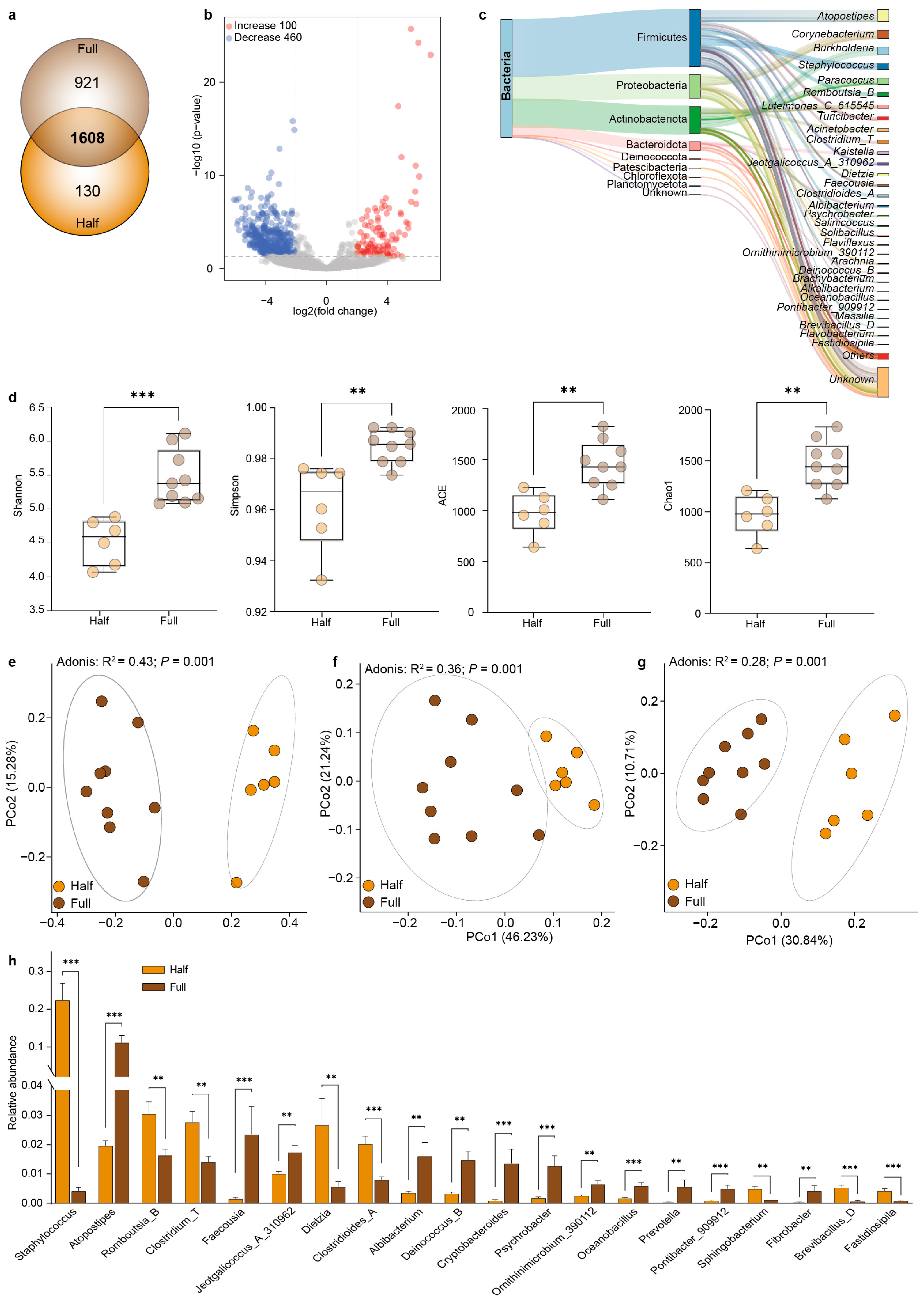
The comparison showed that 1608 OTUs were shared between the Half and Full groups, and a total of 130 and 921 OTUs were unique to the Half group and the Full group, respectively (Figure 2a). The abundances of 100 OTUs and 460 OTUs in the Half group significantly increased and decreased, respectively, in comparison with the Full group (p < 0.05) (Figure 2b). Furthermore, we identified a total of 184 core OTUs that were present in all samples (Figure 2c). These 184 OTUs were assigned to eight phyla, such as Firmicutes, Proteobacteria, Actinobacteota, and Bacteroidota, which accounted for more than 97% microbiota. Moreover, these 184 OTUs were classified into 69 genera, such as Atopostipes spp., Corynebacterium spp., Burkholderia spp., Staphylococcus spp., and Paracoccus spp.
Next, we compared the microbial diversity between the Half and Full groups, and found that the Shannon (p < 0.001), Simpson (p < 0.01), Ace (p < 0.01), and Chao1 (p < 0.01) indices in the Half group were significantly lower than those in the Full group (Figure 2d). The PCoA results revealed the clear and significant separation of microbiota between the Half and Full groups (p < 0.01) based on the Bray–Curtis dissimilarity matrix (Figure 2e), weighted Unifrac distance (Figure 2f), and unweighted Unifrac distance (Figure 2g). We then identified differences in microbial composition during antler velvet regeneration (Figure 2h). The results showed that the relative abundances of Staphylococcus spp., Romboutsia_B, Clostridium_T, Dietzia spp., Clostridioides_A, Sphingobacterium spp., Brevibacillus_D, and Fastidiosipila spp. were significantly increased in the Half group (p < 0.01) compared with the Full group, while the relative abundances of Atopostipes spp., Faecousia spp., Jeotgalicoccus_A_310962, Albibacterium spp., Deinococcus_B, Cryptobacteroides spp., Psychrobacter spp., Ornithinimicrobium_390112, Oceanobacillus spp., Prevotella spp., Pontibacter_909912, and Fibrobacter spp. were lower (p < 0.01).

3.3. Co-Occurrence Network Analysis of the Microbial Interactions

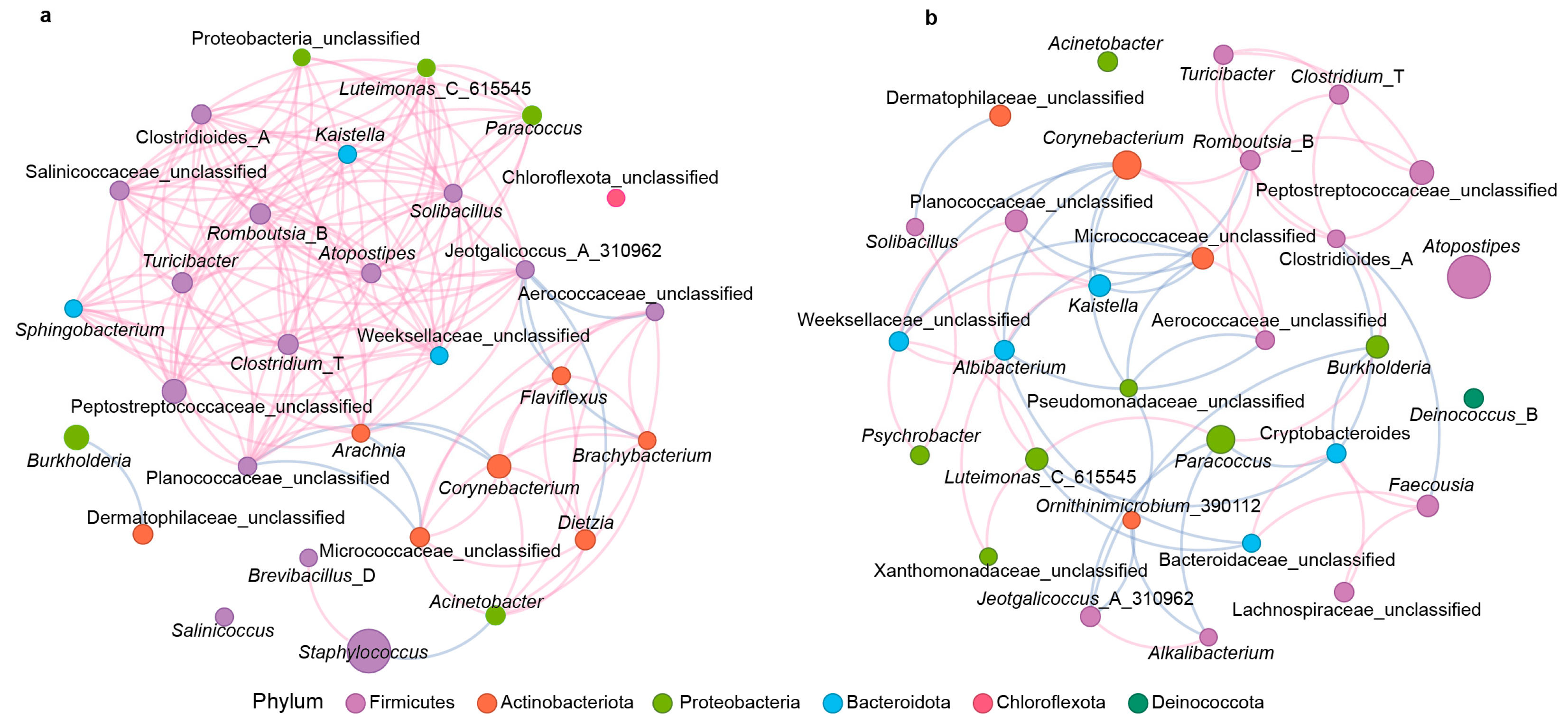
The co-occurrence network showed that there was a strong positive correlation among microbiota in the Half group (148 edges, positive edges = 138, negative edges = 10; Figure 3a), while the co-occurrence network in the Full group was relatively relaxed (64 edges, positive edges = 37, negative edges = 27; Figure 3b). In the largest positive correlation module, a total of 17 microbial genera, such as Clostridides_A, Clostridium_T, Turicibacter, Romboutsia_B, Sphingobacterium, Arachnia, Atopostipes, Solibacillus, Kaistella, Paracoccus, Luteimonas_C_615545, and Jeotgalicoccus_A_310962, showed significant positive correlations in the Half group, while eight genera such as Clostridides_A, Clostridium_T, Corynebacterium, Turicibacter, and Romboutsia_B were positively correlated in the Full group.

3.4. Changes in the Function of Antler Velvet Microbes Between the Two Groups

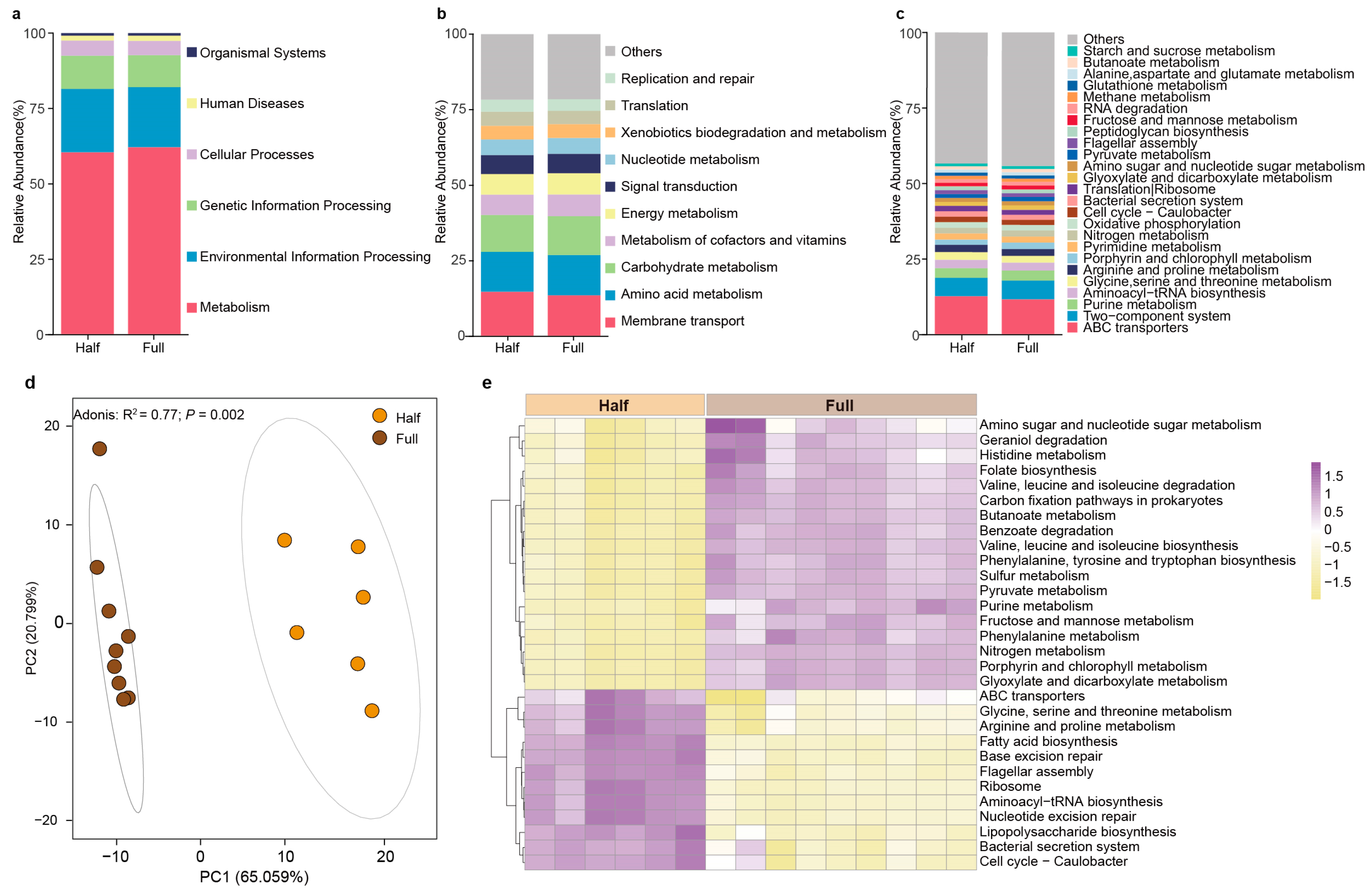
We used Tax4Fun to predict the potential functions of the antler velvet microbiota. The results revealed six pathways in both the Half and Full groups at KEGG level 1, including metabolism, environmental information processing, genetic information processing, cellular processes, human diseases, and organ systems, with metabolism being the most abundant pathway (Figure 4a). At KEGG level 2, the primary predicted pathways included membrane transport, amino acid metabolism, carbohydrate metabolism, metabolism of cofactors and vitamins, energy metabolism, signal transduction, nucleotide metabolism, xenobiotics biodegradation and metabolism, translation, and replication and repair (Figure 4b). At KEGG level 3, the primary predicted pathways included ABC transporters, two-component system, purine metabolism, aminoacyl-tRNA biosynthesis, glycine, serine, and threonine metabolism, arginine and proline metabolism, porphyrin and chlorophyll metabolism, pyrimidine metabolism, nitrogen metabolism, and oxidative phosphorylation (Figure 4c).
We then analyzed the differences in the functions of the antler velvet microbiota between the Half and Full groups at KEGG level 3. The results show that there were distinct differences in microbial functional potential between the Half group and the Full group (Figure 4d, p < 0.05). We found that 30 pathways were significantly different between the Half and Full groups at KEGG level 3, including purine metabolism, geraniol degradation, porphyrin and chlorophyll metabolism, folate biosynthesis, fatty acid biosynthesis, lipopolysaccharide biosynthesis, sulfur metabolism, nitrogen metabolism, pyruvate metabolism, glyoxylate and dicarboxylate metabolism, fructose and mannose metabolism, butanoate metabolism, amino sugar and nucleotide sugar metabolism, valine, leucine and isoleucine degradation, valine, leucine and isoleucine biosynthesis, phenylalanine, tyrosine and tryptophan biosynthesis, phenylalanine metabolism, histidine metabolism, glycine, serine, and threonine metabolism, arginine and proline metabolism, ribosome, bacterial secretion system, ABC transporters, and flagellar assembly (Figure 4e). Among them, the relative abundances of glycine, serine, and threonine metabolism, arginine and proline metabolism, fatty acid biosynthesis, and ABC transporters were significantly increased in the Half group, while five carbohydrate metabolism pathways (pyruvate metabolism, glyoxylate and dicarboxylate metabolism, fructose and mannose metabolism, butanoate metabolism and amino sugar, and nucleotide sugar metabolism) and five amino acid metabolism pathways (valine, leucine, and isoleucine degradation, valine, leucine, and isoleucine biosynthesis, phenylalanine, tyrosine, and tryptophan biosynthesis, phenylalanine metabolism, and histidine metabolism) were significantly increased in the Full group.
Correlation analyses were performed on the differential KEGG pathways and microbial composition at the genus level (Figure S2). The result showed that Staphylococcus spp., Romboutsia_B, Clostridium_T, Dietzia spp., Clostridioides_A, Brevibacillus_D, and Fastidiosipila spp. positively correlated with glycine, serine, and threonine metabolism, arginine and proline metabolism, fatty acid biosynthesis, and ABC transporters (p < 0.05), while Atopostipes spp., Faecousia spp., Albibacterium spp., Cryptobacteroides spp., Psychrobacter spp., Ornithinimicrobium_390112, Oceanobacillus spp., Prevotella spp., Pontibacter_909912, and Fibrobacter spp. positively correlated with five carbohydrate metabolism pathways (pyruvate metabolism, glyoxylate and dicarboxylate metabolism, fructose and mannose metabolism, butanoate metabolism and amino sugar, and nucleotide sugar metabolism) and five amino acid metabolism pathways (valine, leucine, and isoleucine degradation, valine, leucine, and isoleucine biosynthesis, phenylalanine, tyrosine, and tryptophan biosynthesis, phenylalanine metabolism, and histidine metabolism) (p < 0.05).

4. Discussion

In this study, we examined the microbial composition and dynamics of antler velvet during regeneration using 16S rRNA gene sequencing. The prevalent bacterial phyla of antler velvet were Firmicutes, Proteobacteria, Actinobacteriota, and Bacteroidota, which is consistent with the findings of microbial composition on the skin of humans [39], mice [40], dogs [16], pigs [41], sheep [19], and yaks and cattle [42]. We found that the dominant bacterial genera in antler velvet were Staphylococcus spp., Atopostipes spp., Corynebacterium spp., and Burkholderia spp., which is slightly different from previous findings from the skin of other mammals, such as the dominated genera of human skin (Propionibacterium spp., Corynebacterium spp., and Staphylococcus spp.) [39], the udder skin of dairy cows (Corynebacterium spp., Bifidobacterium spp., Staphylococcus spp., and Brachybacterium spp.) [43], and the dorsal and abdominal skin of donkeys (Staphylococcus spp., Planococcus spp., Corynebacterium spp., and Salinicoccus spp.) [44]. This difference is likely related to the living environment (lifestyle, hygiene routine, geographic location, climate, and seasonality) and/or skin physiology (moist, dry, and sebaceous microenvironments) [45,46]. However, the genera Staphylococcus spp. and Corynebacterium spp. were commonly present on the skin of these mammals, suggesting that these bacteria may have a core symbiotic relationship with skin.
We also observed a significant decrease in the microbial diversity of antler velvet in the Half group compared with the Full group. Similar to our findings, previous studies have demonstrated that the microbial diversity of human skin in chronic wounds [47] and burn wounds [48] is also significantly lower than that of healthy skin. Sanjar et al. [49] reported that the microbial diversity of rat burn wounds was lower than that of healthy skin. Similarly, the microbial diversity observed in horse limb and thoracic wounds during the early healing stages (1 week and 2 weeks post-wound) is lower than that observed during the fully healed stage [50]. A decrease in microbial diversity and richness is often associated with skin diseases and inflammation such as psoriasis [51], atopic dermatitis [52], and vitiligo [53]. Furthermore, Gupta et al. (2005) found that Pseudomonas spp. in burn wounds significantly inhibited growth of the opportunistic fungus Candida spp. [54]. It was reported that antler velvet regeneration is associated with an attenuated immune response [8]. These results indicate that the immune response may be an important factor influencing the microbial composition of antler velvet.
Compared to the Full group, the relative abundances of Staphylococcus spp., Romboutsia_B, and Dietzia spp. increased significantly in the Half group, while Atopostipes spp., Psychrobacter spp., and Faecousia spp. increased in the Full group. Similar to our results, Sanjar et al. [49] found that the proportion of Staphylococcus spp. in rat burn wounds was significantly higher than that in sham burns. Hannigan et al. (2014) demonstrated that the relative abundance of Staphylococcus spp. in the center of open fracture wounds increased significantly with the course of injury [55]. The genus Romboutsia has multiple metabolic capacities, such as anaerobic respiration, single amino acid fermentation, and the production of metabolic end products [56]. Recent studies showed that Romboutsia spp. were drastically reduced in adenomatous polyps and cancerous mucosa [57]. Dietzia spp. can colonize human skin and are enriched in lipid metabolism [58,59]. Moreover, the microbiota correlations in the Half group were much more complex than those in the Full group. These results suggest that the cooperation of different microbiota likely affects velvet regeneration. Staphylococcus spp. is a Gram-positive bacterium that inhabits the skin and mucous membranes [60] and has been reported to be negatively correlated with ulcer depth and duration [61] and burn infection [62]. S. epidermidis stimulates skin CD8+ T cells, which enhance keratinocyte progression by upregulating Toll-like receptors and modulating TNF-α signaling [63,64]. Additionally, lipoteichoic acid [15] and trace amines [65] produced by S. epidermidis can act upon keratinocytes to inhibit skin inflammation or antagonize the inhibition of cell migration to accelerate wound healing. However, the growth of S. aureus is associated with severe skin infections, primarily due to the production of superantigens [66,67]. These results suggest functional heterogeneity within Staphylococcus spp. Further investigation is warranted to isolate Staphylococcus species and examine their genomic profiles.
The relative abundances of glycine, serine, and threonine metabolism, arginine and proline metabolism were significantly increased in the Half group in comparison with the Full group, and were also positively correlated with Staphylococcus spp., Romboutsia_B and Dietzia spp. Metabolism of nutrients such as amino acids and lipids promotes skin wound healing and reduces scar formation by providing energy and substrate for repair cells [68]. Amino acids are important nutrients required for wound healing promotion and repair of the damaged skin [69]. Arginine can be hydrolyzed to ornithine, which is further metabolized to proline and polyamine. Proline is an important component of collagen, and polyamine is important for cell proliferation and differentiation [70,71]. Previous findings have also shown that the addition of amino acid precursors of collagen (proline, lysine, glycine, and leucine) can promote and accelerate wound healing [72]. These findings suggest that interactions among Staphylococcus spp., Romboutsia spp., and Dietzia spp., and amino acids (such as arginine and proline) can affect wound healing and regeneration of antler velvet. Atopostipes spp. are lactic acid bacteria that can metabolize glucose to formate, acetate, and lactate, and valine to branched-chain fatty acids, which are abundant in the skin and can inhibit the growth of potential pathogens [73,74,75,76]. Psychrobacter spp. are prevalent on the skin of humpback whales, and changes in their prevalence seem to be related to changes in the animal’s metabolic state [77]. Faecousia spp. are often detected in animal feces and are related to carbohydrate degradation [78]. Consistently, the metabolism of carbohydrates was also enhanced in the Full group, suggesting that these microbes might influence the carbohydrate metabolism of antler velvet during the later stages of regeneration. These results indicate possible shifts in the metabolic profiles of the antler velvet microbiota, highlighting the role of the interaction between specific microbiota and amino acids in antler velvet regeneration.

5. Conclusions

Here, we characterized and compared the bacterial communities that are present during the regeneration of antler velvet. Although common microbiota components were observed between antler velvet and the skin of other mammals, antler velvet was also inhabited by a slightly distinct microbiota. The immune response is likely an important factor influencing the dynamics of the antler velvet microbiota. The significantly changed microbial diversity, composition, and potential functions suggest a role for Staphylococcus and arginine and proline metabolism during antler velvet regeneration. Our results expand the current understanding of the link between microbiota and skin regeneration.

Supplementary Materials

The following supporting information can be downloaded at: https://www.mdpi.com/article/10.3390/microorganisms13010036/s1, Table S1. Summarizes data regarding the OTUs and microbial phylum, family, and genus levels. Figure S1. The Half (a) and Full (b) regeneration periods of antler velvet. Figure S2. Correlation analysis among differential pathways and microbial genera.

Author Contributions

Conceptualization, Z.L.; resources, Y.Z. and Z.L.; investigation, Y.L., Y.Z., B.Y., S.Y., S.L. and R.D.; formal analysis, Y.L., Y.Z., H.S. and B.Y.; writing—original draft preparation, Y.L., Y.Z., A.-D.G.W., B.Y. and H.S.; writing—review and editing, H.S. and Z.L.; funding acquisition, Z.L. All authors have read and agreed to the published version of the manuscript.

Funding

This research was supported by the Natural Science Foundation of Jilin Province (20240101012JJ), the National Natural Science Foundation of China (32122083), the Innovation and Entrepreneurship Talent Program from Jilin Province (2023QN02), and the Funding of Joint International Research Laboratory of Modern Agricultural Technology, Ministry of Education (D5110230222).

Data Availability Statement

The sequences in the present study were deposited in the SRA database under accession number SRP520253.

Acknowledgments

We are grateful for the eSwabs provided by Copan Italia SpA Co., Ltd.

Conflicts of Interest

The authors declare no conflicts of interest.

References

  1. Proksch, E.; Brandner, J.M.; Jensen, J.M. The skin: An indispensable barrier. Exp. Dermatol. 2008, 17, 1063–1072. [Google Scholar] [CrossRef] [PubMed]
  2. Stoica, A.E.; Grumezescu, A.M.; Hermenean, A.O.; Andronescu, E.; Vasile, B.S. Scar-free healing: Current concepts and future perspectives. Nanomaterials 2020, 10, 2179. [Google Scholar] [CrossRef] [PubMed]
  3. Monavarian, M.; Kader, S.; Moeinzadeh, S.; Jabbari, E. Regenerative scar-free skin wound healing. Tissue Eng. Part B Rev. 2019, 25, 294–311. [Google Scholar] [CrossRef] [PubMed]
  4. Sen, C.K.; Gordillo, G.M.; Roy, S.; Kirsner, R.; Lambert, L.; Hunt, T.K.; Gottrup, F.; Gurtner, G.C.; Longaker, M.T. Human skin wounds: A major and snowballing threat to public health and the economy. Wound Repair Regen. 2009, 17, 763–771. [Google Scholar] [CrossRef]
  5. Mascharak, S.; desJardins-Park, H.E.; Davitt, M.F.; Griffin, M.; Borrelli, M.R.; Moore, A.L.; Chen, K.; Duoto, B.; Chinta, M.; Foster, D.S.; et al. Preventing Engrailed-1 activation in fibroblasts yields wound regeneration without scarring. Science 2021, 372, eaba2374. [Google Scholar] [CrossRef]
  6. Greenstein, E.S.; Falone, W.; Patterson, T.; Cesario, K.; Mitchell, L.; Martel, T.; Rivera, J.; Vooijs, M.; Norton, S. Treating chronic wounds in an acute care setting: The forgotten diagnosis. Wound Manag. Prev. 2024, 70. [Google Scholar] [CrossRef]
  7. Billingham, R.E.; Mangold, R.; Silvers, W.K. The neogenesis of skin in the antlers of deer. Ann. N. Y. Acad. Sci. 1959, 83, 491–498. [Google Scholar] [CrossRef]
  8. Sinha, S.; Sparks, H.D.; Labit, E.; Robbins, H.N.; Gowing, K.; Jaffer, A.; Kutluberk, E.; Arora, R.; Raredon, M.S.B.; Cao, L.; et al. Fibroblast inflammatory priming determines regenerative versus fibrotic skin repair in reindeer. Cell 2022, 185, 4717–4736.e4725. [Google Scholar] [CrossRef]
  9. Rong, X.; Zhang, G.; Yang, Y.; Gao, C.; Chu, W.; Sun, H.; Wang, Y.; Li, C. Transplanted antler stem cells stimulated regenerative healing of radiation-induced cutaneous wounds in rats. Cell Transplant. 2020, 29, 963689720951549. [Google Scholar] [CrossRef]
  10. Zhang, G.; Wang, D.; Ren, J.; Li, J.; Guo, Q.; Shi, L.; Li, C. Antler stem cell-derived exosomes promote regenerative wound healing via fibroblast-to-myofibroblast transition inhibition. J. Biol. Eng. 2023, 17, 67. [Google Scholar] [CrossRef]
  11. Jiang, W.; Zhang, J.; Zhang, X.; Fan, C.; Huang, J. VAP-PLGA microspheres (VAP-PLGA) promote adipose-derived stem cells (ADSCs)-induced wound healing in chronic skin ulcers in mice via PI3K/Akt/HIF-1α pathway. Bioengineered 2021, 12, 10264–10284. [Google Scholar] [CrossRef] [PubMed]
  12. Kong, H.H.; Segre, J.A. Skin microbiome: Looking back to move forward. J. Investig. Dermatol. 2012, 132, 933–939. [Google Scholar] [CrossRef] [PubMed]
  13. Chen, Y.E.; Fischbach, M.A.; Belkaid, Y. Skin microbiota-host interactions. Nature 2018, 553, 427–436. [Google Scholar] [CrossRef] [PubMed]
  14. Grice, E.A.; Kong, H.H.; Renaud, G.; Young, A.C.; Bouffard, G.G.; Blakesley, R.W.; Wolfsberg, T.G.; Turner, M.L.; Segre, J.A. A diversity profile of the human skin microbiota. Genome Res. 2008, 18, 1043–1050. [Google Scholar] [CrossRef]
  15. Lai, Y.; Di Nardo, A.; Nakatsuji, T.; Leichtle, A.; Yang, Y.; Cogen, A.L.; Wu, Z.R.; Hooper, L.V.; Schmidt, R.R.; von Aulock, S.; et al. Commensal bacteria regulate Toll-like receptor 3-dependent inflammation after skin injury. Nat. Med. 2009, 15, 1377–1382. [Google Scholar] [CrossRef]
  16. Cuscó, A.; Sánchez, A.; Altet, L.; Ferrer, L.; Francino, O. Individual signatures define canine skin microbiota composition and variability. Front. Vet. Sci. 2017, 4, 6. [Google Scholar] [CrossRef]
  17. Zeng, B.; Zhao, J.; Guo, W.; Zhang, S.; Hua, Y.; Tang, J.; Kong, F.; Yang, X.; Fu, L.; Liao, K.; et al. High-altitude living shapes the skin microbiome in humans and pigs. Front. Microbiol. 2017, 8, 1929. [Google Scholar] [CrossRef]
  18. Manus, M.B.; Yu, J.J.; Park, L.P.; Mueller, O.; Windsor, S.C.; Horvath, J.E.; Nunn, C.L. Environmental influences on the skin microbiome of humans and cattle in rural Madagascar. Evol. Med. Public Health 2017, 2017, 144–153. [Google Scholar] [CrossRef]
  19. Jost, S.M.; Cardona, L.; Rohrbach, E.; Mathis, A.; Holliger, C.; Verhulst, N.O. Environment rather than breed or body site shapes the skin bacterial community of healthy sheep as revealed by metabarcoding. Vet. Dermatol. 2024, 35, 273–283. [Google Scholar] [CrossRef]
  20. Kloos, W.E.; Zimmerman, R.J.; Smith, R.F. Preliminary studies on the characterization and distribution of Staphylococcus and Micrococcus species on animal skin. Appl. Environ. Microbiol. 1976, 31, 53–59. [Google Scholar] [CrossRef]
  21. Wang, G.; Sweren, E.; Liu, H.; Wier, E.; Alphonse, M.P.; Chen, R.; Islam, N.; Li, A.; Xue, Y.; Chen, J.; et al. Bacteria induce skin regeneration via IL-1β signaling. Cell Host Microbe 2021, 29, 777–791.e776. [Google Scholar] [CrossRef] [PubMed]
  22. Saheb Kashaf, S.; Proctor, D.M.; Deming, C.; Saary, P.; Hölzer, M.; Taylor, M.E.; Kong, H.H.; Segre, J.A.; Almeida, A.; Finn, R.D. Integrating cultivation and metagenomics for a multi-kingdom view of skin microbiome diversity and functions. Nat. Microbiol. 2022, 7, 169–179. [Google Scholar] [CrossRef] [PubMed]
  23. Bjerre, R.D.; Hugerth, L.W.; Boulund, F.; Seifert, M.; Johansen, J.D.; Engstrand, L. Effects of sampling strategy and DNA extraction on human skin microbiome investigations. Sci. Rep. 2019, 9, 17287. [Google Scholar] [CrossRef] [PubMed]
  24. Li, Z.; Bai, X.; Peng, T.; Yi, X.; Luo, L.; Yang, J.; Liu, J.; Wang, Y.; He, T.; Wang, X.; et al. New insights into the skin microbial communities and skin aging. Front. Microbiol. 2020, 11, 565549. [Google Scholar] [CrossRef]
  25. Mekadim, C.; Skalnikova, H.K.; Cizkova, J.; Cizkova, V.; Palanova, A.; Horak, V.; Mrazek, J. Dysbiosis of skin microbiome and gut microbiome in melanoma progression. BMC Microbiol. 2022, 22, 63. [Google Scholar] [CrossRef]
  26. Takahashi, S.; Tomita, J.; Nishioka, K.; Hisada, T.; Nishijima, M. Development of a prokaryotic universal primer for simultaneous analysis of bacteria and archaea using next-generation sequencing. PLoS ONE 2014, 9, e105592. [Google Scholar] [CrossRef]
  27. Kennedy, K.; Hall, M.W.; Lynch, M.D.; Moreno-Hagelsieb, G.; Neufeld, J.D. Evaluating bias of illumina-based bacterial 16S rRNA gene profiles. Appl. Environ. Microbiol. 2014, 80, 5717–5722. [Google Scholar] [CrossRef]
  28. Ross, A.A.; Müller, K.M.; Weese, J.S.; Neufeld, J.D. Comprehensive skin microbiome analysis reveals the uniqueness of human skin and evidence for phylosymbiosis within the class Mammalia. Proc. Natl. Acad. Sci. USA 2018, 115, E5786–E5795. [Google Scholar] [CrossRef]
  29. Chen, S.; Zhou, Y.; Chen, Y.; Gu, J. fastp: An ultra-fast all-in-one FASTQ preprocessor. Bioinformatics 2018, 34, i884–i890. [Google Scholar] [CrossRef]
  30. Magoč, T.; Salzberg, S.L. FLASH: Fast length adjustment of short reads to improve genome assemblies. Bioinformatics 2011, 27, 2957–2963. [Google Scholar] [CrossRef]
  31. Caporaso, J.G.; Kuczynski, J.; Stombaugh, J.; Bittinger, K.; Bushman, F.D.; Costello, E.K.; Fierer, N.; Peña, A.G.; Goodrich, J.K.; Gordon, J.I.; et al. QIIME allows analysis of high-throughput community sequencing data. Nat. Methods 2010, 7, 335–336. [Google Scholar] [CrossRef] [PubMed]
  32. Edgar, R.C. UPARSE: Highly accurate OTU sequences from microbial amplicon reads. Nat. Methods 2013, 10, 996–998. [Google Scholar] [CrossRef] [PubMed]
  33. Edgar, R.C.; Haas, B.J.; Clemente, J.C.; Quince, C.; Knight, R. UCHIME improves sensitivity and speed of chimera detection. Bioinformatics 2011, 27, 2194–2200. [Google Scholar] [CrossRef] [PubMed]
  34. Henderson, G.; Yilmaz, P.; Kumar, S.; Forster, R.J.; Kelly, W.J.; Leahy, S.C.; Guan, L.L.; Janssen, P.H. Improved taxonomic assignment of rumen bacterial 16S rRNA sequences using a revised SILVA taxonomic framework. PeerJ 2019, 7, e6496. [Google Scholar] [CrossRef]
  35. Liu, C.; Cui, Y.; Li, X.; Yao, M. microeco: An R package for data mining in microbial community ecology. FEMS Microbiol. Ecol. 2021, 97, fiaa255. [Google Scholar] [CrossRef]
  36. Aßhauer, K.P.; Wemheuer, B.; Daniel, R.; Meinicke, P. Tax4Fun: Predicting functional profiles from metagenomic 16S rRNA data. Bioinformatics 2015, 31, 2882–2884. [Google Scholar] [CrossRef]
  37. Segata, N.; Izard, J.; Waldron, L.; Gevers, D.; Miropolsky, L.; Garrett, W.S.; Huttenhower, C. Metagenomic biomarker discovery and explanation. Genome Biol. 2011, 12, R60. [Google Scholar] [CrossRef]
  38. Bastian, M.; Heymann, S.; Jacomy, M. Gephi: An open source software for exploring and manipulating networks. In Proceedings of the International AAAI Conference on Web and Social Media, San Jose, CA, USA, 17–20 May 2009; pp. 361–362. [Google Scholar]
  39. Grice, E.A.; Kong, H.H.; Conlan, S.; Deming, C.B.; Davis, J.; Young, A.C.; Bouffard, G.G.; Blakesley, R.W.; Murray, P.R.; Green, E.D.; et al. Topographical and temporal diversity of the human skin microbiome. Science 2009, 324, 1190–1192. [Google Scholar] [CrossRef]
  40. Grice, E.A.; Snitkin, E.S.; Yockey, L.J.; Bermudez, D.M.; Liechty, K.W.; Segre, J.A. Longitudinal shift in diabetic wound microbiota correlates with prolonged skin defense response. Proc. Natl. Acad. Sci. USA 2010, 107, 14799–14804. [Google Scholar] [CrossRef]
  41. McIntyre, M.K.; Peacock, T.J.; Akers, K.S.; Burmeister, D.M. Initial characterization of the pig skin bacteriome and its effect on In vitro models of wound healing. PLoS ONE 2016, 11, e0166176. [Google Scholar] [CrossRef]
  42. Zhang, J.; Ahmad, A.A.; Jia, Y.; Dingkao, R.; Du, M.; Liang, Z.; Zheng, J.; Bature, I.; Yan, P.; Salekdeh, G.H.; et al. Comparison of dynamics of udder skin microbiota from grazing yak and cattle during the perinatal period on the Qinghai-Tibetan Plateau. Front. Vet. Sci. 2022, 9, 864057. [Google Scholar] [CrossRef] [PubMed]
  43. Ekman, L.; Bagge, E.; Nyman, A.; Persson Waller, K.; Pringle, M.; Segerman, B. A shotgun metagenomic investigation of the microbiota of udder cleft dermatitis in comparison to healthy skin in dairy cows. PLoS ONE 2020, 15, e0242880. [Google Scholar] [CrossRef] [PubMed]
  44. Ma, Q.; Yue, Y.; Kou, X.; Hou, W.; Wang, M.; Yang, X.; Liu, G.; Li, Y.; Wang, C. Dynamic distribution of skin microorganisms in donkeys at different ages and various sites of the body. Animals 2023, 13, 1566. [Google Scholar] [CrossRef] [PubMed]
  45. Chen, Y.E.; Tsao, H. The skin microbiome: Current perspectives and future challenges. J. Am. Acad. Dermatol. 2013, 69, 143–155. [Google Scholar] [CrossRef]
  46. Boxberger, M.; Cenizo, V.; Cassir, N.; La Scola, B. Challenges in exploring and manipulating the human skin microbiome. Microbiome 2021, 9, 125. [Google Scholar] [CrossRef]
  47. Gardiner, M.; Vicaretti, M.; Sparks, J.; Bansal, S.; Bush, S.; Liu, M.; Darling, A.; Harry, E.; Burke, C.M. A longitudinal study of the diabetic skin and wound microbiome. PeerJ 2017, 5, e3543. [Google Scholar] [CrossRef]
  48. Lima, K.M.; Davis, R.R.; Liu, S.Y.; Greenhalgh, D.G.; Tran, N.K. Longitudinal profiling of the burn patient cutaneous and gastrointestinal microbiota: A pilot study. Sci. Rep. 2021, 11, 10667. [Google Scholar] [CrossRef]
  49. Sanjar, F.; Weaver, A.J.; Peacock, T.J.; Nguyen, J.Q.; Brandenburg, K.S.; Leung, K.P. Identification of metagenomics structure and function associated with temporal changes in rat (Rattus norvegicus) skin microbiome during health and cutaneous burn. J. Burn Care Res. 2020, 41, 347–358. [Google Scholar] [CrossRef]
  50. Kamus, L.J.; Theoret, C.; Costa, M.C. Use of next generation sequencing to investigate the microbiota of experimentally induced wounds and the effect of bandaging in horses. PLoS ONE 2018, 13, e0206989. [Google Scholar] [CrossRef]
  51. Alekseyenko, A.V.; Perez-Perez, G.I.; De Souza, A.; Strober, B.; Gao, Z.; Bihan, M.; Li, K.; Methé, B.A.; Blaser, M.J. Community differentiation of the cutaneous microbiota in psoriasis. Microbiome 2013, 1, 31. [Google Scholar] [CrossRef]
  52. Williams, M.R.; Gallo, R.L. The role of the skin microbiome in atopic dermatitis. Curr. Allergy Asthma Rep. 2015, 15, 65. [Google Scholar] [CrossRef] [PubMed]
  53. Ganju, P.; Nagpal, S.; Mohammed, M.H.; Nishal Kumar, P.; Pandey, R.; Natarajan, V.T.; Mande, S.S.; Gokhale, R.S. Microbial community profiling shows dysbiosis in the lesional skin of Vitiligo subjects. Sci. Rep. 2016, 6, 18761. [Google Scholar] [CrossRef] [PubMed]
  54. Gupta, N.; Haque, A.; Mukhopadhyay, G.; Narayan, R.P.; Prasad, R. Interactions between bacteria and Candida in the burn wound. Burns 2005, 31, 375–378. [Google Scholar] [CrossRef] [PubMed]
  55. Hannigan, G.D.; Hodkinson, B.P.; McGinnis, K.; Tyldsley, A.S.; Anari, J.B.; Horan, A.D.; Grice, E.A.; Mehta, S. Culture-independent pilot study of microbiota colonizing open fractures and association with severity, mechanism, location, and complication from presentation to early outpatient follow-up. J. Orthop. Res. 2014, 32, 597–605. [Google Scholar] [CrossRef] [PubMed]
  56. Gerritsen, J.; Hornung, B.; Ritari, J.; Paulin, L.; Rijkers, G.T.; Schaap, P.J.; de Vos, W.M.; Smidt, H. A comparative and functional genomics analysis of the genus Romboutsia provides insight into adaptation to an intestinal lifestyle. BioRxiv 2019. [Google Scholar] [CrossRef]
  57. Mangifesta, M.; Mancabelli, L.; Milani, C.; Gaiani, F.; de’Angelis, N.; de’Angelis, G.L.; van Sinderen, D.; Ventura, M.; Turroni, F. Mucosal microbiota of intestinal polyps reveals putative biomarkers of colorectal cancer. Sci. Rep. 2018, 8, 13974. [Google Scholar] [CrossRef]
  58. Dekio, I.; Sakamoto, M.; Hayashi, H.; Amagai, M.; Suematsu, M.; Benno, Y. Characterization of skin microbiota in patients with atopic dermatitis and in normal subjects using 16S rRNA gene-based comprehensive analysis. J. Med. Microbiol. 2007, 56, 1675–1683. [Google Scholar] [CrossRef]
  59. Fang, H.; Xu, J.B.; Nie, Y.; Wu, X.L. Pan-genomic analysis reveals that the evolution of Dietzia species depends on their living habitats. Environ. Microbiol. 2021, 23, 861–877. [Google Scholar] [CrossRef]
  60. Paharik, A.E.; Horswill, A.R. The Staphylococcal biofilm: Adhesins, regulation, and host response. Microbiol. Spectr. 2016, 4. [Google Scholar] [CrossRef]
  61. Gardner, S.E.; Hillis, S.L.; Heilmann, K.; Segre, J.A.; Grice, E.A. The neuropathic diabetic foot ulcer microbiome is associated with clinical factors. Diabetes 2013, 62, 923–930. [Google Scholar] [CrossRef]
  62. Plichta, J.K.; Gao, X.; Lin, H.; Dong, Q.; Toh, E.; Nelson, D.E.; Gamelli, R.L.; Grice, E.A.; Radek, K.A. Cutaneous burn injury promotes shifts in the bacterial microbiome in autologous donor skin: Implications for skin grafting outcomes. Shock 2017, 48, 441–448. [Google Scholar] [CrossRef] [PubMed]
  63. Linehan, J.L.; Harrison, O.J.; Han, S.J.; Byrd, A.L.; Vujkovic-Cvijin, I.; Villarino, A.V.; Sen, S.K.; Shaik, J.; Smelkinson, M.; Tamoutounour, S.; et al. Non-classical immunity controls microbiota impact on skin immunity and tissue repair. Cell 2018, 172, 784–796.e718. [Google Scholar] [CrossRef] [PubMed]
  64. Naik, S.; Bouladoux, N.; Linehan, J.L.; Han, S.J.; Harrison, O.J.; Wilhelm, C.; Conlan, S.; Himmelfarb, S.; Byrd, A.L.; Deming, C.; et al. Commensal-dendritic-cell interaction specifies a unique protective skin immune signature. Nature 2015, 520, 104–108. [Google Scholar] [CrossRef] [PubMed]
  65. Luqman, A.; Muttaqin, M.Z.; Yulaipi, S.; Ebner, P.; Matsuo, M.; Zabel, S.; Tribelli, P.M.; Nieselt, K.; Hidayati, D.; Götz, F. Trace amines produced by skin bacteria accelerate wound healing in mice. Commun. Biol. 2020, 3, 277. [Google Scholar] [CrossRef]
  66. Kim, C.K.; Karau, M.J.; Greenwood-Quaintance, K.E.; Tilahun, A.Y.; Krogman, A.; David, C.S.; Pritt, B.S.; Patel, R.; Rajagopalan, G. Superantigen-producing Staphylococcus aureus elicits systemic immune activation in a murine wound colonization model. Toxins 2015, 7, 5308–5319. [Google Scholar] [CrossRef]
  67. Vojtov, N.; Ross, H.F.; Novick, R.P. Global repression of exotoxin synthesis by Staphylococcal superantigens. Proc. Natl. Acad. Sci. USA 2002, 99, 10102–10107. [Google Scholar] [CrossRef]
  68. Wang, Z.; Zhao, F.; Xu, C.; Zhang, Q.; Ren, H.; Huang, X.; He, C.; Ma, J.; Wang, Z. Metabolic reprogramming in skin wound healing. Burn. Trauma 2024, 12, tkad047. [Google Scholar] [CrossRef]
  69. Solano, F. Metabolism and functions of amino acids in the skin. Adv. Exp. Med. Biol. 2020, 1265, 187–199. [Google Scholar] [CrossRef]
  70. Guccione, E.; Richard, S. The regulation, functions and clinical relevance of arginine methylation. Nat. Rev. Mol. Cell Biol. 2019, 20, 642–657. [Google Scholar] [CrossRef]
  71. Kämpfer, H.; Pfeilschifter, J.; Frank, S. Expression and activity of arginase isoenzymes during normal and diabetes-impaired skin repair. J. Investig. Dermatol. 2003, 121, 1544–1551. [Google Scholar] [CrossRef]
  72. Mariggiò, M.A.; Cassano, A.; Vinella, A.; Vincenti, A.; Fumarulo, R.; Lo Muzio, L.; Maiorano, E.; Ribatti, D.; Favia, G. Enhancement of fibroblast proliferation, collagen biosynthesis and production of growth factors as a result of combining sodium hyaluronate and aminoacids. Int. J. Immunopathol. Pharmacol. 2009, 22, 485–492. [Google Scholar] [CrossRef] [PubMed]
  73. Nicolaides, N.J.S. Skin lipids: Their biochemical uniqueness: Unlike internal organs, the skin biosynthesizes and excretes unusual fat soluble substances. Science 1974, 186, 19–26. [Google Scholar] [CrossRef] [PubMed]
  74. Keseler, I.M.; Mackie, A.; Peralta-Gil, M.; Santos-Zavaleta, A.; Gama-Castro, S.; Bonavides-Martínez, C.; Fulcher, C.; Huerta, A.M.; Kothari, A.; Krummenacker, M.; et al. EcoCyc: Fusing model organism databases with systems biology. Nucleic Acids Res. 2013, 41, D605–D612. [Google Scholar] [CrossRef]
  75. Cho, S.; Hwang, O.; Park, S. Effect of dietary protein levels on composition of odorous compounds and bacterial ecology in pig manure. Asian-Australas. J. Anim. Sci. 2015, 28, 1362–1370. [Google Scholar] [CrossRef]
  76. Cotta, M.A.; Whitehead, T.R.; Collins, M.D.; Lawson, P.A. Atopostipes suicloacale gen. nov., sp. nov., isolated from an underground swine manure storage pit. Anaerobe 2004, 10, 191–195. [Google Scholar] [CrossRef]
  77. Apprill, A.; Robbins, J.; Eren, A.M.; Pack, A.A.; Reveillaud, J.; Mattila, D.; Moore, M.; Niemeyer, M.; Moore, K.M.; Mincer, T.J. Humpback whale populations share a core skin bacterial community: Towards a health index for marine mammals? PLoS ONE 2014, 9, e90785. [Google Scholar] [CrossRef]
  78. Yang, J.; Fan, Y.; Jin, R.; Peng, Y.; Chai, J.; Wei, X.; Zhao, Y.; Deng, F.; Zhao, J.; Li, Y.J.F. Exploring the intestinal microbial community of lantang pigs through metagenome-assembled genomes and carbohydrate degradation Genes. Fermentation 2024, 10, 207. [Google Scholar] [CrossRef]
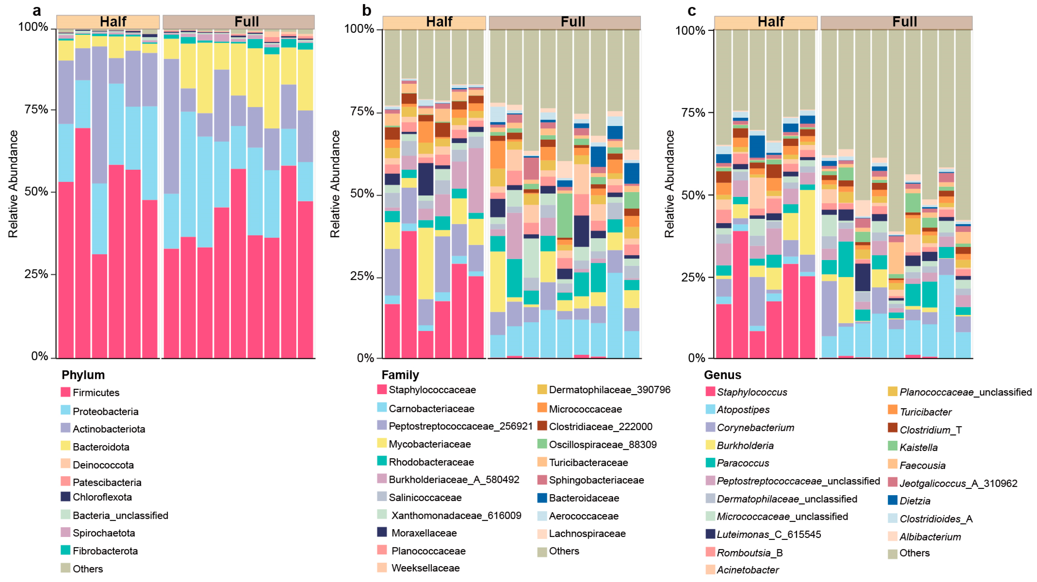
Figure 1. The microbial composition of antler velvet at the (a) phylum, (b) family, and (c) genus levels between the Half and Full groups.
Microorganisms 13 00036 g001
Figure 2. Comparison of microbial community composition between the Half and Full groups. (a) Comparison of the number of OTUs between the Half and Full groups. (b) Volcano plot showing the significantly changed OTUs between the Half and Full groups. (c) Taxonomic classification of the core OTUs. (d) Comparison of alpha diversity indices between the Half and Full groups. PCoA revealing differences in the microbial community based on (e) Bray–Curtis dissimilarity matrix, (f) weighted Unifrac distance, and (g) unweighted Unifrac distance. (h) Significantly different genera between the Half and Full groups. ** p < 0.01, *** p < 0.001.
Microorganisms 13 00036 g002
Figure 3. The co-occurrence network of the microbial community at the genus level based on Spearman correlation in the Half (a) and Full (b) groups (r > 0.6 or r < −0.6, p < 0.05). The pink and blue lines indicate positive and negative correlations, respectively.
Microorganisms 13 00036 g003
Figure 4. Comparison of antler velvet microbial function between the Half and Full groups. The relative abundances of metabolic pathways at KEGG level 1 (a), KEGG level 2 (b), and KEGG level 3 (c); (d) PCoA revealing variations in KEGG level 3 between the Half and Full groups; and (e) the significantly changed pathways based on LEfSe analysis at level 3 (LDA score [log 10] > 2.5, p < 0.05).
Microorganisms 13 00036 g004
Disclaimer/Publisher’s Note: The statements, opinions and data contained in all publications are solely those of the individual author(s) and contributor(s) and not of MDPI and/or the editor(s). MDPI and/or the editor(s) disclaim responsibility for any injury to people or property resulting from any ideas, methods, instructions or products referred to in the content.