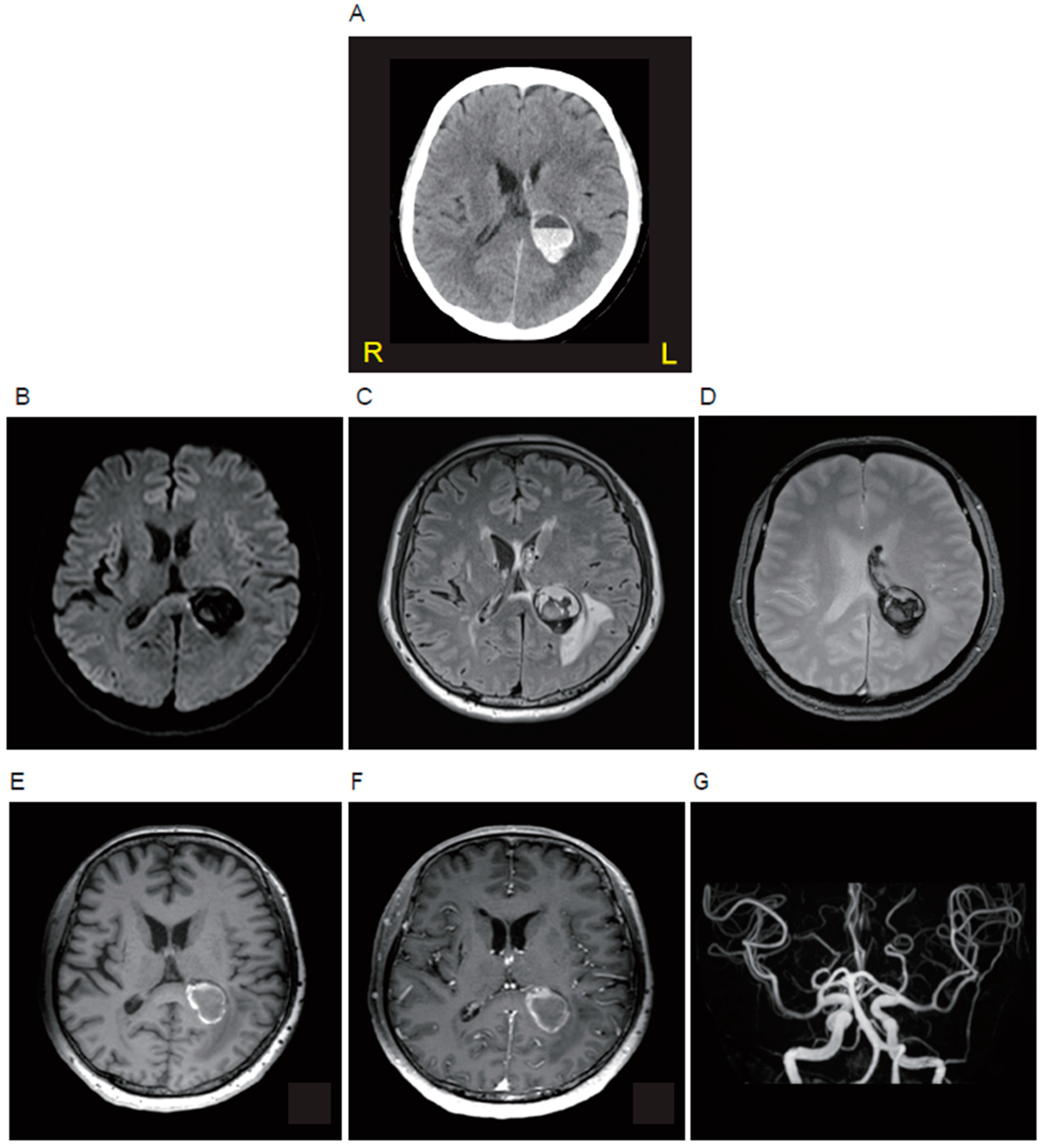
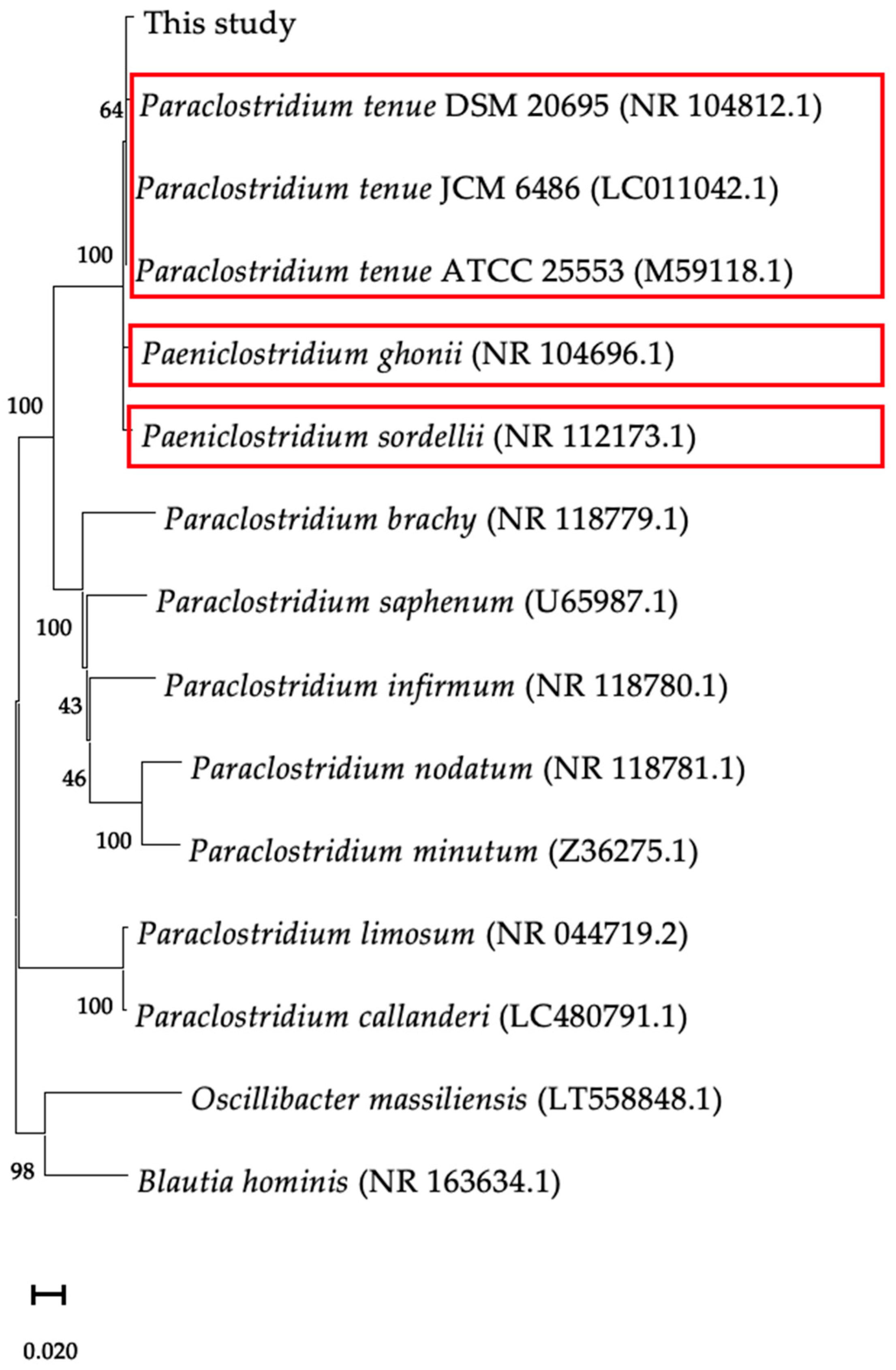
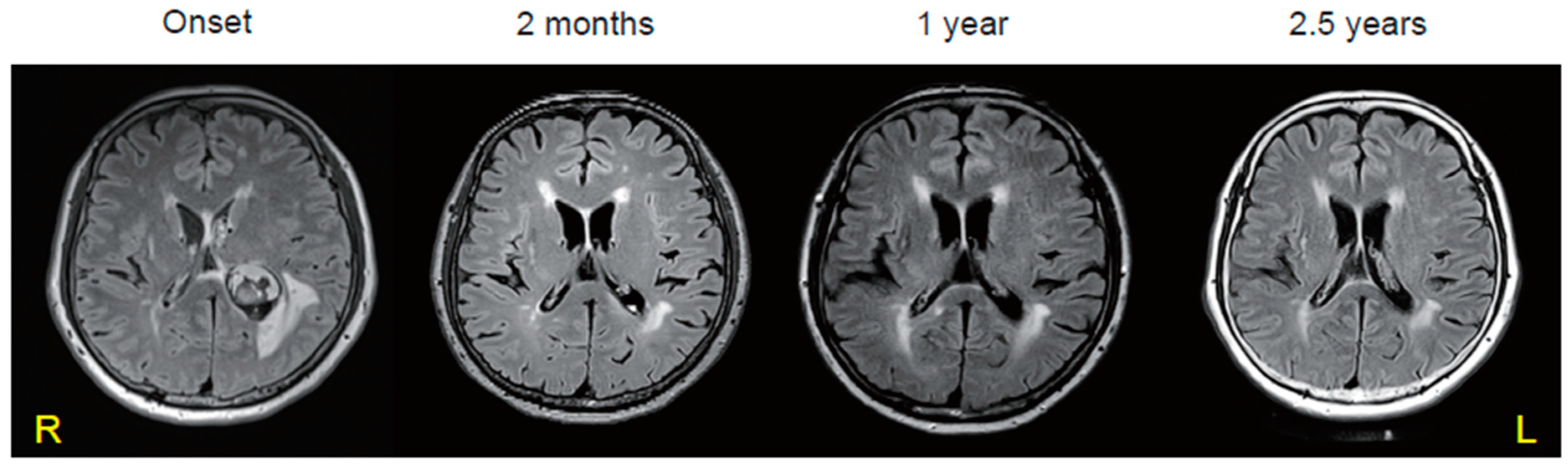
Paraclostridium tenue Causing an Anaerobic Brain Abscess Identified by Whole-Metagenome Sequencing: A Case Report




Abstract
Share and Cite
Chiba, T.; Hattori, Y.; Motooka, D.; Tanaka, T.; Ihara, M. Paraclostridium tenue Causing an Anaerobic Brain Abscess Identified by Whole-Metagenome Sequencing: A Case Report. Microorganisms 2024, 12, 1692. https://doi.org/10.3390/microorganisms12081692
Chiba T, Hattori Y, Motooka D, Tanaka T, Ihara M. Paraclostridium tenue Causing an Anaerobic Brain Abscess Identified by Whole-Metagenome Sequencing: A Case Report. Microorganisms. 2024; 12(8):1692. https://doi.org/10.3390/microorganisms12081692
Chicago/Turabian StyleChiba, Tetsuya, Yorito Hattori, Daisuke Motooka, Tomotaka Tanaka, and Masafumi Ihara. 2024. "Paraclostridium tenue Causing an Anaerobic Brain Abscess Identified by Whole-Metagenome Sequencing: A Case Report" Microorganisms 12, no. 8: 1692. https://doi.org/10.3390/microorganisms12081692
APA StyleChiba, T., Hattori, Y., Motooka, D., Tanaka, T., & Ihara, M. (2024). Paraclostridium tenue Causing an Anaerobic Brain Abscess Identified by Whole-Metagenome Sequencing: A Case Report. Microorganisms, 12(8), 1692. https://doi.org/10.3390/microorganisms12081692