Article

A Study on the Effect of Various Media and the Supplementation of Organic Compounds on the Enhanced Production of Astaxanthin from Haematococcus lacustris (Girod—Chantrans) Rostafinski (Chlorophyta)

1
Department of Biology, Soonchunhyang University, Asan 31538, Chungcheongnam-do, Republic of Korea
2
Korea Fisheries Resources Agency East Sea Branch, Samho-ro, Buk-gu, Pohang 37601, Gyungsangbuk-do, Republic of Korea
3
Department of Environmental Health Science, Soonchunhyang University, Asan 31538, Chungcheongnam-do, Republic of Korea
4
Department of Sports Science, Soonchunhyang University, Asan 31538, Chungcheongnam-do, Republic of Korea
5
Department of Sports Medicine, Soonchunhyang University, Asan 31538, Chungcheongnam-do, Republic of Korea
6
AlgaeBio, Inc., Asan 31459, Chungcheongnam-do, Republic of Korea
7
Research Institute for Basic Science, Soonchunhyang University, Asan 31538, Chungcheongnam-do, Republic of Korea
*
Authors to whom correspondence should be addressed.
These authors contributed equally to this work.
Microorganisms 2024, 12(6), 1040; https://doi.org/10.3390/microorganisms12061040
Submission received: 17 April 2024 / Revised: 14 May 2024 / Accepted: 20 May 2024 / Published: 21 May 2024

Abstract

:
Natural astaxanthin is in high demand due to its multiple health benefits. The microalga Haematococcus lacustris has been used for the commercial production of astaxanthin. In this study, we investigated the effects of six different media with and without a nitrogen source and supplementation with nine organic compounds on the growth and astaxanthin accumulation of H. lacustris. The highest astaxanthin contents were observed in cultures of H. lacustris in Jaworski’s medium (JM), with a level of 9.099 mg/L in JM with a nitrogen source supplemented with leucine (0.65 g/L) and of 20.484 mg/L in JM without a nitrogen source supplemented with sodium glutamate (0.325 g/L). Six of the nine organic compounds examined (leucine, lysine, alanine, sodium glutamate, glutamine, and cellulose) enhanced the production of astaxanthin in H. lacustris, while malic acid, benzoic acid, and maltose showed no beneficial effects.

1. Introduction

Astaxanthin (3,3′-dihydroxy-β-carotene-4,4′-dione) is a red–orange pigment belonging to the carotenoid family with commercial applications in cosmetics, food supplements, livestock feedstuffs, and pharmaceuticals due to its remarkable biochemical characteristics, physiological effects, and physical properties [1,2]. Although astaxanthin is naturally produced by a range of fungal and bacterial species, Haematococcus lacustris (Girod-Chantrans) Rostafinski (Chlorophyta) is considered the chief producer of astaxanthin among microalgae with astaxanthin contents up to 5% of cell dry weight [3]. The large-scale production of astaxanthin from yeasts, fungi, bacteria, shrimp, fish, and so forth is not feasible because of their lower astaxanthin contents (less than 1% of dry weight) [4]. Among microalgae, edible astaxanthin is mainly produced by Haematococcus lacustris, Halochlorella rubescens, Rhexinema sarcinoideum, Chromochloris zofingiensis, and Ettlia carotinosa [5]. Haematococcus lacustris is an important microalga in terms of its astaxanthin production, biomass, and lipid production, which can be utilized for commercial purposes. Astaxanthin, a tetraterpene with linked isoprene units, consists of a linear polyene chain and two terminal β-ionone rings [1,6,7,8,9]. The presence of 13 conjugated double-bond systems causes it to have a pink and red color [1,6]. The global astaxanthin market value was estimated to be USD 1 billion in 2019, which is anticipated to reach USD 3398.8 million by 2027 [8]. Natural astaxanthin has properties that aid growth and enhance immunity as well as antioxidant, antiobesity, anticancer, antidiabetic, and antiinflammatory functions and protective effects against gastric ulcers and a number of ocular diseases. Due to these properties, it is used as a major bioactive component in beauty products, antiaging serums, and sunblock creams and is also used as an oral dietary supplement [6]. H. lacustris cells undergo a number of ultrastructural changes throughout their life cycle depending on exposure to various stress conditions. Typically, the cells are spherical–ovoid in shape, with a diameter of approximately 30 µm. Initially, they are green palmelloid or biflagellate, freely swimming with a single pyrenoid-containing chloroplast. Later, the cells lose their flagella, round up into a nonmotile palmella, and turn into thick-walled aplanospores. The green-stage vegetative cells can reproduce asexually and give rise to 2 to 32 daughter cells. The accumulation of astaxanthin starts during the intermediate stage between days 7 and 10 when H. lacustris cells turn greenish–orange. There is an increase in the cell size and the loss of flagella due to stress conditions. The “red-astaxanthin formation” (red stage) appears between 11 and 14 days, where astaxanthin accumulation continues, with the cells forming cysts at the aplanospore stage [10]. Then, a thick algeenan-containing cell wall is formed, which protects the aplanospore cells from acetolysis via high light exposure or nutrient deprivation. Finally, astaxanthin accumulates in droplets in the perinuclear cytoplasm, and the cells appear bright red in the mature aplanospore stage [10].
Some organic carbon sources, such as glucose and acetate, have been used for the supplementation of media in the commercial production of astaxanthin from H. lacustris (Girod-Chantrans) Rostafinski (Chlorophyta) [11]. Supplementation with sodium acetate as an organic carbon source increases H. lacustris growth and astaxanthin accumulation. The addition of organic carbon sources to phototrophic growth cultures influences photosynthesis and carbon metabolism in H. lacustris [12]. In a recent study, we evaluated the effects of four organic carbon sources: ribose, glycerol, sodium acetate, and sodium gluconate [5]. Astaxanthin accumulation is enhanced in H. lacustris by supplementation with glycerol [13], sodium fumarate [14], trisodium citrate [15], and oxaloacetate [16] under various conditions. Sodium glutamate, or monosodium glutamate (MSG), is a widely used food enhancer that was originally extracted from beet sugar but has also been obtained by the microbial fermentation of potato starch, cassava, wheat, and so forth [17]. Microalgae produce high levels of carotenoids when mariculture wastewater is modified with sodium glutamate residue [18]. Adding sodium glutamate to a medium is beneficial for the growth of Spirulina maxima (now classified as Limnospira maxima) [19,20]. Malic acid is a dicarboxylic acid with four carbons that has biological phytohormone effects and promotes cold resistance and the growth of seedlings [21]. Leucine is an important nutrient in plants that plays a role in signalling for lipid decomposition, protein metabolism, biomass accumulation, and other biological reactions [22]. Glutamine affects gene expression in plants, and at low concentrations, it has been shown to improve the growth of rice seedlings [23]. Benzoic acid inhibits plant growth and plays a major role in crop autotoxicity [24]. Cellulose is a simple polymer and the most abundant carbohydrate produced by plants. It is highly resistant to enzymatic hydrolysis and forms insoluble, crystalline microfibrils. A complete set of enzymes with the ability to effectively degrade cellulose is present only in a few microorganisms, such as eubacteria, fungi, anaerobic cellulose-degrading protozoa, avocado fruit, and slime mold (Dictyostelium discoideum) [25] Moreover, Chlamydomonas reinhardtii, a phototrophic unicellular green microalga belonging to the order Chlamydomonadales and the class Chlorophyceae, has the ability to utilize cellulose for its growth in the absence of other carbon sources and digest exogenous cellulose when grown under CO₂-limiting conditions in the light [26]. Alanine improves lipid production and biomass accumulation in microalgae [27]. Lysine plays a number of different metabolic roles in plants [28]. The effects of maltose have been studied in Auxenochlorella pyrenoidosa (formerly Chlorella pyrenoidosa), and it has been shown to increase the growth of Botryococcus braunii and the cell density of A. pyrenoidosa [29,30].
In this study, we used six different media that have been utilized for the growth of microalgae in recent studies. We compared and evaluated the effects of these media on the growth and astaxanthin accumulation of Haematococcus lacustris (Girod-Chantrans) Rostafinski (Chlorophyta) under laboratory conditions. As our preliminary experiments showed that H. lacustris produced more astaxanthin in Jaworski’s medium (JM) under nitrogen-depleted conditions compared to other media, this was used as the basal medium. The other consideration of using JM was its economical profitability. Next, we examined the effects of the supplementation of JM with different readily available organic compounds on growth and astaxanthin accumulation in H. lacustris.

2. Materials and Methods

2.1. Microalgae and Culture Conditions

H. lacustris (LIMS-PS-1354) was obtained from the Library of Marine Samples at The Korea Institute of Ocean Science and Technology (KIOST), Geoje, Republic of Korea, and cultured and maintained at 21 ± 1 °C under a 12/12 h light–dark cycle and a photon flux density of 37 µmol m⁻2 s⁻1 in (JM).

2.2. Preparation of Growth Media

Six different media were prepared with and without a nitrogen source for the experiment, as described in the cited references: JM [5], Walne/Conway [31], Optimal Haematococcus Medium (OHM) [32], Blue Green-11 (BG-11) [33,34,35,36], modified Chu-13 [37], and Bold’s Basal Medium (BBM) [38].
For the preparation of media without nitrogen sources, Walne/Conway, OHM, modified Chu-13, and JM were prepared without KNO3. Similarly, BG-11, BBM, and JM were prepared without NaNO3. The constituents of the six media are listed in Table 1. The pH of the modified CHU 13, BG-11, JM, BBM, Walne/Conway, and OHM were adjusted to 6.7, 7.5, 7.0, 6.8, 7.5, and 7.0, respectively. The experiments were carried out in three replicates in 40 mL vials containing 30 mL of the medium and 10 mL of the seed culture. A 4-day-old culture was used as the seed culture for the initial inoculum. The media experiment was replicated five times, and the experiments with the supplementation of organic carbon compounds were replicated three times. The experiment was carried out over a period of 14 days.
The effects of supplementation with the following nine organic compounds were examined: sodium glutamate, malic acid, leucine, glutamine, benzoic acid, cellulose, lysine, alanine, and maltose. All nine of these organic compounds are carbon sources, while five (alanine, leucine, lysine, glutamine, and sodium glutamate) are also nitrogen sources. These compounds were added to the media at concentrations of 0 g/L (control), 0.325 g/L, 0.65 g/L, 1.3 g/L, and 2.6 g/L.

2.3. Experimental Design

H. lacustris was cultured under a photon flux density of 40 µmol m–2 s–1. The fluorescent light intensity was measured using a light meter (Fluke-941; Fluke Corp., Everett, WA, USA). The experiments were performed at 16 °C ± 2 °C.

2.4. Analysis of the Cell Number and Astaxanthin Accumulation

A haemocytometer with an improved Neubauer chamber (Marienfeld Superior, Lauda-Konigshofen, Germany) and a light microscope with a 20× objective lens (BX53; Olympus, Tokyo, Japan) were used to estimate the cell number. The analysis of astaxanthin was performed according to the methods described in previous studies [5,6].

2.5. Chemicals and Reagents

Benzoic acid, dl-malic acid, l-alanine, zinc chloride, ammonium molybdate tetrahydrate, chromium (III) oxide, cobalt (II) nitrate hexahydrate (Duksan Reagents, Asansi, Gyeonggi-do, Republic of Korea), cellulose, sodium carbonate, calcium chloride dihydrate, copper(II) sulphate pentahydrate, thiamine hydrochloride, biotin (Sigma-Aldrich, Steinheim, Germany), sodium l(+)-glutamate monohydrate, d(+)-maltose monohydrate, l(+)-glutamine, l-leucine, l-lysine monohydrochloride, potassium nitrate, sodium phosphate dibasic anhydrous, magnesium sulphate heptahydrate, iron (III) citrate hydrate, cyanocobalamin, thiamine, potassium phosphate dibasic anhydrous, chromium (III) oxide, calcium nitrate tetrahydrate, sodium phosphate tribasic anhydrous (Daejung Reagents Chemicals, Gyronhhi-do, Republic of Korea), ferric ammonium citrate (Kisanbio, Seoul, Seocho-gu, Republic of Korea), iron (III) chloride hexahydrate, cobalt (II) chloride hexahydrate (Kanto Chemical Industry Co., Ltd., Chiyoda-Ku, Tokyo, Japan), citric acid anhydrous (Samchun Pure Chemical Co., Ltd., Gangnam-gu, Seoul, Republic of Korea), and zinc sulphate heptahydrate (Junsei Chemical Co., Ltd., Chuo-ku, Tokyo, Japan) were obtained from the sources shown and used in the experiments as received.

2.6. Statistical Analysis

The cell number and pigment data are represented as the mean ± standard deviation. Statistical analyses were performed using one-way ANOVA followed by Tukey’s multiple comparison test using SPSS (version 22; IBM Corp., Armonk, NY, USA). In all analyses, p < 0.001 was taken to indicate statistical significance.

3. Results

3.1. Effect of Media on the Growth of H. lacustris

During the cultivation in BBM with a nitrogen source (+N), the H. lacustris cell cultures were green until day 9 but turned orange on day 12. However, the cultures remained green until day 12 when cultivated in BBM without a nitrogen source (–N) (Figure 1). During cultivation in JM (+N) and JM (−N), the cultures were green until day 6 but turned orange on day 9 and pink on day 12. During cultivation in BG-11 (+N), they were green until day 12. In BG-11 (−N), however, they remained green until day 9 but turned yellowish orange on day 12. During cultivation in Walne/Conway (+N), the cultures were green until day 9 and turned pink on day 12. In Walne/Conway (−N), they remained green until day 6, turned orange on day 9, and turned pink on day 12. In OHM (+N), they were green until day 12. In OHM (−N), they remained green until day 9 but turned yellowish–orange on day 12. In Chu-13 (+N), they were green until day 9 but turned yellowish–orange on day 12. In Chu-13 (−N), they remained green until day 9 and turned orange on day 12.
The initial cell numbers on day 0 for BBM (+N), BG-11 (+N), Chu-13 (+N), Conway (+N), JM (+N), and OHM (+N) were 5.48 × 10⁴ cells/mL, 2.69 × 10⁴ cells/mL, 3.63 × 10⁴ cells/mL, 4.4 × 10⁴ cells/mL, 3.09 × 10⁴ cells/mL, and 2.68 × 10⁴ cells/mL, respectively (Figure 2). On day 6, H. lacustris showed significant increases in the cell number in all media with a nitrogen source. Similarly, the initial cell numbers on day 0 for the culture without N were 2.47 × 10⁴, 2.47 × 10⁴, 2.57 × 10⁴, 2.47 × 10⁴, 2.01 × 10⁴, and 2.51 × 10⁴ cells/mL, respectively. The cell numbers increased significantly on days 6, 9, and 12 for all media. On day 12, there was an increase of 11.99-fold in the cell number for BBM (−N), 9.52-fold for BG-11 (−N), 11.69-fold for Chu-13 (−N), 10.12-fold for Conway (−N), 15.48-fold for JM (−N), and 7.80-fold for OHM (−N).

3.2. Effect of Media on Astaxanthin Levels in H. lacustris

The astaxanthin levels were the highest for JM (−N) and the lowest for OHM (+N). The astaxanthin production by H. lacustris grown in JM (−N) was approximately 15% higher than that in Conway/Walne (−N), 22% higher than that in Chu-13 (−N), 50% higher than that in BBM (−N), 183% higher than that in BG-11 (−N), and 158% higher than that in OHM (−N) (Figure 3).

3.3. Effects of Organic Compounds on H. lacustris Cell Numbers and Astaxanthin Levels

H. lacustris was grown in JM (+N) and JM (−N) supplemented with various organic compounds, and the cell numbers are shown in Table 2, Table 3, Table 4, Table 5, Table 6, Table 7, Table 8, Table 9, Table 10, Table 11, Table 12, Table 13, Table 14, Table 15, Table 16, Table 17, Table 18 and Table 19. When grown in JM (+N), the cell numbers increased significantly under all conditions, being much higher than those in the controls on all days. For example, on day 6, the cell numbers were increased by 85.92-fold in cultures with leucine (0.325 g/L), 48.27-fold in cultures with leucine (0.65 g/L), 37.82-fold in cultures with sodium glutamate (0.325 g/L), 174.38-fold in cultures with maltose (0.325 g/L), 42-fold in cultures with maltose (0.325 g/L), 75-fold in cultures with maltose (0.65 g/L), 58.77-fold in cultures with maltose (1.3 g/L), and 37.82-fold in cultures with maltose (2.6 g/L). The numbers were similarly high for the other days.
There were no differences in the cell number in cultures grown in JM (−N) supplemented with various organic compounds compared to that of JM (+N), with the exception of a 51.35-fold increase on day 6 with sodium glutamate (0.325 g/L), increases of 50.65-fold and 59.35-fold on day 9 with leucine (0.65 g/L) and sodium glutamate (0.325 g/L), respectively, and increases of 64.65-fold, 104.65-fold, 126.15-fold, and 58.35-fold on day 12 with lysine (0.325 g/L), leucine (0.65 g/L), leucine (1.3 g/L), and sodium glutamate (0.325 g/L), respectively. H. lacustris grown in JM (+N) and JM (−N) supplemented with benzoic acid and malic acid at all concentrations died after 3 days of culture.
Astaxanthin extraction and estimation were performed with 14-day cultures of H. lacustris grown in JM (+N) and JM (–N) supplemented with nine organic compounds at concentrations of 0.325 g/L, 0.65 g/J, 1.3 g/L, and 2.6 g/L (Table 2, Table 3, Table 4, Table 5, Table 6, Table 7, Table 8, Table 9, Table 10, Table 11, Table 12, Table 13, Table 14, Table 15, Table 16, Table 17, Table 18 and Table 19). When grown in JM (−N), the astaxanthin contents were higher than those in controls with almost all organic compounds except cellulose (0.325 g/L), malic acid (0.325 g/L, 0.65 g/L, 1.325 g/L, 2.6 g/L), maltose (0.325 g/L, 0.65 g/L, 1.325 g/L, 2.6 g/L), and benzoic acid (0.325 g/L, 0.65 g/L, 1.325 g/L, 2.6 g/L). The levels were higher than 6 mg/L when grown with alanine (0.65 g/L), alanine (1.3 g/L), lysine (2.6 g/L), leucine (0.325 g/L), leucine (0.65 g/L), glutamine (0.325 g/L), glutamine (0.65 g/L), sodium glutamate (0.325 g/L), sodium glutamate (0.65 g/L), and sodium glutamate (1.3 g/L); the highest levels of 9.099 mg/L were observed in the presence of leucine at 0.65 g/L.
In this experiment, astaxanthin levels were higher after growth in JM (+N) than those after growth in JM (−N). The levels grown with lysine (1.3 g/L), sodium glutamate (0.325 g/L), cellulose (0.325 g/L), cellulose (0.65 g/L), and cellulose (2.6 g/L) were higher than the control level of 11.637 mg/L, and the highest astaxanthin content of 20.484 mg/L was observed when grown in the presence of sodium glutamate at 0.325 g/L.

4. Discussion

Cultivation under conditions of nitrogen starvation is considered necessary to produce astaxanthin from H. lacustris. As JM showed the highest concentration of astaxanthin (5.87 mg/mL) under nitrogen-depleted conditions, we found that JM (−N) was the optimal medium for astaxanthin production by H. lacustris. Moreover, another reason for selecting JM was its cost-effectiveness on a larger scale. As we can see, the concentrations of the chemical ingredients required for JM are lower. However, when we compared the cell numbers and astaxanthin production of H. lacustris during the organic compounds supplementation experiment with the media experiment, the results showed that the cell numbers and astaxanthin production of H. lacustris grown under JM (+N) were higher than those under JM (−N). This could be dependent on the initial cell number of H. lacustris. The initial cell numbers of H. lacustris grown under JM (+N) and JM (−N) for the media experiment were higher by 15.45-fold and 10.05-fold, respectively, than for the organic compounds supplementation experiment. Therefore, there is still a further requirement for additional experiments to optimize the initial cell numbers for the commercial production of astaxanthin from H. lacustris. Five of the nine organic carbon sources tested as supplements—alanine, leucine, lysine, glutamine, and sodium glutamate—contain nitrogen. Nitrogen has been shown to play an important role in both cell growth and astaxanthin accumulation by H. lacustris [39]. A previous study examined the effects of four organic carbon sources on astaxanthin production by H. lacustris, i.e., glycerol, sodium acetate, ribose, and sodium gluconate [5]. Nitrogen is a vital nutrient in the growth medium of H. lacustris. Its depletion induces stress responses, as a result of which the green motile cells stop dividing and turn into hematocysts ready for the accumulation of astaxanthin with the increased activity of the Tricarboxylic acid (TCA) cycle [40,41]. The range of optimal light intensity for microalgae is between 26 and 400 µmol photons m−2 s−1 [42]. Nitrogen plays an essential role in the cultivation of microalgae since it is a critical component of biological macromolecules such as DNA, protein, and chlorophyll. Nitrogen deficiency can enhance lipid accumulation and lower the biomass yield [43]. Astaxanthin accumulated in H. lacustris under growth-limiting conditions, such as high salinity, high light intensity, and nutrient deficiency. Nitrogen is also an essential nutrient responsible for enzymatic activities and the cell growth of H. lacustris. Its deficiency inhibits the biosynthesis of chlorophyll and thus impairs photosynthetic function [44]. Moreover, we also evaluated the effects of a low light intensity and lower temperature coupled with different media and supplementation with organic compounds on the growth and astaxanthin accumulation of H. lacustris. Cultures were illuminated with cool-white fluorescent lamps with an intensity of 40 µmol m−2 s−1 [12], and the experiments were performed at the relatively low temperature of 16 °C ± 2 °C.
When H. lacustris was grown in JM (+N) media supplemented with various organic compounds, the greatest increment in cell number (174.38-fold) was observed on day 6 in the culture with maltose (0.325 g/L). On day 9, the greatest increment in the cell number (129.76-fold) was again seen with maltose (0.325 g/L), while on day 12, the greatest increment in the cell number (115.65-fold) was seen with leucine (1.3 g/L). Similarly, when grown in JM (−N) supplemented with various organic compounds, the greatest increment in the cell number (51.35-fold) was seen with sodium glutamate (0.325 g/L) on day 6. The greatest increment in the cell number on day 9 (59.35-fold) was again seen with sodium glutamate (0.325 g/L), while on day 12, the greatest (126.15-fold) was seen with leucine (1.3 g/L). The results showed improvements in astaxanthin production by H. lacustris in JM (−N) supplemented with alanine (0.325 g/L, 0.65 g/L, 1.3 g/L), lysine (2.6 g/L), leucine (0.325 g/L, 0.65 g/L, 1.3 g/L, 2.6 g/L), glutamine (0.325 g/L, 0.65 g/L, 1.3 g/L), and sodium glutamate (0.325 g/L, 0.65 g/L, 1.3 g/L) compared to a previous study that supplemented the culture medium with ribose, sodium acetate, sodium gluconate, and glycerol [5]. Similarly, when cultivated in JM (+N) supplemented with various organic compounds, H. lacustris showed increases in astaxanthin contents of 1.17-fold in a medium supplemented with lysine (1.3 g/L), 1.75-fold in a medium supplemented with sodium glutamate (0.325 g/L), 1.52-fold in a medium supplemented with cellulose (0.325 g/L), 1.04-fold in a medium supplemented with cellulose (0.65 g/L), and 1.08-fold in a medium supplemented with cellulose (2.6 g/L) compared to the control. These observations suggest that astaxanthin production can be improved even without the removal of nitrogen sources from JM.

5. Conclusions

Culture in JM (−N) supplemented with leucine (0.65 g/L) and sodium glutamate (0.325 g/L) significantly enhanced the accumulation of astaxanthin in H. lacustris. Astaxanthin production was increased in JM (+N) supplemented with lysine (1.3 g/L), sodium glutamate (0.325 g/L), cellulose (0.325 g/L), cellulose (0.65 g/L), and cellulose (2.6 g/L). Similarly, when cultured in JM (−N), its production was increased by the supplementation of the medium with alanine (0.325 g/L, 0.65 g/L, 1.3 g/L), lysine (2.6 g/L), leucine (0.325 g/L, 0.65 g/L, 1.3 g/L, 2.6 g/L), glutamine (0.325 g/L, 0.65 g/L, 1.3 g/L), and sodium glutamate (0.325 g/L, 0.65 g/L, 1.3 g/L). Overall, the supplementation of four amino acids (glutamine, alanine, lysine, and leucine) along with sodium glutamate and cellulose enhanced the astaxanthin accumulation in H. lacustris. Glutamate feeding enhanced the yield of astaxanthin in Xanthophyllomyces dendrorhous. Apart from serving as a nitrogen source in a cell, it also participates in the TCA cycle [45]. There is a great deal of interest in astaxanthin production from microalgae due to the health benefits of natural astaxanthin. Further studies are required on the solubility of cellulose in JM and to determine the mechanisms underlying the effects of supplementation with lysine, leucine, glutamine, alanine, and cellulose on the growth and astaxanthin accumulation of H. lacustris at the molecular level.

Author Contributions

Conceptualization, V.R.; methodology, V.R.; software, V.R., T.K. and J.L. (Jihyun Lee); validation, V.R., S.J. and H.S.; formal analysis, V.R., S.J. and H.S.; investigation, V.R.; resources, Y.A., H.B., J.L. (Jihyun Lee), C.K., K.L., S.J. and H.S.; data curation, Y.A., H.B., V.R. and A.C.; writing—original draft preparation, V.R.; writing—review and editing, V.R.; visualization, V.R., T.K., A.C. and S.J.; supervision, S.J. and H.S.; project administration, V.R., J.L. (JongDae Lee), C.K., K.L., S.J. and H.S.; funding acquisition, S.J. and H.S. All authors have read and agreed to the published version of the manuscript.
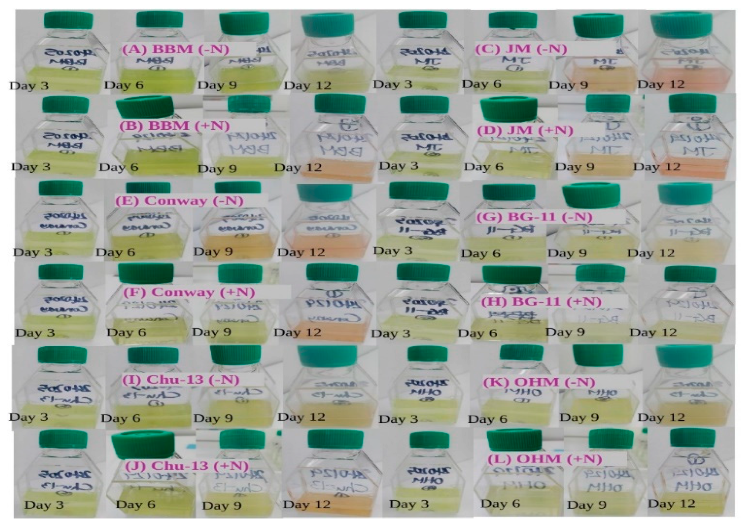
Funding

This research was supported by the “regional innovation mega project” program through the Korea Innovation Foundation funded by the Ministry of Science and ICT (Project Number: 2023-DD-UP-0007) and by the Basic Science Research Program (NRF-2021R1A6A1A03039503). It was also supported by the Korea Institute of Marine Science & Technology Promotion (KIMST) funded by the Ministry of Oceans and Fisheries (20220379, Development of mass production process standardization of xanthophyll astaxanthin) (RS-2022-KS221611). It was also supported by the Soonchunhyang University research fund.

Data Availability Statement

Data are contained within the article.

Conflicts of Interest

Author Jihyun Lee was employed by the company Korea Fisheries Resources Agency East Sea Branch. Author Hyun Woung Shin was employed by the company AlgaeBio, Inc. The remaining authors declare that the research was conducted in the absence of any commercial or financial relationships that could be construed as a potential conflict of interest.

References

  1. Nishida, Y.; Berg, P.C.; Shakersain, B.; Hecht, K.; Takikawa, A.; Tao, R.; Kakuta, Y.; Uragami, C.; Hashimoto, H.; Misawa, N.; et al. Astaxanthin: Past, Present, and Future. Mar. Drugs 2023, 21, 514. [Google Scholar] [CrossRef] [PubMed]
  2. Panis, G.; Carreon, J.R. Commercial astaxanthin production derived by green alga Haematococcus pluvialis: A microalgae process model and a techno-economic assessment all through production line. Algal Res. 2016, 18, 175–190. [Google Scholar] [CrossRef]
  3. Azizi, M.; Hejazi, M.A.; Hashemi, M. Supplementation with polyalcohols and sequential mixotrophy dilution photoinduction strategy boost the accumulation of astaxanthin by Haematococcus pluvialis. Aquaculture 2019, 511, 734225. [Google Scholar] [CrossRef]
  4. Li, Q.; Li, L.; Zhang, Y.; Gao, H.; Zhao, Y.; Yu, X. Chemical inducers regulate ROS signalling to stimulate astaxanthin production in Haematococcus pluvialis under environmental stresses: A review. Trends Food Sci. Technol. 2023, 136, 181–193. [Google Scholar] [CrossRef]
  5. Byeon, H.; An, Y.; Kim, T.; Rayamajhi, V.; Lee, J.; Shin, H.; Jung, S. Effects of Four Organic Carbon Sources on the Growth and Astaxanthin Accumulation of Haematococcus lacustris. Life 2024, 14, 29. [Google Scholar] [CrossRef]
  6. An, Y.; Kim, T.; Byeon, H.; Rayamajhi, V.; Lee, J.; Jung, S.; Shin, H. Improved Production of Astaxanthin from Haematococcus pluvialis Using a Hybrid Open–Closed Cultivation System. Appl. Sci. 2024, 14, 1104. [Google Scholar] [CrossRef]
  7. Elbahnaswy, S.; Elshopakey, G.E. Recent progress in practical applications of a potential carotenoid astaxanthin in aquaculture industry: A review. Fish Physiol. Biochem. 2024, 50, 97–126. [Google Scholar] [CrossRef] [PubMed]
  8. Stachowiak, B.; Szulc, P. Astaxanthin for the Food Industry. Astaxanthin for the Food Industry. Molecules 2021, 26, 2666. [Google Scholar] [CrossRef]
  9. Sun, J.; Yan, J.; Dong, H.; Gao, K.; Yu, K.; He, C.; Mao, X. Astaxanthin with different configurations: Sources, activity, post-modification and application in foods. Curr. Opin. Food Sci. 2022, 49, 100955. [Google Scholar] [CrossRef]
  10. Oslan, S.N.H.; Shoparwe, N.F.; Yusoff, A.H.; Rahim, A.A.; Chang, C.S.; Tan, J.S.; Oslan, S.N.; Arumugam, K.; Ariff, A.B.; Sulaiman, A.Z.; et al. A Review on Haematococcus pluvialis Bioprocess Optimization of Green and Red Stage Culture Conditions for the Production of Natural Astaxanthin. Biomolecules 2021, 11, 256. [Google Scholar] [CrossRef]
  11. Joun, J.; Sirohi, R.; Sim, S.J. The effects of acetate and glucose on carbon fixation and carbon utilization in mixotrophy of Haematococcus pluvialis. Bioresour. Technol. 2023, 367, 128218. [Google Scholar] [CrossRef] [PubMed]
  12. Pang, N.; Chen, S. Effects of C5 organic carbon and light on growth and cell activity of Haematococcus pluvialis under mixotrophic conditions. Algal Res. 2017, 21, 227–235. [Google Scholar] [CrossRef]
  13. Zhang, L.; Zhang, C.; Liu, J.; Yang, N. A strategy for stimulating astaxanthin and lipid production in Haematococcus pluvialis by exogenous glycerol application under low light. Algal Res. 2020, 46, 101779. [Google Scholar] [CrossRef]
  14. Yu, W.; Zhang, L.; Zhao, J.; Liu, J. Exogenous sodium fumarate enhances astaxanthin accumulation in Haematococcus pluvialis by enhancing the respiratory metabolic pathway. Bioresour. Technol. 2021, 341, 125788. [Google Scholar] [CrossRef] [PubMed]
  15. Du, F.; Hu, C.; Xue Sun, X.; Zhang, L.; Xu, N. Transcriptome analysis reveals the promoting effect of trisodium citrate on astaxanthin accumulation in Haematococcus pluvialis under high light condition. Aquaculture 2021, 543, 736978. [Google Scholar] [CrossRef]
  16. Yu, W.; Zhang, L.; Zhao, J.; Liu, J. Enhancement of astaxanthin accumulation in Haematococcus pluvialis by exogenous oxaloacetate combined with nitrogen deficiency. Bioresour. Technol. 2022, 345, 126484. [Google Scholar] [CrossRef] [PubMed]
  17. Zhang, L.; Pei, H.; Yang, Z.; Wang, X.; Chen, S.; Li, Y.; Xie, Z. Microalgae nourished by mariculture wastewater aids aquaculture self-reliance with desirable biochemical composition. Bioresour. Technol. 2019, 278, 205–213. [Google Scholar] [CrossRef] [PubMed]
  18. Prabha, Y.; Tyagi, A.; Soni, S.K. Impact of Monosodium glutamate on the production and chemical constituents of Spirulina maxima. Int. J. Pure App. Biosci. 2016, 4, 180–184. [Google Scholar] [CrossRef]
  19. Singh, S.; Rekha, P.D.; Arun, A.B.; Young, C. Impacts of monosodium glutamate industrial wastewater on plant growth and soil characteristics. Ecol. Eng. 2009, 35, 1559–1563. [Google Scholar] [CrossRef]
  20. Ghanchivahora, S.S. Evaluation of Limnospira Maxima Biomass and Microwave-Assisted Extracts as Biofertilisers to Support Growth of Sorghum Bicolor; Flinders University, College of Medicine and Public Health: Bedford Park, Australia, 2022; p. 8. [Google Scholar]
  21. Si, P.; Shao, W.; Yu, H.; Xu, G.; Du, G. Differences in Microbial Communities Stimulated by Malic Acid Have the Potential to Improve Nutrient Absorption and Fruit Quality of Grapes. Front. Microbiol. 2022, 13, 850807. [Google Scholar] [CrossRef]
  22. Hill, P.G.; Warwick, P.E.; Zubkov, M.V. Low microbial respiration of leucine at ambient oceanic concentration in the mixed layer of the central Atlantic Ocean. Limnol. Oceanogr. 2013, 58, 1569–1604. [Google Scholar] [CrossRef]
  23. Kan, C.C.; Chung, T.Y.; Juo, Y.A.; Hsieh, M.H. Glutamine rapidly induces the expression of key transcription factor genes involved in nitrogen and stress responses in rice roots. BMC Genom. 2015, 16, 731. [Google Scholar] [CrossRef]
  24. Xiang, W.; Wei, X.; Tang, H.; Li, L.; Huang, R. Complete Genome Sequence and Biodegradation Characteristics of Benzoic Acid-Degrading Bacterium Pseudomonas sp. SCB32. Biomed. Res. Int. 2020, 2020, 6146104. [Google Scholar] [CrossRef]
  25. Béguin, P.; Aubert, J.P. The biological degradation of cellulose. FEMS Microbiol. Rev. 1994, 13, 25–58. [Google Scholar] [CrossRef]
  26. Blifernez-Klassen, O.; Klassen, V.; Doebbe, A.; Kersting, K.; Grimm, P.; Wobbe, L.; Kruse, O. Cellulose degradation and assimilation by the unicellular phototrophic eukaryote Chlamydomonas reinhardtii. Nat. Commun. 2012, 3, 1214. [Google Scholar] [CrossRef]
  27. Li, Y.; Zhao, T.; Sun, W.; Gao, R.; Ma, G. Supplementation of alanine improves biomass accumulation and lipid production of Chlorella pyrenoidosa by increasing the respiratory and metabolic processes. J. Ocean. Limnol. 2024, 42, 570–579. [Google Scholar] [CrossRef]
  28. Yang, Q.; Zhao, D.; Liu, Q. Connections Between Amino Acid Metabolisms in Plants: Lysine as an Example. Front. Plant. Sci. 2020, 11, 928. [Google Scholar] [CrossRef]
  29. Zhang, W.; Zhang, P.; Sun, H.; Chen, M.; Lu, S.; Li, P. Effects of various organic carbon sources on the growth and biochemical composition of Chlorella pyrenoidosa. Bioresour. Technol. 2014, 173, 52–58. [Google Scholar] [CrossRef]
  30. Chen, Z.; Su, B. Influence of Medium Frequency Light/Dark Cycles on the Cultivation of Auxenochlorella pyrenoidosa. Appl. Sci. 2020, 10, 5093. [Google Scholar] [CrossRef]
  31. Begum, H. Effects of different salinities on the growth and proximate composition of Dunaliella sp. isolated from South China Sea at different growth phases. Process. Saf. Environ. Prot. 2017, 112, 280–287. [Google Scholar]
  32. Fábregas, J.; Domínguez, A.; Regueiro, M.; Maseda, A.; Otero, A. Optimization of culture medium for the continuous cultivation of the microalga Haematococcus pluvialis. Appl. Microbiol. Biotechnol. 2000, 53, 530–535. [Google Scholar] [CrossRef] [PubMed]
  33. Yang, C.C.; Wen, R.C.; Shen, C.R.; Yao, D.J. Using a Microfluidic Gradient Generator to Characterize BG-11 Medium for the Growth of Cyanobacteria Synechococcus elongatus PCC7942. Micromachines 2015, 6, 1755–1767. [Google Scholar] [CrossRef]
  34. Colusse, G.A.; Duarte, M.E.R.; Carvalho, J.C.; Noseda, D.M. Media effects on laboratory scale production costs of Haematococcus pluvialis biomass. Bioresour. Technol. Rep. 2019, 7, 100236. [Google Scholar] [CrossRef]
  35. Rippka, R.; Deruelles, J.; Waterbury, J.B.; Herdman, M.; Stanier, R.Y. Generic assignments, strain histories, and properties of pure cultures of cyanobacteria. Microbiology 1979, 111, 1–61. [Google Scholar] [CrossRef]
  36. Stanier, R.Y.; Kunisawa, R.; Mandel, M.; Cohen-Bazire, G. Purification and properties of unicellular blue-green algae (order Chroococcales). Bacteriol. Rev. 1971, 35, 171–205. [Google Scholar] [CrossRef] [PubMed]
  37. Yeesang, C.; Cheirsilp, B. Effect of nitrogen, salt, and iron content in the growth medium and light intensity on lipid production by microalgae isolated from freshwater sources in Thailand. Bioresour. Technol. 2011, 102, 3034–3040. [Google Scholar] [CrossRef] [PubMed]
  38. Sorokina, K.N.; Samoylova, Y.V.; Parmon, V.N. Comparative analysis of microalgae metabolism on BBM and municipal wastewater during salt induced lipid accumulation. Bioresour. Technol. Rep. 2020, 11, 100548. [Google Scholar] [CrossRef]
  39. Gutierrez, B.S.H.; Miranda, A.; Meneses, D.; Vargas, G.; Sáez, A. The Effect of Different Concentrations of Nitrogen and Phosphorous on the Production of Lipid Metabolites in Haematococcus Pluvialis UTEX 25051. PREPRINT (Version 1) Available Res. Sq. 2021. preprint. [Google Scholar] [CrossRef]
  40. Ren, Y.; Deng, J.; Huang, J.; Wu, Z.; Yi, L.; Bi, Y.; Chen, F. Using green alga Haematococcus pluvialis for astaxanthin and lipid co-production: Advances and outlook. Bioresour. Technol. 2021, 340, 125736. [Google Scholar] [CrossRef]
  41. Chekanov, K. Diversity and Distribution of Carotenogenic Algae in Europe: A Review. Mar. Drugs 2023, 21, 108. [Google Scholar] [CrossRef]
  42. Maltsev, Y.; Maltseva, K.; Kulikovskiy, M.; Maltseva, S. Influence of Light Conditions on Microalgae Growth and Content of Lipids, Carotenoids, and Fatty Acid Composition. Biology 2021, 10, 1060. [Google Scholar] [CrossRef] [PubMed]
  43. Zarrinmehr, M.J.; Farhadian, O.; Heyrati, F.P.; Keramat, J.; Koutra, E.; Kornaros, M.; Daneshvar, E. Effect of nitrogen concentration on the growth rate and biochemical composition of the microalga, Isochrysis galbana. Egypt. J. Aquat. Res. 2020, 46, 153–158. [Google Scholar] [CrossRef]
  44. Feng, L.; Minggang, C.; Yanqi, W.; Qingsheng, L.; Zuyuan, Q.; Jiansen, L.; Yulei, Z.; Ning, Z.; Changling, L.; Xianghu, H. Effects of Nitrogen and Light Intensity on the Astaxanthin Accumulation in Motile Cells of Haematococcus pluvialis. Front. Mar. Sci. 2022, 9, 909237. [Google Scholar]
  45. Wang, B.; Pan, X.; Jia, J.; Xiong, W.; Manirafasha, E.; Ling, X.; Yinghua, L. Strategy and regulatory mechanisms of glutamate feeding to enhance astaxanthin yield in Xanthophyllomyces dendrorhous. Enzym. Microb. Technol. 2019, 125, 45–52. [Google Scholar] [CrossRef] [PubMed]
Figure 1. Appearances of H. lacustris cultures grown in different media (Day 3, 6, 9, and 12). (A) H. lacustris cultures grown in BBM (−N). (B) H. lacustris cultures grown in BBM (+N). (C) H. lacustris cultures grown in JM (−N). (D) H. lacustris cultures grown in JM (+N). (E) H. lacustris cultures grown in Conway (−N). (F) H. lacustris cultures grown in Conway (+N). (G) H. lacustris cultures grown in BG-11 (−N). (H) H. lacustris cultures grown in BG-11 (+N). (I) H. lacustris cultures grown in CHU-13 (−N). (J) H. lacustris cultures grown in CHU-13 (+N). (K) H. lacustris cultures grown in OHM (−N). (L) H. lacustris cultures grown in OHM (+N).
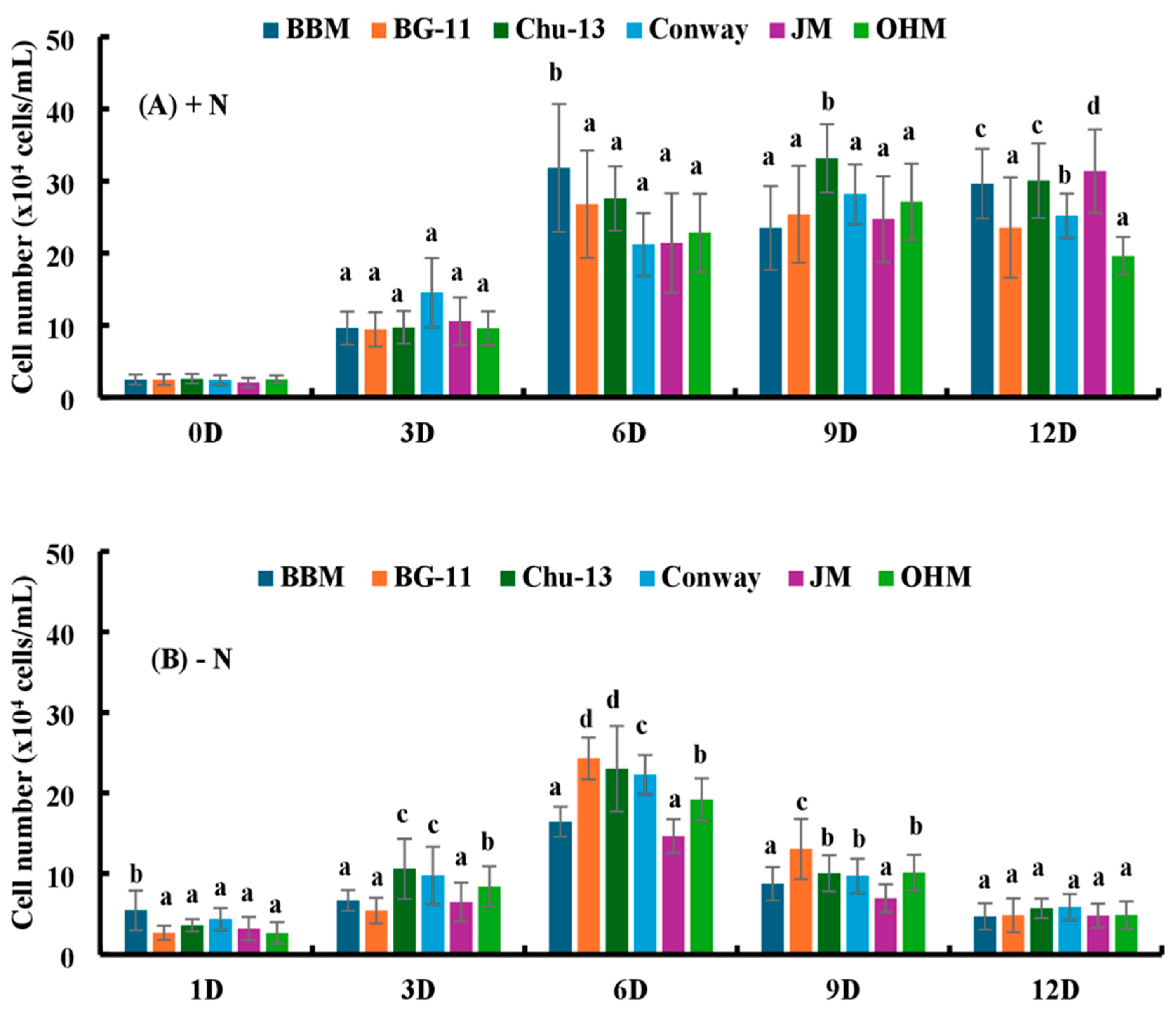
Microorganisms 12 01040 g001
Figure 2. (A) Comparison of the cell number of H. lacustris under the influence of different media with nitrogen source (0D—Day 0, 3D—Day 3, 6D—Day 6, 9D—Day 9, 12D—Day 12). (B) Comparison of the cell number of H. lacustris under the influence of different media without nitrogen source (1D—Day 1, 3D—Day 3, 6D—Day 6, 9D—Day 9, 12D—Day 12). Mean values that do not share the same letter are significantly different at p < 0.001.
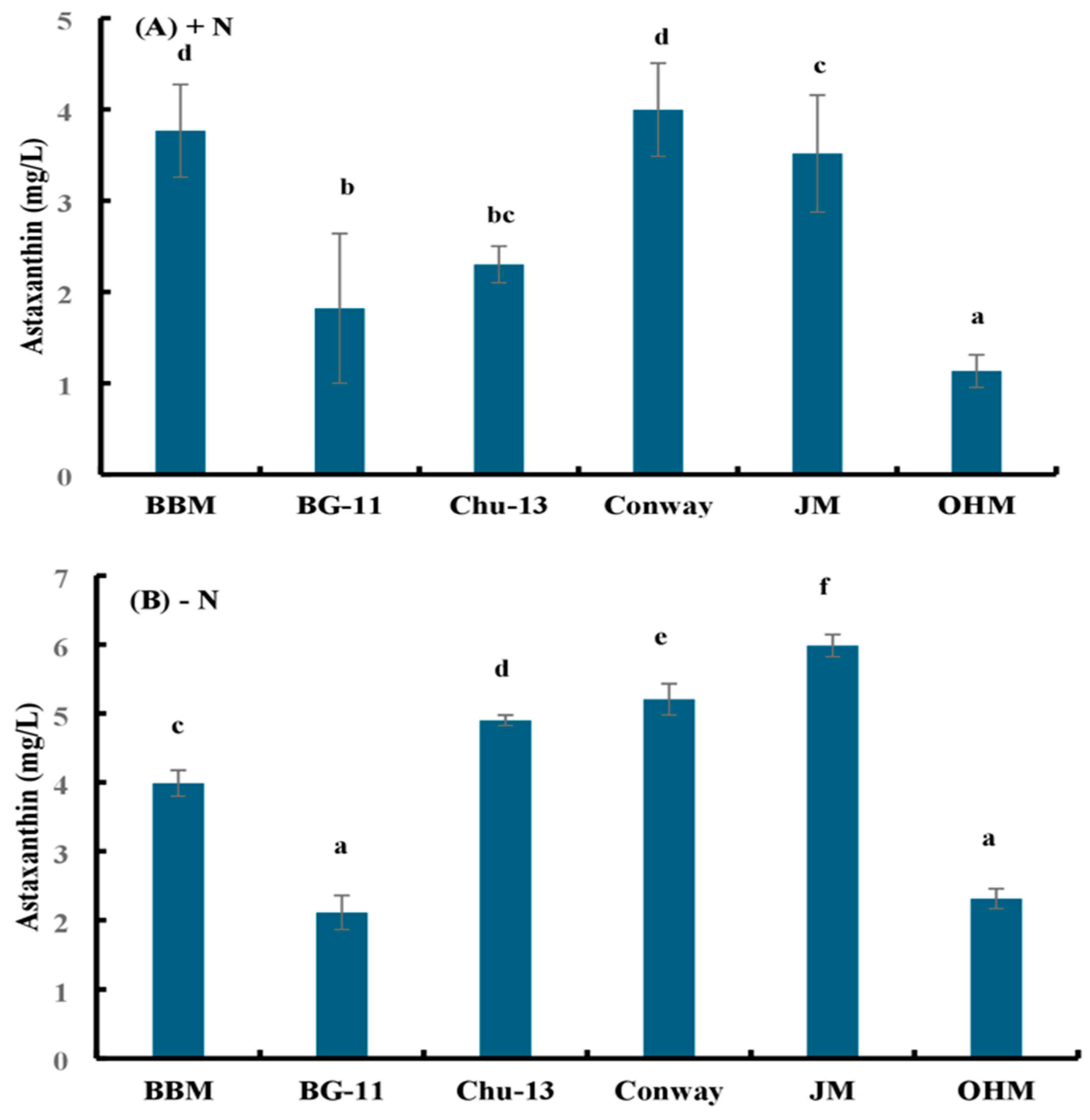
Microorganisms 12 01040 g002
Figure 3. Astaxanthin accumulation by H. lacustris grown in different media in 14 days (A) with a nitrogen source and (B) without a nitrogen source. Mean values that do not share the same letter are significantly different at p < 0.001.
Microorganisms 12 01040 g003
Table 1. Chemical composition of six different media used in this study.
ChemicalWalne/ConwayOHMBG-11Mod. CHU 13BBMJM
KNO3100 g/L410 mg/L-0.2 g/L-0.05 mg/L
NaNO3--1.5 g/L-1.5 g/L0.08 mg/L
Ca(NO3)2·4H2O-----0.02 mg/L
Ferric ammonium citrate--0.012 g/L-6 mg/L-
Na2EDTA--0.001 g/L-1 mg/L2.25 µg/L
K2HPO4--0.04 g/L0.04 g/L40 mg/L0.0124 mg/L
Na3PO420 g/L-----
Na2HPO4-30 mg/L---0.036 mg/L
EDTAFeNa-----2.25 µg/L
Na2CO3--0.02 g/L---
MgSO4·7H2O-246.5 mg/L36.1 mg/L0.1 g/L75 mg/L-
CaCl2·2H2O-110.9 mg/L27.2 mg/L0.054 g/L36 mg/L-
Citric acid---100.1 mg/L6 mg/L-
Sodium citrate--8.82 mg/L---
NaHCO3--0.036 g/L--15.9 µg/L
FeC6H5O7·5H2O-2.62 mg/L-0.01 g/L--
Na2H2EDTA.2H2O45 g/L-----
FeCl3·6H2O1.3 g/L-----
ZnCl24.2 g/L-----
MnCl2·4H2O0.36 g/L0.989 mg/L1.81 mg/L1.8 mg/L1.81 mg/L0.139 µg/L
CoCl2·6H2O4 g/L0.011 mg/L-0.08 mg/L--
CuSO4·5H2O4 g/L0.012 mg/L0.079 mg/L0.08 mg/L0.08 mg/L-
Cr2O3-0.076 mg/L----
Na2MoO4·2H2O-0.12 mg/L0.39 mg/L0.05 mg/L0.39 mg/L-
(NH4)6Mo7O24·4H2O1.8 g/L----0.001 mg/L
H3BO333.4 g/L-2.86 mg/L2.85 mg/L2.86 mg/L2.48 µg/L
Thiamine—HCl200 mg/L----0.04 µg/L
Cyanocobalamin10 mg/L15 µg/mL---0.04 µg/L
Biotin 25 µg/mL----0.04 µg/L
Thiamine-17.5 µg/mL----
ZnSO4·7H2O--0.222 mg/L0.02 mg/L0.222 mg/L-
Co(NO3)2·6H2O--0.049 mg/L---
Table 2. Cell number of H. lacustris grown in JM (+N) media (Day 1, Day 3, Day 6, Day 9, Day 12) and astaxanthin contents of H. lacustris on Day 14 grown with the supplementation of benzoic acid. Data are presented as the mean ± standard deviation.
H. lacustris Culture Grown with the Supplementation of Benzoic AcidDay 1 Cell Number (×104 Cells/mL)Day 3 Cell Number (×104 Cells/mL)Day 6 Cell Number (×104 Cells/mL)Day 9 Cell Number (×104 Cells/mL)Day 12 Cell Number (×104 Cells/mL)Day 14 Astaxanthin Content (mg/L)
Control0.20 ± 0.0003.20 ± 0.56612.93 ± 1.32011.47 ± 1.67622.40 ± 5.57111.64 ± 0.077
Benzoic acid (0.325 g/L)0.20 ± 0.0000.07 ± 0.0940.00 ± 0.0000.00 ± 0.0000.00 ± 0.0000.00 ± 0.000
Benzoic acid (0.65 g/L)0.20 ± 0.0000.00 ± 0.0000.00 ± 0.0000.00 ± 0.0000.00 ± 0.0000.00 ± 0.000
Benzoic acid (1.3 g/L)0.13 ± 0.0940.00 ± 0.0000.00 ± 0.0000.00 ± 0.0000.00 ± 0.0000.00 ± 0.000
Benzoic acid (2.6 g/L)0.27 ± 0.2490.00 ± 0.0000.00 ± 0.0000.00 ± 0.0000.00 ± 0.0000.00 ± 0.000
Table 3. Cell number of H. lacustris grown in JM (+N) media (Day 1, Day 3, Day 6, Day 9, Day 12) and astaxanthin contents of H. lacustris on Day 14 grown with the supplementation of cellulose. Data are presented as the mean ± standard deviation. Lowercase letters indicate significant differences (p < 0.001).
H. lacustris Culture Grown with the Supplementation of CelluloseDay 1 Cell Number (×104 Cells/mL)Day 3 Cell Number (×104 Cells/mL)Day 6 Cell Number (×104 Cells/mL)Day 9 Cell Number (×104 Cells/mL)Day 12 Cell Number (×104 Cells/mL)Day 14 Astaxanthin Content (mg/L)
Control0.20 ± 0.0003.20 ± 0.56612.93 ± 1.320 b11.47 ± 1.67622.40 ± 5.571 ab11.64 ± 0.077 a
Cellulose (0.325 g/L)0.27 ± 0.0943.93 ± 0.66010.93 ± 1.268 ab14.47 ± 3.15129.40 ± 0.432 b17.76 ± 0.067 d
Cellulose (0.65 g/L)0.40 ± 0.2832.93 ± 0.4119.93 ± 1.76 ab13.27 ± 2.13124.33 ± 0.772 ab12.17 ± 0.017 b
Cellulose (1.3 g/L)0.27 ± 0.0949.93 ± 1.7618.33 ± 0.525 a12.40 ± 0.58920.73 ± 0.340 a11.79 ± 0.048 ab
Cellulose (2.6 g/L)0.13 ± 0.0942.47 ± 0.4119.00 ± 0.993 ab13.20 ± 2.59220.13 ± 0.838 a12.60 ± 0.294 c
Table 4. Cell number of H. lacustris grown in JM (+N) media (Day 1, Day 3, Day 6, Day 9, Day 12) and astaxanthin contents of H. lacustris on Day 14 grown with the supplementation of glutamine. Data are presented as the mean ± standard deviation. Lowercase letters indicate significant differences (p < 0.001).
H. lacustris Culture Grown with the Supplementation of GlutamineDay 1 Cell Number (×104 Cells/mL)Day 3 Cell Number (×104 Cells/mL)Day 6 Cell Number (×104 Cells/mL)Day 9 Cell Number (×104 Cells/mL)Day 12 Cell Number (×104 Cells/mL)Day 14 Astaxanthin Content (mg/L)
Control0.20 ± 0.0003.20 ± 0.566 b12.93 ± 1.320 c11.47 ± 1.676 c22.40 ± 5.571 c11.64 ± 0.077 e
Glutamine (0.325 g/L)0.47 ± 0.0943.07 ± 0.822 b6.87 ± 1.389 b3.13 ± 0.736 a1.73 ± 0.411 b8.80 ± 0.106 d
Glutamine (0.65 g/L)0.60 ± 0.4321.27 ± 0.189 a2.60 ± 0.589 a2.20 ± 0.909 a0.40 ± 0.163 a5.92 ± 0.006 c
Glutamine (1.3 g/L)0.33 ± 0.0940.33 ± 0.094 a0.07 ± 0.0940.07 ± 0.0940.07 ± 0.0941.31 ± 0.029 b
Glutamine (2.6 g/L)0.33 ± 0.0940.73 ± 0.411 a0.00 ± 0.0000.00 ± 0.0000.00 ± 0.0000.01 ± 0.003 a
Table 5. Cell number of H. lacustris grown in JM (+N) media (Day 1, Day 3, Day 6, Day 9, Day 12) and astaxanthin contents of H. lacustris on Day 14 grown with the supplementation of alanine. Data are presented as the mean ± standard deviation. Lowercase letters indicate significant differences (p < 0.001).
H. lacustris Culture Grown with the Supplementation of AlanineDay 1 Cell Number (×104 Cells/mL)Day 3 Cell Number (×104 Cells/mL)Day 6 Cell Number (×104 Cells/mL)Day 9 Cell Number (×104 Cells/mL)Day 12 Cell Number (×104 Cells/mL)Day 14 Astaxanthin Content (mg/L)
Control0.20 ± 0.0003.20 ± 0.56612.93 ± 1.320 d11.47 ± 1.676 c22.40 ± 5.571 c11.64 ± 0.077 c
Alanine (0.325 g/L)0.33 ± 0.0943.40 ± 0.86410.27 ± 0.249 c5.27 ± 0.660 b4.60 ± 0.163 b7.03 ± 0.048 b
Alanine (0.65 g/L)0.47 ± 0.0940.07 ± 0.0943.00 ± 0.432 b3.00 ± 0.748 a0.73 ± 0.249 a7.05 ± 0.006 b
Alanine (1.3 g/L)0.13 ± 0.0940.07 ± 0.0940.07 ± 0.094 a0.00 ± 0.0000.07 ± 0.0941.34 ± 0.076 a
Alanine (2.6 g/L)0.40 ± 0.1630.07 ± 0.0940.00 ± 0.0000.00 ± 0.0000.00 ± 0.0000.23 ± 0.017 a
Table 6. Cell number of H. lacustris grown in JM (+N) media (Day 1, Day 3, Day 6, Day 9, Day 12) and astaxanthin contents of H. lacustris on Day 14 grown with the supplementation of leucine. Data are presented as the mean ± standard deviation. Lowercase letters indicate significant differences (p < 0.001).
H. lacustris Culture Grown with the Supplementation of LeucineDay 1 Cell Number (×104 Cells/mL)Day 3 Cell Number (×104 Cells/mL)Day 6 Cell Number (×104 Cells/mL)Day 9 Cell Number (×104 Cells/mL)Day 12 Cell Number (×104 Cells/mL)Day 14 Astaxanthin Content (mg/L)
Control0.20 ± 0.0003.20 ± 0.566 ab12.93 ± 1.320 a11.47 ± 1.676 a22.40 ± 5.57111.64 ± 0.077 e
Leucine (0.325 g/L)0.27 ± 0.0943.47 ± 0.525 b23.20 ± 2.953 b23.27 ± 0.806 c21.40 ± 2.67311.48 ± 0.073 d
Leucine (0.65 g/L)0.33 ± 0.0942.93 ± 0.680 ab15.93 ± 1.746 a15.80 ± 1.766 b19.47 ± 1.6367.33 ± 0.017 c
Leucine (1.3 g/L)0.20 ± 0.1631.60 ± 0.163 a10.27 ± 1.310 a19.73 ± 0.411 c23.13 ± 3.4655.33 ± 0.011 b
Leucine (2.6 g/L)0.27 ± 0.0942.27 ± 0.618 ab12.33 ± 1.087 a15.07 ± 1.676 ab20.60 ± 0.7485.18 ± 0.006 a
Table 7. Cell number of H. lacustris grown in JM (+N) media (Day 1, Day 3, Day 6, Day 9, Day 12) and astaxanthin contents of H. lacustris on Day 14 grown with the supplementation of lysine. Data are presented as the mean ± standard deviation. Lowercase letters indicate significant differences (p < 0.001).
H. lacustris Culture Grown with the Supplementation of LysineDay 1 Cell Number (×104 Cells/mL)Day 3 Cell Number (×104 Cells/mL)Day 6 Cell Number (×104 Cells/mL)Day 9 Cell Number (×104 Cells/mL)Day 12 Cell Number (×104 Cells/mL)Day 14 Astaxanthin Content (mg/L)
Control0.20 ± 0.0003.20 ± 0.566 b12.93 ± 1.320 b11.47 ± 1.676 bc22.40 ± 5.571 bc11.64 ± 0.077 d
Lysine (0.325 g/L)0.27 ± 0.0944.67 ± 1.087 b12.73 ± 0.411 b14.60 ± 0.589 c20.73 ± 2.205 bc0.04 ± 0.000 a
Lysine (0.65 g/L)0.80 ± 0.3274.13 ± 0.806 b12.87 ± 1.668 b26.47 ± 3.356 d38.67 ± 11.927 c10.15 ± 0.006 c
Lysine (1.3 g/L)0.27 ± 0.0941.07 ± 0.525 a1.93 ± 0.618 a7.00 ± 1.395 b15.27 ± 0.660 ab13.62 ± 0.061 e
Lysine (2.6 g/L)0.20 ± 0.0000.07 ± 0.094 a0.13 ± 0.094 a0.53 ± 0.340 a1.00 ± 0.327 a7.65 ± 0.013 b
Table 8. Cell number of H. lacustris grown in JM (+N) media (Day 1, Day 3, Day 6, Day 9, Day 12) and astaxanthin contents of H. lacustris on Day 14 grown with the supplementation of malic acid.
H. lacustris Culture Grown with the Supplementation of Malic AcidDay 1 Cell Number (×104 Cells/mL)Day 3 Cell Number (×104 Cells/mL)Day 6 Cell Number (×104 Cells/mL)Day 9 Cell Number (×104 Cells/mL)Day 12 Cell Number (×104 Cells/mL)Day 14 Astaxanthin Content (mg/L)
Control0.20 ± 0.0003.20 ± 0.56612.93 ± 1.32011.47 ± 1.67622.40 ± 5.57111.64 ± 0.077
Malic acid (0.325 g/L)0.47 ± 0.0940.00 ± 0.0000.00 ± 0.0000.00 ± 0.0000.00 ± 0.0000.13 ± 0.006
Malic acid (0.65 g/L)0.67 ± 0.0940.00 ± 0.0000.00 ± 0.0000.00 ± 0.0000.00 ± 0.0000.23 ± 0.048
Malic acid (1.3 g/L)0.13 ± 0.0940.00 ± 0.0000.00 ± 0.0000.00 ± 0.0000.00 ± 0.0000.03 ± 0.006
Malic acid (2.6 g/L)0.20 ± 0.1630.00 ± 0.0000.00 ± 0.0000.00 ± 0.0000.00 ± 0.0000.00 ± 0.000
Table 9. Cell number of H. lacustris grown in JM (+N) media (Day 1, Day 3, Day 6, Day 9, Day 12) and astaxanthin contents of H. lacustris on Day 14 grown with the supplementation of maltose. Data are presented as the mean ± standard deviation. Lowercase letters indicate significant differences (p < 0.001).
H. lacustris Culture Grown with the Supplementation of MaltoseDay 1 Cell Number (×104 Cells/mL)Day 3 Cell Number (×104 Cells/mL)Day 6 Cell Number (×104 Cells/mL)Day 9 Cell Number (×104 Cells/mL)Day 12 Cell Number (×104 Cells/mL)Day 14 Astaxanthin Content (mg/L)
Control0.20 ± 0.0003.20 ± 0.56612.93 ± 1.320 a11.47 ± 1.67622.40 ± 5.571 b11.64 ± 0.077
Maltose (0.325 g/L)0.13 ± 0.0942.00 ± 0.16322.67 ± 2.391 b16.87 ± 2.58422.13 ± 4.620 b0.00 ± 0.000
Maltose (0.65 g/L)0.40 ± 0.1633.40 ± 0.71216.80 ± 1.337 a13.40 ± 0.58914.67 ± 1.236 ab0.00 ± 0.000
Maltose (1.3 g/L)0.20 ± 0.1633.33 ± 0.34015.00 ± 2.007 a13.13 ± 0.6809.60 ± 1.020 a0.00 ± 0.000
Maltose (2.6 g/L)0.27 ± 0.0943.60 ± 0.84916.20 ± 2.068 a12.73 ± 2.55311.87 ± 2.007 ab0.00 ± 0.000
Table 10. Cell number of H. lacustris grown in JM (+N) media (Day 1, Day 3, Day 6, Day 9, Day 12) and astaxanthin contents of H. lacustris on Day 14 grown with the supplementation of sodium glutamate. Data are presented as the mean ± standard deviation. Lowercase letters indicate significant differences (p < 0.001).
H. lacustris Culture Grown with the Supplementation of Sodium GlutamateDay 1 Cell Number (×104 Cells/mL)Day 3 Cell Number (×104 Cells/mL)Day 6 Cell Number (×104 Cells/mL)Day 9 Cell Number (×104 Cells/mL)Day 12 Cell Number (×104 Cells/mL)Day 14 Astaxanthin Content (mg/L)
Control0.20 ± 0.0003.20 ± 0.566 a12.93 ± 1.320 c11.47 ± 1.676 c22.40 ± 5.571 b11.64 ± 0.077 d
Sodium Glutamate (0.325 g/L)0.40 ± 0.0001.93 ± 0.340 c15.13 ± 0.998 c12.73 ± 0.411 c25.00 ± 2.592 b20.48 ± 0.017 e
Sodium Glutamate (0.65 g/L)0.33 ± 0.0941.40 ± 0.163 bc6.87 ± 0.957 b5.53 ± 0.573 b5.27 ± 0.754 a8.30 ± 0.048 c
Sodium Glutamate (1.3 g/L)0.40 ± 0.1630.80 ± 0.000 ab3.07 ± 0.943 a3.33 ± 0.411 b0.53 ± 0.094 a5.62 ± 0.011 b
Sodium Glutamate (2.6 g/L)0.53 ± 0.2490.07 ± 0.094 a0.53 ± 0.189 a0.20 ± 0.000 a0.13 ± 0.094 a0.55 ± 0.019 a
Table 11. Cell number of H. lacustris grown in JM (−N) media (Day 1, Day 3, Day 6, Day 9, Day 12) and astaxanthin contents of H. lacustris on Day 14 grown with the supplementation of benzoic acid. Data are presented as the mean ± standard deviation.
H. lacustris Culture Grown with the Supplementation of Benzoic AcidDay 1 Cell Number (×104 Cells/mL)Day 3 Cell Number (×104 Cells/mL)Day 6 Cell Number (×104 Cells/mL)Day 9 Cell Number (×104 Cells/mL)Day 12 Cell Number (×104 Cells/mL)Day 14 Astaxanthin Content (mg/L)
Control0.20 ± 0.1630.20 ± 0.1630.53 ± 0.1890.87 ± 0.0940.80 ± 0.1630.91 ± 0.035
Benzoic acid (0.325 g/L)0.13 ± 0.0940.13 ± 0.0940.20 ± 0.1630.07 ± 0.0940.00 ± 0.0000.00 ± 0.000
Benzoic acid (0.65 g/L)0.20 ± 0.1630.07 ± 0.0940.13 ± 0.0940.13 ± 0.0940.00 ± 0.0000.00 ± 0.000
Benzoic acid (1.3 g/L)0.20 ± 0.1630.27 ± 0.2490.13 ± 0.1890.13 ± 0.1890.00 ± 0.0000.00 ± 0.000
Benzoic acid (2.6 g/L)0.20 ± 0.0000.13 ± 0.0940.00 ± 0.0000.00 ± 0.0000.00 ± 0.0000.00 ± 0.000
Table 12. Cell number of H. lacustris grown in JM (−N) media (Day 1, Day 3, Day 6, Day 9, Day 12) and astaxanthin contents of H. lacustris on Day 14 grown with the supplementation of cellulose. Data are presented as the mean ± standard deviation. Lowercase letters indicate significant differences (p < 0.001).
H. lacustris Culture Grown with the Supplementation of CelluloseDay 1 Cell Number (×104 Cells/mL)Day 3 Cell Number (×104 Cells/mL)Day 6 Cell Number (×104 Cells/mL)Day 9 Cell Number (×104 Cells/mL)Day 12 Cell Number (×104 Cells/mL)Day 14 Astaxanthin Content (mg/L)
Control0.20 ± 0.1630.20 ± 0.1630.53 ± 0.189 a0.87 ± 0.0940.80 ± 0.1630.91 ± 0.035 a
Cellulose (0.325 g/L)0.27 ± 0.0940.47 ± 0.2490.67 ± 0.094 a1.07 ± 0.0941.47 ± 0.8220.90 ± 0.006 a
Cellulose (0.65 g/L)0.20 ± 0.0000.40 ± 0.1630.73 ± 0.094 a0.93 ± 0.1891.00 ± 0.5891.04 ± 0.028 b
Cellulose (1.3 g/L)0.07 ± 0.0940.47 ± 0.2491.38 ± 0.189 b1.00 ± 0.1631.13 ± 0.3771.40 ± 0.023 c
Cellulose (2.6 g/L)0.07 ± 0.0940.53 ± 0.3401.47 ± 0.189 b1.87 ± 0.6602.13 ± 0.5252.02 ± 0.017 d
Table 13. Cell number of H. lacustris grown in JM (−N) media (Day 1, Day 3, Day 6, Day 9, Day 12) and astaxanthin contents of H. lacustris on Day 14 grown with the supplementation of glutamine. Data are presented as the mean ± standard deviation. Lowercase letters indicate significant differences (p < 0.001).
H. lacustris Culture Grown with the Supplementation of GlutamineDay 1 Cell Number (×104 Cells/mL)Day 3 Cell Number (×104 Cells/mL)Day 6 Cell Number (×104 Cells/mL)Day 9 Cell Number (×104 Cells/mL)Day 12 Cell Number (×104 Cells/mL)Day 14 Astaxanthin Content (mg/L)
Control0.20 ± 0.1630.20 ± 0.163 a0.53 ± 0.189 ab0.87 ± 0.094 c0.80 ± 0.163 b0.91 ± 0.035 a
Glutamine (0.325 g/L)0.20 ± 0.0001.20 ± 0.163 b1.40 ± 0.283 c0.53 ± 0.189 b0.13 ± 0.189 a8.60 ± 0.046 e
Glutamine (0.65 g/L)0.13 ± 0.0940.13 ± 0.094 a0.87 ± 0.094 bc0.13 ± 0.094 a0.00 ± 0.0007.61 ± 0.011 d
Glutamine (1.3 g/L)0.07 ± 0.0940.20 ± 0.163 a0.67 ± 0.094 ab0.07 ± 0.094 a0.00 ± 0.0004.86 ± 0.006 c
Glutamine (2.6 g/L)0.13 ± 0.0940.33 ± 0.094 a0.20 ± 0.283 a0.00 ± 0.0000.00 ± 0.0001.63 ± 0.025 b
Table 14. Cell number of H. lacustris grown in JM (−N) media (Day 1, Day 3, Day 6, Day 9, Day 12) and astaxanthin contents of H. lacustris on Day 14 grown with the supplementation of alanine. Data are presented as the mean ± standard deviation. Lowercase letters indicate significant differences (p < 0.001).
H. lacustris Culture Grown with the Supplementation of AlanineDay 1 Cell Number (×104 Cells/mL)Day 3 Cell Number (×104 Cells/mL)Day 6 Cell Number (×104 Cells/mL)Day 9 Cell Number (×104 Cells/mL)Day 12 Cell Number (×104 Cells/mL)Day 14 Astaxanthin Content (mg/L)
Control0.20 ± 0.1630.20 ± 0.163 a0.53 ± 0.1890.87 ± 0.0940.80 ± 0.1630.91 ± 0.035 a
Alanine (0.325 g/L)0.20 ± 0.0001.20 ± 0.163 b1.60 ± 0.5891.20 ± 0.5661.07 ± 0.4715.76 ± 0.011 c
Alanine (0.65 g/L)0.27 ± 0.0940.27 ± 0.094 a1.67 ± 0.4991.00 ± 0.1630.87 ± 0.0946.06 ± 0.092 d
Alanine (1.3 g/L)0.07 ± 0.0940.40 ± 0.163 a0.60 ± 0.1630.87 ± 0.0941.93 ± 0.5256.13 ± 0.011 d
Alanine (2.6 g/L)0.20 ± 0.0000.53 ± 0.499 ab0.53 ± 0.1890.87 ± 0.0941.47 ± 0.3771.80 ± 0.013 b
Table 15. Cell number of H. lacustris grown in JM (−N) media (Day 1, Day 3, Day 6, Day 9, Day 12) and astaxanthin contents of H. lacustris on Day 14 grown with the supplementation of leucine. Data are presented as the mean ± standard deviation. Lowercase letters indicate significant differences (p < 0.001).
H. lacustris Culture Grown with the Supplementation of LeucineDay 1 Cell Number (×104 Cells/mL)Day 3 Cell Number (×104 Cells/mL)Day 6 Cell Number (×104 Cells/mL)Day 9 Cell Number (×104 Cells/mL)Day 12 Cell Number (×104 Cells/mL)Day 14 Astaxanthin Content (mg/L)
Control0.20 ± 0.1630.20 ± 0.163 a0.53 ± 0.189 a0.87 ± 0.094 a0.80 ± 0.163 a0.91 ± 0.035 a
Leucine (0.325 g/L)0.07 ± 0.0940.93 ± 0.189 b1.73 ± 0.411 b3.13 ± 0.660 ab10.80 ± 0.589 c8.30 ± 0.090 d
Leucine (0.65 g/L)0.20 ± 0.0000.67 ± 0.094 ab2.47 ± 0.618 bc10.13 ± 0.618 bc20.93 ± 2.614 e9.10 ± 0.029 e
Leucine (1.3 g/L)0.13 ± 0.0940.47 ± 0.094 ab3.27 ± 0.249 c5.93 ± 1.112 b16.40 ± 0.993 d5.82 ± 0.032 c
Leucine (2.6 g/L)0.07 ± 0.0940.53 ± 0.189 ab2.73 ± 0.499 bc4.93 ± 1.543 b7.07 ± 0.660 b2.17 ± 0.011 b
Table 16. Cell number of H. lacustris grown in JM (−N) media (Day 1, Day 3, Day 6, Day 9, Day 12) and astaxanthin contents of H. lacustris on Day 14 grown with the supplementation of lysine. Data are presented as the mean ± standard deviation. Lowercase letters indicate significant differences (p < 0.001).
H. lacustris Culture Grown with the Supplementation of LysineDay 1 Cell Number (×104 Cells/mL)Day 3 Cell Number (×104 Cells/mL)Day 6 Cell Number (×104 Cells/mL)Day 9 Cell Number (×104 Cells/mL)Day 12 Cell Number (×104 Cells/mL)Day 14 Astaxanthin Content (mg/L)
Control0.20 ± 0.1630.20 ± 0.1630.53 ± 0.189 a0.87 ± 0.094 ab0.80 ± 0.163 a0.91 ± 0.035 a
Lysine (0.325 g/L)0.20 ± 0.0000.40 ± 0.1631.47 ± 0.249 b7.53 ± 0.499 c12.93 ± 1.236 c1.99 ± 0.061 c
Lysine (0.65 g/L)0.07 ± 0.0940.20 ± 0.1630.93 ± 0.411 ab1.67 ± 0.499 b3.73 ± 0.499 b2.12 ± 0.006 d
Lysine (1.3 g/L)0.07 ± 0.0940.20 ± 0.1630.27 ± 0.377 a0.07 ± 0.094 a0.07 ± 0.0941.85 ± 0.000 b
Lysine (2.6 g/L)0.13 ± 0.0940.07 ± 0.0940.13 ± 0.189 a0.00 ± 0.0000.00 ± 0.0007.36 ± 0.023 e
Table 17. Cell number of H. lacustris grown in JM (−N) media (Day 1, Day 3, Day 6, Day 9, Day 12) and astaxanthin contents of H. lacustris on Day 14 grown with the supplementation of malic acid. Data are presented as the mean ± standard deviation. Lowercase letters indicate significant differences (p < 0.001).
H. lacustris Culture Grown with the Supplementation of Malic AcidDay 1 Cell Number (×104 Cells/mL)Day 3 Cell Number (×104 Cells/mL)Day 6 Cell Number (×104 Cells/mL)Day 9 Cell Number (×104 Cells/mL)Day 12 Cell Number (×104 Cells/mL)Day 14 Astaxanthin Content (mg/L)
Control0.20 ± 0.1630.20 ± 0.1630.53 ± 0.1890.87 ± 0.0940.80 ± 0.1630.91 ± 0.035 c
Malic acid (0.325 g/L)0.07 ± 0.0940.07 ± 0.0941.27 ± 0.1890.40 ± 0.3270.00 ± 0.0000.07 ± 0.000 a
Malic acid (0.65 g/L)0.20 ± 0.0000.00 ± 0.0001.00 ± 0.4320.13 ± 0.0940.00 ± 0.0000.00 ± 0.000
Malic acid (1.3 g/L)0.13 ± 0.0940.00 ± 0.0000.67 ± 0.3400.07 ± 0.0940.00 ± 0.0000.00 ± 0.000
Malic acid (2.6 g/L)0.13 ± 0.0940.00 ± 0.0000.93 ± 0.4110.07 ± 0.0940.00 ± 0.0000.89 ± 0.055 b
Table 18. Cell number of H. lacustris grown in JM (−N) media (Day 1, Day 3, Day 6, Day 9, Day 12) and astaxanthin contents of H. lacustris on Day 14 grown with the supplementation of maltose. Data are presented as the mean ± standard deviation. Lowercase letters indicate significant differences (p < 0.001).
H. lacustris Culture Grown with the Supplementation of MaltoseDay 1 Cell Number (×104 Cells/mL)Day 3 Cell Number (×104 Cells/mL)Day 6 Cell Number (×104 Cells/mL)Day 9 Cell Number (×104 Cells/mL)Day 12 Cell Number (×104 Cells/mL)Day 14 Astaxanthin Content (mg/L)
Control0.20 ± 0.1630.20 ± 0.163 a0.53 ± 0.1890.87 ± 0.094 ab0.80 ± 0.1630.91 ± 0.035 a
Maltose (0.325 g/L)0.13 ± 0.0940.47 ± 0.094 ab0.87 ± 0.2490.47 ± 0.189 a0.73 ± 0.0940.89 ± 0.055 a
Maltose (0.65 g/L)0.20 ± 0.0000.67 ± 0.189 ab1.00 ± 0.3270.60 ± 0.283 a0.73 ± 0.0940.73 ± 0.000 b
Maltose (1.3 g/L)0.13 ± 0.0941.20 ± 0.566 b1.27 ± 0.4991.33 ± 0.249 b1.27 ± 1.0870.70 ± 0.006 ab
Maltose (2.6 g/L)0.20 ± 0.0000.80 ± 0.189 ab0.87 ± 0.1891.13 ± 0.094 ab0.60 ± 0.0940.62 ± 0.046 a
Table 19. Cell number of H. lacustris grown in JM (−N) media (Day 1, Day 3, Day 6, Day 9, Day 12) and astaxanthin contents of H. lacustris on Day 14 grown with the supplementation of sodium glutamate. Data are presented as the mean ± standard deviation. Lowercase letters indicate significant differences (p < 0.001).
H. lacustris Culture Grown with the Supplementation of Sodium GlutamateDay 1 Cell Number (×104 Cells/mL)Day 3 Cell Number (×104 Cells/mL)Day 6 Cell Number (×104 Cells/mL)Day 9 Cell Number (×104 Cells/mL)Day 12 Cell Number (×104 Cells/mL)Day 14 Astaxanthin Content (mg/L)
Control0.20 ± 0.1630.20 ± 0.163 a0.53 ± 0.189 a0.87 ± 0.094 a0.80 ± 0.163 a0.91 ± 0.035 a
Sodium Glutamate (0.325 g/L)0.20 ± 0.0001.27 ± 0.340 b10.27 ± 0.618 c11.87 ± 1.087 c11.67 ± 0.525 b6.03 ± 0.017 c
Sodium Glutamate (0.65 g/L)0.13 ± 0.0941.27 ± 0.249 b2.87 ± 0.499 c1.93 ± 0.411 b0.73 ± 0.189 a8.01 ± 0.092 d
Sodium Glutamate (1.3 g/L)0.13 ± 0.0940.33 ± 0.094 a0.33 ± 0.094 a0.20 ± 0.000 a0.07 ± 0.094 a8.45 ± 0.017 e
Sodium Glutamate (2.6 g/L)0.07 ± 0.0940.33 ± 0.340 a0.27 ± 0.094 a0.27 ± 0.094 a0.13 ± 0.189 a2.00 ± 0.017 b
Disclaimer/Publisher’s Note: The statements, opinions and data contained in all publications are solely those of the individual author(s) and contributor(s) and not of MDPI and/or the editor(s). MDPI and/or the editor(s) disclaim responsibility for any injury to people or property resulting from any ideas, methods, instructions or products referred to in the content.

