Article

A Novel Regulator PepR Regulates the Expression of Dipeptidase Gene pepV in Bacillus thuringiensis

1
State Key Laboratory for Biology of Plant Diseases and Insect Pests, Institute of Plant Protection, Chinese Academy of Agricultural Sciences, Beijing 100193, China
2
College of Life Science, Northeast Agricultural University, Harbin 150030, China
*
Author to whom correspondence should be addressed.
These authors contributed equally to this work.
Microorganisms 2024, 12(3), 579; https://doi.org/10.3390/microorganisms12030579
Submission received: 23 February 2024 / Revised: 11 March 2024 / Accepted: 12 March 2024 / Published: 14 March 2024
(This article belongs to the Special Issue Transcriptional Regulation in Bacteria)

Abstract

:
Bacillus thuringiensis produces insecticidal crystal proteins encoded by cry or cyt genes and targets a variety of insect pests. We previously found that a strong promoter of a DeoR family transcriptional regulator (HD73_5014) can efficiently drive cry1Ac expression in B. thuringiensis HD73. Here, we investigated the regulation of neighbor genes by HD73_5014. The HD73_5014 homologs are widely distributed in Gram-positive bacterial species. Its neighbor genes include pepV, rsuA, and ytgP, which encode dipeptidase, rRNA pseudouridine synthase and polysaccharide biosynthesis protein, respectively. The four open reading frames (ORFs) are organized to be a pepR gene cluster in HD73. RT-PCR analysis revealed that the rsuA and ytgP genes formed a transcriptional unit (rsuA-ytgP operon), while pepV formed a transcriptional unit in HD73. Promoter-lacZ fusion assays showed that the pepV and rsuA-ytgP promoters are regulated by HD73_5014. EMSA experiments showed that HD73_5014 directly binds to the pepV promoter region but not to the rusA-ytgP promoter region. Thus, the HD73_5014 transcriptional regulator, which controls the expression of the dipeptidase pepV, was named PepR (dipeptidase regulator). We also confirmed the direct regulation between PepR and PepV by the increased sensitivity to vancomycin in ΔpepV and ΔpepR mutants compared to HD73.

1. Introduction

Bacillus thuringiensis (Bt) is a very important microorganism in the biological control of plant pests and diseases [1]. During sporulation, it can produce crystals which are mainly formed by the insecticidal crystal proteins and are toxic to many pests, including more than 500 species in nine orders such as Lepidoptera, Hymenoptera, Diptera, Coleoptera, Trichoptera, Orthoptera, and so on [2]. As of an update dated 22 February 2024, approximately 858 cry and cyt genes encoding crystal proteins have been discovered (http://www.lifesci.sussex.ac.uk/home/Neil_Crickmore/Bt/). The cry genes express during the stationary phase, and crystal production constitutes 20 to 30% of the dry weight of sporulating cells [3].
Several cry and non-cry gene promoters have been identified to effectively direct cry gene expression. For example, the cry1Ac promoter can direct the expression of a variety of cry-like genes [4], such as cry1Ac, cry1Ab, cry1Ac5, cry1Ac-av3, cry2Ab27, and cry8. Similarly, the expression of Cry1AbMod/Cry1AcMod [5], Cry1Ac [6], Cry1c [7], Cry3A [8], Cry8Ga [9], and Cry69Aa1 [10] can be directed by the cry3A promoter. Recent reports have shown that some non-cry gene promoters with high-level activity were utilized for the expression of cry genes [11,12]. PexsY is a strong promoter of the exosporium basal layer structural gene exsY in late sporulation which has been used to express cry1Ac genes in Bt [11]. P5014, a non-cry gene strong promoter controlled by Sigma E, was used to strongly direct cry1Ac expression in Bt HD73. The HD73_5014 gene with promoter P5014 was annotated as the DeoR family transcriptional regulator [12]. However, the function and the target gene of HD73_5014 as transcriptional regulator are still unknown.
In the HD73 genome, the upstream of the HD73_5014 gene was the pepV gene, which encodes a dipeptidase. Dipeptidases are involved in the final breakdown of protein degradation fragments produced by other peptidases (e.g., aminopeptidasesare PepN, PepC, PepP, PepX, PepA [13] and endopeptidasesare PepO1, PepO2, PepF1 and PepF2 [14,15]). In Lactococcus lactis MG1363, PepV has been found to be involved in resistance to vancomycin. The transcription of pepV in L. helveticus was regulated by a CodY-like regulator and the BCAA-responsive transcriptional regulator which binds adjacent to the pepV promoter region [16,17]. However, whether PepV had a similar function and its transcription regulated by PepR (HD73_5014, encoding dipeptidase regulator) required further investigation in B. thuringiensis.

2. Materials and Methods

2.1. Bacterial Strains, Plasmids, and Growth Conditions

Bacillus thuringiensis HD73 and its derivatives were cultured at 30 °C in Luria–Bertani (LB) medium (1% tryptone, 0.5% yeast extract, and 0.5% NaCl) or on solid LB medium supplemented with 1.5% agar. Schaeffer’s sporulation medium [18] (SSM; 0.8% nutrient broth, 0.012% MgSO4, 0.1% KCl, 0.5 mM NaOH, 1 mM Ca(NO3)2, 0.01 μM MnCl2, and 1 μM FeSO4) was used to observe the development of bacterial cells. Escherichia coli TG1 was used for molecular cloning experiments, and E. coli ET 12567 was used for producing non-methylated plasmid DNA for B. thuringiensis transformations [19,20]. These strains were cultured at 37 °C in LB medium. When required, antibiotics were added at the following concentrations for growth of B. thuringiensis: 5 μg/mL erythromycin, 50 μg/mL kanamycin, and 6 ng/mL vancomycin. For growth of E. coli, 100 μg/mL ampicillin was added. The bacterial strains and plasmids used in this study are summarized in Table 1.

2.2. DNA Manipulation and Transformation

Plasmid DNA was extracted from E. coli cells with a Plasmid Miniprep Kit (Axygen, Beijing, China). Restriction enzymes and T4 DNA ligase (Takara Biotechnology Corporation, Dalian, China) were used according to the manufacturer’s instructions. PCR was performed with high-fidelity PrimeSTAR HS DNA polymerase (Takara Biotechnology Corporation, Dalian, China) or Taq DNA polymerase (BioMed, Beijing, China). DNA fragments were purified from 1% agarose gels using an AxyPrep DNA Gel Extraction Kit (Axygen). Standard procedures were followed for E. coli transformation [25], and Bt HD73 cells were transformed by electroporation, as previously described [26].

2.3. RNA Isolation and Reverse Transcription PCR (RT-PCR) of pepR Neighbor Genes

The HD73 strain was cultured in SSM at 30 °C and harvested at T6. Total RNA was extracted using the RNAprep Pure Bacteria Kit (Aidlab, Beijing, China). The RNA (500 ng) was used for reverse transcription using the HiScript III 1st Strand cDNA Synthesis Kit (Vazyme, Nanjing, China). The primers were used to detect the expression of pepR and neighbor genes locus, as shown in Table 2. To confirm the absence of DNA contamination, all RNA samples were routinely subjected to 16S rRNA gene PCR using 16S rRNA-F/16S rRNA-R primers.

2.4. β-Galactosidase Activity Assays

The pepV (PpepV, 360 bp) and rsuA-ytgP (PrsuA-ytgP, 402 bp) promoters were amplified from HD73 genomic DNA using primers PpepV-F/PpepV-R and PrsuA-ytgP-F/PrsuA-ytgP-R, respectively. PpepV and PrsuA-ytgP were digested with PstI and BamHI sites, followed by ligation into the linearized pHT304-18Z plasmid, which harbors a promoterless lacZ [22], to obtain the recombinant plasmid, 304PpepV and 304PrsuA-ytgP. The 304PpepV and 304PrsuA-ytgP plasmid were introduced into HD73 and ∆pepR, resulting in HD (PpepV), ∆pepR (PpepV), HD (PrsuA-ytgP), and ∆pepR (PrsuA-ytgP), respectively. The HD (PpepV), ∆pepR (PpepV), HD (PrsuA-ytgP), and ∆pepR (PrsuA-ytgP) strains were validated by erythromycin and PCR.
To detect the transcriptional activity of the PpepV and PrsuA-ytgP promoters in HD73 and ΔpepR strains, HD (PpepV), ∆pepR (PpepV), HD (PrsuA-ytgP), and ∆pepR (PrsuA-ytgP) were cultured in SSM medium at 30 °C with shaking 220 rpm. Two milliliters of culture were collected at 1 h intervals from T0 to T7 (T0 indicates the end of the exponential growth phase and Tn indicates n hours after time T0). The cells were centrifuged (12,000× g, 1 min), and the pellets were stored at −20 °C until use. β-galactosidase activities were measured as previously described [24]. Values are reported as the mean and standard error of at least three independent assays.

2.5. Expression and Purification of PepR

The pETpepR plasmid containing pepR from HD73 was constructed by amplifying pepR with primers pETpepR-F/pETpepR-R and cloning into BamHI/SalI-digested pET21b. The pETpepR was transferred into the E. coli BL21(DE3), and the positive transformants were BL21 (pETpepR). The transformants were grown to the OD600nm of approximately 1.0 in LB medium supplemented with ampicillin; then, they were incubated with isopropyl-β-D-thiogalactoside (IPTG) at a final concentration of 1 mM in an 18 °C shaking incubator for 12 h. Total cell proteins from each optimization experiment were analyzed by sodium dodecyl sulfate–polyacrylamide gel electrophoresis (SDS-PAGE). The expression and purification of the PepR-His protein was performed as previously described [27].

2.6. Electrophoresis Mobility Shift Assays

The pepV fragment was PCR amplified from HD73 genomic DNA by specific primers labeled with a 5′-end FAM modification and confirmed by pepV sequencing. Electrophoresis mobility shift assays (EMSA) [28] were performed as described to analyze the binding of purified PepR.

2.7. Vancomycin Sensitivity Assay

The ∆pepR, ∆pepV, and HD73 strains were grown in LB medium until they reached an optical density of approximately 1.0 at 600 nm (OD 600 nm). Then, each strain suspension was divided into triplicates and transferred to 100 mL of SSM medium with a vancomycin concentration of 6 ng/mL. The OD 600 nm was measured every hour at 30 °C.

3. Results

3.1. PepR Homologs Are Widely Distributed in Various Gram-Positive Bacteria

In a previous study, we found that the pepR gene promoter can efficiently direct cry1Ac expression in HD73 [12]. However, the function of the pepR gene and the target gene of PepR as a transcriptional regulator are still unknown. In this study, we analyzed PepR homologs through a phylogenetic evolutionary tree. PepR homologs with amino acid similarity greater than 70% and 100% amino acid coverage from the NCBI database were screened (Table S1). The PepR homologs were searched with an E-value lower than 2.07 × 10−45. The phylogenetic tree showed that the PepR homologs were present in 143 bacterial species, all of which were Gram-positive and mainly included Anoxybacillus spp., Bacillus spp., Caldibacillus spp., Cytobacillus spp., Domibacillus spp., Ectobacillus spp., Heyndrickxia spp., Jeotgalibacillus spp., Kurthia spp., Lysinibacillus spp., Metabacillus spp., Neobacillus spp., Paenisporosarcina spp., Peribacillus spp., Planococcus spp., Psychrobacillus spp., Sporosarcina spp., and Sutcliffiella spp., etc. (Figure 1). This finding suggests that PepR homologs are highly conserved across a wide range of Gram-positive bacteria.

3.2. Characterization of the Transcription Units in the pepR Gene Cluster

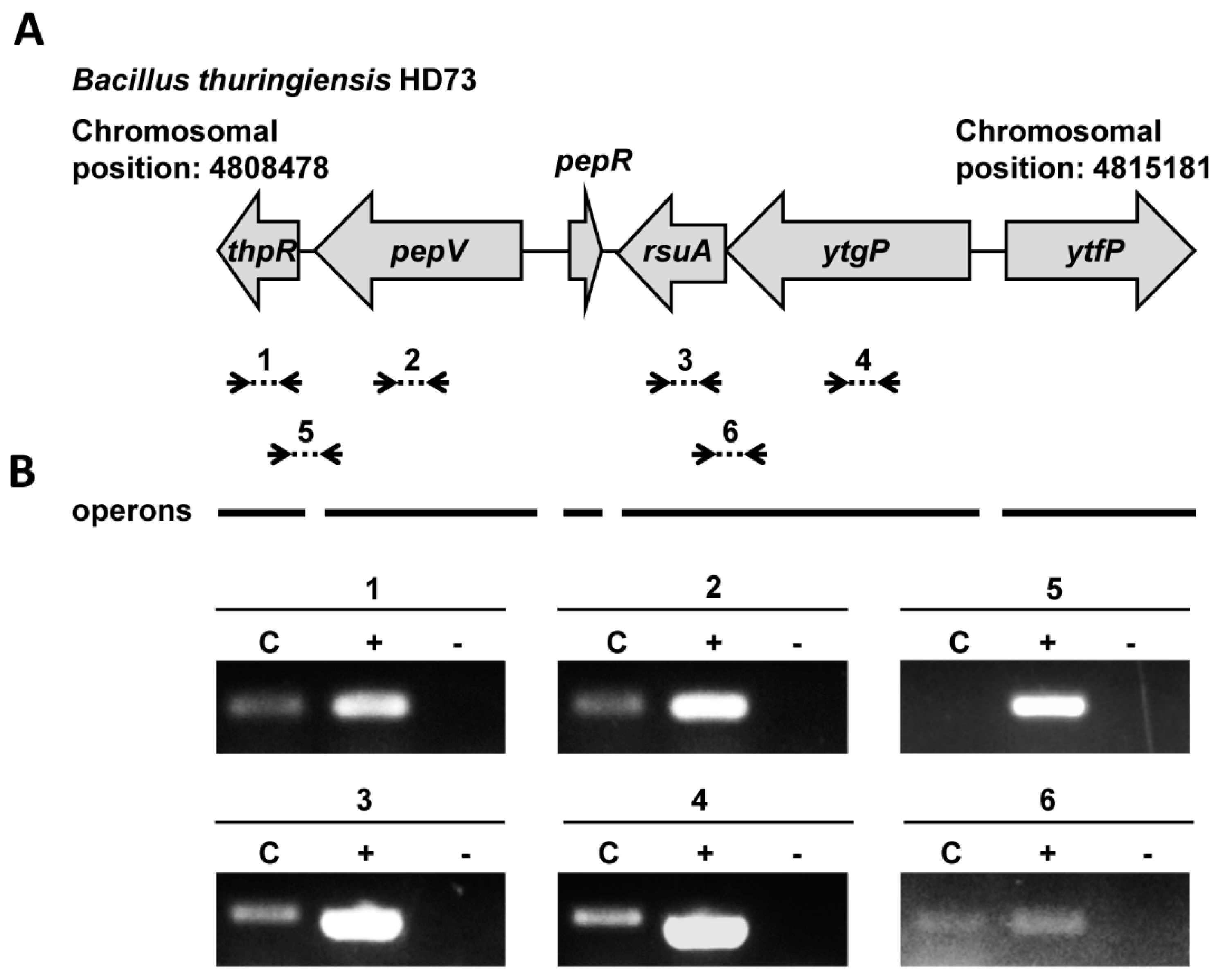
The nucleotide sequence of the pepR gene cluster (4489 bp) of HD73 is comprised of four ORFs, which are annotated as dipeptidase (pepV, HD73_5013), DeoR family transcriptional regulator (pepR, HD73_5014), rRNA pseudouridine synthase (rsuA, HD73_5015), and polysaccharide biosynthesis protein (ytgP, HD73_5016) (Figure 2A). To determine the transcription units of the pepR cluster, a series of primers were designed. The total RNA was extracted at T6 from B. thuringiensis HD73 cultures grown in SSM. RT-PCR results showed mRNA overlapping the rsuA and ytgP genes (Figure 2B). However, no mRNA overlapping thpR and pepV was transcribed. These results indicate that rsuA and ytgP genes form a transcriptional unit (rsuA-ytgP operon), while thpR and pepV each form a transcriptional unit independently.

3.3. PepR Positively Regulates pepV and rsuA-ytgP

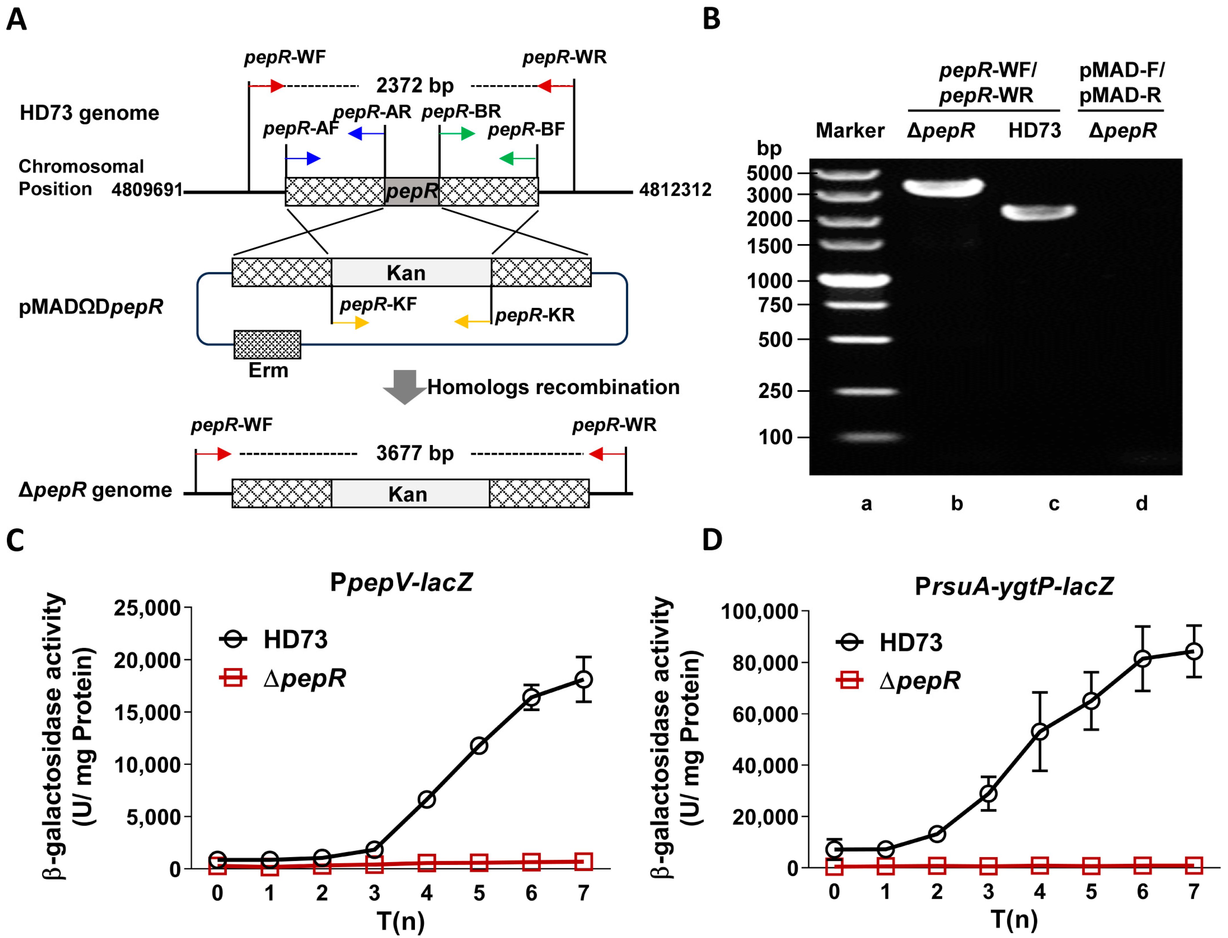
To verify whether PepR regulates the transcription of pepV and rsuA-ytgP, we first constructed a pepR deletion mutant. The upstream (pepR-A, 1065 bp) and downstream (pepR-B, 1057 bp) homologous fragments of pepR were amplified from the HD73 genome DNA. Then, the pepR-A, pepR-B, and kanamycin resistance gene (pepR-K, 1506 bp) fragments were ligated together by overlapping PCR. The resulting 3563-bp fragment was inserted into the temperature-sensitive pMAD plasmid, leading to pMADΩDpepR plasmid. The pMADΩDpepR plasmid was electroporated into HD73. This transformant was then screened at 37 °C to identify the pepR deletion mutant, which lacked erythromycin resistance but was resistant to kanamycin. The diagram shows that the kanamycin resistance gene on the recombinant plasmid was exchanged by homolog recombination with pepR on the HD73 chromosome (Figure 3A). HD73 and ΔpepR strains were confirmed by PCR using primers of pepR-WF/pepR-WR. PCR products with the size of 3677 bp from ΔpepR and 2372 bp from HD73 were detected by agarose gel electrophoresis (Figure 3B). The ΔpepR DNA as template was amplified by PCR using primers (pMAD-F/pMAD-R) for detecting the pMAD plasmid, resulting in no PCR products (Figure 3B). It proved that the ΔpepR mutant was obtained.
Then, PpepV and PrsuA-ytgP promoter activity was measured in HD73 and ∆pepR. The PpepV and PrsuA-ytgP promoters were fused with the lacZ reporter gene and transformed into HD73 and ∆pepR, respectively. β-galactosidase activity showed that the activity of PpepV increased from T0 to T7 and reached the highest level at T7 in HD73. However, the transcriptional activity of PpepV was significantly reduced from T0 to T7 in ∆pepR (Figure 3C). The result revealed that the PpepV promoter is regulated by PepR. Similarly, the transcriptional activity of PrsuA-ytgP in the pepR mutant strain was also significantly decreased compared to that in HD73 from T0 to T7 (Figure 3D), which suggested that the rsuA-ytgP operon was also regulated by PepR.

3.4. PepR Binds to the pepV Promoter

To determine whether the PepR protein directly binds to the pepV or rsuA-ytgP promoter regions, the electrophoretic mobility shift assay (EMSA) was performed. We constructed the recombinant plasmid, pETpepR, which was able to express the His-tagged PepR protein and introduced it into the E. coli BL21(DE3). The PepR-His protein with a molecular weight of approximately 10.06 kDa was expressed in E. coli BL21(pETpepR) and purified by Ni2+-affinity chromatography (Figure 4A). Then, we tested the binding of the PepR protein to the pepV or rsuA-ytgP promoter regions. A 0.21 nM PpepV-labeled probe was used to bind to PepR (low concentrations of 34.79 μM). The PpepV-labeled probe at 0.21 nM was strongly shifted with a 44.73 nM PepR (Figure 4B). Notably, a 200-fold excess of unlabeled probe competed with the labeled probe, confirming the specific binding (Figure 4B). These results demonstrated that PepR directly binds to the pepV promoter region. However, when 0.19 nM of the PrsuA-ytfP probe was exposed to PepR concentrations ranging from 14.91 μM to 44.73 μM, no bind-shift could be detected, indicating that PepR cannot interact with the rusA-ytfP promoter region (Figure 4C). These results strongly support the theory that PepR directly regulates the expression of pepV and indirectly regulates rusA-ytfP expression.

3.5. ΔpepR and ΔpepV Mutants Are More Sensitive to Vancomycin Than HD73

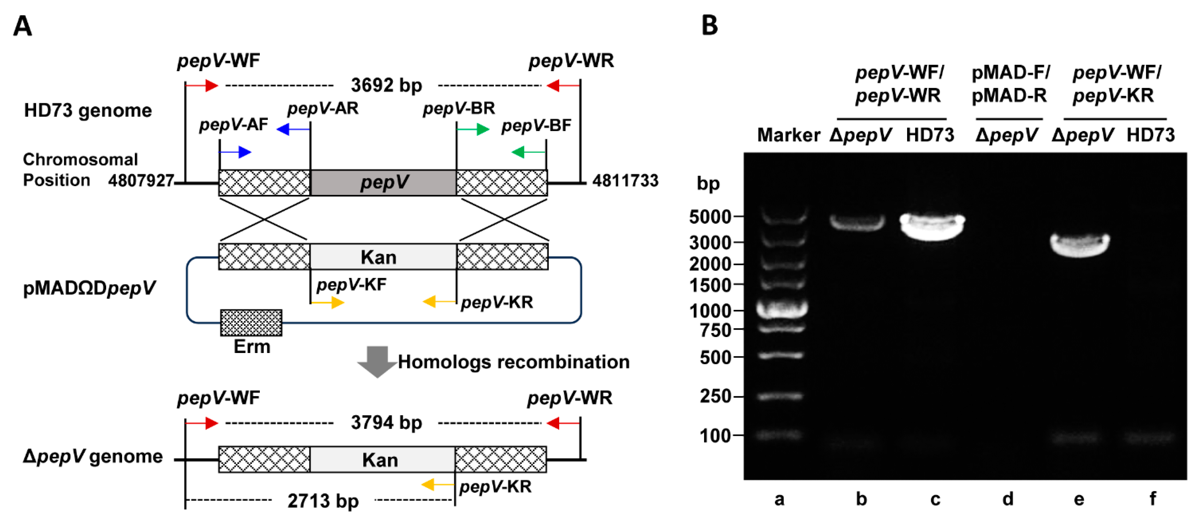
To characterize the function of the pepV gene, we constructed a pepV deletion mutant. The homolog arms on both sides of pepV were pepV-A (993 bp) and pepV-B (1023 bp) amplified from HD73 using primers pepV-AF/pepV-AR and pepV-BF/pepV-BR, respectively. The pepV-K fragment was obtained using PCR with pepV-KF/pepV-KR. The overlapping PCR products of pepV-A, pepV-B, and pepV-K were amplified with pepV-AF/pepV-BR primers, resulting in a 3452 bp fragment. The fragment was digested with BamHI and EcoRI sites and ligated to the temperature-sensitive vector of pMAD to obtain recombinant plasmid pMAD∆pepV. The recombinant plasmid pMAD∆pepV was electroporated into HD73. Transformants were selected for anti-sensitivity to erythromycin and kanamycin (Figure 5A). Positive transformants were verified at 37 °C. Colonies lacking erythromycin resistance and containing kanamycin resistance were selected for ∆pepV.
The pepV deletion mutant and HD73 strains were confirmed by PCR using pepV-WF/pepV-WR primers. PCR was performed with HD73 and ∆pepV chromosomal DNA as template. The product from ∆pepV was a 3794 bp fragment, while the product from HD73 was a 3692 bp fragment (Figure 5B, Lanes b and c). The ΔpepV mutant was confirmed using PCR with pMAD plasmid primers of pMAD-F/pMAD-R, which did not produce any PCR product (Figure 5B, Lane d). In addition, PCR products were generated using HD73 and ∆pepV chromosomal DNA as templates with primers (pepV-WF/pepV-KR). There was a 2713 bp fragment from ∆pepV, while no product from HD73 was obtained (Figure 5B, Lanes f and e). The result confirms that the ΔpepV mutant was successfully obtained.
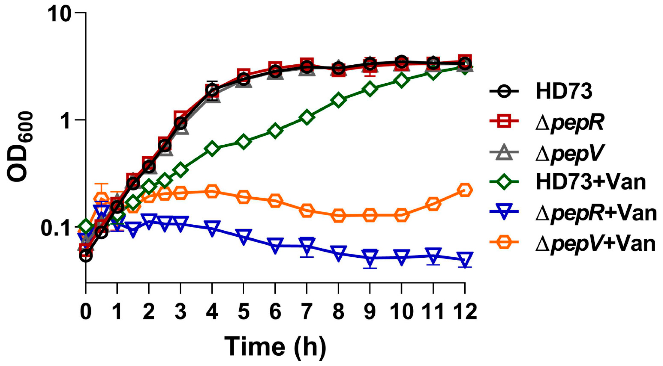
It has been demonstrated that PepV is related to vancomycin resistance in L. lactis MG1363 [29]. A vancomycin resistance test was performed by inoculating ∆pepR, ∆pepV, and HD73 in SSM medium with 6 ng/mL concentrations of vancomycin. The result showed that the absence of vancomycin had no effect on the growth of ∆pepR, ∆pepV, and HD73 strains. However, the addition of vancomycin slowed HD73 growth. ∆pepV growth stagnated while ∆pepR lysed and died after vancomycin was added (Figure 6). The result suggests that ∆pepR and ∆pepV were more sensitive to vancomycin than HD73, and ∆pepR was more sensitive to vancomycin than ∆pepV.

4. Discussion

In our previous study, we validated the strong promoter activity from the pepR gene which encodes a DeoR family transcriptional regulator [12]. The pepR promoter is activated by Sigma E and can direct cry1Ac expression efficiently [12]. However, the functions of the pepR gene and its targets were unknown. In this study, we found that PepR homologs are widely distributed in various Gram-positive bacteria, and the amino acid similarities are highly conserved in different bacteria (Figure 1). We selected 18 strains that have been extensively studied for exhaustive analyses of the pepR genetic loci. We found that pepV, pepR and rusA-ytgP were closely linked in some strains, such as B. thuringiensis HD73, B. cereus ATCC 14579, B. cytotoxicus, B. anthracis Ames, Planococcus antarcticus, Ectobacillus sp. JY-23, Sporosarcina psychrophile, and Sutcliffiella horikoshii, while in some other strains they were not closely linked, such as in Anoxybacillus caldiproteolyticus, A. flavithermus, Priestia koreensis, Mangrovibacillus cuniculi, P. megaterium QM B1551, B. pumilus, Lysinibacillus fusiformis, Aeribacillus pallidus KCTC 3564, Metabacillus sediminilitoris, and Cytobacillus spongiae (Figure S1). In HD73, pepV and rusA-ytgP were two independent operons, and their promoters failed to transcript in the pepR knockout strain (Figure 2 and Figure 3), suggesting these genes are all controlled by transcriptional regulator PepR. We further found that PepR directly binds to the pepV promoter but not to the rusA-ytgP promoter (Figure 4 and Figure 5), revealing that pepV acts as a downstream target of pepR. The pepV promoter regions were 98.95% similar in different B. thuringiensis strains, and we determined that the expression of pepV is regulated by PepR. Whether PepR homologs also regulate pepV transcription in other bacteria remains to be confirmed. In addition, other targets of PepR should be investigated using RNA-seq and ChIP-seq methods in following studies.
PepV is highly conserved within the dipeptidase M20 family in the metallopeptidases [30]. It has been reported that the active sites and conserved domains of all PepVs are similar [30,31,32]. We analyzed the amino acid sequence of the PepV homologs and found that the identity varies in B. subtilis (57.36%), L. delbrueckii (34.62%), L. helveticus (33.12%), S. gordonii (40.86%) and L. lactis (38.92%) compared to HD73 (Figure S2). This result suggests that PepV in HD73 may have a similar function in catalyzing substrates to that reported in other bacteria. It is worth mentioning that the deletion of pepV in HD73 increased the susceptibility to vancomycin, whereas the deletion of pepV in L. lactis MG1363 decreased the susceptibility of L. lactis to vancomycin [29]. In addition to BCARR regulating pepV expression in the presence of BCAAs in L. helveticus [17] and a CodY-like regulatory system controlling the expression of pepV in L. helveticus CM4 [16], pepV might have multiple functions in responding to different signals in different bacteria strains.

Supplementary Materials

The following supporting information can be downloaded at: https://www.mdpi.com/article/10.3390/microorganisms12030579/s1, Figure S1: The pepR gene cluster is a conserved in bacteria; Figure S2: Comparison of the amino acid sequences revealed a conserved PepV homologs in bacteria; Table S1: List of species in Figure 1.

Author Contributions

F.S. and X.Z. designed the experiments. X.Z., H.W., Y.C. and T.Y. performed the experiments. X.Z., Q.P. and F.S. analyzed the results. X.Z. wrote the manuscript. F.S. revised the manuscript. All authors have read and agreed to the published version of the manuscript.

Funding

This work was supported by grants from the National Key R&D Program of China (Grant No. 2022YFE0116500) and the National Natural Science Foundation of China (General Program, grants No. 32072499 and No. 32372623).

Data Availability Statement

Data are contained within the article.

Conflicts of Interest

The authors declare no conflict of interest.

References

  1. Argôlo-Filho, R.C.; Loguercio, L.L. Bacillus thuringiensis Is an Environmental Pathogen and Host-Specificity Has Developed as an Adaptation to Human-Generated Ecological Niches. Insects 2013, 5, 62–91. [Google Scholar] [CrossRef] [PubMed]
  2. Palma, L.; Muñoz, D.; Berry, C.; Murillo, J.; Caballero, P. Bacillus thuringiensis toxins: An overview of their biocidal activity. Toxins 2014, 6, 3296–3325. [Google Scholar] [CrossRef] [PubMed]
  3. Agaisse, H.; Lereclus, D. How does Bacillus thuringiensis produce so much insecticidal crystal protein? J. Bacteriol. 1995, 177, 6027–6032. [Google Scholar] [CrossRef] [PubMed]
  4. Peng, Q.; Yu, Q.; Song, F. Expression of cry genes in Bacillus thuringiensis biotechnology. Appl. Microbiol. Biot. 2019, 103, 1617–1626. [Google Scholar] [CrossRef]
  5. García-Gómez, B.I.; Sánchez, J.; Martínez de Castro, D.L.; Ibarra, J.E.; Bravo, A.; Soberón, M. Efficient production of Bacillus thuringiensis Cry1AMod toxins under regulation of cry3Aa promoter and single cysteine mutations in the protoxin region. Appl. Environ. Microb. 2013, 79, 6969–6973. [Google Scholar] [CrossRef] [PubMed]
  6. Chaoyin, Y.; Wei, S.; Sun, M.; Lin, L.; Faju, C.; Zhengquan, H.; Ziniu, Y. Comparative study on effect of different promoters on expression of cry1Ac in Bacillus thuringiensis chromosome. J. Appl. Microbiol. 2007, 103, 454–461. [Google Scholar] [CrossRef]
  7. Sanchis, V.; Agaisse, H.; Chaufaux, J.; Lereclus, D. Construction of new insecticidal Bacillus thuringiensis recombinant strains by using the sporulation non-dependent expression system of cryIIIA and a site specific recombination vector. J. Bacteriol. 1996, 48, 81–96. [Google Scholar] [CrossRef]
  8. de Souza, M.T.; Lecadet, M.M.; Lereclus, D. Full expression of the cryIIIA toxin gene of Bacillus thuringiensis requires a distant upstream DNA sequence affecting transcription. J. Bacteriol. 1993, 175, 2952–2960. [Google Scholar] [CrossRef]
  9. Jia, Y.; Zhao, C.; Wang, Q.; Shu, C.; Feng, X.; Song, F.; Zhang, J. A genetically modified broad-spectrum strain of Bacillus thuringiensis toxic against Holotrichia parallela, Anomala corpulenta and Holotrichia oblita. World J. Microb. Biot. 2014, 30, 595–603. [Google Scholar] [CrossRef]
  10. Guan, P.; Dai, X.; Zhu, J.; Li, Q.; Li, S.; Wang, S.; Li, P.; Zheng, A. Bacillus thuringiensis subsp. sichuansis strain MC28 produces a novel crystal protein with activity against Culex quinquefasciatus larvae. World J. Microb. Biot. 2014, 30, 1417–1421. [Google Scholar] [CrossRef]
  11. Zheng, Q.; Wang, G.; Zhang, Z.; Qu, N.; Zhang, Q.; Peng, Q.; Zhang, J.; Gao, J.; Song, F. Expression of cry1Ac gene directed by PexsY promoter of the exsY gene encoding component protein of exosporium basal layer in Bacillus thuringiensis. Acta Microbiol. Sin. 2014, 54, 1138–1145. [Google Scholar]
  12. Zhang, X.; Gao, T.; Peng, Q.; Song, L.; Zhang, J.; Chai, Y.; Sun, D.; Song, F. A strong promoter of a non-cry gene directs expression of the cry1Ac gene in Bacillus thuringiensis. Appl. Microbiol. Biot. 2018, 102, 3687–3699. [Google Scholar] [CrossRef]
  13. Guédon, E.; Renault, P.; Ehrlich, S.D.; Delorme, C. Transcriptional pattern of genes coding for the proteolytic system of Lactococcus lactis and evidence for coordinated regulation of key enzymes by peptide supply. J. Bacteriol. 2001, 183, 3614–3622. [Google Scholar] [CrossRef]
  14. Mierau, I.; Tan, P.S.; Haandrikman, A.J.; Mayo, B.; Kok, J.; Leenhouts, K.J.; Konings, W.N.; Venema, G. Cloning and sequencing of the gene for a lactococcal endopeptidase, an enzyme with sequence similarity to mammalian enkephalinase. J. Bacteriol. 1993, 175, 2087–2096. [Google Scholar] [CrossRef]
  15. Nardi, M.; Renault, P.; Monnet, V. Duplication of the pepF gene and shuffling of DNA fragments on the lactose plasmid of Lactococcus lactis. J. Bacteriol. 1997, 179, 4164–4171. [Google Scholar] [CrossRef]
  16. Wakai, T.; Yamaguchi, N.; Hatanaka, M.; Nakamura, Y.; Yamamoto, N. Repressive processing of antihypertensive peptides, Val-Pro-Pro and Ile-Pro-Pro, in Lactobacillus helveticus fermented milk by added peptides. J. Biosci. Bioeng. 2012, 114, 133–137. [Google Scholar] [CrossRef]
  17. Wakai, T.; Yamamoto, N. A novel branched chain amino acids responsive transcriptional regulator, BCARR, negatively acts on the proteolytic system in Lactobacillus helveticus. PLoS ONE 2013, 8, e75976. [Google Scholar] [CrossRef] [PubMed]
  18. Schaeffer, P.; Millet, J.; Aubert, J.P. Catabolic repression of bacterial sporulation. Proc. Natl. Acad. Sci. USA 1965, 54, 704–711. [Google Scholar] [CrossRef]
  19. Macaluso, A.; Mettus, A.M. Efficient transformation of Bacillus thuringiensis requires nonmethylated plasmid DNA. J. Bacteriol. 1991, 173, 1353–1356. [Google Scholar] [CrossRef] [PubMed]
  20. Wang, G.; Zhang, J.; Song, F.; Wu, J.; Feng, S.; Huang, D. Engineered Bacillus thuringiensis GO33A with broad insecticidal activity against lepidopteran and coleopteran pests. Appl. Microbiol. Biot. 2006, 72, 924–930. [Google Scholar] [CrossRef] [PubMed]
  21. Munson, R.S., Jr.; Sasaki, K. Protein D, a putative immunoglobulin D-binding protein produced by Haemophilus influenzae, is glycerophosphodiester phosphodiesterase. J. Bacteriol. 1993, 175, 4569–4571. [Google Scholar] [CrossRef]
  22. Agaisse, H.; Lereclus, D. Structural and functional analysis of the promoter region involved in full expression of the cryIIIA toxin gene of Bacillus thuringiensis. Mol. Microbiol. 1994, 13, 97–107. [Google Scholar] [CrossRef]
  23. Arnaud, M.; Chastanet, A.; Débarbouillé, M. New vector for efficient allelic replacement in naturally nontransformable, low-GC-content, gram-positive bacteria. Appl. Environ. Microb. 2004, 70, 6887–6891. [Google Scholar] [CrossRef]
  24. Yang, H.; Wang, P.; Peng, Q.; Rong, R.; Liu, C.; Lereclus, D.; Zhang, J.; Song, F.; Huang, D. Weak transcription of the cry1Ac gene in nonsporulating Bacillus thuringiensis cells. Appl. Environ. Microb. 2012, 78, 6466–6474. [Google Scholar] [CrossRef]
  25. Sambrook, B.J.; Russell, D.W. Molecular Cloning: A Laboratory Manual; Cold Spring Harbor Laboratory Press: Cold Spring Harbor, NY, USA, 2015. [Google Scholar]
  26. Lereclus, D.; Arantes, O.; Chaufaux, J.; Lecadet, M. Transformation and expression of a cloned delta-endotoxin gene in Bacillus thuringiensis. FEMS Microbiol. Lett. 1989, 51, 211–217. [Google Scholar]
  27. Peng, Q.; Yang, M.; Wang, W.; Han, L.; Wang, G.; Wang, P.; Zhang, J.; Song, F. Activation of gab cluster transcription in Bacillus thuringiensis by γ-aminobutyric acid or succinic semialdehyde is mediated by the Sigma 54-dependent transcriptional activator GabR. BMC Microbiol. 2014, 14, 306. [Google Scholar] [CrossRef]
  28. Li, R.; Liu, G.; Xie, Z.; He, X.; Chen, W.; Deng, Z.; Tan, H. PolY, a transcriptional regulator with ATPase activity, directly activates transcription of polR in polyoxin biosynthesis in Streptomyces cacaoi. Mol. Microbiol. 2010, 75, 349–364. [Google Scholar] [CrossRef]
  29. Huang, C.; Hernandez-Valdes, J.A.; Kuipers, O.P.; Kok, J. Lysis of a Lactococcus lactis Dipeptidase Mutant and Rescue by Mutation in the Pleiotropic Regulator CodY. Appl. Environ. Microb. 2020, 86, e02937-19. [Google Scholar] [CrossRef]
  30. Goldstein, J.M.; Kordula, T.; Moon, J.L.; Mayo, J.A.; Travis, J. Characterization of an extracellular dipeptidase from Streptococcus gordonii FSS2. Infect. Immun. 2005, 73, 1256–1259. [Google Scholar] [CrossRef]
  31. Jozic, D.; Bourenkow, G.; Bartunik, H.; Scholze, H.; Dive, V.; Henrich, B.; Huber, R.; Bode, W.; Maskos, K. Crystal structure of the dinuclear zinc aminopeptidase PepV from Lactobacillus delbrueckii unravels its preference for dipeptides. Structure 2002, 10, 1097–1106. [Google Scholar] [CrossRef]
  32. Mori, S.; Miyamoto, M.; Kaneko, S.; Nirasawa, S.; Komba, S.; Kasumi, T. Characterization and kinetic analysis of enzyme-substrate recognition by three recombinant lactococcal PepVs. Arch. Biochem. Biophys. 2006, 454, 137–145. [Google Scholar] [CrossRef] [PubMed]
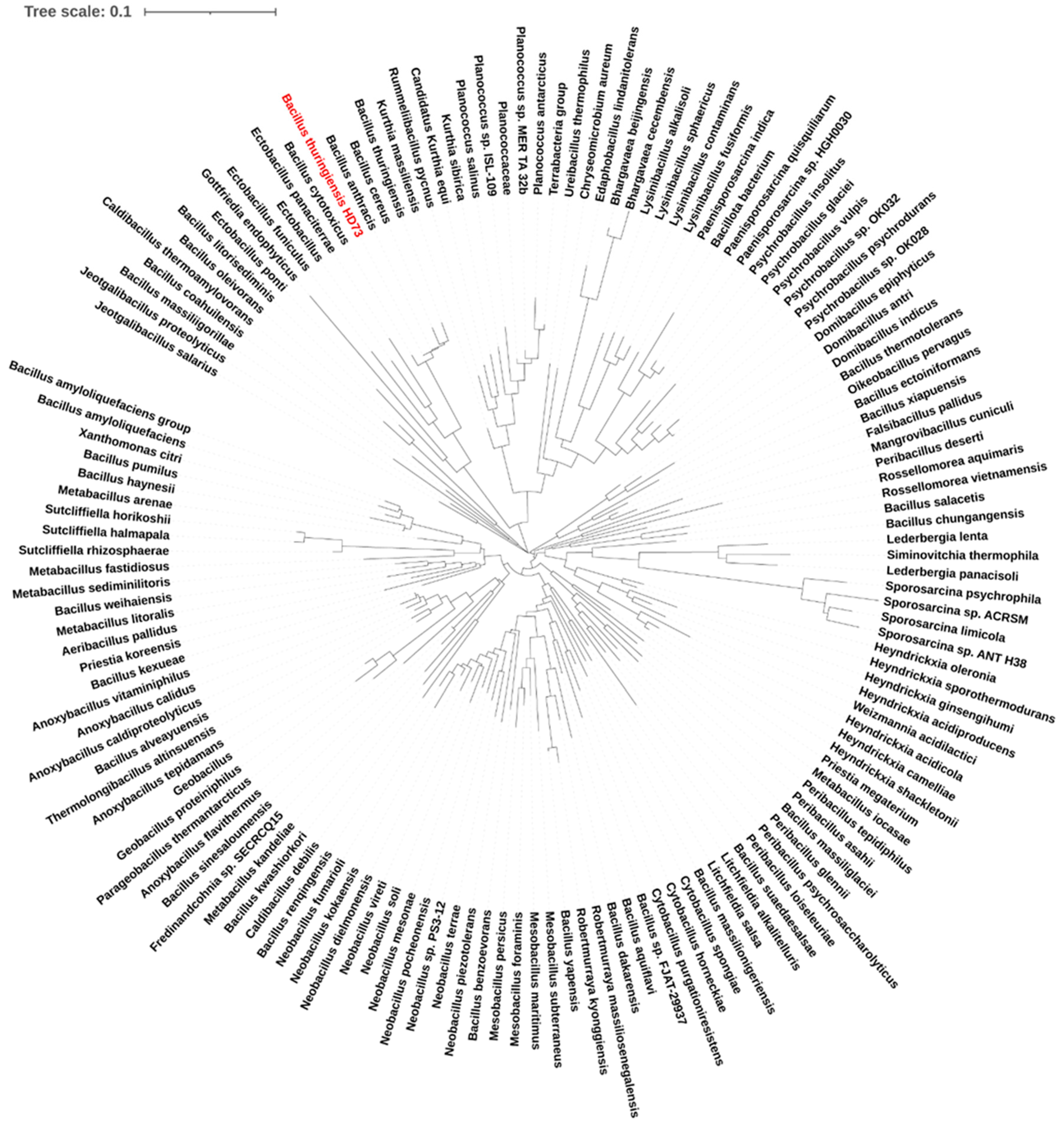
Figure 1. Evolution of PepR homologs in bacteria. Evolutionary analyses were conducted in MEGA7 using the neighbor-joining method. A total of 143 amino acid sequences were analyzed and an optimal tree was shown. Red text indicates the B. thuringiensis HD73 strain from which the reference sequence of the PepR homologs originated.
Microorganisms 12 00579 g001
Figure 2. RT-PCR identified the pepV and rsuA-ytgP transcription units at the pepR gene locus in B. thuringiensis HD73. (A) Genetic organization of the pepR locus. Open reading frames (ORFs) are represented by grey arrows. The RT-PCR amplicons (see lanes in panel (B)) correspond to the dashed lines with small black arrows. The solid lines below the ORFs represent the operons. (B) RT-PCR analyzed the transcription units of pepR neighboring genes. RNA samples were prepared at T6 of bacterial culture in SSM medium. The RT-PCR labelled “C” was performed with 500 ng RNA. Positive controls are labelled “+” (PCR with 100 ng of genomic DNA). Negative controls are labelled “−” (RT-PCR with 500 ng RNA using heat-inactivated reverse transcriptase). Numbers represent different RT-PCR amplicons: numbers 1–4 represent thpR, pepV, rsuA, and ytgP; numbers 5 and 6 represent thpR-pepV and rsuA-ytgP, respectively.
Microorganisms 12 00579 g002
Figure 3. Transcription of pepV and rsuA-ygtP depended on pepR. (A) Construction of an in-frame deletion pepR mutant in HD73. The pepR gene was substituted by a kanamycin-resistant gene through double homolog recombination events (B) Identification of ΔpepR knockout mutant by PCR. PCR products were amplified from ΔpepR (Lane b) and HD73 (Lane c) with primer pair pepR-WF/pepR-WR. To confirm the presence of the pMADΩDpepR plasmid in the ΔpepR genome, PCR was performed using pMAD plasmid universal primers (pMAD-F/pMAD-R) (Lane d). The numbers indicate the size of the DNA standards in kilobase pairs (Lane a). (C) β-galactosidase activity of PpepV-lacZ in HD73 and ΔpepR. (D) β-galactosidase activity of PrsuA-ygtP-lacZ in HD73 and ΔpepR. The promoters of pepV and rsuA-ygtP were fused with the lacZ reporter and transformed into HD73 and ΔpepR, respectively. The β-galactosidase activities of three clones were determined at the indicated times after growing the cells in SSM at 30 °C. Each value represents the mean and standard error of at least three independent replicates.
Microorganisms 12 00579 g003
Figure 4. PepR directly bound pepV but not the rsuA-ygtP promoter region. (A) SDS-PAGE confirmed the expression and purification of the PepR-His protein by Ni2+-affinity chromatography. Lane a, standard proteins (PageRuler Prestained Protein Ladder 26632, Thermo, Rockford, IL, USA). Lane b, purified PepR from the BL21(pETpepR) strain. (B) EMSA detecting protein–DNA interactions using FAM-labeled PpepV and increasing concentrations of recombinant PepR. The lanes contained 0, 14.91, 24.85, 34.79, and 44.73 μM of the PepR protein. The last lane was employed for the 200-fold unlabeled probe. Protein–DNA complexes were separated by native-PAGE and FAM-labeled bands scanned using Typhoon9410 (Cytiva, Marlborough, MA, USA). (C) EMSA detecting protein–DNA interactions using FAM-labeled PrusA-ytgP and increasing concentrations of recombinant PepR.
Microorganisms 12 00579 g004
Figure 5. Construction of an in-frame deletion pepV mutant in HD73. (A) Construction of an in-frame deletion pepV mutant. The pepV gene was substituted by a kanamycin-resistant gene through double homolog recombination events. (B) Identification of the ΔpepR knockout mutant by PCR. PCR products were amplified from HD73 and ΔpepV strains using the primer pairs of pepV-WF/pepV-WR (Lanes b and c) and pepV-WF/pepV-KR (Lanes e and f). To confirm the presence of the pMADΩDpepV plasmid in the ΔpepV genome, PCR was performed using pMAD plasmid universal primers (pMAD-F/pMAD-R) (Lane d). The numbers indicate the size of the DNA standards in kilobase pairs (Lane a).
Microorganisms 12 00579 g005
Figure 6.pepR and ΔpepV mutant strains were more sensitive to vancomycin compared to HD73. Bacteria growth was monitored every hour after inoculation to culture medium with vancomycin at 6 ng/mL. Growth curves represent means of three clones.
Microorganisms 12 00579 g006
Table 1. Strains and plasmids used in this study.
Strain or PlasmidRelevant Details aReference or Source
E. coli strains
E. coli TG1∆(lac-proAB) supE thi hsd-5 (F′ traD36 proA+ proB+ lacIq lacZ∆M15), general purpose cloning hostLaboratory collection
E. coli ET 12567F dam-13::Tn 9 dcm-6 hsdM hsdR recF143 zjj-202::Tn10 galK2 galT22 ara14 pacY1 xyl-5 leuB6 thi-1, for generation of unmethylated DNALaboratory collection
BL21 (DE3)F dcmopmT hsds (rB mB) galλ(DE3)[21]
BL21 (pETpepR)BL21(DE3) with pETpepR plasmidThis study
B. thuringiensis strains
HD73Wild-type strain containing plasmid pHT73 carrying cry1Ac geneLaboratory collection
pepRHD73 mutant, pepR gene was deleted by homologs recombinationThis study
pepVHD73 mutant, pepV gene was deleted by homologs recombinationThis study
HD (PpepV)HD73 strain containing plasmid pHT304-PpepV-18ZThis study
∆pepR (PpepV)pepR mutant containing plasmid pHT304-PpepV-18ZThis study
HD (PrsuA-ytgP)HD73 strain containing plasmid pHT304- PrsuA-ytgP-18ZThis study
∆pepR (PrsuA-ytgP)pepR mutant containing plasmid pHT304- PrsuA-ytgP-18ZThis study
Plasmids
pHT304-18ZE. coli-Bt shuttle vector with promoter-less lacZ reporter, AmpR, ErmR[22]
pET-21bExpression vector; Ampr 5.4 kbLaboratory collection
pMADAmpR, ErmR, temperature-sensitive E. coli-B. thuringiensis shuttle vector[23]
pMADΩDspo0ApMAD with spo0A deletion fragment, AmpR, ErmR, KanR[24]
pMADΩDpepRpMAD with pepR deletion fragment, AmpR, ErmR, KanRThis study
pMADΩDpepVpMAD with pepV deletion fragment, AmpR, ErmR, KanRThis study
pETpepRpET-21b containing pepR gene; AmprThis study
304PpepVpHT304-18Z carrying PpepV, AmpR, ErmRThis study
304PrsuA-ytgPpHT304-18Z carrying PrsuA-ytgP, AmpR, ErmRThis study
a Antibiotic resistance cassettes are indicated as follows: ErmR, erythromycin resistance; KanR, kanamycin resistance; AmpR, ampicillin resistance.
Table 2. Oligonucleotide primers used in this study.
Primer NameSequence (5′–3′) aRestriction Site
pETpepR-FCGGGATCCGTTGAAACCTACAACTACTCGBamHI
pETpepR-RACGCGTCGACTGAGGTCATTCTCACTTTCSalI
pepV-AFGTACCCGGGAGCTCGAATTCCGAAATGTCCGACTTGTTCCATACGEcoRI
pepV-ARTCACCTCAAATGGTTCGCTGGAATTAGCGAAGTAATGGATATATAAATAATGTCCGCTC
pepV-KFGAGCGGACATTATTTATATATCCATTACTTCGCTAATTCCAGCGAACCATTTGAGGTGA
pepV-KRGAAGGATGGATGCGTGATGTCAGCAATTAATTGGAAATTCCTCGTAGGCGCTCG
pepV-BFCGAGCGCCTACGAGGAATTTCCAATTAATTGCTGACATCACGCATCCATCCTTC
pepV-BRCGTCGGGCGATATCGGATCCGCACACGTTGCAGGAGTAGTAACAGAAGBamHI
pepV-WFGATACACAGCACCTAAATCTGTACCTTCG
pepV-WRCCCAGTTGGACGACTTGATATCGATACGGAAGG
pepR-AFGTACCCGGGAGCTCGAATTCAATCAAATGAAACAAGTTCATEcoRI
pepR-ARTCAAATGGTTCGCTGAATGCGTGTTAGCATACGAG
pepR-KFATGCTAACACGCATTCAGCGAACCATTTGAGGTGA
pepR-KRTTATGAGGTCATTCTAAATTCCTCGTAGGCGCTCG
pepR-BFGCCTACGAGGAATTTAGAATGACCTCATAATGAAA
pepR-BRCGTCGGGCGATATCGGATCCCAATTTGTGCAGGTATTGGCBamHI
pepR-WFCTTCTCCTGTAATAACCGCTTCTGC
pepR-WRCTAACACTTATAATGGTCGTTGCTG
pMAD-FCCAAATTTCCTCTGGCCATT
pMAD-RCCTATACCTTGTCTGCCTCC
PpepV-FCGCGGATCCCATCACGCATCCATCCTTCACTBamHI
PpepV-RAACTGCAGGAGAAAACCACTCCTCTACPstI
PrsuA-ytgP-FAACTGCAGCTGCAGCACCAATTGCAGCCATTAGPstI
PrsuA-ytgP-RCGCGGATCCGTAACAATAAGCGTTCCGCGCAGBamHI
RT-thpR-FACGAGCAACGGTAATA
RT-thpR-RGGGCGAAGGTAAGTGA
RT-pepV-FTGATGTGGGCGAGAAT
RT-pepV-RACGGTGGTAACTTAGGG
RT-thpR-pepV-FGTACATCTTCTCCTTCTTTC
RT-thpR-pepV-RGGCCCGCTATTCCCTGG
RT-rsuA-FCATCCTCTTCTGTTACTACTCC
RT-rsuA-RCAGCGACGGAAGATGATAATC
RT-ytgP-FGCTCCTACTGTACCGAAATAACG
RT-ytgP-RGATTCAGATCCACTAGGTGGAC
RT-rsuA-ytgP-FGTTTTTCACCTTCTTTATTTATC
RT-rsuA-ytgP-RCATGATTTCGCCTGATGGCCG
RT-16S-FTCGCATTAGCTAGTTGGTGAG
RT-16S-RTCTTCCCTAACAACAGAGTTT
a Restriction enzyme sites are underlined.
Disclaimer/Publisher’s Note: The statements, opinions and data contained in all publications are solely those of the individual author(s) and contributor(s) and not of MDPI and/or the editor(s). MDPI and/or the editor(s) disclaim responsibility for any injury to people or property resulting from any ideas, methods, instructions or products referred to in the content.