
A Pseudovirus-Based Neutralization Assay for SARS-CoV-2 Variants: A Rapid, Cost-Effective, BSL-2–Based High-Throughput Assay Useful for Vaccine Immunogenicity Evaluation

 , ,
, ,
Abstract
1. Introduction
2. Materials and Methods
2.1. Cell Lines
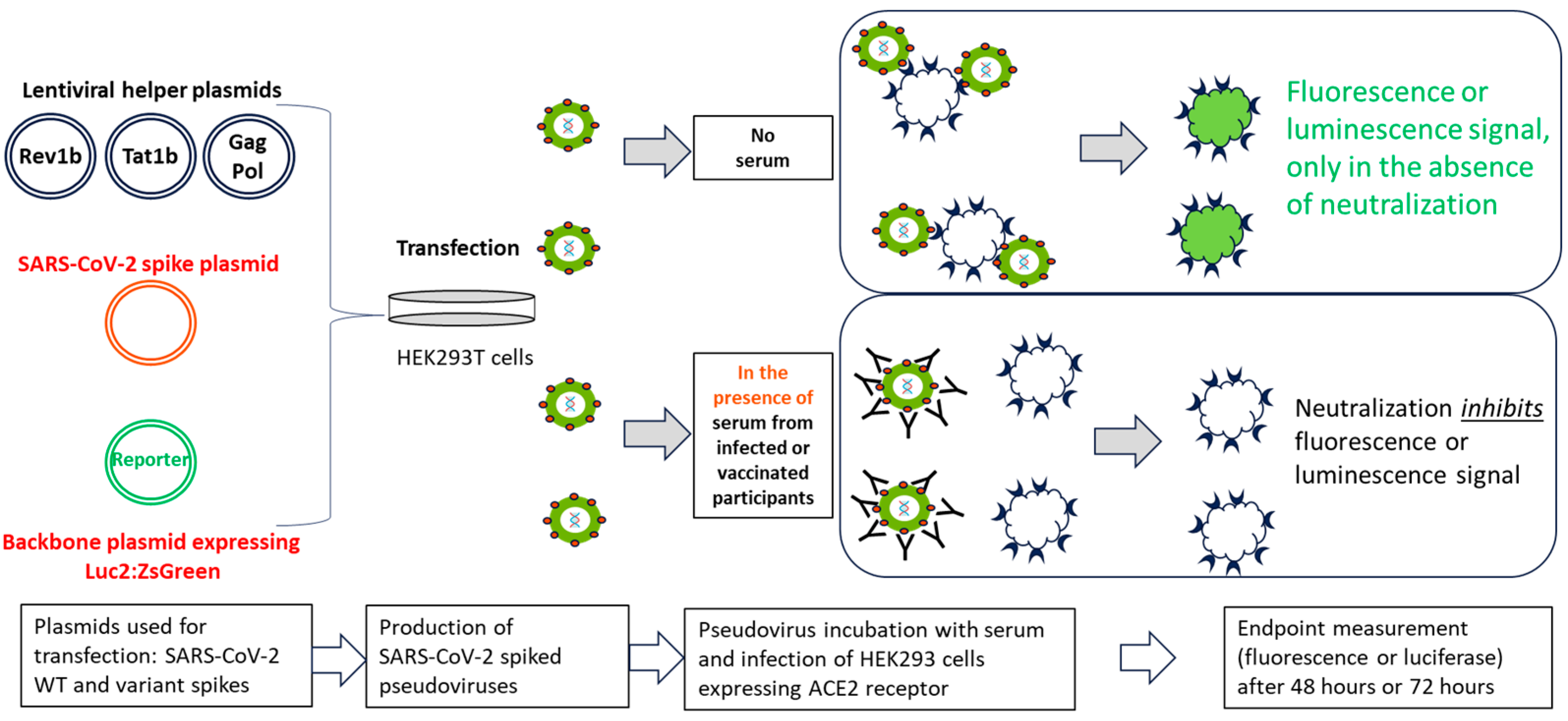
2.2. Pseudoviruses
2.3. Assay Procedure
2.4. Serum Samples
2.5. PNT Evaluation
2.6. Correlation Analyses
2.7. Clinical Utility
3. Results
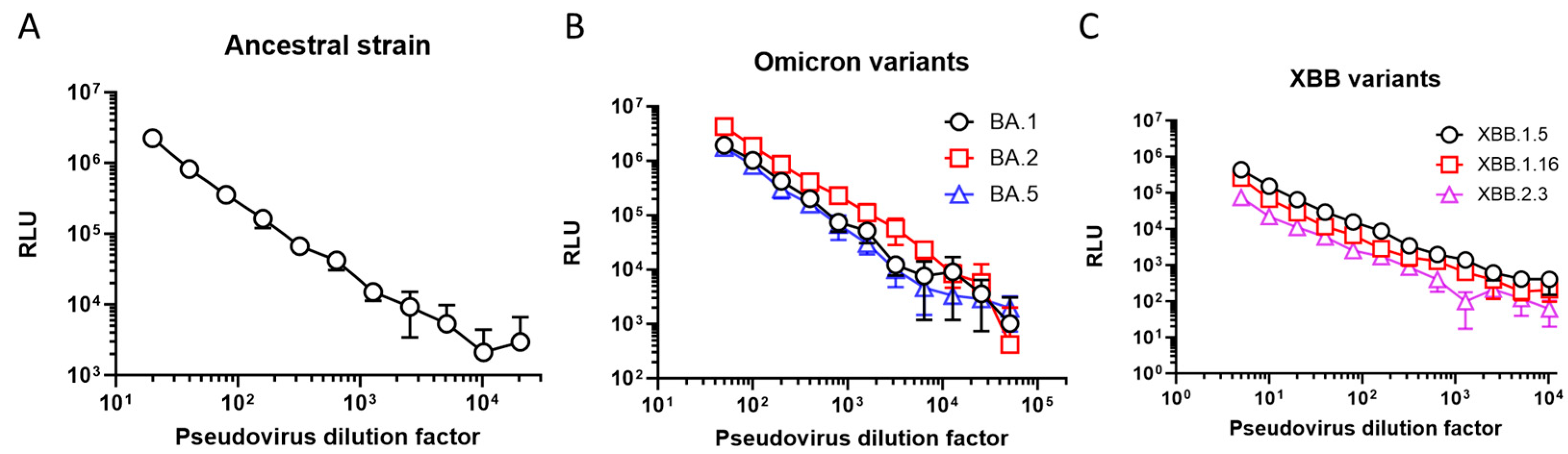
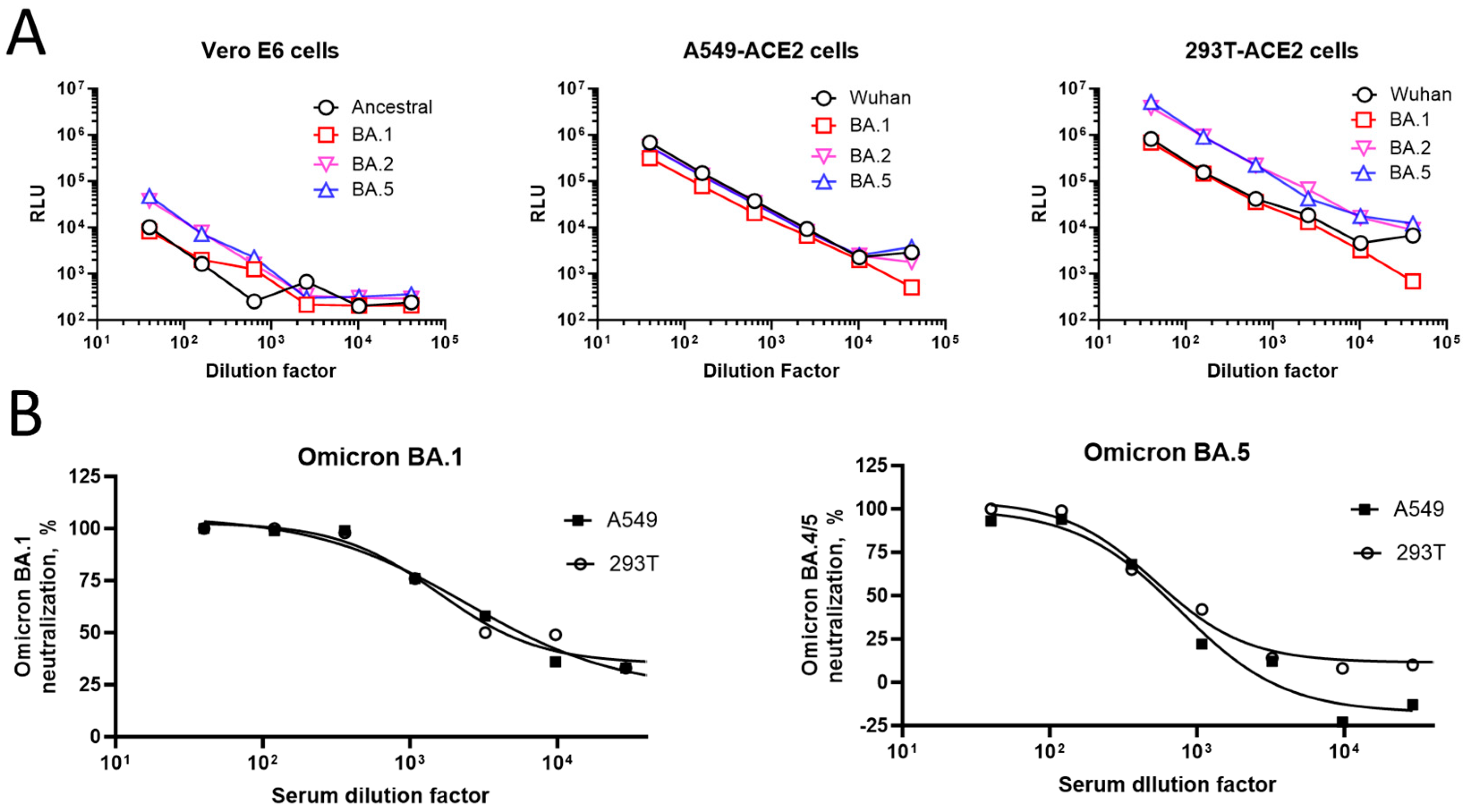
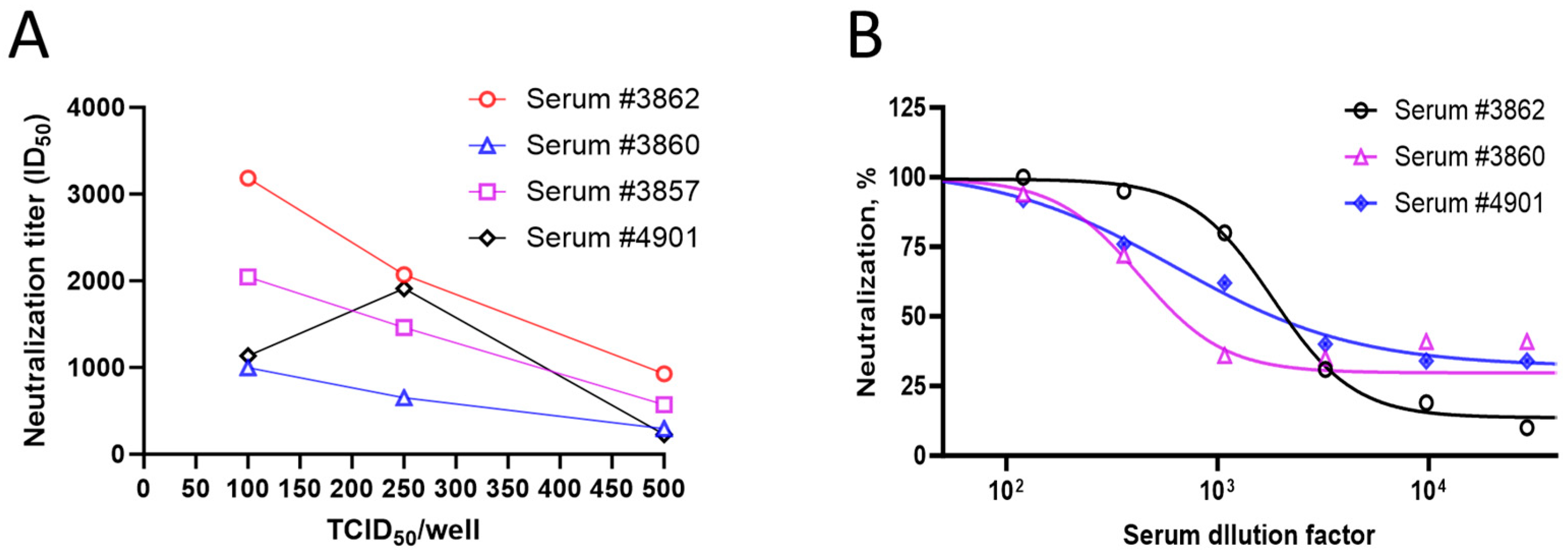
3.1. Assay Development/Optimization
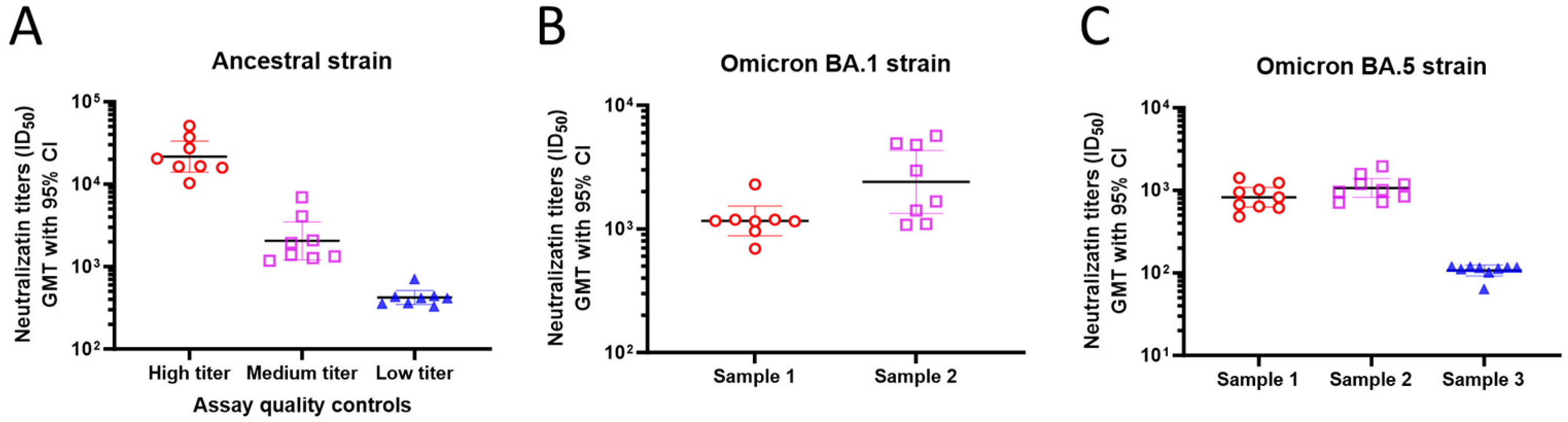
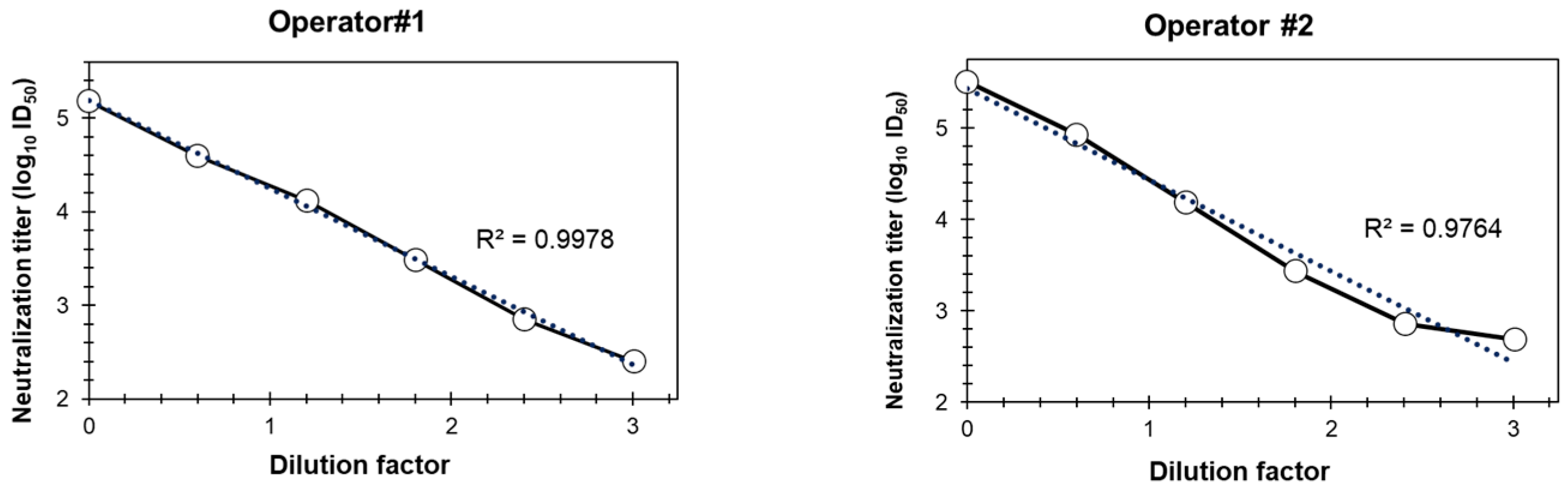
3.2. Assay Quality
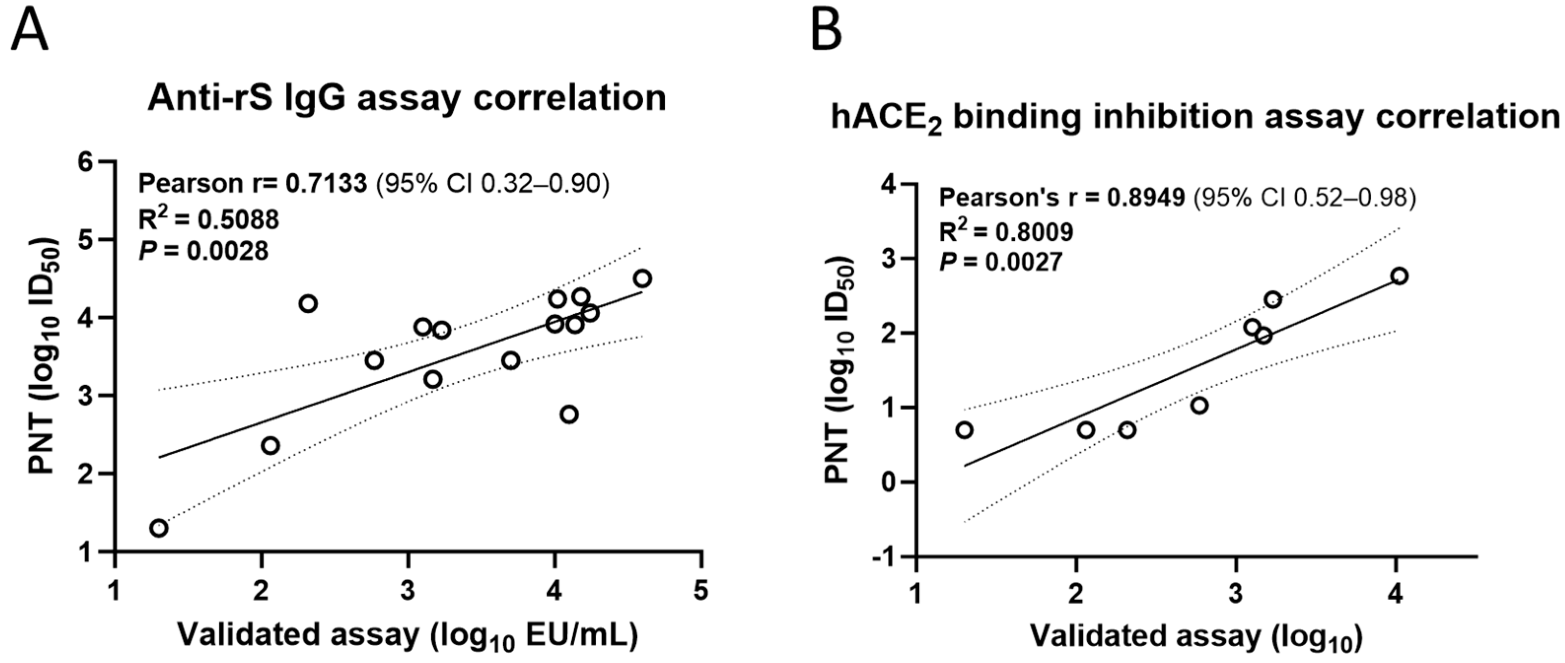
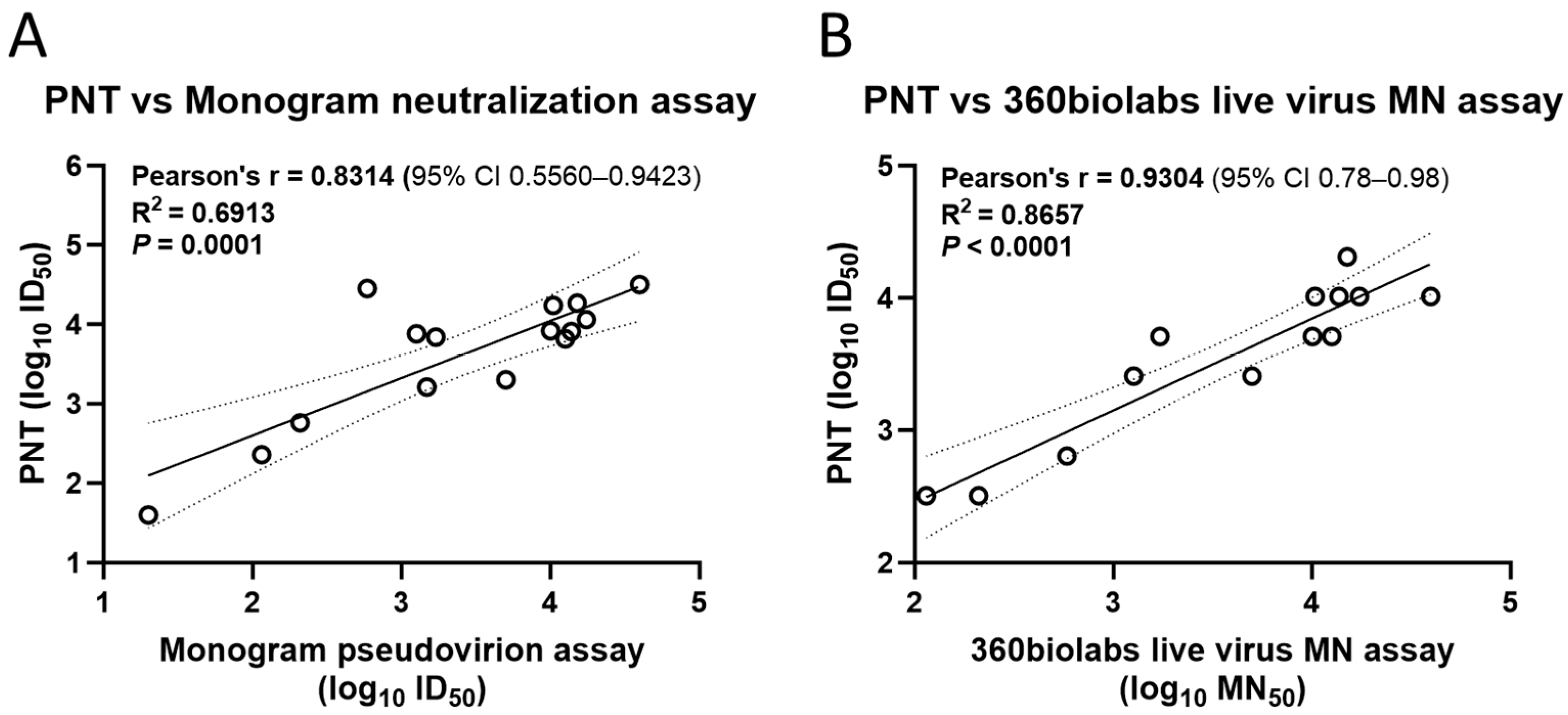
3.3. Correlation with Other Markers
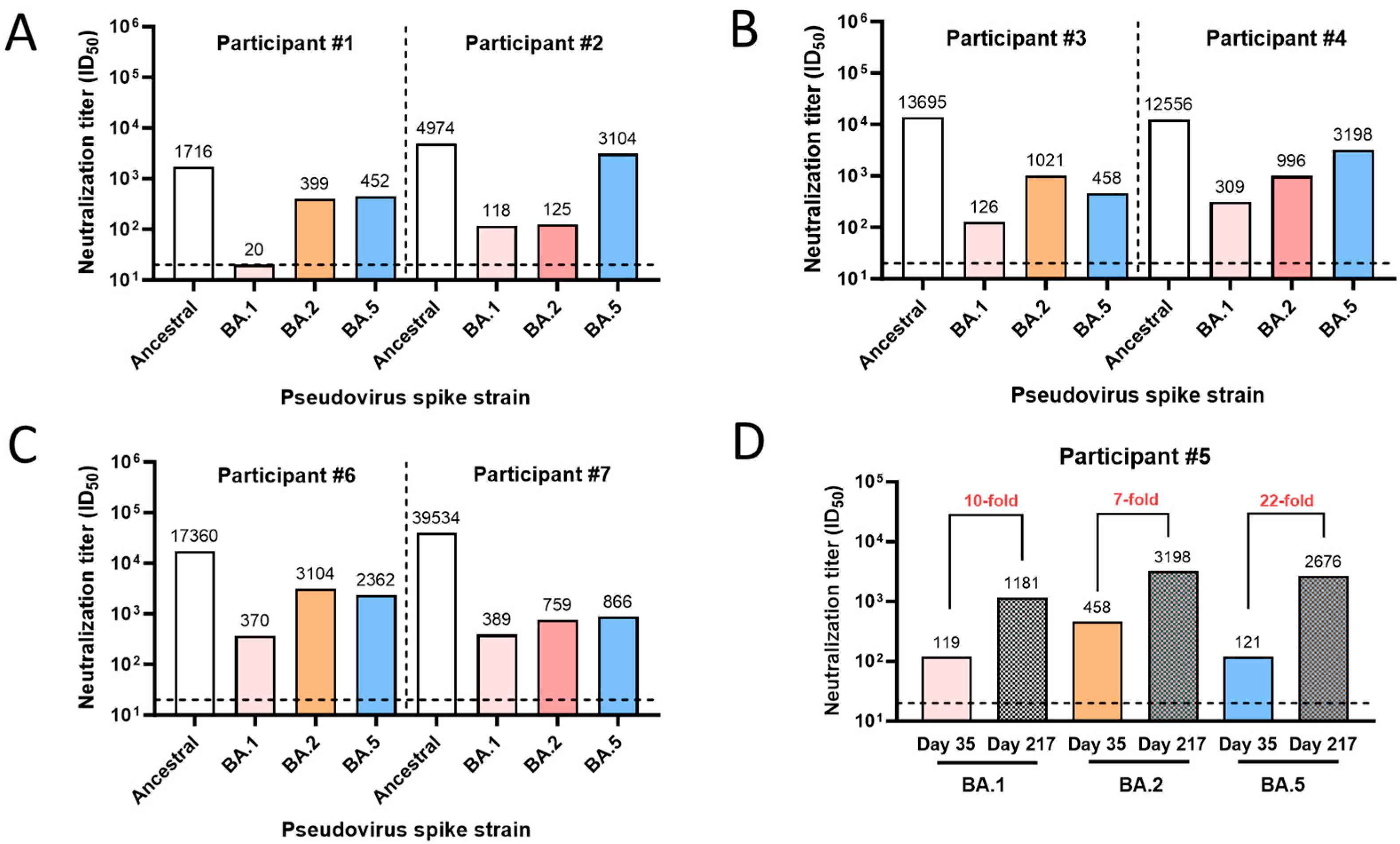
3.4. Clinical Utility
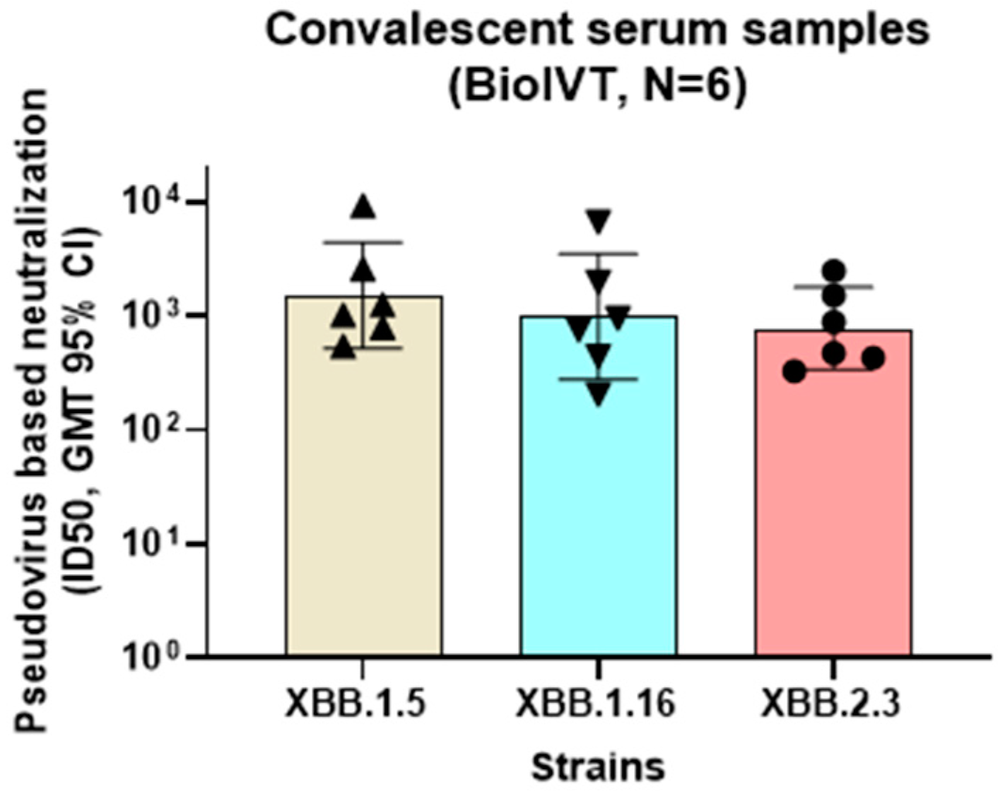
3.5. Neutralization Titers in Convalescent Human Serum Samples against XBB Variants
4. Discussion
5. Conclusions
Author Contributions
Funding
Institutional Review Board Statement
Informed Consent Statement
Data Availability Statement
Acknowledgments
Conflicts of Interest
References
- Khoury, D.S.; Cromer, D.; Reynaldi, A.; Schlub, T.E.; Wheatley, A.K.; Juno, J.A.; Subbarao, K.; Kent, S.J.; Triccas, J.A.; Davenport, M.P. Neutralizing antibody levels are highly predictive of immune protection from symptomatic SARS-CoV-2 infection. Nat. Med. 2021, 27, 1205–1211. [Google Scholar] [CrossRef]
- Abebe, E.C.; Dejenie, T.A. Protective roles and protective mechanisms of neutralizing antibodies against SARS-CoV-2 infection and their potential clinical implications. Front. Immunol. 2023, 14, 1055457. [Google Scholar] [CrossRef]
- Fong, Y.; Huang, Y.; Benkeser, D.; Carpp, L.N.; Áñez, G.; Woo, W.; McGarry, A.; Dunkle, L.M.; Cho, I.; Houchens, C.R.; et al. Immune correlates analysis of the PREVENT-19 COVID-19 vaccine efficacy clinical trial. Nat. Commun. 2023, 14, 331. [Google Scholar] [CrossRef] [PubMed]
- World Health Organization. Tracking SARS-CoV-2 variants. Available online: https://www.who.int/activities/tracking-SARS-CoV-2-variants (accessed on 31 January 2024).
- Stanford University Coronavirus Antiviral and Resistance Database. SARS-CoV-2 Variants. Available online: https://covdb.stanford.edu/variants/omicron_xbb/ (accessed on 31 January 2024).
- Faraone, J.N.; Qu, P.; Goodarzi, N.; Zheng, Y.M.; Carlin, C.; Saif, L.J.; Oltz, E.M.; Xu, K.; Jones, D.; Gumina, R.J.; et al. Immune evasion and membrane fusion of SARS-CoV-2 XBB subvariants EG.5.1 and XBB.2.3. Emerg. Microbes Infect. 2023, 12, 2270069. [Google Scholar] [CrossRef]
- Fendler, A.; de Vries, E.G.E.; GeurtsvanKessel, C.H.; Haanen, J.B.; Wörmann, B.; Turajlic, S.; von Lilienfeld-Toal, M. COVID-19 vaccines in patients with cancer: Immunogenicity, efficacy and safety. Nat. Rev. Clin. Oncol. 2022, 19, 385–401. [Google Scholar] [CrossRef] [PubMed]
- Chen, M.; Zhang, X. Construction and applications of SARS-CoV-2 pseudoviruses: A mini review. Int. J. Biol. Sci. 2021, 17, 1574–1580. [Google Scholar] [CrossRef] [PubMed]
- Centers for Disease Control and Prevention. Quick Learn Lesson: Recognizing the Biosafety Levels. Available online: https://www.cdc.gov/training/quicklearns/biosafety/ (accessed on 12 August 2023).
- Gattinger, P.; Ohradanova-Repic, A.; Valenta, R. Importance, Applications and Features of Assays Measuring SARS-CoV-2 Neutralizing Antibodies. Int. J. Mol. Sci. 2023, 24, 5352. [Google Scholar] [CrossRef] [PubMed]
- Xiang, Q.; Li, L.; Wu, J.; Tian, M.; Fu, Y. Application of pseudovirus system in the development of vaccine, antiviral-drugs, and neutralizing antibodies. Microbiol. Res. 2022, 258, 126993. [Google Scholar] [CrossRef] [PubMed]
- Nie, J.; Li, Q.; Wu, J.; Zhao, C.; Hao, H.; Liu, H.; Zhang, L.; Nie, L.; Qin, H.; Wang, M.; et al. Establishment and validation of a pseudovirus neutralization assay for SARS-CoV-2. Emerg. Microbes Infect. 2020, 9, 680–686. [Google Scholar] [CrossRef] [PubMed]
- Crawford, K.H.D.; Eguia, R.; Dingens, A.S.; Loes, A.N.; Malone, K.D.; Wolf, C.R.; Chu, H.Y.; Tortorici, M.A.; Veesler, D.; Murphy, M.; et al. Protocol and Reagents for Pseudotyping Lentiviral Particles with SARS-CoV-2 Spike Protein for Neutralization Assays. Viruses 2020, 12, 513. [Google Scholar] [CrossRef] [PubMed]
- Kulkarni, P.S.; Kadam, A.; Godbole, S.; Bhatt, V.; Raut, A.; Kohli, S.; Tripathi, S.; Kulkarni, P.; Ludam, R.; Prabhu, M.; et al. Safety and immunogenicity of SII-NVX-CoV2373 (COVID-19 vaccine) in adults in a phase 2/3, observer-blind, randomised, controlled study. Lancet Reg. Health-Southeast Asia 2023, 10, 100139. [Google Scholar] [CrossRef] [PubMed]
- Zhu, M.; Cloney-Clark, S.; Feng, S.L.; Parekh, A.; Gorinson, D.; Silva, D.; Skonieczny, P.; Wilson, A.; Kalkeri, R.; Woo, W.; et al. A Severe Acute Respiratory Syndrome Coronavirus 2 Anti-Spike Immunoglobulin G Assay: A Robust Method for Evaluation of Vaccine Immunogenicity Using an Established Correlate of Protection. Microorganisms 2023, 11, 1789. [Google Scholar] [CrossRef] [PubMed]
- Plested, J.S.; Zhu, M.; Cloney-Clark, S.; Massuda, E.; Patel, U.; Klindworth, A.; Massare, M.J.; Cai, R.; Fries, L.; Glenn, G.; et al. Severe Acute Respiratory Syndrome Coronavirus 2 Receptor (Human Angiotensin-Converting Enzyme 2) Binding Inhibition Assay: A Rapid, High-Throughput Assay Useful for Vaccine Immunogenicity Evaluation. Microorganisms 2023, 11, 368. [Google Scholar] [CrossRef] [PubMed]
- Huang, K.A.; Chen, X.; Mohapatra, A.; Nguyen, H.T.V.; Schimanski, L.; Tan, T.K.; Rijal, P.; Vester, S.K.; Hills, R.A.; Howarth, M.; et al. Structural basis for a conserved neutralization epitope on the receptor-binding domain of SARS-CoV-2. Nat. Commun. 2023, 14, 311. [Google Scholar] [CrossRef] [PubMed]
- Zhou, D.; Ren, J.; Fry, E.E.; Stuart, D.I. Broadly neutralizing antibodies against COVID-19. Curr. Opin. Virol. 2023, 61, 101332. [Google Scholar] [CrossRef] [PubMed]
- Chen, C.; Liang, J.; Hu, H.; Li, X.; Wang, L.; Wang, Z. Research progress in methods for detecting neutralizing antibodies against SARS-CoV-2. Anal. Biochem. 2023, 673, 115199. [Google Scholar] [CrossRef] [PubMed]
- Valcourt, E.J.; Manguiat, K.; Robinson, A.; Lin, Y.C.; Abe, K.T.; Mubareka, S.; Shigayeva, A.; Zhong, Z.; Girardin, R.C.; DuPuis, A.; et al. Evaluating Humoral Immunity against SARS-CoV-2: Validation of a Plaque-Reduction Neutralization Test and a Multilaboratory Comparison of Conventional and Surrogate Neutralization Assays. Microbiol. Spectr. 2021, 9, e0088621. [Google Scholar] [CrossRef] [PubMed]


| Cell Line | TCID50/mL | |||
|---|---|---|---|---|
| Ancestral | BA.1 | BA.2 | BA.5 | |
| Vero-E6 | 20,380 | 17,820 | 46,240 | 53,120 |
| A549/hACE2-TMPRSS2 | 498,600 | 398,480 | 367,420 | 411,280 |
| 293T/hACE2 | 543,700 | 663,660 | 1,249,340 | 834,400 |
| Sample ID | PNT ID50 BA.1 Titer | PNT ID50 BA.5 Titer | ||
|---|---|---|---|---|
| 293T | A549 | 293T | A549 | |
| Serum #8522 | <40 | <40 | NT | NT |
| Serum #2127 | 4223 | 2905 | 674 | 603 |
| Serum #2124 | 4782 | 5811 | 1950 | 1524 |
| Serum #7781 | <40 | <40 | 40 | 43 |
| Serum #8061 | NT | NT | 102 | 126 |
| Virus/Well (µL) | Ancestral | BA.1 | ||
|---|---|---|---|---|
| 48 h | 72 h | 48 h | 72 h | |
| 50 | 735 | 329 | 636 | 427 |
| 25 | 339 | 161 | 224 | 147 |
| 12.5 | 152 | 39 | 65 | 72 |
| 6.25 | 97 | 32 | 32 | 34 |
| 3.13 | 66 | 15 | 18 | 18 |
Disclaimer/Publisher’s Note: The statements, opinions and data contained in all publications are solely those of the individual author(s) and contributor(s) and not of MDPI and/or the editor(s). MDPI and/or the editor(s) disclaim responsibility for any injury to people or property resulting from any ideas, methods, instructions or products referred to in the content. |
© 2024 by the authors. Licensee MDPI, Basel, Switzerland. This article is an open access article distributed under the terms and conditions of the Creative Commons Attribution (CC BY) license (https://creativecommons.org/licenses/by/4.0/).
Share and Cite
Cai, Z.; Kalkeri, R.; Zhu, M.; Cloney-Clark, S.; Haner, B.; Wang, M.; Osman, B.; Dent, D.; Feng, S.-L.; Longacre, Z.; et al. A Pseudovirus-Based Neutralization Assay for SARS-CoV-2 Variants: A Rapid, Cost-Effective, BSL-2–Based High-Throughput Assay Useful for Vaccine Immunogenicity Evaluation. Microorganisms 2024, 12, 501. https://doi.org/10.3390/microorganisms12030501
Cai Z, Kalkeri R, Zhu M, Cloney-Clark S, Haner B, Wang M, Osman B, Dent D, Feng S-L, Longacre Z, et al. A Pseudovirus-Based Neutralization Assay for SARS-CoV-2 Variants: A Rapid, Cost-Effective, BSL-2–Based High-Throughput Assay Useful for Vaccine Immunogenicity Evaluation. Microorganisms. 2024; 12(3):501. https://doi.org/10.3390/microorganisms12030501
Chicago/Turabian StyleCai, Zhaohui, Raj Kalkeri, Mingzhu Zhu, Shane Cloney-Clark, Benjamin Haner, Mi Wang, Bahar Osman, Dominic Dent, Sheau-Line Feng, Zach Longacre, and et al. 2024. "A Pseudovirus-Based Neutralization Assay for SARS-CoV-2 Variants: A Rapid, Cost-Effective, BSL-2–Based High-Throughput Assay Useful for Vaccine Immunogenicity Evaluation" Microorganisms 12, no. 3: 501. https://doi.org/10.3390/microorganisms12030501
APA StyleCai, Z., Kalkeri, R., Zhu, M., Cloney-Clark, S., Haner, B., Wang, M., Osman, B., Dent, D., Feng, S.-L., Longacre, Z., Glenn, G., & Plested, J. S. (2024). A Pseudovirus-Based Neutralization Assay for SARS-CoV-2 Variants: A Rapid, Cost-Effective, BSL-2–Based High-Throughput Assay Useful for Vaccine Immunogenicity Evaluation. Microorganisms, 12(3), 501. https://doi.org/10.3390/microorganisms12030501