Article

Facultatively Anaerobic Staphylococci Enable Anaerobic Cutibacterium Species to Grow and Form Biofilms Under Aerobic Conditions

1
Laboratory for Skin Research, Institute for Medical Research, Galilee Medical Center, Nahariya 2210001, Israel
2
The Azrieli Faculty of Medicine, Bar-Ilan University, Safed 1311502, Israel
3
Kane Biotech Inc., Winnipeg, MB R3T 6G2, Canada
*
Author to whom correspondence should be addressed.
Microorganisms 2024, 12(12), 2601; https://doi.org/10.3390/microorganisms12122601 (registering DOI)
Submission received: 20 November 2024 / Revised: 9 December 2024 / Accepted: 13 December 2024 / Published: 16 December 2024
(This article belongs to the Collection Feature Paper in Biofilm)

Abstract

:
Facultatively anaerobic Staphylococcus spp. and anaerobic Cutibacterium spp. are among the most prominent bacteria on human skin. Although skin microbes generally grow as multispecies biofilms, few studies have investigated the interaction between staphylococci and Cutibacterium spp. in dual-species biofilms. Here, we measured the mono- and dual-species biofilm formation of four staphylococcal species (S. epidermidis, S. hominis, S. capitis, and S. aureus) and two Cutibacterium spp. (C. acnes and C. avidum) cultured in vitro under both aerobic and anaerobic conditions. The biofilms were quantitated by rinsing them to remove planktonic cells, detaching the biofilm bacteria via sonication, and enumerating the cells by dilution plating. When cultured alone, staphylococci formed biofilms under both aerobic and anaerobic conditions, whereas Cutibacterium spp. formed biofilms only under anaerobic conditions. In co-culture, staphylococcal biofilm formation was unaffected by the presence of Cutibacterium spp., regardless of oxygen availability. However, Cutibacterium spp. biofilm formation was significantly enhanced in the presence of staphylococci, enabling robust growth under both anaerobic and aerobic conditions. Fluorescence confocal microscopy of the aerobic dual-species biofilms suggested that staphylococci create anaerobic niches at the base of the biofilm where C. acnes can grow. These findings demonstrate that staphylococci facilitate the colonization of Cutibacterium spp. in oxygen-rich environments, potentially explaining their presence in high numbers on the oxygen-exposed stratum corneum.

1. Introduction

The Gram-positive bacteria Cutibacterium acnes and Staphylococcus epidermidis are among the most abundant members of the human skin microbiome [1,2]. Although both species are considered beneficial commensals because they help maintain homeostasis of the skin microbiota and control pathogens such as S. aureus via colonization resistance [3,4,5], both species can also act as opportunistic pathogens. C. acnes causes invasive infections of the skin, soft tissue, and cardiovascular system [6], while S. epidermidis is associated with bacteremia in preterm infants and atopic dermatitis [5]. Both species also cohabitate in acne vulgaris lesions [7,8] and cause serious infections of implanted medical devices [6,9]. C. acnes and S. epidermidis are frequently co-isolated from clinical skin samples [10] and medical device infections [11].
Because of their shared abundance in the skin microbiome community, C. acnes and S. epidermidis likely evolved a close relationship based on competition and mutualism. For example, previous studies showed that C. acnes competes with S. epidermidis in hair follicles through the production of the antimicrobial peptide cutimycin [12]. C. acnes may also inhibit S. epidermidis biofilm formation and sensitize S. epidermidis to antibiotic killing through the production of short-chain fatty acids [13]. S. epidermidis, however, can counteract this competition through the production of antimicrobial peptides and the fermentation of glycerol into short-chain fatty acids that suppress C. acnes growth [14].
In addition to interspecies competition, C. acnes and S. epidermidis likely evolved the ability to mutually coexist in polymicrobial biofilms, which are the primary mode of growth for bacteria on human skin [15]. Biofilms are defined as densely packed layers of bacterial cells that grow attached to a tissue or surface [16]. Bacteria in a biofilm are encased in a self-synthesized, extracellular polymeric matrix that holds the cells together in a mass, attaches them to the underlying surface, and protects them from killing by antimicrobial agents and host immunity. In vivo, C. acnes biofilms have been observed in acne lesions and on implanted medical devices [17,18], and S. epidermidis biofilms have been observed on skin, in sweat glands, and on implanted medical devices [19,20]. Both C. acnes and S. epidermidis form biofilms in vitro [6,9]. Biofilms play a role in many chronic infections which are often difficult to treat because of the protective nature of biofilms [16].
Among the few studies that have investigated C. acnes/S. epidermidis dual-species biofilms, the biofilms were cultured under anaerobic conditions [21,22,23]. This is because C. acnes is an aerotolerant anaerobe whose growth is inhibited by oxygen. No studies investigating C. acnes/S. epidermidis dual-species biofilms under aerobic conditions have been reported. Aerobiosis more closely mimics the environment on the stratum corneum, which is directly exposed to atmospheric oxygen, and in hair follicles and sebaceous glands, where oxygen levels are often only moderately hypoxic due to diffusion of oxygen from the atmosphere and tissues [24].
In the present study, we assessed the ability of S. epidermidis and C. acnes, either individually or jointly, to form biofilms in vitro. Since S. epidermidis is a facultative anaerobe and C. acnes is an aerotolerant anaerobe, we incubated culture vessels under both aerobic and anaerobic conditions to determine the role of atmosphere in biofilm formation. Our findings suggest that S. epidermidis significantly enhances growth and biofilm formation by C. acnes under both anaerobic and aerobic conditions. These findings may be clinically relevant to the interaction between staphylococci and Cutibacterium spp. on human skin.

2. Materials and Methods

2.1. Bacterial Strains

The bacterial strains used in this study are listed in Table 1. C. acnes strain HL086PA1 was obtained from BEI Resources (Manassas, VA, USA) as part of the NIAID NIH Human Microbiome Project. C. acnes strains 2, 7, and 14, and C. avidum strain 42, were provided by Ronen Hazan and Amit Rimon (Hebrew University of Jerusalem, Jerusalem, Israel). S. epidermidis strain 5 was obtained from Irina Sadovskaya (Université du Littoral-Côte d’Opale, Boulogne-sur-mer, France). S. epidermidis strain 1457 and green fluorescent protein-expressing strain 1457/pCM29-GFP were provided by Holger Rohde (Universitätsklinikum Hamburg-Eppendorf, Hamburg, Germany). S. aureus strain JE2 was kindly provided by Alex Horswill (University of Colorado, Boulder, CO, USA). Staphylococcal laboratory strains JK4, JK5, JK6, JK7, JK9, and JK10 were identified at the species level using MALDI-TOF mass spectrometry.

2.2. Media and Growth Conditions

The media used were tryptic soy agar (TSA) and tryptic soy broth (TSB), both purchased in powder form from Becton, Dickinson and Company. All bacteria were cultured at 37 °C. The reference stock cultures were maintained at −80 °C in 20% glycerol/80% TSB. The working stock cultures were restreaked weekly on TSA. Staphylococcal working stocks were incubated for 1 d in air. Cutibacterium working stocks were incubated for 3 d under anaerobic conditions generated with a BD GasPak EZ Anaerobe sachet system.

2.3. Test Tube and Microtiter Plate Biofilm Assays

A loopful of cells from a fresh agar plate was transferred to a 1.5 mL microcentrifuge tube containing 200 µL of sterile TSB. The cells were mixed by vortex agitation and then diluted to 106–107 CFU/mL in filter-sterilized TSB (1:1000 dilution). Dual-species biofilms were inoculated at a C. acnes/S. epidermidis ratio of 1–8:1. Inocula were aliquoted into 13 × 100 mm glass tubes (1 mL/tube), 15 mL conical-bottom polypropylene tubes (1 mL/tube), 96-well round-bottom non-tissue-culture-treated polystyrene microtiter plates (0.2 mL/well), or 24-well flat-bottom tissue-culture-treated polystyrene microtiter plates (1 mL/well). Cultures were incubated statically for 24–96 h in air or under anaerobic conditions as described above. In some experiments, polypropylene tubes were incubated under dynamic conditions by clamping them in a nearly horizontal position in a roller apparatus set to 40 rpm, which inhibited the settling of cells at the bottom of the tube and biofilm formation.

2.4. NHEK Cell Culture

Normal human epidermal keratinocytes (NHEKs) were purchased from ATCC (# PCS-200-011). The cells were cultured in 24-well tissue-culture-treated microtiter plates. Then, the cells were seeded at a density of 5 × 10⁴ cells per well in 500 µL of dermal cell basal medium (ATCC # PCS-200-030) supplemented with Keratinocyte Growth Kit components (ATCC # PCS-200-040) and antibiotics, as per the supplier’s recommendations. The cells were incubated at 37 °C in a humidified atmosphere with 5% CO2. The medium was replaced every 48 h. Monolayers were allowed to reach approximately 100% confluence, which typically occurred in 5–6 d. Prior to bacterial inoculation, the cells were washed twice with sterile phosphate-buffered saline (PBS) to remove residual medium, antibiotics, and non-adherent cells.

2.5. Enumeration of Biofilm Bacteria

For the glass tubes, the biofilms were rinsed three times with 3 mL of PBS, and the tubes were filled with 1 mL of PBS. The biofilm bacteria were detached from the walls of the tubes by sonication for 30 s at 50% power/50% duty cycle using an IKA Labortechnik sonicator equipped with a microtip probe. Control experiments showed that this sonication treatment did not affect the viability of the C. acnes, S. epidermidis, or S. aureus cells. The sonicates were serially diluted in PBS. To selectively enumerate staphylococcal CFUs, dilutions were plated on TSA and incubated in air. To selectively enumerate Cutibacterium CFUs, dilutions were plated on TSA supplemented with 20 µg/mL erythromycin and incubated anaerobically. All Cutibacterium strains used in this study were erythromycin-resistant, and all staphylococcal strains were erythromycin-susceptible (Table 1). For the polypropylene tubes, the tubes were mixed for 10 s at a high speed in a Vortex Genie 2 mixer and then subjected to sonication for 15 s at 50% power in a Qsonica CL188 sonicator equipped with a microtip probe. Control experiments showed that the 10 s vortex agitation step removed nearly all staphylococcal biofilm cells and approximately 10% of the Cutibacterium biofilm cells from the polypropylene tubes, and that the 15 s sonication step efficiently detached the remaining C. acnes biofilm cells without affecting the staphylococcal or Cutibacterium cell viability The sonicates were diluted and plated on selective agar for CFU enumeration, as described above. For the 96-well microtiter plates, the contents of each well were mixed vigorously by pipetting up and down 20 times in combination with stirring and scraping the bottoms of the wells using the pipette tips. The contents of each well were then diluted and plated on selective agar for CFU enumeration as described above. For the 24-well microtiter plates, the biofilms were scraped from the bottom of the well with a cell scraper and then mixed by vortex agitation, diluted, and plated on selective agar for CFU enumeration, as described above.

2.6. Treatment of Biofilms with Enzymes

S. epidermidis strain 5/C. acnes strain HL086PA1 dual-species biofilms were cultured aerobically in glass tubes. After 72 h, the biofilms were rinsed with water to remove planktonic cells, and then treated with PBS supplemented with 80 µg/mL dispersin B (Kane Biotech, Winnipeg, MB, Canada) or 100 µg/mL DNase I (Sigma-Aldrich, St. Louis, MO, USA). Control biofilms were treated with PBS alone. After 15 min at 37 °C, the biofilms were rinsed vigorously with tap water to remove detached cells and then stained for 1 min with 1 mL of Gram’s crystal violet. The tubes were then rinsed with tap water to remove the unbound dye, and then air-dried and photographed.

2.7. Colony Biofilm Assay

Steam-sterilized 25 mm diameter, 0.45 µm pore-size nylon filters (#66607, Pall Corp., Port Washington, NY, USA) were transferred to the surface of a TSA plate. A volume of 20 µL of inoculum was pipetted in the center of each filter. The inocula were C. acnes strain 2 alone (ca. 105 CFUs), or C. acnes strain 2 plus S. epidermidis strain JK4 (ca. 105 CFUs each). The plates were incubated aerobically or anaerobically for 3 d. The filters were transferred to 50 mL conical-bottom polypropylene centrifuge tubes containing 5 mL of PBS and sonicated for 20 s at 50% power in a Qsonica CL188 sonicator equipped with a microtip probe. The sonicates were diluted and plated on selective agar for C. acnes CFU enumeration, as described above.

2.8. Fluorescence Confocal Microscopy

A total of 4 mL of inoculum containing C. acnes strain 2 (5 × 10⁶ CFU/mL), S. epidermidis strain 1457/pCM29-GFP (5 × 10⁶ CFU/mL), or a mixture of both strains (5 × 10⁶ CFU/mL of each species) was transferred to a 35 mm diameter glass-bottom Petri dish. C. acnes mono-species cultures were incubated anaerobically, and S. epidermidis mono-species and C. acnes/S. epidermidis dual-species cultures were incubated aerobically. After 3 d, the biofilms were rinsed vigorously with water and treated with 2 mL of FluoroMounter with DAPI aqueous mounting medium (Bio SB, Santa Barbara, CA, USA) overnight at 4 °C. The biofilms were viewed under a Lieca DMi8 inverted confocal microscope using a 63× oil objective. The GFP was visualized using a 488 nm laser. The DAPI was excited using an 405 nm laser, and its emission was recorded at 470 nm.

2.9. Statistics and Reproducibility of Results

Biofilm assays were performed in triplicate test tubes, in duplicate or triplicate microtiter plate wells, or on duplicate filters for the colony biofilm assay. All experiments were repeated at least three times on different days. All repetitions exhibited a similar range of values and similarly significant differences in the mean CFU/tube, CFU/well, and CFU/filter values among the experimental groups. The results from the representative experiments are shown in the figures. The significance of the differences between the means was calculated using a Student’s t-test. A p value < 0.01 was considered significant.

3. Results

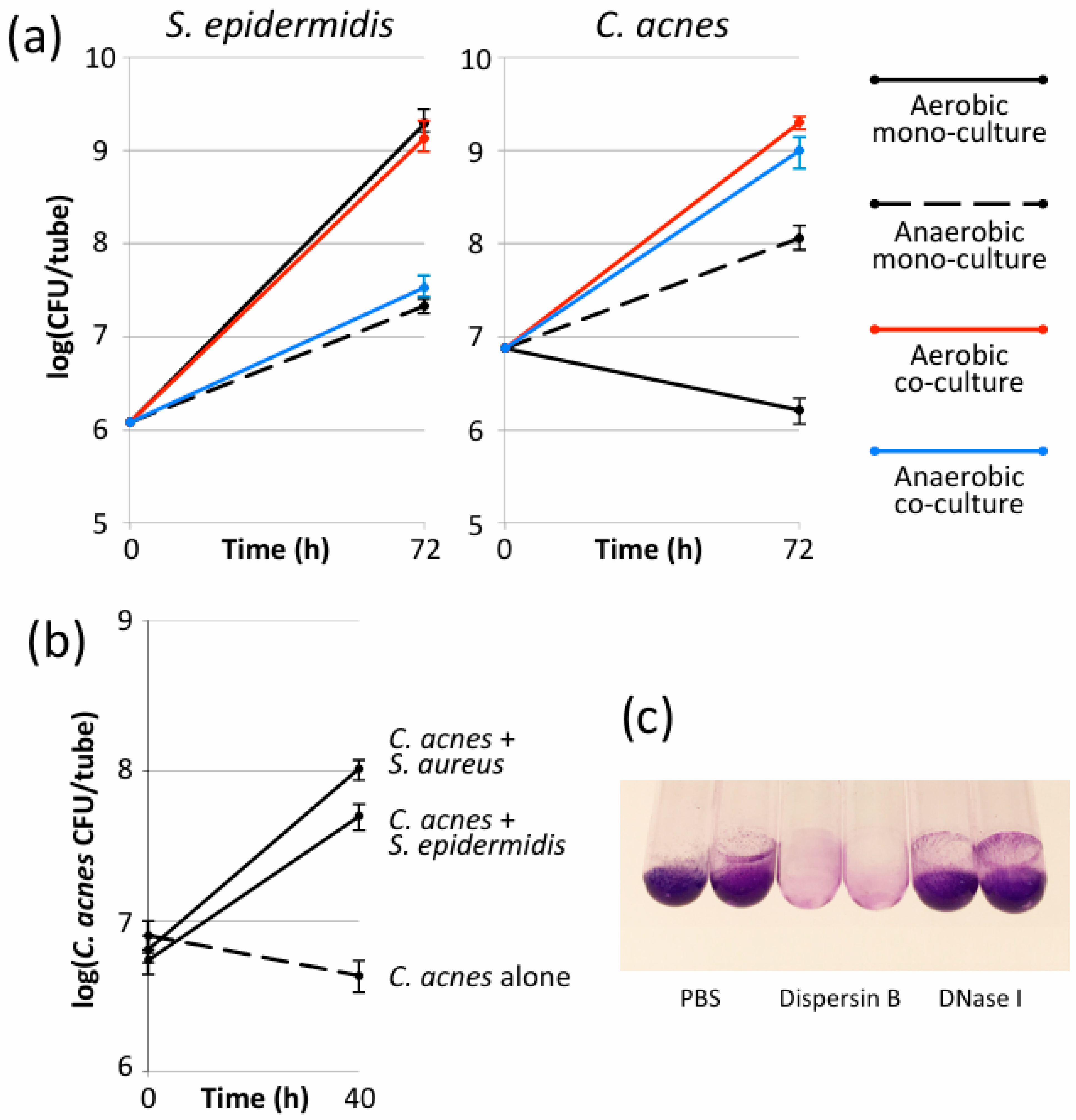
3.1. S. epidermidis Enabled C. acnes to Form Biofilms in Glass Tubes Under Aerobic Conditions

Preliminary experiments were performed with C. acnes strain HL086PA1, an acne isolate, and S. epidermidis strain 5, an implant isolate (Table 1). When cultured individually in 13 × 100 mm glass tubes, S. epidermidis strain 5 formed biofilms under both anaerobic and aerobic conditions, whereas C. acnes strain HL086PA1 formed biofilms only under anaerobic conditions (Figure 1a). S. epidermidis strain 5 formed significantly more ßbiofilm under aerobic conditions than under anaerobic conditions (p < 0.01). When co-cultured, the presence of C. acnes strain HL086PA1 had no significant effect on biofilm formation by S. epidermidis strain 5 under either anaerobic or aerobic conditions (Figure 1a, left panel). However, the presence of S. epidermidis strain 5 significantly increased C. acnes strain HL086PA1 biofilm formation under anaerobic conditions (p < 0.01) and enabled C. acnes strain HL086PA1 to form robust biofilms under aerobic conditions (Figure 1a, right panel).

3.2. S. aureus Enabled C. acnes to Form Biofilms in Glass Tubes Under Aerobic Conditions

C. acnes also grew and formed biofilms in glass tubes under aerobic conditions when co-cultured with S. aureus strain JE2, a biofilm-forming MRSA strain (Figure 1b). This finding suggests that the biofilm enabling phenotype exhibited by S. epidermidis is a general phenomenon and not due to a specific interaction between S. epidermidis and C. acnes.

3.3. Aerobic S. epidermidis/C. acnes Biofilms Contained Poly-N-Acetylglucosamine Exopolysaccharide

Previous studies have shown that the exopolysaccharide poly-N-acetylglucosamine (PNAG) and extracellular double-stranded DNA (eDNA) function as adhesive components in both S. epidermidis and C. acnes biofilms [32,33,34]. To determine whether PNAG or eDNA contribute to biofilm cohesion in aerobic S. epidermidis/C. acnes dual-species biofilms, the biofilms were treated with the PNAG-degrading enzyme dispersin B [35], or with DNase I (Figure 1c). The treatment of dual-species biofilms with dispersin B, but not DNase I, resulted in a significant decrease in the crystal violet stainable biomass, suggesting that PNAG contributed to the cohesion of S. epidermidis/C. acnes dual-species biofilm under the conditions tested.

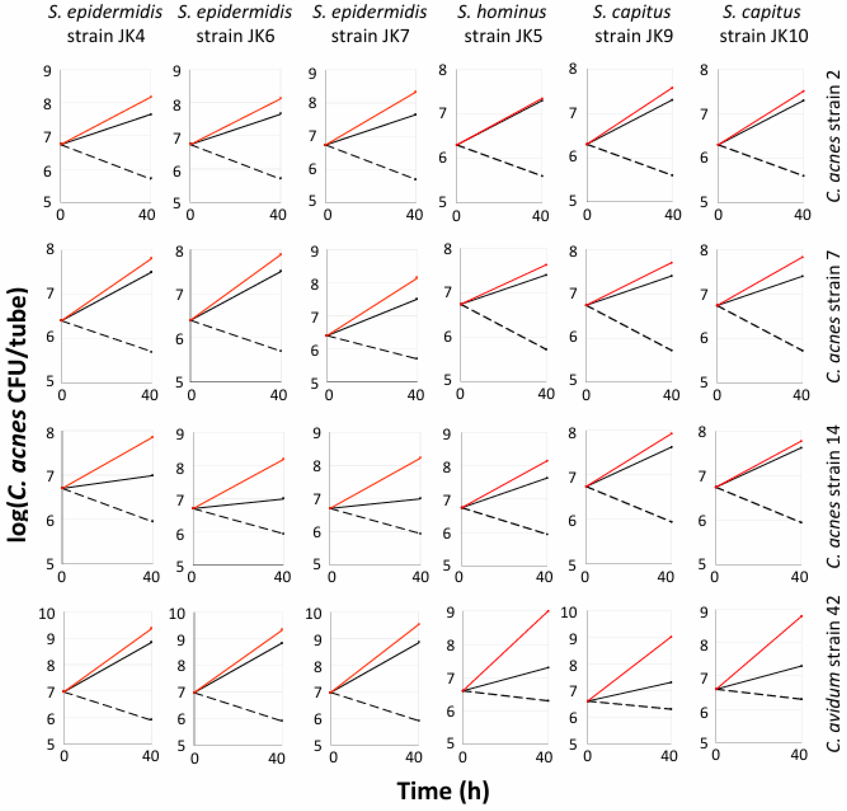
3.4. Multiple Staphylococcal Species Promoted Aerobic Biofilm Formation by C. acnes and C. avidum in Polypropylene Tubes

To investigate the generalizability of these results, we measured the biofilm formation of four Cutibacterium spp. strains (C. acnes strains 2, 7, and 14; C. avidum strain 42) when cultured alone under aerobic or anaerobic conditions, and when co-cultured with six different staphylococcal strains (S. epidermidis strains JK4, JK5, and JK7; S. capitis strains JK9 and JK10; S. hominis strain JK6) under aerobic conditions (Figure 2). These assays were carried out in 15 mL conical-bottom polypropylene tubes. As expected, when cultured alone, all four Cutibacterium spp. strains formed biofilms under anaerobic conditions but not under aerobic conditions. However, all four Cutibacterium spp. strains produced robust biofilms under aerobic conditions when co-cultured with any of the six staphylococcal strains. For all 24 combinations of strains, the amount of biofilm formed by Cutibacterium under aerobic conditions when co-cultured with Staphylococcus was equal to or greater than the amount formed in mono-culture under anaerobic conditions (Figure 2). This same phenotype was exhibited by C. acnes strain HL086PA1 and S. epidermidis strain 5 when co-cultured in glass tubes (Figure 1a). These results suggest that enabling Cutibacterium spp. to form biofilms under aerobic conditions is a common phenotype among staphylococci.

3.5. Aerobic Growth of C. acnes Depended on Biofilm Formation and Required Live S. epidermidis Cells

Further experiments were performed utilizing C. acnes strain 2 (acne isolate) and S. epidermidis strain JK4 (healthy isolate). C. acnes strain 2 formed biofilms in air when co-cultured with S. epidermidis strain JK4 in polypropylene tubes only if the tubes were incubated under static conditions and not under dynamic conditions (in a roller apparatus), which inhibited biofilm formation (Figure 3a). Similarly, C. acnes strain 2 was unable to form biofilms under aerobic conditions when co-cultured with S. epidermidis strain JK4 if erythromycin was added to the culture medium to inhibit S. epidermidis growth (Figure 3b), or if the S. epidermidis inoculum was heat-killed prior to incubation (Figure 3c). These results suggest that the ability of Staphylococcus spp. to promote aerobic Cutibacterium biofilm formation requires live S. epidermidis cells and depends on de novo biofilm formation.

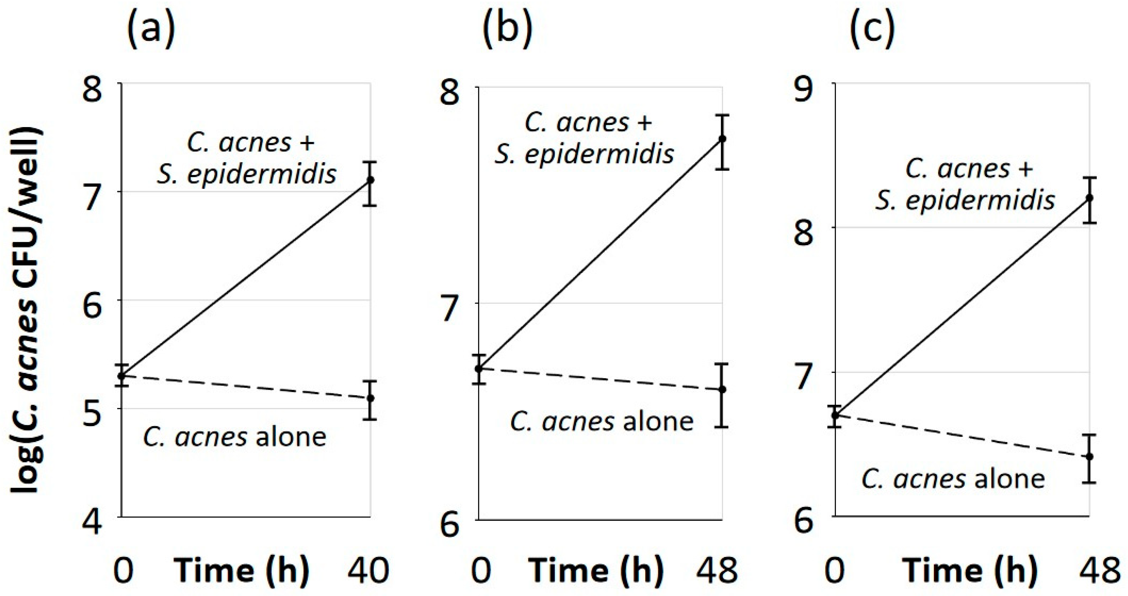
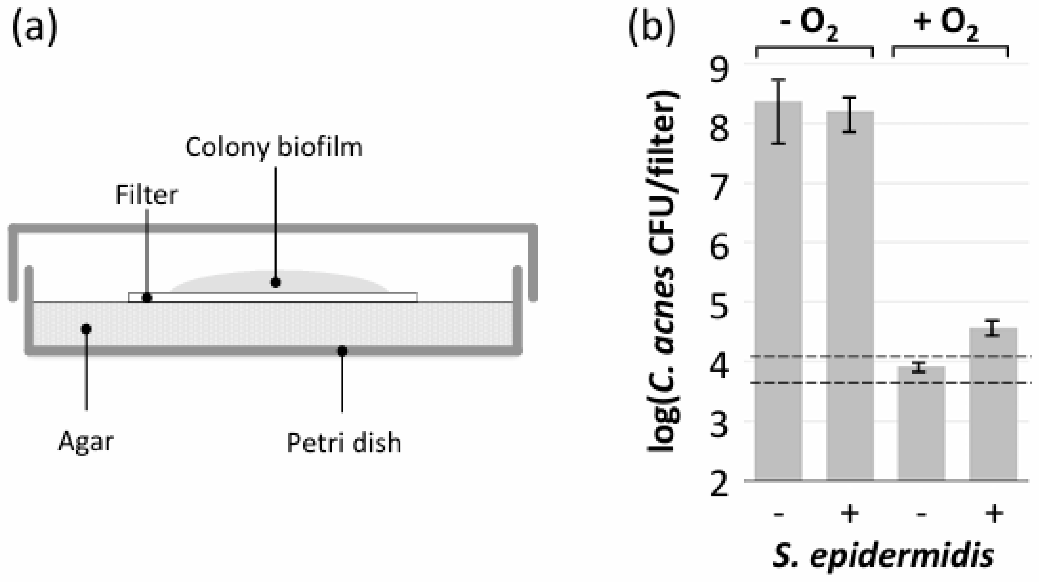
3.6. S. epidermidis Promoted Aerobic Growth of C. acnes in Polystyrene Microtiter Plate Wells but Not in Colony Biofilms

C. acnes strain 2 exhibited robust growth under aerobic conditions when co-cultured with S. epidermidis strain JK4 in 96-well, non-tissue-culture-treated round-bottom polystyrene microtiter plates (Figure 4a), in 24-well, tissue-culture-treated flat-bottom microtiter plates (Figure 4b), and in 24-well microtiter plates that were seeded with NHEK cell monolayers (Figure 4c). The presence of C. acnes and S. epidermidis did not affect the morphology of NHEK cell monolayers as assessed by light microscopy. These findings demonstrate that S. epidermidis enabled the aerobic growth of C. acnes in multiple culture vessels and on multiple surfaces, including glass, polypropylene, polystyrene, and NHEK cell monolayers.
Colony biofilms are biofilms that form on semipermeable membranes placed on the surface of an agar plate (Figure 5a). C. acnes strain 2 displayed robust growth in colony biofilms when cultured alone or co-cultured with S. epidermidis strain JK4 under anaerobic conditions (Figure 5b). However, under aerobic conditions, C. acnes strain 2 showed no growth in colony biofilms when cultured alone and only minimal growth when co-cultured with S. epidermidis strain JK4 (Figure 5b). Since colony biofilms are exposed to oxygen that diffuses through the agar and semipermeable membrane, this suggests that S. epidermidis requires an inert surface such as glass, polypropylene, or polystyrene to facilitate the aerobic growth of C. acnes.

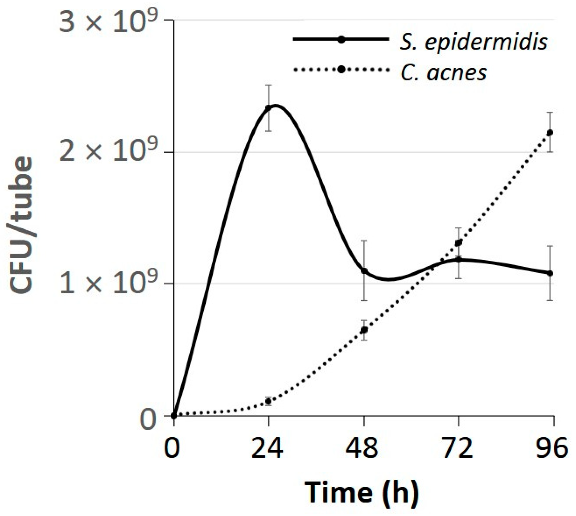
3.7. C. acnes Outcompeted S. epidermidis After 3 Days of Growth Under Aerobic Conditions

Time course studies revealed that after 3 d of aerobic growth, C. acnes strain 2 reproducibly achieved CFU/tube values that were equal to or greater than those achieved by S. epidermidis strain JK4 (Figure 6). The decrease in S. epidermidis CFU/tube values from 24–48 h occurred even in the absence of C. acnes strain 2 and was highly reproducible.

3.8. Aerobic C. acnes/S. epidermidis Dual-Species Biofilms Exhibited a Bilayer Structure

To investigate the structure of aerobic C. acnes/S. epidermidis dual-species biofilms, C. acnes strain 2 was co-cultured aerobically in glass-bottom Petri dishes with S. epidermidis strain 1457/pCM29-GFP, which expresses green fluorescent protein (GFP). As controls, C. acnes strain 2 was cultured alone under anaerobic conditions, and S. epidermidis strain 1457/pCM29-GFP was cultured alone under aerobic conditions. After 3 d, biofilms were stained with the DNA-binding dye DAPI, which fluoresces blue, and then analyzed by fluorescence confocal microscopy (Figure 7). As expected, anaerobic C. acnes strain 2 mono-species biofilms exhibited a signal for DAPI (blue) but not for GFP (Figure 7a). Aerobic S. epidermidis strain 1457/pCM29-GFP mono-species biofilms exhibited a bilayer structure, with green fluorescing aggregates comprising the entire top layer and blue fluorescing aggregates located exclusively in the bottom layer attached to the glass coverslip (Figure 7b). Aerobic C. acnes strain 2/S. epidermidis strain 1457/pCM29-GFP dual-species biofilms exhibited a bilayer structure like that exhibited by aerobic S. epidermidis strain 1457/pCM29-GFP mono-species biofilms (Figure 7c). In all biofilms, the predominant structures observed were independent green and blue fluorescing pillar- and tower-shaped aggregates in a 20 µm thick formation. Since eDNA is a component of C. acnes biofilms [32,36,37,38,39,40], these observations are consistent with the hypothesis that aerobic staphylococcal biofilms (green stain) create anaerobic niches at the base of the biofilm (blue stain) where C. acnes can grow protected from the bacteriostatic effects of oxygen.

4. Discussion

Cutibacterium acnes is one of the most prominent members of the human skin microbiome [41]. Ever since C. acnes was first isolated from acne lesions and grown in pure culture by the French physician Raymond Sabouraud in 1897 [42], it has always been cultured in the laboratory under anaerobic conditions, such as in a glove box or anaerobic jar. In this study, we found that anaerobic C. acnes and C. avidum strains can grow and form luxuriant biofilms in air when co-cultured with skin-associated staphylococci on glass, polypropylene, and polystyrene surface, and on NHEK cell monolayers. Remarkably, even though C. acnes is an anaerobe with a slow growth rate, it outcompeted fast-growing facultative S. epidermidis after three days of growth when both species were co-cultured in polypropylene tubes under aerobic conditions (Figure 5).
Although previous studies have demonstrated that under aerobic conditions, the growth of aerobic bacteria or fungi reduces the level of oxygen and thereby facilitates the growth of strictly anaerobic bacteria in both the planktonic and biofilm modes of growth [43,44,45,46,47,48,49], most of these studies were carried out using oral or gut microbes. Bernard et al. [44] showed that three different strains of Candida albicans increased biofilm formation by C. acnes strains ATCC 6919 (acne isolate) and CE1 (shoulder isolate) by 1-log-unit over 24 h when biofilms were cultured in brain heart infusion broth on polystyrene disks under aerobic conditions. C. albicans did not increase C. acnes biofilm formation under anaerobic conditions. It is difficult to compare these results to those of the present study because Bernard et al. [44] found that C. acnes strain ATCC 6919 alone formed equally robust biofilms under both anaerobic and aerobic conditions. Our findings are the first to demonstrate the aerobic growth of C. acnes strains that are unable to grow alone in air, and the first to show that an anaerobic bacterium can outcompete an aerobic bacterium under aerobic conditions.
One possible explanation for the phenotype observed in the present study is that rapidly growing staphylococcal biofilms create anaerobic niches where C. acnes is protected from the bacteriostatic effects of oxygen, thereby enabling C. acnes to grow. This model is supported by the fact that S. epidermidis does not enable the aerobic growth of C. acnes in tubes incubated under dynamic conditions (Figure 3a) or in colony biofilms that are exposed to oxygen from both above and below (Figure 5b), and that aerobic C. acnes/S. epidermidis dual-species biofilms exhibit a bilayer structure, with C. acnes evidently growing only at the base of the biofilm (Figure 7). This model is consistent with the observations of Fox et al. [47], who showed that C. albicans biofilms created locally hypoxic microenvironments that enabled the growth of anaerobic Clostridium perfringens and Bacillus fragilis in 6-well, polystyrene microtiter plates under aerobic conditions. Similarly, C. albicans biofilms were shown to protect anaerobic Porphyromonas gingivalis from unfavorable oxic environments when cultured on glass slides and in 96-well, polystyrene microtiter plates [43], and Enterococcus faecalis appeared to shield P. gingivalis and support its continued growth in polystyrene microtiter plates under aerobic conditions [49].
Previous studies have shown that poly-N-acetylglucosamine exopolysaccharide (PNAG) and double-stranded DNA (eDNA) function as adhesive components in both S. epidermidis and C. acnes biofilms in vitro [32,33,34]. Although DNase I was shown to detach pre-formed C. acnes mono-species biofilms [32,36,39], DNase I did not detach S. epidermidis strain 5/C. acnes strain HL086PA1 dual-species biofilms (Figure 1c). Instead, the PNAG-degrading enzyme dispersin B efficiently detached S. epidermidis strain 5/C. acnes strain HL086PA1 dual-species biofilms (Figure 1c), suggesting that PNAG is the major intercellular adhesion on these biofilms. Although S. epidermidis strain 5 has been shown to produce PNAG [28], it is not known whether C. acnes strain HL086PA1 or other strains used in our study produce PNAG. Therefore, more experiments are needed to determine whether S. epidermidis or C. acnes PNAG, or both, contribute to biofilm cohesion in Staphylococcus/Cutibacterium dual-species biofilms.

5. Conclusions

Our major finding was that staphylococci enabled C. acnes to form luxuriant biofilms under aerobic conditions. This phenotype was exhibited by multiple staphylococcal and Cutibacterium species and strains in glass tubes, polypropylene tubes, and polystyrene microtiter plate wells. Confocal fluorescence microscopy revealed that staphylococci may contain anaerobic niches at the base of the biofilm where C. acnes can grow. Aerobic Cutibacterium/Staphylococcus dual-species biofilms provide a novel method for culturing anaerobic Cutibacterium spp. in vitro. This dual-species biofilm model may also replicate the symbiotic relationship between Cutibacterium and Staphylococcus on human skin. It may also offer insight into how C. acnes colonizes oxic and moderately hypoxic sites on the skin.

Author Contributions

J.B.K. designed the experiments. J.B.K., M.A. and N.M. performed the experiments. All authors helped design the study, analyze the data, and write the paper. All authors have read and agreed to the published version of the manuscript.

Funding

This study was funded by Kane Biotech Inc. (Winnipeg, MB, Canada) and the Health Corporation of the Galilee Medical Center.

Data Availability Statement

The authors confirm that the data supporting the findings of this study are available within the article. No new datasets were created during the course of the study.

Acknowledgments

The authors thank Ronen Hazan and Amit Rimon (Hebrew University of Jerusalem), Irina Sadovskaya (Université du Littoral-Côte d’Opale, Boulogne-sur-Mer, France), Holger Rohde (Universitätsklinikum Hamburg-Eppendorf, Hamburg, Germany), and Alex Horswill (University of Colorado, Aurora, CO, USA) for providing bacterial strains. We also thank Hagai Rechnitzer and Larisa Belavsky of the Clinical Microbiology Laboratory at the Galilee Medical Center performing MALTI-TOF mass spectrometry analysis of bacterial strains.

Conflicts of Interest

J.B.K. serves as an advisor for, owns equity in, and receives royalties from Kane Biotech Inc., Winnipeg, Canada. M.S. and S.R. are employees of Kane Biotech Inc., manufacturer of dispersin B (DispersinB®), and own company stocks and stock options. This company provided partial funding for this study. M.A., M.N. and K.K. declare no competing interests.

References

  1. Ahle, C.M.; Stødkilde, K.; Poehlein, A.; Bömeke, M.; Streit, W.R.; Wenck, H.; Reuter, J.H.; Hüpeden, J.; Brüggemann, H. Interference and co-existence of staphylococci and Cutibacterium acnes within the healthy human skin microbiome. Commun. Biol. 2022, 5, 923. [Google Scholar] [CrossRef] [PubMed]
  2. Fournière, M.; LaVre, T.; Souak, D.; Feuilloley, M.G.J.; Bedoux, G. Staphylococcus epidermidis and Cutibacterium acnes: Two major sentinels of skin microbiota and the influence of cosmetics. Microorganisms 2020, 8, 1752. [Google Scholar] [CrossRef]
  3. Francuzik, W.; Franke, K.; Schumann, R.R.; Heine, G.; Worm, M. Propionibacterium acnes abundance correlates inversely with Staphylococcus aureus: Data from atopic dermatitis skin microbiome. Acta Derm. Venereol. 2018, 98, 490–495. [Google Scholar] [CrossRef]
  4. Rozas, M.; de Ruijter, A.H.; Fabrega, M.J.; Zorgani, A.; Guell, M.; Berhard Paetzold, B.; Franois Brillet, F. From dysbiosis to healthy skin: Major contributions of Cutibacterium acnes to skin homeostasis. Microorganisms 2021, 9, 628. [Google Scholar] [CrossRef] [PubMed]
  5. Severn, M.M.; Horswill, A.R. Staphylococcus epidermidis and its dual lifestyle in skin health and infection. Nat. Rev. Microbiol. 2023, 21, 97–111. [Google Scholar] [CrossRef] [PubMed]
  6. Achermann, Y.; Goldstein, E.J.; Coenye, T.; Shirtliff, M.E. Propionibacterium acnes: From commensal to opportunistic biofilm-associated implant pathogen. Clin. Microbiol. Rev. 2014, 27, 419–440. [Google Scholar] [CrossRef] [PubMed]
  7. Bek-Thomsen, M.; Lomholt, H.B.; Kilian, M. Acne is not associated with yet-uncultured bacteria. J. Clin. Microbiol. 2008, 46, 3355–3360. [Google Scholar] [CrossRef] [PubMed]
  8. McLaughlin, J.; Watterson, S.; Layton, A.M.; Bjourson, A.J.; Barnard, E.; McDowell, A. Propionibacterium acnes and acne vulgaris: New insights from the integration of population genetic, multi-omic, biochemical and host-microbe studies. Microorganisms 2019, 7, 128. [Google Scholar] [CrossRef] [PubMed]
  9. Otto, M. Staphylococcus epidermidis—The “accidental” pathogen. Nat. Rev. Microbiol. 2009, 7, 555–567. [Google Scholar] [CrossRef]
  10. Dréno, B.; Dagnelie, M.A.; Khammari, A.; Corvec, S. The skin microbiome: A new actor in inflammatory acne. Am. J. Clin. Dermatol. 2020, 21 (Suppl. S1), 18–24. [Google Scholar] [CrossRef] [PubMed]
  11. Tyner, H.; Patel, R. Propionibacterium acnes biofilm—A sanctuary for Staphylococcus aureus? Anaerobe 2016, 40, 63–67. [Google Scholar] [CrossRef] [PubMed]
  12. Claesen, J.; Spagnolo, J.B.; Ramos, S.F.; Kurita, K.L.; Byrd, A.L.; Aksenov, A.A.; Melnik, A.V.; Wong, W.R.; Wang, S.; Hernandez, R.D.; et al. A Cutibacterium acnes antibiotic modulates human skin microbiota composition in hair follicles. Sci. Transl. Med. 2020, 12, 5445. [Google Scholar] [CrossRef] [PubMed]
  13. Nakamura, K.; O’Neill, A.M.; Williams, M.R.; Cau, L.; Nakatsuji, T.; Horswill, A.R.; Gallo, R.L. Short chain fatty acids produced by Cutibacterium acnes inhibit biofilm formation by Staphylococcus epidermidis. Sci. Rep. 2020, 10, 21237. [Google Scholar] [CrossRef]
  14. Wang, Y.; Kuo, S.; Shu, M.; Yu, J.; Huang, S.; Dai, A.; Two, A.; Gallo, R.L.; Huang, C.-M. Staphylococcus epidermidis in the human skin microbiome mediates fermentation to inhibit the growth of Propionibacterium acnes: Implications of probiotics in acne vulgaris. Appl. Microbiol. Biotechnol. 2014, 98, 411–424. [Google Scholar] [CrossRef] [PubMed]
  15. Brandwein, M.; Steinberg, D.; Meshner, S. Microbial biofilms and the human skin microbiome. NPJ Biofilms Microbiomes 2016, 2, 3. [Google Scholar] [CrossRef] [PubMed]
  16. Penesyan, A.; Paulsen, I.T.; Kjelleberg, S.; Gillings, M.R. Three faces of biofilms: A microbial lifestyle, a nascent multicellular organism, and an incubator for diversity. NPJ Biofilms Microbiomes 2021, 7, 80. [Google Scholar] [CrossRef] [PubMed]
  17. Bayston, R.; Ashraf, W.; Barker-Davies, R.; Tucker, E.; Clement, R.; Clayton, J.; Freeman, B.J.C.; Nuradeen, B. Biofilm formation by Propionibacterium acnes on biomaterials in vitro and in vivo: Impact on diagnosis and treatment. J. Biomed. Mater. Res. A 2006, 81, 705–709. [Google Scholar] [CrossRef]
  18. Jahns, A.C.; Lundskog, B.; Ganceviciene, R.; Palmer, R.H.; Golovleva, I.; Zouboulis, C.C.; McDowell, A.; Patrick, S.; Alexeyev, O.A. An increased incidence of Propionibacterium acnes biofilms in acne vulgaris: A case-control study. Br. J. Dermatol. 2012, 167, 50–58. [Google Scholar] [CrossRef]
  19. Allen, H.B.; Mueller, J.L. A novel finding in atopic dermatitis: Film-producing Staphylococcus epidermidis as an etiology. Int. J. Dermatol. 2011, 50, 992–993. [Google Scholar] [CrossRef]
  20. Bügner, H.; Mack, D.; Rohde, H. Structural basis of Staphylococcus epidermidis biofilm formation: Mechanisms and molecular interactions. Front. Cell Infect. Microbiol. 2015, 5, 14. [Google Scholar] [CrossRef]
  21. Gannesen, A.V.; Lesouhaitier, O.; Racine, P.J.; Barreau, M.; Netrusov, A.I.; Plakunov, V.K.; Feuilloley, M.G.J. Regulation of monospecies and mixed biofilms formation of skin Staphylococcus aureus and Cutibacterium acnes by human natriuretic peptides. Front. Microbiol. 2018, 9, 2912. [Google Scholar] [CrossRef] [PubMed]
  22. Ovcharova, M.A.; Geraskina, O.V.; Danilova, N.D.; Botchkova, E.A.; Martyanov, S.V.; Feofanov, A.V.; Plakunov, V.K.; Gannesen, A.V. Atrial natriuretic peptide affects skin commensal Staphylococcus epidermidis and Cutibacterium acnes dual-species biofilms. Microorganisms 2021, 9, 552. [Google Scholar] [CrossRef] [PubMed]
  23. Ovcharova, M.A.; Schelkunov, M.I.; Geras’kina, O.V.; Makarova, N.E.; Sukhacheva, M.V.; Martyanov, S.V.; Nevolina, E.D.; Zhurina, M.V.; Feofanov, A.V.; Botchkova, E.A.; et al. C-Type natriuretic peptide acts as a microorganism-activated regulator of the skin commensals Staphylococcus epidermidis and Cutibacterium acnes in dual-species biofilms. Biology 2023, 12, 436. [Google Scholar] [CrossRef] [PubMed]
  24. Acosta, E.A.; Little, K.A.; Bratto, B.P.; Lopez, J.G.; Mao, X.; Payne, A.S.; Donia, M.; Devenport, D.; Gitai, Z. Bacterial DNA on the skin surface overrepresents the viable skin microbiome. eLife 2023, 12, RP87192. [Google Scholar] [CrossRef]
  25. Sheffer-Levi, S.; Rimon, A.; Lerer, V.; Shlomov, T.; Coppenhagen-Glazer, S.; Rakov, C.; Zeiter, T.; Nir-Paz, R.; Hazan, R.; Molho-Pessach, V. Antibiotic susceptibility of Cutibacterium acnes strains isolated from Israeli acne patients. Acta Derm. Venereol. 2020, 100, adv00295. [Google Scholar] [CrossRef]
  26. Fitz-Gibbon, S.; Tomida, S.; Chiu, B.-H.; Nguyen, L.; Du, C.; Liu, M.; Elashoff, D.; Erfe, M.C.; Loncaric, A.; Kim, J.; et al. Propionibacterium acnes strain populations in the human skin microbiome associated with acne. J. Investig. Dermatol. 2013, 133, 2152–2160. [Google Scholar] [CrossRef] [PubMed]
  27. Miller, L.G.; Perdreau-Remington, F.; Rieg, G.; Mehdi, S.; Perlroth, J.; Bayer, A.S.; Tang, A.W.; Phung, T.O.; Spellberg, B. Necrotizing fasciitis caused by community-associated methicillin-resistant Staphylococcus aureus in Los Angeles. N. Engl. J. Med. 2005, 352, 1445–1453. [Google Scholar] [CrossRef] [PubMed]
  28. Chaignon, P.; Sadovskaya, I.; Ragunath, C.; Ramasubbu, N.; Kaplan, J.B.; Jabbouri, S. Susceptibility of staphylococcal biofilms to enzymatic treatments depends on their chemical composition. Appl. Microbiol. Biotechnol. 2007, 75, 125–132. [Google Scholar] [CrossRef]
  29. Mack, D.; Siemssen, N.; Laufs, R. Parallel induction by glucose of adherence and a polysaccharide antigen specific for plastic-adherent Staphylococcus epidermidis: Evidence for functional relation to intercellular adhesion. Infect. Immun. 1992, 60, 2048–2057. [Google Scholar] [CrossRef] [PubMed]
  30. Malone, C.L.; Boles, B.R.; Lauderdale, K.J.; Thoendel, M.; Kavanaugh, J.S.; Horswill, A.R. Fluorescent reporters for Staphylococcus aureus. J. Microbiol. Methods 2009, 77, 251–260. [Google Scholar] [CrossRef]
  31. Scholz, C.F.P.; Jensen, A.; Lomholt, H.B.; Brüggemann, H.; Kilian, M. A novel high-resolution single locus sequence typing scheme for mixed populations of Propionibacterium acnes in vivo. PLoS ONE 2014, 9, e104199. [Google Scholar] [CrossRef]
  32. Kaplan, J.B.; Cywes-Bentley, C.; Pier, G.B.; Yakandawala, N.; Sailer, M.; Edwards, M.S.; Kridin, K. Poly-b(1-6)-N-acetyl-D-glucosamine (PNAG) mediates surface attachment, biofilm formation, and biocide resistance in Cutibacterium acnes. Front. Microbiol. 2024, 15, 1386017. [Google Scholar] [CrossRef] [PubMed]
  33. Izano, E.A.; Amarante, M.A.; Kher, W.B.; Kaplan, J.B. Differential roles of poly-N-acetylglucosamine surface polysaccharide and extracellular DNA in Staphylococcus aureus and Staphylococcus epidermidis biofilms. Appl. Environ. Microbiol. 2008, 74, 470–476. [Google Scholar] [CrossRef] [PubMed]
  34. Kaplan, J.B.; Jabbouri, S.; Sadovskaya, I. Extracellular DNA-dependent biofilm formation by Staphylococcus epidermidis RP62A in response to subminimal inhibitory concentrations of antibiotics. Res. Microbiol. 2011, 162, 535–541. [Google Scholar] [CrossRef] [PubMed]
  35. Kaplan, J.B.; Sukhishvili, S.A.; Sailer, M.; Kridin, K.; Ramasubbu, N. Aggregatibacter actinomycetemcomitans dispersin B: The quintessential antibiofilm enzyme. Pathogens 2024, 13, 668. [Google Scholar] [CrossRef] [PubMed]
  36. Fang, J.-Y.; Chou, W.-L.; Lin, C.-F.; Sung, C.T.; Alalaiwe, A.; Yang, S.-C. Facile biofilm penetration of cationic liposomes loaded with DNase I/proteinase K to eradicate Cutibacterium acnes for treating cutaneous and catheter infections. Int. J. Nanomed. 2021, 16, 8121–8138. [Google Scholar] [CrossRef] [PubMed]
  37. Gannesen, A.V.; Zdorovenko, E.L.; Botchkova, E.A.; Hardouin, J.; Massier, S.; Kopitsyn, D.S.; Gorbachevskii, M.V.; Kadykova, A.A.; Shashkov, A.S.; Zhurina, M.V.; et al. Composition of the biofilm matrix of Cutibacterium acnes acneic strain RT5. Front. Microbiol. 2019, 10, 1284. [Google Scholar] [CrossRef]
  38. Jahns, A.C.; Eilers, H.; Alexeyev, O.A. Transcriptomic analysis of Propionibacterium acnes biofilms in vitro. Anaerobe 2016, 42, 111–118. [Google Scholar] [CrossRef]
  39. Kuehnast, T.; Cakar, F.; Weinhäupl, T.; Pilz, A.; Selak, S.; Schmidt, M.A.; Rüter, C.; Schild, S. Comparative analyses of biofilm formation among different Cutibacterium acnes isolates. Int. J. Med. Microbiol. 2018, 308, 1027–1035. [Google Scholar] [CrossRef] [PubMed]
  40. Okuda, K.I.; Nagahori, R.; Yamada, S.; Sugimoto, S.; Sato, C.; Sato, M.; Iwase, T.; Hashimoto, K.; Mizunoe, Y. The composition and structure of biofilms developed by Propionibacterium acnes isolated from cardiac pace-maker devices. Front. Microbiol. 2018, 9, 182. [Google Scholar] [CrossRef]
  41. Byrd, A.L.; Belkaid, Y.; Segre, J.A. The human skin microbiome. Nature Rev. Microbiol. 2018, 16, 143–155. [Google Scholar] [CrossRef]
  42. Tilles, G. Acne pathogenesis: History of concepts. Dermatology 2014, 229, 1–46. [Google Scholar] [CrossRef] [PubMed]
  43. Bartnicka, D.; Karkowska-Kuleta, J.; Zawrotniak, M.; Satała, D.; Michalik, K.; Zielinska, G.; Bochenska, O.; Kozik, A.; Ciaston, I.; Koziel, J.; et al. Adhesive protein-mediated cross-talk between Candida albicans and Porphyromonas gingivalis in dual species biofilm protects the anaerobic bacterium in unfavorable oxic environment. Sci. Rep. 2019, 9, 4376. [Google Scholar] [CrossRef] [PubMed]
  44. Bernard, C.; Lemoine, V.; Hoogenkamp, M.A.; Girardot, M.; Krom, B.P.; Imbert, C. Candida albicans enhances initial biofilm growth of Cutibacterium acnes under aerobic conditions. Biofouling 2019, 35, 350–360. [Google Scholar] [CrossRef]
  45. Bradshaw, D.J.; Marsh, P.D.; Allison, C.; Schilling, K.M. Effect of oxygen, inoculum composition and flow rate on development of mixed-culture oral biofilms. Microbiology 1996, 142, 623–629. [Google Scholar] [CrossRef] [PubMed]
  46. Diaz, P.I.; Zilm, P.S.; Rogers, A.H. Fusobacterium nucleatum supports the growth of Porphyromonas gingivalis in oxygenated and carbon-dioxide-depleted environments. Microbiology 2002, 148, 467–472. [Google Scholar] [CrossRef] [PubMed]
  47. Fox, E.P.; Cowley, E.S.; Nobile, C.J.; Hartooni, N.; Newman, D.K.; Johnson, A.D. Anaerobic bacteria grow within Candida albicans biofilms and induce biofilm formation in suspension cultures. Curr. Biol. 2014, 24, 2411–2416. [Google Scholar] [CrossRef] [PubMed]
  48. Janus, M.M.; Crielaard, W.; Volgenant, C.M.C.; van der Veen, M.H.; Brandt, B.W.; Krom, B.P. Candida albicans alters the bacterial microbiome of early in vitro oral biofilms. J. Oral. Microbiol. 2017, 9, 1270613. [Google Scholar] [CrossRef] [PubMed]
  49. Tan, H.C.; Cheung, G.S.P.; Chang, J.W.W.; Zhang, C.; Lee, A.H.C. Enterococcus faecalis shields Porphyromonas gingivalis in dual-species biofilm in oxic condition. Microorganisms 2022, 10, 1729. [Google Scholar] [CrossRef] [PubMed]
Figure 1. Cutibacterium acnes and Staphylococcus spp. mono- and dual-species biofilms grown in glass tubes under aerobic and anaerobic conditions. (a) Biofilm formation by S. epidermidis strain 5 (left panel) and C. acnes strain HL086PA1 (right panel) cultured alone (mono-culture—black solid and dashed lines) or together (co-culture—red and blue lines) under aerobic and anaerobic conditions. After 72 h, tubes were rinsed to remove planktonic cells, biofilms were detached by sonication, and species were individually enumerated by dilution plating on selective agar. Graphs show mean CFU/tube values for triplicate tubes. Error bars indicate sd. (b) Biofilm formation by C. acnes strain HL086PA1 cultured alone (dashed line) or co-cultured with S. epidermidis strain 5 or S. aureus strain JE2 (solid lines) for 40 h under aerobic conditions. Tubes were processed as in panel (a). Graph shows mean CFU/tube values for triplicate tubes. Error bars indicate sd. (c) Photographs of 72-h-old S. epidermdis strain 5/C. acnes strain HL086PA1 dual-species biofilms after treating them for 15 min with phosphate-buffered saline (PBS), PBS supplemented with dispersin B (80 µg/mL), or PBS supplemented with DNase I (100 µg/mL), and then staining them with crystal violet. Duplicate tubes for each treatment are shown.
Microorganisms 12 02601 g001
Figure 2. Growth of Cutibacterium spp. in 15 mL conical-bottom polypropylene tubes. Four Cutibacterium spp. (species and strain names indicated at right) were cultured alone anaerobically (solid black line), alone aerobically (dashed black line), or co-cultured aerobically with each staphylococcal species and strain indicated at top (red lines). Tubes were sonicated after 40 h of incubation, and sonicates were diluted and plated on agar plates selective for C. acnes. Graphs show mean C. acnes CFU/tube values for triplicate tubes. Error bars were omitted for clarity.
Microorganisms 12 02601 g002
Figure 3. Growth of C. acnes strain 2 co-cultured with S. epidermidis strain JK4 in polypropylene tubes under aerobic conditions. After 40 h, tubes were sonicated, and sonicates were diluted and plated on selective agar for C. acnes CFU enumeration. (a) Tubes were incubated under static conditions or under dynamic conditions in a roller apparatus. (b). Media were supplemented with 20 µg/mL erythromycin or no antibiotic. (c) S. epidermidis was heat killed prior to inoculation. Graphs show mean CFU/tube values for triplicate tubes and error bars indicate sd.
Microorganisms 12 02601 g003
Figure 4. Growth of C. acnes strain 2 co-cultured with S. epidermidis strain JK4 in polystyrene microtiter plate wells. (a) C. acnes strain 2 was cultured alone (dashed line) or co-cultured with S. epidermidis strain JK4 (solid line) in 96-well, non-tissue-culture-treated, round-bottom microtiter plates under aerobic conditions. After 48 h, wells were scraped with a pipette tip, and contents of wells were diluted and plated on selective agar for C. acnes CFU enumeration. Graphs show mean CFU/well values for triplicate wells, and error bars indicate sd. (b) Same as (a), except bacteria were cultured in 24-well, tissue-culture-treated microtiter plate wells. (c) Same as (b), except surfaces of wells were seeded with NHEK cell monolayers.
Microorganisms 12 02601 g004
Figure 5. Colony biofilm assay. (a) Colony biofilms were cultured on a 25 mm diameter, 0.45 µm pore-size nylon filter placed on the surface of a tryptic soy agar plate. (b) C. acnes strain 2 was inoculated alone or in combination with S. epidermidis strain JK4 (indicated at bottom) onto filters. Plates were incubated anaerobically or aerobically (indicated at top) for 48 h. Filters were then transferred to 5 mL of saline and sonicated. Sonicates were diluted and plated on selective agar to enumerate C. acnes CFUs. Graph shows mean C. acnes CFU/filter values from a total of six filters from three independent experiments for each condition. Error bars indicate sd. Horizontal dashed lines indicate average starting C. acnes inoculum per filter (±1 sd).
Microorganisms 12 02601 g005
Figure 6. Growth of C. acnes strain 2 co-cultured with S. epidermidis strain JK4 in 15 mL polypropylene tubes under aerobic conditions. Values show mean CFU/tube of each species for triplicate tubes at each time point. Error bars indicate sd.
Microorganisms 12 02601 g006
Figure 7. Cross-sections of C. acnes strain 2 and S. epidermidis strain 1457/pCM29-GFP biofilms grown on glass coverslips, stained with DAPI, and visualized by fluorescence confocal microscopy. (a) Anaerobic C. acnes mono-species biofilm. (b) Aerobic S. epidermidis mono-species biofilm. (c) Aerobic C. acnes/S. epidermidis dual-species biofilm. Biofilm thickness ≈ 20 µm. Green, GFP; blue, DAPI.
Microorganisms 12 02601 g007
Table 1. Bacterial strains.
SpeciesStrainCharacteristics *Reference
C. acnes2Moderate acne, SLST A1, Ermr[25]
C. acnes7Moderate acne, SLST C1, Ermr[25]
C. acnes14Mild acne, SLST D1, Ermr[25]
C. acnesHL086PA1Severe acne, SLST E4, Ermr[26]
C. avidum42Acne patient, Ermr[25]
S. aureusJE2Necrotizing fasciitis, MRSA,
USA300 clone, Erms
[27]
S. capitisJK9Healthy subject, ErmsLaboratory strain
S. capitisJK10Healthy subject, ErmsLaboratory strain
S. epidermidis5Implant infection, Erms[28]
S. epidermidis1457Implant infection, Erms[29]
S. epidermidis1457/pCM29-GFPStrain 1457 expressing GFP, Erms[30]
S. epidermidisJK4Healthy subject, ErmsLaboratory strain
S. epidermidisJK6Healthy subject, ErmsLaboratory strain
S. epidermidisJK7Healthy subject, ErmsLaboratory strain
S. hominisJK5Healthy subject, ErmsLaboratory strain
* SLST, single-locus sequence type [31]; Ermr, erythromycin-resistant; Erms, erythromycin-sensitive; GFP, green fluorescent protein.
Disclaimer/Publisher’s Note: The statements, opinions and data contained in all publications are solely those of the individual author(s) and contributor(s) and not of MDPI and/or the editor(s). MDPI and/or the editor(s) disclaim responsibility for any injury to people or property resulting from any ideas, methods, instructions or products referred to in the content.

